Embed Size (px)
Citation preview
Effect of Cardiac Glycosides on Action PotentialCharacteristics and Contractility in Cat Ventricular Myocytes:Role of Calcium Overload
STUART R. RUCH, MANABU NISHIO, and J ANDREW WASSERSTROM
Departments of Medicine (Cardiology) and Molecular Pharmacology and Biological Chemistry and the Feinberg Cardiovascular ResearchInstitute, Northwestern University Medical School, Chicago, Illinois
Received January 21, 2003; accepted August 5, 2003
ABSTRACTThere is increasing evidence that cardiac glycosides actthrough mechanisms distinct from inhibition of the sodiumpump but which may contribute to their cardiac actions. Tomore fully define differences between agents indicative of mul-tiple sites of action, we studied changes in contractility andaction potential (AP) configuration in cat ventricular myocytesproduced by six cardiac glycosides (ouabain, ouabagenin, di-hydroouabain, actodigin, digoxin, and resibufogenin). AP short-ening was observed only with ouabain and actodigin. Therewas extensive inotropic variability between agents, with somegiving full inotropic effects before automaticity occurredwhereas others produced minimal inotropy before toxicity. APshortening was not a result of alterations in calcium current orthe inward rectifier potassium current, but correlated with anincrease in steady-state outward current (Iss), which was sen-sitive to KB-R7943, a Na�-Ca2� exchange (NCX) inhibitor.
Interestingly, Iss was observed following exposure to ouabainand dihydroouabain, suggesting that an additional mechanismis operative with dihydroouabain that prevents AP shortening.Further investigation into differences in inotropy betweenouabagenin, dihydroouabain and ouabain revealed almostidentical responses under AP voltage clamp. Thus all agentsappear to act on the sodium pump and thereby secondarilyincrease the outward reverse mode NCX current, but the extentof AP duration shortening and positive inotropy elicited by eachagent is limited by development of their toxic actions. Thequantitative differences between cardiac glycosides suggestthat mechanisms independent of sodium pump inhibition mayresult from an altered threshold for calcium overload possiblyinvolving direct or indirect effects on calcium release from thesarcoplasmic reticulum.
The well described action of cardiac glycosides to inhibitthe Na�,K�-ATPase (Na� pump, sodium pump) is believedby many investigators to be the mechanism of central impor-tance in both inotropic and toxic effects of these agents(Akera et.al.,1970; Lee and Dagostino, 1982; Gadsby et al.,1985; Steimers et al., 1990). Indeed, studies supporting thesodium pump inhibition hypothesis are so frequent in theliterature that the implication is that the action of cardiacglycosides on the sodium pump is the only significant effectmediated by cardiac glycosides in the myocardium. However,there is also substantial evidence that cardiac glycosides mayact at sites other than the sodium pump, and that these
alternative sites may account for important differences be-tween agents. Support for alternative mechanisms underly-ing inotropic and/or toxic effects of cardiac glycosides in-cludes findings that cardiac glycoside analogs differsignificantly in their effects on intracellular [Na�], actionand resting membrane potentials (Wasserstrom et al., 1991),and toxic to therapeutic ratios in both isolated preparations(Karagueuzian and Katzung, 1981) and whole animals (Men-dez et.al., 1974). In addition, studies directed at definingalternative cardiac glycoside mechanisms have revealed in-tracellular sites of action independent of the sodium pump,including the sarcoplasmic reticulum (Fujino and Fujino,1982; Isenberg, 1984) and its calcium release channel (Rar-don and Wasserstrom, 1990; McGarry and Williams, 1993;Sagawa et al., 2002).
Despite growing evidence in support of alternative mech-anisms and their role in determining differences in the ef-fects of cardiac glycoside analogs, few studies have compared
Dr. Stuart Ruch was supported in part by a National Institutes of Health(NIH) Program Project Grant awarded to the University of Illinois at Chicago,Departments of Physiology and Cardiology. Additional support for this workwas provided by NIH Grant 30724 (J.A.W.).
Article, publication date, and citation information can be found athttp://jpet.aspetjournals.org.
DOI: 10.1124/jpet.103.049189.
ABBREVIATIONS: AP, action potential; APD, action potential duration; KHB, Krebs-Henseleit buffer; DHO, dihydroouabain; NCX, sodium-calciumexchanger; RMP, resting membrane potential; Iss, outward steady-state current; ICa, calcium current (L-type); IK1, inward rectifying potassiumcurrent; BAPTA, 1,2-bis(2-aminophenoxy)ethane-N,N,N�,N�-tetraacetic acid.
0022-3565/03/3071-419–428$7.00THE JOURNAL OF PHARMACOLOGY AND EXPERIMENTAL THERAPEUTICS Vol. 307, No. 1Copyright © 2003 by The American Society for Pharmacology and Experimental Therapeutics 49189/1103551JPET 307:419–428, 2003 Printed in U.S.A.
419
at ASPE
T Journals on Septem
ber 16, 2016jpet.aspetjournals.org
Dow
nloaded from
cardiac glycoside analog actions using isolated cardiac myo-cytes. In contrast to multicellular preparations, the use ofsingle cardiac cells provides considerable advantages in eval-uating cardiac glycoside effects. Of key importance is theelimination of extracellular diffusion barriers that confoundmeasurement of membrane currents and other cellular pro-cesses dependent on ion gradients. In addition, nearly allprevious studies have used a variety of multicellular prepa-rations under an equally varied number of experimentalconditions, making comparisons between agents extremelydifficult. The purpose of this study was to characterize theelectrophysiological and inotropic effects of different cardiacglycosides in isolated cardiac myocytes in a standardizedmanner to determine whether there are indeed differences incellular actions of these agents. Six cardiac glycoside analogswere chosen for study based on specific differences in theirmolecular structures. The study was designed first to char-acterize differences between these six cardiac glycoside ana-logs on action potential (AP) configuration and contractilityin isolated cat ventricular myocytes, and then to define theunderlying mechanisms that account for the differences intheir actions.
Materials and MethodsIsolation of Cat Ventricular Myocytes. After anesthesia by
sodium pentobarbital (approximately 45 mg/kg, i.p.), the hearts fromadult cats were excised and perfused through the aorta and coronaryarteries with Ca2�-free modified Krebs-Henseleit buffer (KHB) for 5min. The heart was then perfused with 0.08% collagenase in KHBsolution (Worthington CLS II) for 8 to 10 min at 36°C. Ventriculartissue was minced and incubated in a shaker bath for 5 min inKHB-collagenase solution at 36°C. Cells were then filtered (200-�mnylon sieve), washed free of collagenase and placed in KHB contain-ing 1% albumin and 1 mM Ca2� for 15 min. Cells were stored in 1.8mM Ca2� Tyrode’s solution at room temperature and used up to 36 hafter isolation. Typical cell yields are 60 to 70% with this technique.Only rod-shaped, Ca2�-tolerant myocytes with visible cross-stria-tions were used for study.
Solutions and Drugs. (Modified) Tyrode’s solution: 140 mMNaCl, 5.4 mM KCl, 0.5 mM MgCl2, 5 mM HEPES, 0.4 mM NaH2PO4,11 mM glucose, 1.8 mM CaCl2, pH � 7.4 (NaOH). ICa extracellularsolution: 0.2 mM BaCl2 added to Tyrode’s solution to block IK1. Rampprotocol extracellular solution: 0.1 mM CdCl2 added to Tyrode’ssolution to block ICa. High resistance microelectrode solution: 500mM KCl. Whole-cell pipette solution: 10 mM NaCl, 120 mM potas-sium-aspartate, 25 mM KCl, 20 mM HEPES, 0.5 mM MgCl2, 4 mMK2ATP, 0.056 or 1.0 mM EGTA. Cardiac glycosides were purchasedfrom Sigma-Aldrich (St. Louis, MO) with the exception of resibufo-genin, which was obtained from Tientsin First Central Pharmaceu-tical (Tientsin, China). Cardiac glycoside stock solutions of 1 to 10
mM were made in distilled water [dihydroouabain (DHO), resibufo-genin], or 50% EtOH/water (ouabain, ouabagenin, actodigin, digox-in). KB-R7943 was a gift from Kanebo, Ltd. (Osaka, Japan). In theexperiments in which ICa was measured, a low concentration of Ba2�
(0.2 mM) was sufficient to block background K� current but had littleeffect on magnitude and kinetics of ICa in the presence of 1.8 mMCaCl2. We have used this approach previously (Nishio et al., 2002)and found it to be a useful means to measure ICa under conditions inwhich normal physiological conditions are otherwise maintained(physiological [Ca2�], temperature, K� gradient).
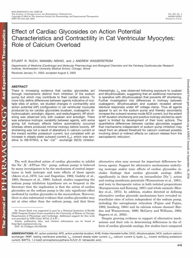
Molecular Structures of the Six Cardiac Glycosides Stud-ied. To more accurately define differences between cardiac glycosideagents and further evaluate cardiac glycoside structure-activity re-lationships, six cardiac glycosides (ouabain, ouabagenin, DHO, ac-todigin, digoxin, and resibufogenin) were chosen for electrophysiol-ogy and contractility studies in single cat ventricular myocytes.Figure 1 shows the molecular structure of these agents. Of particularnote, DHO and ouabagenin differ from the prototypical analogouabain by saturation of the lactone ring and omission of the carbo-hydrate moiety, respectively. Actodigin differs from ouabain in thesite of attachment of the lactone ring at C-17 of the steroid nucleus.Resibufogenin, which unlike the other analogs is isolated from ananimal source (frog skin), has a unique 6-membered lactone ring atC-17, and also lacks the sugar at C-3. Digoxin, chosen primarily forits importance in clinical medicine, shares many similarities withouabain but differs in the type of carbohydrate at C-3, and thenumber and position of hydroxyl groups attached to the steroidnucleus, thereby making it the most hydrophobic of the group.
Measurement of Voltage, Current, and Fractional Shorten-ing in Single Ventricular Myocytes. Several drops of cell suspen-sion were placed in an experimental chamber (0.5 ml volume)mounted on the stage of an inverted microscope (Nikon Diaphot;Nikon, Tokyo, Japan). After allowing 10 min for the cells to adhere tothe chamber bottom, they were superfused (2–3 ml/min) with Ty-rode’s solution maintained at 36 � 1°C using a Peltier device.
For experiments that included measurement of fractional short-ening, the video image of the long axis of the cell was aligned withthe rasters of the video edge detector (Crescent Electronics, SaltLake City, UT), which continuously monitored cell length along asingle line. Fractional shortening was defined as the ratio of cellshortening to resting cell length. Intracellular access was then ob-tained using either high-resistance microelectrodes or whole-cellpatch pipettes.
High-Resistance Microelectrode Method. A microelectrode(15–30 M� resistance) was pressed gently against the cell surfaceforming a relatively low-resistance seal. Access was then obtainedby briefly turning up the capacitance compensation, causing theamplifier circuitry to ring. Immediately following access, negativeholding current (�1 nA) was applied to maintain a resting mem-brane potential (RMP) of �70 to �80 mV. A high-resistance sealspontaneously formed within a few minutes, and RMP becamemore negative, indicating successful impalement. Backgroundholding current was then turned off. The primary advantage of
Fig. 1. Molecular structure of the six cardiac glycosides used in the study.
420 Rush et al.
at ASPE
T Journals on Septem
ber 16, 2016jpet.aspetjournals.org
Dow
nloaded from
this technique is that it allows electrical access of the cell withoutappreciable diffusion between the cytoplasm and the electrodesolution. The result is greater stability in measurement of ICa andcontractility.
Whole-Cell Suction Pipette Method. A 2 to 3 M� pipette waspressed gently against the cell surface, and suction was applied(50 –100 cm of H2O), allowing formation of a gigaohm seal. Appli-cation of a brief suction pulse then ruptured the membrane patchgiving electrical and physical access to the cell interior. Advan-tages of this technique include low-resistance access, and theability to control the ionic composition of the cytoplasm (diffusionthrough a relatively wide-bore pipette). Disadvantages include ICa
“run-down” that interferes with stable measurement of ICa andcontractility.
After obtaining access to the cell interior, voltage- and current-clamp protocols were directed by pCLAMP6 software (Axon In-struments Inc., Union City, CA). For voltage-clamp experiments,discontinuous single-electrode voltage-clamp mode of the Axoc-lamp-2A was used, which allowed simultaneous measurement ofboth current and actual (not command) membrane potential. Inaddition, series resistance compensation is unnecessary becausecurrent passage does not occur during voltage measurement sinceeach task is performed during alternate phases of every dutycycle. Voltage, current, and cell-length signals were digitized by aTL-1 DMA interface (Axon Instruments Inc.) at 7 to 10 kHz andchanneled into the Clampex data acquisition program, for lateranalysis using the Clampfit program (pCLAMP6). Additional de-tails of the use of the switch clamp with high-resistance electrodescan be found in previous publications (see Salata and Wasser-strom, 1988; Wasserstrom and Salata, 1988).
Statistical Analysis. Concentration-response curves were an-alyzed using one-way analysis of variance followed by the Stu-dent-Newman-Keuls test if criteria for significance were met.Data sets that did not fit parametric criteria were analyzed usingnonparametric rank analysis, followed by Dunn’s test where ap-propriate. All means represent the results of five to eleven exper-iments, unless stated otherwise. Data were considered significantwhen p � 0.05.
ResultsEffect of Cardiac Glycosides on Contractility and Ac-
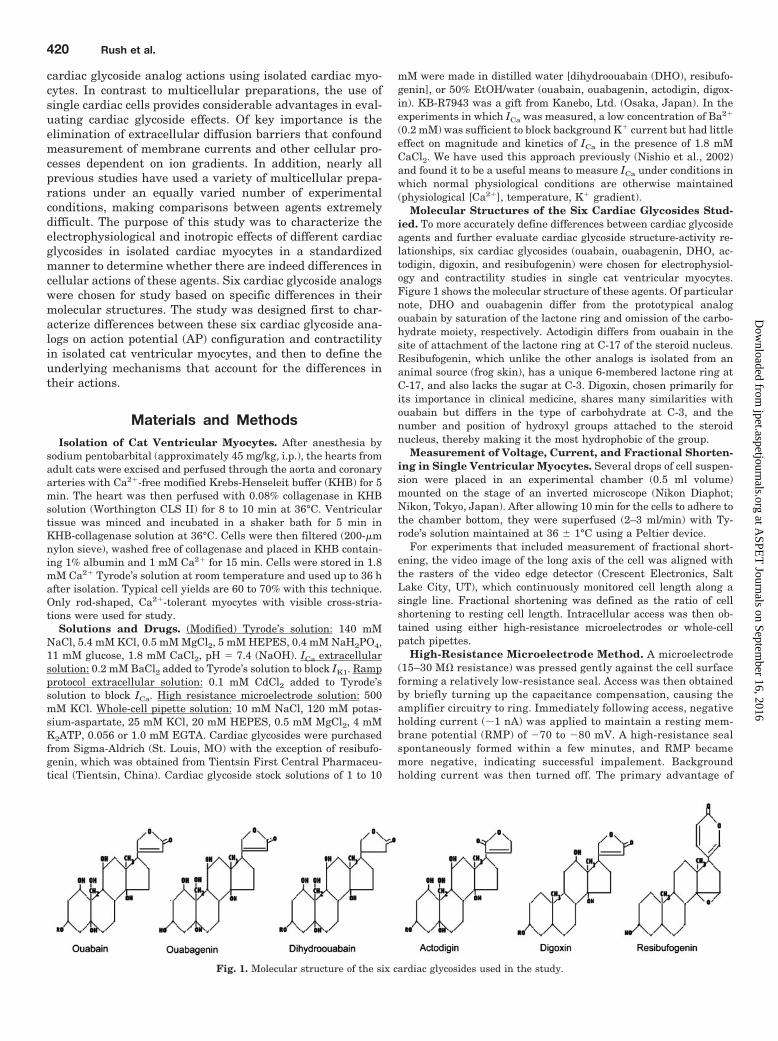
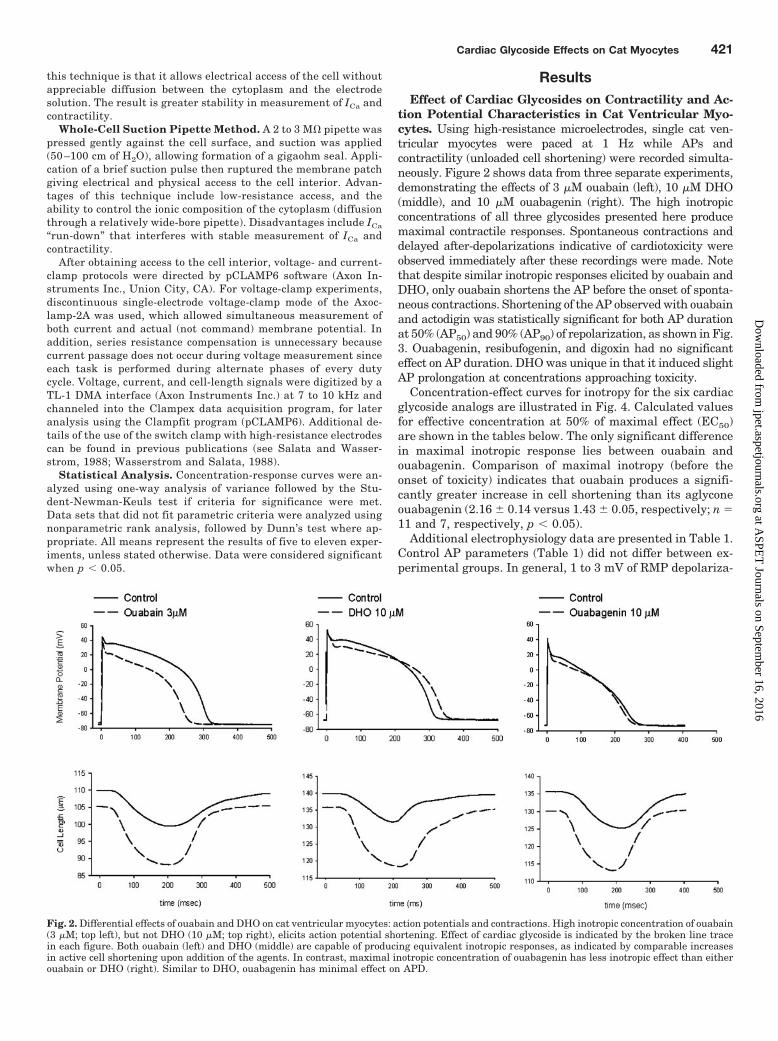
tion Potential Characteristics in Cat Ventricular Myo-cytes. Using high-resistance microelectrodes, single cat ven-tricular myocytes were paced at 1 Hz while APs andcontractility (unloaded cell shortening) were recorded simulta-neously. Figure 2 shows data from three separate experiments,demonstrating the effects of 3 �M ouabain (left), 10 �M DHO(middle), and 10 �M ouabagenin (right). The high inotropicconcentrations of all three glycosides presented here producemaximal contractile responses. Spontaneous contractions anddelayed after-depolarizations indicative of cardiotoxicity wereobserved immediately after these recordings were made. Notethat despite similar inotropic responses elicited by ouabain andDHO, only ouabain shortens the AP before the onset of sponta-neous contractions. Shortening of the AP observed with ouabainand actodigin was statistically significant for both AP durationat 50% (AP50) and 90% (AP90) of repolarization, as shown in Fig.3. Ouabagenin, resibufogenin, and digoxin had no significanteffect on AP duration. DHO was unique in that it induced slightAP prolongation at concentrations approaching toxicity.
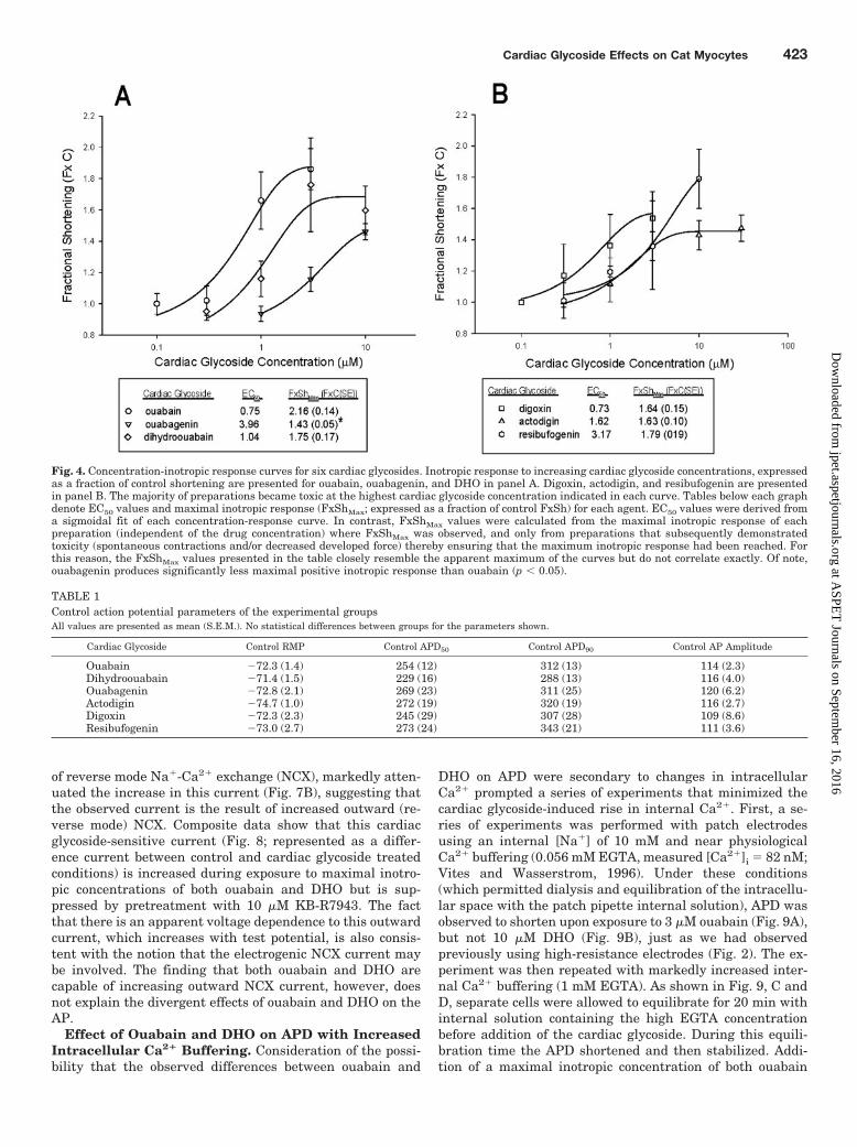
Concentration-effect curves for inotropy for the six cardiacglycoside analogs are illustrated in Fig. 4. Calculated valuesfor effective concentration at 50% of maximal effect (EC50)are shown in the tables below. The only significant differencein maximal inotropic response lies between ouabain andouabagenin. Comparison of maximal inotropy (before theonset of toxicity) indicates that ouabain produces a signifi-cantly greater increase in cell shortening than its aglyconeouabagenin (2.16 � 0.14 versus 1.43 � 0.05, respectively; n �11 and 7, respectively, p � 0.05).
Additional electrophysiology data are presented in Table 1.Control AP parameters (Table 1) did not differ between ex-perimental groups. In general, 1 to 3 mV of RMP depolariza-
Fig. 2. Differential effects of ouabain and DHO on cat ventricular myocytes: action potentials and contractions. High inotropic concentration of ouabain(3 �M; top left), but not DHO (10 �M; top right), elicits action potential shortening. Effect of cardiac glycoside is indicated by the broken line tracein each figure. Both ouabain (left) and DHO (middle) are capable of producing equivalent inotropic responses, as indicated by comparable increasesin active cell shortening upon addition of the agents. In contrast, maximal inotropic concentration of ouabagenin has less inotropic effect than eitherouabain or DHO (right). Similar to DHO, ouabagenin has minimal effect on APD.
Cardiac Glycoside Effects on Cat Myocytes 421
at ASPE
T Journals on Septem
ber 16, 2016jpet.aspetjournals.org
Dow
nloaded from
tion and a 5 to 10 mV decrease in AP amplitude was observedat maximal cardiac glycoside concentrations, with no signif-icant differences noted between agents.
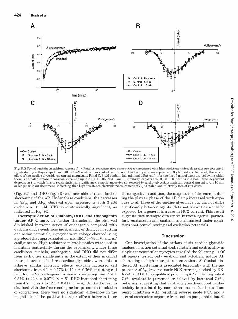
Effect of Ouabain on ICa. Finding significant differencesbetween cardiac glycosides on AP shortening prompted aninvestigation into membrane currents that might explainthese observed differences. Voltage-clamp studies were con-ducted to evaluate effects of ouabain on ICa as a possiblemechanism for AP shortening using high-resistance micro-electrodes. As demonstrated in Fig. 5A, exposure to 3 �Mouabain has little effect on the magnitude of ICa, although inthis example the rate of inactivation appears to be increased.
With longer exposure times (10 min), there is a small de-crease in ICa; however, this decrease was not statisticallysignificant (Fig. 5B). DHO shows a similar effect on ICa (Fig.5C), with a small time-dependent decrease in magnitude.Thus the difference in action potential changes induced bythese agents cannot be explained on the basis of alterationsin ICa. Further experiments evaluating the stability of ICa
measurements under control conditions (without cardiac gly-coside) showed no time-dependent diminution of ICa (Fig. 5D)demonstrating that the slight decrease in current magnitudeis in fact a result of actions of both drugs.
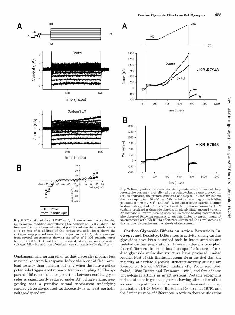
Effect of Ouabain on IK1. Further voltage-clamp studieswere conducted to evaluate effects of ouabain on the inwardrectifier current (IK1) as a possible mechanism for actionpotential shortening. Figure 6A shows representative IK1
data obtained with high-resistance electrodes. The voltage-clamp protocol is presented in the top trace. From a holding
potential of �40 mV, hyperpolarizing and depolarizing volt-age steps were used to assess both the inward and outwardIK1 currents, measured at the end of the test voltage step.Although ICa was not blocked in these experiments, valuesfor IK1 at voltages positive to �40 mV were assumed to befairly accurate at the end of the voltage-clamp steps after ICa
was mostly inactivated (300 ms). Current recordings at hy-perpolarizing potentials show large inward currents; depo-larizing pulses yielded progressively diminishing outwardcurrents typical of cat ventricular IK1. Exposure to 3 �Mouabain caused no change in inward currents and a slowlydeveloping outward current during depolarization This out-ward current component increased with exposure time andwas investigated further using a ramp protocol, discussedbelow. Composite voltage-current data (Fig. 6B) again showsno change in steady-state IK1 over the voltage range of �140to 30 mV, with a small increase in outward current at voltagesteps above 40 mV, which did not achieve statistical signifi-cance.
Effect of Ouabain and DHO on the Steady-State Out-ward Current (Iss). A ramp protocol was used to evaluatethe late outward current component noted above using high-resistance electrodes. The voltage protocol and representa-tive data are presented in Fig. 7. Cd2� (0.1 mM) was added toblock ICa. As illustrated in Fig. 7A, 10 min of exposure to 3�M ouabain induced a pronounced increase in outward cur-rent (previously termed “outward steady-state current”, Iss;Levi, 1993) that is observed at voltages above �20 mV. How-ever, pretreatment of cells with 10 �M KB-R7943, a blocker
Fig. 3. Concentration-APD response curves for six cardiac glycosides. Effect of increasing cardiac glycoside concentration on APD50 (panels A and C)and APD90 (panels B and D) is shown. Ouabain, DHO, and ouabagenin concentration-response curves are presented in the upper panels (A and B).Digoxin, actodigin, and resibufogenin are presented in lower panels (C and D). APD values are given as a fraction of control values (error bars �S.E.M.). As noted, ouabain and actodigin elicit action potential shortening, whereas DHO elicits a small but significant prolongation of the AP at highinotropic concentrations. �, p � 0.05 indicates significant difference from control.
422 Rush et al.
at ASPE
T Journals on Septem
ber 16, 2016jpet.aspetjournals.org
Dow
nloaded from
of reverse mode Na�-Ca2� exchange (NCX), markedly atten-uated the increase in this current (Fig. 7B), suggesting thatthe observed current is the result of increased outward (re-verse mode) NCX. Composite data show that this cardiacglycoside-sensitive current (Fig. 8; represented as a differ-ence current between control and cardiac glycoside treatedconditions) is increased during exposure to maximal inotro-pic concentrations of both ouabain and DHO but is sup-pressed by pretreatment with 10 �M KB-R7943. The factthat there is an apparent voltage dependence to this outwardcurrent, which increases with test potential, is also consis-tent with the notion that the electrogenic NCX current maybe involved. The finding that both ouabain and DHO arecapable of increasing outward NCX current, however, doesnot explain the divergent effects of ouabain and DHO on theAP.
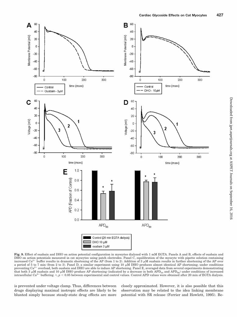
Effect of Ouabain and DHO on APD with IncreasedIntracellular Ca2� Buffering. Consideration of the possi-bility that the observed differences between ouabain and
DHO on APD were secondary to changes in intracellularCa2� prompted a series of experiments that minimized thecardiac glycoside-induced rise in internal Ca2�. First, a se-ries of experiments was performed with patch electrodesusing an internal [Na�] of 10 mM and near physiologicalCa2� buffering (0.056 mM EGTA, measured [Ca2�]i � 82 nM;Vites and Wasserstrom, 1996). Under these conditions(which permitted dialysis and equilibration of the intracellu-lar space with the patch pipette internal solution), APD wasobserved to shorten upon exposure to 3 �M ouabain (Fig. 9A),but not 10 �M DHO (Fig. 9B), just as we had observedpreviously using high-resistance electrodes (Fig. 2). The ex-periment was then repeated with markedly increased inter-nal Ca2� buffering (1 mM EGTA). As shown in Fig. 9, C andD, separate cells were allowed to equilibrate for 20 min withinternal solution containing the high EGTA concentrationbefore addition of the cardiac glycoside. During this equili-bration time the APD shortened and then stabilized. Addi-tion of a maximal inotropic concentration of both ouabain
Fig. 4. Concentration-inotropic response curves for six cardiac glycosides. Inotropic response to increasing cardiac glycoside concentrations, expressedas a fraction of control shortening are presented for ouabain, ouabagenin, and DHO in panel A. Digoxin, actodigin, and resibufogenin are presentedin panel B. The majority of preparations became toxic at the highest cardiac glycoside concentration indicated in each curve. Tables below each graphdenote EC50 values and maximal inotropic response (FxShMax; expressed as a fraction of control FxSh) for each agent. EC50 values were derived froma sigmoidal fit of each concentration-response curve. In contrast, FxShMax values were calculated from the maximal inotropic response of eachpreparation (independent of the drug concentration) where FxShMax was observed, and only from preparations that subsequently demonstratedtoxicity (spontaneous contractions and/or decreased developed force) thereby ensuring that the maximum inotropic response had been reached. Forthis reason, the FxShMax values presented in the table closely resemble the apparent maximum of the curves but do not correlate exactly. Of note,ouabagenin produces significantly less maximal positive inotropic response than ouabain (p � 0.05).
TABLE 1Control action potential parameters of the experimental groupsAll values are presented as mean (S.E.M.). No statistical differences between groups for the parameters shown.
Cardiac Glycoside Control RMP Control APD50 Control APD90 Control AP Amplitude
Ouabain �72.3 (1.4) 254 (12) 312 (13) 114 (2.3)Dihydroouabain �71.4 (1.5) 229 (16) 288 (13) 116 (4.0)Ouabagenin �72.8 (2.1) 269 (23) 311 (25) 120 (6.2)Actodigin �74.7 (1.0) 272 (19) 320 (19) 116 (2.7)Digoxin �72.3 (2.3) 245 (29) 307 (28) 109 (8.6)Resibufogenin �73.0 (2.7) 273 (24) 343 (21) 111 (3.6)
Cardiac Glycoside Effects on Cat Myocytes 423
at ASPE
T Journals on Septem
ber 16, 2016jpet.aspetjournals.org
Dow
nloaded from
(Fig. 9C) and DHO (Fig. 9D) was now able to cause furthershortening of the AP. Under these conditions, the decreasesin AP50 and AP90 observed upon exposure to both 3 �Mouabain or 10 �M DHO were statistically significant, asindicated in Fig. 9E.
Inotropic Action of Ouabain, DHO, and Ouabageninunder AP Clamp. To further characterize the observeddiminished inotropic action of ouabagenin compared withouabain under conditions independent of changes in restingand action potentials, myocytes were voltage-clamped usinga protocol that approximated normal RMP (�78 mV) and APconfiguration. High-resistance microelectrodes were used tomaintain contractility during the experiment. Under theseconditions, ouabain, ouabagenin, and DHO did not differfrom each other significantly in the extent of their maximalinotropic action; all three cardiac glycosides were able toachieve similar inotropic effects; ouabain increased cellshortening from 4.1 � 0.77% to 10.4 � 0.76% of resting celllength (n � 9); ouabagenin increased shortening from 4.9 �0.87% to 11.4 � 0.27% (n � 5); DHO increased shorteningfrom 4.7 � 0.27% to 12.1 � 0.61% (n � 4). Unlike the resultsobtained with the free-running action potential stimulationof contraction, there were no significant differences in themagnitude of the positive inotropic effects between these
three agents. In addition, the magnitude of the current dur-ing the plateau phase of the AP clamp increased with expo-sure to all three of the cardiac glycosides but did not differsignificantly between agents (data not shown) as would beexpected for a general increase in NCX current. This resultsuggests that inotropic differences between agents, particu-larly ouabagenin and ouabain, are minimized under condi-tions that control resting and excitation potentials.
DiscussionOur investigation of the actions of six cardiac glycoside
analogs on action potential configuration and contractility insingle cat ventricular myocytes revealed the following. 1) Ofall agents tested, only ouabain and actodigin induce APshortening at high inotropic concentrations. 2) Ouabain-in-duced AP shortening is associated temporally with the ap-pearance of ISS (reverse mode NCX current, blocked by KB-R7943). 3) DHO is capable of producing AP shortening only ifCa2� overload is prevented or delayed by increased Ca2�
i
buffering, suggesting that cardiac glycoside-induced cardio-toxicity is mediated by more than one mechanism-sodiumpump inhibition with resulting reverse mode NCX and asecond mechanism separate from sodium pump inhibition. 4)
Fig. 5. Effect of ouabain on calcium current (ICa). Panel A, representative current traces measured with high-resistance microelectrodes are presented.ICa elicited by voltage steps from �40 to 0 mV is shown for control condition and following a 5-min exposure to 3 �M ouabain. As noted, there is noeffect of the cardiac glycoside on current magnitude. Panel C, 3 �M ouabain has minimal effect on ICa for the first 5 min of exposure, following whichthere is a small decrease in maximal current amplitude (p 0.05, NS). Panel D, similarly, exposure to 10 �M DHO results in a small, time-dependentdecrease in ICa, which fails to reach statistical significance. Panel B, myocytes not exposed to cardiac glycosides maintain control current levels 10 minor longer without decrement, indicating that high-resistance electrode measurement of ICa is stable and relatively free of run-down.
424 Rush et al.
at ASPE
T Journals on Septem
ber 16, 2016jpet.aspetjournals.org
Dow
nloaded from
Ouabagenin and certain other cardiac glycosides produce lessmaximal contractile response before the onset of Ca2� over-load toxicity than ouabain but only when the native actionpotentials trigger excitation-contraction coupling. 5) The ap-parent difference in inotropic action between cardiac glyco-sides is significantly reduced under AP voltage clamp, sug-gesting that a putative second mechanism underlyingcardiac glycoside-induced cardiotoxicity is at least partiallyvoltage-dependent.
Cardiac Glycoside Effects on Action Potentials, In-otropy, and Toxicity. Differences in activity among cardiacglycosides have been described both in intact animals andisolated cardiac preparations. However, attempts to explainthese differences in action based on specific features of car-diac glycoside molecular structure have produced limitedresults. Part of this limitation stems from the fact that themajority of cardiac glycoside structure-activity studies arefocused on Na�/K�-ATPase binding (De Pover and God-fraind, 1982; Brown and Erdmann, 1984), and few addressphysiological actions in intact systems. Notable exceptionsinclude studies in guinea pig atria showing stimulation of thesodium pump at low concentrations of ouabain and ouabage-nin, but not DHO (Ghysel-Burton and Godfraind, 1979), andthe demonstration of differences in toxic to therapeutic ratios
Fig. 6. Effect of ouabain and DHO on IK1. A, raw current traces showingIK1 in control condition and following the addition of 3 �M ouabain. Theincrease in outward current noted at positive voltage steps develops over5 to 10 min after addition of the cardiac glycoside. Inset shows thevoltage-clamp protocol used for IK1 experiments. B, IK1 data averagedfrom several experiments showing the effect of 3 �M ouabain (errorbars � S.E.M.). The trend toward increased outward current at positivevoltages following addition of ouabain was not statistically significant.
Fig. 7. Ramp protocol experiments: steady-state outward current. Rep-resentative current traces elicited by a voltage-clamp ramp protocol (in-set). As indicated, the protocol consisted of a step to �40 mV for 200 ms,then a ramp up to �80 mV over 500 ms before returning to the holdingpotential of �70 mV. Cd2� and Ba2� were added to the external solutionto diminish ICa and K� currents. Panel A, 10-min exposure to 3 �Mouabain produced a dramatic increase in steady-state outward current.An increase in inward current upon return to the holding potential wasalso observed following exposure to ouabain (noted by arrow). Panel B,pretreatment with KB-R7943 effectively eliminated the development ofthis cardiac glycoside-sensitive steady-state current.
Cardiac Glycoside Effects on Cat Myocytes 425
at ASPE
T Journals on Septem
ber 16, 2016jpet.aspetjournals.org
Dow
nloaded from

of cardiac glycosides in intact dogs, dependent on the positionof the attachment of the lactone ring (Mendez et.al., 1974).However, none of these studies (individually or collectively)provides a comprehensive foundation for understanding car-diac glycoside structure-activity relationships, and it is likelythat multiple factors in structure determine the specific re-sponse elicited by a given agent. Information provided by ourinvestigation indicates that subtle differences in structure(including a simple saturation of the lactone ring) causesignificant differences in physiological activity. If intracellu-lar sites are indeed important in determining differences inaction between cardiac glycoside agents (Fujino and Fujino,1982; Isenberg, 1984; Sagawa et al., 2002), including thethreshold for Ca2� overload, one could speculate that therelative ability of a given glycoside to cross the sarcolemma(either passively or actively; Nunez-Duran et al., 1988) andaffinity for specific intracellular sites would be importantadditional determinants of action. In addition, if the ryano-dine receptor is an important determinant of this intracellu-lar mechanism as postulated by some investigators (Isen-berg, 1984; Rardon and Wasserstrom, 1990; McGarry andWilliams, 1993; Sagawa et al., 2002), then differences inbinding affinity to this receptor between cardiac glycosidesmight contribute to the observed differences in action.
Mechanisms Underlying Shortening of APD. Manystudies evaluating the effects of cardiac glycosides on isolatedmyocardial preparations or single cells have shown a short-ening of APD (McDonald et al., 1975; Levi, 1993). Explana-tions for this finding are reflected in contrasting theories,which attempt to characterize a cardiac glycoside-inducedoutward current. The first theory (Luk and Carmeliet, 1990)invokes an outward current that is increased by 10 to 100 �Mouabain in guinea pig cardiac myocytes with similar proper-ties to a single channel current found in inside-out patches.This current is proposed to be a Na�-activated K� current byvirtue of the fact that it is dependent on the presence of Na�
i,displays rectification that is dependent on the K� gradient,
and has a reversal potential near the K� equilibrium poten-tial. The second theory (Levi, 1993) suggests the involvementof an outward current elicited by 50 �M strophanthidin alsoin guinea pig cardiac cells with reversal potential of �54 mV.This current was shown to be consistent with reverse modeNCX by being more pronounced at positive voltages, abol-ished by removing external Ca2� or addition of Ni2�, andunaffected by increasing intracellular Ca2� buffering(BAPTA) or addition of K� current blockers (Ba2�, TEA, and4-AP). Data from our experiments indicate the cardiac gly-coside-induced outward current is most likely due to in-creased reverse mode NCX because it does not develop in thepresence of KB-R7943, a reverse mode NCX blocker (Kimuraet al., 1999). In addition, AP shortening appears to be anaction of some but not all cardiac glycosides. This could be aresult of other drug actions, including the possibility thatcertain glycosides might also block IKs, the slowly activatingcomponent of the delayed rectifier current (Rocchetti et al.,2003), which could explain why DHO (and not ouabain oractodigin) caused AP prolongation. The balance of opposingdirect and indirect actions on net membrane current couldthen be responsible for the variety of glycoside effects on APduration, especially in different species with varying depen-dencies of repolarization on IKs.
Role of Ca2� Overload in Development of APD Short-ening and Inotropy. Data from this study suggest thatCa2� overload is a primary determinant both for maximalinotropic response and alterations in AP configuration andcould account for differences in response between cardiacglycoside analogs. This is not a new concept, particularlywith regard to inotropic action as addressed by other inves-tigators (Capogrossi et al., 1988). The basic theory, supportedby experimental data, contends that the myocardium is ca-pable of increasing inotropy by Ca2�-dependent mechanismsuntil toxicity develops as indicated by spontaneous sarcoplas-mic reticulum Ca2� release and spontaneous contractions.This spontaneous, uncoordinated release of Ca2� depletesthe sarcoplasmic reticulum of Ca2� for the next contraction,thereby reducing the inotropic state as well as any contribu-tions of Ca2�-dependent conductances (including NCX cur-rent) to AP duration.
We found that Ca2� overload in fact does influencewhether or not cardiac glycosides induced AP abbreviation.Although direct evidence for sodium pump inhibition was notevaluated, the presence of a robust increase in reverse modeNCX current within the inotropic range of both ouabain andDHO would suggest that sodium pump inhibition is an im-portant mechanism in the inotropic response of cardiac gly-cosides in vitro. In addition, the data suggest that a secondcardiac glycoside mechanism (independent of sodium pumpinhibition) influences the threshold for Ca2� overload andthereby determines the maximal inotropic response ob-served. For example, the weaker inotropic response and lackof AP shortening observed with ouabagenin compared withouabain is likely the result of earlier spontaneous SR Ca2�
release (lower toxicity threshold) elicited by ouabagenin priorto accumulation of equal levels of intracellular calcium.
How voltage alters spontaneous release from the SR andthereby alters the maximal inotropic response of ouabagenin(as demonstrated in the AP clamp experiments) can only bespeculated. The most likely reason is that the positive feed-back between SR Ca2� release and membrane depolarization
Fig. 8. Effect of DHO and ouabain on cardiac glycoside-sensitive steady-state outward current. Averaged data from ramp protocol experiments ispresented. Glycoside-sensitive current is defined as a subtraction (exper-imental minus control) current. The data summarize the effect of ouabain(3 �M, empty circles) and DHO (10 �M, filled circles) on cardiac glycoside-sensitive current; 10 min of exposure demonstrates the time-dependentdevelopment of the current. Pretreatment with 10 �M KB-R7943 elimi-nates the effect of ouabain on development of outward steady-state cur-rent (triangles) producing significantly less current at all test voltages (�,p � 0.05 compared with ouabain alone).
426 Rush et al.
at ASPE
T Journals on Septem
ber 16, 2016jpet.aspetjournals.org
Dow
nloaded from
is prevented under voltage clamp. Thus, differences betweendrugs displaying maximal inotropic effects are likely to beblunted simply because steady-state drug effects are more
closely approximated. However, it is also possible that thisobservation may be related to the idea linking membranepotential with SR release (Ferrier and Howlett, 1995). Be-
Fig. 9. Effect of ouabain and DHO on action potential configuration in myocytes dialyzed with 1 mM EGTA. Panels A and B, effects of ouabain andDHO on action potentials measured in cat myocytes using patch electrodes. Panel C, equilibration of the myocyte with pipette solution containingincreased Ca2� buffer results in dramatic shortening of the AP (from 1 to 2). Addition of 3 �M ouabain results in further shortening of the AP overa period of 5 to 7 min (from 2 to 3). Panel D, a similar experiment using 10 �M DHO produces almost identical AP shortening; under conditionspreventing Ca2� overload, both ouabain and DHO are able to induce AP shortening. Panel E, averaged data from several experiments demonstratingthat both 3 �M ouabain and 10 �M DHO produce AP shortening (indicated by a decrease in both APD50 and APD90) under conditions of increasedintracellular Ca2� buffering. �, p � 0.05 between experimental and control values. Control APD values were obtained after 20 min of EGTA dialysis.
Cardiac Glycoside Effects on Cat Myocytes 427
at ASPE
T Journals on Septem
ber 16, 2016jpet.aspetjournals.org
Dow
nloaded from
cause the threshold for spontaneous calcium release from theSR during diastole is a primary determinant of cardiac gly-coside inotropy, it may be that voltage clamp prevents ordelays diastolic calcium release. This implies that small volt-age perturbations during diastole may contribute to sponta-neous release or that spontaneous release during diastole isat least partially dependent on membrane potential.
Effects of Low Internal Free Mg2� Concentration onExperimental Results. It is possible that certain of ourexperimental conditions might influence the results found inthis study. One such issue is the low free [Mg2�] concentra-tion in the internal solution (about 10�5 M). This is likely tohave important effects on Mg2�-dependent process as in thecell. However, it should be noted that even with this low[Mg2�], there are still pronounced differences between differ-ent cardiac glycosides just as expected from data obtainedusing high-resistance microelectrodes in which the intracel-lular environment is closer to physiological. In addition, therectifying characteristics of IK1 are largely unaffected by thebuffering of internal Mg2� (data not shown), suggesting thateffective concentrations in critical regions of the cytoplasmremain at normal regulatory levels despite calculatedchanges in bulk concentration. This fact suggests that it isdifficult to extrapolate bulk calculated [Mg2�] to true freeconcentration at regulatory sites.
ReferencesAkera T, Larson FS, and Brody TM (1970) Correlation of cardiac sodium- and
potassium-activated adenosine triphosphatase activity with ouabain-induced ino-tropic stimulation. J Pharmacol Exp Ther 173:145–151.
Brown L and Erdmann E (1984) Binding of digitalis derivatives to beef, cat andhuman cardiac (Na� � K�)-ATPase. Affinity and kinetic constants. Arch IntPharmacodyn 271:229–240.
Capogrossi MC, Stern MD, Spurgeon HA, and Lakatta EG (1988) Spontaneous Ca2�
release from the sarcoplasmic reticulum limits Ca2�-dependent twitch potentia-tion in individual cardiac myocytes. J Gen Physiol 91:133–155.
De Pover A and Godfraind T (1982) Influence of 16� formylation on Na,K-ATPaseinhibition by cardiac glycosides. Naunyn Schmiedeberg’s Arch Pharmacol 321:135–139.
Ferrier GR and Howlett SE (1995) Contractions in guinea-pig ventricular myocytestriggered by a calcium-release mechanism separate from Na� and L-currents.J Physiol (Lond) 484:107–122.
Fujino S and Fujino M (1982) Ouabain potentiation and Ca release from sarcoplas-mic reticulum in cardiac and skeletal muscle. Can J Physiol Pharmacol 60:542–555.
Gadsby DC, Kimura J, and Noma A (1985) Voltage dependence of the Na/K pumpcurrent in isolated heart cells. Nature (Lond) 315:63–65.
Ghysel-Burton J and Godfraind T (1979) Stimulation and inhibition of the sodium
pump by cardioactive steroids in relation to their binding sites and their inotropiceffect on guinea pig isolated atria. Br J Pharmacol 66:175–184.
Isenberg G (1984) Contractility of isolated bovine ventricular myocytes is enhancedby intracellular injection of cardioactive glycosides. Evidence for an intracellularmode of action, in Cardiac Glycoside Receptors and Positive Inotropy (Erdmann Eed) pp 56–71, Steinkopff, Darmstadt.
Karagueuzian HS and Katzung BG (1981) Relative inotropic and arrhythmogeniceffects of five cardiac steroids in ventricular myocardium: Oscillatory afterpoten-tials and the role of endogenous catecholamines. J Pharmacol Exp Ther 218:348–356.
Kimura J, Watano T, Kawahara M, Saki E, and Yatabe J (1999) Direction-independent block of bi-directional Na�/Ca2� exchange current by KB-R7943 inguinea-pig cardiac myocytes. Br J Pharmacol 128:969–974.
Lee C and Dagostino M (1982) Effect of strophanthidin on intracellular Na� ionactivity and twitch tension of constantly driven canine cardiac Purkinje fibers.Biophys J 40:185–198.
Levi AJ (1993) A role for sodium/calcium exchange in the action potential shorteningcaused by strophanthidin in guinea pig ventricular myocytes. Cardiovasc Res27:471–481.
Luk HN and Carmeliet E (1990) Na�-activated K� current in cardiac cells: rectifi-cation, open probability, block and role in digitalis toxicity. Pflugers Arch 416:766–768.
McDonald TF, Nawrath H, and Trautwein W (1975) Membrane currents and tensionin cat ventricular muscle treated with cardiac glycosides. Circ Res 37:674–682.
McGarry SJ and Williams AJ (1993) Digoxin activates sarcoplasmic reticulum Ca2�-release channels: a possible role in cardiac inotropy. Br J Pharmacol 108:1043–1050.
Mendez R, Pastelin G, and Kabela E (1974) The influence of the position of attach-ment of the lactone ring to the steroid nucleus on the action of cardiac glycosides.J Pharmacol Exp Ther 188:189–197.
Nishio M, Ruch SR, and Wasserstrom JA (2002) Positive inotropic effects of ouabainin isolated cat ventricular myocytes in sodium-free conditions. Am J Physiol283:H2045–H2053.
Nunez-Duran H, Riboni L, Ubaldo E, Kabela E, and Barcenas-Ruiz L (1988) Ouabainuptake by endocytosis in isolated guinea pig atria. Am J Physiol 255:C479–C485.
Rardon DP and Wasserstrom JA (1990) Cardiotonic steroids activate cardiac sarco-plasmic reticulum calcium release channels (Abstract). Circulation 82:III–342.
Rocchetti M, Besana A, Mostacciuolo G, Ferrari P, Micheletti R, and Zaza A (2003)Diverse toxicity associated with cardiac Na�/K� pump inhibition: evaluation ofelectrophysiological mechanisms. J Pharmacol Exp Ther 305:765–771.
Sagawa T, Sagawa K, Kelly JE, Tsushima RG, and Wasserstrom JA (2002) Activa-tion of cardiac ryanodine receptors by cardiac glycosides. Am J Physiol 282:H1118–H1126.
Salata JJ and Wasserstrom JA (1988) Effects of quinidine on action potentials andionic currents in isolated canine ventricular myocytes. Circ Res 62:324–337.
Steimers JR, Lobaugh LA, Liu S, Shigeto N, and Lieberman M (1990) Intracellularsodium affects ouabain interaction with the Na/K pump in cultured chick cardiacmyocytes. J Gen Physiol 95:77–95.
Vites AM and Wasserstrom JA (1996) Fast sodium influx provides an initial step totrigger contractions in cat ventricle. Am J Physiol 271:H674–H686.
Wasserstrom JA, Farkas DE, Norell MA, and Vereault DV (1991) Effects of differentcardiac steroids on intracellular sodium, inotropy and toxicity in sheep Purkinjefibers. J Pharmacol Exp Ther 258:918–925.
Wasserstrom JA and Salata JJ (1988) Basis for tetrodotoxin and lidocaine effects onaction potentials in dog ventricular myocytes. Am J Physiol 254:H1157–H1166.
Address correspondence to: Dr. J. Andrew Wasserstrom, Division of Car-diology—S203, Ward 3-105, Northwestern University Medical School, 303 E.Chicago Ave., Chicago, IL 60611. E-mail: [email protected]
428 Rush et al.
at ASPE
T Journals on Septem
ber 16, 2016jpet.aspetjournals.org
Dow
nloaded from


