Embed Size (px)
Citation preview

This content has been downloaded from IOPscience. Please scroll down to see the full text.
Download details:
This content was downloaded by: theo15
IP Address: 195.251.7.233
This content was downloaded on 13/11/2015 at 08:01
Please note that terms and conditions apply.
Non-covalent nanodiamond–polymer dispersions and electrostatic immobilization of bovine
serum albumin protein
View the table of contents for this issue, or go to the journal homepage for more
2015 Mater. Res. Express 2 115005
(http://iopscience.iop.org/2053-1591/2/11/115005)
Home Search Collections Journals About Contact us My IOPscience

Mater. Res. Express 2 (2015) 115005 doi:10.1088/2053-1591/2/11/115005
PAPER
Non-covalent nanodiamond–polymer dispersions and electrostaticimmobilization of bovine serum albumin protein
TSkaltsas, S Pispas andNTagmatarchisTheoretical and Physical Chemistry Institute, NationalHellenic Research Foundation, 48Vassileos ConstantinouAvenue, 11635Athens,Greece
E-mail: [email protected] and [email protected]
Keywords: diamond, dispersions, proteins
Supplementarymaterial for this article is available online
AbstractNanodiamonds (NDs) lack efficient dispersion, not only in solvents but also in aqueousmedia. Thelatter is of great importance, considering the inherent biocompatibility ofNDs and the plethora ofsuitable strategies for immobilizing functional biomolecules. In this work, a series of polymers wasnon-covalently interactedwithNDs, formingND–polymer ensembles, and their dispersibility andstability was examined. Dynamic light scattering gave valuable information regarding the size of theensembles in liquid phase, while theirmorphology was further examined by high-resolutiontransmission electronmicroscopy imaging. In addition, thermal analysismeasurements were appliedto collect information on the thermal behavior ofNDs and their ensembles and to calculate theamount of polymer interactingwith theNDs, as well as the dispersibility values of theND–polymerensembles. Finally, the bovine serum albumin proteinwas electrostatically bound to aND–polymerensemble inwhich the polymericmoiety was carrying quaternized pyridine units.
1. Introduction
Nanodiamond (ND) particles are an all-carbonmaterial [1], though less studied as comparedwith other carbon-based nanostructures such as fullerenes, nanotubes and graphene. NDs prepared by detonationmethods in theabsence of oxygen [2] possess outstanding properties such as high surface area and amenability to diverseorganic transformations [3], and are potentially suitable for diverse applications, for example in optics,photonics and catalysis [4].Moreover, NDs are less toxic than the aforementioned carbon nanostructures [5, 6],and are certainlymore appropriate as far as biological andmedicinal applications are concerned [7–14].However, similar to the other nanocarbonmaterials, multiple surface-based van derWaals interactionsdominate in pristineNDs, resulting in aggregation, responsible for the formation of large particles. Thishandicaps efforts to fully exploit the intriguing properties ofNDs and take full advantage of them in terms ofdeveloping their applicability. Seeking strategies for dispersingNDs in organic solvents and/orwater is thereforeof primary importance. Along these lines, de-aggregation ofNDswas achieved by either ball-milling or bymicrobead-assisted ultrasonic disintegration [15], or salt- and sugar-assistedmilling [16]. However,disadvantages such as contaminationwith the beadmaterial and generation of graphitic layers on theNDssurface always exist [17]. Alternatively, (ultra)centrifugationwas also applied to separateNDparticles intofractions byweight and size, as a contamination-free approach in contrast with the beadmilling technique, albeitwith the drawback of yielding a limited amount ofmaterial [18–21].
Therefore, complementary to the aforementioned procedures, treatment ofNDswith polymers could beanother strategy for achieving disintegratedNDs and forming stable dispersions. Actually, themajority of suchstudies deal with covalently functionalizedNDswith polymers, based on the so-called ‘grafting to’ [22] or‘grafting from’ [23, 24] approach, whileND ensembles incorporating non-covalent decorationwith polymersalso exist [25]. Briefly, covalent functionalization leads tomaterials carrying functionalities stronglyimmobilized and anchored onto the skeleton ofNDs.On the other hand, with non-covalent functionalization
RECEIVED
6May 2015
REVISED
20August 2015
ACCEPTED FOR PUBLICATION
14October 2015
PUBLISHED
13November 2015
© 2015 IOPPublishing Ltd

the structure ofNDs remains unaffected, while the added functionalities are less tightly bound as comparedwiththe correspondingmaterials prepared followed covalent functionalization protocols. Hence, for example,polyacrylonitrile and polyamidewere reinforcedwith high loadings ofNDs, thus creating uniform films thatoffer protection fromultraviolet irradiation [26]. Furthermore, NDs could show enhancedmechanicalproperties when used asfillers in polyacrylonitrile films [27].
Considering the high durability and inherent biocompatibility ofNDs, functional biomolecules such asproteins and enzymes could be integrated, forming functional nanoensembles. In this context, enzymes suitablycondensed via covalent bondingwith soft polymeric units, directly interactingwithNDs, describe an efficientnanosystem inwhich the biomolecules retain their structure and functionality [28]. Furthermore, ND-basedbiosensors incorporating proteins and/or antibodies, either in a covalent [29–31] or non-covalent fashion[32–37]were developed.
Herein, the dispersibility ofNDs upon non-covalent interactions with polymers and the integration ofbovine serum albumin (BSA) proteinwas examined. The aimof this study is three-fold, namely, (i) to investigatethe dispersibility and stability of a series ofND–polymer ensembles in a variety of organic solvents aswell aswater, (ii) tomorphologically and thermally evaluate theND–polymer ensembles, and (iii) to electrostaticallyintegrate the BSA proteinwithin anND–polymer ensemble.
2. Experimental
2.1. InstrumentationDynamic light scattering (DLS)measurements were performed on anALV/CGS-3CompactGoniometerSystem (ALVGmbH,Germany), equippedwith a JDSUniphase 22MwHe–Ne laser, operating at 632.8 nm,interfacedwith anALV-5000/EPPmulti-tau digital correlator with 288 channels and anALV/LSE-5003 lightscattering electronics unit for a steppermotor drive and limiting switch control. The scattering intensity andcorrelation functionsweremeasured at 90°. Correlation functionswere collected 10 times andwere analysed bythe cumulantmethod and theCONTIN software, which provide the apparent hydrodynamic radii distributionsby Laplace inversion of the correlation function andwith the aid of the Stokes–Einstein relationship.
HR-TEMmeasurements were carried out using a JEM-2100F (JEOL)high-resolution field-emission gunTEMoperated at 80 keV at room temperature and under a pressure of 10−6 Pa.HR-TEM images were recordedwith a charge-coupled device with an exposure time of typically 1s.
The thermogravimetric analysis was performed using a TGAQ500V20.2 Build 3 instrument by TA in aninert atmosphere of nitrogen. In a typical experiment, 1 mg of thematerial was placed in the sample pan and thetemperaturewas equilibrated at 40 °C. Subsequently, the temperature was increased to 900 °C at a rate of10 °Cmin−1 and theweight changes were recorded as a function of temperature.
The ζ-Potentialmeasurements were performedwith a ZetasizerNanoZS fromMalvern Instruments (UK)equippedwith aHe–Ne laser (632.8 nm) and using a non-invasive back scatter (NIBS) technology. ζ-Potentialvalueswere determined using the Smolukowski equation relating the ionicmobilities with surface charge, andare reported as averages of ten repeatedmeasurements.
2.2.Materials and reagentsNanodiamond powder (<10 nmTEM, 95% tracemetals basis, batch no: 636444-1G), bovine serum albumin,lysozyme from chicken eggwhite and all solvents were purchased fromAldrich and usedwithout furtherpurification.Quaternized poly(2-vinylpyridine) abbreviated as qP2VP, poly(N-isopropylacrylamide)abbreviated as PNIPAM, poly(ethylene oxide) abbreviated as PEO, poly[n-butylacrylate-b-N-isopropylacrylamide] abbreviated as PnBA-b-PNIPAM, poly(2-vinylpyridine) abbreviated as P2VP,poly[styrene-b-(ethylene oxide)] abbreviated as PS-b-PEO, poly[styrene-b-(2-vinylpyridine)] abbreviated asPS-b-P2VP and poly(methacrylic acid) abbreviated as PMAAwere synthesized by anionic or RAFTpolymerization followed by post polymerization functionalizationwhere necessary, as exemplified previouslyin the literature (see supporting information, table S1, for theirmolecular characteristics) [38–46].
2.3. Preparation ofND–polymer dispersionsNanodiamond dispersions were prepared by adding 5 mg of commercially available nanodiamond powder in5 mLof the examined solvent, namely, water (H2O), methanol (MeOH), chloroform (CHCl3) andtetrahydrofuran (THF) under bath sonication conditions for 5–30 s. Then, 25 mg of each polymer (qP2VP,PNIPAM, PEO, PnBA-b-PNIPAM, P2VP, PS-b-PEO, PS-b-P2VP and PMAA)was added in 5 mLof eachsolvent, H2O,MeOH,CHCl3 andTHF and themixture left standing overnight. For the preparation ofnanodiamond/polymer dispersions, the dispersedNDswere added in the polymer solution and themixturewas
2
Mater. Res. Express 2 (2015) 115005 T Skaltsas et al

bath sonicated for 5–30 s and left standing overnight to reach equilibrium. For the salinity studies, the initialdispersion ofND–qP2VPwas diluted so that the final polymer concentration to be adjusted at 0.1 mg mL−1.
2.4. Complexation of BSA toND–qP2VPInitially, 20 mgNDswere introduced in 20 mLqP2VP aqueous solution (5 mgmL−1) and themixture wassonicated for 30 s. The dispersionwas centrifuged (2500 rpm for 10 min), and the lower 1/3 of the supernatant’svolumewas pipetted andwas used for the BSA additions. Four different amounts of BSA from a stock BSAsolution (5 mgmL−1)were added inND–qP2VP samples, so that the BSA concentrationwas adjusted to 0.17,0.42, 0.83 and 1.67 mg mL−1 respectively.
3. Results and discussion
Initially, the dispersibility and stability of pristineNDs andND–polymer ensembles in different solvents (H2O,MeOH,CHCl3 andTHF)was examined. The stability of the dispersions was evaluated after 1, 2, and 7 days.Optical examination revealed that four samples afforded stable dispersions for longer than 7 days andwerecharacterized as possessing ‘good stability’. These were the intact NDs inH2O andMeOH, aswell asND–qP2VPandND–PNIPAMensembles inH2O andMeOH, respectively (figure 1(a), left vial). Dispersions thatwere stablebetween 2 to 7 days were characterized as possessing ‘average stability’ and include the intact NDs andND–PNIPAMensembles in THF (figure 1(b), middle vial), as well as theND–P2VP,ND–PNIPAMandND–PS-P2VP ensembles inCHCl3. The remaining dispersions thatwere stable only for a day (or even less, i.e. few hours)were described as possessing ‘poor stability’ (figure 1(c), right vial). Collectively, the stability results for intactNDs, as well asND/polymer ensembles are presented in table 1. Thus, without a doubt, it is evident that intactNDs formed stable dispersions inH2O andMeOH, as compared inCHCl3 andTHF, whileND–qP2VP andND–PNIPAMensembles formed stable dispersions inH2O andMeOH, respectively.
Figure 1.Digital photos ofND–PNIPAMensembles after (a) 7 days, (b) 2 days, and (c) 1 day, inMeOH, THF andH2O (left,middleand right image, respectively).
3
Mater. Res. Express 2 (2015) 115005 T Skaltsas et al

Dynamic light scattering (DLS) is a powerful tool to further analyze theNDs dispersions and obtaininformation about their size in liquid phase. To this end, intact NDs andND–polymer ensembles were bathsonicated for 30 s and left standing overnight to reach an equilibrium state prior to theDLSmeasurements. Thedatawere collected and presented in table 1. Intact NDs dispersed inwater consist of a dominant population(R 127 nm,h
1 = 93%), which is, however, attributed to secondary aggregatedNDs, and a smaller population(12 nm, 5%), whileNDs inmethanol are completely de-aggregated, forming smallerparticles withR 25 nmh
1 = . Then, upon incorporation of qP2VPblock copolymer, one scattering population is evident inND–qP2VP ensemble inwater (R 246 nm .h
1 )= The latter suggests the formation ofND aggregates, in contrastwith intact NDs dispersed inwater, inwhich a yet smaller fraction of de-aggregatedNDs is also present. On theother hand, whenPNIPAMwas added toNDs inmethanol, three different populations were found. Althoughthe dominant fraction (68%) represents NDparticles havingRh around 30 nm, a value that is very close to theone observed for intactNDs inmethanol (i.e. R 25 nm ,h
1 )= an even smaller fraction (R 9 nm,h1 = 16%)was
also formed.Hence, it is reasonable to assume that PNIPAMbetter aids de-aggregation ofNDparticles ascompared to qP2VP. The aggregation observed in the qP2VP case is rationalized by considering π–π stackinginteractions developed between the pyridinemoieties present in qP2VPwith the partially graphitic structure ofNDs [47]. In addition, large aggregates for theND–PNIPAMensemble were also evident (R 285 nm,h
3 = 15%).Similarly, the dispersions ofNDs in other solvents upon interactions with different polymers were examined(table 1). However, theywere not further evaluated, since themost stable dispersions were achieved only forND–qP2VP andND–PNIPAMensembles.
Furthermore, theRh of theND–qP2VP ensemble inH2Owas investigatedwhile changing the salinity of thedispersion. At this point, it should bementioned that the initial ND–qP2VPdispersionwas diluted by 1:3 v/v inorder to proceed to the salinity study. That was necessary since qP2VPpolymer chains swell or create aggregatesafter the salt additions, and the solution turns from transparent to opaque,making the light scatteringmeasurements impossible. Briefly, 0.1, 0.5 and 1.0 mLof aqueousNaCl (1M)was added to a 1 mL aliquot of thedilutedND–qP2VP dispersion. Evidently, as theNaCl concentrationwas increased, theRh increased, while thenormalized scattering intensity was decreased (table 2). Conclusively, the increase ofRh can only be attributed totheND–qP2VP swelling and not to aggregation phenomena, since the scattering intensity decreases. The lattercan be further rationalized by considering that the qP2VP cation interacts with theNDs surface, thus preventingany additional aggregation phenomena.
Themorphology of intactNDs andND–polymer ensembles was further examined byHR-TEM.Notably, itshould be highlighted that themorphological imaging results fromHR-TEMcannot directly be correlatedwiththe ones derived by theDLSmeasurements, since the latter reflect interactions with solvent in liquid phase,which are obviously absent when performing TEM imaging in the solid state. Examination of intactNDs
Table 1.Apparent hydrodynamic radius of intactNDs andND–polymer ensembles dispersed inH2O,MeOH,CHCl3 andTHF (in parenthesis the intensity averaged percentage of each population of particlesin the dispersions).
Material Solvent Stability Rh1 (nm) Rh
2 (nm) Rh3 (nm)
NDs H2O Good 12 (5%) 127 (93%)ND–qP2VP H2O Good 246
NDs MeOH Good 25
ND–PNIPAM MeOH Good 9 (16%) 30 (68%) 285 (15%)NDs THF Average 107 (20%) 363 (43%) 5290 (37%)ND–PNIPAM THF Average 127 (26%) 482 (73%)ND–qP2VP CHCl3 Average 115 (11%) 566 (88%)ND–PNIPAM CHCl3 Average 440
ND–PS-qP2VP CHCl3 Average 654
ND–PNIPAM H2O Poor 184 (5%) 941 (93%)ND–PEO H2O Poor 128 (44%) 1162 (56%)ND–PEO MeOH Poor 343 (3%) 1266 (96%)ND–PS-qP2VP THF Poor 17 (25%) 57 (75%)ND–PnBA-b-PNIPAM THF Poor 10 (13%) 25 (87%)ND–PMAA THF Poor 60
ND–qP2VP THF Poor 138 (29%) 556 (71%)ND–PEO THF Poor 182 (5%) 1529 (95%)ND–PS-PEO THF Poor 210
NDs CHCl3 Poor 75
ND–PEO CHCl3 Poor 394
ND–PnBA-b-PNIPAM CHCl3 Poor 80 (18%) 270 (82%)ND–PS-PEO CHCl3 Poor 640 (6%) 2000 (93%)
4
Mater. Res. Express 2 (2015) 115005 T Skaltsas et al

dispersed either inH2OorMeOHbyHR-TEM revealed the presence of pseudo-spherical NDparticles ofaround 5 nm in diameter (figures 2(a) and (b), respectively), highly interacting together and in accordance withthe observation of largeND aggregates byDLS (cf table 2). On the other hand,HR-TEM imaging ofND–polymer ensembles revealed that the size of the aggregates remains practically unaltered, around∼6 nm forND–qP2VP. The slight increase of the diameter ismost likely caused byπ–π interactions between the pyridinemoieties present in the qP2VP coating and the sp2 hybridized carbon surface of theNDs. Similarly, the diameterofND–PNIPAMremains unchanged as comparedwith that of intactNDs, around 3 nm (figures 2(c) and (d),respectively). Notably, the polymer coating around theNDparticles was clearly visible under theHR-TEMimaging in bothND–qP2VP andND–PNIPAMensembles, despite the low contrast betweenNDs and eachpolymer.
Thermogravimetric analysis (TGA)was used in order to collect information on the thermal behavior ofNDsand their ensembles with the qP2VPpolymer. Furthermore, the thermal analysismeasurements were used tocalculate the amount of polymer interactingwith theNDs, within the nanoensemble, as well as the dispersibilityvalues of theND–polymer ensembles. Generally, intactNDs are thermally stable up to 500 °Cunder an inertatmosphere. In the case of qP2VP, a two-step degradationwas evident, starting at 200 °C, showing aweight lossof 47%up to 270 °C,while a secondweight loss of another 48% starting at 280 °Cwhichwas completed at400 °C,was identified (figure 3(a)). Similar thermal behaviorwas observed forND–qP2VP ensemble, provingthat∼87%of theweight ofND–qP2VP is due to qP2VP, with the remaining 13%accounting for theNDs.Onthe other hand, thermal decomposition of PNIPAM, calculated at 95.5%of its initial weight, occurred in thetemperature range from305–440 °C. Likewise, based onTGAperformed under nitrogen, the amount ofNDs inND–PNIPAMamounts to 15%, since the remaining 85%due to PNIPAMwas found to decompose ataround 400 °C.
Moving a step forward, by analyzing the aforementioned results, the dispersibility values for both intact NDsandND–polymers were calculated (table 3). Thus, the dispersibility ofNDs inH2Owas found to be0.45 mgmL−1, while inMeOH it was 0.41 mg mL−1. However, addingNDs in aqueous qP2VP resulted in an
Table 2.Effect of salinity onND–qP2VP ensemble dispersed inH2O.
Ensemble CNaCl (M) Rh1 (nm) Rh
2 (nm) Intensity (kHz)
ND–qP2VP — 13 (4%) 143 (94%) 747
ND–qP2VP 0.10 13 (3,6%) 158 (93,8%) 591
ND–qP2VP 0.33 11 (2%) 206 (96%) 354
ND–qP2VP 0.50 9 (1%) 222 (99%) 192.5
Figure 2.HR-TEM images of (a, b) pristineNDs from a sample dispersed inH2O, andMeOH, respectively, and (c)ND–qP2VPensemble from a sample dispersed inH2O, and (d)ND–PNIPAMensemble from a sample dispersed inMeOH.
5
Mater. Res. Express 2 (2015) 115005 T Skaltsas et al
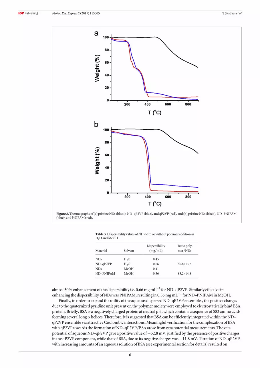
almost 50% enhancement of the dispersibility i.e. 0.66 mg mL−1 forND–qP2VP. Similarly effective inenhancing the dispersibility ofNDswas PNIPAM, resulting in 0.56 mgmL−1 forND–PNIPAM inMeOH.
Finally, in order to expand the utility of the aqueous dispersedND–qP2VP ensembles, the positive chargesdue to the quaternized pyridine unit present on the polymermoiety were employed to electrostatically bind BSAprotein. Briefly, BSA is a negatively charged protein at neutral pH, which contains a sequence of 583 amino acidsforming several longα helices. Therefore, it is suggested that BSA can be efficiently integratedwithin theND–qP2VP ensemble via attractive Coulombic interactions.Meaningful verification for the complexation of BSAwith qP2VP towards the formation ofND–qP2VP/BSA arose from zeta potentialmeasurements. The zetapotential of aqueousND–qP2VP gave a positive value of+52.8 mV, justified by the presence of positive chargesin the qP2VP component, while that of BSA, due to its negative charges was−11.8 mV. Titration ofND–qP2VPwith increasing amounts of an aqueous solution of BSA (see experimental section for details) resulted on
Figure 3.Thermographs of (a) pristineNDs (black), ND–qP2VP (blue), and qP2VP (red), and (b)pristineNDs (black), ND–PNIPAM(blue), and PNIPAM (red).
Table 3.Dispersibility values ofNDswith orwithout polymer addition inH2O andMeOH.
Material Solvent
Dispersibility
(mg/mL)Ratio poly-
mer/NDs
NDs H2O 0.45
ND–qP2VP H2O 0.66 86.8/13.2
NDs MeOH 0.41
ND–PNIPAM MeOH 0.56 85.2/14.8
6
Mater. Res. Express 2 (2015) 115005 T Skaltsas et al

decreasing the zeta potential, which eventually reached a value of+37.6 mV, indicating efficient electrostaticinteractions between the two oppositely charged components. Importantly, this was not the case when areference studywas conducted, utilizing lysozyme, a positively charged protein at neutral pH.When the latterwas added into theND–qP2VP ensemble (ca. 1.2 mg lysozyme in 3 mLof aqueousND–qP2VP) the zetapotential of the systemwasmarginally changed (i.e.+49.3 mV), hence suggesting that the repulsive forcesbetween the same charged lysozyme and q2PVPprevented its integration, in contrast with the case where BSAwas used (figure 4).
The formation of the three-component nanosystemwas also examined byDLS. The results together withthose of zeta potential corroborated the formation ofND–qP2VP/BSAnanoensembles. Despite the fact that nosignificant changes of theRhwere observed (figure 5(a)), by adding different amounts of BSA, the increase of thenormalized scattering intensity from36 000 kHz to 63 000 kHz (figure 5(b)) suggests themass increase of thedispersed hybrid particles, thus confirming the integration of BSAonto theND–qP2VPnanoensembles. Theinvariability ofRhmust be attributed to the small size of BSA, which after complexation does not contributesignificantly to the total size of the assembly.Moreover, turning our attention on the reference experiment withthe lysozyme, the intensity ofND–qP2VP/lysozymemixed solutionswas found at 35 000 kHz, which is almostthe same value that wasmeasured for theND–qP2VPnanoensembles alone (i.e. 36 000 kHz). The latterobservation suggests that there is no complexation between the positively charged protein lysozyme and thepositively chargedND–qP2VPnanoensemble, as expected. Practically, this corroborates that BSAwassuccessfully bounded onto theND–qP2VPnanoensemble via electrostatic interactions between the oppositecharges of the components.
4. Conclusions
The dispersibility and stability of nanodiamonds and their ensembles with several polymers in different solventswere evaluatedwith the aid of dynamic light scattering. In particular, although pristineNDs formed stabledispersions inwater aswell as inmethanol, theND–qP2VP andND–PNIPAMensembles were stable in aqueousmedia andMeOH, respectively.Moreover, the dispersibility value ofNDswas increased after the qP2VP orPNIPAMaddition inH2O andMeOH, respectively. Themorphology of pristineNDs and the formation ofND–polymer ensembles were observed byTEM,where primaryNDs particles with 3–5 nmdiameter were evident.Furthermore, the polymer coating ofND–polymerwas also evident. The thermal stability of pristineNDs andtheir ensembles with polymers was tested by TGA,while the amount of polymerwhichwas adsorbed onto theNDs surface in each case was determined to be 85–87%of the total nanoensemblemass. Finally,moving a stepforward and inspired by the possible utilization ofND–polymer ensembles as nanocarriers, bovine serumalbumin, which is a negatively charged protein, was successfully bounded via Coulombic interactions onto thepositively chargedND–qP2VP ensemble. The results were confirmed by zeta potential andDLSmeasurements,suggesting the ability of these novel chimericmaterials to be utilized in awide range of biomedical applications.
Figure 4.Zeta potential changes ofND–qP2VP/BSA as obtained upon titration of an aqueous dispersion ofND–qP2VPwith aqueousBSA.
7
Mater. Res. Express 2 (2015) 115005 T Skaltsas et al

Acknowledgments
Weare deeply indebted to Prof. Hisanori Shinohara and his group for kindly providing us access toHR-TEMfacilities at the ChemistryDepartment, NagoyaUniversity, Japan. Partialfinancial support from theGreekGeneral Secretariat for Research andTechnology and the EuropeanCommission, through the European Fundfor Regional Development, NSRF 2007–2013 action ‘Development of ResearchCentres—KPHPIS’, project447963 ‘NewMultifunctional NanostructuredMaterials andDevices—POLYNANO’ is acknowledged.
References
[1] MochalinVN, ShenderovaO,HoDandGogotsi Y 2012The properties and applications of nanodiamondsNat. Nano. 7 11–23[2] Krueger A and LangD 2012 Functionality is key: recent progress in the surfacemodification of nanodiamondAdv. Funct.Mater. 22
890–906[3] GreinerNR, PhillipsD S, Johnson JD andVolk F 1988Diamonds in detonation sootNature 333 440–2[4] Krueger A 2011 Beyond the shine: recent progress in applications of nanodiamond J.Mater. Chem. 21 12571–8[5] SchrandAM,Hens S AC and ShenderovaOA2009Nanodiamond particles: properties and perspectives for bioapplicationsCrit. Rev.
Solid StateMater. Sci. 34 18–74[6] SchrandAM,HuangH,CarlsonC, Schlager J J,Ōsawa E,Hussain SMandDai L 2006Are diamond nanoparticles cytotoxic? J. Phys.
Chem.B 111 2–7[7] HuangH, Pierstorff E, Osawa E andHoD2007Active nanodiamond hydrogels for chemotherapeutic deliveryNano Lett. 7 3305–14[8] ChenM, Pierstorff ED, LamR, Li S-Y,HuangH,Osawa E andHoD2009Nanodiamond-mediated delivery of water-insoluble
therapeuticsACSNano 3 2016–22
Figure 5. (a)The hydrodynamic radii of ND–qP2VP/BSA as obtained fromDLSmeasurements as a function of the BSAconcentration, and (b) the normalized scattering intensity versus the BSA concentration.
8
Mater. Res. Express 2 (2015) 115005 T Skaltsas et al

[9] HuangH,ChenM, Bruno P, LamR, Robinson E, GruenD andHoD2009Ultrananocrystalline diamond thinfilms functionalizedwiththerapeutically active collagen networks J. Phys. Chem.B 113 2966–71
[10] Faklaris O et al 2009 Photoluminescent diamond nanoparticles for cell labeling: study of the uptakemechanism inmammalian cellsACSNano 3 3955–62
[11] Barras A,Martin FA, BandeO, Baumann J-S, Ghigo J-M, BoukherroubR, BeloinC, Siriwardena A and Szunerits S 2013Glycan-functionalized diamond nanoparticles as potent E. coli anti-adhesivesNanoscale 5 2307–16
[12] Moore L, EK-HC,Osawa E, Bishop JMandHoD2013Diamond-lipid hybrids enhance chemotherapeutic tolerance andmediatetumor regressionAdv.Mater. 25 3532–41
[13] Xing Y andDai L 2009Nanodiamonds for nanomedicineNanomedicine 4 207–18[14] MohanN,ChenC-S,HsiehH-H,WuY-C andChangH-C 2010 In Vivo Imaging andToxicity Assessments of Fluorescent
Nanodiamonds inCaenorhabditis elegansNano Lett. 10 3692–9[15] OzawaM, InagumaM, TakahashiM, Kataoka F, Krüger A andŌsawa E 2007 Preparation and behavior of brownish, clear
nanodiamond colloidsAdv.Mater. 19 1201–6[16] Pentecost A, Gour S,MochalinV, Knoke I andGogotsi Y 2010Deaggregation of nanodiamond powders using salt- and sugar-assisted
millingACSAppl.Mater. Interfaces 2 3289–94[17] Ōsawa E 2007Recent progress and perspectives in single-digit nanodiamondDiamondRelat.Mater. 16 2018–22[18] ShenderovaO, Petrov I,Walsh J, GrichkoV,GrishkoV, Tyler T andCunninghamG2006Modification of detonation nanodiamonds
by heat treatment in airDiamondRelat.Mater. 15 1799–803[19] Larionova I, KuznetsovV, Frolov A, ShenderovaO,Moseenkov S andMazov I 2006 Properties of individual fractions of detonation
nanodiamondDiamondRelat.Mater. 15 1804–8[20] Morita Y, Takimoto T, YamanakaH,KumekawaK,Morino S, Aonuma S, Kimura T andKomatsuN2008A facile and scalable process
for size-controllable separation of nanodiamond particles as small as 4 nm Small 4 2154–7[21] PengW,Mahfouz R, Pan J,HouY, Beaujuge PMandBakrOM2013Gram-scale fractionation of nanodiamonds by density gradient
ultracentrifugationNanoscale 5 5017–26[22] Cheng J,He J, Li C andYang Y 2008 Facile approach to functionalize nanodiamond particles with v-shaped polymer brushesChem.
Mater. 20 4224–30[23] MatrabT, ChehimiMM, Boudou J P, Benedic F,Wang J, NaguibNNandCarlisle J 2006A Surface functionalization of
ultrananocrystalline diamond using atom transfer radical polymerization (ATRP) initiated by electro-grafted aryldiazonium saltsDiamondRelat.Mater. 15 639–44
[24] ZhangR, Shi Z, Liu Y andYin J 2012 Synthesis and characterization of polybenzimidazole–nanodiamond hybrids via in situpolymerizationmethod J. Appl. Polym. Sci. 125 3191–9
[25] LiH, Song S I, SongGY andKim I 2014Non-covalently functionalized carbon nanostructures for synthesizing carbon-based hybridnanomaterials J. Nanosci. Nanotechnol. 14 1425–40
[26] Behler KD, Stravato A,MochalinV, KornevaG, YushinG andGogotsi Y 2009Nanodiamond-polymer composite fibers and coatingsACSNano 3 363–9
[27] BransonBT, SeifMA,Davidson J L and Lukehart CM2011 Fabrication andmacro/nanoscale characterization of aggregated andhighly de-aggregated nanodiamond/polyacrylonitrile composite thickfilms J.Mater. Chem. 21 18832–9
[28] Reitinger AA,HutterNA,Donner A, SteenackersM,WilliamsOA, StutzmannM, JordanR andGarrido J 2013A functional polymerbrushes on diamond as a platform for immobilization and electrical wiring of biomoleculesAdv. Funct.Mater. 23 2979–86
[29] Krueger A, Stegk J, Liang Y, Lu L and JarreG 2008 Biotinylated nanodiamond: simple and efficient functionalization of detonationdiamond Langmuir 24 4200–4
[30] Perevedentseva E, Cai P J, Chiu YC andChengCL 2010Characterizing protein activities on the lysozyme and nanodiamond complexprepared for bio applications Langmuir 27 1085–91
[31] Dahoumane SA,NguyenMN,Thorel A, Boudou J-P, ChehimiMMandMangeneyC 2009 Protein-functionalized hairy diamondnanoparticles Langmuir 25 9633–8
[32] Huang LCL andChangH-C 2004Adsorption and immobilization of cytochrome c onnanodiamonds Langmuir 20 5879–84[33] NguyenTT-B, ChangH-C andWuVW-K2007Adsorption and hydrolytic activity of lysozyme on diamond nanocrystallitesDiamond
Relat.Mater. 16 872–6[34] Shimkunas RA, Robinson E, LamR, Lu S, XuX, ZhangX-Q,HuangH,Osawa E andHoD2009Nanodiamond–insulin complexes as
pH-dependent protein delivery vehiclesBiomaterials 30 5720–8[35] Chung PH, Perevedentseva E, Tu J S, ChangCC andChengCL 2006 Spectroscopic study of bio-functionalized nanodiamonds
DiamondRelat.Mater. 15 622–5[36] Tzeng Y-K, Faklaris O, Chang B-M,KuoY,Hsu J-H andChangH-C 2011 Superresolution imaging of albumin-conjugated fluorescent
nanodiamonds in cells by stimulated emission depletionAngew. Chem., Int. Ed. 50 2262–5[37] Lee JW, Lee S, Jang S, HanKY,KimY,Hyun J, Kim SK and Lee Y 2013 Preparation of non-aggregated fluorescent nanodiamonds
(FNDs) by non-covalent coatingwith a block copolymer and proteins for enhancement of intracellular uptakeMol. BioSyst. 9 1004–11[38] Hadjichristidis N, IatrouH, Pispas S andPitsikalisM2000Anionic polymerization: high vacuum techniques J. of Polymer Science Part
A: Polymer Chemistry 38 3211–34[39] Smith AE, XuX andMcCormick C L 2010 Stimuli-responsive amphiphilic (co)polymers via RAFTpolymerization Prog. Polym. Sci. 35
45–93[40] OrfanouK, TopouzaD, SakellariouG and Pispas S 2003Graftlike interpolymer complexes frompoly(2-vinylpyridine) and end-
sulfonic acid polystyrene and polyisoprene: Intermediates to noncovalently bonded block copolymer-likemicelles J. of Polymer SciencePart A: Polymer Chemistry 41 2454–61
[41] Pispas S andHadjichristidis N 2003Micellization behavior of poly(butadiene-b-sodiummethacrylate) copolymers in dilute aqueousmediaMacromolecules 36 8732–7
[42] Pispas S 2006 Soluble complexes of sodiumpoly(isoprene-b-methacrylate)micelles with cationic surfactants in aqueousmedia J. Phys.Chem.B 110 2649–55
[43] MountrichasG, Pispas S, Xenogiannopoulou E, Aloukos P andCouris S 2007Aqueous dispersions of C60 fullerene by use ofamphiphilic block copolymers: preparation andnonlinear optical properties J. Phys. Chem.B 111 4315–9
[44] TalelliM and Pispas S 2008Complexes of cationic block copolymermicelles with dna: histone/dna complexmimeticsMacromol.Biosci. 8 960–7
[45] Zhao J, ZhangG and Pispas S 2009Morphological transitions in aggregates of thermosensitive poly(ethylene oxide)-b-poly(N-isopropylacrylamide) block copolymers prepared via RAFTpolymerization J. of Polymer Science Part A: Polymer Chemistry 47 4099–110
9
Mater. Res. Express 2 (2015) 115005 T Skaltsas et al

[46] Škvarla J, Zedník J, ŠloufM, Pispas S and ŠtěpánekM2014Poly(N-isopropyl acrylamide)-block-poly(n-butyl acrylate)thermoresponsive amphiphilic copolymers: Synthesis, characterization and self-assembly behavior in aqueous solutions Eur. Polym. J.61 124–32
[47] Krueger A 2008Diamond nanoparticles: jewels for chemistry and physicsAdv.Mater. 20 2445–9
10
Mater. Res. Express 2 (2015) 115005 T Skaltsas et al


