Embed Size (px)
Citation preview
Molecular Biology of the CellVol. 15, 1172–1184, March 2004
Novel Kelch-like Protein, KLEIP, Is Involved in ActinAssembly at Cell-Cell Contact Sites of Madin-DarbyCanine Kidney CellsTakahiko Hara, Hiroshi Ishida, Razi Raziuddin, Stephan Dorkhom,Keiju Kamijo, and Toru Miki*
Molecular Tumor Biology Section, Basic Research Laboratory, National Cancer Institute, Bethesda,Maryland, 20892-4255
Submitted July 25, 2003; Revised October 29, 2003; Accepted November 17, 2003Monitoring Editor: Martin A. Schwartz
Dynamic rearrangements of cell-cell adhesion underlie a diverse range of physiological processes, but their precisemolecular mechanisms are still obscure. Thus, identification of novel players that are involved in cell-cell adhesion wouldbe important. We isolated a human kelch-related protein, Kelch-like ECT2 interacting protein (KLEIP), which contains thebroad-complex, tramtrack, bric-a-brac (BTB)/poxvirus, zinc finger (POZ) motif and six-tandem kelch repeats. KLEIPinteracted with F-actin and was concentrated at cell-cell contact sites of Madin-Darby canine kidney cells, where itcolocalized with F-actin. Interestingly, this localization took place transiently during the induction of cell-cell contact andwas not seen at mature junctions. KLEIP recruitment and actin assembly were induced around E-cadherin–coated beadsplaced on cell surfaces. The actin depolymerizing agent cytochalasin B inhibited this KLEIP recruitment aroundE-cadherin–coated beads. Moreover, constitutively active Rac1 enhanced the recruitment of KLEIP as well as F-actin to theadhesion sites. These observations strongly suggest that KLEIP is localized on actin filaments at the contact sites. We alsofound that N-terminal half of KLEIP, which lacks the actin-binding site and contains the sufficient sequence for thelocalization at the cell-cell contact sites, inhibited constitutively active Rac1-induced actin assembly at the contact sites.We propose that KLEIP is involved in Rac1-induced actin organization during cell-cell contact in Madin-Darby caninekidney cells.
INTRODUCTION
Cell-cell adhesion is crucial for development and survival ofmulticellular organism (Adams et al., 1998). In epithelialtissues, cadherins are required for the assembly of cells intomultilayer and the establishment and maintenance of theepithelial phenotype (Braga et al., 1997). Cadherins are trans-membrane proteins that bind to the same type of cadherinsin adjacent cells. The exodomain of cadherins interacts witheach other by calcium-dependent homophilic interaction,whereas the cytoplasmic domain binds to �-catenin, and thiscomplex is linked to the actin filaments by �-catenin (Va-sioukhin and Fuchs, 2001). Functional cadherin receptorscan influence the reorganization of the actin filaments andother cytoskeletal components, and the distribution of trans-membrane proteins in a polarized manner, and the forma-tion of the tight junctions and other adhesive junctions. It istherefore important to understand how cadherin-mediatedcell adhesion is regulated. Although several different mech-anisms have been proposed for the regulation, the regula-tory mechanisms of cadherin-mediated cell adhesion are not
yet fully understood (Fukata et al., 2001; Sahai and Marshall,2002).
Recent studies revealed that Rho family small GTPases areinvolved in the cadherin-mediated cell adhesion (Braga,2002b). It has been reported that Rac1 promotes E-cadherin–dependent cell adhesion in Madin-Darby canine kidney(MDCK) cells (Takaishi et al., 1997; Jou and Nelson, 1998).Rac1 is activated by the formation of E-cadherin–based celladhesion (Nakagawa et al., 2001). It has been demonstratedthat Rac1 is spatiotemporally recruited at the sites of newestcell-cell adhesion and regulates the formation of lamellipo-dia, which are actin-based membrane protrusion duringcell-cell adhesion (Ehrlich et al., 2002). By contrast, RhoAactivity was suppressed via p190RhoGAP during cell-cellcontact (Noren et al., 2003). Thus, Rac1 is likely to fulfill thetask of coordinating cell adhesion and actin-based cytoskel-etal remodeling. IQGAP1, one of the targets of Cdc42 orRac1, was shown to regulate the linkage between cadherinsand the actin filaments (Fukata et al., 2001). The IQGAP1-based model supports a positive role for Rac1 in regulatingthe cadherin–mediated cell contacts (Fukata and Kaibuchi,2001). However, some contradictions still remain. For exam-ple, dominant negative Rac1 has no effect on cell-cell contactformation or cadherin–catenins complex recruitment, al-though it inhibits the actin-accumulation in myogenic C2cells (Lambert et al., 2002). Although the modulation of thecadherin-mediated cell adhesion by Rac1 might depend onthe cell type, these findings suggest that there might beadditional mechanisms of the cadherin-actin linkage.
Article published online ahead of print. Mol. Biol. Cell 10.1091/mbc.E03–07–0531. Article and publication date are available at www.molbiolcell.org/cgi/doi/10.1091/mbc.E03–07–0531.
* Corresponding author. E-mail address: [email protected] used: CA, constitutively active; ECT2, epithelialcell transforming gene 2; GFP, green fluorescence protein; GST,glutathione S-transferase; KLEIP, Kelch-like ECT2 interactingprotein; PBS, phosphate-buffered saline; TBS, Tris-buffered sa-line.
1172 © 2004 by The American Society for Cell Biology http://www.molbiolcell.org/content/suppl/2003/12/10/E03-07-0531.DC1.htmlSupplemental Material can be found at:
The Drosophila Kelch protein is an actin filament cross-linking protein and plays an important role on the mainte-nance of ring canals that regulate cytoplasmic transportfrom nurse cells to the developing oocyte within an eggchamber (Kelso et al., 2002). Kelch has two sequence motifs(Xue and Cooley, 1993). The first motif, broad-complex,tramtrack, bric-a-brac (BTB)/poxvirus, zinc finger (POZ) do-main has been proposed to function as a protein–proteininteraction interface (Godt et al., 1993; Albagli et al., 1995).The second motif, the kelch repeats contain repeated se-quences, each of which is composed of 40–50 amino acids(Xue and Cooley, 1993). The kelch repeats are required for itsinteraction with actin cytoskeleton (Kelso et al., 2002). Sev-eral mammalian proteins that contain the kelch repeats withthe BTB/POZ motif are known (Adams et al., 2000), andsome of them are actin-binding proteins. Like DrosophilaKelch, Kelch-related proteins might be involved in organi-zation of actin cytoskeleton in various situations. However,proteins containing the kelch repeats have diverse cellularfunctions (Robinson and Cooly, 1997). Moreover, low aminoacid identity of Kelch with other members of the BTB/Kelchsubfamily makes it difficult to presume their functions(Bomont and Koeing, 2003).
In this study, we identified Kelch-like ECT2 interactingprotein (KLEIP) as a protein that can associate with ECT2, aRho nucleotide exchange factor involved in cytokinesis(Miki et al., 1993; Tatsumoto et al., 1999). Whereas KLEIPseems to regulate cytokinesis, we show that KLEIP is alsoinvolved in actin assembly at the sites of cell-cell adhesion.
MATERIALS AND METHODS
Isolation of KLEIP cDNAThe original KLEIP cDNA was isolated from a human testis cDNA library ina yeast two-hybrid screening by using the N-terminal half of human ECT2 asbait. A cDNA clone containing the entire open reading frame was isolated bypolymerase chain reaction (PCR) by using the sequence of the kelch repeats-containing protein (GenBank accession no. AB02610). PCR was performed ina 25-�l mixture containing 10 mM Tris-HCl, 1.5 mM MgCl2, 50 mM KCl, 10mM each dNTPs, 2.5 U of high-fidelity Taq polymerase (BD BiosciencesClontech, Palo Alto, CA), 30 mM each forward primer and reverse primer,and 0.5 �g of cDNA from testis. The sequence of the forward- and reverse-primers were 5�-GGGAGATCTATGGAAGGAAAGCCAATGCGC-3� and 5�-GGGTCGACTCACCAAATATGGGATTCACA-3�, respectively. The mixturewas heated at 95°C for 5 min and then subjected to 35 cycles of amplificationat 94°C for 45 s, 56°C for 45 s, and 72°C for 2 min. PCR products wereseparated in 2% agarose gels and purified using Ultra clean (MO BIO Labo-ratory, Salana Beach, CA) and then subcloned in pGEX-T vector (Promega,Madison, WI). The DNA sequences of the isolated KLEIP cDNA were thesame as that of AB026190 except for position 1726, where G has been replacedby T in the KLEIP cDNA, resulting in replacement of Trp at the amino acid593 by Gly. Because our sequence was identical to the corresponding positionof another entry of this gene (GenBank accession no. AK001430), this may notrepresent a mutation caused by PCR.
Construction of Expression VectorsFull-length KLEIP, KLEIP-N (aa 1–303), and -C (aa 301–609) cDNA weresubcloned in the pEGFP-C1 expression vector (BD Biosciences Clontech),which contains the green fluorescent protein (GFP) or the pCEV32-F3, whichcontains the FLAG-epitope.
Preparation of AntibodyKLEIP-N or KLEIP-C was introduced into pCEV30G vector, which containsglutathione S-transferase (GST). GST-KLEIP fusion protein was expressed inEscherichia coli with 0.4 mM isopropyl-�-d-thiogalactopyranoside at 25°C for2 h. GST-KLEIP-N fusion protein was purified using glutathione-Sepharose4B (Amersham Biosciences, Piscataway, NJ) and then eluted with 20 mMreduced glutathione from the beads. Because GST-KLEIP-C was in insolublefraction, it was separated on SDS-PAGE gels and purified by electroelution.The samples were dialyzed in phosphate-buffered saline (PBS). The purifiedGST-KLEIP-N or -C fusion protein was used for immunizing rabbits. Weraised two antisera against each of KLEIP-N and -C. The IgG fraction waspurified from the rabbit antisera by using a protein A-Sepharose column(Amersham Biosciences). For affinity purification of anti-KLEIP antibodies,
KLEIP-N and KLEIP-FL was subcloned into pET-32 vector (Novagen, Darm-stadt, Germany) and pMAL2c-E (New England Biolabs, Beverly, MA), respec-tively. His-tagged KLEIP-N and maltose binding protein (MBP)-fusedKLEIP-FL were expressed in E. coli as described above, and purified usingNi-NTA resin (Novagen) and amylose resin (New England Biolabs), respec-tively. Each purified fusion protein was coupled with cyanogen bromide-activated beads (Amersham Biosciences) for affinity chromatography. Anti-KLEIP-N and anti-KLEIP-C were purified through His-KLEIP-N and MBP-KLEIP-FL affinity columns, respectively.
The following antibodies were from commercial sources: anti-�-tubulin(Sigma-Aldrich, St. Louis, MO), anti-FLAG antibody (M2 FLAG antibody;Sigma-Aldrich), anti-ZO1 (Zymed Laboratories, South San Francisco, CA),and anti-E-cadherin and �-catenin (BD Transduction Laboratories, San Diego,CA).
Cell CultureMDCK, HeLa, HEK293T, COS, Swiss 3T3, and NIH 3T3 cells were grown in35- or 100-mm dish with DMEM (Invitrogen, Carlsbad, CA) containing 10%FBS. U2OS cells were cultured with McCoy’s 5A medium (Invitrogen) con-taining 10% fetal bovine serum (FBS). MDCK, HeLa, and HEK293 cells are ofepithelial origin, whereas Swiss 3T3 and NIH3T3 cells are mouse fibroblasts.COS cells and U2OS cells are derived from kidney of monkey and humanosteosarcoma, respectively. HEK293T is a human embryonic kidney cells.
For calcium-switch assays, MDCK cells were grown in low calcium-con-taining media (calcium-free S-MEN containing 2% dialyzed FBS) overnightand then incubated normal media for several hours.
For transfection, LipofectAMINE 2000 (Invitrogen) was used for the tran-sient transfection according to the manufacturer’s protocols.
ImmunoblottingCells were lysed in lysis buffer (50 mM Tris-HCl, pH 7.5, 1% NP-40, 150 mMNaCl, 2 mM EDTA, 2 mM phenylmethylsulfonyl fluoride, 0.1 mg/ml apro-tinin, 0.1 mg/ml leupeptin) at 4°C for 30 min. Total cell lysates were clarifiedby centrifugation at 14,000 � g for 20 min at 4°C and then the proteinconcentration of each sample was determined using BCA assay (Pierce Chem-ical, Rockford, IL). Samples were separated and analyzed on 8–16% gradientSDS-PAGE gels (Invitrogen) and then transferred to polyvinylidene difluo-ride membranes (Invitrogen). The membranes were blocked with Tris-buff-ered saline containing 3% milk and 0.1% Tween 20 and then probed withprimary antibodies. The membranes were washed with Tris-buffered salinecontaining 0.1% Tween 20 for 7 min three times and probed with horseradishperoxidase-conjugated secondary antibody. Immunoreactive bands were vi-sualized using ECL (Amersham Biosciences).
ImmunoprecipitationAn identical amount (390 �g of protein) of protein from each sample wasprecleared by incubation with protein G-Sepharose (Amersham Biosciences)for 1 h at 4°C. Immunoprecipitation of antigen–antibody complex was accom-plished by incubation for 4 h at 4°C. The antigen–antibody complex wasincubated with protein G-Sepharose for 1 h at 4°C. Bound proteins weresolubilized in Laemmli buffer and analyzed by immunoblotting.
mRNA Expression AnalysisMTC panel single-strand cDNA (BD Biosciences Clontech) was used formRNA expression analysis. PCR was performed in a 25-�l mixture containing10 mM Tris-HCl, 1.5 mM MgCl2, 50 mM KCl, 10 mM each dNTPs, 2.5 U of Taqpolymerase (Promega), 30 mM each forward primer and reverse primer, and2.5 �l of MTC panel single-strand cDNA. PCR primer sets were as follows: forKLEIP, the forward primer was 5�-GGGAGATCTCTAATGCAAGGAC-CAAGGACG-3� and the reverse primer was 5�-GCATAAAGAAAGCCTC-CAAGTACT-3�; and for glyceraldehyde-3-phosphate dehydrogenase, a for-ward primer (5�-TGAAGGTCGGAGTCAACGGATTTGGT-3�) and a reverseprimer (5�-CATGTGGGC CATGAGGTCCACCAC-3�) were used. The mix-tures were heated at 95°C for 5 min and then subjected to 26-cycle amplifi-cation at 94°C for 45 s, 60°C for 45 s, and 72°C for 1 min. PCR products wereseparated on 2% agarose gel and visualized using ethidium bromide. Thesequences of the PCR products were confirmed using automated DNA se-quencer.
Cross-Linking of MBP-KLEIP-N DimersThe MBP-KLEIP-N fusion protein and MBP were expressed and purified asdescribed above. The protein was incubated in a buffer containing 20 mMHEPES, 150 mM NaCl, and 10 mM dithiobis-(propionic acid n-hydroxysuc-cinimide ester) (DTSP), a bifunctional protein cross-linking reagent, freshlyprepared as a 450 mM stock in dimethyl sulfoxide (Sigma-Aldrich). Cross-linking was carried out at room temperature for 30 min and terminated byadding 2� Laemmli buffer either with or without 50 mM dithiothreitol (DTT).Proteins were boiled for 5 min, resolved on an 8–16% gradient gel, andvisualized by Coomassie Blue staining.
Novel Kelch-related Protein, KLEIP
Vol. 15, March 2004 1173
Actin Cosedimentation AssaysActin cosedimentation assays were performed using the actin binding proteinbiochem kit (Cytoskeleton, Denver, CO). Briefly, rabbit muscle actin waspolymerized at 24°C for 1 h under 5 mM Tris-HCl, pH 8.0, 0.2 mM CaCl2, 50mM KCl, 2 mM MgCl2, and 1 mM ATP. MBP-fusion proteins were expressedand purified as described above. Purified proteins (2 �M) were added andincubated at 24°C for 30 min with or without the prepared polymerized actin.The samples were centrifuged at 24°C, 150,000 � g for 90 min. The precipi-tates were suspended in Laemmli buffer. The supernatants and precipitateswere analyzed using SDA-PAGE and visualized by Coomassie Blue staining.
Immunofluorescence MicroscopyCells were grown on coverslips and fixed for 20 min with 4% paraformalde-hyde (PFA) in PBS, and then cells were permeabilized for 7 min with 0.1%Triton X-100 in PBS and washed with PBS twice. For staining against E-cadherin, cells were fixed for 7 min with 100% methanol at –20°C. Sampleswere incubated with PBS containing 2% horse serum for 1 h and then withprimary antibody for 1 h. The cells were washed in PBS three times andincubated with Cy3- (Jackson ImmunoResearch Laboratories, West Grove,PA) or Alexa 488-conjugated second antibody (Molecular Probes, Eugene,OR). To detect actin filaments, cells were incubated with Alexa 350 or 488-conjugated phalloidin (Molecular Probes) for 20 min. Cells were observedwith fluorescence microscopy. For deconvolution microscopy (Figures 4Aand 5, A and D), the image of each focal plane was taken, and each image wascleaned by removing blurs from other focal planes using Open Lab software(Improvision, Lexington, MA).
To evaluate KLEIP recruitment at the sites of cell-cell contact, the cells thatexhibited at least one clear line-like staining pattern of KLEIP along thecell-cell border were scored.
When the effects of constitutive active Rac or KLEIP-N were examined, cellswith increased staining patterns of KLEIP or F-actin at the cell-cell contactsites compared with those of surrounding cells were scored positive.
E-Cadherin/Fc Beads AssaysConcanavalin A (ConA; Sigma-Aldrich) or human E-cadherin/Fc chimera(Sigma-Aldrich) was immobilized with latex beads as described previously(Burbelo et al., 1995; Lambert et al., 2000). Briefly, 200 �l of Polystyrene latexbeads (6.4-�m average diameter; Sigma-Aldrich) were coated with either 100�g/ml ConA or 100 �g/ml E-Cadherin/Fc in 0.1 M borate buffer (pH 8.0) atroom temperature for 18 h. The beads were washed with PBS twice and thenincubated with 2 mg/ml bovine serum albumin at room temperature for 2 h.The beads were washed using PBS three times again and suspended in 200 �lof PBS containing 1 mM CaCl2. An aliquot of beads (5 �l) was used for a35-mm culture dish. MDCK cells were incubated in the presence of beads for2 h at 37°C and analyzed by immunofluorescence. The beads placed on cellsurfaces were identified by phase contrast image, and examined for rim-likestaining of �-catenin, F-actin and KLEIP around the beads. To analyze theeffect of F-actin on KLEIP recruitment, MDCK cells were incubated withE-cadherin/Fc chimera-coated beads in the presence of vehicle alone (di-methyl sulfoxide) or 10 �g/ml cytochalasin B (Sigma-Aldrich) for 2 h at 37°C.
RESULTS
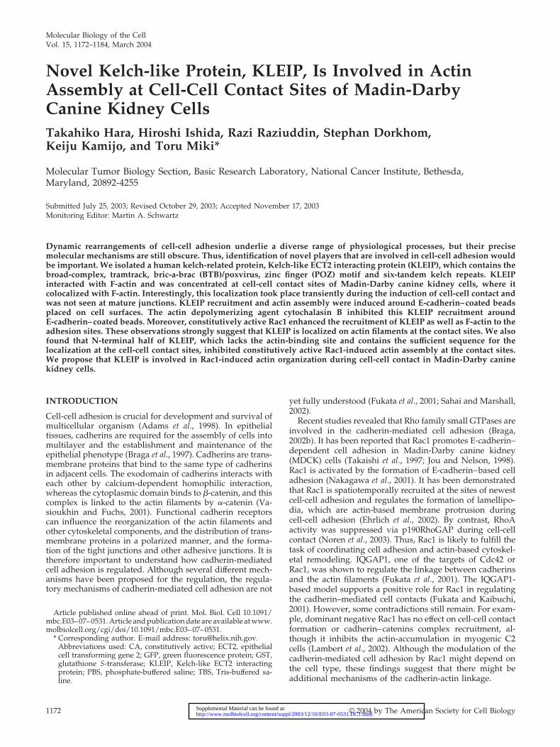
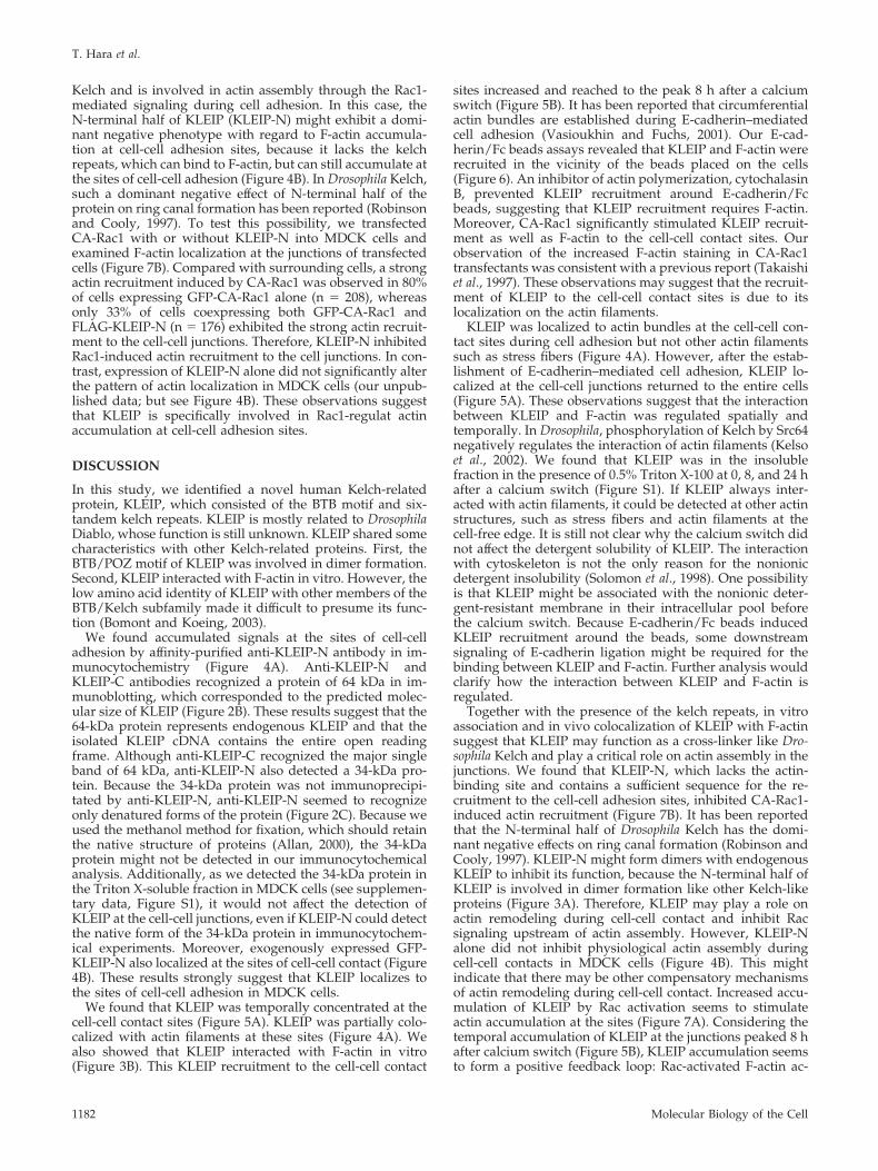
KLEIP Encodes a Kelch-like ProteinWe identified a human kelch repeat-containing protein, des-ignated KLEIP, in a yeast two-hybrid screening by usingECT2 as bait. A database search revealed that KLEIP isnearly identical to a Kelch-related protein of unknown func-tion (GenBank accession no. AB026190). A full-length cDNAwas isolated by PCR cloning from a human cDNA library asa template. The predicted KLEIP protein contains a BTB/POZ domain and six-tandem kelch repeats of 40–50-aminoacid sequence that contained highly conserved sequencesamong the repeats (Figure 1). It has been reported that thekelch repeats forms a �-sheet propeller structure (Adams etal., 2000).
Database search also revealed two proteins closely relatedto KLEIP. Drosophila Diablo (GenBank accession no.AF237711) exhibited amino acid identities of 80.8% to KLEIP(Figure 1C). In contrast, the sequence identity between hu-man KLEIP and Drosophila Kelch was 43.0%. Therefore, Dia-blo seems to be the Drosophila orthologue of human KLEIP.Although the biological function of Drosophila Kelch is welldocumented, the function of Diablo is still unknown. An-other protein in Anopheles (GenBank accession no.
EAA05692) also exhibited a high amino acid identity toKLEIP (79.8%), and could be the Anopheles orthologue ofhuman KLEIP. The biological function of the AnophelesKelch-like protein is also not known.
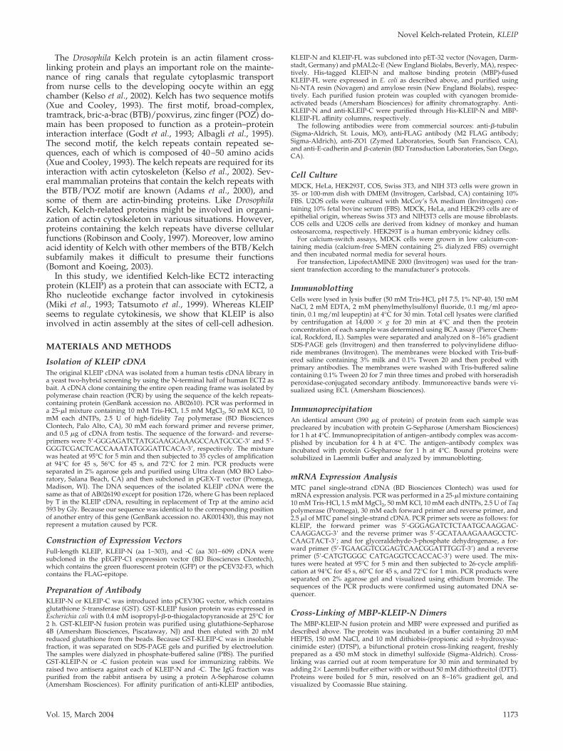
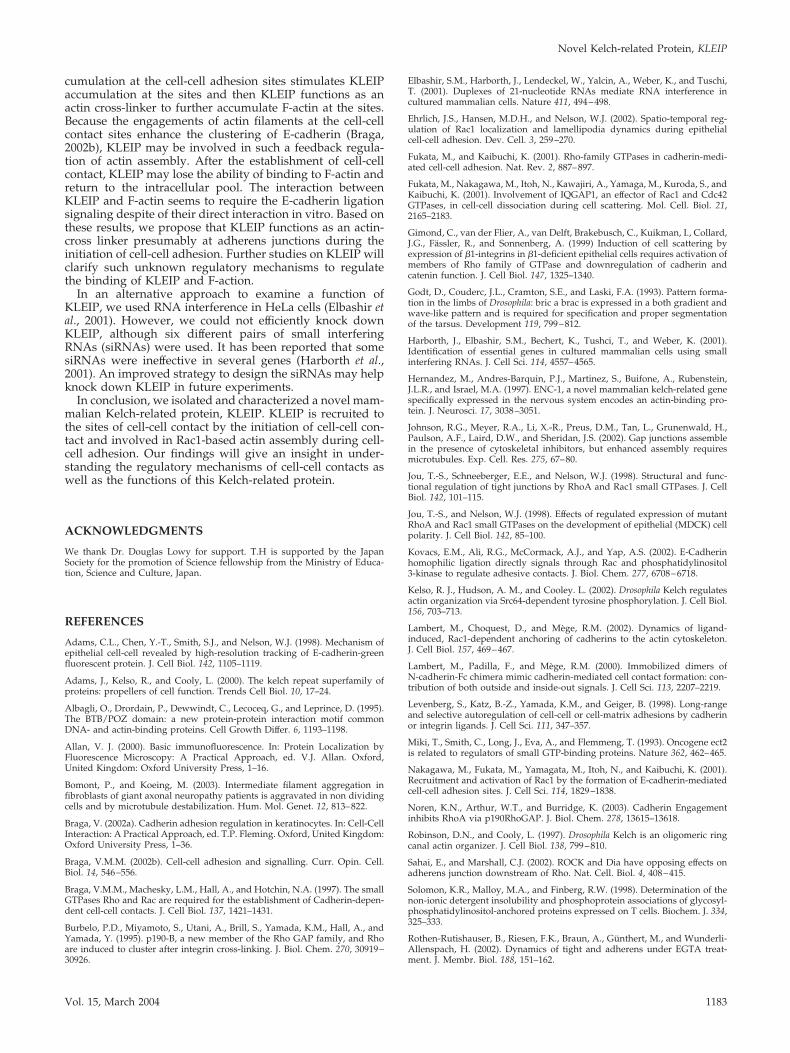
Identification and Expression of Endogenous KLEIPTo analyze endogenous KLEIP, we raised antisera againstthe N-terminal (KLEIP-N) and C-terminal (KLEIP-C) half ofKLEIP (Figure 1A) and affinity purified the antibodies. Totest these antibodies, we expressed FLAG-tagged KLEIP-Nand full-length KLEIP (KLEIP-FL) in COS cells (Figure 2A).Anti-FLAG antibody detected major bands of �40- and 64-kDa in FLAG-KLEIP-N and FLAG-KLEIP-FL transfectants,respectively (Figure 2A, right). The sizes of these proteinscorresponded to the predicted sizes of FLAG-KLEIP-N and-FL. Anti-FLAG antibody also detected several minor bands,three of which were common in the lysates of KLEIP-N andKLEIP-FL transfectants, suggesting that these bands repre-sented degradation products. Anti-KLEIP-N antibody de-tected similar proteins in addition to the band whose mo-bility was slightly lower than that of FLAG-KLEIP-FL(Figure 2A, middle). This band seemed to be endogenousKLEIP, because it was commonly observed in KLEIP-N,KLEIP-FL, and vector alone transfectants. Anti-KLEIP-Nalso detected degradation products of exogenous proteins inKLEIP-N and KLEIP-FL transfectants. However, anti-KLEIP-N also detected bands of 34 kDa in all three lanes(Figure 2, A and B). Preimmune sera did not detect anyprotein (Figure 2A, left). In contrast, anti-KLEIP-C recog-nized only a protein of 64 kDa (Figure 2B), supporting thatthe 64-kDa protein is endogenous KLEIP. It is possible thatthe 34-kDa protein might be an isoform of KLEIP that lacksthe C-terminal domain, because anti-KLEIP-C did notrecognize the protein. Alternatively, it might represent anunrelated protein recognized by a cross-reactivity of anti-KLEIP-N. When anti-KLEIP-N was used for immunopre-cipitation, the 64-kDa protein was detected in the immuno-precipitate, whereas the 34-kDa protein was not (Figure 2C).The 64-kDa protein, but not the 34-kDa protein, was alsodetected by anti-KLEIP-C after immunoprecipitation by an-ti-KLEIP-N (our unpublished data). These results suggestthat the 64-kDa protein represents endogenous KLEIP, andthe 34-kDa protein is recognized by anti-LEIP-N only whenit is denatured. We also performed immunoblotting andimmunoprecipitation against anti-KLEIP-N by using lysatesof MDCK cells and obtained the similar results to those ofHeLa cells (see supplemental data, Figure S1).
Next, we examined the expression of KLEIP in severalcultured cells (Figure 2D). The highest expression of KLEIPwas observed in MDCK cells among the cultured cells ex-amined. KLEIP expression was also detected in HeLa andSwiss 3T3 cells but not significantly in HEK293T andNIH3T3 cells. We also examined mRNA expression levels ofKLEIP in several human tissues by a PCR-based analysis.KLEIP mRNA was detected among a number of adult tissuesexamined (Figure 2E), suggesting that KLEIP is ubiquitouslyexpressed in various cell types.
KLEIP Can Form Dimers and Associate with F-ActinIt has been reported that some Kelch-related proteins withthe BTB/POZ motif can form homodimers through theirBTB/POZ domains (Hernandez et al., 1997; Soltysik-Es-panola et al., 1999). To determine whether KLEIP can formdimers, affinity-purified MBP-fused KLEIP-N or MBP alonewas cross-linked by the bifunctional protein cross-linkerDTSP and analyzed by SDS-PAGE. DTSP forms stable link-ages between free amine groups of interacting proteins, and
T. Hara et al.
Molecular Biology of the Cell1174
Figure 1. KLEIP belongs to the superfamilyof Kelch-like proteins. Predicted structure ofKLEIP and its derivatives. (A) Schematic rep-resentation of the structure of KLEIP and itsderivatives. KLEIP contains the N-terminalBTB/POZ motif (aa 53–165) and C-terminalkelch repeat domains. (aa 316–609). The re-gions carried by KLEIP-N (aa 1–303) and -C(aa 301–609) are shown. (B) Alignment of thekelch repeats of human KLEIP. Conservedamino acids are shown in bold. The bottomline shows the consensus sequence of thekelch repeats. The C-terminal 16 amino acidresidues (aa 598–613) is a part of the firstkelch repeat (Adams et al., 2000). (C) Aminoacid comparison of human KLEIP, DrosophilaDiablo, the Anopheles Kelch-like protein, andDrosophila Kelch. Consensus sequences of theBTB motif and the kelch repeats are shown atthe top.
Novel Kelch-related Protein, KLEIP
Vol. 15, March 2004 1175
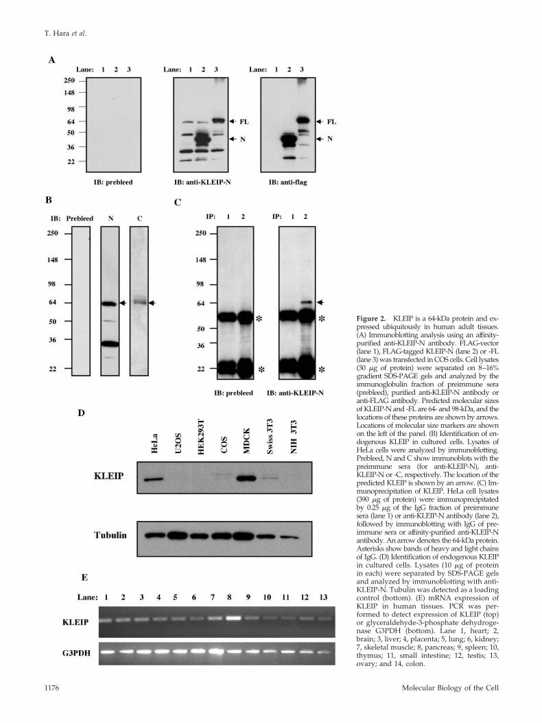
Figure 2. KLEIP is a 64-kDa protein and ex-pressed ubiquitously in human adult tissues.(A) Immunoblotting analysis using an affinity-purified anti-KLEIP-N antibody. FLAG-vector(lane 1), FLAG-tagged KLEIP-N (lane 2) or -FL(lane 3) was transfected in COS cells. Cell lysates(30 �g of protein) were separated on 8–16%gradient SDS-PAGE gels and analyzed by theimmunoglobulin fraction of preimmune sera(prebleed), purified anti-KLEIP-N antibody oranti-FLAG antibody. Predicted molecular sizesof KLEIP-N and -FL are 64- and 98-kDa, and thelocations of these proteins are shown by arrows.Locations of molecular size markers are shownon the left of the panel. (B) Identification of en-dogenous KLEIP in cultured cells. Lysates ofHeLa cells were analyzed by immunoblotting.Prebleed, N and C show immunoblots with thepreimmune sera (for anti-KLEIP-N), anti-KLEIP-N or -C, respectively. The location of thepredicted KLEIP is shown by an arrow. (C) Im-munoprecipitation of KLEIP. HeLa cell lysates(390 �g of protein) were immunoprecipitatedby 0.25 �g of the IgG fraction of preimmunesera (lane 1) or anti-KLEIP-N antibody (lane 2),followed by immunoblotting with IgG of pre-immune sera or affinity-purified anti-KLEIP-Nantibody. An arrow denotes the 64-kDa protein.Asterisks show bands of heavy and light chainsof IgG. (D) Identification of endogenous KLEIPin cultured cells. Lysates (10 �g of proteinin each) were separated by SDS-PAGE gelsand analyzed by immunoblotting with anti-KLEIP-N. Tubulin was detected as a loadingcontrol (bottom). (E) mRNA expression ofKLEIP in human tissues. PCR was per-formed to detect expression of KLEIP (top)or glyceraldehyde-3-phosphate dehydroge-nase G3PDH (bottom). Lane 1, heart; 2,brain; 3, liver; 4, placenta; 5, lung; 6, kidney;7, skeletal muscle; 8, pancreas; 9, spleen; 10,thymus; 11, small intestine; 12, testis; 13,ovary; and 14, colon.
T. Hara et al.
Molecular Biology of the Cell1176
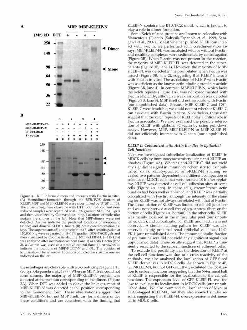
these linkages are cleavable with a S-S reducing reagent DTT(Soltysik-Espanola et al., 1999). Whereas MBP itself could notform dimers, the majority of MBP-KLEIP-N protein wasdetected at the position corresponding to the dimers (Figure3A). When DTT was added to cleave the linkages, most ofMBP-KLEIP-N was detected at the position correspondingto the monomeric form. These observations suggest thatMBP-KLEIP-N, but not MBP itself, can form dimers underthese conditions and are consistent with the finding that
KLEIP-N contains the BTB/POZ motif, which is known toplay a role in dimer formation.
Some Kelch-related proteins are known to colocalize withfilamentous (F)-actin (Soltysik-Espanola et al., 1999, Sasa-gawa et al., 2002). To test whether purified KLEIP can inter-act with F-actin, we performed actin cosedimentation as-says. MBP-KLEIP-FL was incubated with or without F-actin,and resulting complexes were sedimented by centrifugation(Figure 3B). When F-actin was not present in the reaction,the majority of MBP-KLEIP-FL was detected in the super-natants (Figure 3B, lane 1). However, the majority of MBP-KLEIP-FL was detected in the precipitates, when F-actin wasmixed (Figure 3B, lane 2), suggesting that KLEIP interactswith F-actin in vitro. The association of KLEIP with F-actinwas as efficient as the known actin-binding protein �-actinin(Figure 3B, lane 4). In contrast, MBP-KLEIP-N, which lacksthe kelch repeats (Figure 1A), was not cosedimented withF-actin efficiently, although a weak association was detected(Figure 3B, lane 3). MBP itself did not associate with F-actin(our unpublished data). Because MBP-KLEIP-C and GST-KLEIP-C were insoluble, we could not test whether KLEIP-Ccan associate with F-actin in vitro. Nonetheless, these datasuggest that the kelch repeats of KLEIP play a critical role inF-actin association. We also examined the possible interac-tion of KLEIP with globular (G)-actin by using pull-downassays. However, MBP, MBP-KLEIP-N or MBP-KLEIP–FLdid not efficiently interact with G-actin (our unpublisheddata).
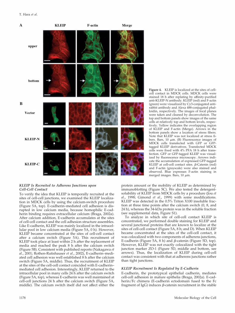
KLEIP Is Colocalized with Actin Bundles in EpithelialCell JunctionsNext, we investigated subcellular localization of KLEIP inMDCK cells by immunocytochemistry using anti-KLEIP an-tibodies (Figure 4A). Whereas anti-KLEIP-C did not yieldany significant signal in immunocytochemistry (our unpub-lished data), affinity-purified anti-KLEIP-N staining re-vealed two patterns dependent on a different compaction ofcolonies of MDCK cells that were formed 18 h after replat-ing. KLEIP was detected at cell-cell adhesion sites of somecells (Figure 4A, top). In these cells, circumference actinbundles had been well established, and KLEIP was partiallycolocalized with F-actin, although the intensity of the stain-ing for KLEIP was not always correlated with that of F-actin.The accumulation of KLEIP was limited to cell-cell junctionsand was not observed at cell-free edges or stress fibers on thebottom of cells (Figure 4A, bottom). In the other cells, KLEIPwas mainly localized in the intracellular pool (our unpub-lished data), and colocalization of KLEIP and F-actin was notobserved. A similar staining pattern for KLEIP was alsoobserved in pig proximal renal epithelial cell lines, LLC-PK-1 (our unpublished data). The immunoglobulin fractionof preimmune sera did not yield any significant signal (ourunpublished data). These results suggest that KLEIP is tran-siently recruited to the cell-cell junctions of adherent cells.
To exclude the possibility that the detection of KLEIP atthe cell-cell junctions was due to a cross-reactivity of theantibody, we also analyzed the localization of GFP-fusedKLEIP derivatives in MDCK cells. As shown in Figure 4B,GFP-KLEIP-N, but not GFP-KLEIP-C, exhibited the localiza-tion to cell-cell junctions, suggesting that the N-terminal halfof KLEIP is responsible for the localization to the cell-celljunctions. The expression level of GFP-KLEIP-FL was toolow to evaluate its localization in MDCK cells (our unpub-lished data). We also examined the localization of Myc- orFLAG-tagged KLEIP-FL, but they also showed similar re-sults, suggesting that KLEIP-FL overexpression is detrimen-tal to MDCK cells.
Figure 3. KLEIP forms dimers and interacts with F-actin in vitro.(A) Homodimer-formation through the BTB/POZ domain ofKLEIP. MBP and MBP-KLEIP-N were cross-linked by DTSP in PBS.The cross-linkage was cleavable with DTT. Both reduced and non-reduced samples were separated on 8–16% gradient SDS-PAGE gelsand then visualized by Coomassie staining. Locations of molecularmakers are shown at the left. Note that MBP-dimers were notdetected. Arrows indicate the predicted locations of monomeric(Mono) and dimeric KLEIP (Dimer). (B) Actin cosedimentation as-says. The supernatants (S) and precipitates (P) after centrifugation at150,000 � g were separated on 8–16% gradient SDS-PAGE gels andthen visualized by Coomassie staining. MBP-KLEIP-FL (�115 kDa)was analyzed after incubation without (lane 1) or with F-actin (lane2). �-Actinin was used as a positive control (lane 4). Arrowheadsindicate the locations of MBP-KLEIP-N and -FL. The position ofactin is shown by an arrow. Locations of molecular size markers areindicated on the left.
Novel Kelch-related Protein, KLEIP
Vol. 15, March 2004 1177
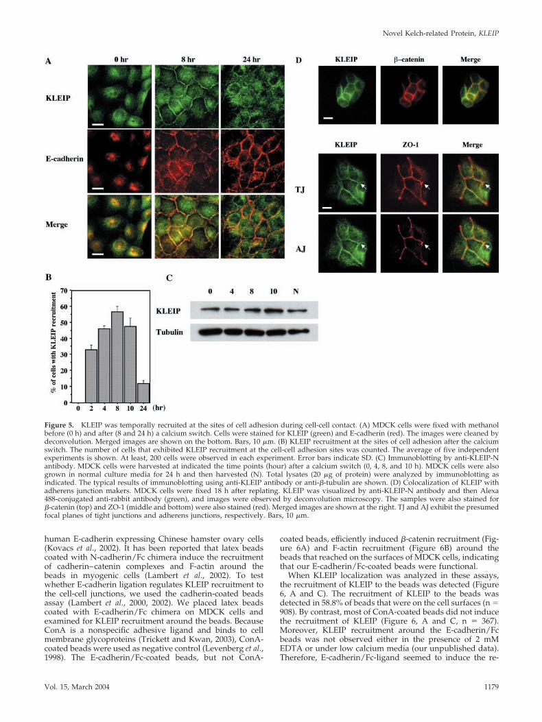
KLEIP Is Recruited to Adherens Junctions uponCell-Cell ContactTo verify the idea that KLEIP is temporally recruited at thesites of cell-cell junctions, we examined the KLEIP localiza-tion in MDCK cells by using the calcium-switch procedure(Figure 5A, top). E-cadherin–mediated cell adhesion is dis-rupted in low calcium media, because homophilic E-cad-herin binding requires extracellular calcium (Braga, 2002a).After calcium addition, E-cadherin accumulates at the sitesof cell-cell contact and the cell adhesion structure assembles.Like E-cadherin, KLEIP was mainly localized in the intracel-lular pool in low calcium media (Figure 5A, 0 h). However,KLEIP became concentrated at the sites of cell-cell contactafter a calcium switch (Figure 5A). This recruitment ofKLEIP took place at least within 2 h after the replacement ofmedia and reached the peak 8 h after the calcium switch(Figure 5B). Consistent with published reports (Nakagawa etal., 2001; Rothen-Rutishauser et al., 2002), E-cadherin–medi-ated cell adhesion was well established 8 h after the calciumswitch (Figure 5A, middle). Thus, the recruitment of KLEIPat the sites of the cell-cell contact coincided with E-cadherin–mediated cell adhesion. Interestingly, KLEIP returned to theintracellular pool in many cells 24 h after the calcium switch(Figure 5A, top), whereas E-cadherin was well maintained atcell-cell junctions 24 h after the calcium switch (Figure 5A,middle). The calcium switch itself did not affect either the
protein amount or the mobility of KLEIP as determined byimmunoblotting (Figure 5C). We also tested the detergent-solubility of KLEIP from MDCK cells by a procedure (Jou etal., 1998; Gimond et al., 1999) with some modifications.KLEIP was detected in the 0.5% Triton X100 insoluble frac-tion at three time points after the calcium switch (0, 8, and24 h), whereas the 34-kDa protein was in the soluble fraction(see supplemental data, Figure S1).
To analyze in which site of cell-cell contact KLEIP isconcentrated, we performed double staining for KLEIP andseveral junctional proteins that are known to localize at thesites of cell-cell contact (Figure 5A, 8 h; and D). When KLEIPbecame concentrated at the sites of the cell-cell contact, itwas colocalized with two components of adherens junctions,E-cadherin (Figure 5A, 8 h) and �-catenin (Figure 5D, top).However, KLEIP was not exactly colocalized with the tightjunction marker ZO-1 (Figure 5D, middle and bottom, seearrows). Thus, the localization of KLEIP during cell-cellcontact was consistent with that at adherens junctions ratherthan tight junctions.
KLEIP Recruitment Is Regulated by E-CadherinE-cadherin, the prototypical epithelial cadherin, mediatescell-cell adhesion in mature epithelia (Braga, 2002a). E-cad-herin/Fc chimera (E-cadherin ectodomain fused to the Fcfragment of IgG) induces �-catenin recruitment in the stable
Figure 4. KLEIP is localized at the sites of cell-cell contact in MDCK cells. MDCK cells werestained 18 h after replating by affinity-purifiedanti-KLEIP-N antibody. KLEIP (red) and F-actin(green) were visualized by Cy3-conjugated anti-rabbit antibody and Alexa 488-conjugated phal-loidin, respectively. The images of focal planeswere taken and cleaned by deconvolution. Thetop and bottom panels show images of the samecells at relatively top and bottom levels, respec-tively. Yellow indicates the overlapping regionof KLEIP and F-actin (Merge). Arrows in thebottom panels show a location of stress fibers.Note that KLEIP was not localized at stress fi-bers. Bars, 10 �m. (B) Fluorescence images ofMDCK cells transfected with GFP or GFP-tagged KLEIP derivatives. Transfected MDCKcells were fixed with 4% PFA 18 h after trans-fection. GFP or GFP-tagged KLEIP was visual-ized by fluorescence microscopy. Arrows indi-cate the accumulation of expressed GFP-taggedKLEIP at cell-cell contact sites. �-Catenin (red)and F-actin (grayscale) were also stained andobserved. Blue expresses F-actin staining inmerged images. Bars, 10 �m.
T. Hara et al.
Molecular Biology of the Cell1178
human E-cadherin expressing Chinese hamster ovary cells(Kovacs et al., 2002). It has been reported that latex beadscoated with N-cadherin/Fc chimera induce the recruitmentof cadherin–catenin complexes and F-actin around thebeads in myogenic cells (Lambert et al., 2002). To testwhether E-cadherin ligation regulates KLEIP recruitment tothe cell-cell junctions, we used the cadherin-coated beadsassay (Lambert et al., 2000, 2002). We placed latex beadscoated with E-cadherin/Fc chimera on MDCK cells andexamined for KLEIP recruitment around the beads. BecauseConA is a nonspecific adhesive ligand and binds to cellmembrane glycoproteins (Trickett and Kwan, 2003), ConA-coated beads were used as negative control (Levenberg et al.,1998). The E-cadherin/Fc-coated beads, but not ConA-
coated beads, efficiently induced �-catenin recruitment (Fig-ure 6A) and F-actin recruitment (Figure 6B) around thebeads that reached on the surfaces of MDCK cells, indicatingthat our E-cadherin/Fc-coated beads were functional.
When KLEIP localization was analyzed in these assays,the recruitment of KLEIP to the beads was detected (Figure6, A and C). The recruitment of KLEIP to the beads wasdetected in 58.8% of beads that were on the cell surfaces (n �908). By contrast, most of ConA-coated beads did not inducethe recruitment of KLEIP (Figure 6, A and C, n � 367).Moreover, KLEIP recruitment around the E-cadherin/Fcbeads was not observed either in the presence of 2 mMEDTA or under low calcium media (our unpublished data).Therefore, E-cadherin/Fc-ligand seemed to induce the re-
Figure 5. KLEIP was temporally recruited at the sites of cell adhesion during cell-cell contact. (A) MDCK cells were fixed with methanolbefore (0 h) and after (8 and 24 h) a calcium switch. Cells were stained for KLEIP (green) and E-cadherin (red). The images were cleaned bydeconvolution. Merged images are shown on the bottom. Bars, 10 �m. (B) KLEIP recruitment at the sites of cell adhesion after the calciumswitch. The number of cells that exhibited KLEIP recruitment at the cell-cell adhesion sites was counted. The average of five independentexperiments is shown. At least, 200 cells were observed in each experiment. Error bars indicate SD. (C) Immunoblotting by anti-KLEIP-Nantibody. MDCK cells were harvested at indicated the time points (hour) after a calcium switch (0, 4, 8, and 10 h). MDCK cells were alsogrown in normal culture media for 24 h and then harvested (N). Total lysates (20 �g of protein) were analyzed by immunoblotting asindicated. The typical results of immunoblotting using anti-KLEIP antibody or anti-�-tubulin are shown. (D) Colocalization of KLEIP withadherens junction makers. MDCK cells were fixed 18 h after replating. KLEIP was visualized by anti-KLEIP-N antibody and then Alexa488-conjugated anti-rabbit antibody (green), and images were observed by deconvolution microscopy. The samples were also stained for�-catenin (top) and ZO-1 (middle and bottom) were also stained (red). Merged images are shown at the right. TJ and AJ exhibit the presumedfocal planes of tight junctions and adherens junctions, respectively. Bars, 10 �m.
Novel Kelch-related Protein, KLEIP
Vol. 15, March 2004 1179
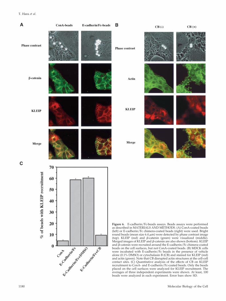
Figure 6. E-cadherin/Fc-beads assays. Beads assays were performedas described in MATERIALS AND METHODS. (A) ConA-coated beads(left) or E-cadherin/Fc chimera-coated beads (right) were used. Brightround beads (mean size 6.4 �m) were detected by phase contrast image(top). KLEIP (red) and �-catenin (green) were visualized (middle).Merged images of KLEIP and �-catenin are also shown (bottom). KLEIPand �-catenin were recruited around the E-cadherin/Fc chimera-coatedbeads on the cell surfaces, but not ConA-coated beads. (B) MDCK cellswere incubated with E-cadherin/Fc beads in the presence of vehiclealone (0.1% DMSO) or cytochalasin B (CB) and stained for KLEIP (red)and actin (green). Note that CB disrupted actin structures at the cell-cellcontact sites. (C) Quantitative analysis of the effects of CB on KLEIPrecruitment to ConA- and E-cadherin/Fc-coated beads. Only the beadsplaced on the cell surfaces were analyzed for KLEIP recruitment. Theaverages of three independent experiments were shown. At least, 100beads were analyzed in each experiment. Error bars show SD.
T. Hara et al.
Molecular Biology of the Cell1180
cruitment of KLEIP around the beads. Such the recruitmentof KLEIP around E-cadherin/Fc-coated beads was also ob-served in LLC-PK1 cells (our unpublished data). These re-sults suggest that KLEIP recruitment is induced by E-cad-herin, possibly by their ligation, and spatially limited to thevicinity of E-cadherin molecules.
F-Actin Is Required for KLEIP Recruitment to the Cell-Cell JunctionsBecause KLEIP bound to F-actin (Figure 3B), we tested whetherF-actin are required for this KLEIP recruitment around theE-cadherin/Fc-coated beads. To inhibit the formation of actinfilaments, we used cytochalasin B (CB), an inhibitor of actinpolymerization (Johnson et al., 2002). MDCK cells were incu-bated with E-cadherin/Fc beads in the presence or absence ofCB (Figure 6, B and C). In the absence of CB, 60.3% of E-cadherin/Fc beads placed on the cell surface exhibited KLEIPrecruitment as well as F-actin recruitment around the beads(n � 431). In contrast, only 9.6% of E-cadherin/Fc-coated beadsplaced on the cells induced F-actin and KLEIP recruitmentaround the beads in the presence of CB (n � 611). CB alsodisrupted de novo actin filaments of MDCK cells. These resultssuggest that F-actin is required for the KLEIP recruitmentsaround E-cadherin/Fc beads.
Rac1, a small GTPase of the Rho family, is activated byE-cadherin-mediated cell adhesion in MDCK cells (Naka-
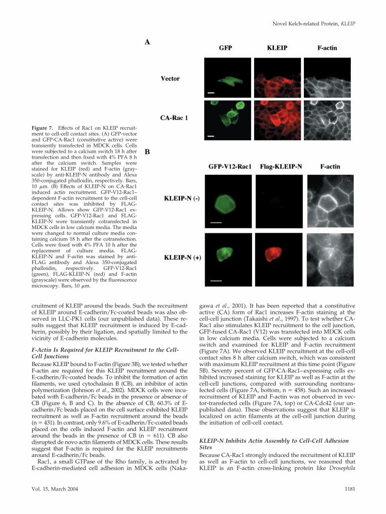
gawa et al., 2001). It has been reported that a constitutiveactive (CA) form of Rac1 increases F-actin staining at thecell-cell junction (Takaishi et al., 1997). To test whether CA-Rac1 also stimulates KLEIP recruitment to the cell junction,GFP-fused CA-Rac1 (V12) was transfected into MDCK cellsin low calcium media. Cells were subjected to a calciumswitch and examined for KLEIP and F-actin recruitment(Figure 7A). We observed KLEIP recruitment at the cell-cellcontact sites 8 h after calcium switch, which was consistentwith maximum KLEIP recruitment at this time point (Figure5B). Seventy percent of GFP-CA-Rac1–expressing cells ex-hibited increased staining for KLEIP as well as F-actin at thecell-cell junctions, compared with surrounding nontrans-fected cells (Figure 7A, bottom, n � 458). Such an increasedrecruitment of KLEIP and F-actin was not observed in vec-tor-transfected cells (Figure 7A, top) or CA-Cdc42 (our un-published data). These observations suggest that KLEIP islocalized on actin filaments at the cell-cell junction duringthe initiation of cell-cell contact.
KLEIP-N Inhibits Actin Assembly to Cell-Cell AdhesionSitesBecause CA-Rac1 strongly induced the recruitment of KLEIPas well as F-actin to cell-cell junctions, we reasoned thatKLEIP is an F-actin cross-linking protein like Drosophila
Figure 7. Effects of Rac1 on KLEIP recruit-ment to cell-cell contact sites. (A) GFP-vectorand GFP-CA-Rac1 (constitutive active) weretransiently transfected in MDCK cells. Cellswere subjected to a calcium switch 18 h aftertransfection and then fixed with 4% PFA 8 hafter the calcium switch. Samples werestained for KLEIP (red) and F-actin (gray-scale) by anti-KLEIP-N antibody and Alexa350-conjugated phalloidin, respectively. Bars,10 �m. (B) Effects of KLEIP-N on CA-Rac1induced actin recruitment. GFP-V12-Rac1–dependent F-actin recruitment to the cell-cellcontact sites was inhibited by FLAG-KLEIP-N. Allows show GFP-V12-Rac1 ex-pressing cells. GFP-V12-Rac1 and FLAG-KLEIP-N were transiently cotransfected inMDCK cells in low calcium media. The mediawere changed to normal culture media con-taining calcium 18 h after the cotransfection.Cells were fixed with 4% PFA 10 h after thereplacement of culture media. FLAG-KLEIP-N and F-actin was stained by anti-FLAG antibody and Alexa 350-conjugatedphalloidin, respectively. GFP-V12-Rac1(green), FLAG-KLEIP-N (red) and F-actin(grayscale) were observed by the fluorescencemicroscopy. Bars, 10 �m.
Novel Kelch-related Protein, KLEIP
Vol. 15, March 2004 1181
Kelch and is involved in actin assembly through the Rac1-mediated signaling during cell adhesion. In this case, theN-terminal half of KLEIP (KLEIP-N) might exhibit a domi-nant negative phenotype with regard to F-actin accumula-tion at cell-cell adhesion sites, because it lacks the kelchrepeats, which can bind to F-actin, but can still accumulate atthe sites of cell-cell adhesion (Figure 4B). In Drosophila Kelch,such a dominant negative effect of N-terminal half of theprotein on ring canal formation has been reported (Robinsonand Cooly, 1997). To test this possibility, we transfectedCA-Rac1 with or without KLEIP-N into MDCK cells andexamined F-actin localization at the junctions of transfectedcells (Figure 7B). Compared with surrounding cells, a strongactin recruitment induced by CA-Rac1 was observed in 80%of cells expressing GFP-CA-Rac1 alone (n � 208), whereasonly 33% of cells coexpressing both GFP-CA-Rac1 andFLAG-KLEIP-N (n � 176) exhibited the strong actin recruit-ment to the cell-cell junctions. Therefore, KLEIP-N inhibitedRac1-induced actin recruitment to the cell junctions. In con-trast, expression of KLEIP-N alone did not significantly alterthe pattern of actin localization in MDCK cells (our unpub-lished data; but see Figure 4B). These observations suggestthat KLEIP is specifically involved in Rac1-regulat actinaccumulation at cell-cell adhesion sites.
DISCUSSION
In this study, we identified a novel human Kelch-relatedprotein, KLEIP, which consisted of the BTB motif and six-tandem kelch repeats. KLEIP is mostly related to DrosophilaDiablo, whose function is still unknown. KLEIP shared somecharacteristics with other Kelch-related proteins. First, theBTB/POZ motif of KLEIP was involved in dimer formation.Second, KLEIP interacted with F-actin in vitro. However, thelow amino acid identity of KLEIP with other members of theBTB/Kelch subfamily made it difficult to presume its func-tion (Bomont and Koeing, 2003).
We found accumulated signals at the sites of cell-celladhesion by affinity-purified anti-KLEIP-N antibody in im-munocytochemistry (Figure 4A). Anti-KLEIP-N andKLEIP-C antibodies recognized a protein of 64 kDa in im-munoblotting, which corresponded to the predicted molec-ular size of KLEIP (Figure 2B). These results suggest that the64-kDa protein represents endogenous KLEIP and that theisolated KLEIP cDNA contains the entire open readingframe. Although anti-KLEIP-C recognized the major singleband of 64 kDa, anti-KLEIP-N also detected a 34-kDa pro-tein. Because the 34-kDa protein was not immunoprecipi-tated by anti-KLEIP-N, anti-KLEIP-N seemed to recognizeonly denatured forms of the protein (Figure 2C). Because weused the methanol method for fixation, which should retainthe native structure of proteins (Allan, 2000), the 34-kDaprotein might not be detected in our immunocytochemicalanalysis. Additionally, as we detected the 34-kDa protein inthe Triton X-soluble fraction in MDCK cells (see supplemen-tary data, Figure S1), it would not affect the detection ofKLEIP at the cell-cell junctions, even if KLEIP-N could detectthe native form of the 34-kDa protein in immunocytochem-ical experiments. Moreover, exogenously expressed GFP-KLEIP-N also localized at the sites of cell-cell contact (Figure4B). These results strongly suggest that KLEIP localizes tothe sites of cell-cell adhesion in MDCK cells.
We found that KLEIP was temporally concentrated at thecell-cell contact sites (Figure 5A). KLEIP was partially colo-calized with actin filaments at these sites (Figure 4A). Wealso showed that KLEIP interacted with F-actin in vitro(Figure 3B). This KLEIP recruitment to the cell-cell contact
sites increased and reached to the peak 8 h after a calciumswitch (Figure 5B). It has been reported that circumferentialactin bundles are established during E-cadherin–mediatedcell adhesion (Vasioukhin and Fuchs, 2001). Our E-cad-herin/Fc beads assays revealed that KLEIP and F-actin wererecruited in the vicinity of the beads placed on the cells(Figure 6). An inhibitor of actin polymerization, cytochalasinB, prevented KLEIP recruitment around E-cadherin/Fcbeads, suggesting that KLEIP recruitment requires F-actin.Moreover, CA-Rac1 significantly stimulated KLEIP recruit-ment as well as F-actin to the cell-cell contact sites. Ourobservation of the increased F-actin staining in CA-Rac1transfectants was consistent with a previous report (Takaishiet al., 1997). These observations may suggest that the recruit-ment of KLEIP to the cell-cell contact sites is due to itslocalization on the actin filaments.
KLEIP was localized to actin bundles at the cell-cell con-tact sites during cell adhesion but not other actin filamentssuch as stress fibers (Figure 4A). However, after the estab-lishment of E-cadherin–mediated cell adhesion, KLEIP lo-calized at the cell-cell junctions returned to the entire cells(Figure 5A). These observations suggest that the interactionbetween KLEIP and F-actin was regulated spatially andtemporally. In Drosophila, phosphorylation of Kelch by Src64negatively regulates the interaction of actin filaments (Kelsoet al., 2002). We found that KLEIP was in the insolublefraction in the presence of 0.5% Triton X-100 at 0, 8, and 24 hafter a calcium switch (Figure S1). If KLEIP always inter-acted with actin filaments, it could be detected at other actinstructures, such as stress fibers and actin filaments at thecell-free edge. It is still not clear why the calcium switch didnot affect the detergent solubility of KLEIP. The interactionwith cytoskeleton is not the only reason for the nonionicdetergent insolubility (Solomon et al., 1998). One possibilityis that KLEIP might be associated with the nonionic deter-gent-resistant membrane in their intracellular pool beforethe calcium switch. Because E-cadherin/Fc beads inducedKLEIP recruitment around the beads, some downstreamsignaling of E-cadherin ligation might be required for thebinding between KLEIP and F-actin. Further analysis wouldclarify how the interaction between KLEIP and F-actin isregulated.
Together with the presence of the kelch repeats, in vitroassociation and in vivo colocalization of KLEIP with F-actinsuggest that KLEIP may function as a cross-linker like Dro-sophila Kelch and play a critical role on actin assembly in thejunctions. We found that KLEIP-N, which lacks the actin-binding site and contains a sufficient sequence for the re-cruitment to the cell-cell adhesion sites, inhibited CA-Rac1-induced actin recruitment (Figure 7B). It has been reportedthat the N-terminal half of Drosophila Kelch has the domi-nant negative effects on ring canal formation (Robinson andCooly, 1997). KLEIP-N might form dimers with endogenousKLEIP to inhibit its function, because the N-terminal half ofKLEIP is involved in dimer formation like other Kelch-likeproteins (Figure 3A). Therefore, KLEIP may play a role onactin remodeling during cell-cell contact and inhibit Racsignaling upstream of actin assembly. However, KLEIP-Nalone did not inhibit physiological actin assembly duringcell-cell contacts in MDCK cells (Figure 4B). This mightindicate that there may be other compensatory mechanismsof actin remodeling during cell-cell contact. Increased accu-mulation of KLEIP by Rac activation seems to stimulateactin accumulation at the sites (Figure 7A). Considering thetemporal accumulation of KLEIP at the junctions peaked 8 hafter calcium switch (Figure 5B), KLEIP accumulation seemsto form a positive feedback loop: Rac-activated F-actin ac-
T. Hara et al.
Molecular Biology of the Cell1182
cumulation at the cell-cell adhesion sites stimulates KLEIPaccumulation at the sites and then KLEIP functions as anactin cross-linker to further accumulate F-actin at the sites.Because the engagements of actin filaments at the cell-cellcontact sites enhance the clustering of E-cadherin (Braga,2002b), KLEIP may be involved in such a feedback regula-tion of actin assembly. After the establishment of cell-cellcontact, KLEIP may lose the ability of binding to F-actin andreturn to the intracellular pool. The interaction betweenKLEIP and F-actin seems to require the E-cadherin ligationsignaling despite of their direct interaction in vitro. Based onthese results, we propose that KLEIP functions as an actin-cross linker presumably at adherens junctions during theinitiation of cell-cell adhesion. Further studies on KLEIP willclarify such unknown regulatory mechanisms to regulatethe binding of KLEIP and F-action.
In an alternative approach to examine a function ofKLEIP, we used RNA interference in HeLa cells (Elbashir etal., 2001). However, we could not efficiently knock downKLEIP, although six different pairs of small interferingRNAs (siRNAs) were used. It has been reported that somesiRNAs were ineffective in several genes (Harborth et al.,2001). An improved strategy to design the siRNAs may helpknock down KLEIP in future experiments.
In conclusion, we isolated and characterized a novel mam-malian Kelch-related protein, KLEIP. KLEIP is recruited tothe sites of cell-cell contact by the initiation of cell-cell con-tact and involved in Rac1-based actin assembly during cell-cell adhesion. Our findings will give an insight in under-standing the regulatory mechanisms of cell-cell contacts aswell as the functions of this Kelch-related protein.
ACKNOWLEDGMENTS
We thank Dr. Douglas Lowy for support. T.H is supported by the JapanSociety for the promotion of Science fellowship from the Ministry of Educa-tion, Science and Culture, Japan.
REFERENCES
Adams, C.L., Chen, Y.-T., Smith, S.J., and Nelson, W.J. (1998). Mechanism ofepithelial cell-cell revealed by high-resolution tracking of E-cadherin-greenfluorescent protein. J. Cell Biol. 142, 1105–1119.
Adams, J., Kelso, R., and Cooly, L. (2000). The kelch repeat superfamily ofproteins: propellers of cell function. Trends Cell Biol. 10, 17–24.
Albagli, O., Drordain, P., Dewwindt, C., Lecoceq, G., and Leprince, D. (1995).The BTB/POZ domain: a new protein-protein interaction motif commonDNA- and actin-binding proteins. Cell Growth Differ. 6, 1193–1198.
Allan, V. J. (2000). Basic immunofluorescence. In: Protein Localization byFluorescence Microscopy: A Practical Approach, ed. V.J. Allan. Oxford,United Kingdom: Oxford University Press, 1–16.
Bomont, P., and Koeing, M. (2003). Intermediate filament aggregation infibroblasts of giant axonal neuropathy patients is aggravated in non dividingcells and by microtubule destabilization. Hum. Mol. Genet. 12, 813–822.
Braga, V. (2002a). Cadherin adhesion regulation in keratinocytes. In: Cell-CellInteraction: A Practical Approach, ed. T.P. Fleming. Oxford, United Kingdom:Oxford University Press, 1–36.
Braga, V.M.M. (2002b). Cell-cell adhesion and signalling. Curr. Opin. Cell.Biol. 14, 546–556.
Braga, V.M.M., Machesky, L.M., Hall, A., and Hotchin, N.A. (1997). The smallGTPases Rho and Rac are required for the establishment of Cadherin-depen-dent cell-cell contacts. J. Cell Biol. 137, 1421–1431.
Burbelo, P.D., Miyamoto, S., Utani, A., Brill, S., Yamada, K.M., Hall, A., andYamada, Y. (1995). p190-B, a new member of the Rho GAP family, and Rhoare induced to cluster after integrin cross-linking. J. Biol. Chem. 270, 30919–30926.
Elbashir, S.M., Harborth, J., Lendeckel, W., Yalcin, A., Weber, K., and Tuschi,T. (2001). Duplexes of 21-nucleotide RNAs mediate RNA interference incultured mammalian cells. Nature 411, 494–498.
Ehrlich, J.S., Hansen, M.D.H., and Nelson, W.J. (2002). Spatio-temporal reg-ulation of Rac1 localization and lamellipodia dynamics during epithelialcell-cell adhesion. Dev. Cell. 3, 259–270.
Fukata, M., and Kaibuchi, K. (2001). Rho-family GTPases in cadherin-medi-ated cell-cell adhesion. Nat. Rev. 2, 887–897.
Fukata, M., Nakagawa, M., Itoh, N., Kawajiri, A., Yamaga, M., Kuroda, S., andKaibuchi, K. (2001). Involvement of IQGAP1, an effector of Rac1 and Cdc42GTPases, in cell-cell dissociation during cell scattering. Mol. Cell. Biol. 21,2165–2183.
Gimond, C., van der Flier, A., van Delft, Brakebusch, C., Kuikman, I., Collard,J.G., Fassler, R., and Sonnenberg, A. (1999) Induction of cell scattering byexpression of �1-integrins in �1-deficient epithelial cells requires activation ofmembers of Rho family of GTPase and downregulation of cadherin andcatenin function. J. Cell Biol. 147, 1325–1340.
Godt, D., Couderc, J.L., Cramton, S.E., and Laski, F.A. (1993). Pattern forma-tion in the limbs of Drosophila: bric a brac is expressed in a both gradient andwave-like pattern and is required for specification and proper segmentationof the tarsus. Development 119, 799–812.
Harborth, J., Elbashir, S.M., Bechert, K., Tushci, T., and Weber, K. (2001).Identification of essential genes in cultured mammalian cells using smallinterfering RNAs. J. Cell Sci. 114, 4557–4565.
Hernandez, M., Andres-Barquin, P.J., Martinez, S., Buifone, A., Rubenstein,J.L.R., and Israel, M.A. (1997). ENC-1, a novel mammalian kelch-related genespecifically expressed in the nervous system encodes an actin-binding pro-tein. J. Neurosci. 17, 3038–3051.
Johnson, R.G., Meyer, R.A., Li, X.-R., Preus, D.M., Tan, L., Grunenwald, H.,Paulson, A.F., Laird, D.W., and Sheridan, J.S. (2002). Gap junctions assemblein the presence of cytoskeletal inhibitors, but enhanced assembly requiresmicrotubules. Exp. Cell. Res. 275, 67–80.
Jou, T.-S., Schneeberger, E.E., and Nelson, W.J. (1998). Structural and func-tional regulation of tight junctions by RhoA and Rac1 small GTPases. J. CellBiol. 142, 101–115.
Jou, T.-S., and Nelson, W.J. (1998). Effects of regulated expression of mutantRhoA and Rac1 small GTPases on the development of epithelial (MDCK) cellpolarity. J. Cell Biol. 142, 85–100.
Kovacs, E.M., Ali, R.G., McCormack, A.J., and Yap, A.S. (2002). E-Cadherinhomophilic ligation directly signals through Rac and phosphatidylinositol3-kinase to regulate adhesive contacts. J. Biol. Chem. 277, 6708–6718.
Kelso, R. J., Hudson, A. M., and Cooley. L. (2002). Drosophila Kelch regulatesactin organization via Src64-dependent tyrosine phosphorylation. J. Cell Biol.156, 703–713.
Lambert, M., Choquest, D., and Mege, R.M. (2002). Dynamics of ligand-induced, Rac1-dependent anchoring of cadherins to the actin cytoskeleton.J. Cell Biol. 157, 469–467.
Lambert, M., Padilla, F., and Mege, R.M. (2000). Immobilized dimers ofN-cadherin-Fc chimera mimic cadherin-mediated cell contact formation: con-tribution of both outside and inside-out signals. J. Cell Sci. 113, 2207–2219.
Levenberg, S., Katz, B.-Z., Yamada, K.M., and Geiger, B. (1998). Long-rangeand selective autoregulation of cell-cell or cell-matrix adhesions by cadherinor integrin ligands. J. Cell Sci. 111, 347–357.
Miki, T., Smith, C., Long, J., Eva, A., and Flemmeng, T. (1993). Oncogene ect2is related to regulators of small GTP-binding proteins. Nature 362, 462–465.
Nakagawa, M., Fukata, M., Yamagata, M., Itoh, N., and Kaibuchi, K. (2001).Recruitment and activation of Rac1 by the formation of E-cadherin-mediatedcell-cell adhesion sites. J. Cell Sci. 114, 1829–1838.
Noren, K.N., Arthur, W.T., and Burridge, K. (2003). Cadherin Engagementinhibits RhoA via p190RhoGAP. J. Biol. Chem. 278, 13615–13618.
Robinson, D.N., and Cooly, L. (1997). Drosophila Kelch is an oligomeric ringcanal actin organizer. J. Cell Biol. 138, 799–810.
Sahai, E., and Marshall, C.J. (2002). ROCK and Dia have opposing effects onadherens junction downstream of Rho. Nat. Cell. Biol. 4, 408–415.
Solomon, K.R., Malloy, M.A., and Finberg, R.W. (1998). Determination of thenon-ionic detergent insolubility and phosphoprotein associations of glycosyl-phosphatidylinositol-anchored proteins expressed on T cells. Biochem. J. 334,325–333.
Rothen-Rutishauser, B., Riesen, F.K., Braun, A., Gunthert, M., and Wunderli-Allenspach, H. (2002). Dynamics of tight and adherens under EGTA treat-ment. J. Membr. Biol. 188, 151–162.
Novel Kelch-related Protein, KLEIP
Vol. 15, March 2004 1183
Sasagawa, K., Mastudo, Y., Kang, M., Fujimura, L., Iitsuka, Y., Okada, S.,Ochiai, T., Tokuhisa, T., and Hatano, M. (2002). Identification of Nd1, a novelmurine kelch family protein, involved in stabilization of actin filaments.J. Biol. Chem. 277, 44140–44146.
Soltysik-Espanola, M., Rogers, R.A., Jiang, S., Kim, T., Gaedigk, R., White, R.A.,and Avraham, H. (1999). Characterization of Mayven, a novel actin-bindingprotein predominantly expressed in brain. Mol. Biol. Cell 10, 2361–2375.
Takaishi, K., Sasaki, T., Kotani, H., Nishioka, H., and Takai, Y. (1997). Regu-lation of cell-cell adhesion by Rac and Rho small G proteins in MDCK cells.J. Cell Biol. 139, 1047–1059.
Tatsumoto, T., Xie, X., Blumenthal, R., Okamoto, I., and Miki, T. (1999).Human ECT2 is an exchange factor for Rho GTPase, phosphorylated in G2/Mphases, and involved in cytokinesis. J. Cell Biol. 147, 921–927.
Trickett, A., and Kwan, Y.L. (2003). T cell stimulation and expansion usinganti-CD3/28 beads. J. Immunol. Methods 275, 251–255.
Vasioukhin, V., and Fuchs, E. (2001). Actin-dynamics and cell-cell adhesion inepithelia. Curr. Opin. Cell Biol. 13, 76–84.
Xue, F., and Cooley, L. (1993). Kelch encodes a component of intercellularbridges in Drosophila egg chambers. Cell 72, 681–693.
T. Hara et al.
Molecular Biology of the Cell1184


