Embed Size (px)
Citation preview
The
Jour
nal o
f Cel
l Bio
logy
The Rockefeller University Press, 0021-9525/2003/10/155/10 $8.00The Journal of Cell Biology, Volume 163, Number 1, October 13, 2003 155–164http://www.jcb.org/cgi/doi/10.1083/jcb.200304053
JCB
Article
155
MAL regulates clathrin-mediated endocytosis at the apical surface of Madin–Darby canine kidney cells
Fernando Martín-Belmonte,
1
José A. Martínez-Menárguez,
2
Juan F. Aranda,
1
José Ballesta,
2
María C. de Marco,
1
and Miguel A. Alonso
1
1
Centro de Biología Molecular “Severo Ochoa”, Universidad Autónoma de Madrid and Consejo Superior de Investigaciones Científicas, Cantoblanco, Madrid, 28049 Spain
2
Departamento de Biología Celular, Facultad de Medicina, Universidad de Murcia, Murcia, 30071 Spain
AL is an integral protein component of the machin-ery for apical transport in epithelial Madin–Darbycanine kidney (MDCK) cells. To maintain its distri-
bution, MAL cycles continuously between the plasmamembrane and the Golgi complex. The clathrin-mediatedroute for apical internalization is known to differ from thatat the basolateral surface. Herein, we report that MALdepends on the clathrin pathway for apical internalization.Apically internalized polymeric Ig receptor (pIgR), whichuses clathrin for endocytosis, colocalized with internalized
MAL in the same apical vesicles. Time-lapse confocal
M
microscopic analysis revealed cotransport of pIgR andMAL in the same endocytic structures. Immunoelectronmicroscopic analysis evidenced colabeling of MAL withapically labeled pIgR in pits and clathrin-coated vesicles.Apical internalization of pIgR was abrogated in cells withreduced levels of MAL, whereas this did not occur eitherwith its basolateral entry or the apical internalization ofglycosylphosphatidylinositol-anchored proteins, which doesnot involve clathrin. Therefore, MAL is critical for efficientclathrin-mediated endocytosis at the apical surface inMDCK cells.
Introduction
In polarized epithelial cells, the composition of the apicaland basolateral membrane subdomains is maintained byspecialized exocytic and endocytic pathways that sort newlysynthesized and internalized proteins, respectively, specificallyto each surface subdomain (Mostov et al., 2000). Clathrin-mediated endocytosis takes place at both the apical andbasolateral domains but the process differs at both membranesurfaces (Naim et al., 1995). The mechanisms of exocytictransport are also different for each membrane surface(Matter and Mellman, 1994). In epithelial MDCK cells,sorting to the basolateral surface is mediated by sequences inthe cytoplasmic tail of membrane proteins that might ormight not be related to tyrosine- and dileucine-based sortingsignals (Mellman et al., 1993). In contrast, a direct pathwayof transport to the apical membrane appears to be mediatedby integration of cargo proteins into specialized glycolipid-
enriched membrane microdomains or rafts (Simons andWandinger-Ness, 1990).
MAL is a raft-associated integral membrane protein of 17kD, which has been demonstrated to be an essential elementof the machinery for efficient apical transport in MDCKcells (Cheong et al., 1999; Puertollano et al., 1999; Martin-Belmonte et al., 2000, 2001). Despite the intense vesiculartraffic to deliver newly synthesized apical cargo, MAL accu-mulation at the plasma membrane is prevented by continu-ous recycling to the Golgi complex via the endosomal sys-tem, which, in addition, allows its participation in newrounds of cargo transport (Puertollano and Alonso, 1999).At steady state,
�
12% of MAL is detected in the plasmamembrane including clathrin-coated pits and buds inMDCK cells (Puertollano et al., 2001). Here, we have ana-lyzed the clathrin requirement for MAL internalization andinvestigated the role of MAL in the internalization of apicalproteins. As model proteins, we used the polymeric Ig receptor(pIgR) and the transferrin receptor, which are known tointernalize from both the apical and basolateral surface
The online version of this article contains supplemental material.Address correspondence to Miguel A. Alonso, Centro de Biología Mo-lecular “Severo Ochoa”, Universidad Autónoma de Madrid and Con-
sejo Superior de Investigaciones Científicas, Cantoblanco, Madrid,28049 Spain. Tel.: 34-91-397-8037. Fax: 34-91-397-8087. email:[email protected] words: apical endocytosis; protein machinery; polarized transport;epithelial cells; lipid rafts
Abbreviations used in this paper: FR, folate receptor; GPI, glycosyl-phosphatidylinositol; NHS-SS-biotin, sulfosuccinimidyl 2-(6-(biotina-mido)hexanoamido)ethyl-1-3
�
-dithiopropionate; pIgR, polymeric Igreceptor; sulfo-NHS-biotin, sulfo-
N
-hydroxyl-succinimido-biotin.
on February 1, 2014
jcb.rupress.orgD
ownloaded from
Published October 6, 2003
http://jcb.rupress.org/content/suppl/2003/10/03/jcb.200304053.DC1.html Supplemental Material can be found at:
The
Jour
nal o
f Cel
l Bio
logy
156 The Journal of Cell Biology
|
Volume 163, Number 1, 2003
through a clathrin-mediated pathway (Odorizzi et al., 1996;Altschuler et al., 1999), and glycosylphosphatidylinositol(GPI)-anchored proteins, such as placental alkaline phos-phatase and folate receptor (FR), which use clathrin-inde-pendent mechanisms (Nichols and Lippincott-Schwartz,2001). Our results indicate that MAL uses clathrin for apicalendocytosis and that MAL is required for internalizationof pIgR and transferrin receptor from the apical, but notfrom the basolateral, surface or apical endocytosis of GPI-anchored proteins.
Results
Apical endocytosis of MAL takes place by a clathrin-dependent pathway in MDCK cells
To detect MAL on the cell surface and to track its internal-ization, we prepared stable transfectants of MDCK cells(MDCK/MAL-FLAG cells) that stably expressed a MALprotein bearing a FLAG tag in its last extracellular/luminal
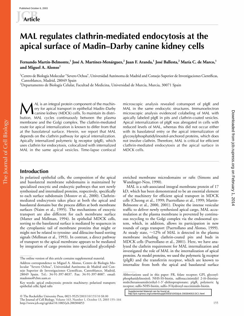
loop (MAL-FLAG). The tag makes MAL-FLAG accessibleto anti-FLAG antibodies from the extracellular space (Puer-tollano and Alonso, 1999). To interfere with clathrin-medi-ated endocytosis, we transiently expressed GFP-DPF coil,an EH-deleted form of the Eps 15 protein tagged with theGFP, which behaves as a dominant-negative mutant andprovokes a block of clathrin-coated pit endocytosis (Ben-merah et al., 1999). MDCK/MAL-FLAG cells were trans-fected with GFP-DPF coil, plated, and left to polarize for48 h in tissue culture filter inserts. The transepithelial resis-tance of the cell monolayer was measured to ensure propertight junction formation. As a control for cell polarization,the distributions of gp135 and E-cadherin, chosen as apicaland basolateral markers, respectively, were determined inparallel cultures. Fig. 1 A shows that, consistent with thepresence of MAL in surface clathrin pits and buds (Puertol-lano et al., 2001), overexpression of GFP-DPF coil led to acomplete block of MAL-FLAG apical internalization. Ascontrols, we observed in parallel that overexpression of
Figure 1. Effect of a dominant-negative mutant of Eps15 on apical internalization of MAL. MDCK/MAL-FLAG, MDCK/pIgR or MDCK/FR cells, which stably express MAL-FLAG, pIgR and FR, respectively, were transiently transfected with GFP-DPF coil cDNA. The cells were plated onto tissue culture filters and left to polarize for 48 h at 37�C. Cells were incubated with (A) Alexa-594–labeled anti-FLAG, (B) anti-pIgR, or (C) anti-FR antibodies from the apical surface at 4�C for 30 min, washed, and fixed (time 0) or shifted to 37�C for 30 min to allow internalization before fixation. Cells were then analyzed by confocal immunofluorescence microscopy. Optical x-y and x-z sections are presented in A, whereas only x-y sections are shown in B and C. As an internal control, internalization of MAL-FLAG and pIgR was observed in cells not expressing GFP-DPF coil (arrowheads). MAL-FLAG internalization in the absence of GFP-DPF coil is clearly detected in the corresponding x-z view (arrowhead). FR internalized regardless of the expression of GFP-DPF coil. Note that, although the cell monolayer was tightly confluent, only the cells expressing the exogenous products are stained. As a control of polarization, the cell monolayers were stained after fixation and permeabilization for gp135 and E-cadherin, as markers for the apical and basolateral surface, respectively. Bar, 10 �m. (D) The percentage of untransfected (black bars) and GFP-DPF coil-transfected (gray bars) cells with internalized MAL-FLAG, pIgR or FR is shown. The values shown are the mean of three independent replicates. Greater than 100 cells from each case were analyzed.
on February 1, 2014
jcb.rupress.orgD
ownloaded from
Published October 6, 2003
The
Jour
nal o
f Cel
l Bio
logy
An essential role for MAL in apical endocytosis |
Martín-Belmonte et al. 157
GFP-DPF coil halted the apical internalization of pIgR(Fig. 1 B) but did not affect that of FR (Fig. 1 C) inMDCK/pIgR and MDCK/FR cells that stably express pIgRand FR, respectively. Quantitative analysis showed that thegreat majority of cells positive for GFP-DPF coil were de-fective for endocytosis of MAL and pIgR (Fig. 1 D),whereas they remained competent at internalizing the GPI-anchored FR molecule. Antibody internalization was dem-onstrated by examining x-z sections of the cells (Fig. 1 A,MAL-FLAG) and by resistance to acid washes (unpublisheddata). Finally, as a further control, we observed that en-docytosis of all MAL-FLAG, pIgR, and FR (unpublisheddata) was blocked by expression of a dynamin mutant,dyn
K44A
, defective in GTP binding and hydrolysis (Damkeet al., 1994), which impairs both clathrin-dependent and-independent pathways (McNiven et al., 2000).
MAL and pIgR are present in the same apical endocytic vesicles
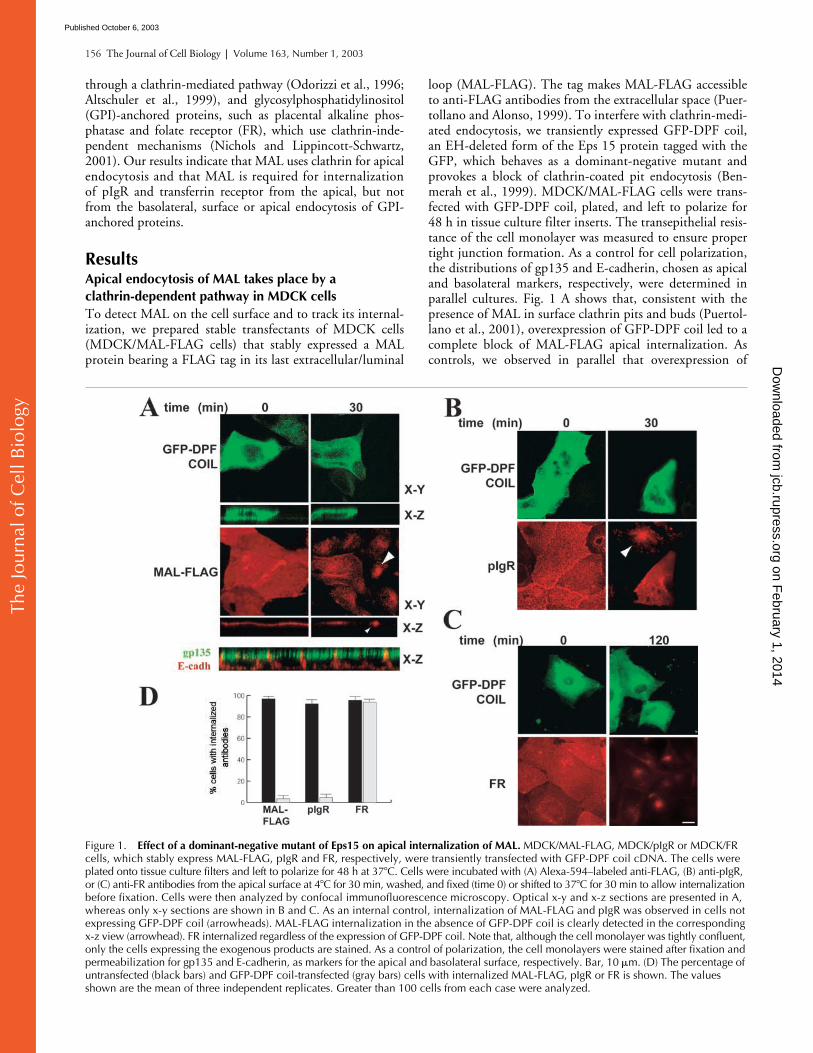
Because both MAL and pIgR require a functional clathrin-dependent pathway for apical endocytosis, we wonderedwhether MAL and pIgR are included in the same vesiclesduring internalization. Keeping in mind that MAL internal-izes constitutively without cross-linking with antibodies(Puertollano and Alonso, 1999), to overcome the use of theanti-FLAG antibodies, a construct expressing a MAL pro-tein fused at its amino-terminal end to GFP (GFP-MAL)was used to trace in living cells the movement of MAL fromthe surface to the cell’s interior. The presence of the GFP
moiety did affect neither the incorporation of MAL intorafts nor its subcellular distribution (unpublished data).MDCK/pIgR cells grown on coverslips were transientlytransfected with GFP-MAL and, after 48 h, cells were incu-bated with anti-pIgR antibodies coupled to Alexa-594 fluo-rochrome at 4
�
C for 30 min. Cells were then shifted to37
�
C, and series of images of GFP-MAL and pIgR com-plexed to the fluorescent anti-pIgR antibodies were analyzedby time-lapse fluorescence confocal microscopy (Fig. 2 andVideo 1, available at http://www.jcb.org/cgi/content/full/jcb.200304053/DC1). Within a 5-min period, a significantnumber of vesicles translocating GFP-MAL and pIgR to-gether from the plasma membrane to the cells’s interiorwere visualized. The GFP-MAL label, which did not ema-nate from the surface, was distributed in vesicles scatteredthroughout the cytoplasm and the Golgi complex. Vesiclescontaining only internalizing pIgR were also identified (un-published data). The existence of endocytic vesicles withboth MAL and pIgR or with pIgR alone in the nonpolarizedMDCK cell cultures used are probably reminiscent of theapical and basolateral vesicles, respectively, evidenced in po-larized cells. To examine this possibility, we scrutinized theapical and basolateral endocytic vesicles in polarized MDCKcells for the presence of internalized MAL-FLAG and/orpIgR. MDCK cells stably expressing both MAL-FLAG andpIgR (MDCK/MAL-FLAG/pIgR cells) were plated and po-larized in culture inserts for 72 h. The apical or the basal sideof the cells were incubated with anti-FLAG antibodies cou-pled to Alexa-488 and with anti-pIgR antibodies coupled to
Figure 2. Time-lapse confocal microscopic analysis of MAL and pIgR internalization. MDCK/pIgR cells were transiently transfected with GFP-MAL and grown on coverslips. 48 h after transfection, cells were incubated at 4�C for 30 min with antibodies anti-pIgR coupled to Alexa-594, and then shifted to 37�C. Sequential images of GFP-MAL and pIgR bound to the antibody were acquired at 5-s intervals using a confocal fluorescence microscope (see Video 1, available at http://www.jcb.org/cgi/content/full/jcb.200304053/DC1). The left panel shows two membrane regions (1 and 2) selected to illustrate endocytic vesicles in which pIgR and GFP-MAL cointernalization was detected (right panels). Some representative frames obtained in a plane close to the coverslip are shown. The time frame intervals are indicated. The outlined circles and the arrowheads label single vesicles carrying both GFP-MAL and pIgR. Bar, 10 �m.
on February 1, 2014
jcb.rupress.orgD
ownloaded from
Published October 6, 2003
The
Jour
nal o
f Cel
l Bio
logy
158 The Journal of Cell Biology
|
Volume 163, Number 1, 2003
Alexa-594 at 4
�
C for 30 min. After 3 min at 37
�
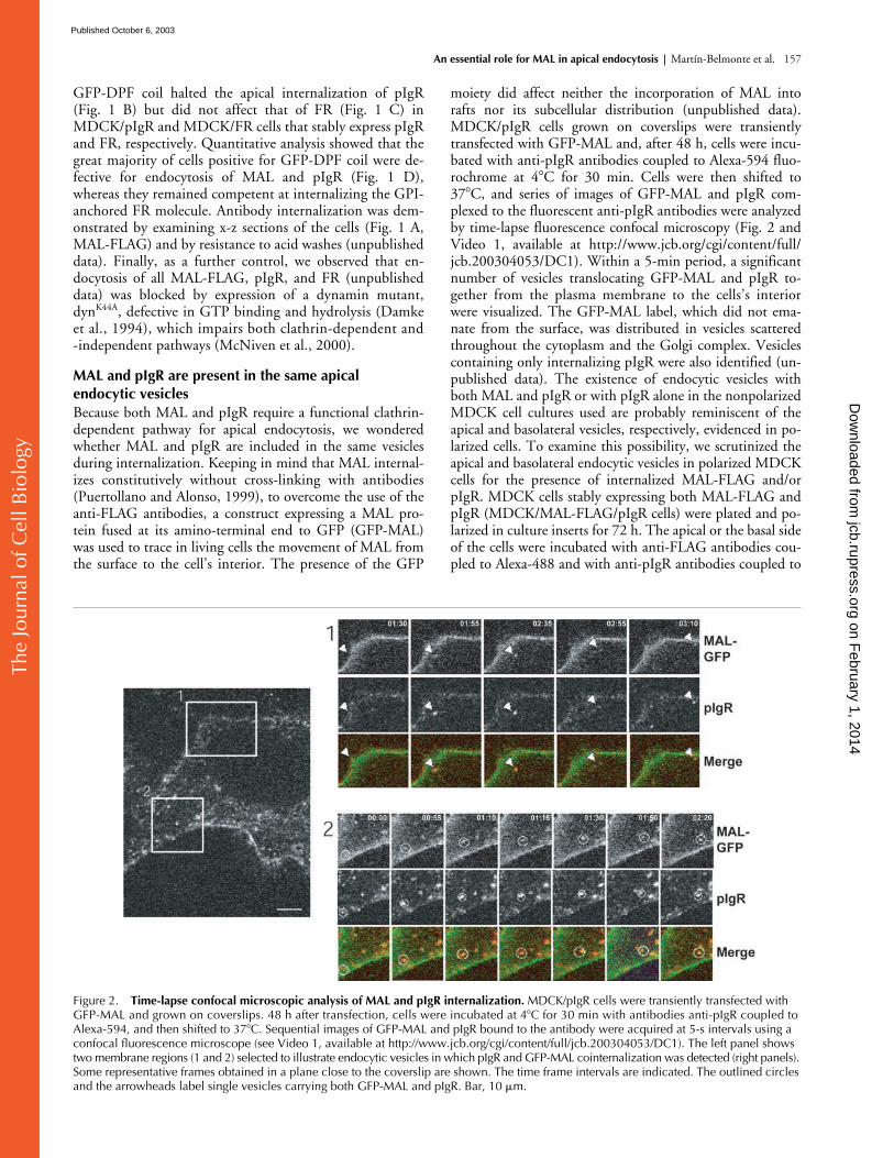
C to allowinternalization, cells were fixed and analyzed by confocal mi-croscopy. Fig. 3 shows subapical (A) and basal (B) confocalplanes of the vesicular structures containing internalizedpIgR and/or MAL-FLAG. Quantitative analysis showed that85% of the apical structures containing internalized pIgRwere also positive for MAL-FLAG, whereas fewer than 2%of the vesicles with pIgR internalized from the basal surfacewere stained for internalized MAL-FLAG (Fig. 3 C).
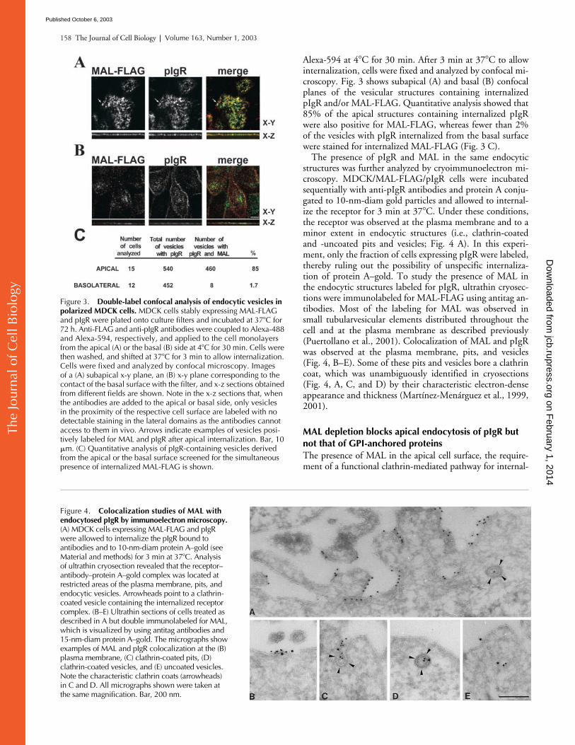
The presence of pIgR and MAL in the same endocyticstructures was further analyzed by cryoimmunoelectron mi-croscopy. MDCK/MAL-FLAG/pIgR cells were incubatedsequentially with anti-pIgR antibodies and protein A conju-gated to 10-nm-diam gold particles and allowed to internal-ize the receptor for 3 min at 37
�
C. Under these conditions,the receptor was observed at the plasma membrane and to aminor extent in endocytic structures (i.e., clathrin-coatedand -uncoated pits and vesicles; Fig. 4 A). In this experi-ment, only the fraction of cells expressing pIgR were labeled,thereby ruling out the possibility of unspecific internaliza-tion of protein A–gold. To study the presence of MAL inthe endocytic structures labeled for pIgR, ultrathin cryosec-tions were immunolabeled for MAL-FLAG using antitag an-tibodies. Most of the labeling for MAL was observed insmall tubularvesicular elements distributed throughout thecell and at the plasma membrane as described previously(Puertollano et al., 2001). Colocalization of MAL and pIgRwas observed at the plasma membrane, pits, and vesicles(Fig. 4, B–E). Some of these pits and vesicles bore a clathrincoat, which was unambiguously identified in cryosections(Fig. 4, A, C, and D) by their characteristic electron-denseappearance and thickness (Martínez-Menárguez et al., 1999,2001).
MAL depletion blocks apical endocytosis of pIgR but not that of GPI-anchored proteins
The presence of MAL in the apical cell surface, the require-ment of a functional clathrin-mediated pathway for internal-
Figure 3. Double-label confocal analysis of endocytic vesicles in polarized MDCK cells. MDCK cells stably expressing MAL-FLAG and pIgR were plated onto culture filters and incubated at 37�C for 72 h. Anti-FLAG and anti-pIgR antibodies were coupled to Alexa-488 and Alexa-594, respectively, and applied to the cell monolayers from the apical (A) or the basal (B) side at 4�C for 30 min. Cells were then washed, and shifted at 37�C for 3 min to allow internalization. Cells were fixed and analyzed by confocal microscopy. Images of a (A) subapical x-y plane, an (B) x-y plane corresponding to the contact of the basal surface with the filter, and x-z sections obtained from different fields are shown. Note in the x-z sections that, when the antibodies are added to the apical or basal side, only vesicles in the proximity of the respective cell surface are labeled with no detectable staining in the lateral domains as the antibodies cannot access to them in vivo. Arrows indicate examples of vesicles posi-tively labeled for MAL and pIgR after apical internalization. Bar, 10 �m. (C) Quantitative analysis of pIgR-containing vesicles derived from the apical or the basal surface screened for the simultaneous presence of internalized MAL-FLAG is shown.
Figure 4. Colocalization studies of MAL with endocytosed pIgR by immunoelectron microscopy. (A) MDCK cells expressing MAL-FLAG and pIgR were allowed to internalize the pIgR bound to antibodies and to 10-nm-diam protein A–gold (see Material and methods) for 3 min at 37�C. Analysis of ultrathin cryosection revealed that the receptor–antibody–protein A–gold complex was located at restricted areas of the plasma membrane, pits, and endocytic vesicles. Arrowheads point to a clathrin-coated vesicle containing the internalized receptor complex. (B–E) Ultrathin sections of cells treated as described in A but double immunolabeled for MAL, which is visualized by using antitag antibodies and 15-nm-diam protein A–gold. The micrographs show examples of MAL and pIgR colocalization at the (B) plasma membrane, (C) clathrin-coated pits, (D) clathrin-coated vesicles, and (E) uncoated vesicles. Note the characteristic clathrin coats (arrowheads) in C and D. All micrographs shown were taken at the same magnification. Bar, 200 nm.
on February 1, 2014
jcb.rupress.orgD
ownloaded from
Published October 6, 2003
The
Jour
nal o
f Cel
l Bio
logy
An essential role for MAL in apical endocytosis |
Martín-Belmonte et al. 159
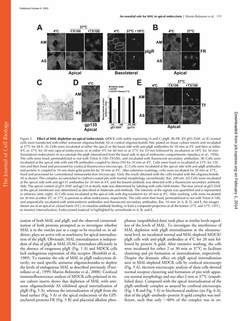
ization of both MAL and pIgR, and the observed cointernal-ization of both proteins prompted us to investigate whetherMAL is in the vesicles just as a cargo to be recycled or, in ad-dition, plays an active role as machinery for apical internaliza-tion of the pIgR. Obviously, MAL internalization is indepen-dent of that of pIgR as MAL-FLAG internalizes efficiently inthe absence of exogenous pIgR (Fig. 1 A) and MDCK cellslack endogenous expression of this receptor (Breitfeld et al.,1989). To examine the role of MAL in pIgR endocytosis di-rectly, we used specific antisense oligonucleotides to depletethe levels of endogenous MAL as described previously (Puer-tollano et al., 1999; Martin-Belmonte et al., 2000). Confocalimmunofluorescence analysis of MDCK cells polarized in tis-sue culture inserts shows that depletion of MAL with anti-sense oligonucleotide AS inhibited apical internalization ofpIgR (Fig. 5 A), whereas the internalization of pIgR from thebasal surface (Fig. 5 A) or the apical endocytosis of the GPI-anchored proteins FR (Fig. 5 B) and placental alkaline phos-
phatase (unpublished data) took place at similar levels regard-less of the levels of MAL. To investigate the interference ofMAL depletion with pIgR internalization at the ultrastruc-tural level, we incubated normal and MAL-depleted MDCK/pIgR cells with anti-pIgR antibodies at 4
�
C for 20 min fol-lowed by protein A–gold. After extensive washing, the cellswere incubated for either 2 or 30 min at 37
�
C to facilitateclustering and pit formation or internalization, respectively.Despite the dramatic effect on pIgR apical internalizationseen in MAL-depleted MDCK cells by confocal microscopy(Fig. 5 A), electron microscopic analysis of these cells showednormal receptor clustering and formation of pits with appar-ent normal morphology and size after 2 min at 37
�
C (unpub-lished data). Compared with the apical internalization of thepIgR–antibody complex as assayed by confocal microscopic(Fig. 1 B and Fig. 5 A) or biochemical analyses (see Fig. 6 A),that of the pIgR–antibody–protein A–gold complex was inef-ficient, such that only
�
40% of the complex was in en-
Figure 5. Effect of MAL depletion on apical endocytosis. MDCK cells stably expressing (A and C) pIgR, (B) FR, (D) gD1-DAF, or (E) normal cells were transfected with either antisense oligonucleotide AS or control oligonucleotide AM, plated on tissue culture inserts and incubated at 37�C for 48 h. (A) Cells were incubated at either the apical or the basal side with anti-pIgR antibodies for 30 min at 4�C and then at either 4�C or 37�C for 30 min (apical endocytosis) or at either 4�C for 60 min or at 37�C for 10 min followed by incubation at 18�C for 50 min (basolateral endocytosis) to accumulate the pIgR internalized from the basal side in apical endosome compartments (Apodaca et al., 1994). The cells were fixed, permeabilized or not with Triton X-100 (TX100), and incubated with fluorescent secondary antibodies. (B) Cells were incubated at the apical side with anti-FR antibodies coupled to Alexa-594 for 30 min at 4�C. Cells were fixed or incubated at 37�C for 120 min and then fixed and processed for confocal fluorescence microscopy. (C) Cells were incubated at the apical side with anti-pIgR antibodies and protein A coupled to 10-nm-diam gold particles for 30 min at 4�C. After extensive washings, cells were incubated for 30 min at 37�C, fixed and processed for conventional immunoelectron microscopy. Only the result obtained with the cells treated with the oligonucleotide AS is shown. The complex accumulated in clathrin-coated pits with normal morphology (arrowheads). Bar, 200 nm. (D) Cells were incubated at the apical side with anti-gp135 antibodies for 30 min at 4�C and the bound antibody was detected with a fluorescent secondary antibody (left). The apical content of gD1-DAF and gp114 at steady state was determined by labeling with sulfo-NHS-biotin. The new arrival of gD1-DAF at the apical membrane was determined as described in Materials and methods. The intensity of the signals was quantified and is represented in arbitrary units (right). (E) Cells were incubated at the apical side with dog transferrin for 30 min at 4�C. After washing, cells were incubated for 30 min at either 4�C or 37�C to prevent or allow endocytosis, respectively. The cells were then fixed, permeabilized or not with Triton X-100, and sequentially incubated with antitransferrin antibodies and fluorescent secondary antibodies. Bar, 10 mm. In A, B, D, and E, the images shown are of an apical or a basal frame (4�C), to visualize antibody binding, or from a composite projection of all the frames (37�C and 37�C–18�C), to monitor internalization. Endocytosed material is highlighted by arrowheads in A, B, and E.
on February 1, 2014
jcb.rupress.orgD
ownloaded from
Published October 6, 2003

The
Jour
nal o
f Cel
l Bio
logy
160 The Journal of Cell Biology
|
Volume 163, Number 1, 2003
docytic compartments after 30 min at 37
�
C in normal cells.Consistent with the requirement of MAL for efficient apicalpIgR endocytosis, in cells with reduced levels of MAL, weobserved less internalization and a twofold accumulation ofthe pIgR–antibody–protein A–gold complex in clathrin-con-taining pits and vesicles (Fig. 5 C; Table I). Importantly, theratio between the total number (including structures not la-beled with gold particles) of coated pits and coated vesicles atthe apical membrane increased from 0.82 in normal cells to1.44 in MAL-depleted cells. This suggests an impairment inthe maturation of the pits to produce vesicles in the cells withreduced levels of MAL.
It is of note that, despite the reduction of MAL levelsslowing down delivery of apical cargo through the directtransport route (Cheong et al., 1999; Puertollano et al.,
1999; Martin-Belmonte et al., 2000), this treatment did notaffect the steady-state levels of the GPI-anchored proteinsFR (Fig. 5 B) and gD1-DAF (Lisanti et al., 1989), or thetransmembrane gp135 and gp114 apical markers (Fig. 5 D),as analyzed by confocal microscopy (FR and gp135) or selec-tive biotinylation of the apical domain (gD1-DAF andgp114). This leaky transport to the apical surface is probablythe result of the remaining MAL molecules being able to de-liver cargo, albeit inefficiently, as is shown for gD1-DAF(Fig. 5 D) and was described previously for the influenza vi-rus hemagglutinin (Puertollano et al., 1999). The transferrinreceptor uses clathrin for both apical and basolateral inter-nalization (Odorizzi et al., 1996). To determine whetherapical internalization of other molecules using clathrin isalso dependent on MAL levels, we analyzed the apical en-docytosis of iron-loaded transferrin mediated by the endoge-nous transferrin receptor in MDCK cells (Apodaca et al.,1994). Fig. 5 E shows that whereas transferrin endocytosistook place efficiently in cells transfected with control oligo-nucleotides AM, most of the transferrin remained at theapical surface in cells whose MAL levels were reduced bytreatment with antisense oligonucleotide AS. Transferrin in-ternalization from the basolateral surface was unaffected bythe treatment (unpublished data). These results indicate amore general role for MAL in clathrin-mediated apical en-docytosis.
To analyze the effect of MAL depletion further, the apicalinternalization of the pIgR molecule was also investigated bybiochemical means (Fig. 6). MDCK/pIgR cells were trans-
Table I.
Distribution of apically internalized pIgR in normal and MAL-depleted MDCK cells
Control MAL-depleted
Plasma membraneexcluding coated pits
58.9
�
3.0 75.9
�
10.2
Clathrin-coated pitsand vesicles
5.0
�
2.2 11.7
�
8.0
Rest of endocyticcompartments
36.1
�
5.2 12.4
�
2.3
Numbers represent the percentages (mean
�
SEM) of the pIgR–antibody–protein A–gold complex over the distinct membrane categories. At least200 gold particles were scored for each type of cell.
Figure 6. Biochemical analysis of the effect of MAL depletion on apical endocytosis of the pIgR. (A) MDCK or MDCK/MAL-�N cells stably expressing pIgR were transfected with either antisense oligonucleotide AS or control oligonucleotide AM and plated on tissue culture inserts. After 48 h at 37�C, the apical surface was labeled with NHS-SS-biotin, and the cells were incubated at 4�C or 37�C. After 30 min, the label remaining on the cell surface was removed by glutathione treatment. After immunoprecipitation with specific antibodies, internalized pIgR was visualized by immunoblotting with streptavidin peroxidase. (B) Endogenous MAL levels were quantified by densitometric scanning of the immunoblots of the initial lysates with anti–dog MAL 2E5 mAb (left). As the anti–dog MAL antibody does not react with the human protein (Puertollano et al., 1999), tagged human MAL was detected with antitag antibodies (right). One representative experiment out of six performed is shown. (C) Quantitative analysis of the effect of MAL depletion on apical endocytosis of pIgR. The intensity of the protein bands obtained in the different experiments was quantified by densitometric analysis. The values shown are the mean values expressed as a percentage of the biotinylated pIgR present on the apical surface at 4�C in the absence of glutathione treatment.
on February 1, 2014
jcb.rupress.orgD
ownloaded from
Published October 6, 2003
The
Jour
nal o
f Cel
l Bio
logy
An essential role for MAL in apical endocytosis |
Martín-Belmonte et al. 161
fected with either antisense oligonucleotide AS or controloligonucleotide AM and plated in tissue culture inserts. Af-ter 48 h at 37
�
C, the apical surface was labeled at 4
�
C withsulfosuccinimidyl 2-(6-(biotinamido)hexanoamido)ethyl-1-39-dithiopropionate (NHS-SS-biotin), a reducible ana-logue of sulfo-
N
-hydroxyl-succinimido-biotin (sulfo-NHS-biotin). Cells were then incubated for 30 min at 37
�
C to allowinternalization and the biotin label that remained on the cellsurface was removed by treating the cells with glutathione,leaving the endocytosed label protected from the reducingagent (Lisanti et al., 1990). After immunoprecipitation, in-ternalized pIgR was visualized by immunoblotting withstreptavidin peroxidase. An example of the efficiency of theglutathione treatment is shown in the left panel of Fig. 6 A.The vast majority (
�
90%) of apical pIgR was endocytosedafter 30 min in control MDCK cells (Fig. 6 A, top left),whereas only a minor portion (
�
5%) was internalized (Fig.6 A, top middle) in cells with whose MAL levels were re-duced to
�
5% of those in control cells, as quantified bydensitometric scanning of the immunoblots obtained withanti-MAL mAb 2E5 (Fig. 6 B). To confirm that these effectswere due to MAL depletion and not to spurious effects ofthe antisense oligonucleotide, we used MDCK/MAL-
�
Ncells expressing a truncated form of human MAL (MAL-
�
N) that is resistant to the depletion treatment as its mRNAcannot pair with the antisense oligonucleotide (Fig. 6 B,right; Martin-Belmonte et al., 2000). The exogenous expres-sion of MAL-
�
N allowed normal apical endocytosis of pIgR(Fig. 6 A, right) despite the drop in the amount of endoge-nous MAL (Fig. 6 B, left). A quantitative analysis of the ef-fect of MAL depletion on apical internalization of pIgR isshown in Fig. 6 C.
Discussion
MAL is essential for efficient endocytosis of the pIgR from the apical surface
Although apical endocytosis is a fundamental process bywhich polarized epithelia take up soluble molecules, macro-molecules and even entire pathogens on the luminal side, itsmechanism is still poorly understood. An important featureof the apical membrane is its high raft lipid content (Simonsand van Meer, 1988). This makes the apical membranemore rigid compared with the fluid structure of the plasmamembrane of nonpolarized cells and the basolateral mem-brane. The process of clathrin-mediated endocytosis is ap-proximately five times slower at the apical than at the baso-lateral surface (Naim et al., 1995). Coated pits at the apicalsurface recognize cargo proteins as efficiently as at the baso-lateral surface, but their maturation into transport vesicles ismuch slower. Apparently, the limiting step occurs before thedynamin-directed fission of the vesicle takes place. To ex-plain this difference the existence was proposed of an apicalmembrane factor that either controls the addition of clathrintriskelions into the lattice or regulates membrane curvature(Naim et al., 1995). Consistent with the reported presenceof MAL in clathrin-coated pits (Puertollano et al., 2001),herein we show that MAL endocytosis is blocked by expres-sion of a dominant negative Eps15 mutant known to inter-fere specifically with clathrin-mediated endocytosis, such as
apical internalization of pIgR. In addition to having thesame requirements, the cotransport of pIgR and MAL ob-served in living cells by time-lapse confocal immunofluores-cence analysis and the presence of internalized pIgR inMAL-containing pits and vesicles as revealed by electron mi-croscopic analysis were consistent with a possible role forMAL in apical endocytosis of the pIgR. The involvement ofMAL in this process was demonstrated by the block of pIgRinternalization observed in the apical surface, but not in thebasolateral, in cells in which the levels of MAL were reducedby treatment with specific antisense oligonucleotides. It is ofnote that the apical endocytosis of GPI-anchored proteinsFR and placental alkaline phosphatase were unaffected byMAL depletion, whereas internalization of the transferrin re-ceptor from the apical surface, a process known to requireclathrin (Odorizzi et al., 1996), was also dependent on nor-mal MAL levels. This is consistent with the fact that apicalinternalization of GPI-anchored protein occurs by a differ-ent pathway that does not involve clathrin and that, in con-trast to apical internalization of pIgR and transferrin recep-tor, which requires MAL and clathrin, appears to usecaveolin-1 as the machinery for the formation of endocyticpits and intracellular transport vesicles resembling caveolae(Verkade et al., 2000).
A direct role or a secondary effect?
The reduction of MAL content slows down the delivery ofapical cargo but apical transport is not completely ablated,probably because of the remaining MAL molecules (Puertol-lano et al., 1999; Martin-Belmonte et al., 2000). As a conse-quence of this “leaky” transport, the steady-state content ofapical proteins is similar in both normal and MAL-depletedMDCK cells (Martin-Belmonte et al., 2000). Consistentwith this, we observed similar levels of FR, placental alkalinephosphatase, gD1-DAF, gp135, and gp114 in the apicalmembrane regardless of the levels of MAL, although we havepreviously shown that GPI-anchored cargo proteins andtransmembrane proteins require MAL for efficient apicaltransport (Martin-Belmonte et al., 2000). Therefore, al-though it cannot be ruled out, it is unlikely that MAL deple-tion reduces specifically the steady-state apical levels of a hy-pothetical cargo molecule important for apical endocytosisof pIgR but not for that of GPI-anchored proteins. The sim-plest explanation for all our observations is that the reduc-tion of the MAL content directly affects apical internaliza-tion of pIgR.
MAL as machinery for the formation of clathrin-coated apical endocytic vesicles
The observations that (1) MAL is required for apical trans-port (Cheong et al., 1999; Puertollano et al., 1999; Martin-Belmonte et al., 2000); (2) apical cargo accumulates in theGolgi complex in MDCK cells with reduced levels of MAL(Cheong et al., 1999; Martin-Belmonte et al., 2001); and (3)overexpression of MAL results in de novo formation of alarge number of intracellular vesicles (Puertollano et al.,1997), led to the proposal that MAL is an element of the ma-chinery involved in the formation of apical transport vesicles.We have not observed significant differences in the initialprocesses of pIgR clustering and formation of coated pits at
on February 1, 2014
jcb.rupress.orgD
ownloaded from
Published October 6, 2003
The
Jour
nal o
f Cel
l Bio
logy
162 The Journal of Cell Biology
|
Volume 163, Number 1, 2003
the apical surface between normal MDCK cells and cells withreduced levels of MAL, although an accumulation of thepIgR in clathrin pits and buds was observed at later times inMAL-depleted cells, which is consistent with apical internal-ization of the pIgR being blocked by MAL depletion. Wehave recently demonstrated that MAL2, a novel raft proteinof the MAL family (Perez et al., 1997), is essential for the exitof vesicular carriers from perinuclear endosomes so that theymay travel to the apical membrane during transcytotic trans-port in hepatoma HepG2 cells (de Marco et al., 2002). It hasbeen proposed that synaptophysin, a tetraspan protein of thephysin family, participates in the biogenesis of the synapticvesicles (Thiele et al., 2000; Huttner and Zimmerberg,2001). It is of note that MAL family proteins share a proteinsequence motif, referred to as the MARVEL motif, withphysins and gyrins that it is thought to play a role in mem-brane trafficking (Sánchez-Pulido et al., 2002). Therefore, wefind it plausible that MAL, the other members of the MALfamily, and probably other proteins containing a MARVELdomain could serve as machinery for lipid remodelling dur-ing biogenesis of transport vesicles. Consistent with this, ourpresent results indicate that MAL is essential for the processof clathrin-mediated internalization of the pIgR in the raft-enriched apical surface and that the maturation process ofapical-coated pits to produce vesicles appears to be impairedin cells with reduced levels of MAL.
Materials and methods
Materials
The mouse hybridoma that produces mAb 9E10 to the c-Myc epitope waspurchased from the American Type Culture Collection. Rabbit polyclonalantibodies to pIgR were purchased from DakoCytomation (Glostrup). Per-oxidase-conjugated anti–mouse or anti–rabbit IgG antibodies, peroxi-dase-coupled streptavidin, NHS-SS-biotin, sulfo-
N
-hydroxyl-succinimido-phenyl-propionate, and sulfo-NHS-biotin were purchased from PierceChemical Co. Fluorochrome-conjugated secondary antibodies were pur-chased from Southern Biotechnology Associates, Inc. The Alexa fluor–488 and–594 antibody labeling kits were from Molecular Probes. The anti–humanFR antibodies and the human FR cDNA construct were donations from L.Kremer (Centro Nacional de Biotecnología, Madrid, Spain); the antibodiesto the gp135 and gp114 apical markers were gifts from E. Rodriguez-Bou-lan (Cornell University, New York, NY). The anti–dog transferrin antibod-ies were donated by K. Mostov (University of San Francisco, San Fran-cisco, CA). Anti-FLAG M2 antibodies and dog transferrin were obtainedfrom Sigma-Aldrich. Protein A–gold was obtained from the Department ofCell Biology at Utrecht University.
Cell culture conditions, transfections, and DNA constructs
Epithelial MDCK II cells were grown on Petri dishes, glass coverslips, ortissue culture inserts in DME supplemented with 10% FBS (Sigma-Aldrich),50 units/ml penicillin, and 50
�
g/ml streptomycin, at 37
�
C in an atmo-sphere of 5% CO
2
/95% air.The MAL-FLAG construct, encoding MAL modified at its last extracellu-
lar loop by insertion of the sequence DYKDDDDK, which contains theFLAG epitope (DYKD), and the construct expressing a human MAL proteinlacking the four amino acids contiguous with the initial methionine resi-due and bearing the 9E10 c-Myc epitope (MAL-
�
N) have been describedpreviously (Puertollano and Alonso, 1999; Martin-Belmonte et al., 2000).The construct expressing MAL fused at its amino terminus to GFP (GFP-MAL) was generated by cloning a DNA fragment with the MAL coding se-quence, except the ATG translation initiation site, in-frame with the lastcodon of the GFP coding sequence contained in the pEGFP-C1 DNA vec-tor (BD Biosciences). Transfections were done by electroporation using anelectroporation instrument (model ECM 600; BTX). Selection of stableMDCK cell transfectants was performed by treatment with either 0.5 mg/ml G418 sulfate (Sigma-Aldrich), or 0.5
�
g/ml puromycin (Sigma-Aldrich)for 4 wk after transfection. Drug-resistant clones were picked up with clon-
ing rings, and individual clones were screened by immunofluorescenceanalysis with the appropriate antibodies. The clones that proved to be pos-itive were maintained in drug-free medium. After several passages throughthis medium,
�
60–70% of cells within the selected positive clones re-tained expression of the exogenous product. The 19-mer phosphorothioateoligonucleotide AS, complementary to canine MAL mRNA, and the oligo-nucleotide control AM, similar in composition to AS but with some re-placements to prevent pairing with endogenous MAL mRNA, have beendescribed previously (Puertollano et al., 1999; Martin-Belmonte et al.,2000). The GFP-DPF coil DNA construct, the EH deleted form of the Eps15protein coupled to GFP was a gift of A. Sorkin (University of Colorado,Denver, CO).
Domain-selective labeling with biotin-containing reagents
For separate access to apical or basolateral domains, MDCK cells wereseeded at confluent levels on 24-mm polyester tissue culture inserts of 0.4
�
m pore size (Transwell; Costar Inc.). The integrity of the cell monolayerwas monitored by measuring the transepithelial electrical resistance usingthe Millicell ERS apparatus (Millipore). To follow the endocytosis of pIgRand FR, we used the protocol described previously by Lisanti et al. (1990).In brief, 0.5 mg/ml NHS-SS-biotin was added either to the apical or baso-lateral compartment of the filter chamber. After 30 min at 4
�
C, the solutionwas removed and remaining unreacted biotin quenched by incubationwith ice cold serum-free culture medium. Cells were then incubated at37
�
C in prewarmed normal medium to allow endocytosis. At the indicatedtimes, the biotin label remaining on the cell surface was stripped by reduc-tion with two sequential treatments with 50 mM glutathione at 4
�
C. Cellmonolayers were extensively washed and extracted with 0.5 ml of 25 mMTris-HCl, pH 7.5, 150 mM NaCl, 5 mM EDTA, 1% Triton X-100, and 60mM octyl-glucoside for 30 min on ice. Extracts were immunoprecipitated,and fractionated by SDS-PAGE. Biotinylated proteins were detected by im-munoblotting with streptavidin peroxidase.
The steady-state levels of gD1-DAF and gp114 at the apical surface ofMDCK cells stably expressing exogenous gD1-DAF were determined bylabeling the apical surface with 0.5 mg/ml sulfo-NHS-biotin. To determinethe delivery of gD1-DAF to the apical surface, cells were pretreated at theapical compartment with 0.5 mg/ml sulfo-
N-hydroxyl-succinimido-phe-nyl-propionate, which lacks the biotin moiety, for 10 min to quench freeamino groups. The solution was removed and the treatment was repeatedfive times to quench residual free amino groups (Lisanti et al., 1990). Afterincubation for 7 h at 37�C, the appearance of newly delivered moleculeson the cell surface was monitored by domain-selective labeling with sulfo-NHS-biotin. Finally, lysates were subjected to immunoprecipitation withthe appropriate antibody coupled to protein G–Sepharose, and immuno-blotting with streptavidin peroxidase. Using this procedure, it was deter-mined previously that 95% of the newly arrived population of gD1-DAF isdue to biosynthetic delivery, whereas the remaining 5% may represent arecycling pool (Lisanti et al., 1990).
Immunoprecipitation and immunoblot analysesFor use in immunoprecipitation studies, antibodies were prebound over-night to protein G–Sepharose in 10 mM Tris-HCl, pH 8.0, 0.15 M NaCl,and 1% Triton X-100 at 4�C. Postnuclear supernatants, prepared with 1%Triton X-100 plus 60 mM octyl-glucoside, were incubated at 4�C for 4 hwith a control antibody bound to protein G–Sepharose. After centrifuga-tion, the supernatant was immunoprecipitated by incubation 4�C for 4 hwith the appropriate antibody bound to protein G–Sepharose. The immu-noprecipitates were collected, washed six times with 1 ml of 10 mM Tris-HCl, pH 8.0, 0.15 M NaCl, and 1% Triton X-100, and fractionated by SDS-PAGE. For immunoblot analysis, samples were transferred to Immobilon-Pmembranes (Millipore) and blocked with 5% nonfat dry milk and 0.05%Tween 20 in PBS. The blots were then incubated with the indicated pri-mary antibody. After several washings, blots were incubated for 1 h withsecondary anti-IgG antibodies coupled to HRP, washed extensively, anddeveloped using an ECL Western blotting kit (Amersham Biosciences).
Immunofluorescence and time-lapse confocal microscopic analysesFor immunofluorescence microscopy, cells grown on culture filters or oncoverslips were washed twice with PBS, fixed in 4% PFA for 15 min, rinsed,and treated with 10 mM glycine for 10 min to quench the aldehyde groups.Then, cells were permeabilized or not with 0.2% Triton X-100 and were in-cubated with 3% BSA for 20 min. After incubation with the appropriate pri-mary and secondary antibodies, images were obtained using a confocallaser microscope (model radiance 2000; Bio-Rad Laboratories) or a con-ventional fluorescence microscope (Carl Zeiss MicroImagining, Inc.). In
on February 1, 2014
jcb.rupress.orgD
ownloaded from
Published October 6, 2003
The
Jour
nal o
f Cel
l Bio
logy
An essential role for MAL in apical endocytosis | Martín-Belmonte et al. 163
some of the experiments presented here, a specific fluorescent primary anti-body was used in living cells to monitor internalization and the cells wereprocessed for immunofluorescence analysis directly after fixation. For time-lapse confocal fluorescence microscopy, MDCK cells expressing GFP-MALand pIgR grown on coverslips were incubated with anti-pIgR antibodiescoupled to Alexa-594 for 30 min at 4�C, washed, and incubated at 37�C ina thermal-controlled chamber coupled to the confocal microscope. GFP-MAL and pIgR images were transferred to a computer workstation runningMetaMorph imaging software (Universal Imaging Corp.). Cells images werecaptured at 5-s intervals using a 63 lens. Controls to assess the specificityand the lack of cross-labeling, included incubations with control primaryantibodies or omission of either of the primary antibodies.
Immunoelectron microscopyMDCK/MAL-FLAG/pIgR cells grown on culture filters were incubated atthe apical side with anti-pIgR antibodies followed by protein A coupled to10-nm gold particles, extensively washed and incubated at 37�C for 3 min.Thereafter, the cells were fixed 2 h with 2% PFA and 0.2% glutaraldehydein 0.1 M phosphate buffer, removed from the filters, and processed for cryo-sectioning as described previously (Martínez-Menárguez et al., 1999,2001). In brief, the cells were collected by centrifugation, embedded in10% gelatin, and cut into small blocks. The blocks were infused overnightwith 2.3 M sucrose, mounted in aluminum pins, frozen in liquid nitrogen,and stored until cryoultramicrotomy. Ultrathin cryosections were immuno-labeled with antitag antibody followed by a bridging rabbit anti–mouseIgG antibody and protein A coupled to 15-nm gold particles. For conven-tional electron microscopic studies, normal or MAL-depleted cells grownon culture filters were incubated at the apical side with anti-pIgR antibod-ies followed by protein A coupled to 10-nm gold particles, extensivelywashed and incubated for either 2 or 30 min at 37�C. Cells were then fixedfor 2 h with 2% PFA and 0.2% glutaraldehyde, fixed after for 2 h with 1%osmium, and embedded in Epon using standard techniques. To quantifythe relative distribution of the internalized pIgR–antibody–protein A–goldcomplex, ultrathin sections of Epon-embedded normal and MAL-depletedMDCK cell monolayers were scanned directly along their apical portionunder the electron microscope at a magnification of 20,000. Gold particles(n � 200) were ascribed to one of the after categories: plasma membrane(excluding coated pits), clathrin-coated pits and vesicles, and the rest ofendocytic membranes (noncoated vesicles and endosomes). The numberof gold particles found over each compartment was expressed as percent-age of the total number of particles scored (Table I). In addition, cells fromthe same experiment were scanned along their apical area to measure theratio between clathrin-coated pits and clathrin-coated vesicles. This wasdone directly from the screen of the microscope working at a magnifica-tion of 30,000. Only coated structures found on the apical membrane or inthe cytoplasm within a distance of 300 nm from the apical membrane (ap-proximately threefold the typical thickness of a coated vesicle) were con-sidered. A total number of 100 coated structures (labeled or not) fromfields randomly sampled were scored, and the ratio between pits and vesi-cles was calculated in normal and MAL-depleted MDCK cells.
Online supplemental materialVideo 1 accompanies Fig. 2 and shows two examples of cointernalizationof pIgR–antibody complexes (red) and MAL-GFP (green) in MDCK cells.Frames were collected every 5 s. The display rate is 9.5 frames/s. Onlinesupplemental material is available at http://www.jcb.org/cgi/content/full/jcb.200304053/DC1.
We thank S. Gómez and C. Sánchez for technical help.This work was supported by grants from the Comunidad de Madrid
(08.5/0066/2001.1), Ministerio de Ciencia y Tecnología (PM99-0092 andBMC2003-03297), Fondo de Investigación Sanitaria (01/0085-01), andFundación Eugenio Rodríguez Pascual to M.A. Alonso; from Ministerio deCiencia y Tecnología (PM99-0137 and BFI2000-0156) to J. Ballesta; andFundación Séneca (PB/49/FS/02) to J.A. Martínez-Menárguez. An institu-tional support from Fundación Ramón Areces is also acknowledged.
Submitted: 10 April 2003Accepted: 14 August 2003
ReferencesAltschuler, Y., S. Liu, L. Katz, K. Tang, S. Hardy, F. Brodsky, G. Apodaca, and K.
Mostov. 1999. ADP-ribosylation factor 6 and endocytosis at the apical sur-face of Madin-Darby canine kidney cells. J. Cell Biol. 147:7–12.
Apodaca, G., L.A. Katz, and K.E. Mostov. 1994. Receptor-mediated transcytosis ofIgA in MDCK cells is via apical recycling endosomes. J. Cell Biol. 125:67–86.
Benmerah, A., M. Bayrou, N. Cerf-Bensussan, and A. Dautry-Varsat. 1999. Inhi-bition of clathrin-coated pit assembly by an Eps15 mutant. J. Cell Sci. 112:1303–1311.
Breitfeld, P.P., J.M. Harris, and K.E. Mostov. 1989. Postendocytic sorting of theligand for the polymeric immunoglobulin receptor in Madin-Darby caninekidney cells. J. Cell Biol. 109:475–486.
Cheong, K.H., D. Zacchetti, E.E. Schneeberger, and K. Simons. 1999. VIP17/MAL, a lipid raft-associated protein, is involved in apical transport inMDCK cells. Proc. Natl. Acad. Sci. USA. 96:6241–6248.
Damke, H., T. Baba, D.E. Warnock, and S.L. Schmid. 1994. Induction of mutantdynamin specifically blocks endocytic coated vesicle formation. J. Cell Biol.127:915–934.
de Marco, M.C., F. Martín-Belmonte, L. Kramer, J.P. Albar, I. Correas, J.P. Vaer-man, M. Marazuela, J.A. Byrne, and M.A. Alonso. 2002. MAL2, a novel raftprotein of the MAL family, is an essential component of the machinery fortranscytosis. J. Cell Biol. 159:37–44.
Huttner, W.B., and J. Zimmerberg. 2001. Implications of lipid microdomains formembrane curvature, budding and fission. Curr. Opin. Cell Biol. 13:478–484.
Lisanti, M.P., I.W. Caras, M.A. Davitz, and E. Rodriguez-Boulan. 1989. A glyco-phospholipid membrane anchor acts as an apical targeting signal in polarizedepithelial cells. J. Cell Biol. 109:2145–2156.
Lisanti, M.P., I.W. Caras, T. Gilbert, D. Hanzel, and E. Rodriguez-Boulan. 1990.Vectorial apical delivery and slow endocytosis of a glycolipid-anchored fu-sion protein in transfected MDCK cells. Proc. Natl. Acad. Sci. USA. 87:7419–7423.
Martin-Belmonte, F., R. Puertollano, J. Millan, and M.A. Alonso. 2000. The MALproteolipid is necessary for the overall apical delivery of membrane proteinsin the polarized epithelial Madin-Darby canine kidney and Fischer rat thy-roid cell lines. Mol. Biol. Cell. 11:2033–2045.
Martin-Belmonte, F., P. Arvan, and M.A. Alonso. 2001. MAL mediates apicaltransport of secretory proteins in polarized epithelial Madin-Darby caninekidney cells. J. Biol. Chem. 276:49337–49342.
Martínez-Menárguez, J.A., H.J. Geuze, J.W. Slot, and J. Klumperman. 1999. Ve-sicular tubular clusters between the ER and the Golgi mediate concentrationof soluble secretory proteins by exclusion from COPI-coated vesicles. Cell.98:81–90.
Martínez-Menárguez, J.A., R. Prekeris, V.M. Oorshot, R. Scheller, H.J. Geuze,J.W. Slot, and J. Klumperman. 2001. Peri-Golgi vesicles contain retrogradebut not anterograde proteins consistent with the cisternal-progression modelof intra-Golgi transport. J. Cell Biol. 155:1213–1224.
Matter, K., and I. Mellman. 1994. Mechanisms of cell polarity: sorting and trans-port in epithelial cells. Curr. Opin. Cell Biol. 6:545–554.
McNiven, M.A., H. Cao, K.R. Pitts, and Y. Yoon. 2000. The dynamin family ofmechanoenzymes: pinching in new places. Trends Biochem. Sci. 25:115–120.
Mellman, I., E. Yamamoto, J.A. Whitney, M. Kim, W. Hunzinker, and K. Matter.1993. Molecular sorting in polarized and non-polarized cells: commonproblems, common solutions. J. Cell Sci. Suppl. 17:1–7.
Mostov, K.E., M. Verges, and Y. Altschuler. 2000. Membrane traffic in polarizedepithelial cells. Curr. Opin. Cell Biol. 12:483–490.
Naim, H.Y., D.T. Dodds, C.B. Brewer, and M.G. Roth. 1995. Apical- and baso-lateral-coated pits of MDCK cells differ in their rates of maturation intocoated vesicles, but not in the ability to distinguish between mutant hemag-glutinin proteins with different internalization signals. J. Cell Biol. 129:1241–1250.
Nichols, B.J., and J. Lippincott-Schwartz. 2001. Endocytosis without clathrincoats. Trends Cell Biol. 11:406–412.
Odorizzi, G., A. Pearse, D. Domingo, I.S. Trowbridge, and C.R. Hopkins. 1996.Apical and basolateral endosomes of MDCK cells are interconnected andcontain a polarized sorting mechanism. J. Cell Biol. 135:139–152.
Perez, P., R. Puertollano, and M.A. Alonso. 1997. Structural and biochemical sim-ilarities reveal a family of proteins related to the MAL proteolipid, a compo-nent of detergent-insoluble membrane microdomains. Biochem. Biophys. Res.Commun. 232:618–621.
Puertollano, R., and M.A. Alonso. 1999. MAL, an integral element of the apicalsorting machinery, is an itinerant protein that cycles between the trans-Golginetwork and the plasma membrane. Mol. Biol. Cell. 10:3435–3447.
Puertollano, R., S. Li, M.P. Lisanti, and M.A. Alonso. 1997. Recombinant expres-sion of the MAL proteolipid, a component of glycolipid-enriched membrane
on February 1, 2014
jcb.rupress.orgD
ownloaded from
Published October 6, 2003
The
Jour
nal o
f Cel
l Bio
logy
164 The Journal of Cell Biology | Volume 163, Number 1, 2003
microdomains, induces the formation of vesicular structures in insect cells. J.Biol. Chem. 272:18311–18315.
Puertollano, R., F. Martin-Belmonte, J. Millan, M.C. de Marco, J.P. Albar, L. Kre-mer, and M.A. Alonso. 1999. The MAL proteolipid is necessary for normalapical transport and accurate sorting of the influenza virus hemagglutinin inMadin-Darby canine kidney cells. J. Cell Biol. 145:141–151.
Puertollano, R., J.A. Martínez-Menárguez, A. Batista, J. Ballesta, and M.A. Alonso.2001. An intact dilysine-like motif in the carboxyl terminus of MAL is re-quired for normal apical transport of the influenza virus hemagglutinincargo protein in epithelial Madin-Darby canine kidney cells. Mol. Biol. Cell.12:1869–1883.
Sánchez-Pulido, L., F. Martín-Belmonte, A. Valencia, and M.A. Alonso. 2002.
MARVEL: a conserved domain involved in membrane apposition events.Trends Biochem. Sci. 27:599–601.
Simons, K., and G. van Meer. 1988. Lipid sorting in epithelial cells. Biochemistry.27:6197–6202.
Simons, K., and A. Wandinger-Ness. 1990. Polarized sorting in epithelia. Cell. 62:207–210.
Thiele, C., M.J. Hannah, F. Fahrenholz, and W.B. Huttner. 2000. Cholesterolbinds to synaptophysin and is required for biogenesis of synaptic vesicles.Nat. Cell Biol. 2:42–49.
Verkade, P., T. Harder, F. Lafont, and K. Simons. 2000. Induction of caveolae inthe apical membrane of Madin-Darby canine kidney cells. J. Cell Biol. 148:727–739.
on February 1, 2014
jcb.rupress.orgD
ownloaded from
Published October 6, 2003


