Embed Size (px)
Citation preview

Lasers in Surgery and Medicine 43:686–695 (2011)
Novel Methods to Incorporate Photosensitizers IntoNanocarriers for Cancer Treatment byPhotodynamic Therapy
Shouyan Wang, PhD,1 Wenzhe Fan, PhD,1 Gwangseong Kim, PhD,1 Hoe Jin Hah, PhD,1 Yong-Eun Koo Lee, PhD,1
Raoul Kopelman, PhD,1� Manivannan Ethirajan, PhD,2 Anurag Gupta, BS,2 Lalit N. Goswami, PhD,2
Paula Pera, MS,2 Janet Morgan, PhD,2 and Ravindra K. Pandey, PhD2��
1Department of Chemistry, University of Michigan, Ann Arbor, Michigan 481092PDT Center and Department of Dermatology, Roswell Park Cancer Institute, Buffalo, New York 14263
Objective: A hydrophobic photosensitizer, 2-[1-hexy-loxyethyl]-2-devinyl pyropheophorbide-a (HPPH), wasloaded into nontoxic biodegradable amine functiona-lized polyacrylamide (AFPAA) nanoparticles using threedifferent methods (encapsulation, conjugation, andpost-loading), forming a stable aqueous dispersion. Eachformulation was characterized for physicochemicalproperties as well as for photodynamic performance soas to determine the most effective nanocarrier formula-tion containing HPPH for photodynamic therapy (PDT).Materials and Methods: HPPH or HPPH-linkedacrylamide was added into monomer mixture and poly-merized in a microemulsion for encapsulation and conju-gation, respectively. For post-loading, HPPH was addedto an aqueous suspension of pre-formed nanoparticles.Those nanoparticles were tested for optical characteris-tics, dye loading, dye leaching, particle size, singletoxygen production, dark toxicity, in vitro photodynamiccell killing, whole body fluorescence imaging and in vivoPDT.Results: HPPH was successfully encapsulated, conjugat-ed or post-loaded into the AFPAA nanoparticles. Theresultant nanoparticles were spherical with a mean diam-eter of 29 � 3 nm. The HPPH remained intact afterentrapment and the HPPH leaching out of nanoparticleswas negligible for all three formulations. The highestsinglet oxygen production was achieved by the post-loaded formulation, which caused the highest photo-toxicity in in vitro assays. No dark toxicity was observed.Post-loaded HPPH AFPAA nanoparticles were localizedto tumors in a mouse colon carcinoma model, enablingfluorescence imaging, and producing a similar photo-dynamic tumor response to that of free HPPH in equiva-lent dose.Conclusions: Post-loading is the promising method forloading nanoparticles with hydrophobic photosensitizersto achieve effective in vitro and in vivo PDT. Lasers Surg.Med. 43:686–695, 2011. � 2011 Wiley-Liss, Inc.
Key words: HPPH; post-loading; polyacrylamide; biode-gradable; photodynamic therapy; cancer treatment
INTRODUCTION
Cancer continues to be one of the world’s most devastat-ing categories of diseases with estimates of more than12 million new cases every year [1]. Currently, the mostcommon cancer treatments include surgical intervention,radiation, and chemotherapy. Unfortunately destructionof surrounding normal tissue, due to these treatments,kill and damage healthy cells, causing toxic side effects.Effective and less harmful alternatives to these classicaltherapies are persistently sought [2]. Photodynamic ther-apy (PDT), one such alternative is considered an innova-tive and attractive modality for treating localized andsuperficial tumors [3,4]. PDT utilizes the combined actionof photosensitizers and specific light wavelengths for thetreatment of various cancers. Following the activation ofthe photosensitizers by light, reactive oxygen species aregenerated from the prevailing molecular oxygen, mediat-ing the destruction of cancer cells [3–5]. Important advan-tages of PDT over other therapies are that: (1) it isprecisely targeted by selective illumination, (2) it canbe repeated at the same site if needed, (3) it has low mor-bidity, and (4) it is much less invasive than surgery [4].For successful PDT, the availability of suitable photo-
sensitizers and an appropriate formulation are of crucialimportance. Photofrin (porfimer sodium), one of the photo-sensitizers approved for cancer therapy in the UnitedStates, Europe, Japan, and Canada, causes cutaneousphotosensitivity lasting 1–3 months [6]. Prolonged skin
Contract grant sponsor: NIH/NCI (R. Kopelman); Contractgrant number: R33CA125297; Contract grant sponsor: NCI/NIHNanoplatform Grant; Contract grant number: CA 119358;Contract grant sponsor: RPCI Cancer Center; Contract grantnumbers: P30CA16056, R33CA125297, R33CA125297-03s1.
*Corresponding to: Raoul Kopelman, PhD, University ofMichigan Chemistry, 930 North University Avenue, Ann Arbor,MI 48109. E-mail: [email protected]
**Corresponding to: Ravindra K. Pandey, PhD, Roswell ParkCancer Institute, Elm and Carlton Streets, Buffalo, NY 14263.E-mail: [email protected] 7 July 2011Published online 15 August 2011 in Wiley Online Library(wileyonlinelibrary.com).DOI 10.1002/lsm.21113
� 2011 Wiley-Liss, Inc.

photosensitivity has also been reported for the second-generation photosensitizer Foscan (tetra [m-hydroxyphe-nyl]chlorin (mTHPC)) [6]. The new photosensitizer, 2-devinyl-2-(1-hexyloxyethyl) pyropheophorbide (HPPH) isa highly lipophilic drug with a log P (the logarithm of par-tition coefficient) of 5.6 at physiological pH [7]. Its largemolar extinction coefficient in the red region of the visiblespectrum (e665 nm � 47,500 M�1 cm�1) and singlet oxygenquantum yield of 0.48 make it an attractive candidate forthe PDT of malignant tumors [8–10]. HPPH, already usedin several clinical trials [6,9,11–13] and currently inPhase I human clinical trials for Head and Neck cancer,shows only mild skin photosensitivity for a few days postPDT and elicits considerably less potential for cutaneousphototoxicity than in patients receiving Photofrin orFoscan [6].PDT drugs in their free state need to be lipophilic to
pass through cellular membranes and reach subcellularsites sensitive to the initial oxidative damage that willsubsequently destroy cells. However, HPPH has poor sol-ubility under physiological conditions, thus its formula-tion for intravenous delivery for in vivo PDT requires 1%Tween 80/5% dextrose formulation. Recent advances innanochemistry offer exciting opportunities for the designand fabrication of a wide variety of delivery vehicleswhich enable a stable dispersion of lipophilic drugs intoaqueous systems [14–17]. Nanoparticles are formulatedfrom a range of biocompatible materials and can be engi-neered to carry an array of substances in a controlled andtargeted manner. Upon systemic administration, suchdrug-doped carriers are preferentially taken up by tumortissues by virtue of the ‘‘enhanced permeability and reten-tion effect,’’ which is the tendency of such tissues to retaincirculating macromolecules and particles owing to their‘‘leaky’’ vasculature and poor drainage [18,19]. There areseveral delivery strategies known to stabilize PDTdrugs in aqueous systems, such as liposomes, polymericmicelles, polymer nanoparticles, gold nanoparticles, andcolloidal silica-based nanoparticles [17,20–30]. Most ofthe commonly used synthetic polymers employed to pre-pare nanoparticles for drug delivery are biodegradable[16].It has been demonstrated that polyacrylamide (PAA)
nanoparticles are promising vehicles for photodynamicdrug delivery [25,26,29]. The PAA nanoparticle matrix,generally a porous hydrogel protects the embedded activeform of photosensitizers from enzymatic or environmentaldegradation and permits molecular oxygen diffusionthrough the pores [26,29]. Activation of the photosensitiz-er by light can lead to the formation of singlet oxygen, byenergy transfer from the excited photosensitizer to themolecular oxygen, which can then diffuse out of the po-rous PAA matrix to produce a cytotoxic effect in tumorcells.Studies on the biodistribution and excretion pathways
of PAA nanoparticles following intravenous administra-tion show that they are biodegradable and exhibit bi-expo-nential kinetics and differential excretion profiles whichindicate that the clearance of the nanoparticles is bi-
phasic with the greatest amount excreted in the first24 hours [31]. Our previous work shows that photosensi-tizers encapsulated in PAA nanoparticles retain theirPDT effectiveness, but loading of the reactive agent waslow and thus relatively large amounts of nanoparticleswere needed for significant cell kill [23–25,32].
Here we report on a nontoxic biodegradable nanocar-rier, consisting of amine functionalized polyacrylamidenanoparticles, synthesized by a micro-emulsion polymeri-zation method, for PDT drug delivery. Photosensitizerdoped nanoparticles were prepared by encapsulation, con-jugation, or post-loading to find the most efficient loadingmethod for achieving high PDT efficiency. The singletoxygen production efficiency of the formulations wastested in solution using a chemical probe [22,25,29]and in vitro cell kill and in vivo tumor response wascompared.
MATERIALS AND METHODS
Chemicals
2-[1-hexyloxyethyl]-2-devinyl pyropheophorbide-a(HPPH) was synthesized as previously described [29,30].All photophysical experiments were carried out usingspectroscopic grade solvents. The reactions weremonitored by thin-layer chromatography (TLC) and/orspectrophotometry. TLC was performed on ANALTECHpre-coated silica gel GF PE sheets (Cat. 159017, layerthickness 0.25 mm). Column chromatography was per-formed either over silica gel 60 (70–230 mesh) or neutralalumina (Brockmann grade III, 50 mesh). In some casespreparative TLC plates were used for purification(ANALTECH precoated silica gel GF glass plate, Cat.02013, layer thickness 1.0 mm). Dichloromethane wasdried over P2O5 under a N2 atmosphere. The syntheticintermediates and the final products were characterizedby NMR (400 MHz) and mass spectrometry (EIMS orHRMS). NMR spectra were recorded on a Bruker DRX400 MHz spectrometer at 303 K in a CDCl3 solution andreferenced to residual CHCl3 (7.26 ppm). EI-Mass spectrawere run on a Brucker Esquire ion-trap mass spectrome-ter equipped with a pneumatically assisted electrosprayionization source, operating in positive mode. UV–visiblespectra were recorded on a Varian Cary 50 Bio UV–Visible spectrophotometer using dichloromethane ormethanol as the solvent.
In case of nanoparticle synthesis, acrylamide,N,N,N0,N0-tetraethylmethylenediamine (TEMED), ammo-nium persulfate (APS), polyethylene glycol dodecyl ether(Brij 30), 3-(acryloyloxy)-2-hydroxypropyl methacrylate(AHM), N-ethyl-N0-(3-dimethylaminopropyl) carbodiimidehydrochloride, 4-dimethylamino pyridine, hexane, di-methyl sulfoxide (DMSO), and dioctyl sulfosuccinate(AOT) were purchased from Sigma Aldrich (St. Louis,MO, USA). 3-(aminopropyl) methacrylamide (APMA) wasobtained from Polysciences Inc. (Warrington, PA, USA)and ethanol (190 proof) was obtained from Fisher Scientif-ic (Suwanee, GA, USA). All solutions were prepared with
BIODEGRADABLE NANOCARRIERS FOR PHOTODYNAMIC THERAPY 687

18 MV water purified by a Millipore Milli-Q AdvantageA10 water purification system.
Preparation of Nanoparticles
Polymerization of blank biodegradable AFPAAnanoparticles. Hexane (45 ml) was added to a dry100 ml round bottom flask and stirred under a constantpurge of argon. Suitable amounts of AOT (1.6 g) and Brij30 (3.1 g) were added to the reaction flask and stirringwas continued under argon protection for 20 minutes. Ac-rylamide (711 mg), APMA (89 mg), and biodegradableAHM (428 mg) were dissolved in phosphate buffered sa-line (2 ml) (PBS, 10 mM pH ¼ 7.4) in a glass vial by soni-cation to obtain a uniform solution. The solution was thenadded to the hexane reaction mixture and vigorouslystirred for another 20 minutes at room temperature. Poly-merization was initiated by adding freshly prepared am-monium persulfate aqueous solution (10% w/v aqueoussolution, 40 mL) and TEMED (40 mL). The resulting solu-tion was stirred vigorously overnight. At the completionof polymerization, hexane was removed by rotary evapo-ration and the particles were precipitated by addition ofethanol (50 ml). The surfactant and residual monomerswere washed away from the particles with ethanol(150 ml) followed by washing with water (100 ml) 5 timeseach in an Amicon ultra-filtration cell equipped with aBiomax 500 kDa cutoff membrane. The concentratednanoparticles were lyophilized for two days for storage,and reconstituted by suspending in PBS before use.
HPPH encapsulated AFPAA nanoparticles. Thepolymerization procedures were the same except thatHPPH (4.0 mg in 50 mL DMSO) was added to the mono-mer mixture before injection into the solvent phase, andthe production process was protected from light.
HPPH conjugated AFPAA nanoparticles. For thepreparation of PS conjugated PAA NPs, the procedures asdiscussed above was followed by using the HPPH-modi-fied monomer (5.0 mg) instead of HPPH, which was syn-thesized by following the reaction sequences depicted inScheme 1.
Synthesis of HPPH-N-(3-aminopropyl) methyl-acrylamide. HPPH (50 mg, 0.078 mmol) was added to a dryround-bottomed flask and dissolved in dry dichloromethane(30.0 ml). To this, N-(3-Aminopropyl) methyl-acrylamidehydrochloride (20.9 mg, 0.117 mmol), N-Ethyl-N0-(3-dimethylaminopropyl) carbodiimide hydrochloride (30.0 mg,0.157 mmol) and 4-dimethylamino pyridine (19.1 mg,0.157 mmol) were added and the resultant mixture wasstirred for 12 hours at room temperature under N2
atmosphere. The reaction mixture was then dilutedwith dichloromethane (50 ml) and washed with brine(50 ml). The organic layer was separated, dried oversodium sulfate and concentrated. The product waspurified over a silica gel column using 1–3% methanol-dichloromethane as mobile phase. Yield: 55.0 mg (91.9%);UV–vis lmax (in CH2Cl2): 661 nm (e 5.3 � 104), 604 nm (e0.8 � 104), 537 nm (e 1.0 � 104), 505 nm (e 1.0 � 104),and 410 nm (e 11.0 � 104); 1H NMR (400 MHz, CDCl3): d9.78 (singlet, 1H, meso-H), 9.38 (split singlet, 1H, meso-
H), 8.52 (singlet, 1H, meso-H), 6.49 (m, 1H, NH), 5.92–5.86 (m, 2H, CH3CHOhexyl & NH), 5.66 (s, 1H,–C––CHH), 5.29 (d, 1H, 151-CHH, J ¼ 19.6 Hz), 5.26 (s,1H, –C––CHH), 5.10 (d, 1H, 151-CHH, J ¼ 19.6 Hz), 4.51(q, 1H, 17-H, J ¼ 6.4 Hz), 4.33 (d, 1H, H-18, J ¼ 7.6 Hz),3.68–3.62 (m, 4H, 8-CH2CH3 & –OCH2-Hexyl), 3.50 (splitsinglet, 3H, ring-CH3), 3.38 (singlet, 3H, ring-CH3), 3.27(singlet, 3H, ring-CH3), 3.11–3.01 (m, 4H, –2NHCH2),2.67 (m, 1H, 172-CHH), 2.42 (m, 1H, 172-CHH), 2.31 (m,1H, 171-CHH), 2.12 (d, 3H, CH3CH-Ohexyl, J ¼ 6.8 Hz),2.03 (m, 1H, 171-CHH), 1.90 (s, 3H, CH3-Acrylic chain),1.81 (d, 3H, 18-CH3, J ¼ 6.0 Hz), 1.75 (t, 2H, CH2-Acrylicchain, J ¼ 8.4 Hz), 1.71 (t, 3H, 8-CH2CH3, J ¼ 6.8 Hz),1.40–1.36 (m, 4H, -2CH2-Hexyl), 1.29–1.22 (m, 4H,–2CH2-Hexyl), 0.80 (t, 3H, CH3-Hexyl, J ¼ 5.6 Hz), 0.49(brs, 1H, NH), –1.56 (brs, 1H, NH). EIMS: 784 (Mþ þ Na).HRMS: calculated for C46H60N6O4: 760.4676. Found:760.4153.HPPH post-loaded AFPAA nanoparticles.
Lyophilized blank AFPAA nanoparticles were suspendedin 1% Tween 80 solution, 50 mL of HPPH (20 mg/ml inDMSO) was added and the mixture was stirred at roomtemperature for at least 2 hours, followed by washingwith PBS in an Amicon Ultra Centrifugal Filter Device(Amicon Ultra-15, 30 kDa filter cutoff (Millipore, Biller-ica, MA, USA) for 20 minutes at 5,000g) to remove theDMSO and any unincorporated photosensitizer. The fil-trate was measured spectrophotometrically to measurethe unloaded HPPH. The retentate was brought up to theoriginal volume with PBS and recentrifuged repeatedlyuntil no absorbance signal was detected in the filtrate.The nanoparticles were reconstituted to the originalvolume, syringe filtered with a 0.2 mm filter and stored at48C for future use.
Characterization of the Nanoparticles
Size and dispersity. The particles were sized bydynamic light scattering (DLS, DelsaTM Nano, BeckmanCoulter, Inc., Brea, CA, USA) and scanning electronmicroscope (SEM, Philips ESEM XL30).Quantification of dye loading. A suitable amount
of dye loaded nanoparticles was suspended in 1%Tween 80 in water solution to measure the UV–visabsorbance (Shimadzu UV-1601 UV–VIS Spectrometer,Shimadzu Scientific Instruments Inc, Columbia, MD,USA). The amount of photosensitizer loaded was calculat-ed by the Beer–Lambert law. Fluorescence spectroscopy(FluoroMax-3, Jobin Yvon/SPEX Division, Instruments S.A. Inc., Edison, NJ, USA) was also conducted to verifyphotosensitizer integrity after entrapment.Singlet oxygen production and efficiency.
Generation of singlet oxygen on light activation of the photo-sensitizer in the different formulations was measured using achemical probe disodium 9,10-anthracenedipropionic acid(ADPA) as a detector, and its decay rate constant as the rela-tive singlet oxygen production rate, when tested at the sameconcentration of photosensitizer as described previously[22,25,29].
688 WANG ET AL.

Dye leaching test. Photosensitizer loaded nanopar-ticles (50 mg) were suspended in 1% Tween 80 in wateror 9% BSA in PBST (PBS containing 1% Tween 80)(2–2.5 ml) by sonication. After 2 hours incubation at
378C, the nanoparticles were washed with 1% Tween 80in water 5 times in an Amicon Stirred Cell equipped witha 100 KDa cutoff membrane. This enables any micellarHPPH, associated with the Tween 80 and not with the
Scheme 1. Preparation of modified HPPH, blank PAA NPs, and the corresponding
nanoformulations.
BIODEGRADABLE NANOCARRIERS FOR PHOTODYNAMIC THERAPY 689

nanoparticles, to be removed. The absorbance of HPPH inthe filtrates was monitored during the washing process.
In Vitro Studies: Comparative PDT
Colon26 cells were maintained in RPMI supplementedwith 10% bovine calf serum, l-glutamine and antibioticsat 378C, 5% CO2, 95% air, and 100% humidity. For thephototoxicity assay, Colon26 cells were seeded in 96-wellplates at a density of 5,000 cells/well. After overnight in-cubation at 378C HPPH loaded nanoparticles were addedat a range of concentrations and incubated at 378C for4 hours in the dark. The cells were washed with PBS,fresh growth medium was added, and the plates were ir-radiated with 0.25–1 J/cm2 of broadband (400–750 nm)light at 3.2 mW/cm2, from a halogen lamp source. Cellswere incubated for an additional 48 hours and in the last4 hours the MTT assay was performed to determine cellgrowth. Briefly, 20 ml of 4.0 mg/ml of 3-[4,5-dimethylthia-zol-2-yl]-2,5-diphenyltetrazoliumbromide (MTT) in PBSwas added to the wells. After 4 hours incubation themedium was removed and 100 ml DMSO was added to sol-ubilize the formazan crystals formed by cellular dehydro-genase activity. Absorbance was read on a microtiterplate reader at 550 nm. Experiments were performed 3times. Cell growth after PDT was expressed relative tothe growth of untreated controls.
In Vivo Studies
All animal work was performed under protocols pre-approved by the Roswell Park Cancer Institute IACUC inaccordance with the Guide for the Use of LaboratoryAnimals. Eight to twelve-week-old BALB/cAnNCr mice(Jackson Laboratory, Bar Harbor, ME) were inoculatedintradermally on the shoulder with 1 � 106 Colon26 cells.When the tumor volume reached 40–70 mm3 based on theformula volume ¼ length � width2/2, they were injectedi.v. via lateral tail vein with the therapeutic dose of photo-sensitizer diluted in D5W (0.47 mmol/kg) in the form ofHPPH or HPPH post-loaded AFPAA NPs. Before imagingand PDT, the area around the tumor was shaved anddepilated with the depilatory cream Nair.
Planar fluorescence imaging of HPPH and HPPHpost-loaded AFPAA NPs. BALB/c mice in groups ofthree, were anesthetized with ketamine/xylazine (100/10 mg/kg i.p.) prior to imaging and imaged 24 hours postinjection. Whole body fluorescence images were acquiredwith a 12 Bit monochrome Nuance CCD Camera (CRI,Woburn, MA). A continuous wave dye laser set to 665 nmwas used to excite the photosensitizer and the fluores-cence emission was collected beyond 695 nm with a695 nm long-pass filter. The HPPH fluorescence intensitywas measured in a region of interest located over thetumor, background due to autofluorescence in the controlmouse was subtracted, and the mean and standard devia-tion of the three mice was calculated. The fluorescenceimages of control mice, HPPH and HPPH post-loadedPAA nanoparticles were scaled to the same range inImageJ (NIH) for comparison.
In vivo PDT. BALB/c mice in groups of 10 were immo-bilized in plexi-glass holders 24 hours post i.v. injectionand tumors were irradiated at 665 nm with a fluence of135 J/cm2 at a fluence rate of 75 mW/cm2 using a pumpedargon-dye laser. Mice were monitored for 60 days postPDT treatment for tumor regrowth. Tumors that regrewwere measured with calipers three times per week untilthe volume reached the endpoint of >400 mm3, when theanimals were euthanized.
RESULTS AND DISCUSSION
A major challenge in PDT is the preparation of suitablepharmaceutical formulations for lipophilic photosensi-tizers, which have inherently poor water solubility, andthus tend to aggregate under physiological conditions.Aggregated photosensitizers lose their photosensitizingactivity because the singlet oxygen quantum yield is lowerthan the monomeric forms [33]. This may be ascribed tothe fast radiation-less excitation energy relaxation thatprevails within aggregates, due to interactions betweenphotosensitizer molecules that shorten the lifetime of theexcited state of the photosensitizer, thus reducing theprobability of the molecule spin-converting to the tripletlevel, from which most photochemical processes start [34].Embedding photosensitizers in AFPAA nanoparticlesmay prevent them from aggregating. In the encapsulationand conjugation methods the free photosensitizer orthe photosensitizer-monomer conjugates mix with thepolymer precursor solution and the hydrogel networkformation and drug incorporation are accomplishedsimultaneously, maintaining the monomeric form of thephotosensitizer. In the post-loading method, after the hy-drogel networks are formed, maintenance of the originalmonomeric photosensitizer form occurs by the charge-charge or hydrophobic interactions between the hydrogeland the photosensitizer.Figure 1a shows an SEM micrograph of as-prepared en-
capsulated nanoparticles. The blank, HPPH-conjugatedand HPPH-post-loaded nanoparticles exhibited a similarsize and morphology. The nanoparticles have a diameterof 29 � 3 nm, with a fairly uniform size distribution. TheDLS measurements in aqueous solution (Fig. 1b) indicat-ed good monodispersity (Polydispersity index ¼ 0.111) ofthe AFPAA blank nanoparticles, but an average diameterof 39 nm. The difference may be attributed to swelling,which is typical for hydrogels [35–37].Figure 2 and the inset show the UV–vis spectra of:
4.0 mM free HPPH (in 1% Tween 80 aqueous solution),blank AFPAA nanoparticles and dye loaded nanopar-ticles. The free HPPH has a strong absorbance peak at665 nm. Importantly, blank AFPAA nanoparticles showminimal absorbance in the wavelength range studied,including the 600–900 nm range which is considered theuseful therapeutic region for photodynamic therapy, dueto the high tissue penetration of light at these wave-lengths [22,38]. This ensures that AFPAA particles aresuitable as drug carriers in photodynamic therapy. Thespectral similarity between the free HPPH and the HPPH
690 WANG ET AL.

encapsulated or post-loaded nanoparticles in aqueous sol-utions, confirms that there are no chemical changes inHPPH after being entrapped inside the AFPAA nanopar-ticles, irrespective of the method of preparation. TheHPPH conjugated nanoparticles show a strong absor-bance peak near 662 nm, which is the same as that of theprecursor used for conjugation (not shown). The HPPHloading per nanoparticle varies with the preparationmethod because of their different loading capacities.Based on the absorbance peak values and the extinction
coefficient of HPPH in 1% Tween 80 solution, and relativeto a nanoparticle concentration of 1.0 mg/ml, the post-loaded nanoparticles have the highest HPPH loading,15.0 mM, followed by conjugated nanoparticles, 1.2 mM,and then encapsulated nanoparticles, 0.2 mM.
A similar order was observed for loading efficiency—de-fined as the percentage of the loaded amount of HPPH pernanoparticle with respect to the input amount of HPPH—that was 95%, 16%, and 4%, for the post-loaded, conjugat-ed and encapsulated nanoparticles, respectively. Notethat the spectra of HPPH encapsulated and conjugatedAFPAA nanoparticles show slight light scattering phe-nomena (demonstrated by the broadening of the peak) inthe therapeutic window, probably because of the relative-ly higher nanoparticle concentration than that of HPPHpost-loaded nanoparticles. The greater drug loading andefficiency, and the ease of preparation of the post-loadedform compared to the other nanoparticles, indicates thatpost-loading is both a convenient and efficient way forloading a large amount of a hydrophobic photosensitizerinto the AFPAA nanoparticle matrix.
Figure 3 presents the fluorescence emission spectra of0.5 mM HPPH, and the as-prepared drug loaded nanopar-ticles in 1% Tween 80 in water solution, excited at415 nm. All preparations demonstrated typical HPPHfluorescence spectra, indicating the photosensitizer wasintact.
Photosensitizer Leaching
Anticipating in vivo applications, the nanoparticleswere incubated in PBS containing 9% BSA, mimickingphysiological condition, to determine the retention ofHPPH in biological fluids. The nanoparticles were washedwith 1% Tween 80 in PBS to prevent possible aggregationof HPPH in PBS. No HPPH was detectable in the filtratesof either the HPPH encapsulated or conjugated AFPAAnanoparticles, under these conditions. For HPPH post-loaded nanoparticles, HPPH did not leach into a 1%
Fig. 1. SEM micrograph (a) and DLS (b) of as-synthesized HPPH encapsulated AFPAA
nanoparticles.
Fig. 2. UV–vis spectra of blank AFPAA nanoparticles
(1.0 mg/ml in 1%Tween 80 in water solution, red dotted line),
free HPPH (1.0 mM, same as below, green dashed line),
HPPH encapsulated AFPAA nanoparticles (5 mg/ml, black
solid line, 1.0 mM of HPPH), HPPH conjugated AFPAA nano-
particles (1.0 mg/ml, pink short dash dotted line, 1.0 mM
of HPPH) and HPPH post-loaded AFPAA nanoparticles
(0.067 mg/ml, blue dash dotted line, 1.0 mM of HPPH). Inset:
Enlarged spectra in the 600–700 nm range.
BIODEGRADABLE NANOCARRIERS FOR PHOTODYNAMIC THERAPY 691

Tween 80 water solution presumably because they had agreater affinity for the hydrogel. In contrast, washingwith 95% ethanol completely leached HPPH, which canbe ascribed to the higher solubility of HPPH in the organ-ic solvent and the precipitation of the AFPAA hydrogel in95% ethanol. The amount of HPPH collected in all thefiltrates after washing post-loaded AFPAA nanoparticlesin the presence of 9% BSA PBST, amounted to approxi-mately 0.2% of the loaded photosensitizer, thus indicatingstable retention of HPPPH under these conditions.
Singlet Oxygen Generation on Light Activation
Figure 4 shows the generation of singlet oxygen inHPPH post-loaded nanoparticles as an example. The rela-tive rate constant for singlet oxygen production measuredby the decay of ADPA was 1.51 � 10�4/s for free HPPH at1.0 mM in 1% Tween 80 water. For HPPH at 1.0 mMentrapped in nanoparticles, the relative rate constantswere 0.48 � 10�4/s for HPPH encapsulated in nanopar-ticles at 5.0 mg/ml, 1.23 � 10�4/s for HPPH conjugatednanoparticles at 1.0 mg/ml, and 1.41 � 10�4/s for post-loaded nanoparticles at 0.067 mg/ml (Fig. 4). Thus singletoxygen production efficiency is approximately 32%, 81%,and 93%, compared to the free HPPH drug, for encapsu-lated, conjugated and post-loaded nanoparticles, respec-tively. These results again indicate that the HPPH isintact after post-loading without serious degree of aggre-gation. Furthermore, the singlet oxygen produced is freeto diffuse out of the hydrogel nanoparticles and react withthe ADPA [22].
In Vitro PDT
The phototoxicity assay (Fig. 5) demonstrates that, forequivalent molar doses of HPPH in the AFPAA nanpar-ticles, the post-loaded formulation was the most effectiveat inducing phototoxicity. A comparative study of HPPHpost-loaded in PAA NPs with free HPPH did not show anysignificant difference in photosensitizing efficacy. Howev-er, the HPPH encapsulated form, or conjugated inside thePAA NPs produced less phototoxicity. No significant darktoxicity was observed with all formulations.
Planar Fluorescence Imaging
Among the nanoparticle formulations, HPPH post-loaded AFPAA nanoparticles provided the best PDTresponse in Colon26 cells. Therefore the accumulation ofHPPH post-loaded AFPAA nanoparticles was comparedwith free HPPH in tumors in vivo by whole body fluores-cence imaging of BALB/c mice bearing Colon26 tumors.The imaging was performed 24 hours post i.v. injection,the time at which maximum accumulation of free HPPHwithin the tumor is found. Figure 6 shows that theaverage fluorescence intensity of HPPH post-loaded nano-particles is slightly higher (1,984 � 657) than that ofHPPH alone (1,359 � 671) at the therapeutic dose for freeHPPH (0.47 mmol/kg), but the difference is not significant(P > 0.05, 2-tailed t-test, n ¼ 3). This indicates that thenanoparticles are capable of delivering HPPH to the siteof the tumor.
In Vivo PDT
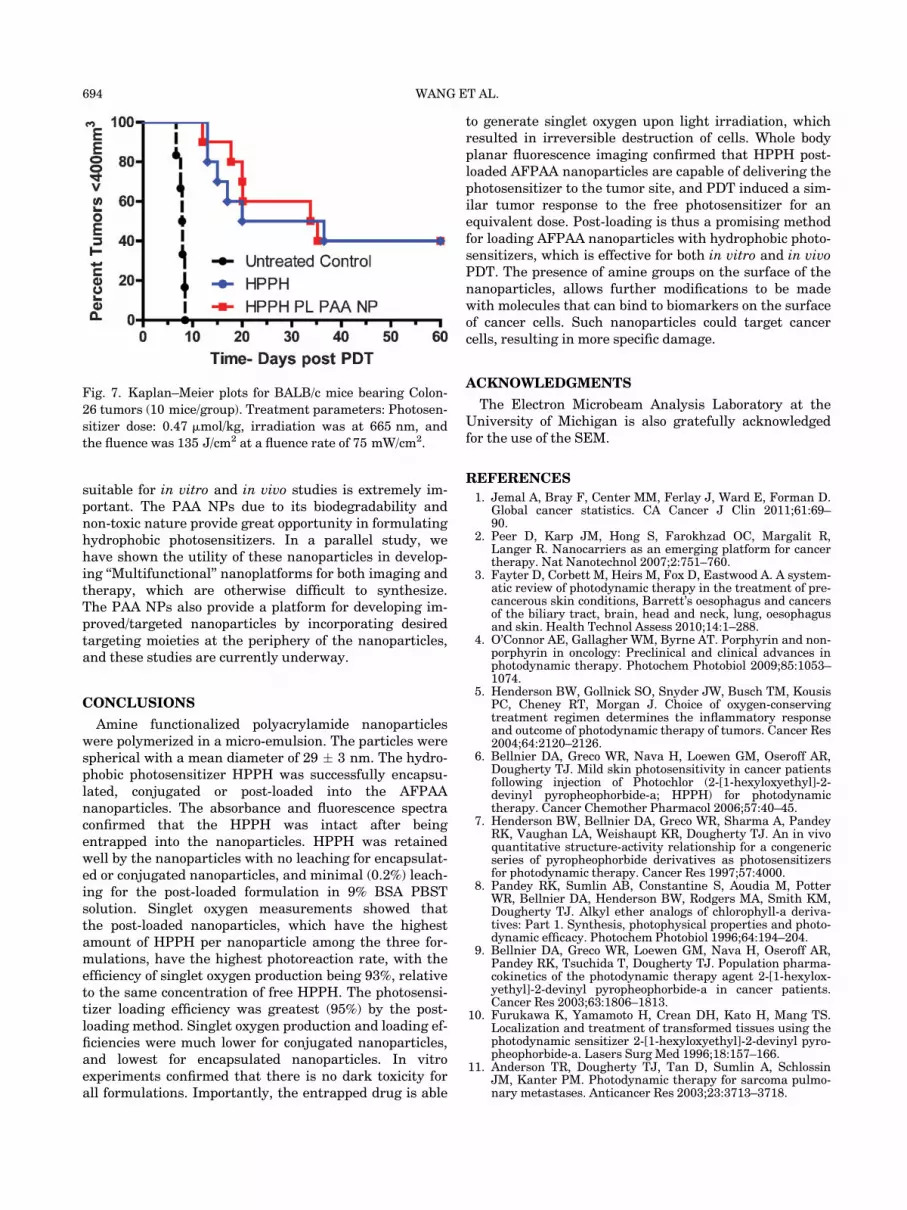
The in vivo PDT efficacy of the HPPH post-loadedAFPAA nanoparticles was next compared to free HPPHin BALB/c mice bearing Colon26 tumors. The tumorresponse was 40% for both groups as shown in Figure 7. Asimilar tumor response was expected since the estimatedHPPH in the tumors was not significantly different basedon the fluorescence imaging data. It is possible that the
Fig. 3. Fluorescence emission spectra of free HPPH (0.5 mM,
green dashed line), HPPH encapsulated (2.5 mg/ml, black
solid line, 0.5 mM of HPPH), HPPH conjugated AFPAA nano-
particles (0.5 mg/ml, pink short dash dotted line, 0.5 mM of
HPPH) and HPPH post-loaded AFPAA nanoparticles
(0.033 mg/ml, blue dash dotted line, 0.5 mM of HPPH), all
excited at 415 nm in 1% Tween 80. The arrows indicate the
corresponding axes.
Fig. 4. Spectra of ADPA from a mixture of ADPA (4 mM) and
HPPH post-loaded nanoparticles (0.067 mg/ml) illuminated
at 665 nm for 0, 5, 10, and 20 minutes. The excitation slit
was 10 nm and the emission slit was 2 nm.
692 WANG ET AL.

pharmacokinetic properties of free HPPH may vary in itsPAA NPs formulation. Therefore, it is important to inves-tigate the in vivo PDT efficacy of HPPH-PAA NPs formu-lation in larger group of mice at different drug/light doses
and variable time intervals, and these studies are cur-rently in progress.
Most of the porphyrin-based compounds are insolublein water and the selection of an appropriate formulation
Fig. 5. Phototoxicity of Colon26 cells after incubation with the different preparations of
AFPAA nanoparticles for 4 hours followed by light at a series of doses at a fluence rate of
3.2 mW/cm2. Growth was assayed by the MTT method. (A) Encapsulated HPPH (PAA-E),
(B) Conjugated HPPH (PAA-CONJ), (C) Post-loaded HPPH (PAA-PL), (D) Free HPPH. Val-
ues are the mean � standard deviation of 3–4 experiments.
Fig. 6. Fluorescence optical imaging of BALB/c mice bearing Colon26 tumors. (A) Control
mouse, no drug, black circle indicates location of tumor (B) 24 hours post-injection of HPPH
(0.47 mmol/kg) and (C) 24 hours post-injection of HPPH post-loaded PAA nanoparticles
(0.47 mmol/kg). The false-colored images are scaled to the same intensity range and show
representative images with fluorescence intensity closest to the average values. The ROI
over the tumor for measuring fluorescence intensity is indicated in A. The bar graph shows
the fluorescence intensity in the ROI (average � standard deviation of three samples).
BIODEGRADABLE NANOCARRIERS FOR PHOTODYNAMIC THERAPY 693
suitable for in vitro and in vivo studies is extremely im-portant. The PAA NPs due to its biodegradability andnon-toxic nature provide great opportunity in formulatinghydrophobic photosensitizers. In a parallel study, wehave shown the utility of these nanoparticles in develop-ing ‘‘Multifunctional’’ nanoplatforms for both imaging andtherapy, which are otherwise difficult to synthesize.The PAA NPs also provide a platform for developing im-proved/targeted nanoparticles by incorporating desiredtargeting moieties at the periphery of the nanoparticles,and these studies are currently underway.
CONCLUSIONS
Amine functionalized polyacrylamide nanoparticleswere polymerized in a micro-emulsion. The particles werespherical with a mean diameter of 29 � 3 nm. The hydro-phobic photosensitizer HPPH was successfully encapsu-lated, conjugated or post-loaded into the AFPAAnanoparticles. The absorbance and fluorescence spectraconfirmed that the HPPH was intact after beingentrapped into the nanoparticles. HPPH was retainedwell by the nanoparticles with no leaching for encapsulat-ed or conjugated nanoparticles, and minimal (0.2%) leach-ing for the post-loaded formulation in 9% BSA PBSTsolution. Singlet oxygen measurements showed thatthe post-loaded nanoparticles, which have the highestamount of HPPH per nanoparticle among the three for-mulations, have the highest photoreaction rate, with theefficiency of singlet oxygen production being 93%, relativeto the same concentration of free HPPH. The photosensi-tizer loading efficiency was greatest (95%) by the post-loading method. Singlet oxygen production and loading ef-ficiencies were much lower for conjugated nanoparticles,and lowest for encapsulated nanoparticles. In vitroexperiments confirmed that there is no dark toxicity forall formulations. Importantly, the entrapped drug is able
to generate singlet oxygen upon light irradiation, whichresulted in irreversible destruction of cells. Whole bodyplanar fluorescence imaging confirmed that HPPH post-loaded AFPAA nanoparticles are capable of delivering thephotosensitizer to the tumor site, and PDT induced a sim-ilar tumor response to the free photosensitizer for anequivalent dose. Post-loading is thus a promising methodfor loading AFPAA nanoparticles with hydrophobic photo-sensitizers, which is effective for both in vitro and in vivoPDT. The presence of amine groups on the surface of thenanoparticles, allows further modifications to be madewith molecules that can bind to biomarkers on the surfaceof cancer cells. Such nanoparticles could target cancercells, resulting in more specific damage.
ACKNOWLEDGMENTS
The Electron Microbeam Analysis Laboratory at theUniversity of Michigan is also gratefully acknowledgedfor the use of the SEM.
REFERENCES
1. Jemal A, Bray F, Center MM, Ferlay J, Ward E, Forman D.Global cancer statistics. CA Cancer J Clin 2011;61:69–90.
2. Peer D, Karp JM, Hong S, Farokhzad OC, Margalit R,Langer R. Nanocarriers as an emerging platform for cancertherapy. Nat Nanotechnol 2007;2:751–760.
3. Fayter D, Corbett M, Heirs M, Fox D, Eastwood A. A system-atic review of photodynamic therapy in the treatment of pre-cancerous skin conditions, Barrett’s oesophagus and cancersof the biliary tract, brain, head and neck, lung, oesophagusand skin. Health Technol Assess 2010;14:1–288.
4. O’Connor AE, Gallagher WM, Byrne AT. Porphyrin and non-porphyrin in oncology: Preclinical and clinical advances inphotodynamic therapy. Photochem Photobiol 2009;85:1053–1074.
5. Henderson BW, Gollnick SO, Snyder JW, Busch TM, KousisPC, Cheney RT, Morgan J. Choice of oxygen-conservingtreatment regimen determines the inflammatory responseand outcome of photodynamic therapy of tumors. Cancer Res2004;64:2120–2126.
6. Bellnier DA, Greco WR, Nava H, Loewen GM, Oseroff AR,Dougherty TJ. Mild skin photosensitivity in cancer patientsfollowing injection of Photochlor (2-[1-hexyloxyethyl]-2-devinyl pyropheophorbide-a; HPPH) for photodynamictherapy. Cancer Chemother Pharmacol 2006;57:40–45.
7. Henderson BW, Bellnier DA, Greco WR, Sharma A, PandeyRK, Vaughan LA, Weishaupt KR, Dougherty TJ. An in vivoquantitative structure-activity relationship for a congenericseries of pyropheophorbide derivatives as photosensitizersfor photodynamic therapy. Cancer Res 1997;57:4000.
8. Pandey RK, Sumlin AB, Constantine S, Aoudia M, PotterWR, Bellnier DA, Henderson BW, Rodgers MA, Smith KM,Dougherty TJ. Alkyl ether analogs of chlorophyll-a deriva-tives: Part 1. Synthesis, photophysical properties and photo-dynamic efficacy. Photochem Photobiol 1996;64:194–204.
9. Bellnier DA, Greco WR, Loewen GM, Nava H, Oseroff AR,Pandey RK, Tsuchida T, Dougherty TJ. Population pharma-cokinetics of the photodynamic therapy agent 2-[1-hexylox-yethyl]-2-devinyl pyropheophorbide-a in cancer patients.Cancer Res 2003;63:1806–1813.
10. Furukawa K, Yamamoto H, Crean DH, Kato H, Mang TS.Localization and treatment of transformed tissues using thephotodynamic sensitizer 2-[1-hexyloxyethyl]-2-devinyl pyro-pheophorbide-a. Lasers Surg Med 1996;18:157–166.
11. Anderson TR, Dougherty TJ, Tan D, Sumlin A, SchlossinJM, Kanter PM. Photodynamic therapy for sarcoma pulmo-nary metastases. Anticancer Res 2003;23:3713–3718.
Fig. 7. Kaplan–Meier plots for BALB/c mice bearing Colon-
26 tumors (10 mice/group). Treatment parameters: Photosen-
sitizer dose: 0.47 mmol/kg, irradiation was at 665 nm, and
the fluence was 135 J/cm2 at a fluence rate of 75 mW/cm2.
694 WANG ET AL.
12. Loewen G, Pandey R, Belliner D, Henderson B, DoughertyT. Endobronchial photodynamic therapy for lung cancer.Lasers Surg Med 2006;38:364–370.
13. Sunar U, Rohrbach D, Rigual N, Tracy E, Keymel K, CooperMT, Baumann H, Henderson BW. Monitoring photobleach-ing and hemodynamic responses to HPPH-mediated photo-dynamic therapy of head and neck cancer: A case report. OptExpress 2010;18:14969–14978.
14. Baba K, Pudavar HE, Roy I, Ohulchanskyy TY, Chen YH,Pandey RK, Prasad PN. New method for delivering a hydro-phobic drug for photodynamic therapy using pure nanocrys-tal form of the drug. Mol Pharm 2007;4:289–297.
15. Wong J, Brugger A, Khare A, Chaubal M, Papadopoulos P,Rabinow B, Kipp J, Ning J. Suspensions for intravenous (IV)injection: A review of development, preclinical and clinicalaspects. Adv Drug Deliv Rev 2008;60:939–954.
16. Koo YE L, Reddy GR, Bhojani M, Schneider R, Philbert MA,Rehemtulla A, Ross BD, Kopelman R. Brain cancer diagnosisand therapy with nanoplatforms. Adv Drug Deliv Rev2006;58:1556–1577.
17. Cinteza LO, Ohulchanskyy TY, Sahoo Y, Bergey EJ, PandeyRK, Prasad PN. Diacyllipid micelle-based nanocarrier formagnetically guided delivery of drugs in photodynamic ther-apy. Mol Pharm 2006;3:415–423.
18. Matsumura Y, Maeda H. A new concept for macromoleculartherapeutics in cancer chemotherapy-mechanism of tumori-tropic accumulation of proteins and the antitumor agentsmancs. Cancer Res 1986;46:6387–6392.
19. Maeda H. The enhanced permeability and retention (epr) ef-fect in tumor vasculature: the key role of tumor-selectivemacromolecular drug targeting. Advan Enzyme Regul 2001;41:189–207.
20. Cheng Y, Samia AC, Meyers JD, Panagopoulos I, Fei BW,Burda C. Highly efficient drug delivery with gold nanoparti-cle vectors for in vivo photodynamic therapy of cancer. J AmChem Soc 2008;130:10643–10647.
21. Yan F, Kopelman R. The embedding of meta-tetra(hydroxy-phenyl)-chlorin into silica nanoparticle platforms for photo-dynamic therapy and their singlet oxygen production andpH-dependent optical properties. Photochem Photobiol 2003;78:587–591.
22. Roy I, Ohulchanskyy TY, Pudavar HE, Bergey EJ, OseroffAR, Morgan J, Dougherty TJ, Prasad PN. Ceramic-basednanoparticles entrapping water-insoluble photosensitizinganticancer drugs: A novel drug-carrier system for photody-namic therapy. J Am Chem Soc 2003;125:7860–7865.
23. Gao D, Agayan RR, Xu H, Philbert MA, Kopelman R. Nano-particles for two-photon photodynamic therapy in livingcells. Nano Lett 2006;6:2383–2386.
24. Gao D, Xu H, Philbert MA, Kopelman R. Ultrafine hydrogelnanoparticles: synthetic approach and therapeutic applica-tion in living cells. Ange Chem Int Ed 2007;46:2224–2227.
25. Tang W, Xu H, Kopelman R, Philbert MA. Photodynamiccharacterization and in vitro application of methylene blue-containing nanoparticle platforms. Photochem Photobiol2005;81:242–249.
26. Tang W, Xu H, Park EJ, Philbert MA, Kopelman R. Encap-sulation of methylene blue in polyacrylamide nanoparticleplatforms protects its photodynamic effectiveness. BiochemBiophys Res Commun 2008;369:579–583.
27. Kim S, Ohulchanskyy TY, Pudavar HE, Pandey RK,Prasad PN. Organically modified silica nanoparticlesco-encapsulating photosensitizing drug and aggregation-en-hanced two-photon absorbing fluorescent dye aggregates fortwo-photon photodynamic therapy. J Am Chem Soc 2007;129:2669–2675.
28. Vargas A, Eid M, Fanchaouy M, Gurny R, Delie F. In vivophotodynamic activity of photosensitizer-loaded nanopar-ticles: formulation properties, administration parametersand biological issues involved in PDT outcome. Eur J PharmBiopharm 2008;69:43–53.
29. Moreno MJ, Monson E, Reddy RG, Rehemtulla A, RossBD, Philbert M, Schneider RJ, Kopelman R. Production ofsinglet oxygen by Ru(dpp(SO3)(2))(3) incorporated in poly-acrylamide PEBBLES. Sens Actuator B Chem 2003;90:82–89.
30. Harrell JA, Kopelman R. Biocompatible probes measure in-tracellular activity. Biophoton Int 2000;7:22.
31. Wenger Y, Schneider IIRJ, Reddy GR, Kopelman R, JollietO, Philbert MA. Tissue distribution and pharmacokinetics ofstable polyacrylamide nanoparticles following intravenousinjection in the rat. Toxicol Appl Pharmacol 2011;251:181–190.
32. Reddy GR, Bhojani MS, McConville P, Moody J, MoffatBA, Hall DE, Kim G, Koo YEL, Woolliscroft MJ, Sugai JV,Johnson TD, Philbert MA, Kopelman R, Rehemtulla A,RossBD. Vascular targeted nanoparticles for imaging andtreatment of brain tumors. Clin Cancer Res 2006;12:6677–6686.
33. Lambert CR, Reddi E, Spikes JD, Rodgers MAJ, Jori G. Theeffects of porphyrin structure and aggregation state on pho-tosensitized processes in aqueous and micellar media. Photo-chem Photobiol 1986;44:595–601.
34. Kelbauskas L, Dietel W. Internalization of aggregated photo-sensitizers by tumor cells: subcellular time-resolved fluores-cence spectroscopy on derivatives of pyropheophorbide-aethers and chlorin e6 under femtosecond one- and two-pho-ton excitations. Photochem Photobiol 2002;76:686–694.
35. Zhang Y, Zhu W, Wang BB, Ding JD. A novel microgel andassociated post-fabrication encapsulation technique of pro-teins. J Controlled Release 2005;105:260.
36. Chiu HC, Lin YF, Hung SH. Equilibrium swelling of copoly-merized acrylic acid – Methacrylated dextran networks:Effects of pH and neutral salt. Macromolecules 2002;35:5235–5242.
37. Tan HP, Chu CR, Payne KA, Marra KG. Injectable in situforming biodegradable chitosan-hyaluronic acid basedhydrogels for cartilage tissue engineering. Biomaterials2009;30:2499–2506.
38. Pallenberg AJ, Dobhal MP, Pandey RK. Efficient synthesisof pyropheophorbide-a and its derivatives. Org Proc Res Dev2004;8:287–290.
BIODEGRADABLE NANOCARRIERS FOR PHOTODYNAMIC THERAPY 695


![Synthesis and Properties of Benzo[ a ]phenoxazinium Chalcogen Analogues as Novel Broad-Spectrum Antimicrobial Photosensitizers](https://img.pdfslide.net/doc/110x75/6355492c25e9052f090c9d2c/synthesis-and-properties-of-benzo-a-phenoxazinium-chalcogen-analogues-as-novel.jpg)


