Embed Size (px)
Citation preview
1© 2014 Wiley-VCH Verlag GmbH & Co. KGaA, Weinheim wileyonlinelibrary.com
Photodynamic Therapy
In Vivo Studies of Nanostructure-Based Photosensitizers for Photodynamic Cancer Therapy Siew Hui Voon , Lik Voon Kiew , Hong Boon Lee , Siang Hui Lim , Mohamed Ibrahim Noordin , Anyanee Kamkaew , Kevin Burgess , and Lip Yong Chung *
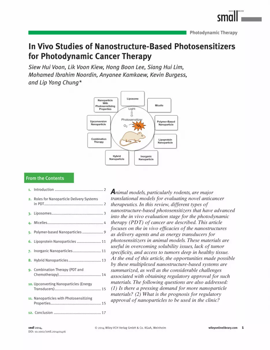
Animal models, particularly rodents, are major translational models for evaluating novel anticancer therapeutics. In this review, different types of nanostructure-based photosensitizers that have advanced into the in vivo evaluation stage for the photodynamic therapy (PDT) of cancer are described. This article focuses on the in vivo effi cacies of the nanostructures as delivery agents and as energy transducers for photosensitizers in animal models. These materials are useful in overcoming solubility issues, lack of tumor specifi city, and access to tumors deep in healthy tissue. At the end of this article, the opportunities made possible by these multiplexed nanostructure-based systems are summarized, as well as the considerable challenges associated with obtaining regulatory approval for such materials. The following questions are also addressed: (1) Is there a pressing demand for more nanoparticle materials? (2) What is the prognosis for regulatory approval of nanoparticles to be used in the clinic?
1. Introduction .............................................. 2
2. Roles for Nanoparticle Delivery Systems in PDT ........................................................ 2
3. Liposomes ................................................. 3
4. Micelles ..................................................... 4
5. Polymer-based Nanoparticles .................... 9
6. Lipoprotein Nanoparticles ....................... 11
7. Inorganic Nanoparticles ........................... 11
8. Hybrid Nanoparticles ............................... 13
9. Combination Therapy (PDT and Chemotherapy) ........................................ 14
10. Upconverting Nanoparticles (Energy Transducers) ............................................ 15
11. Nanoparticles with Photosensitizing Properties ................................................ 15
12. Conclusion ............................................. 17
From the Contents
small 2014, DOI: 10.1002/smll.201401416

S. H. Voon et al.
2 www.small-journal.com © 2014 Wiley-VCH Verlag GmbH & Co. KGaA, Weinheim
reviews
DOI: 10.1002/smll.201401416
S. H. Voon, Dr. L. V. Kiew Department of Pharmacology Faculty of Medicine University of Malaya 50603 , Kuala Lumpur , Malaysia Dr. H. B. Lee, S. H. Lim, Dr. M. I. Noordin, Prof. L. Y. Chung Department of Pharmacy Faculty of Medicine University of Malaya 50603 , Kuala Lumpur , Malaysia E-mail: [email protected] ; [email protected] Dr. H. B. Lee, S. H. Lim Cancer Research Initiatives Foundation (CARIF) Subang Jaya Medical Center 47500 , Subang Jaya , Selangor , Malaysia A. Kamkaew, Prof. K. Burgess Department of Chemistry Texas A&M University Box 30012, College Station , Texas 77842 , USA
1. Introduction
Photodynamic therapy (PDT) emerged as a treatment modality for neoplastic and non-malignant lesions after it was reported that a combination of red light and porfi mer sodium (hematoporphyrin derivatives) could eradicate mouse mam-mary tumor growth. [ 1 ] Several clinical trials were then initi-ated, and PDT was shown to be successful in treating patients with bladder cancer and skin tumors. [ 2 ] Subsequent clinical studies involved other types of cancers, including those of the breast, colon, prostate, squamous cells, basal cells, endome-trium, malignant melanoma, mycosis fungoides, chondrosar-coma and angiosarcoma. [ 3 ]
PDT applications involve a photosensitizer, light and molecular oxygen. Upon light irradiation, the photosensi-tizer is activated and generates highly reactive singlet oxygen from molecular oxygen. [ 3a , 4 ] Due to its short half-life of about 3 µs, damage caused by singlet oxygen is highly localized. [ 5 ] In cancer treatment, PDT can destroy tumor cells directly, damage vasculature surrounding tumor cells and activates immunological responses against tumors. [ 6 ]
PDT using porfi mer sodium (Photofrin; Axcan Pharma Inc., Mont-Saint-Hilaire, Canada) was approved for the treatment of bladder cancer in Canada in 1993. Since that time, other anticancer photosensitizing drugs (photosen-sitizers) have obtained clinical approval, including Foscan (temoporfi n, meta-tetrahydroxyphenylchlorin; Biolitec AG), Visudyne (verteporfi n, benzoporphyrin derivative monoacid ring A; Novartis Pharmaceuticals), Levulan (5-aminolevulinic acid; DUSA Pharmaceuticals, Inc.), and most recently, Metvix (methyl aminolevulinate; PhotoCure ASA.).
PDT has remained at best a fringe cancer treatment option as it is limited by inaccessibility of deep seated tumors, skin photosensitivity for prolonged periods following treat-ment, high initial setup cost and lack of standard protocols established by randomized trials. [ 7 ] For instance, porfi mer sodium causes lasting skin photosensitivity for up to 4–12 weeks at its therapeutic dose. [ 8 ] Many of the photosensitizers currently used cannot be excited by light of wavelengths >700 nm; this limits their use because tissue penetration of light into deeper tissues is only possible for long wavelength excitation sources. Although light-tissue interaction is not fully understood, wavelengths of less than 600 nm penetrate tissue to about 0.5 cm while slightly longer wavelengths up can double tissue penetration to 1 cm. [ 9 ] In the case of porfi mer sodium, the relatively low maximum absorption wavelength of 630 nm has the challenge of poor tissue penetration of light while the very low extinction coeffi cient (1.170 M −1 cm −1 ) [ 4 ] at this Q-band wavelength would increase the dose of drug and/or light required to give adequate phototherapeutic response: neither of these are the characteristics of an ideal clinical photosensitizer. [ 10 ] Moreover, most photosensitizer molecules are hydrophobic and aggregate easily in aqueous media, leading to reduced singlet oxygen generation [ 11 ] and diffi culties with intravenous delivery strategies. [ 12 ] The other limitations may remain in the initial period until the avail-ability of cheaper lasers and optic fi ber equipment to reduce the high initial setup cost and standardization of treatment protocols for a greater range of target organs. [ 7 ]
Ideal photosensitizers would be activated at >700 nm and have better tumor specifi cities, which would reduce the generalized photosensitivities. [ 13 ] Newer photosensitizers in clinical trials include tin ethyl etiopurpurin (SnET2), mono-L-aspartyl chlorin e 6 (Npe 6 ), benzoporphyrin derivative (BPD), and lutetium texaphyrin (Lu-Tex); these have absorp-tion bands at 660, 664, 690 and 732 nm, respectively, and pro-voke only mild and transient skin photosensitivity. However, clinical studies using these photosensitizers are restricted to the treatment of cutaneous cancer, [ 14 ] early squamous cell carcinoma of the lung, [ 15 ] and recurrent prostate cancer; [ 16 ] therefore, full understanding of the relative merits of these sensitizers and approval for general clinical use are still pending. Figure 1 presents chemical structures for many of the photosensitizers discussed in this review.
2. Roles for Nanoparticle Delivery Systems in PDT
The effectiveness of PDT is not only largely determined by the effi ciency of singlet oxygen generation [ 17 ] upon light acti-vation, but also the effi ciency and selectivity to which thera-peutic concentrations of the photosensitizer is delivered to the targeted tumor site with minimal uptake by non-targeted normal cells. [ 18 ] Incorporating PDT agents into nanoparticles can alter these properties to ultimately improve clinical outcome. Covalent conjugation or physical inclusion of photosensitizers to nanoparticle carrier systems can surround hydrophobic photosensitizers with a hydrophilic environ-ment, improving their solubilities and preventing aggrega-tion in blood. This process may also reduce the non-specifi c phototoxicity to the skin and eyes, enhance the antitumor effi cacy, [ 19 ] and increase passive tumor accumulation via an enhanced permeability and retention (EPR) effect. [ 7 ] This will elevate levels of nanostructure-based photosensitizers either in proximity to the tumor tissue or inside the cells
small 2014, DOI: 10.1002/smll.201401416

In Vivo Studies of Photosensitizers for Cancer Therapy
3www.small-journal.com© 2014 Wiley-VCH Verlag GmbH & Co. KGaA, Weinheim
Lik Voon Kiew obtained his doctorate in biopolymer technology and therapeutics from the University of Malaya, Malaysia. He is currently senior lecturer at the Pharmacol-ogy department, Faculty of Medicine of the University of Malaya, Malaysia. His current research interests include the development and in vitro/in vivo evaluation of targetable anticancer polymer therapeutics, renal target-ing drug carriers, anticancer photodynamic therapeutics, and bioactive compounds for chronic disease treatment.
Siew Hui Voon obtained her BSc in Bio-medical Science from National University of Malaysia in 2009. She received a MyPhD scholarship from Ministry of Education Malaysia in 2012 to pursue her PhD under the guidance of Prof. Lip Yong Chung at the University of Malaya. Her current research of interest is in the fi eld of nanomaterials for drug delivery in photodynamic cancer therapy.
Lip Yong Chung received his doctorate in pharmacy from the University of Cardiff and joined Cardiff University as a research asso-ciate focusing on bioactive polymers and cell regeneration, and is currently at the Faculty of Medicine of the University of Malaya, Ma-laysia as a Professor in Pharmaceutical Sci-ences. His current research interests include the discovery of anticancer and CNS active compounds, and development of nanodrug conjugates for therapy.
after internalization. Majority of cellular internalization of nanoparticles occur through pinocytosis known as clathrin-mediated endocytosis (CME). [ 20 ] The attachment of cancer-targeting moieties such as folic acid, [ 21 ] F3 peptide [ 22 ] or RGD peptide [ 23 ] to the nano-drug carriers can further enhance the selectivity for tumors. Chemical modifi cation of the structures of the nano-drug carriers may also confer alternate means for energy transfer to photosensitizers, which enables the use of deep tissue-penetrating light for the activation of the photo-sensitizers to treat deeply seated tumors. [ 24 ]
The concept of subcellular targeted nanoparticles is being developed and only a limited number of strategies has been identifi ed for endoplasmic reticulum, mitochondria and nucleus targeting. Further investigations are necessary to evaluate its impact and relevance for future clinical applica-tions. The design of subcellular targeted nanoparticles with non-immunogenic stealth character, with the ability to over-come biological barriers and target site specifi city, remains necessary. [ 20a ]
This review describes examples of how photosensitizers in nanostructures have advanced into the in vivo evaluation stage, focusing on the in vivo effi cacies of the nanostructures as delivery agents and as energy transducers for photosen-sitizers in animal models ( Table 1 ). We also summarize the in vivo characteristics of nanostructures in terms of biodeg-radability, clearance and side effect as well as the regulatory approval status for clinical use ( Table 2 ). This article com-plements previous reviews on the design, synthesis, physico-chemical properties, and in vitro therapeutic effi ciencies of photosensitizers. [ 25 ]
3. Liposomes
Liposomes are spherical, closed membranes composed of concentric phospholipid bilayers with an aqueous inner com-partment. [ 26 ] They can accommodate lipophilic photosen-sitizers by incorporating them into the lipid bilayer, which renders them hydrosoluble and prevents aggregation in an aqueous environment ( Figure 2 ). [ 27 ]
These structures naturally tend to accumulate in tumors, which enhances the ability of incorporated photosensitizers to target the tumor tissue. [ 28 ] Several studies have demon-strated a higher accumulation of photosensitizers and better PDT effi cacy in tumor tissues for photosensitizer-containing liposomes compared to free photosensitizers. [ 29 ] Thus, the incorporation of Photofrin into liposomes resulted in 6 and 2.4-fold increases in in vivo tumoral accumulation for 9L gliosarcoma and U87 glioma nude mice models, respectively, compared to the free Photofrin control in a dextrose for-mulation. Tissue necrosis was signifi cantly increased in both tumor types after PDT compared to the controls. [ 30 ]
Enhanced tumor accumulation of hypocrellin A was also observed when a liposome-hypocrellin A nanoconstruct was tested in an S-180 sarcoma Kunming mouse model, with max-imum accumulation in tumor at 12 h (0.48 µg/g) compared to the group treated with hypocrellin A-DMSO (0.14 µg/g). [ 31 ] This observation is consistent with the higher tumor regres-sion at day 7 after PDT (relative regression percent of tumor,
RRP = 87%) and with the PDT effi cacy exhibited by the liposome-hypocrellin A-treated group relative to the hypo-crellin A-DMSO treatment group (RRP = 14%).
Metatetra(hydroxyphenyl) chlorin formulated in uni-lamellar dipalmitoylphosphatidylcholine/dipalmitoylphos-phatidylglycerol liposomal nanoconstructs has been applied to female Foxn1nu/nu mice implanted with EMT-6 mam-mary tumor cells. In the four month follow-up period, 79% of the primary tumors were cured and showed no sign of recurrence. [ 32 ]
Visudyne, a commercially available liposomal formula-tion of benzoporphyrin derivative monoacid ring A (BPD-MA) indicated for age-related macular degeneration [ 33 ] when applied to Meth-A sarcoma-bearing mice using antiangio-genic scheduling (short drug-light interval of 15 min) showed greater tumor suppression compared to conventional sched-uling (longer drug-light interval of 3 h). In this, 40 and 60% of the tumor of Meth-A sarcoma-bearing mice were cured
small 2014, DOI: 10.1002/smll.201401416

S. H. Voon et al.
4 www.small-journal.com © 2014 Wiley-VCH Verlag GmbH & Co. KGaA, Weinheim
reviews
at 0.25 and 0.5 mg/kg BPD-MA, respectively. These suggest Visudyne is effective in cancer PDT using antiangiogenic scheduling. [ 34 ]
These liposomes of 70–100 nm led to an increase in pas-sive accumulation in the tumors via the enhanced permea-bility and retention effect. However, the main drawback of conventional liposomes is that they are easily taken up by cells of the reticuloendothelial system after systemic admin-istration. This results in their rapid removal from the blood into the liver and spleen, which in turn reduces the accumu-lation in the tumor tissue. [ 35 ] Some liposome modifi cations have been reported to counteract these delivery problems. For instance, polyethylene glycol has been added to lipo-somal surfaces to create more stable “sheathed” forms. Such PEGylation may protect liposomes from being recognized by opsonins and taken up by the reticuloendothelial system. [ 36 ] However, the increased stability provided by this type of
modifi cation of the liposomes may also decrease the lipo-some-cell interactions and reduce the transfer of the photo-sensitizer payload into tumor cells. [ 37 ]
4. Micelles
Micelles are aggregates of surfactant molecules in liquid-col-loid dispersions in which the hydrophilic head regions are in contact with surrounding solvent and the hydrophobic tails are in the center. The particle size range is normally within 5–100 nm. They are widely used to carry hydrophobic drugs, physically entrapped in or covalently bound to the hydro-phobic core and delivered to tumors via passive or active tar-geting strategies. [ 38 ] Micelles can be classifi ed according to the nature of the amphiphilic core: polymeric or lipid micelles. Polymeric micelles are formed from block copolymers,
Figure 1. Some photosensitizers featured in this review.
small 2014, DOI: 10.1002/smll.201401416

In Vivo Studies of Photosensitizers for Cancer Therapy
5www.small-journal.com© 2014 Wiley-VCH Verlag GmbH & Co. KGaA, Weinheim
Table 1. In Vivo Studies Reported on Nanostructure-Based Photosensitizer Formulations.
Nanostructure Photosensitizer Animal model
LIPOSOMES Photofrin (porfi mer sodium); hypro-crellin A; metatetra(hydroxyphenyl) chlorin (mTHPC); Verteporfi n
Athymic (nude) rats (strain Cr: NIH-rna) implanted with U87 glioma cells through craniectomy; [30a] male Kunming mice transplanted with S-180 sarcoma; [31] female Foxn1 nu/nu mice injected subcutaneously with EMT6 cells; [32] male BALB/c mice injected subcutaneously with Meth-A sarcoma cells [34]
MICELLES
Polymer Micelles Aluminum chloride phthalocyanine (AlClPc); protophorphyrin IX (PpIX)
Mice with squamous cell carcinoma (SCC7) tumor; [40] Balb/c mice implanted with EMT-6 tumors [41]
PEG-Lipid Micelles Verteporfi n; meso -tetraphenylporphine (TPP)
Female C57BL/6 mice injected subcutaneously with LLC cells; [43a[ male DBA/2 mice implanted with rhabdomyosarcoma (M1) tumor cells [46]
Cremophor EL Temocene; azabodipy DBA/2 mice inoculated with the DBA/2 mastocytoma cell line P815; [53] Balb/c mice inoculated with the Balb/c colon adenocarci-noma cell line CT26.CL25 (ATCC, CRL-2639); [53] female Balb/c nu/nu mice injected with MDA-MB-231-GFP cells [51]
POLYMER-BASED NANOPARTICLES
Poly(lactic- co -glycolic acid) (PLGA) Zinc phthalocyanine; verteporfi n Female albino mice injected with Ehrlich ascites carcinoma cells; [56] male DBA/2 mice implanted with rhabdomyosarcoma (M1) tumor [18]
Dendrimers Aminolevulinic acid (ALA); phthalocyanine
Male BALB/c mice injected with the LM3 cell line; [60] female nude mice (BALB/c nu/nu) subcutaneously transplanted with subcutaneous A549 tumor cells [61]
Chitosan Protophorphyrin IX (PpIX); chlorin e 6 Athymic nude mice inoculated with SCC7 cells; [65] athymic nude mice injected with HT-29 human colon adenocarcinoma cells [67,70[
LIPOPROTEIN NANOPARTICLES Bacteriochlorin e 6 bisoleate (Bchl-BOA)
Female athymic nude mice inoculated with HepG2 cells [77]
INORGANIC NANOPARTICLES
Silica Protophorphyrin IX (PpIX); methylene blue; zinc phthalocyanine
Male athymic Nude-Foxn1 nu mice subcutaneously implanted with glioblastoma multiforme; [82] female athymic Swiss nude mice and female athymic Naval Medical Research Institute nude mice injected subcutaneously with HCT 116 and A549 cells, respectively; [82] male athymic BALB/c (Balb/C-nu) mice inoculated subcutaneously with Hela cells; [80] female BALB/c nude mice implanted with H22 cells; [84] female Swiss nude mice xenografted with HCT-116 cells [85]
Gold Silicon phthalocyanine 4; porphyrin-brucine conjugates; Chlorin e 6
NuNu mice subcutaneously injected with basaloid squamous cell carcinoma PE/CA-PJ34 cells; [90a] mice with MDA-MB-435 tumor [92]
Calcium Phosphosilicate Indocyanine green (ICG) Female C3H/HeJ mice injected with 32D-p210-GFP cells [94]
HYBRID NANOPARTICLES
Polyacrylamide Photofrin (porfi mer sodium) Male Fischer 344 rats implanted with rat 9L glioma cells [22]
COMBINATION THERAPY (PDT & CHEMOTHERAPY)
N -(2-Hydroxypropyl)methacrylamide (HPMA) meso -chlorin e 6 monoethylene diamine (Mce 6 )
Female nu/nu mice with OVCAR-3 solid tumor [102b]
Liposome Doxorubicin (Chemotherapeutic agent); Verteporfi n
Female BALB/c mice inoculated subcutaneously with murine colon carcinoma Colo 26; [103] Male Copenhagen rats implanted subcutane-ously and orthotopically with R3327-MatLyLu Dunning prostate tumor cells [104]
UPCONVERSION NANOPARTICLES (ENERGY TRANSDUCER)
Chlorin e 6 ; merocyanine 540 (MC540); zinc phthalocyanine (ZnPc)
Female Balb/c mice injected subcutaneously with 4T1 cells; [24b] C57BL/6 female mice implanted subcutaneously with B16-F0 cells [21]
NANOPARICLES WITH PHOTOSENSITIZING PROPERTIES
Titanium Dioxide None Nude mice injected subcutaneously with T-24 cells; [108] female BALB/c-nude mice inoculated with U87 cells [110]
Fullerenes (C 60 ) None Female CDF1 mice inoculated with Meth A fi brosarcoma cells; [118a] female CDF1 mice inoculated with Meth AR1fi brosarcoma cells; [118b] male BALB/c mice inoculated with CT26-Luc cells; [122] mice xeno-grafted with human-melanoma (COLO679) [123]
small 2014, DOI: 10.1002/smll.201401416

S. H. Voon et al.
6 www.small-journal.com © 2014 Wiley-VCH Verlag GmbH & Co. KGaA, Weinheim
reviews Table 2. In Vivo Characteristics of Nanostructures and Their Regulatory Approval Status for Clinical Use.
Nanostructure Biodegradability Metabolism & Clearance Side Effects FDA Approval Status for PDT use
Regulatory Approval Status for other anticancer drugs
LIPOSOMES Yes Vesicle opsonization by serum protein and subsequent uptake by the reticuloendo-thelial system; Complement-mediated phagocytosis by Kupffer and endothelial cells of the liver as well as other phagocytic cells of the reticulo-endothelial system [128]
NA Yes Clinically approved: Doxil (Doxorubicin) [US, EU] [129]
Clinically approved: Visudyne (Verteporfi n) to treat age-related macular degeneration
DaunoXome (Daunorubicin citrate) [US] [129]
Phase I & II clinical trials: CGP 55847 (liposomal zinc(II)-phthalocyanine) for cancer treatment [125]
Depocyt (Cytarabine) [US, EU] [130]
Mepact (Mifamurtide) [EU] [131]
Marqibo (Vincristine sulfate) [On market] [132]
MICELLES
Polymer Micelles No Degradation of polymer micelles, resulting in the formation of block copolymer unimers, which can be removed via renal excre-tion if the polymer chains are designed with a lower molecular weight than the critical value for renal fi ltration less than ∼20–40 kDa. [133]
Slow extravazation; Risk of chronic liver toxicity due to prolonged circulation and slower metabolism of drug which may exhibit toxic side effects [133]
No Clinically approved: Taxotere (Docetaxel) [EU, US] [129a]
Phase II clinical trials: SP1049C (Pluronic block-copolymer doxorubicin) [129a , 134]
PEG-Lipid Micelles NA NA NA No NA
Cremophor EL Slow May be largely degraded in the blood compartment by serum carboxylesterase-induced degradation, causing a gradual release of the ricinoleic acid residues attached to the triglyceride structure; Hepato-biliary elimination; Less than 0.1% of administered dose via urinary excretion [52]
Associated with severe ana-phylactoid hypersensitivity reactions, hyperlipidaemia, abnormal lipoprotein patterns, aggregation of erythrocytes and peripheral neuropathy [52]
No Clinically approved: Taxol (Paclitaxel) [US] [129a]
Phase II Clinical Trials: WST09 (TOOKAD) in Cremophor EL formulation [126a]
Phase I Clinical Trias: Silicon Phthalocyanine Pc 4 in Cremophor EL formulation [126b]
POLYMER-BASED NANOPARTICLES
Poly(lactic- co -glycolic acid) (PLGA)
Yes Undergo hydrolysis and biode-grades into lactic and glycolic acids. Lactic acid enter the tricarboxylic acid cycle and is metabolized and subsequently eliminated from the body as carbon dioxide and water. Glycolic acid is either excreted unchanged in the kidney or enters the tricarboxylic acid cycle and is eventually elimi-nated as carbon dioxide and water [55a]
NA Approved by the US FDA for the use of drug delivery [135]
NA
small 2014, DOI: 10.1002/smll.201401416

In Vivo Studies of Photosensitizers for Cancer Therapy
7www.small-journal.com© 2014 Wiley-VCH Verlag GmbH & Co. KGaA, Weinheim
Table 2. Continued
Nanostructure Biodegradability Metabolism & Clearance Side Effects FDA Approval Status for PDT use
Regulatory Approval Status for other anticancer drugs
Dendrimers NA Renal clearance for dendrimers with diameter 3–10 nm [128]
NA No Phase III clinical trials: SH L 643A (Gadolinium) for diag-nostic imaging [136]
Chitosan Yes Mainly degraded by lysozyme through the hydrolysis of the acetylated residues [137]
NA No NA
LIPOPROTEIN NANOPARTICLES
Yes Catabolized by the endothe-lium-associated lipoprotein lipase, thereby generating free fatty acids, which are taken up by the liver, muscle, and adipose tissues [138]
NA No NA
INORGANIC NANOPARTICLES
Silica Slow Gradual biodegradation results in the formation of water-sol-uble salts of silicic acid, which are excreted by the kidney; Hepatobiliary excretion [139]
Caused granuloma formation in the organs of the reticulo-endothelial system, such as liver and spleen [139,140]
No NA
Gold No Renal clearance for particles with diameter < 10 nm [128]
NA No Phase I clinical trials: AuroShell (gold nanoparticle) for laser therapy [141]
Calcium Phosphosilicate
No Hepatobiliary clearance with minimal acute renal involvement [93a]
NA No NA
HYBRID NANOPARTICLES
Polyacrylamide Yes Phagocytosis by Kupffer cells followed by hepatobiliary and renal clearance [97]
NA No NA
COMBINATION THERAPY (PDT & CHEMOTHERAPY)
N -(2-Hydroxypropyl)methacrylamide (HPMA)
No Blood clearance by spleen macrophage and eliminated by glomerular fi ltration in the kidney [137]
NA No Phase II clinical trials: ProLindac (HPMA copolymer–DACH platinate) [142]
UPCONVERSION NANOPAR-TICLES (ENERGY TRANSDUCER)
No Hepatobiliary clearance [24b] NA No NA
NANOPARICLES WITH PHOTOSENSI-TIZING PROPERTIES
Titanium Dioxide No 94% and 2% of administered TiO 2 was found in the liver and spleen and did not decrease up to 30 days after administration [143]
Can induce pathological lesions of the liver, spleen, kidneys, and brain [144]
No NA
Fullerenes (C 60 ) No Increased hepatobiliary accumulation and excrete via urinary tract [114,128]
NA No NA
NA = not available
small 2014, DOI: 10.1002/smll.201401416

S. H. Voon et al.
8 www.small-journal.com © 2014 Wiley-VCH Verlag GmbH & Co. KGaA, Weinheim
reviews
whereas lipid micelles are prepared from water-soluble poly-mers conjugated to lipids.
4.1. Polymer Micelles
Polymer micelles are formed in an aqueous solution from amphiphilic block or graft copolymers consisting of hydro-philic and hydrophobic monomeric units, which form their corona and core, respectively ( Figure 3 a). These self-assem-bling structures have the ability to solubilize drugs with poor water solubility, [ 39 ] and uptake by the reticuloendothelial system is substantially reduced by the hydrophilic palisades of tethered polymer chains surrounding the drug-loaded core. [ 39d ]
The uptake of pH-responsive methoxy polyethylene glycol-poly(β-amino ester) block copolymer micelles (pH-PMs) encapsulating protophorphyrin IX (PpIX) by the retic-uloendothelial system has been shown to be reduced. [ 40 ] In SCC7 tumor-bearing mice, biodistribution evaluation meas-ured by photon counts showed that the PpIX level in tumors for the PpIX-pH-PMs treated group were 10× higher than that of free PpIX treated group at 48 h post-injection. Ex vivo imaging of the excised organs (liver, lung, spleen, kidney, heart) and tumors showed the strongest fl uorescence inten-sity in the tumors, while PpIX uptake in other normal organs was not signifi cant except in liver and kidney, organs where PpIX is rapidly metabolized. In contrast, when free PpIX was delivered, strong fl uorescent signals were observed mainly in the liver, whereas the tumor only presented a very weak fl uo-rescent signal that could not be clearly distinguished from the body. In this study, complete ablation of tumors occurred in mice treated with PpIX-pH-PMs, whereas tumor growth con-tinued in free PpIX-treated group. Histological examinations revealed that most of the tumor cells in the mice treated with PpIX-pH-PMs were severely damaged or destroyed at day 10 after treatment, while incomplete tumor cell death was observed in the mice treated with free PpIX.
N -iso-Propylacrylamide (NIPAM) copolymer micelles have been co-polymerized with a pH-sensitive moiety, methacrylic acid and N -vinyl-2-pyrrolidone units to impart
tumor-targeting properties and stealth properties, respec-tively. [ 41 ] The methacrylic acid is believed to increase tumoral localization, while the N -vinyl-2-pyrrolidone provides a non-ionic hydrophilic shield to reduce uptake by the mononuclear phagocytes, thus increasing the time the micelles remain cir-culating in the blood. When loaded with aluminum chloride phthalocyanine (AlClPc), these AlClPc-polymer micelle for-mulations did not show increased tumoral uptake of AlClPc over the Cremophor® EL control formulation (1.8–2% vs. 2.4% AlClPc of injected dose/g, respectively). Both formula-tions showed similar PDT effi cacy with no tumor recurrence in 80% of mice bearing intradermal EMT-6 tumors. However, the low toxicity of these modifi ed NIPAM polymeric micelles and their strong affi nity for AlClPc makes them a good alter-native to Cremophor EL for the administration of poorly water-soluble phthalocyanines for PDT.
4.2. Polyethylene Glycol–Lipid Micelles
Polyethylene glycol-lipid micelles consist of polyethylene glycol conjugated to a relatively short but hydrophobic diacyl phospholipid moiety such as phosphatidyl ethanolamine (Figure 3 b). [ 42 ] In this construct, the phosphatidyl ethanola-mine binds to the hydrophobic photosensitizers [ 43 ] and the
Figure 2. Liposome. Size: 70–100 nm.
Figure 3. (a) Polymer micelle. Size: 30–122 nm; (b) PEG-lipid micelles. Size 13–30 nm; (c) Cremophor EL. Size: 30 nm.
small 2014, DOI: 10.1002/smll.201401416

In Vivo Studies of Photosensitizers for Cancer Therapy
9www.small-journal.com© 2014 Wiley-VCH Verlag GmbH & Co. KGaA, Weinheim
polyethylene glycol prevents the rapid uptake of the micelle nanoparticles by the reticuloendothelial system. [ 44 ]
Roby et al. [ 43a ] modifi ed the polyethylene glycol-lipid micelles by attaching antibodies to the micellar surface as tumor-targeting ligands for meso-tetraphenylporphine (TPP) delivery. Specifi cally, they used the monoclonal antibody 2C5 (MAb 2C5) to target surface-bound nucleosomes, which are released from apoptotically dying cancer cells. Two-to-three-fold improved accumulation of TPP in tumors was achieved compared to the non-targeted polyethylene glycol-phosphatidyl ethanolamine micelles in female C57BL/6 mice injected subcutaneously with Lewis lung carcinoma cells. The antibody-targeted micelles resulted in complete inhibition of tumor growth until day 35 after PDT treat-ment, while those lacking the antibody only reduced tumor growth by 50%. Treatment with free TPP only caused slight tumor growth suppression compared to the untreated con-trol. Histological examination of the tumors showed sig-nifi cantly higher tumor cell death in the mice treated with TPP-loaded MAb 2C5-polyethylene glycol-phosphatidyl ethanolamine-immunomicelles.
Formulation of benzoporphyrin photosensitizers in methoxypoly(ethylene glycol)-distearoylphosphatidyleth-anolamine (mPEG-DSPE) micelles also increased their solubility. [ 45 ] An intravenous dose of 1.4 µmol/kg of an mPEG2000-DSPE micellar formulation of an A-ring benzo-porphyrin derivative at a 6:1 lipid/drug ratio caused tumor regression in male DBA/2 mice implanted with rhadbomyo-sarcoma (M1) tumor cells. The mice remained free of tumors up to 20 days after PDT, while there was no observable tumor reduction in the control mice. [ 46 ]
4.3. Cremophor EL
Cremophor EL is a self-assembling micelle containing glycerol polyethylene glycol ricinoleate used to solubilize hydrophobic photosensitizing agents and promote their dis-tribution into plasma lipoproteins (Figure 3 c). [ 47 ] It has been used in combination with ethanol to solubilize lipophilic photosensitizers, such as TOOKAD, [ 48 ] phthalocyanine, [ 49 ] purpurin [ 50 ] and azabodipy-based photosensitizers. [ 51 ] The in vivo use of Cremophor EL has been associated with anaphy-lactic hypersensitivity reaction and neurotoxicity, [ 52 ] but it is widely used in early preclinical studies because it conveni-ently and effi ciently emulsifi es lipophilic entities.
Temocene incorporated in Cremophor EL micelles showed a better in vivo response than free temocene. Total regression of the principal tumors and remission of tumors in Balb/c mice subcutaneously inoculated with CT26.CL25 tumor cells and DBA/2 mice inoculated with mastocytoma cells P815 for 40 and 60 days, respectively, were observed. [ 53 ] The best PDT response was obtained if the photoactivation was carried out after a short drug-light interval of 15 min after injection of the temocene-loaded micelles. Tumor regres-sion was due to tumor-associated vascular damage leading to tumor hypoxia. The kinetics of the drug accumulation suggests that the small Cremophor EL micellar size (30 nm) confers fast tumoral accumulation, and the micelles are
then rapidly cleared by the reticuloendothelial system. This temocene-loaded Cremophor EL micellar formulation signif-icantly improves the PDT effect of temocene and is particu-larly suited for vascular-targeted treatments.
Cremophor EL micelles were also used for the delivery of a lead BF2-azadipyrromethene molecule, ADMP06, targeting the mammary tumor vasculature [ 51 ] and were evaluated in nude mice inoculated subcutaneously with MDA-MB-231-GFP cells. After PDT treatment, tumor abla-tion was demonstrated for 71% of the treated nude mice with no recurrence for 6 months.
5. Polymer-based Nanoparticles
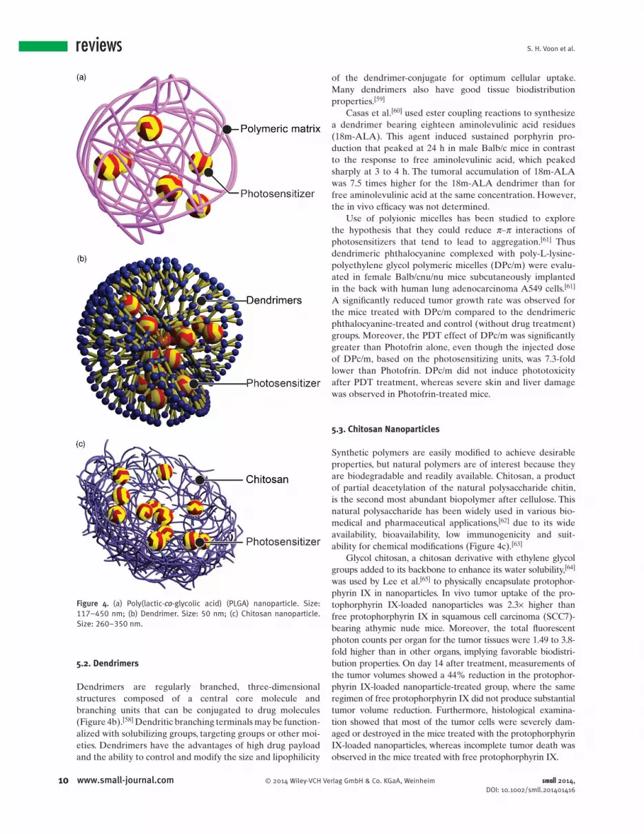
Some polymer-based nanoparticles can have good aqueous dispersions, high drug-loading capacities, and extended release properties. Modifi cations are possible to alter their particle size, surface characteristics, and ability to achieve active or passive targeting. [ 54 ] Several categories of polymeric nanoparticles have been studied as carriers for PDT agents, including synthetic polymers e.g., poly(lactide- co -glycolide) copolymers (PLGA) and dendrimers, and natural polymers such as chitosan.
5.1. Poly(lactic- co -Glycolic Acid) (PLGA)
PLGAs are copolymers of lactic and glycolic acids. These structures can have excellent biocompatibility, biodegrada-bility and mechanical strength. They have been formulated into various delivery systems for carrying a variety of active agents such as vaccines, peptides, proteins, and macromol-ecules ( Figure 4 a). Some of these agents are now approved by the US Food and Drug Administration for use in drug delivery. [ 55 ]
Fadel et al. [ 56 ] formulated PLGA nanoparticles to enhance tissue uptake, permeation, and targeting of zinc (II) phthalocyanine for photodynamic therapy. Female albino mice implanted with Ehrlich ascites carcinoma cells and treated with nanoparticle formulated-zinc (II) phthalocya-nine survived signifi cantly longer than those treated with free drug (mean = 60 and 25 days, respectively), while the mean survival time of an untreated control group was 15 days. Two weeks after the treatment, the mean tumor volumes for the nanoparticle formulated-zinc (II) phthalocyanine group were found to be 1.5 times smaller than those in animals treated with the free drug, and the animals in the former group exhibited a longer tumor growth delay of 39 days.
PLGA nanoparticles have also been used to carry Verte-porfi n. Free Verteporfi n causes extensive skin photosen-sitivity, [ 57 ] but male DBA/2 mice treated with Verteporfi n loaded in PLGA nanoparticles showed only a very mild and short period of skin photosensitivity as measured by ery-thema/eschar formation and edema observations 24 h after PDT in a rhabdomyosarcoma (M1) tumor model. [ 18 ] Total tumor eradication with no tumor regrowth was observed up to 14 days after PDT and on day 20, only 34% of the mice showed tumor regrowth.
small 2014, DOI: 10.1002/smll.201401416
S. H. Voon et al.
10 www.small-journal.com © 2014 Wiley-VCH Verlag GmbH & Co. KGaA, Weinheim
reviews
5.2. Dendrimers
Dendrimers are regularly branched, three-dimensional structures composed of a central core molecule and branching units that can be conjugated to drug molecules (Figure 4 b). [ 58 ] Dendritic branching terminals may be function-alized with solubilizing groups, targeting groups or other moi-eties. Dendrimers have the advantages of high drug payload and the ability to control and modify the size and lipophilicity
of the dendrimer-conjugate for optimum cellular uptake. Many dendrimers also have good tissue biodistribution properties. [ 59 ]
Casas et al. [ 60 ] used ester coupling reactions to synthesize a dendrimer bearing eighteen aminolevulinic acid residues (18m-ALA). This agent induced sustained porphyrin pro-duction that peaked at 24 h in male Balb/c mice in contrast to the response to free aminolevulinic acid, which peaked sharply at 3 to 4 h. The tumoral accumulation of 18m-ALA was 7.5 times higher for the 18m-ALA dendrimer than for free aminolevulinic acid at the same concentration. However, the in vivo effi cacy was not determined.
Use of polyionic micelles has been studied to explore the hypothesis that they could reduce π–π interactions of photosensitizers that tend to lead to aggregation. [ 61 ] Thus dendrimeric phthalocyanine complexed with poly-L-lysine-polyethylene glycol polymeric micelles (DPc/m) were evalu-ated in female Balb/cnu/nu mice subcutaneously implanted in the back with human lung adenocarcinoma A549 cells. [ 61 ] A signifi cantly reduced tumor growth rate was observed for the mice treated with DPc/m compared to the dendrimeric phthalocyanine-treated and control (without drug treatment) groups. Moreover, the PDT effect of DPc/m was signifi cantly greater than Photofrin alone, even though the injected dose of DPc/m, based on the photosensitizing units, was 7.3-fold lower than Photofrin. DPc/m did not induce phototoxicity after PDT treatment, whereas severe skin and liver damage was observed in Photofrin-treated mice.
5.3. Chitosan Nanoparticles
Synthetic polymers are easily modifi ed to achieve desirable properties, but natural polymers are of interest because they are biodegradable and readily available. Chitosan, a product of partial deacetylation of the natural polysaccharide chitin, is the second most abundant biopolymer after cellulose. This natural polysaccharide has been widely used in various bio-medical and pharmaceutical applications, [ 62 ] due to its wide availability, bioavailability, low immunogenicity and suit-ability for chemical modifi cations (Figure 4 c). [ 63 ]
Glycol chitosan, a chitosan derivative with ethylene glycol groups added to its backbone to enhance its water solubility, [ 64 ] was used by Lee et al. [ 65 ] to physically encapsulate protophor-phyrin IX in nanoparticles. In vivo tumor uptake of the pro-tophorphyrin IX-loaded nanoparticles was 2.3× higher than free protophorphyrin IX in squamous cell carcinoma (SCC7)-bearing athymic nude mice. Moreover, the total fl uorescent photon counts per organ for the tumor tissues were 1.49 to 3.8-fold higher than in other organs, implying favorable biodistri-bution properties. On day 14 after treatment, measurements of the tumor volumes showed a 44% reduction in the protophor-phyrin IX-loaded nanoparticle-treated group, where the same regimen of free protophorphyrin IX did not produce substantial tumor volume reduction. Furthermore, histological examina-tion showed that most of the tumor cells were severely dam-aged or destroyed in the mice treated with the protophorphyrin IX-loaded nanoparticles, whereas incomplete tumor death was observed in the mice treated with free protophorphyrin IX.
Figure 4. (a) Poly(lactic- co -glycolic acid) (PLGA) nanoparticle. Size: 117–450 nm; (b) Dendrimer. Size: 50 nm; (c) Chitosan nanoparticle. Size: 260–350 nm.
small 2014, DOI: 10.1002/smll.201401416
In Vivo Studies of Photosensitizers for Cancer Therapy
11www.small-journal.com© 2014 Wiley-VCH Verlag GmbH & Co. KGaA, Weinheim
Nanoparticles in which the cargo is non-covalently incorporated can exhibit burst release profi les; such sudden liberation of cargo can reduce tumor targeting and increase damage to normal tissues. [ 66 ] To circumvent this, Lee et al. [ 67 ] used chemical conjugation rather than physical loading to prepare protophorphyrin IX-conjugated glycol chitosan nanoparticles. These particles exhibited desirable “safety catch” photodynamic properties because the highly dense protophorphyrin IX cores remain in a quenched ‘off’ state until they are taken up into cells; in their off-state they may not generate suffi cient singlet oxygen to produce phototoxicity. [ 68 ] After cellular uptake, the dense proto-phorphyrin IX cores disintegrate in the intracellular envi-ronment, [ 69 ] permitting the protophorphyrin IX to revert to a photocytotoxic ‘on’ state. Thus, in mice bearing the HT-29 human colon adenocarcinoma, the photon counts in the tumor tissues of mice treated with the protophorphyrin IX-conjugated glycol chitosan nanoparticles were 1.4 or 2.1-fold higher than in the tumors of mice treated with protophorphyrin IX physically loaded into glycol chitosan nanoparticles and free protophorphyrin IX, respectively. The tumor sizes were at least 5.2-fold smaller in the mice treated with the protophorphyrin IX-conjugated glycol chitosan nanoparticles than in those treated with free pro-tophorphyrin IX. Furthermore, histological examination revealed that little apoptosis and necrosis was observed in the tumors of mice treated with free protophorphyrin IX, in contrast to the large amount of cell death in the tumor tissues of those treated with the protophorphyrin IX-conju-gated glycol chitosan nanoparticles.
Lee et al. [ 70 ] supported his fi ndings above by comparing the antitumor effi cacy of chlorin e 6 of chemically conju-gated and physically loaded glycol-chitosan nanoparticles in HT-29 human colon adenocarcinoma tumor-bearing mice. Severe photo-induced tumor necrosis was induced in the mice treated with the conjugated chlorin e 6 -glycol-chitosan nanoparticles whereas the group treated with the physi-cally loaded chlorin e 6 -glycol-chitosan nanoparticles failed to show noticeable phototoxicity in the tumor tissues. Final tumor volumes in the mice treated with the conjugated chlorin e 6 -glycol-chitosan-nanoparticles were approxi-mately 160 mm 3 at 20 days post-injection, which was signifi -cantly smaller than the size of the tumors (approximately 560 mm 3 ) in the mice treated with the physically loaded chlorin e 6 -glycol-chitosan-nanoparticles. The improved antitumor effi cacy of conjugated chlorin e 6 -glycol-chitosan nanoparticles in these experiments was attributed to more effi cient accumulation of these particles in the tumors. [ 70 ] The fl uorescence of the conjugated chlorin e 6 -glycol-chitosan-nanoparticles in the tumor tissues persisted for two days after administration. In contrast, the physically loaded chlorin e 6 -glycol-chitosan nanoparticles showed no specifi c tumor accumulation: instead, an intense whole-body fl uores-cence at 3 h post-injection was followed by a rapid decrease in fl uorescence. Furthermore, the total photon counts of the chemically conjugated chlorin e 6 -glycol-chitosan nano-particles in tumor tissue were approximately 6 to 7-fold higher than that of the physically loaded chlorin e 6 -glycol-chitosan nanoparticles.
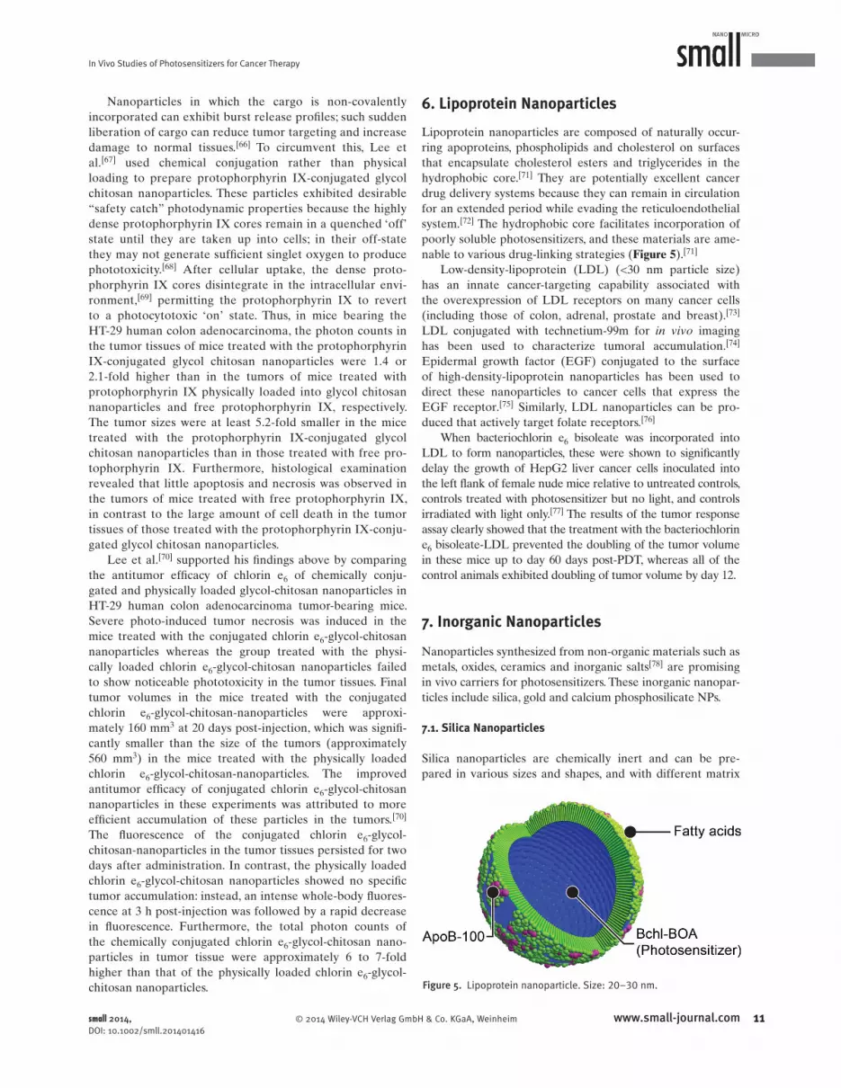
6. Lipoprotein Nanoparticles
Lipoprotein nanoparticles are composed of naturally occur-ring apoproteins, phospholipids and cholesterol on surfaces that encapsulate cholesterol esters and triglycerides in the hydrophobic core. [ 71 ] They are potentially excellent cancer drug delivery systems because they can remain in circulation for an extended period while evading the reticuloendothelial system. [ 72 ] The hydrophobic core facilitates incorporation of poorly soluble photosensitizers, and these materials are ame-nable to various drug-linking strategies ( Figure 5 ). [ 71 ]
Low-density-lipoprotein (LDL) (<30 nm particle size) has an innate cancer-targeting capability associated with the overexpression of LDL receptors on many cancer cells (including those of colon, adrenal, prostate and breast). [ 73 ] LDL conjugated with technetium-99m for in vivo imaging has been used to characterize tumoral accumulation. [ 74 ] Epidermal growth factor (EGF) conjugated to the surface of high-density-lipoprotein nanoparticles has been used to direct these nanoparticles to cancer cells that express the EGF receptor. [ 75 ] Similarly, LDL nanoparticles can be pro-duced that actively target folate receptors. [ 76 ]
When bacteriochlorin e 6 bisoleate was incorporated into LDL to form nanoparticles, these were shown to signifi cantly delay the growth of HepG2 liver cancer cells inoculated into the left fl ank of female nude mice relative to untreated controls, controls treated with photosensitizer but no light, and controls irradiated with light only. [ 77 ] The results of the tumor response assay clearly showed that the treatment with the bacteriochlorin e 6 bisoleate-LDL prevented the doubling of the tumor volume in these mice up to day 60 days post-PDT, whereas all of the control animals exhibited doubling of tumor volume by day 12.
7. Inorganic Nanoparticles
Nanoparticles synthesized from non-organic materials such as metals, oxides, ceramics and inorganic salts [ 78 ] are promising in vivo carriers for photosensitizers. These inorganic nanopar-ticles include silica, gold and calcium phosphosilicate NPs.
7.1. Silica Nanoparticles
Silica nanoparticles are chemically inert and can be pre-pared in various sizes and shapes, and with different matrix
Figure 5. Lipoprotein nanoparticle. Size: 20–30 nm.
small 2014, DOI: 10.1002/smll.201401416

S. H. Voon et al.
12 www.small-journal.com © 2014 Wiley-VCH Verlag GmbH & Co. KGaA, Weinheim
reviewsporosities and dispersion characteristics. The surfaces of silica nanoparticles can be modifi ed with specifi c biomolecules to target tumor cells. [ 79 ] Despite being non-biodegradable, these materials do not affect the photoreactivity of the perma-nently entrapped photosensitizers because the singlet oxygen generated during PDT can diffuse to the surrounding tissue through the porous matrix. [ 80 ] Moreover, silica nanoparticles are well tolerated in biological systems ( Figure 6 a). [ 81 ]
Simon et al. [ 82 ] conducted a biodistribution study of Pro-toporphyrin IX silica nanoparticles loaded with dioctadecyl tetramethyl indodicarbocyanine chlorobenzene (DID) as a tracer in glioblastoma tumor-bearing male athymic nude Foxn1 mice. The nanoparticles at each time point showed higher tumoral accumulation than the control DID tracer, which indicates higher tumor selectivity for the protopor-phyrin IX silica nanoparticles loaded with DID. Similar patterns were observed when the protoporphyrin IX silica nanoparticles and DID tracer control were evaluated in HCT 116 tumor-bearing athymic nude mice and A549 tumor-bearing athymic nude mice.
The surfaces of silica nanoparticles have been modifi ed with a phosphonate termination to produce particles with a greater negative charge (−44.0 mV) than the unmodifi ed particles (−29.1 mV). [ 80 ] This change enhances the stability of the carrier system and reduces aggregation. Entrapment of methylene blue during the syntheses of these particles gave a bi-functional hybrid carrier for imaging and PDT in which fl uorescence was used as a marker for tumor site-directed irradiation to activate the methylene blue. Tumor necrosis gradually developed in the group treated with the methylene blue-encapsulated photosensitizer nanoparticles, whereas the tumors remained intact in the control group treated with light irradiation alone. One fl aw in this approach is that methylene blue emits at a relatively short wavelength and cannot be vis-ualized in deep tissue.
Mesoporous silica is a matrix containing a regular arrange-ment of pores that can be fi lled with various cargoes, including photosensitizers. It has been claimed [ 83 ] that this material is “generally recognized as safe” by the US Food and Drug Administration (FDA) for use as a drug carrier. Tu et al. [ 84 ] used mesoporous silica nanoparticles further functional-ized with polyethylenimine (PEI) and poly(ethylene glycol) (PEG) for delivery of zinc phthalocyanine (ZnPc). The PEI was added to aid particle endocytosis and subsequent escape from the endosomes in the cell. PEG also increases the nanoparticles’ biocompatibility and reduces the PEI’s cationic charge-related cytotoxicity. Biodistribution studies showed that in H22 tumor-bearing mice, the PEG-PEI/ZnPc mesoporous silica nanoparticles exhibited tumor selectivity with the fastest and greatest uptake in the tumors 1 h post-injection. In contrast, tumoral accumulation of unmodifi ed mesoporous silica nanoparticles containing ZnPc continued for 12 h and then reached a plateau at a lower concentration than that of PEG-PEI/ZnPc mesoporous silica nanoparticles. Tumor volumes decreased by day 2 in PEG-PEI/ZnPc par-ticles in the PDT-treated group, and on day 12, the tumor growth was signifi cantly suppressed. In contrast, rapid tumor growth was observed in the control groups treated with PEG-PEI/ZnPc particles administered under dark conditions
or with light irradiation alone. The average survival time for the mice in the control group was 16 days, while the mice treated with PEG-PEI/ZnPc mesoporous silica nanoparticles and PDT survived over 40 days without a single death. These
Figure 6. (a) Silica nanoparticle. Size: 50–118 nm; (b) Gold nanoparticle. Size: 5–14.7 nm; (c) Calcium phosphosilicate nanoparticle. Size: 16 nm.
small 2014, DOI: 10.1002/smll.201401416

In Vivo Studies of Photosensitizers for Cancer Therapy
13www.small-journal.com© 2014 Wiley-VCH Verlag GmbH & Co. KGaA, Weinheim
fi ndings were consistent with the histological examination, which revealed apparent prominent necrosis in the tumors of test group whereas necrosis was less apparent in tumors in the control mice.
Mesoporous silica nanoparticles have been adapted for two-photon excitation. [ 85 ] Two-photon PDT combines the energy of two identical photons (long wavelength NIR light 780–950 nm) arriving at the photosensitizers at the same time to provide the energy of a single photon of approximately half the wavelength, which excites the photosensitizers to the fi rst excited singlet state. [ 86 ] The advantage of two-photon techniques is that the long wavelength excitation source penetrates tissue well, but the disadvantage is that the cross-section for overall uptake of photons into the PDT agents is relatively low.
Gary-Bobo et al. encapsulated a two-photon chromo-phore made from 9,9-dibutyl-2,7-diiodo-9H-fl uorene [ 87 ] into mesoporous silica nanoparticles with and without the addi-tion of mannose to the particle surfaces. The mannose was added to target lectins, which are overexpressed by some cancer cells such as hepatocellular carcinoma. In nude mice with HCT-116 xenografts, two-photon PDT of the animals treated with the lectin-targeting particles showed a reduc-tion in tumor weight by approximately 70% relative to the controls that were treated with saline solution alone. No mor-tality was observed in the treated group up to 30 days post-injection. Furthermore, in the treated group, macrometastasis formation in the liver and the colon was impaired compared to the control groups. This is likely related to the decrease of the subcutaneous tumor growth and of its associated neo-angiogenesis, both of which can delay the spread of cancer cells and subsequent distal metastasis. [ 85 ]
7.2. Gold Nanoparticles
Gold nanoparticles are inert and non-biodegradable, and they can be chemically modifi ed for delivery of drugs, including photosensitizers (Figure 6 b). [ 88 ] The size of the gold nano-particles can be fl exibly tuned from 2 to 100 nm to enhance selective tumor vascular extravasation and tissue permeation and accumulation via the EPR effect. [ 89 ] Modifi cation of gold nanoparticles with water-soluble polyethylene glycol further stabilizes them by inhibiting colloid aggregation and reducing reticuloendothelial system uptake. [ 90 ]
Cheng et al. [ 90a ] conjugated silicon phthalocyanine to PEGylated gold nanoparticles, and found this signifi cantly shortened the delivery time of silicon phthalocyanine to tumor in vivo (2 h compared to 2 days for free silicon phth-alocyanine), without affecting the singlet oxygen yield of sil-icon phthalocyanine in nude mice subcutaneously implanted with the basaloid squamous cell carcinoma, PE/CA-PJ34 cells.
Porphyrin-brucine conjugated to gold nanoparticles retarded tumor growth in the same nude mouse model bearing basaloid squamous cell carcinoma cells. All of the tumors were eliminated by day 8 and no detectable relapse of the primary tumor was observed. [ 88 ] In contrast, animals treated with unconjugated porphyrin-brucine exhibited only
a transient regression in tumor size and the primary tumors gradually, but perceptibly, began to regrow after day 18. Whereas, upon photoexcitation, unconjugated gold nano-particles treated control tumor-bearing mice exhibited only slight tumor growth retardation. This is most likely due to the thermal energy or heat generated by the gold nanoparticles which disrupts the plasma membrane of target cells leading to cell lysis and consequently cell death. [ 91 ]
The use of chlorin e 6 -functionalized gold nanostars to coordinate PDT with plasmonic photothermal therapy (PPTT) by a single continuous wave laser was recently reported. [ 92 ] An intratumoral dose of chlorin e 6 -functional-ized gold nanostars, followed by 6 min exposure to 671 nm laser at 1.0 W/cm 2 in MDA-MB-435 tumor bearing mice caused complete tumor regression, However, the free chlorin e 6 and unconjugated gold nanostars treated groups only showed delay in tumor growth. This suggests combined PDT/PPTT treatment protocol improves anticancer effect by syn-ergistically damaging tumour cells through cytotoxic singlet oxygen produced by chlorin e 6 and heat generated by gold nanostars.
7.3. Calcium Phosphosilicate
Calcium phosphosilicate nanoparticles are non-toxic, col-loidal stable nanoscale vehicles that can deliver imaging agents, drugs and other molecules (Figure 6 c). Indocyanine green loaded into these nanoparticles, which were further functionalized with polyethylene glycol, was found to accu-mulate in breast and pancreatic tumors in subcutaneous xenografts (MDA-MB-231 cells in female nude mice, and BxPC-3 cells in the pancreas of athymic mice). [ 93 ]
The possibility of using PEGylated calcium phospho-silicate loaded with indocyanine green nanoparticles to treat leukemia, a form of blood cancer that is not commonly addressed using PDT, was then explored. The particles were conjugated with antibodies that recognize CD117 recep-tors, which are abundantly expressed on leukemic stem cells. CD117 is a tyrosine kinase receptor for stem cell factor and is important to the pro-growth signaling mechanisms. Near-infrared laser irradiation was directed at the spleen 30 min after injection of the nanoparticles into the leukemic C3H/HeJ mice. The treatments were repeated every 3 days and the leukemia burden was monitored in blood collected from tail pricks and analyzed for GFP+ cells using fl ow cytometry. In vivo PDT effi cacy of indocyanine green delivered in this manner was signifi cantly enhanced, resulting in 29% leu-kemia-free survival. [ 94 ]
8. Hybrid Nanoparticles
Hybrid particles encapsulate two different constituents that can be used for synergistic therapeutics (PDT and chemo-therapy), PDT (two different photosensitizers) or theranos-tics (e.g., PDT and imaging technique, such as MRI). The main hybrids that have been reported to improve in vivo PDT are mesoporous silica (as described above), upconversion
small 2014, DOI: 10.1002/smll.201401416

S. H. Voon et al.
14 www.small-journal.com © 2014 Wiley-VCH Verlag GmbH & Co. KGaA, Weinheim
reviews
nanoparticles (described below) and polyacrylamide-based hybrid nanoparticles ( Figure 7 ).
Polyacrylamide is a nontoxic and biologically inert poly mer of repeating acrylamide subunits (-CH 2 CHCONH 2 -). [ 95 ] Nan-oparticles from this polymer can be formed as small hydrogels approximately 100 nm in diameter. These particles have good loading characteristics for various chemicals and biological molecules. [ 96 ] Furthermore, they can be made biodegradable by introducing appropriate cross-linkers. [ 97 ]
Photofrin-loaded polyacrylamide particles have been combined with the MRI contrast agent iron oxide (Fe 2 O 3 ). [ 22,23,98 ] Reddy et al. [ 22 ] used this combination for simultaneous tumor imaging and PDT of male Fischer 344 rats implanted with rat 9L glioma cells in the right forebrain. The hybrid particles were coated with polyethylene glycol to impart stealth properties, and the F3 targeting peptide was used to direct them to the tumors. The latter is a vascular homing peptide with cell penetrating properties. [ 99 ] This form of delivery reduced the PDT drug-light interval of photofrin from 24 to 1 h, prolonged the NPs’ intracranial tumor transit time by 3-fold, and resulted in a 2-fold increase in MRI contrast-to-noise ratio at both 1 h and 2 h post-administra-tion compared to non-targeted hybrid particles. The median survival period (33 days) in these experiments was 2.5× increased relative to the control group treated with free Pho-tofrin (median survival period of 13 days). Three of the rats that received F3 targeted-hybrid PAA nanoparticles survived for >60 days, and no tumors were present in two of the three rats after 6 months.
9. Combination Therapy (PDT and Chemotherapy)
N - (2-Hydropropyl)methacrylamide (HPMA) is a water-soluble, biocompatible polymeric drug carrier that can be easily formulated to include targeting ligands [ 100 ] and has been shown to preferentially accumulate in tumors via the enhanced permeability and retention effect. [ 101 ] HPMA copolymer-bound drugs have been developed for the combination of PDT and chemotherapy ( Figure 8 ). [ 102 ]
Peterson et al. [ 102a ] designed a com-bination of two HPMA nanoparticle sys-tems to co-deliver meso-chlorin e 6 and
adriamycin to nude mice with human epithelial ovarian car-cinoma. The HPMA copolymer-adriamycin conjugate with 2.2 mg/kg adriamycin equivalent plus HPMA copolymer-meso-chlorin e 6 at 1.5 and 8.7 mg/kg meso-chlorin e 6 equiva-lent with light resulted in signifi cant tumor regression relative to receiving either agent and/or with light alone. No shock syndrome was observed in the mice; this can result from mas-sive agent-stimulated prostaglandin release, which causes platelet aggregation and damage and leads to obstruction of blood fl ow. This study suggests that an HPMA copolymer carrier can extend the safety margin of meso-chlorin e 6 in vivo in PDT because free meso-chlorin e 6 at between 2.5 and 10 mg/kg not only caused tumor regression but also caused shock syndrome.
Synder et al. developed a combination therapy using PDT to enhance the delivery and effi cacy of macromolecule-based cancer drug such as Doxil a pegylated liposome encapsulated doxorubicin with an average diameter of 100 nm. [ 103 ] Prior to the administration of Doxil, murine Colo 26 tumor-bearing mice was treated with the photosensitizer 2-[1-hexyloxyethyl]-2-devinyl pyropheophorbide-a (HPPH) at 0.4 mol/kg and subjected to PDT at 48 J/cm 2 and 14 mW/cm 2 . This increased the accumulation of doxorubicin in transplanted Colo 26 tumors signifi cantly without concomitant enhancement of sys-temic or local toxicity. Eighty percent of tumor-bearing mice treated with the combination of PDT and Doxil at 5 mg/kg were cured compared to the untreated tumor-bearing control mice. On the other hand, a dose of just 5 mg/kg Doxil without PDT only showed modest tumor growth delay without com-plete remission. Furthermore, the combination of PDT and Doxil treatment also reduced skin toxicity in terms of ery-thema, broken vessels and light eschar formation.
This phenomenon is supported by Bin Chen et al.’s fi nd-ings in relation to subcutaneous and orthotopic MatLyLu rat prostate models, where tumor uptake of macromolecules such as Evans blue-albumin and high molecular weight FITC-labeled dextran was signifi cantly increased following PDT treatment. This suggests that PDT increased vascular barrier dysfunction in the MatLyLu tumors. The observation on the adherence of blood cells to vessel wall shortly after PDT further supports the loss of endothelial integrity in the blood vessels. [ 104 ]
Figure 7. Hybrid nanoparticle. Size: 100–200 nm.
Figure 8. Combination therapy. Size: 25 nm.
small 2014, DOI: 10.1002/smll.201401416

In Vivo Studies of Photosensitizers for Cancer Therapy
15www.small-journal.com© 2014 Wiley-VCH Verlag GmbH & Co. KGaA, Weinheim
10. Upconverting Nanoparticles (Energy Transducers) One way to overcome the limitations imposed by poor tissue penetration of shorter wavelength (<700 nm) is via upconver-sion processes that converts near infra-red (NIR) light in the optical tissue penetration window (700–1000 nm) to visible photons. The visible photons liberated can then activate the photosensitizers adsorbed on nanoparticles via resonance energy transfer and generate reactive oxygen species (ROS). The most effi cient upconverting phosphors discovered to date are Yb 3+ - and Er 3+ -doped sodium yttrium fl uoride (NaYF 4 ) ( Figure 9 ). [ 105 ] A successful example of the use of the doped NaYF 4 combined this phosphor with Zn (II) phthalocyanine, which was reported to show signifi cant in vitro HT29 human colonic adenocarcinoma cell killing.
The fi rst in vivo application of upconverting nanoparticles in PDT was by Wang et al. [ 24b ] In this study, a polyethylene glycol-coated upconverting nanoparticle loaded with chlorin e 6 was given to Balb/c mice bearing 4T1 tumors, followed by exposure of the tumor to NIR light (980 nm). Excel-lent PDT effi cacy was achieved: the tumors on seven out of ten mice were completely eliminated two weeks after PDT, and no tumor regrowth was found. In contrast, no delay in tumor growth was observed in the control mice treated in the dark with upconverting nanoparticle-chlorin e 6 injection by itself or NIR-laser irradiation alone. Hematoxylin and eosin staining of the tumor slices collected from upconverting nanoparticle-chlorin e 6 -treated mice on day 7 showed severe tumor tissue damage, while none was found in control tumor tissue slices. Moreover, seven of the treated mice showed complete remission 60 days post-PDT, but the control mice had an average life-span of only 23 days post-treatment.
Improvements to the anticancer effi cacies of upconverting nanoparticles have been achieved by incorporating dual pho-tosensitizers and active targeting ligands into the nanoparti-cles. Idris et al. [ 21 ] used two photosensitizers, merocyanine 540 and zinc phthalocyanine, incorporated into sodium yttrium fl uoride (NaYF 4 ) upconverting nanoparticles coated with a mesoporous silica shell. Irradiation of these particles with a 980 nm laser resulted in the emission of upconverted visible fl uorescence at green (∼540 nm) and red (∼660 nm) wave-lengths that could be absorbed by two photosensitizers. The
PDT effi cacy of these upconverting nanoparticles was evalu-ated in vivo in B16-F0 tumor-bearing C57BL/6 mice. The co-loaded upconverting nanoparticles were injected directly into the melanoma then irradiated with a 980 nm laser 4 h after the injection. Signifi cantly slower tumor growth rates were observed at day 6 and 8 in the treated group compared to the phosphate buffered saline (PBS) control group. On Day 11 post-treatment, biodistribution studies in mice that received only intratumoral injection of the co-loaded upcon-verting nanoparticles revealed yttrium only in the tumor tissues, indicating minimal diffusion to other organs in the body.
Idris et al. also examined the targeted PDT effi cacy of folic acid and polyethylene glycol-conjugated upconverting nanoparticles because folate receptors are overexpressed in B16-F0 melanoma cells. [ 21,106 ] The mice bearing the B16-F0 melanoma tumors were intravenously injected with folic acid-polyethylene glycol-upconverting nanoparticles and then irradiated with a 980 nm laser at 4 h after injection. The treated mice showed a signifi cant reduction in tumor growth compared to control mice treated with PBS. The antitumor effect was less signifi cant in laser-irradiated mice intrave-nously injected with unmodifi ed upconverting nanoparticles. This suggests that modifying the surfaces of these nanoparti-cles to actively target cancer-specifi c ligands has an important role in improving the therapeutic effi cacy. However, in this study, the tumors did not completely regress by the end of the treatment.
11. Nanoparticles with Photosensitizing Properties
Certain materials that generate singlet oxygen upon photoac-tivation can be fashioned into nanoparticles. Titanium oxide and fullerene are members of this category that have reached the in vivo pre-clinical stage in anticancer PDT development.
11.1. Titanium Dioxide
Titanium dioxide (TiO 2 ) is biocompatible [ 107 ] and non-toxic in the dark to experimental animals. [ 108 ] However, upon pho-tocatalytic activation, TiO 2 becomes highly cytotoxic via gen-eration of reactive oxygen species including superoxide and hydroxyl radicals ( Figure 10 a). [ 109 ]
Kubota et al. [ 108 ] injected TiO 2 nanoparticles into nude mice implanted with T-24 human bladder cancer cells. On illumination with UV light at 300–400 nm, tumor growth was drastically delayed for up to 30 days compared to untreated controls. Without modifi cation, this modality is limited by the short wavelength required, which would only be suitable for treating superfi cial tumors in organs amenable to direct light exposure such as the skin, oral cavity, gastrointestinal tract, trachea and urinary bladder.
The effectiveness of nano-TiO 2 -based PDT was further explored in a glioma model by Wang et al. [ 110 ] Female Balb/c nude mice with glioma (U87) xenografts in the subscap-ular subcutaneous tissue were treated with TiO 2 and UVA Figure 9. Upconversion nanoparticle. Size: 30 nm.
small 2014, DOI: 10.1002/smll.201401416

S. H. Voon et al.
16 www.small-journal.com © 2014 Wiley-VCH Verlag GmbH & Co. KGaA, Weinheim
reviews
irradiation. The treatment reduced tumor growth compared to the control group treated with TiO 2 alone. The survival duration of the TiO 2 + UVA-treated mice (57 ± 1.0 days) was signifi cantly longer than that of the TiO 2 -treated control group (42 ± 0.9 days), and the mean tumoral necrotic area of the TiO 2 + UVA treated group was signifi cantly greater than that of the TiO 2 control. At the periphery of the necrotic areas, the apoptotic indices in the TiO 2 + UVA group were 7.4-fold higher than the TiO 2 control group. Overall, these data suggest that nano-TiO 2 -based PDT is effective in reducing tumor growth and promoting survival.
11.2. Fullerenes
The “soccer ball” of 60 carbon atoms (C 60 , arranged in pentagons and hexagons) [ 111 ] a molecule approximately 0.7 nm in diameter and spherical enough to be considered as a nanoparticle, is representative of a larger group of mol-ecules known as the fullerenes (Figure 10 b). Fullerenes have several attributes over conventional photosensitizers; notably, they are more photostable and resistant to photobleaching than tetrapyrroles and organic dyes. They can generate sin-glet oxygen and other reactive free radicals on illumina-tion. [ 112 ] Furthermore, fullerenes can be chemically modifi ed; for instance, incorporation of a light-harvesting antenna can enhance their quantum yield for reactive oxygen species gen-eration. [ 113 ] Fullerenes can also produce self-assembled nano-particles that allow selective delivery with different tissue targeting properties. [ 114 ]
Fullerenes also have some severe limitations as PDT agents. Their extreme hydrophobicity means they aggregate easily in aqueous environments. Consequently, it has been deemed necessary to overcome this issue by formulating fullerenes in liposomes, [ 115 ] micelles, [ 116 ] dendrimers, [ 117 ] and by modifying them to PEGylated forms. [ 118 ] Moreover, fullerenes predominantly absorb blue–green visible light, at wavelengths too short for PDT. This problem has been cir-cumvented by attaching light harvesting antennae to the fullerenes, [ 119 ] using an optical clearing agent such as glycerol or glucose to reduce light scattering leading to tissue trans-parency to improve light penetration [ 120 ] or using two-photon PDT. [ 86,121 ]
The PEGylated fullerenes (e.g., C 60 -PEG) studied by Tabata et al. [ 118a ] showed prolonged retention and higher accumulation in tumors compared to skin, muscle and other tissues in mice bearing Meth A fi brosarcoma tumors. [ 118a ] This agent also resulted in higher suppression of tumor growth than the Photofrin control. The PDT effect of Photo-frin on the tumor was less than that of the conjugate despite the fact that 10-times higher doses were used. Photofrin (620 nm) and the fullerene (400–505 nm) were photoac-tivated at different wavelengths but with equivalent total irradiation power. Histological examination also revealed that the injected PEGylated fullerenes upon light irradiation strongly induced tumor necrosis without damage to the over-lying normal skin, while injection of the PEGylated fullerene alone did not induce tumor necrosis. This suggests that the PEGylated fullerene was not toxic in the dark, and thus light irradiation was essential to generate singlet oxygen to cause destruction of the tumor tissue.
Liu et al. [ 118b ] added diethylenetriaminepentaacetic acid (DTPA) to the terminal group of polyethylene glycol (PEG) on C 60 and combined this construct with gadolinium acetate (Gd) to obtain Gd 3+ -chelated C 60 -PEG. Both C 60 -PEG-Gd and Magnevist, a gadolinium-based MRI contrast agent, showed enhanced MRI signal intensity in Meth AR1 fi bro-sarcomas in CDF1 mice, but the C 60 -based agent persisted in the tumor tissue for a longer time than the Magnevist. Moreover, the MRI signal contrast between the tumor tissue and normal muscle reached a peak approximately 3 h after administration of the C 60 -based agent and was maintained at
Figure 10. (a) Titanium oxide nanoparticle. Size: 0.03–10 nm; (b) Fullerene. Size: 0.7 nm.
small 2014, DOI: 10.1002/smll.201401416
In Vivo Studies of Photosensitizers for Cancer Therapy
17www.small-journal.com© 2014 Wiley-VCH Verlag GmbH & Co. KGaA, Weinheim
a high level even after 24 h, while Magnevist showed contrast enhancement for only 1 h. Further studies using 59 Fe-radiola-beled C 60 -PEG-DTPA also indicated elevated and prolonged tumoral accumulation. Meth AR1 fi brosarcoma-bearing CDF1 mice treated with C 60 -PEG-Gd followed by light irra-diation demonstrated in vivo anti-tumor activity based on the tumor volume ratio before and after treatment, whereas the tumor volume increased with time in the control groups (mice injected with C 60 -PEG-Gd without light irradiation and mice injected with saline).
Intraperitoneal injection of N -methylpyrrolidinium-fullerene entrapped in Cremophor EL micelles followed by white-light illumination delivered through the peritoneal wall of Balb/c mice with abdominal adenocarcinomas signifi cantly prolonged the survival compared to the untreated control group. [ 122 ] A colon adenocarcinoma cell line (CT26) stably expressing luciferase was used to monitor the intraperito-neal tumor burden in Balb/c mice by non-invasive, real-time optical imaging using a sensitive low light camera. This study showed that the N-methylpyrrolidinium-fullerene formulated in Cremophor® EL micelles followed by white-light illumi-nation produced a statistically signifi cant reduction in biolu-minescence and a survival advantage.
Glucose has been incorporated into C 60 to produce C 60 -glucose with which to explore targeting of the glucose receptors that are overexpressed on cancer cells. [ 123 ] Glucose conjugation also increases the aqueous solubility of fuller-enes. Glycoconjugated-C 60 was found to produce selective phototoxicity (after irradiation with UVA1) towards cancer cells compared with normal fi broblasts, showing the impor-tance of targeting glucose receptors. PDT with glycoconju-gated C 60 suppressed tumor growth in human melanoma (COLO679)-xenograft-bearing mice.
12. Conclusion
Currently, there are no clinically approved nanostructure-based photosensitizers for use in anticancer PDT. [ 124 ] In general, the only clinically approved PDT nanoparticle for-mulation is Visudyne (liposomal verteporfi n), approved for the wet form of age-related macular degeneration, patholog-ical myopia and ocular histoplasmosis syndrome. Liposomal-formulated zinc phthalocyanine has been reported to be in clinical trials to treat squamous carcinomas. [ 125 ] Cremophor EL-formulated silicon phthalocyanine and Cremophor EL-formulated TOOKAD have also undergone preliminary clin-ical investigation for primary or metastatic cutaneous cancers and prostate cancers, respectively. [ 126 ] The other nanostruc-tures from different categories reviewed above, illustrating promising approaches to develop useful nanostructure-based photosensitizers for PDT, are still undergoing pre-clinical development. Thus, in research studies it is possible to solve solubility, stability, targeting, optical issues (e.g., via upcon-version nanoparticles), and undesirable skin phototoxicity. Moreover, it is relatively convenient to combine therapeu-tics and diagnostics in one platform to obtain “theranostics”. Overall, it is obvious from reviewing these materials that a lot is yet to be understood with regards to their biocompatibility,
pharmacokinetic properties, in vivo toxicities (Table 3 ), which has been refl ected in the low number of nano-PDT drugs approved thus far.
At best, the process to obtain approval of drugs for human use by the US Food and Drug Administration (FDA) through the Center for Drug Evaluation and Research (CDER) is time-consuming and costly, and the approval rates are low. The strength of nanostructures for PDT is also their weakness: multiplexed formulations (e.g., combinations of photosensitizers, targeting moiety and nanostructure scaffold material) facilitate designs to overcome challenges in pre-clinical PDT studies, but make the approval process harder to negotiate. Approval agencies must be satisfi ed that every component in the nanoparticle is safe, the physiological prop-erties of the nanoparticle as a whole do not cause adverse effects, and the overall formulation can be made to repro-ducibly high standards of quality control. [ 127 ] In general, it is easier to obtain FDA-approval for a small molecule than a nanoparticle composite.
Our perspective on this area is as follows. As always, the easiest research challenges will be addressed fi rst. There is so much momentum behind “nanochemistry” that it is inevitable that rapid development will continue to generate nanoparticles from new materials, displaying a variety of different physical and chemical properties. Simultaneously, synthetic chemistry will provide new alternatives for PDT-active dyes that absorb in the near-IR region. Consequently, progress in this fi eld is not limited by the need to design new nanoparticles and PDT agents. On the other hand, we see a much greater need for progress in the following areas:
• development of more nanoparticles that are discrete mole-cules (i.e., non-polymeric materials with one well-defi ned molecular mass) with minimal high-risk properties that might adversely affect their approval as clinical agents; and related to this,
• studies that facilitate a greater understanding of the toxi-cology of nanoparticles, particularly with respect to size, morphology, composition of the polymer matrix, and formulation;
• development of a wider selection of targeting molecules that can be attached to nanoparticles to direct them to dif-ferent types of cancer cells and away from healthy tissue;
• development of informative in vivo systems particularly those incorporating minimally invasive imaging technolo-gies to study how nanoparticles distribute and their effects on whole animals;
• involvement of a multidisciplinary group of researchers from the early development of nanoparticle photosensitiz-ers, recognizing that successful development will require input from multiple disciplines of science;
• greater integration of PDT with endoscopic surgery to allow excitation of deep tissue via fi ber optics, possibly guid-ed by outputs from theranostic nanoparticles; and,
• a review of the regulatory procedures to focus conditions for approval on the most high-risk aspects of nanoparticle development, recognizing that these may not be exactly the same as for small molecule and biological drugs.
small 2014, DOI: 10.1002/smll.201401416

S. H. Voon et al.
18 www.small-journal.com © 2014 Wiley-VCH Verlag GmbH & Co. KGaA, Weinheim
reviews
Acknowledgements
We would like to thank HIR-MoHE grants (UM.C/625/1/HIR/MOHE/MED/17 and UM.C/625/1/HIR/MOHE/MED/33), Ministry of Higher Education, Malaysia for fi nancial support.
[1] T. J. Dougherty , G. B. Grindey , R. Fiel , K. R. Weishaupt , D. G. Boyle , J. Natl. Cancer Inst. 1975 , 55 , 115 .
[2] a) J. F. Kelly , M. E. Snell , J. Urol. 1976 , 115 , 150; b) T. J. Dougherty , J. E. Kaufman , A. Goldfarb , K. R. Weishaupt , D. Boyle , A. Mittleman , Cancer Res. 1978 , 38 , 2628 .
[3] a) P. Agostinis , K. Berg , K. A. Cengel , T. H. Foster , A. W. Girotti , S. O. Gollnick , S. M. Hahn , M. R. Hamblin , A. Juzeniene , D. Kessel , M. Korbelik , J. Moan , P. Mroz , D. Nowis , J. Piette , B. C. Wilson , J. Golab , CA. Cancer J. Clin. 2011 , 61 , 250; b) D. Fayter , M. Corbett , M. Heirs , D. Fox , A. Eastwood , Health Technol. Assess. 2010 , 14 , 1; c) F. Gao , Y. Bai , S. R. Ma , F. Liu , Z. S. Li , J. Hepatobiliary Pancreat. Sci. 2010 , 17 , 125 .
[4] D. E. Dolmans , D. Fukumura , R. K. Jain , Nat. Rev. Cancer 2003 , 3 , 380 .
[5] J. Moan , K. Berg , Photochem. Photobiol. 1991 , 53 , 549 . [6] A. Juarranz , P. Jaen , F. Sanz-Rodriguez , J. Cuevas , S. Gonzalez ,
Clin. Transl. Oncol. 2008 , 10 , 148 . [7] D. K. Chatterjee , L. S. Fong , Y. Zhang , Adv. Drug Delivery
Rev. 2008 , 60 , 1627 . [8] M. Triesscheijn , P. Baas , J. H. Schellens , F. A. Stewart , Oncolo-
gist 2006 , 11 , 1034 . [9] K. Plaetzer , B. Krammer , J. Berlanda , F. Berr , T. Kiesslich , Lasers
Med. Sci. 2009 , 24 , 259 . [10] A. Juzeniene , K. P. Nielsen , J. Moan , J. Environ. Pathol. Toxicol.
Oncol. 2006 , 25 , 7 . [11] N. A. Kuznetsova , N. S. Gretsova , V. M. Derkacheva , O. L. Kaliya ,
E. A. Lukyanets , J. Porphyrins Phthalocyanines 2003 , 7 , 147 . [12] T. Trindade , P. O’Brien , N. L. Pickett , Chem. Mater. 2001 , 13 , 3843 . [13] L. B. Josefsen , R. W. Boyle , Metal-based Drugs 2008 , 2008 ,
276109 . [14] a) A. L. Chan , M. Juarez , R. Allen , W. Volz , T. Albertson , Pho-
todermatol. Photoimmunol. Photomed. 2005 , 21 , 72; b) M. J. Kaplan , R. G. Somers , R. H. Greenberg , J. Ackler , J. Surg. Oncol. 1998 , 67 , 121; c) H. Lui , L. Hobbs , W. D. Tope , P. K. Lee , C. Elmets , N. Provost , A. Chan , H. Neyndorff , X. Y. Su , H. Jain , I. Hamzavi , D. McLean , R. Bissonnette , Arch. Dermatol. 2004 , 140 , 26; d) T. S. Mang , R. Allison , G. Hewson , W. Snider , R. Moskowitz , Cancer J. Sci. Am. 1998 , 4 , 378; e) S. W. Taber , V. H. Fingar , C. T. Coots , T. J. Wieman , Clin. Cancer Res. 1998 , 4 , 2741 .
[15] H. Kato , K. Furukawa , M. Sato , T. Okunaka , Y. Kusunoki , M. Kawahara , M. Fukuoka , T. Miyazawa , T. Yana , K. Matsui , T. Shiraishi , H. Horinouchi , Lung Cancer (Amsterdam, Nether-lands) 2003 , 42 , 103 .
[16] K. Verigos , D. C. Stripp , R. Mick , T. C. Zhu , R. Whittington , D. Smith , A. Dimofte , J. Finlay , T. M. Busch , Z. A. Tochner , S. Malkowicz , E. Glatstein , S. M. Hahn , J. Environ. Pathol. Tox-icol. Oncol. 2006 , 25 , 373 .
[17] W. Chen , J. Zhang , J. Nanosci. Nanotechnol. 2006 , 6 , 1159 . [18] Y. N. Konan-Kouakou , R. Boch , R. Gurny , E. Allémann , J. Control.
Release 2005 , 103 , 83 . [19] a) N. Solban , I. Rizvi , T. Hasan , Lasers Surg. Med. 2006 , 38 ,
522; b) S. Verma , G. M. Watt , Z. Mai , T. Hasan , Photochem. Pho-tobiol. 2007 , 83 , 996 .
[20] a) B. Yameen , W. I. Choi , C. Vilos , A. Swami , J. Shi , O. C. Farokhzad , J. Control. Release 2014 ; b) O. Harush-Frenkel ,
N. Debotton , S. Benita , Y. Altschuler , Biochem. Biophys. Res. Commun. 2007 , 353 , 26; c) J. K. Vasir , V. Labhasetwar , Bio-materials 2008 , 29 , 4244; d) D. M. Huang , Y. Hung , B. S. Ko , S. C. Hsu , W. H. Chen , C. L. Chien , C. P. Tsai , C. T. Kuo , J. C. Kang , C. S. Yang , C. Y. Mou , Y. C. Chen , FASEB J. 2005 , 19 , 2014 .
[21] N. M. Idris , M. K. Gnanasammandhan , J. Zhang , P. C. Ho , R. Mahendran , Y. Zhang , Nat. Med. 2012 , 18 , 1580 .
[22] G. R. Reddy , M. S. Bhojani , P. McConville , J. Moody , B. A. Moffat , D. E. Hall , G. Kim , Y.-E. L. Koo , M. J. Woolliscroft , J. V. Sugai , T. D. Johnson , M. A. Philbert , R. Kopelman , A. Rehemtulla , B. D. Ross , Clin. Cancer Res. 2006 , 12 , 6677 .
[23] R. Kopelman , Y.-E. Lee Koo , M. Philbert , B. A. Moffat , G. Ramachandra Reddy , P. McConville , D. E. Hall , T. L. Chenevert , M. S. Bhojani , S. M. Buck , A. Rehemtulla , B. D. Ross , J. Magn. Magn. Mater. 2005 , 293 , 404 .
[24] a) D. K. Chatterjee , Z. Yong , Nanomed. 2008 , 3 , 73; b) C. Wang , H. Tao , L. Cheng , Z. Liu , Biomaterials 2011 , 32 , 6145 .
[25] a) A. Master , M. Livingston , A. Sen Gupta , J. Control. Release 2013 , 168 , 88; b) D. Bechet , P. Couleaud , C. Frochot , M. L. Viriot , F. Guillemin , M. Barberi-Heyob , Trends Bio-technol. 2008 , 26 , 612; c) A. M. Bugaj , Photochem. Photo-biol. Sci. 2011 , 10 , 1097; d) Y.-Y. Huang , S. K. Sharma , T. Dai , H. Chung , A. Yaroslavsky , M. Garcia-Diaz , J. Chang , L. Y. Chiang , M. R. Hamblin , Nanotechnology Rev. 2012 , 1 , 111 .
[26] D. Kozlowska , P. Foran , P. MacMahon , M. J. Shelly , S. Eustace , R. O’Kennedy , Adv. Drug Delivery Rev. 2009 , 61 , 1402 .
[27] A. Samad , Y. Sultana , M. Aqil , Curr. Drug Deliv. 2007 , 4 , 297 . [28] M. L. Immordino , F. Dosio , L. Cattel , Int. J. Nanomedicine 2006 ,
1 , 297 . [29] a) A. S. L. Derycke , P. A. M. de Witte , Adv. Drug Delivery
Rev. 2004 , 56 , 17; b) B. Chen , B. W. Pogue , T. Hasan , Expert Opin. Drug Deliv. 2005 , 2 , 477 .
[30] a) F. Jiang , L. Lilge , J. Grenier , Y. Li , M. D. Wilson , M. Chopp , Lasers Surg. Med. 1998 , 22 , 74; b) F. Jiang , L. Lilge , B. Logie , Y. Li , M. Chopp , Photochem. Photobiol. 1997 , 65 , 701 .
[31] Z.-J. Wang , Y.-Y. He , C.-G. Huang , J.-S. Huang , Y.-C. Huang , J.-Y. An , Y. Gu , L.-J. Jiang , Photochem. Photobiol. 1999 , 70 , 773 .
[32] H.-P. Lassalle , D. Dumas , S. Gräfe , M.-A. D’Hallewin , F. Guillemin , L. Bezdetnaya , J. Control. Release 2009 , 134 , 118 .
[33] T. A. Ciulla , R. P. Danis , M. Criswell , L. M. Pratt , Expert Opin. Investig. Drugs 1999 , 8 , 2173 .
[34] K. Ichikawa , Y. Takeuchi , S. Yonezawa , T. Hikita , K. Kurohane , Y. Namba , N. Oku , Cancer Lett. 2004 , 205 , 39 .
[35] D. D. Lasic , F. J. Martin , A. Gabizon , S. K. Huang , D. Papahadjopoulos , Biochim. Biophys. Acta 1991 , 1070 , 187 .
[36] J. Huwyler , J. Drewe , S. Krahenbuhl , Int. J. Nanomedicine 2008 , 3 , 21 .
[37] A. Gijsens , A. Derycke , L. Missiaen , D. De Vos , J. Huwyler , A. Eberle , P. de Witte , Int. J. Cancer 2002 , 101 , 78 .
[38] a) V. P. Torchilin , CMLS, Cell. Mol. Life Sci. 2004 , 61 , 2549; b) C. F. van Nostrum , Adv. Drug Delivery Rev. 2004 , 56 , 9 .
[39] a) G. S. Kwon , T. Okano , Adv. Drug Delivery Rev. 1996 , 21 , 107; b) C. Allen , D. Maysinger , A. Eisenberg , Colloids Surf. B. Biointerfaces 1999 , 16 , 3; c) M.-C. Jones , J.-C. Leroux , Eur. J. Pharm. Biopharm. 1999 , 48 , 101; d) K. Kataoka , A. Harada , Y. Nagasaki , Adv. Drug Delivery Rev. 2001 , 47 , 113 .
[40] H. Koo , H. Lee , S. Lee , K. H. Min , M. S. Kim , D. S. Lee , Y. Choi , I. C. Kwon , K. Kim , S. Y. Jeong , Chem. Commun. 2010 , 46 , 5668 .
[41] D. Le Garrec , J. Taillefer , J. E. Van Lier , V. Lenaerts , J.-C. Leroux , J. Drug Target. 2002 , 10 , 429 .
[42] A. N. Lukyanov , V. P. Torchilin , Adv. Drug Delivery Rev. 2004 , 56 , 1273 .
[43] a) A. Roby , S. Erdogan , V. P. Torchilin , Cancer Biol. Ther. 2007 , 6 , 1136; b) A. Roby , S. Erdogan , V. P. Torchilin , Eur. J. Pharm. Biopharm. 2006 , 62 , 235; c) I. Skidan , P. Dholakia , V. Torchilin , J. Drug Target. 2008 , 16 , 486 .
small 2014, DOI: 10.1002/smll.201401416
In Vivo Studies of Photosensitizers for Cancer Therapy
19www.small-journal.com© 2014 Wiley-VCH Verlag GmbH & Co. KGaA, Weinheim
[44] a) A. L. Klibanov , K. Maruyama , V. P. Torchilin , L. Huang , FEBS Lett. 1990 , 268 , 235; b) J. Senior , C. Delgado , D. Fisher , C. Tilcock , G. Gregoriadis , Biochim. Biophys. Acta 1991 , 1062 , 77 .
[45] G.-D. Zhang , A. Harada , N. Nishiyama , D.-L. Jiang , H. Koyama , T. Aida , K. Kataoka , J. Control. Release 2003 , 93 , 141 .
[46] J. X. Zhang , C. B. Hansen , T. M. Allen , A. Boey , R. Boch , J. Con-trol. Release 2003 , 86 , 323 .
[47] D. Kessel , Photochem. Photobiol. 1992 , 56 , 447 . [48] A. Rück , A. Böhmler , R. Steiner , Photodiagnosis Photodyn.
Ther. 2005 , 2 , 79 . [49] C. M. Whitacre , D. K. Feyes , T. Satoh , J. Grossmann ,
J. W. Mulvihill , H. Mukhtar , N. L. Oleinick , Clin. Cancer Res. 2000 , 6 , 2021 .
[50] D. Kessel , Photochem. Photobiol. 1989 , 50 , 169 . [51] A. T. Byrne , A. E. O’Connor , M. Hall , J. Murtagh , K. O’Neill ,
K. M. Curran , K. Mongrain , J. A. Rousseau , R. Lecomte , S. McGee , J. J. Callanan , D. F. O’Shea , W. M. Gallagher , Br. J. Cancer 2009 , 101 , 1565 .
[52] H. Gelderblom , J. Verweij , K. Nooter , A. Sparreboom , Eur. J. Cancer 2001 , 37 , 1590 .
[53] M. García-Díaz , M. Kawakubo , P. Mroz , M. L. Sagristà , M. Mora , S. Nonell , M. R. Hamblin , J. Control. Release 2012 , 162 , 355 .
[54] a) M. L. Hans , A. M. Lowman , Current Opinion in Solid State and Materials Science 2002 , 6 , 319; b) C. E. Mora-Huertas , H. Fessi , A. Elaissari , Int. J. Pharm. 2010 , 385 , 113; c) A. Kumari , S. K. Yadav , S. C. Yadav , Colloids Surf. B. Biointer-faces 2010 , 75 , 1; d) H. Maeda , Adv. Enzyme Regul. 2001 , 41 , 189 .
[55] a) M. Muthu , Asian Journal of Pharmaceutics 2009 , 3 , 266; b) M. Stevanovic , D. Uskokovic , Current Nanoscience 2009 , 5 , 1; c) R. A. Jain , Biomaterials 2000 , 21 , 2475; d) C. E. Lowe , (E.I.du Pont de Nemours and Company) US.2668162, 1954 ; e) D. K. Gilding , A. M. Reed , Polymer 1979 , 20 , 1459; f) I. Bala , S. Hariharan , M. N. Kumar , Crit. Rev. Ther. Drug Carrier Syst. 2004 , 21 , 387 .
[56] M. Fadel , K. Kassab , D. Abdel Fadeel , Lasers Med. Sci. 2010 , 25 , 283 .
[57] A. M. Richter , S. Yip , E. Waterfi eld , P. M. Logan , C. E. Slonecker , J. G. Levy , Photochem. Photobiol. 1991 , 53 , 281 .
[58] a) U. Boas , P. M. Heegaard , Chem. Soc. Rev. 2004 , 33 , 43; b) J. B. Wolinsky , M. W. Grinstaff , Adv. Drug Delivery Rev. 2008 , 60 , 1037; c) C. C. Lee , J. A. MacKay , J. M. Fréchet , F. C. Szoka , Nat. Biotechnol. 2005 , 23 , 1517 .
[59] a) T. D. McCarthy , P. Karellas , S. A. Henderson , M. Giannis , D. F. O’Keefe , G. Heery , J. R. A. Paull , B. R. Matthews , G. Holan , Mol. Pharm. 2005 , 2 , 312; b) S. C. Lee , Dendrimers and Other Dendritic Polymers , John Wiley & Sons, Ltd ., Chichester, UK 2002 .
[60] A. Casas , S. Battah , G. Di Venosa , P. Dobbin , L. Rodriguez , H. Fukuda , A. Batlle , A. J. MacRobert , J. Control. Release 2009 , 135 , 136 .
[61] N. Nishiyama , Y. Nakagishi , Y. Morimoto , P.-S. Lai , K. Miyazaki , K. Urano , S. Horie , M. Kumagai , S. Fukushima , Y. Cheng , W.-D. Jang , M. Kikuchi , K. Kataoka , J. Control. Release 2009 , 133 , 245 .
[62] J. H. Park , G. Saravanakumar , K. Kim , I. C. Kwon , Adv. Drug Delivery Rev. 2010 , 62 , 28 .
[63] a) M. N. V. R. Kumar , R. A. A. Muzzarelli , C. Muzzarelli , H. Sashiwa , A. J. Domb , Chem. Rev. 2004 , 104 , 6017; b) L. Ilium , Pharm. Res. 1998 , 15 , 1326; c) O. Felt , P. Buri , R. Gurny , Drug Dev. Ind. Pharm. 1998 , 24 , 979 .
[64] K. Kim , J. H. Kim , H. Park , Y.-S. Kim , K. Park , H. Nam , S. Lee , J. H. Park , R.-W. Park , I.-S. Kim , K. Choi , S. Y. Kim , K. Park , I. C. Kwon , J. Control. Release 2010 , 146 , 219 .
[65] S. J. Lee , K. Park , Y.-K. Oh , S.-H. Kwon , S. Her , I.-S. Kim , K. Choi , S. J. Lee , H. Kim , S. G. Lee , K. Kim , I. C. Kwon , Biomate-rials 2009 , 30 , 2929 .
[66] Y. H. Bae , H. Yin , J. Control. Release 2008 , 131 , 2 . [67] S. J. Lee , H. Koo , D.-E. Lee , S. Min , S. Lee , X. Chen , Y. Choi ,
J. F. Leary , K. Park , S. Y. Jeong , I. C. Kwon , K. Kim , K. Choi , Bio-materials 2011 , 32 , 4021 .
[68] J. F. Lovell , J. Chen , M. T. Jarvi , W.-G. Cao , A. D. Allen , Y. Liu , T. T. Tidwell , B. C. Wilson , G. Zheng , J. Phys. Chem. B 2009 , 113 , 3203 .
[69] a) R. Savic , T. Azzam , A. Eisenberg , D. Maysinger , Lang-muir 2006 , 22 , 3570; b) H. Chen , S. Kim , W. He , H. Wang , P. S. Low , K. Park , J.-X. Cheng , Langmuir 2008 , 24 , 5213 .
[70] S. J. Lee , H. Koo , H. Jeong , M. S. Huh , Y. Choi , S. Y. Jeong , Y. Byun , K. Choi , K. Kim , I. C. Kwon , J. Control. Release 2011 , 152 , 21 .
[71] K. K. Ng , J. F. Lovell , G. Zheng , Acc. Chem. Res. 2011 , 44 , 1105 . [72] S. Eisenberg , H. G. Windmueller , R. I. Levy , J. Lipid Res. 1973 ,
14 , 446 . [73] I. R. Corbin , G. Zheng , Nanomed. 2007 , 2 , 375 . [74] E. Ponty , G. Favre , R. Benaniba , A. Boneu , H. Lucot , M. Carton ,
G. Soula , Int. J. Cancer 1993 , 54 , 411 . [75] a) Z. Zhang , J. Chen , L. Ding , H. Jin , J. F. Lovell , I. R. Corbin ,
W. Cao , P.-C. Lo , M. Yang , M.-S. Tsao , Q. Luo , G. Zheng , Small 2010 , 6 , 430; b) H. Jin , J. Lovell , J. Chen , K. Ng , W. Cao , L. Ding , Z. Zhang , G. Zheng , Cancer Nanotechnol. 2010 , 1 , 71 .
[76] G. Zheng , J. Chen , H. Li , J. D. Glickson , Proc. Natl. Acad. Sci. USA 2005 , 102 , 17757 .
[77] D. E. Marotta , W. Cao , E. P. Wileyto , H. Li , I. Corbin , E. Rickter , J. D. Glickson , B. Chance , G. Zheng , T. M. Busch , Nanomed. 2011 , 6 , 475 .
[78] C. N. R. Rao , H. S. S. Ramakrishna Matte , R. Voggu , A. Govindaraj , Dalton Trans. 2012 , 41 , 5089 .
[79] S. Wang , R. Gao , F. Zhou , M. Selke , J. Mater. Chem. 2004 , 14 , 487 .
[80] X. He , X. Wu , K. Wang , B. Shi , L. Hai , Biomaterials 2009 , 30 , 5601 .
[81] a) D. Tarn , C. E. Ashley , M. Xue , E. C. Carnes , J. I. Zink , C. J. Brinker , Acc. Chem. Res. 2013 , 46 , 792; b) J. Lu , M. Liong , Z. Li , J. I. Zink , F. Tamanoi , Small 2010 , 6 , 1794 .
[82] V. Simon , C. Devaux , A. Darmon , T. Donnet , E. Thiénot , M. Germain , J. Honnorat , A. Duval , A. Pottier , E. Borghi , L. Levy , J. Marill , Photochem. Photobiol. 2010 , 86 , 213 .
[83] Y. Piao , A. Burns , J. Kim , U. Wiesner , T. Hyeon , Adv. Funct. Mater. 2008 , 18 , 3745 .
[84] J. Tu , T. Wang , W. Shi , G. Wu , X. Tian , Y. Wang , D. Ge , L. Ren , Bio-materials 2012 , 33 , 7903 .
[85] M. Gary-Bobo , Y. Mir , C. Rouxel , D. Brevet , I. Basile , M. Maynadier , O. Vaillant , O. Mongin , M. Blanchard-Desce , A. Morère , M. Garcia , J.-O. Durand , L. Raehm , Angew. Chem. Int. Ed. 2011 , 50 , 11425 .
[86] J. D. Bhawalkar , N. D. Kumar , C. F. Zhao , P. N. Prasad , J. Clin. Laser Med. Surg. 1997 , 15 , 201 .
[87] C. Rouxel , M. Charlot , Y. Mir , C. Frochot , O. Mongin , M. Blanchard-Desce , New J. Chem. 2011 , 35 , 1771 .
[88] K. Zaruba , J. Kralova , P. Rezanka , P. Pouckova , L. Veverkova , V. Kral , Org. Biomol. Chem. 2010 , 8 , 3202 .
[89] D. Demberelnyamba , M. Ariunaa , Y. K. Shim , Int. J. Mol. Sci. 2008 , 9 , 864 .
[90] a) Y. Cheng , A. C. Samia , J. D. Meyers , I. Panagopoulos , B. Fei , C. Burda , J. Am. Chem. Soc. 2008 , 130 , 10643; b) Y. Liu , M. K. Shipton , J. Ryan , E. D. Kaufman , S. Franzen , D. L. Feldheim , Anal. Chem. 2007 , 79 , 2221; c) R. B. Greenwald , Y. H. Choe , J. McGuire , C. D. Conover , Adv. Drug Delivery Rev. 2003 , 55 , 217 .
[91] a) D. P. O’Neal , L. R. Hirsch , N. J. Halas , J. D. Payne , J. L. West , Cancer Lett. 2004 , 209 , 171; b) P. Rezanka , K. Záruba , V. Král , Tetrahedron Lett. 2008 , 49 , 6448; c) N. F. Gamaleia , E. D. Shishko , G. A. Dolinsky , A. B. Shcherbakov , A. V. Usatenko , V. V. Kholin , Exp. Oncol. 2010 , 32 , 44 .
small 2014, DOI: 10.1002/smll.201401416

S. H. Voon et al.
20 www.small-journal.com © 2014 Wiley-VCH Verlag GmbH & Co. KGaA, Weinheim
reviews[92] S. Wang , P. Huang , L. Nie , R. Xing , D. Liu , Z. Wang , J. Lin ,
S. Chen , G. Niu , G. Lu , X. Chen , Adv. Mater. 2013 , 25 , 3055 . [93] a) E. I. Altinoglu , T. J. Russin , J. M. Kaiser , B. M. Barth ,
P. C. Eklund , M. Kester , J. H. Adair , ACS Nano 2008 , 2 , 2075; b) B. M. Barth , R. Sharma , E. I. Altinoglu , T. T. Morgan , S. S. Shanmugavelandy , J. M. Kaiser , C. McGovern , G. L. Matters , J. P. Smith , M. Kester , J. H. Adair , ACS Nano 2010 , 4 , 1279 .
[94] B. M. Barth , E. i. Altinoglu , S. S. Shanmugavelandy , J. M. Kaiser , D. Crespo-Gonzalez , N. A. DiVittore , C. McGovern , T. M. Goff , N. R. Keasey , J. H. Adair , T. P. Loughran , Jr. , D. F. Claxton , M. Kester , ACS Nano 2011 , 5 , 5325 .
[95] a) F. A. Anderson , Int. J. Toxicol. 2005 , 24 Suppl 2 , 21; b) D. J. King , R. R. Noss , Rev. Environ. Health 1989 , 8 , 3 .
[96] a) H. A. Clark , R. Kopelman , R. Tjalkens , M. A. Philbert , Anal. Chem. 1999 , 71 , 4837; b) E. J. Park , M. Brasuel , C. Behrend , M. A. Philbert , R. Kopelman , Anal. Chem. 2003 , 75 , 3784; c) S. M. Buck , H. Xu , M. Brasuel , M. A. Philbert , R. Kopelman , Talanta 2004 , 63 , 41; d) M. King , R. Kopelman , Sens. Actuat. B: Chem. 2003 , 90 , 76 .
[97] Y.-E. L. Koo , G. R. Reddy , M. Bhojani , R. Schneider , M. A. Philbert , A. Rehemtulla , B. D. Ross , R. Kopelman , Adv. Drug Delivery Rev. 2006 , 58 , 1556 .
[98] a) B. Ross , A. Rehemtulla , Y.-E. L. Koo , R. Reddy , G. Kim , C. Behrend , S. Buck , R. J. Schneider , M. A. Philbert , R. Weissleder , in Nanobiophotonics and Biomedical Appli-cations , Vol. 5331 (Ed: A. N. Cartwright ), SPIE Proceedings , San Jose, USA 2004; b) H. Xu , S. M. Buck , R. Kopelman , M. A. Philbert , M. Brasuel , B. D. Ross , A. Rehemtulla , Isr. J. Chem. 2004 , 44 , 317 .
[99] a) K. Porkka , P. Laakkonen , J. A. Hoffman , M. Bernasconi , E. Ruoslahti , Proc. Natl. Acad. Sci. USA 2002 , 99 , 7444; b) M. E. Akerman , W. C. Chan , P. Laakkonen , S. N. Bhatia , E. Ruoslahti , Proc. Natl. Acad. Sci. USA 2002 , 99 , 12617; c) E. Ruoslahti , T. Duza , L. Zhang , Curr. Pharm. Des. 2005 , 11 , 3655 .
[100] K. Ulbrich , V. Šubr , Adv. Drug Delivery Rev. 2010 , 62 , 150 . [101] J. Kopecek , P. Kopecková , T. Minko , Z.-R. Lu , Eur. J. Pharm. Biop-
harm. 2000 , 50 , 61 . [102] a) C. M. Peterson , J. M. Lu , Y. Sun , C. A. Peterson , J. G. Shiah ,
R. C. Straight , J. Kopecek , Cancer Res. 1996 , 56 , 3980; b) N. L. Krinick , Y. Sun , D. Joyner , J. D. Spikes , R. C. Straight , J. Kopecek , J. Biomater. Sci. Polym. Ed. 1994 , 5 , 303 .
[103] J. W. Snyder , W. R. Greco , D. A. Bellnier , L. Vaughan , B. W. Henderson , Cancer Res. 2003 , 63 , 8126 .
[104] B. Chen , B. W. Pogue , J. M. Luna , R. L. Hardman , P. J. Hoopes , T. Hasan , Clin. Cancer Res. 2006 , 12 , 917 .
[105] N. Menyuk , K. Dwight , J. W. Pierce , Appl. Phys. Lett. 1972 , 21 , 159 .
[106] S. Mohapatra , S. Mallick , T. Maiti , S. Ghosh , P. Pramanik , Nano-technology 2007 , 18 , 385102 .
[107] A. Hertz , I. J. Bruce , Nanomed. 2007 , 2 , 899 . [108] Y. Kubota , T. Shuin , C. Kawasaki , M. Hosaka , H. Kitamura ,
R. Cai , H. Sakai , K. Hashimoto , A. Fujishima , Br. J. Cancer 1994 , 70 , 1107 .
[109] a) A. Fujishima , T. N. Rao , D. A. Tryk , J. Photochem. Photobiol. C: Photochem. Rev. 2000 , 1 , 1; b) J. J. Wang , B. J. S. Sanderson , H. Wang , Mutation Research/Genetic Toxicology and Environ-mental Mutagenesis 2007 , 628 , 99 .
[110] C. Wang , S. Cao , X. Tie , B. Qiu , A. Wu , Z. Zheng , Mol. Biol. Rep. 2011 , 38 , 523 .
[111] P. Mroz , G. P. Tegos , H. Gali , T. Wharton , T. Sarna , M. R. Hamblin , Photochem. Photobiol. Sci. 2007 , 6 , 1139 .
[112] a) Y. Yamakoshi , N. Umezawa , A. Ryu , K. Arakane , N. Miyata , Y. Goda , T. Masumizu , T. Nagano , J. Am. Chem. Soc. 2003 , 125 , 12803; b) R. Bakry , R. M. Vallant , M. Najam-ul-Haq , M. Rainer , Z. Szabo , C. W. Huck , G. K. Bonn , Int. J. Nanomedicine 2007 , 2 , 639 .
[113] L. Y. Chiang , P. A. Padmawar , J. E. Rogers-Haley , G. So , T. Canteenwala , S. Thota , L.-S. Tan , K. Pritzker , Y.-Y. Huang , S. K. Sharma , D. B. Kurup , M. R. Hamblin , B. Wilson , A. Urbas , J. Mater. Chem. 2010 , 20 , 5280 .
[114] S. K. Sharma , L. Y. Chiang , M. R. Hamblin , Nanomed. 2011 , 6 , 1813 .
[115] a) S. Kato , H. Aoshima , Y. Saitoh , N. Miwa , Toxicol. Ind. Health 2009 , 25 , 197; b) S. Kato , H. Aoshima , Y. Saitoh , N. Miwa , J. Photochem. Photobiol. B: Biol. 2010 , 98 , 99; c) Y. Doi , A. Ikeda , M. Akiyama , M. Nagano , T. Shigematsu , T. Ogawa , T. Takeya , T. Nagasaki , Chem. Eur. J. 2008 , 14 , 8892 .
[116] a) A. Yan , A. Von Dem Bussche , A. B. Kane , R. H. Hurt , Carbon N. Y. 2007 , 45 , 2463; b) M. Akiyama , A. Ikeda , T. Shintani , Y. Doi , J. Kikuchi , T. Ogawa , K. Yogo , T. Takeya , N. Yamamoto , Org. Biomol. Chem. 2008 , 6 , 1015 .
[117] a) C. Kojima , Y. Toi , A. Harada , K. Kono , Bioconjug. Chem. 2008 , 19 , 2280; b) B. Pan , D. Cui , P. Xu , C. Ozkan , G. Feng , M. Ozkan , T. Huang , B. Chu , Q. Li , R. He , G. Hu , Nanotechnology 2009 , 20 , 125101 .
[118] a) Y. Tabata , Y. Murakami , Y. Ikada , Jpn. J. Cancer Res. 1997 , 88 , 1108; b) J. Liu , S.-i. Ohta , A. Sonoda , M. Yamada , M. Yamamoto , N. Nitta , K. Murata , Y. Tabata , J. Control. Release 2007 , 117 , 104; c) J. B. Hooper , D. Bedrov , G. D. Smith , Langmuir 2008 , 24 , 4550; d) N. Nitta , A. Seko , A. Sonoda , S. Ohta , T. Tanaka , M. Takahashi , K. Murata , S. Takemura , T. Sakamoto , Y. Tabata , Cardiovasc. Intervent. Radiol. 2008 , 31 , 359 .
[119] a) P. D. Boyd , C. A. Reed , Acc. Chem. Res. 2005 , 38 , 235; b) M. E. El-Khouly , Y. Araki , O. Ito , S. Gadde , A. L. McCarty , P. A. Karr , M. E. Zandler , F. D’Souza , Phys. Chem. Chem. Phys. 2005 , 7 , 3163; c) H. Imahori , Org. Biomol. Chem. 2004 , 2 , 1425; d) D. I. Schuster , P. Cheng , P. D. Jarowski , D. M. Guldi , C. Luo , L. Echegoyen , S. Pyo , A. R. Holzwarth , S. E. Braslavsky , R. M. Williams , G. Klihm , J. Am. Chem. Soc. 2004 , 126 , 7257; e) S. A. Vail , D. I. Schuster , D. M. Guldi , M. Isosomppi , N. Tkachenko , H. Lemmetyinen , A. Palkar , L. Echegoyen , X. Chen , J. Z. H. Zhang , J. Phys. Chem. B 2006 , 110 , 14155 .
[120] a) S. C. Gebhart , W. C. Lin , A. Mahadevan-Jansen , Phys. Med. Biol. 2006 , 51 , 2011; b) V. V. Tuchin , R. K. Wang , A. T. Yeh , J. Biomed. Opt. 2008 , 13 , 021101; c) J. Jiang , M. Boese , P. Turner , R. K. Wang , J. Biomed. Opt. 2008 , 13 , 021105 .
[121] a) M. Göppert-Mayer , Annalen der Physik 1931 , 401 , 273; b) A. Karotki , M. Khurana , J. R. Lepock , B. C. Wilson , Photo-chem. Photobiol. 2006 , 82 , 443; c) K. S. Samkoe , A. A. Clancy , A. Karotki , B. C. Wilson , D. T. Cramb , J. Biomed. Opt. 2007 , 12 , 034025; d) K. S. Samkoe , D. T. Cramb , J. Biomed. Opt. 2003 , 8 , 410 .
[122] P. Mroz , Y. Xia , D. Asanuma , A. Konopko , T. Zhiyentayev , Y.-Y. Huang , S. K. Sharma , T. Dai , U. J. Khan , T. Wharton , M. R. Hamblin , Nanomedicine: Nanotechnol. Biol. Medi-cine 2011 , 7 , 965 .
[123] E. Otake , S. Sakuma , K. Torii , A. Maeda , H. Ohi , S. Yano , A. Morita , Photochem. Photobiol. 2010 , 86 , 1356 .
[124] L. Zhang , F. X. Gu , J. M. Chan , A. Z. Wang , R. S. Langer , O. C. Farokhzad , Clin. Pharmacol. Ther. 2008 , 83 , 761 .
[125] a) U. Isele , P. van Hoogevest , H. Leuenberger , H.-G. Capraro , K. Schieweck , in Photodynamic Therapy of Cancer , Vol. 2078 (Eds: G. Jori , J. Moan , W. M. Star ), SPIE Conf. Proc., The Interna-tional Society for Optical Engineering , Budapest, Hungary 1994 , 397; b) M. Ochsner , J. Photochem. Photobiol. B 1996 , 32 , 3 .
[126] a) R. A. Weersink , A. Bogaards , M. Gertner , S. R. Davidson , K. Zhang , G. Netchev , J. Trachtenberg , B. C. Wilson , J. Photo-chem. Photobiol. B 2005 , 79 , 211; b) T. J. Kinsella , E. D. Baron , V. C. Colussi , K. D. Cooper , C. L. Hoppel , S. T. Ingalls , M. E. Kenney , X. Li , N. L. Oleinick , S. R. Stevens , S. C. Remick , Front. Oncol. 2011 , 1 , 14 .
[127] A. Eifl er , C. S. Thaxton , in Biomedical Nanotechnology , Vol. 726 (Ed: S. J. Hurst ), Humana Press , New York City, USA 2011 , Ch. 21 .
small 2014, DOI: 10.1002/smll.201401416

In Vivo Studies of Photosensitizers for Cancer Therapy
21www.small-journal.com© 2014 Wiley-VCH Verlag GmbH & Co. KGaA, Weinheim
[128] M. Longmire , P. L. Choyke , H. Kobayashi , Nanomedicine 2008 , 3 , 703 .
[129] a) J. D. Heidel , M. E. Davis , Pharm. Res. 2011 , 28 , 187; b) A. Z. Wang , R. Langer , O. C. Farokhzad , Annu. Rev. Med. 2012 , 63 , 185 .
[130] M. J. Glantz , S. LaFollette , K. A. Jaeckle , W. Shapiro , L. Swinnen , J. R. Rozental , S. Phuphanich , L. R. Rogers , J. C. Gutheil , T. Batchelor , D. Lyter , M. Chamberlain , B. L. Maria , C. Schiffer , R. Bashir , D. Thomas , W. Cowens , S. B. Howell , J. Clin. Oncol. 1999 , 17 , 3110 .
[131] P. M. Anderson , M. Tomaras , K. McConnell , Drugs of Today 2010 , 46 , 327 .
[132] a) W. C. Zamboni , Oncologist 2008 , 13 , 248; b) M. A. Rodriguez , R. Pytlik , T. Kozak , M. Chhanabhai , R. Gascoyne , B. Lu , S. R. Deitcher , J. N. Winter , Cancer 2009 , 115 , 3475 .
[133] M. Yokoyama , J. Experimental Clinical Medicine 2011 , 3 , 151 .
[134] J. W. Valle , A. Armstrong , C. Newman , V. Alakhov , G. Pietrzynski , J. Brewer , S. Campbell , P. Corrie , E. K. Rowinsky , M. Ranson , Invest. New Drugs 2011 , 29 , 1029 .
[135] H. K. Makadia , S. J. Siegel , Polymers 2011 , 3 , 1377 .
[136] C. U. Herborn , J. Barkhausen , I. Paetsch , P. Hunold , M. Mahler , K. Shamsi , E. Nagel , Radiology 2003 , 229 , 217 .
[137] E. Markovsky , H. Baabur-Cohen , A. Eldar-Boock , L. Omer , G. Tiram , S. Ferber , P. Ofek , D. Polyak , A. Scomparin , R. Satchi-Fainaro , J. Control. Release 2012 , 161 , 446 .
[138] B. C. Kwan , F. Kronenberg , S. Beddhu , A. K. Cheung , J. Am. Soc. Nephrol. 2007 , 18 , 1246 .
[139] S. Ivanov , S. Zhuravsky , G. Yukina , V. Tomson , D. Korolev , M. Galagudza , Materials 2012 , 5 , 1873 .
[140] R. Kumar , I. Roy , T. Y. Ohulchanskky , L. A. Vathy , E. J. Bergey , M. Sajjad , P. N. Prasad , ACS Nano 2010 , 4 , 699 .
[141] S. Jain , D. G. Hirst , J. M. O’Sullivan , Br. J. Radiol. 2012 , 85 , 101 . [142] D. P. Nowotnik , E. Cvitkovic , Adv. Drug Deliv. Rev. 2009 , 61 , 1214 . [143] N. Shinohara , N. Danno , T. Ichinose , T. Sasaki , H. Fukui ,
K. Honda , M. Gamo , Nanotoxicology 2014 , 8 , 132 . [144] H. Shi , R. Magaye , V. Castranova , J. Zhao , Part. Fibre Tox-
icol. 2013 , 10 , 15 .
Received: May 21, 2014 Revised: July 26, 2014 Published online:
small 2014, DOI: 10.1002/smll.201401416


