Embed Size (px)
Citation preview
Engineered nonviral nanocarriers for intracellular gene delivery applications
This article has been downloaded from IOPscience. Please scroll down to see the full text article.
2012 Biomed. Mater. 7 054106
(http://iopscience.iop.org/1748-605X/7/5/054106)
Download details:
IP Address: 132.166.20.57
The article was downloaded on 17/12/2012 at 08:02
Please note that terms and conditions apply.
View the table of contents for this issue, or go to the journal homepage for more
Home Search Collections Journals About Contact us My IOPscience
IOP PUBLISHING BIOMEDICAL MATERIALS
Biomed. Mater. 7 (2012) 054106 (13pp) doi:10.1088/1748-6041/7/5/054106
Engineered nonviral nanocarriers forintracellular gene delivery applicationsIsaac Ojea-Jimenez1,4, Olivia Tort2, Julia Lorenzo2
and Victor F Puntes1,3
1 Institut Catala de Nanotecnologia (ICN), UAB Campus, 08193 Cerdanyola del Valles, Barcelona, Spain2 Institut de Biotecnologia i de Biomedicina and Departament de Bioquımica i Biologia Molecular,Universitat Autonoma de Barcelona, 08193 Bellaterra, Spain3 Institut Catala de Recerca i Estudis Avancats (ICREA), 08010 Barcelona, Spain
E-mail: [email protected] and [email protected]
Received 10 January 2012Accepted for publication 12 March 2012Published 12 September 2012Online at stacks.iop.org/BMM/7/054106
AbstractThe efficient delivery of nucleic acids into mammalian cells is a central aspect of cell biologyand of medical applications, including cancer therapy and tissue engineering. Non-viralchemical methods have been received with great interest for transfecting cells. However,further development of nanocarriers that are biocompatible, efficient and suitable for clinicalapplications is still required. In this paper, the different material platforms for gene deliveryare comparatively addressed, and the mechanisms of interaction with biological systems arediscussed carefully.
(Some figures may appear in colour only in the online journal)
1. Introduction
Since its development, gene delivery has become a commonlyused technique in a wide range of biological domains such as(i) transforming plasmids into bacteria to obtain high quantitiesof DNA or recombinant proteins, (ii) transducing cells withviruses to obtain stable cell lines for long-term studies or(iii) for genetic therapies in medicine [1]. The major aim ofgene therapy is to deliver genetic material into somatic cellsof a patient, which results into a therapeutic effect by eithercorrecting a genetic defect, by overexpressing proteins that aretherapeutically useful, or by suppressing the expression of agene. On the one hand, the ability to introduce functional genesinto mammalian cells offers many new possibilities for thetreatment of commonly acquired and inherited human diseasessuch as cancers [2], autoimmune disorders, monogenicand cardiovascular diseases and neurodegenerative diseases,among others. For an in-depth overview of disease targetsfor gene therapy, see [3]. On the other hand, for regenerativemedicine and tissue engineering, genetic modification maybe preferable over the exposure of cells to growth factors
4 Author to whom any correspondence should be addressed.
and cytokines, since the half-life and body clearance of thesemolecules may imply the use of either high non-physiologicalconcentrations or repeated administrations to produce thedesired therapeutic effect [4].
Gene therapy approaches currently under developmentcan be classified as in vivo and ex vivo. The in vivo strategyconsists in delivering genes directly to the patient, locallyor systemically, by direct injection or through controlledrelease from a scaffold. However, using this method, it isdifficult to avoid transfection in secondary tissues unless somecell targeting strategy is added to the system. For example,cancer is one of the most popular targets for gene deliverysystems in vivo and addresses much research on siRNAdelivery treatments, which implies silencing of the abnormaloverexpression of defined genes developed in tumors [5, 6].The ex vivo or cell-mediated gene transfer method involvesthe in vitro genetic manipulation of cells followed by thereintroduction of the cells into the injury site. This strategy isespecially suitable for tissue engineering applications, wherestem cells could be grown in cell culture dishes or already in3D environments (such as a matrix or biodegradable scaffoldfor wound repair) and then be implanted in the patient body[4, 7, 8]. After implantation, the transfected cells could recruit
1748-6041/12/054106+13$33.00 1 © 2012 IOP Publishing Ltd Printed in the UK & the USA
Biomed. Mater. 7 (2012) 054106 I Ojea-Jimenez et al
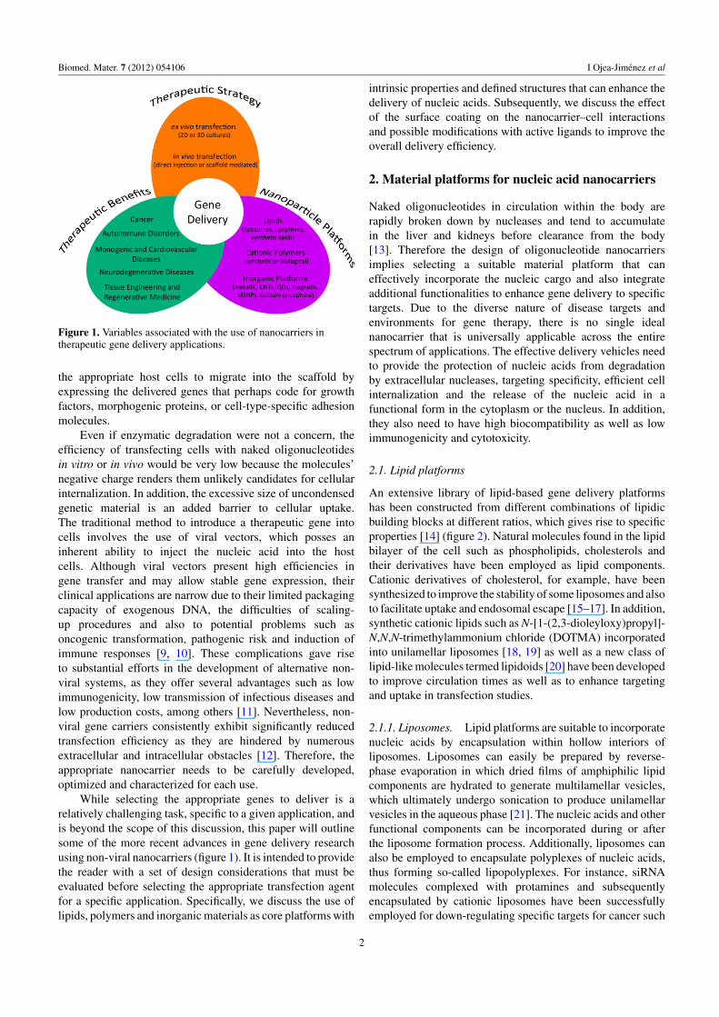
Figure 1. Variables associated with the use of nanocarriers intherapeutic gene delivery applications.
the appropriate host cells to migrate into the scaffold byexpressing the delivered genes that perhaps code for growthfactors, morphogenic proteins, or cell-type-specific adhesionmolecules.
Even if enzymatic degradation were not a concern, theefficiency of transfecting cells with naked oligonucleotidesin vitro or in vivo would be very low because the molecules’negative charge renders them unlikely candidates for cellularinternalization. In addition, the excessive size of uncondensedgenetic material is an added barrier to cellular uptake.The traditional method to introduce a therapeutic gene intocells involves the use of viral vectors, which posses aninherent ability to inject the nucleic acid into the hostcells. Although viral vectors present high efficiencies ingene transfer and may allow stable gene expression, theirclinical applications are narrow due to their limited packagingcapacity of exogenous DNA, the difficulties of scaling-up procedures and also to potential problems such asoncogenic transformation, pathogenic risk and induction ofimmune responses [9, 10]. These complications gave riseto substantial efforts in the development of alternative non-viral systems, as they offer several advantages such as lowimmunogenicity, low transmission of infectious diseases andlow production costs, among others [11]. Nevertheless, non-viral gene carriers consistently exhibit significantly reducedtransfection efficiency as they are hindered by numerousextracellular and intracellular obstacles [12]. Therefore, theappropriate nanocarrier needs to be carefully developed,optimized and characterized for each use.
While selecting the appropriate genes to deliver is arelatively challenging task, specific to a given application, andis beyond the scope of this discussion, this paper will outlinesome of the more recent advances in gene delivery researchusing non-viral nanocarriers (figure 1). It is intended to providethe reader with a set of design considerations that must beevaluated before selecting the appropriate transfection agentfor a specific application. Specifically, we discuss the use oflipids, polymers and inorganic materials as core platforms with
intrinsic properties and defined structures that can enhance thedelivery of nucleic acids. Subsequently, we discuss the effectof the surface coating on the nanocarrier–cell interactionsand possible modifications with active ligands to improve theoverall delivery efficiency.
2. Material platforms for nucleic acid nanocarriers
Naked oligonucleotides in circulation within the body arerapidly broken down by nucleases and tend to accumulatein the liver and kidneys before clearance from the body[13]. Therefore the design of oligonucleotide nanocarriersimplies selecting a suitable material platform that caneffectively incorporate the nucleic cargo and also integrateadditional functionalities to enhance gene delivery to specifictargets. Due to the diverse nature of disease targets andenvironments for gene therapy, there is no single idealnanocarrier that is universally applicable across the entirespectrum of applications. The effective delivery vehicles needto provide the protection of nucleic acids from degradationby extracellular nucleases, targeting specificity, efficient cellinternalization and the release of the nucleic acid in afunctional form in the cytoplasm or the nucleus. In addition,they also need to have high biocompatibility as well as lowimmunogenicity and cytotoxicity.
2.1. Lipid platforms
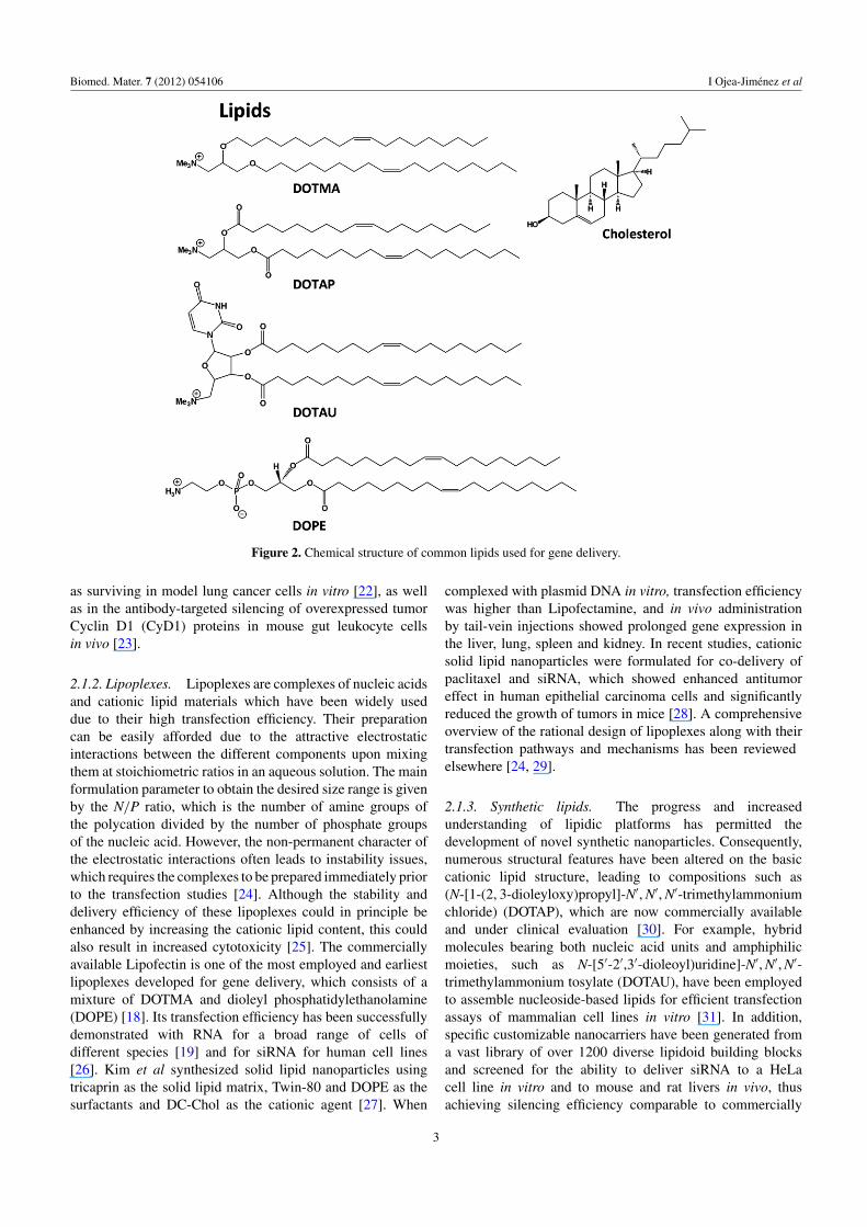
An extensive library of lipid-based gene delivery platformshas been constructed from different combinations of lipidicbuilding blocks at different ratios, which gives rise to specificproperties [14] (figure 2). Natural molecules found in the lipidbilayer of the cell such as phospholipids, cholesterols andtheir derivatives have been employed as lipid components.Cationic derivatives of cholesterol, for example, have beensynthesized to improve the stability of some liposomes and alsoto facilitate uptake and endosomal escape [15–17]. In addition,synthetic cationic lipids such as N-[1-(2,3-dioleyloxy)propyl]-N,N,N-trimethylammonium chloride (DOTMA) incorporatedinto unilamellar liposomes [18, 19] as well as a new class oflipid-like molecules termed lipidoids [20] have been developedto improve circulation times as well as to enhance targetingand uptake in transfection studies.
2.1.1. Liposomes. Lipid platforms are suitable to incorporatenucleic acids by encapsulation within hollow interiors ofliposomes. Liposomes can easily be prepared by reverse-phase evaporation in which dried films of amphiphilic lipidcomponents are hydrated to generate multilamellar vesicles,which ultimately undergo sonication to produce unilamellarvesicles in the aqueous phase [21]. The nucleic acids and otherfunctional components can be incorporated during or afterthe liposome formation process. Additionally, liposomes canalso be employed to encapsulate polyplexes of nucleic acids,thus forming so-called lipopolyplexes. For instance, siRNAmolecules complexed with protamines and subsequentlyencapsulated by cationic liposomes have been successfullyemployed for down-regulating specific targets for cancer such
2
Biomed. Mater. 7 (2012) 054106 I Ojea-Jimenez et al
Figure 2. Chemical structure of common lipids used for gene delivery.
as surviving in model lung cancer cells in vitro [22], as wellas in the antibody-targeted silencing of overexpressed tumorCyclin D1 (CyD1) proteins in mouse gut leukocyte cellsin vivo [23].
2.1.2. Lipoplexes. Lipoplexes are complexes of nucleic acidsand cationic lipid materials which have been widely useddue to their high transfection efficiency. Their preparationcan be easily afforded due to the attractive electrostaticinteractions between the different components upon mixingthem at stoichiometric ratios in an aqueous solution. The mainformulation parameter to obtain the desired size range is givenby the N/P ratio, which is the number of amine groups ofthe polycation divided by the number of phosphate groupsof the nucleic acid. However, the non-permanent character ofthe electrostatic interactions often leads to instability issues,which requires the complexes to be prepared immediately priorto the transfection studies [24]. Although the stability anddelivery efficiency of these lipoplexes could in principle beenhanced by increasing the cationic lipid content, this couldalso result in increased cytotoxicity [25]. The commerciallyavailable Lipofectin is one of the most employed and earliestlipoplexes developed for gene delivery, which consists of amixture of DOTMA and dioleyl phosphatidylethanolamine(DOPE) [18]. Its transfection efficiency has been successfullydemonstrated with RNA for a broad range of cells ofdifferent species [19] and for siRNA for human cell lines[26]. Kim et al synthesized solid lipid nanoparticles usingtricaprin as the solid lipid matrix, Twin-80 and DOPE as thesurfactants and DC-Chol as the cationic agent [27]. When
complexed with plasmid DNA in vitro, transfection efficiencywas higher than Lipofectamine, and in vivo administrationby tail-vein injections showed prolonged gene expression inthe liver, lung, spleen and kidney. In recent studies, cationicsolid lipid nanoparticles were formulated for co-delivery ofpaclitaxel and siRNA, which showed enhanced antitumoreffect in human epithelial carcinoma cells and significantlyreduced the growth of tumors in mice [28]. A comprehensiveoverview of the rational design of lipoplexes along with theirtransfection pathways and mechanisms has been reviewedelsewhere [24, 29].
2.1.3. Synthetic lipids. The progress and increasedunderstanding of lipidic platforms has permitted thedevelopment of novel synthetic nanoparticles. Consequently,numerous structural features have been altered on the basiccationic lipid structure, leading to compositions such as(N-[1-(2, 3-dioleyloxy)propyl]-N′, N′, N′-trimethylammoniumchloride) (DOTAP), which are now commercially availableand under clinical evaluation [30]. For example, hybridmolecules bearing both nucleic acid units and amphiphilicmoieties, such as N-[5′-2′,3′-dioleoyl)uridine]-N′, N′, N′-trimethylammonium tosylate (DOTAU), have been employedto assemble nucleoside-based lipids for efficient transfectionassays of mammalian cell lines in vitro [31]. In addition,specific customizable nanocarriers have been generated froma vast library of over 1200 diverse lipidoid building blocksand screened for the ability to deliver siRNA to a HeLacell line in vitro and to mouse and rat livers in vivo, thusachieving silencing efficiency comparable to commercially
3
Biomed. Mater. 7 (2012) 054106 I Ojea-Jimenez et al
available Lipofectamine 2000 [20]. Other lipidoids have beenoptimized for the delivery of siRNA to target anti-angiogenicfactor (SHP-1) in human endothelial cell in vitro [32], as wellas silencing Claudin-3, an overexpressed protein in mouseovarian tumors, in vivo [33]. Recently, a co-delivery systemof anticancer drugs and siRNA for effective common therapyhas been reported [34]. The anticancer drug mitoxantrone wasfunctionalized with palmitoleic acid chains to form cationicnanoparticles for siRNA complexation, which showed the highefficiency of the cellular delivery of siRNA and the reductionof tumor cell viability.
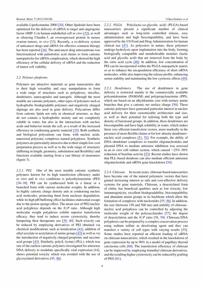
2.2. Polymer platforms
Polymers are attractive materials as gene nanocarriers dueto their high versatility and easy manipulation to forma wide range of structures such as polyplexes, micelles,dendrimers, nanocapsules and nanogels. Although the mostnotable are cationic polymers, other types of polymers such ashydrophobic biodegradable polymers and negatively chargedalginage are also used in gene delivery. Polycations differfrom cationic lipids not only in chemical structure, as theydo not contain a hydrophobic moiety and are completelysoluble in water, but also in the interactions with nucleicacids and behavior inside the cell, as a result of the increasedefficiency in condensing genetic material [35]. Both syntheticand biological polycations can form, with nucleic acids,nanosized polyionic complexes named polyplexes. Syntheticpolymers are particularly attractive due to their simple low-costpreparation process as well as to the wide range of structures(differing in length, geometry, substitution and flexibility) andfunctions available starting from a vast library of monomers(figure 3).
2.2.1. PEI. One of the most notable cationic syntheticpolymers known for its high transfection efficiency underin vitro and in vivo conditions is polyethyleneimine (PEI)[36–38]. PEI can be synthesized both in a linear or abranched form with various molecular weights. In addition,its highly cationic charge density aids at condensing nucleicacid molecules, protecting them from nuclease degradation,while its high pH buffering effect facilitates endosomal escapedue to the proton sponge effect. The mean size of PEI/nucleicacid polyplexes depends on the N/P ratio. Although highmolecular weight polyplexes exhibit superior transfectionefficacy, they tend to induce severe cytotoxicity, therebyhampering their therapeutic use [39, 40]. Such toxicity canbe reduced by employing derivatives of PEI obtained viachemical modifications such as ketalization [41], addition ofethyl acrylate or acetylation of amine groups [42] as well as viathe introduction of negatively charged propionic and succinicacid groups [43]. Similarly, poly(L-lysine) (PLL), which wasone of the earliest cationic polymers investigated for antisenseDNA delivery to modulate specifically viral expression [44],shows potential toxicity which was avoided with the use ofglycosylated derivatives [45, 46].
2.2.2. PLGA. Poly(lactic-co-glycolic acid) (PLGA)-basednanocarriers present a significant number of clinicaladvantages such as long-term controlled release, easyadministration and high biocompatibility, and have beenapproved by the US Food and Drug Administration for humanclinical use [47]. As polyesters in nature, these polymersundergo hydrolysis upon implantation into the body, formingbiologically compatible and metabolizable moieties (lacticacid and glycolic acid) that are removed from the body bythe citric acid cycle [48]. In addition, low concentration ofPEI can be incorporated within the PLGA nanoparticle matrixso as to enhance the encapsulation efficiency of short siRNAmolecules, while also improving the release profile, enhancingserum stability and maintaining the low cytotoxic effects [49].
2.2.3. Dendrimers. The use of dendrimers in genedelivery is restricted mainly to the commercially availablepolyamidoamine (PAMAM) and polypropyleneimine (PPI),which are based on an alkyldiamine core with tertiary aminebranches that give a cationic net surface charge [50]. Thesecationic polymers have generated particular interest in nucleicacid delivery for their customizable architecture and size,as well as their potential for tailoring both the type anddensity of functional groups. In addition, these dendrimers arebiocompatible and have high solubility in water, which makesthem very efficient transfection vectors, more markedly in thepresence of more flexible chains or for low-density dendrimer–nucleic acid complexes [51, 52]. For example, the ability ofDNA–dendrimer complexes to transfer oligonucleotides andplasmid DNA to mediate antisense inhibition was assessedin an in vitro cell culture system, which caused ∼25%–50%reduction of baseline activity [53]. Recent studies have shownthat PLL-based dendrons can also mediate efficient antisenseoligonucleotide and siRNA gene knockdown [54].
2.2.4. Chitosan. In recent years, chitosan-based nanocarriershave become one of the natural polymeric vectors that havegained increasing interest as safe and cost-effective deliverysystems for gene materials. Chitosan, a deacetylated formof chitin, has beneficial qualities such as low toxicity, lowimmunogenicity, excellent biodegradability, biocompatibilityand abundant amine groups in its backbone which allow theformation of complexes with nucleotides [55, 56]. In addition,the size (between 150 and 500 nm) and stability of chitosan–nucleic acid polyplexes can be controlled by adjusting themolecular weight of the polysaccharides [57], the degreeof deacetylation and the N/P ratio [58, 59]. Chitosan/DNApolyplexes can be prepared by a complex coacervation processusing sodium sulfate as desolvating agent and they cantransfect a variety of cell types with varying results [55].Some studies have reported an efficient loading of siRNAon chitosan nanocarriers, which resulted in the knockdown ofgene expression by up to 90% in a model of papillary thyroidcarcinoma cells [60]. The transfection efficiency of chitosancan be improved by employing trimethyl chitosan derivatives,and the resulting higher cytotoxicity can be reduced by graftingof PEG [61].
4
Biomed. Mater. 7 (2012) 054106 I Ojea-Jimenez et al
Figure 3. Chemical structure of the most common synthetic and biological polymers used for gene delivery.
2.2.5. Alginate nanogels. Alginate, a negatively chargedpolysaccharide extracted from brown algae, is anotherbiological polymer widely employed due to its low toxicityand low immunogenicity. The addition of calcium ions to analginate solution under certain conditions leads to gelationand nanoparticle formation as a result of ionic cross-linkagebetween the polysaccharide chains [62]. The resulting Ca–alginate nanoparticles with a mean diameter of 80 nmwere efficiently used as gene carriers. Further investigationsreported a great ability of alginate nanogels to protect antisenseoligonucleotide from degradation in the presence of serum andto modify the biodistribution toward the lungs, the liver andthe spleen after intravenous administration into mice [63]. Theformulation of alginate nanogels often requires the use of apolycation as a platform for gene delivery, which has given riseto diverse studies employing PEI/alginate [64], PLL–alginate[65] and chitosan–alginate nanocomposites [66].
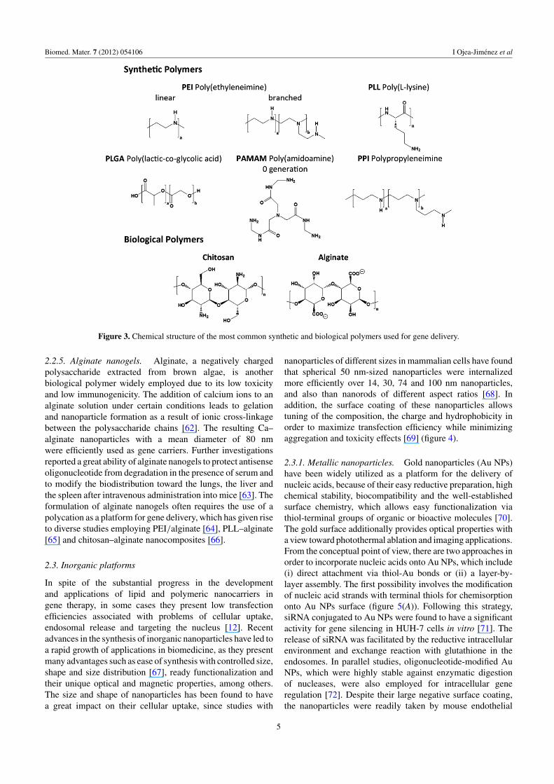
2.3. Inorganic platforms
In spite of the substantial progress in the developmentand applications of lipid and polymeric nanocarriers ingene therapy, in some cases they present low transfectionefficiencies associated with problems of cellular uptake,endosomal release and targeting the nucleus [12]. Recentadvances in the synthesis of inorganic nanoparticles have led toa rapid growth of applications in biomedicine, as they presentmany advantages such as ease of synthesis with controlled size,shape and size distribution [67], ready functionalization andtheir unique optical and magnetic properties, among others.The size and shape of nanoparticles has been found to havea great impact on their cellular uptake, since studies with
nanoparticles of different sizes in mammalian cells have foundthat spherical 50 nm-sized nanoparticles were internalizedmore efficiently over 14, 30, 74 and 100 nm nanoparticles,and also than nanorods of different aspect ratios [68]. Inaddition, the surface coating of these nanoparticles allowstuning of the composition, the charge and hydrophobicity inorder to maximize transfection efficiency while minimizingaggregation and toxicity effects [69] (figure 4).
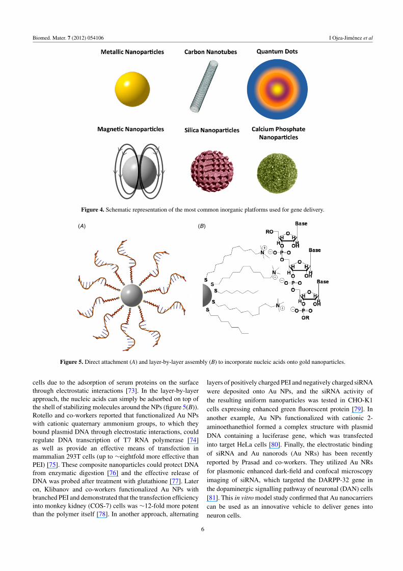
2.3.1. Metallic nanoparticles. Gold nanoparticles (Au NPs)have been widely utilized as a platform for the delivery ofnucleic acids, because of their easy reductive preparation, highchemical stability, biocompatibility and the well-establishedsurface chemistry, which allows easy functionalization viathiol-terminal groups of organic or bioactive molecules [70].The gold surface additionally provides optical properties witha view toward photothermal ablation and imaging applications.From the conceptual point of view, there are two approaches inorder to incorporate nucleic acids onto Au NPs, which include(i) direct attachment via thiol-Au bonds or (ii) a layer-by-layer assembly. The first possibility involves the modificationof nucleic acid strands with terminal thiols for chemisorptiononto Au NPs surface (figure 5(A)). Following this strategy,siRNA conjugated to Au NPs were found to have a significantactivity for gene silencing in HUH-7 cells in vitro [71]. Therelease of siRNA was facilitated by the reductive intracellularenvironment and exchange reaction with glutathione in theendosomes. In parallel studies, oligonucleotide-modified AuNPs, which were highly stable against enzymatic digestionof nucleases, were also employed for intracellular generegulation [72]. Despite their large negative surface coating,the nanoparticles were readily taken by mouse endothelial
5
Biomed. Mater. 7 (2012) 054106 I Ojea-Jimenez et al
Figure 4. Schematic representation of the most common inorganic platforms used for gene delivery.
(A) (B)
Figure 5. Direct attachment (A) and layer-by-layer assembly (B) to incorporate nucleic acids onto gold nanoparticles.
cells due to the adsorption of serum proteins on the surfacethrough electrostatic interactions [73]. In the layer-by-layerapproach, the nucleic acids can simply be adsorbed on top ofthe shell of stabilizing molecules around the NPs (figure 5(B)).Rotello and co-workers reported that functionalized Au NPswith cationic quaternary ammonium groups, to which theybound plasmid DNA through electrostatic interactions, couldregulate DNA transcription of T7 RNA polymerase [74]as well as provide an effective means of transfection inmammalian 293T cells (up to ∼eightfold more effective thanPEI) [75]. These composite nanoparticles could protect DNAfrom enzymatic digestion [76] and the effective release ofDNA was probed after treatment with glutathione [77]. Lateron, Klibanov and co-workers functionalized Au NPs withbranched PEI and demonstrated that the transfection efficiencyinto monkey kidney (COS-7) cells was ∼12-fold more potentthan the polymer itself [78]. In another approach, alternating
layers of positively charged PEI and negatively charged siRNAwere deposited onto Au NPs, and the siRNA activity ofthe resulting uniform nanoparticles was tested in CHO-K1cells expressing enhanced green fluorescent protein [79]. Inanother example, Au NPs functionalized with cationic 2-aminoethanethiol formed a complex structure with plasmidDNA containing a luciferase gene, which was transfectedinto target HeLa cells [80]. Finally, the electrostatic bindingof siRNA and Au nanorods (Au NRs) has been recentlyreported by Prasad and co-workers. They utilized Au NRsfor plasmonic enhanced dark-field and confocal microscopyimaging of siRNA, which targeted the DARPP-32 gene inthe dopaminergic signalling pathway of neuronal (DAN) cells[81]. This in vitro model study confirmed that Au nanocarrierscan be used as an innovative vehicle to deliver genes intoneuron cells.
6
Biomed. Mater. 7 (2012) 054106 I Ojea-Jimenez et al
2.3.2. Carbon nanotubes. The use of carbon-basednanostructures, such as carbon nanotubes (CNTs), inbiomedicine is increasingly attracting attention. One keyadvantage of carbon nanotubes is their ability to translocatethrough plasma membranes by receptor-mediated endocytosis,allowing their use for the delivery of therapeutically activemolecules [82–84]. Moreover, exploitation of their uniqueelectrical, optical, thermal and spectroscopic properties in abiological context is hoped to yield great advances in thedetection, monitoring and therapy of disease. The toxicityinvolving the use of CNTs still remains a controversial issuesince, on the one hand, preliminary toxicology studies on micerevealed no signs of toxicity after four months [85], while onthe other hand, CNTs introduced into the abdominal cavity ofmice showed asbestos-like pathogenicity [86].
The most common strategy to render CNTs viable as aplatform for gene delivery is to use a vector system ableto associate with nucleic acids by self-assembly and assistits intracellular translocation. The surface functionalizationplays an important role since only positively charged CNTs,covalently functionalized with amine-terminal groups, are ableto complex and deliver nucleic acids [87]. CNT-mediatedgene delivery and expression leading to the production ofmarker proteins has been investigated by Kostarelos andco-workers [88, 89], such as expression of β-galactosidasein Chinese hamster ovary (CHO) cells, which was found∼ 5–10-fold higher than the naked pDNA alone. In parallelstudies, Liu and co-workers also reported a non-covalentassociation of pDNA with PEI-functionalized CNTs and foundthat the levels of expression of luciferase in different celllines were much higher for the complexes incorporatingCNTs than pDNA or PEI-pDNA alone [90]. Other verydifferent approaches make use of magnetic CNTs containingNi particles enclosed in their tips [91] or microwave irradiationto open up temporary nanochannels across the cell envelope[92] to deliver pDNA into either mammalian cells or E. coli,respectively. Finally, CNTs have also been conjugated withsiRNA and some promising initial results have been reportedin siRNA-mediated gene silencing. Dai and co-workers havelinked siRNA through disulfide bonds to PEG-functionalizedphospholipids coating the CNT surface [93]. These siRNA-CNT conjugates were internalized by human T-cells andprimary cells, and the siRNA was released intracellularlyupon cleavage of disulfide bonds due to the reducingenvironment of the endosome, which resulted in a 60%–70%knockdown in phenotypic expression [94]. Further examplesemployed cationic amine-functionalized CNTs to complex anddeliver siRNA, which allowed to suppress the expression oftarget proteins in primary cardiomyocytes [95], or suppresstumor growth and prolong survival of tumor-bearing animals[96, 97].
2.3.3. Semiconductor quantum dots. Quantum dots (QDs)are semiconductor nanoparticles that exhibit size-dependentphotoluminiscent properties due to the effect of quantumconfinement, which provides many advantages over traditionalorganic dyes such as narrow emission spectra, largermolar extinction coefficients and enhanced stability with
minimal photobleaching [98, 99]. These QDs can be usedas the core platform onto which nucleic acids and otherfunctional components can be conjugated, therefore givingrise to multifunctional nanocarriers combining therapeutic anddiagnostic imaging functions in a single vehicle toward thetreatment of focal human diseases such as cancer. For example,siRNA and F3 tumor-targeting peptides were conjugated ontoPEGylated QDs using labile and non-labile cross-linkers, andthese nanocarriers were tested toward targeted internalizationand gene knockdown in tumor HeLa cells [100, 101].In addition, QDs covered with proton-absorbing polymericcoatings [102] as well as amphipols [103] have been used forsiRNA delivery in order to facilitate cellular internalizationand endosomal escape.
2.3.4. Magnetic nanoparticles. Magnetic nanoparticleshave also been employed as platforms for gene deliverydue to their appealing intrinsic magnetic properties thatallow both imaging and tracking. Although the synthesisof magnetic nanoparticles with controlled size and shapeis already well established, their stability in physiologicalsolutions and the degree to which their surfaces may bechemically functionalized still remains a big challenge, whichhampers the extent of their biomedical applicability [104].Nevertheless, iron oxide magnetic nanoparticles have beenfunctionalized with siRNA, a dye molecule and a dextrancoating, which allowed monitoring of cell internalizationduring in vitro studies by magnetic resonance imagingand near-infrared fluorescence optical imaging, resulting insuppression of the targeted gene [105]. Similarly, oleic acid-coated magnetic nanoparticles could be stabilized with acationic lipid shell with siRNA molecules, and the resultingformulation could be magnetically guided in vivo to deliverand silence genes in cells and tumors in mice [106]. Furtherstudies by Bhatia and co-workers made use of dendrimer-conjugated magnetofluorescent nanoworms as a modularplatform for siRNA delivery in vivo [107]. SiRNA moleculestargeting against the epidermal growth factor receptor (EGFR)reduced protein levels of EGFR in human glioblastomacells by 70%–80%. Finally, the multifunctional characterof magnetic nanoparticle platforms conjugated with siRNA,cancer-cell-specific targeting moieties and fluorescent dyeswas demonstrated for simultaneous delivery and multimodalimaging of the location and trafficking of siRNA in vivo [108].
2.3.5. Silica nanoparticles. Mesoporous silica nanoparticles(MSNPs) have been investigated as potential carriers of nucleicacids largely due to their inherent biocompatibility and lowtoxicity, easy surface modification through silanol groups onthe surface and the large pore volume which facilitates nucleicacid loading [109]. Diverse polycationic polymers have beenemployed for pore capping of silica nanoparticles [110];among them PAMAM-dendrimer capped MSNPs could bindto plasmid DNA and showed enhanced transfection efficiencyin neural glia and HeLa cells [111]. The same deliveryvehicle was used for the simultaneous delivery of doxorubicinand siRNAs for chemotherapy in multidrug-resistant humanovarian cancer cells [112]. Alternatively, PEI-functionalized
7
Biomed. Mater. 7 (2012) 054106 I Ojea-Jimenez et al
have also been used for the delivery of siRNA resulting in upto 60% knockdown of green fluorescent protein (GFP) whileexhibiting low cytotoxicity [113, 114].
2.3.6. Calcium phosphate nanoparticles. Particles formed bycalcium phosphate present excellent biocompatibility, sincethis inorganic biological material is the main componentin bones and teeth. While the size-controlled synthesisof calcium phosphate nanoparticles still remains elusive,commonly employed methods make use of the high affinityof this material for binding to nucleic acids [115]. Theoligonucleotide is first dissolved in a phosphate buffer andthen a calcium chloride solution is added, resulting in a calciumphosphate precipitate containing the DNA on its surface [116].Neuronal cells are typically transfected by calcium phosphatemethods optimized to decrease toxicity, but only 1%–5%of cells are efficiently transfected. Improvements in thistechnique lead to up to 60% transfection efficiencies in low-density primary neuronal cultures [117]. These improvementsbasically consisted in the formation of smaller precipitateparticles easier to endocytose. For example, the self-assemblyof a PEG-based block copolymer with calcium phosphateentrapping nucleic acids yielded nanocarriers with diametersin the range of hundreds of nanometers [118, 119]. The calciumphosphate core dissociates in the intracellular environment dueto the lowered Ca+2-concentration, thus allowing the release ofthe cargo [120]. Similarly, lipid-coated calcium phosphateformulations for siRNA delivery have also been developedfor controlled disassembly in the acidic environment of theendosomes [121].
3. Binding and uptake of the nanocarriers by thecells
The lipid bilayer membrane is a common obstacle forthe delivery of nucleic acids into the cell. Unlike small-molecule drugs, macromolecules including DNA, RNA andtheir nanocarriers are prohibited from passive diffusion into thecell [122]. While neutral functional groups on nanoparticlesare excellent in preventing nanocarrier–biological interactions,most charged moieties are responsible for active interactionwith cells. It is generally accepted that interaction of lipoplexesand polyplexes with cells in culture, in the absence of serum,is non-specific and involves mainly electrostatic interactionsbetween the positively charged complexes and the negativelycharged cell surface [36, 123]. Endocytosis or endocytosis-like mechanisms have been proposed as the main pathways ofinternalization of lipoplexes and polyplexes rather than fusionof membranes [124], which was evidenced by microscopyanalysis and endocytosis-interfering drugs. In parallel studies,it has been observed that cancer cell membranes absorbmuch less neutral and negatively charged Au NPs, withconsequently lower levels of internalization, than positivelycharged particles [125]. The internalization of negativelycharged nanoparticles is believed to occur through non-specificbinding and clustering of the particles on the cell membrane,which induces endocytosis. By contrast, positively chargednanoparticles are rapidly internalized via clathrin-mediated
pathway [126]. The presence of slight changes in surfacefunctionalities such as hydrophobic structures can lead tovarying amounts of cellular internalization [127].
The ability and the specific roles of different types ofendocytosis in the uptake depend, in part, on the cell type andare important parameters that should be taken into account.For example, in primary cell cultures, such as epithelial airwaycells, the binding and uptake are important rate-limiting stepsfor the transfection efficiency [128, 129], while in establishedcell lines such as fibroblast-like COS or HeLa cells, the uptakeis not a rate-limiting step [130, 131]. The presence of serumhas a dramatic inhibitory effect on the transfection efficiency[132], since polycationic nanocarriers become negativelycharged after interaction with plasma proteins, thus decreasingthe interactions with the cell membrane and consequently theuptake [133].
4. Targeting specificity
The delivery of oligonucleotide nanocarriers to specific tissuesin vivo can be accomplished via two approaches, passive andactive targeting. Passive targeting of tumor tissues can beachieved by the enhanced permeability and retention effect(EPR) [134], in which a combination of leaky vessels and poorlymphatic drainage, both characteristic of the rapid onset ofangiogenesis in solid tumors, results in improved extravasationand enhanced retention in tumor tissues. Nanoparticles witha diameter of less than 500 nm can achieve passive tumortargeting via the hypothetical EPR effect [135]. However,passive targeting is a limited strategy, since not all tumorsexhibit the EPR effect and even amongst those that do, thedegree of permeability may differ significantly within a singletumor.
Alternatively, active targeting of specific tissue and cellscan be achieved by modification of the delivery platform withtarget-recognition molecules that facilitate localization of thenanocarrier via specific ligand–receptor interactions at thecell surface. A great number of ligands have been developedand can be incorporated into nanocarriers for improvedtargeting specificity or enhanced cellular uptake. Among them,antibodies and aptamers are typically used for targeting cell-specific receptors as they present high binding affinities andtarget specificities, while cell-penetrating peptides (CPPs)provide enhanced cellular internalization as they facilitatethe translocation across the membrane. For example, CPPssuch as HIV-TAT protein or VP22 from Herpes simplex virustype I (obtained from the protein transduction domains ofvirus) [136, 137], nuclear-localization-signal (NLS) peptides[138], model proteins such as transferrin [139] as well asother peptide motifs such as Arg–Gly–Asp (RGD) [140], PLL[141] and arginine-rich peptides [142] have been shown aseffective mediators of nanocarrier internalization into variouscell types. Most of such biological motifs have positivelycharged residues assisted by hydrophobic moieties for efficientcellular internalization, which correlates very well with thebehavior of positively charged functionalized nanocarriers[143]. Recently, Wood et al have shown tumor-targeted genedelivery exceeding that of PEI by conjugating a PAMAM
8
Biomed. Mater. 7 (2012) 054106 I Ojea-Jimenez et al
dendrimer with a short peptide ligand that targets the tumorantigen glucose regulated protein-78 [144].
5. Conclusions and perspectives
The development of gene delivery systems using non-viralnanocarriers presents multiple challenges such as overcomingphysiological barriers, conjugating targeting ligands orinteracting with the molecular targets. The mechanisms oftargeting, binding, interaction or release depend on severalfactors, including the vector used, the type of cells and thetransfection conditions employed (e.g. in vitro or in vivo).As a consequence, a wide range of material platforms iscurrently being used as transfection vectors and a hugeamount of fundamental research has been performed in thisdirection. Almost at the same time, many research groups arepushing forward with experiments that have direct clinicalimplications either on the molecular level or in treatmentsin the entire organism. Approaches to eventual therapiesrange from the permanent repair of chromosomal mutationto silencing the expression of a given gene using antisenseoligodeoxynucleotides or siRNA. Unfortunate events inclinical gene delivery trials have raised serious concernsregarding the safety of using gene delivery as medicaltreatment, which has somehow hampered a smooth and rapidprogress in this field.
A rational design of highly efficient nanocarriers requiresa deep understanding of the interactions between the vector andthe nucleic acid, and the cellular pathways and the mechanismsinvolved in the entry into the cell. In this paper, we providean overview of the different material platforms used for genedelivery together with the most recent developments in thisfield, which is under constant progress and expansion. Whilesignificant improvements have been made, there is still muchwork that has to be performed before any nanocarrier can beused as a viable option for the treatment of certain humandiseases.
Acknowledgment
We thank Dr Elena Bellido from Centro de Investigacion enNanociencia y Nanotecnologıa (CIN2, ICN-CSIC) for helpwith figures.
References
[1] Bonetta L 2005 The inside scoop—evaluating gene deliverymethods Nature Methods 2 875–83
[2] Alshamsan A, Hamdy S, Samuel J, El-Kadi A O S,Lavasanifar A and Uludaq H 2010 The induction of tumorapoptosis in B16 melanoma following STAT3 siRNAdelivery with a lipid-substituted polyethylenimineBiomaterials 31 1420–8
[3] Rubanyi G M 2001 The future of human gene therapyMol. Asp. Med. 22 113–42
[4] Heyde M, Partridge K A, Oreffo R O C, Howdle S M,Shakesheff K M and Garnett M C 2007 Gene therapy usedfor tissue engineering applications J. Pharm. Pharmacol.59 329–50
[5] Whitehead K A, Langer R and Anderson D G 2009 Knockingdown barriers: advances in siRNA delivery Nature Rev.Drug Discovery 8 129–38
[6] Tan S J, Kiatwuthinon P, Roh Y H, Kahn J S and Luo D 2011Engineering nanocarriers for siRNA delivery Small7 841–56
[7] Kimelman N, Pelled G, Helm G A, Huard J, Schwarz E Mand Gazit D 2007 Review: gene- and stem cell-basedtherapeutics for bone regeneration and repair Tissue Eng.13 1135–50
[8] Santos J L, Pandita D, Rodrigues J, Pego A P, Granja P Land Tomas H 2011 Non-viral gene delivery tomesenchymal stem cells: methods, strategies andapplication in bone tissue engineering and regenerationCurr. Gene Ther. 11 46–57
[9] Check E 2002 Gene therapy: a tragic setback Nature420 116–8
[10] Li S-D and Huang L 2007 Non-viral is superior to viral genedelivery J. Control. Release 123 181–3
[11] Mintzer M A and Simanek E E 2008 Nonviral vectors forgene delivery Chem. Rev. 109 259–302
[12] Li S and Huang L 2000 Nonviral gene therapy: promises andchallenges Gene Ther. 7 31–4
[13] Braasch D A, Paroo Z, Constantinescu A, Ren G, Oz O K,Mason R P and Corey D R 2004 Biodistribution ofphosphodiester and phosphorothioate siRNA Bioorg. Med.Chem. Lett. 14 1139–43
[14] Kostarelos K and Miller A D 2005 Synthetic, self-assemblyABCD nanoparticles: a structural paradigm for viablesynthetic non-viral vectors Chem. Soc. Rev. 34 970–94
[15] Cooper R G, Etheridge C J, Stewart L, Marshall J,Rudginsky S, Cheng S H and Miller A D 1998 Polyamineanalogues of 3β-[N-(N′,N′-dimethylaminoethane)carbamoyl]cholesterol (DC-Chol) as agents for genedelivery Chem. Eur. J. 4 137–51
[16] Han S-E, Kang H, Shim G Y, Suh M S, Kim S J, Kim J-Sand Oh Y-K 2008 Novel cationic cholesterolderivative-based liposomes for serum-enhanced deliveryof siRNA Int. J. Pharm. 353 260–9
[17] Gao X and Huang L 1995 Cationic liposome-mediated genetransfer Gene Ther. 2 711–22
[18] Felgner P L, Gadek T R, Holm M, Roman R, Chan H W,Wenz M, Northrop J. P, Ringold G M and Danielsen M1987 Lipofection: a highly efficient, lipid-mediatedDNA-transfection procedure Proc. Natl Acad. Sci.84 7413–7
[19] Malone R W, Felgner P L and Verma I M 1989 Cationicliposome-mediated RNA transfection Proc. Natl Acad.Sci. 86 6077–81
[20] Akinc A et al 2008 A combinatorial library of lipid-likematerials for delivery of RNAi therapeutics NatureBiotechnol. 26 561–9
[21] Mevel M et al 2010 DODAG; a versatile new cationic lipidthat mediates efficient delivery of pDNA and siRNAJ. Control. Release 143 222–32
[22] Li S-D and Huang L 2006 Targeted delivery of antisenseoligodeoxynucleotide and small interference RNA intolung cancer cells Mol. Pharm. 3 579–88
[23] Peer D, Park E J, Morishita Y, Carman C V and Shimaoka M2008 Systemic leukocyte-directed siRNA deliveryrevealing cyclin D1 as an anti-inflammatory target Science319 627–30
[24] Elouahabi A and Ruysschaert J-M 2005 Formation andintracellular trafficking of lipoplexes and polyplexesMol. Ther. 11 336–347
[25] Xu Y, Hui S W, Frederik P and Szoka F C Jr 1999Physicochemical characterization and purification ofcationic lipoplexes Biophysics 77 341–53
9
Biomed. Mater. 7 (2012) 054106 I Ojea-Jimenez et al
[26] Beale G, Hollins A J, Benboubetra M, Sohail M, Fox S P,Benter I and Akhtar S 2003 Gene silencing nucleic acidsdesigned by scanning arrays: anti-EGFR activity ofsiRNA, ribozyme and DNA enzymes targeting a singlehybridization-accessible region using the same deliverysystem J. Drug Target. 11 449–56
[27] Choi S H, Jin S-E, Lee M-K, Lim S-J, Park J-S, Kim B-G,Ahn W S and Kim C-K 2008 Novel cationic solid lipidnanoparticles enhanced p53 gene transfer to lung cancercells Eur. J. Pharm. Biopharm. 68 545–54
[28] Yu Y H, Kim E, Park D E, Shim G, Lee S, Kim Y B,Kim C-W and Oh Y-K 2011 Cationic solid lipidnanoparticles for co-delivery of paclitaxel and siRNAEur. J. Pharm. Biopharm. 80 268–73
[29] Zuhorn I S and Hoekstra D 2002 On the mechanism ofcationic amphiphile-mediated transfection. To fuse or notto fuse: is that the question? J. Membr. Biol. 189 167–79
[30] Godbey W T and Mikos A G 2001 Recent progress in genedelivery using non-viral transfer complexes J. Control.Release 72 115–25
[31] Chabaud P, Camplo M, Payet D, Serin G, Moreau L,Barthelemy P and Grinstaff M W 2006 Cationic nucleosidelipids for gene delivery Bioconjug. Chem. 17 466–72
[32] Cho S-W, Goldberg M, Son S M, Xu Q, Yang F, Mei Y,Bogatyrev S, Langer R and Anderson D G 2009 Lipid-likenanoparticles for small interfering RNA delivery toendothelial cells Adv. Funct. Mater. 19 3112–8
[33] Akinc A et al 2009 Development of lipidoid–siRNAformulations for systemic delivery to the liver Mol. Ther.17 872–9
[34] Chang R S et al 2011 Cationic drug-derived nanoparticles formultifunctional delivery of anticancer siRNA Biomaterials32 9785–95
[35] Ruponen M, Yla-Herttuala S and Urtti A 1999 Interactions ofpolymeric and liposomal gene delivery systems withextracellular glycosaminoglycans: physicochemical andtransfection studies Biochim. Biophys. Acta Biomembr.1415 331–41
[36] Boussif O, Lezoualc’h F, Zanta M A, Mergny M D,Scherman D, Demeneix B and Behr J P 1995 A versatilevector for gene and oligonucleotide transfer into cells inculture and in vivo: polyethylenimine Proc. Natl Acad. Sci.92 7297–301
[37] Godbey W T, Wu K K and Mikos A G 1999Poly(ethylenimine) and its role in gene delivery J. Control.Release 60 149–60
[38] Kircheis R, Wightman L and Wagner E 2001 Design andgene delivery activity of modified polyethyleniminesAdv. Drug Deliv. Rev. 53 341–58
[39] Moghimi S M, Symonds P, Murray J C, Hunter A C,Debska G and Szewczyk A 2005 A two-stagepoly(ethylenimine)-mediated cytotoxicity: implicationsfor gene transfer/therapy Mol. Ther. 11 990–5
[40] Godbey W T, Wu K K and Mikos A G 2001Poly(ethylenimine)-mediated gene delivery affectsendothelial cell function and viability Biomaterials22 471–80
[41] Shim M S and Kwon Y J 2008 Controlled Delivery ofplasmid DNA and siRNA to intracellular targets usingketalized polyethylenimine Biomacromolecules 9 444–55
[42] Nimesh S, Aggarwal A, Kumar P, Singh Y, Gupta K Cand Chandra R 2007 Influence of acyl chain length ontransfection mediated by acylated PEI nanoparticlesInt. J. Pharm. 337 265–74
[43] Zintchenko A, Philipp A, Dehshahri A and Wagner E 2008Simple modifications of branched PEI lead to highlyefficient siRNA carriers with low toxicity Bioconjug.Chem. 19 1448–55
[44] Leonetti J P, Rayner B, Lemaitre M, Gagnor C, Milhaud P G,Irnbach J L and Lebleua B 1988 Antiviral activity ofconjugates between poly(l-lysine) and syntheticoligodeoxyribonucleotides Gene 72 323–32
[45] Mahato R I, Takemura S, Akamatsu K, Nishikawa M,Takakura Y and Hashida M 1997 Physicochemical anddisposition characteristics of antisense oligonucleotidescomplexed with glycosylated poly(l-lysine) Biochem.Pharmacol. 53 887–95
[46] Zauner W, Ogris M and Wagner E 1998 Polylysine-basedtransfection systems utilizing receptor-mediated deliveryAdv. Drug Deliv. Rev. 30 97–113
[47] Panyam J and Labhasetwar V 2003 Biodegradablenanoparticles for drug and gene delivery to cells and tissueAdv. Drug Deliv. Rev. 55 329–47
[48] Anderson J M and Shive M S 1997 Biodegradation andbiocompatibility of PLA and PLGA microspheresAdv. Drug Deliv. Rev. 28 5–24
[49] Patil Y and Panyam J 2009 Polymeric nanoparticles forsiRNA delivery and gene silencing Int. J. Pharm.367 195–203
[50] Dufes C, Uchegbu I F and Schatzlein A G 2005 Dendrimersin gene delivery Adv. Drug Deliv. Rev. 57 2177–202
[51] Bielinska A U, Chen C, Johnson J and Baker J R 1999 DNAcomplexing with polyamidoamine dendrimers:implications for transfection Bioconjug. Chem. 10 843–50
[52] Luo D, Haverstick K, Belcheva N, Han E and Saltzman W M2002 Poly(ethylene glycol)-conjugated PAMAMdendrimer for biocompatible, high-efficiency DNAdelivery Macromolecules 35 3456–62
[53] Bielinska A, Kukowska-Latallo J F, Johnson J, Tomalia D Aand Baker J R 1996 Regulation of in vitro gene expressionusing antisense oligonucleotides or antisense expressionplasmids transfected using starburst PAMAM dendrimersNucleic Acids Res. 24 2176–82
[54] Inoue Y, Kurihara R, Tsuchida A, Hasegawa M,Nagashima T, Mori T, Niidome T, Katayama Yand Okitsu O 2008 Efficient delivery of siRNA usingdendritic poly(l-lysine) for loss-of-function analysisJ. Control. Release 126 59–66
[55] Borchard G 2001 Chitosans for gene delivery Adv. DrugDeliv. Rev. 52 145–50
[56] Richardson S W, Kolbe H J and Duncan R 1999 Potential oflow molecular mass chitosan as a DNA delivery system:biocompatibility, body distribution and ability to complexand protect DNA Int. J. Pharm. 178 231–43
[57] Katas H and Alpar H O 2006 Development andcharacterisation of chitosan nanoparticles for siRNAdelivery J. Control. Release 115 216–25
[58] Howard K A et al 2006 RNA interference in vitro and in vivousing a chitosan/siRNA nanoparticle system Mol. Ther.14 476–84
[59] Mao H-Q, Roy K, Troung-Le V L, Janes K A, Lin K Y,Wang Y, August J T and Leong K W 2001 Chitosan–DNAnanoparticles as gene carriers: synthesis, characterizationand transfection efficiency J. Control. Release 70 399–421
[60] de Martimprey H, Bertrand J-R, Fusco A, Santoro M,Couvreur P, Vauthier C and Malvy C 2008 siRNAnanoformulation against the Ret/PTC1 junction oncogeneis efficient in an in vivo model of papillary thyroidcarcinoma Nucleic Acids Res. 36 e2
[61] Germershaus O, Mao S, Sitterberg J, Bakowsky Uand Kissel T 2008 Gene delivery using chitosan, trimethylchitosan or polyethylenglycol-graft-trimethyl chitosanblock copolymers: establishment of structure–activityrelationships in vitro J. Control. Release 125 145–54
[62] You J-O and Peng C-A 2005 Calcium-alginate nanoparticlesformed by reverse microemulsion as gene carriersMacromol. Symp. 219 147–53
10
Biomed. Mater. 7 (2012) 054106 I Ojea-Jimenez et al
[63] Aynie I, Vauthier C, Chacun H, Fattal E and Couvreur P 1999Spongelike alginate nanoparticles as a new potentialsystem for the delivery of antisense oligonucleotidesAntisense Nucleic Acid Drug Dev. 9 301–12
[64] Patnaik S, Aggarwal A, Nimesh S, Goel A, Ganguli M,Saini N, Singh Y and Gupta K C 2006 PEI-alginatenanocomposites as efficient in vitro gene transfectionagents J. Control. Release 114 398–409
[65] Gonzalez Ferreiro M, Tillman L, Hardee G and Bodmeier R2002 Characterization of alginate/poly-l-lysine particlesas antisense oligonucleotide carriers Int. J. Pharm.239 47–59
[66] Douglas K L and Tabrizian M 2005 Effect of experimentalparameters on the formation of alginate–chitosannanoparticles and evaluation of their potential applicationas DNA carrier J. Biomater. Sci. Polym. Ed. 16 43–56
[67] Ojea-Jimenez I, Bastus N G and Puntes V 2011 Influence ofthe sequence of the reagents addition in thecitrate-mediated synthesis of gold nanoparticles J. Phys.Chem. C 115 15752–7
[68] Chithrani B D, Ghazani A A and Chan W C W 2006Determining the size and shape dependence of goldnanoparticle uptake into mammalian cells Nano Lett.6 662–8
[69] Ojea-Jimenez I and Puntes V 2009 Instability of cationic goldnanoparticle bioconjugates: the role of citrate ions J. Am.Chem. Soc. 132 5322–2
[70] Pissuwan D, Niidome T and Cortie M B 2011 Theforthcoming applications of gold nanoparticles in drug andgene delivery systems J. Control. Release 149 65–71
[71] Oishi M, Nakaogami J, Ishii T and Nagasaki Y 2006 SmartPEGylated gold nanoparticles for the cytoplasmic deliveryof siRNA to induce enhanced gene silencing Chem. Lett.35 1046–7
[72] Rosi N L, Giljohann D A, Thaxton C S, Lytton-Jean A K R,Han M S and Mirkin C A 2006 Oligonucleotide-modifiedgold nanoparticles for intracellular gene regulation Science312 1027–30
[73] Giljohann D A, Seferos D S, Patel P C, Millstone J E,Rosi N L and Mirkin C A 2007 Oligonucleotide loadingdetermines cellular uptake of DNA-modified goldnanoparticles Nano Lett. 7 3818–21
[74] McIntosh C M, Esposito E A, Boal A K, Simard J M,Martin C T and Rotello V M 2001 Inhibition of DNAtranscription using cationic mixed monolayer protectedgold clusters J. Am. Chem. Soc. 123 7626–9
[75] Sandhu K K, McIntosh C M, Simard J M, Smith S Wand Rotello V M 2001 Gold nanoparticle-mediatedtransfection of mammalian cells Bioconjug. Chem. 13 3–6
[76] Han G, Martin C T and Rotello V M 2006 Stability of goldnanoparticle-bound DNA toward biological, physical, andchemical agents Chem. Biol. Drug Des. 67 78–82
[77] Han G, Chari N S, Verma A, Hong R, Martin C Tand Rotello V M 2005 Controlled recovery of thetranscription of nanoparticle-bound DNA by intracellularconcentrations of glutathione Bioconjug. Chem. 16 1356–9
[78] Thomas M and Klibanov A M 2003 Conjugation to goldnanoparticles enhances polyethylenimine’s transfer ofplasmid DNA into mammalian cells Proc. Natl Acad. Sci.100 9138–43
[79] Elbakry A, Zaky A, Liebl R, Rachel R, Goepferich Aand Breunig M 2009 Layer-by-layer assembled goldnanoparticles for siRNA delivery Nano Lett. 9 2059–64
[80] Niidome T, Nakashima K, Takahashi H and Niidome Y 2004Preparation of primary amine-modified gold nanoparticlesand their transfection ability into cultivated cells Chem.Commun. 1978–9
[81] Bonoiu A C, Mahajan S D, Ding H, Roy I, Yong K-T,Kumar R, Hu R, Bergey E J, Schwartz S A and Prasad P N
2009 Nanotechnology approach for drug addictiontherapy: gene silencing using delivery of goldnanorod-siRNA nanoplex in dopaminergic neurons Proc.Natl Acad. Sci. 106 5546–50
[82] Kam N W S, Liu Z and Dai H 2006 Carbon nanotubes asintracellular transporters for proteins and dna: aninvestigation of the uptake mechanism and pathwayAngew. Chem. Int. Ed. 45 577–81
[83] Kostarelos K et al 2007 Cellular uptake of functionalizedcarbon nanotubes is independent of functional group andcell type Nature Nanotech. 2 108–13
[84] Lacerda L, Raffa S, Prato M, Bianco A and Kostarelos K2007 Cell-penetrating CNTs for delivery of therapeuticsNano Today 2 38–43
[85] Schipper M L, Nakayama-Ratchford N, Davis C R,Kam N W S, Chu P, Liu Z, Sun X, Dai H and Gambhir S S2008 A pilot toxicology study of single-walled carbonnanotubes in a small sample of mice Nature Nanotech.3 216–21
[86] Poland C A, Duffin R, Kinloch I, Maynard A,Wallace W A H, Seaton A, Stone V, Brown S, MacNee Wand Donaldson K 2008 Carbon nanotubes introduced intothe abdominal cavity of mice show asbestos-likepathogenicity in a pilot study Nature Nanotech. 3 423–8
[87] Gao L, Nie L, Wang T, Qin Y, Guo Z, Yang D and Yan X2006 Carbon nanotube delivery of the GFP gene intomammalian cells Chem. Bio. Chem. 7 239–42
[88] Pantarotto D, Singh R, McCarthy D, Erhardt M, Briand J-P,Prato M, Kostarelos K and Bianco A 2004 Functionalizedcarbon nanotubes for plasmid DNA gene delivery Angew.Chem. Int. Ed. 43 5242–6
[89] Singh R, Pantarotto D, McCarthy D, Chaloin O, Hoebeke J,Partidos C D, Briand J-P, Prato M, Bianco Aand Kostarelos K 2005 Binding and condensation ofplasmid DNA onto functionalized carbon nanotubes:toward the construction of nanotube-based gene deliveryvectors J. Am. Chem. Soc. 127 4388–96
[90] Liu Y, Wu D-C, Zhang W-D, Jiang X, He C-B, Chung T S,Goh S H and Leong K W 2005 Polyethylenimine-graftedmultiwalled carbon nanotubes for secure noncovalentimmobilization and efficient delivery of DNA Angew.Chem. Int. Ed. 44 4782–5
[91] Cai D, Mataraza J M, Qin Z-H, Huang Z, Huang J,Chiles T C, Carnahan D, Kempa K and Ren Z 2005 Highlyefficient molecular delivery into mammalian cells usingcarbon nanotube spearing Nature Methods 2 449–54
[92] Rojas-Chapana J, Troszczynska J, Firkowska I, Morsczeck Cand Giersig M 2005 Multi-walled carbon nanotubes forplasmid delivery into Escherichia coli cells Lab on a Chip5 536–9
[93] Kam N W S, Liu Z and Dai H 2005 Functionalization ofcarbon nanotubes via cleavable disulfide bonds for efficientintracellular delivery of siRNA and potent gene silencingJ. Am. Chem. Soc. 127 12492–3
[94] Liu Z, Winters M, Holodniy M and Dai H 2007 siRNAdelivery into human T-cells and primary cells with carbon-nanotube transporters Angew. Chem. Int. Ed. 46 2023–7
[95] Krajcik R, Jung A, Hirsch A, Neuhuber W and Zolk O 2008Functionalization of carbon nanotubes enablesnon-covalent binding and intracellular delivery of smallinterfering RNA for efficient knock-down of genesBiochem. Biophys. Res. Commun. 369 595–602
[96] Podesta J E, Al-Jamal K T, Herrero M A, Tian B,Ali-Boucetta H, Hegde V, Bianco A, Prato Mand Kostarelos K 2009 Antitumor activity and prolongedsurvival by carbon-nanotube-mediated therapeutic siRNAsilencing in a human lung xenograft model Small5 1176–85
11
Biomed. Mater. 7 (2012) 054106 I Ojea-Jimenez et al
[97] Zhang Z, Yang X, Zhang Y, Zeng B, Wang S, Zhu T,Roden R B S, Chen Y and Yang R 2006 Delivery oftelomerase reverse transcriptase small interfering RNA incomplex with positively charged single-walled carbonnanotubes suppresses tumor growth Clin. Cancer Res.12 4933–9
[98] Bruchez M, Moronne M, Gin P, Weiss S and Alivisatos A P1998 Semiconductor nanocrystals as fluorescent biologicallabels Science 281 2013–6
[99] Chan W C W and Nie S 1998 Quantum dot bioconjugates forultrasensitive nonisotopic detection Science 281 2016–8
[100] Derfus A M, Chen A A, Min D-H, Ruoslahti Eand Bhatia S N 2007 Targeted quantum dot conjugates forsiRNA delivery Bioconjug. Chem. 18 1391–6
[101] Singh N, Agrawal A, Leung A K L, Sharp P Aand Bhatia S N 2010 Effect of nanoparticle conjugation ongene silencing by RNA interference J. Am. Chem. Soc.132 8241–3
[102] Yezhelyev M V, Qi L, O’Regan R M, Nie S and Gao X 2008Proton-sponge coated quantum dots for siRNA deliveryand intracellular imaging J. Am. Chem. Soc. 130 9006–12
[103] Qi L and Gao X 2008 Quantum dot-amphipol nanocomplexfor intracellular delivery and real-time imaging of siRNAACS Nano 2 1403–10
[104] Lu A-H, Salabas E L and Schuth F 2007 Magneticnanoparticles: synthesis, protection, functionalization andapplication Angew. Chem. Int. Ed. 46 1222–44
[105] Medarova Z, Kumar M, Ng S-w, Yang J, Barteneva N,Evgenov N V, Petkova V and Moore A 2008Multifunctional magnetic nanocarriers for image-taggedsiRNA delivery to intact pancreatic islets Transplantation86 1170–7
[106] Namiki Y et al 2009 A novel magnetic crystal-lipidnanostructure for magnetically guided in vivo genedelivery Nature Nanotech. 4 598–606
[107] Agrawal A et al 2009 Functional delivery of siRNA in miceusing dendriworms ACS Nano 3 2495–504
[108] Lee J-H, Lee K, Moon S H, Lee Y, Park T G and Cheon J2009 All-in-one target-cell-specific magneticnanoparticles for simultaneous molecular imaging andsiRNA delivery Angew. Chem. Int. Ed. 48 4174–9
[109] Slowing I I, Trewyn B G, Giri S and Lin V S-Y 2007Mesoporous silica nanoparticles for drug delivery andbiosensing applications Adv. Funct. Mater. 17 1225–36
[110] Kneuer C, Sameti M, Bakowsky U, Schiestel T, Schirra H,Schmidt H and Lehr C-M 2000 A nonviral DNA deliverysystem based on surface modified silica-nanoparticles canefficiently transfect cells in vitro Bioconjug. Chem.11 926–32
[111] Radu D R, Lai C-Y, Jeftinija K, Rowe E W, Jeftinija Sand Lin V S Y 2004 A polyamidoamine dendrimer-cappedmesoporous silica nanosphere-based gene transfectionreagent J. Am. Chem. Soc. 126 13216–7
[112] Chen A M, Zhang M, Wei D, Stueber D, Taratula O, Minko Tand He H 2009 Co-delivery of doxorubicin and Bcl-2siRNA by mesoporous silica nanoparticles enhances theefficacy of chemotherapy in multidrug-resistant cancercells Small 5 2673–7
[113] Hom C, Lu J, Liong M, Luo H, Li Z, Zink J I and Tamanoi F2010 Mesoporous silica nanoparticles facilitate delivery ofsiRNA to shutdown signaling pathways in mammaliancells Small 6 1185–90
[114] Xia T, Kovochich M, Liong M, Meng H, Kabehie S,George S, Zink J I and Nel A E 2009 Polyethyleneiminecoating enhances the cellular uptake of mesoporous silicananoparticles and allows safe delivery of siRNA and DNAconstructs ACS Nano 3 3273–86
[115] Okazaki M, Yoshida Y, Yamaguchi S, Kaneno Mand Elliott J C 2001 Affinity binding phenomena of DNAonto apatite crystals Biomaterials 22 2459–64
[116] Graham F L and van der Eb A J 1973 A new technique forthe assay of infectivity of human adenovirus 5 DNAVirology 52 456–67
[117] Jiang M. and Chen G 2006 High Ca2+-phosphate transfectionefficiency in low-density neuronal cultures Nature Prot.1 695–700
[118] Kakizawa Y, Furukawa S, Ishii A and Kataoka K 2006Organic–inorganic hybrid-nanocarrier of siRNAconstructing through the self-assembly of calciumphosphate and PEG-based block aniomer J. Control.Release 111 368–70
[119] Kakizawa Y, Furukawa S and Kataoka K 2004 Blockcopolymer-coated calcium phosphate nanoparticlessensing intracellular environment foroligodeoxynucleotide and siRNA delivery J. Control.Release 97 345–56
[120] Zhang M, Ishii A, Nishiyama N, Matsumoto S, Ishii T,Yamasaki Y and Kataoka K 2009 PEGylated calciumphosphate nanocomposites as smart environment-sensitivecarriers for siRNA delivery Adv. Mater. 21 3520–5
[121] Li J, Chen Y-C, Tseng Y-C, Mozumdar S and Huang L 2010Biodegradable calcium phosphate nanoparticle with lipidcoating for systemic siRNA delivery J. Control. Release142 416–21
[122] Banerji S K and Hayes M A 2007 Examination ofnonendocytotic bulk transport of nanoparticles acrossphospholipid membranes Langmuir 23 3305–13
[123] Pires P, Simoes S, Nir S, Gaspar R, Duzgunes N andPedroso de Lima M C 1999 Interaction of cationicliposomes and their DNA complexes with monocyticleukemia cells Biochim. Biophys. Acta Biomembr.1418 71–84
[124] Elouahabi A, Thiry M, Pector V, Ruysschaert J-M,Vandenbranden M and Nejat D 2003 Calorimetry ofcationic liposome–DNA complex and intracellularvisualization of the complexes Methods Enzymol.373 312–32
[125] Cho E C, Xie J, Wurm P A and Xia Y 2009 Understandingthe role of surface charges in cellular adsorption versusinternalization by selectively removing gold nanoparticleson the cell surface with a I2/KI etchant Nano Lett.9 1080–4
[126] Harush-Frenkel O, Debotton N, Benita S and Altschuler Y2007 Targeting of nanoparticles to the clathrin-mediatedendocytic pathway Biochem. Biophys. Res. Commun.353 26–32
[127] Zhu Z-J, Ghosh P S, Miranda O R, Vachet R W andRotello V M 2008 Multiplexed screening of cellularuptake of gold nanoparticles using laser desorption/ionization mass spectrometry J. Am. Chem. Soc.130 14139–43
[128] Chu Q, Tousignant J D, Fang S, Jiang C, Eastman S J,Chen L H, Cheng S H and Scheule R K 1999 Binding anduptake of cationic lipid: pDNA complexes by polarizedairway epithelial cells Hum. Gene Ther. 10 25–36
[129] Matsui H, Johnson L G, Randell S H and Boucher R C 1997Loss of binding and entry of liposome-DNA complexesdecreases transfection efficiency in differentiated airwayepithelial cells J. Biol. Chem. 272 1117–26
[130] Bieber T, Meissner W, Kostin S, Niemann Aand Elsasser H-P 2002 Intracellular route andtranscriptional competence of polyethylenimine-DNAcomplexes J. Control. Release 82 441–54
[131] Zabner J, Fasbender A J, Moninger T, Poellinger K A andWelsh M J 1995 Cellular and molecular barriers to genetransfer by a cationic lipid J. Biol. Chem. 270 18997–9007
12
Biomed. Mater. 7 (2012) 054106 I Ojea-Jimenez et al
[132] Escriou V, Ciolina C, Lacroix F, Byk G, Scherman Dand Wils P 1998 Cationic lipid-mediated gene transfer:effect of serum on cellular uptake and intracellular fate oflipopolyamine/DNA complexes Biochim. Biophys. ActaBiomembr. 1368 276–88
[133] Yang J P and Huang L 1997 Overcoming the inhibitory effectof serum on lipofection by increasing the charge ratio ofcationic liposome to DNA Gene Ther. 4 950–60
[134] Iyer A K, Khaled G, Fang J and Maeda H 2006 Exploitingthe enhanced permeability and retention effect for tumortargeting Drug Discovery Today 11 812–8
[135] Maeda H, Wu J, Sawa T, Matsumura Y and Hori K 2000Tumor vascular permeability and the EPR effect inmacromolecular therapeutics: a review J. Control. Release65 271–84
[136] Joliot A and Prochiantz A 2004 Transduction peptides: fromtechnology to physiology Nature Cell. Biol. 6 189–96
[137] Stewart K M, Horton K L and Kelley S O 2008Cell-penetrating peptides as delivery vehicles for biologyand medicine Org. Biomed. Chem. 6 2242–55
[138] Patel P C, Giljohann D A, Seferos D S and Mirkin C A 2008Peptide antisense nanoparticles Proc. Natl Acad. Sci.105 17222–6
[139] Chithrani B D and Chan W C W 2007 Elucidating themechanism of cellular uptake and removal ofprotein-coated gold nanoparticles of different sizes andshapes Nano Lett. 7 1542–50
[140] Green J J, Chiu E, Leshchiner E S, Shi J, Langer Rand Anderson D G 2007 Electrostatic ligand coatings ofnanoparticles enable ligand-specific gene delivery tohuman primary cells Nano Lett. 7 874–9
[141] Mok H, Park J W and Park T G 2008 Enhanced intracellulardelivery of quantum dot and adenovirus nanoparticlestriggered by acidic pH via surface charge reversalBioconjug. Chem. 19 797–801
[142] Sun L, Liu D and Wang Z 2008 Functional goldnanoparticle–peptide complexes as cell-targeting agentsLangmuir 24 10293–7
[143] Thomas M and Klibanov A M 2002 Enhancingpolyethylenimine’s delivery of plasmid DNA intomammalian cells Proc. Natl Acad. Sci. 99 14640–5
[144] Wood K C, Azarin S M, Arap W, Pasqualini R, Langer Rand Hammond P T 2008 Tumor-targeted gene deliveryusing molecularly engineered hybrid polymersfunctionalized with a tumor-homing peptide Bioconjug.Chem. 19 403–5
13


