Embed Size (px)
Citation preview
Novel Role for Proteinase-activated Receptor 2 (PAR2) inMembrane Trafficking of Proteinase-activated Receptor 4(PAR4)*□S
Received for publication, October 21, 2011, and in revised form, March 2, 2012 Published, JBC Papers in Press, March 12, 2012, DOI 10.1074/jbc.M111.315911
Margaret R. Cunningham‡§1, Kathryn A. McIntosh‡, John D. Pediani¶, Joris Robben¶�, Alexandra E. Cooke§,Mary Nilsson‡, Gwyn W. Gould**, Stuart Mundell§, Graeme Milligan¶, and Robin Plevin‡
From the ‡Department of Physiology and Pharmacology, Strathclyde Institute for Biomedical Sciences, Univesity of Strathclyde,27 Taylor Street, Glasgow G4 0NR, Scotland, United Kingdom, the ¶Molecular Pharmacology Group, Institute of Molecular, Cell andSystems Biology, College of Medical, Veterinary and Life Sciences, and **Henry Wellcome Laboratory of Cell Biology, Institute ofMolecular, Cell and Systems Biology, College of Medical, Veterinary and Life Sciences, University of Glasgow, Glasgow G12 8QQ,Scotland, United Kingdom, the �Department of Physiology, Nijmegen Centre for Molecular Life Sciences, Radboud UniversityNijmegen Medical Centre, Nijmegen, Netherlands, and the §School of Physiology and Pharmacology, University of Bristol,Bristol BS8 1TD, United Kingdom
Background: Bioinformatic analysis revealed that PAR4 possesses an ER retention motif.Results: PAR2 both abrogates and facilitates chaperone protein interaction with PAR4 to allow PAR4 to evade ER retention andbe delivered to the plasma membrane.Conclusion: PAR2 regulates PAR4 localization and cell signaling through heterodimerization.Significance: Impact upon understanding PAR2 and PAR4 in inflammation where clear roles are defined.
Proteinase-activated receptors 4 (PAR4) is a classAGprotein-coupled receptor (GPCR) recognized through the ability of ser-ine proteases such as thrombin and trypsin to mediate receptoractivation. Due to the irreversible nature of activation, a freshsupply of receptor is required to bemobilized to the cell surfacefor responsiveness to agonist to be sustained. Unlike other PARsubtypes, the mechanisms regulating receptor trafficking ofPAR4 remain unknown. Here, we report novel features of theintracellular trafficking of PAR4 to the plasmamembrane. PAR4was poorly expressed at the plasma membrane and largelyretained in the endoplasmic reticulum (ER) in a complex withtheCOPIprotein subunit�-COP1.Analysis of thePAR4proteinsequence identified an arginine-based (RXR) ER retentionsequence located within intracellular loop-2 (R183AR 3A183AA), mutation of which allowed efficient membrane deliv-ery of PAR4. Interestingly, co-expression with PAR2 facilitatedplasmamembrane delivery of PAR4, an effect produced throughdisruption of �-COP1 binding and facilitation of interactionwith the chaperone protein 14-3-3�. Intermolecular FRET stud-ies confirmed heterodimerization between PAR2 and PAR4.PAR2 also enhanced glycosylation of PAR4 and activation ofPAR4 signaling. Our results identify a novel regulatory rolefor PAR2 in the anterograde traffic of PAR4. PAR2 was shown toboth facilitate and abrogate protein interactions with PAR4,impacting upon receptor localization and cell signal transduc-
tion. This work is likely to impact markedly upon the under-standing of the receptor pharmacology of PAR4 in normal phys-iology and disease.
Proteinase-activated receptors (PARs)2 are a class A GPCRfamily comprised of four family members, PAR1 through toPAR4, which play key roles in aspects of both physiology andpathophysiology including platelet aggregation, wound heal-ing, and various aspects of inflammation (1–3). Detailed char-acterization of the protein structure of the PAR family has iden-tified proteolytic cleavage sites at the receptor N-terminal(4–7). For each receptor a unique tethered ligand is exposedwithin the N-terminal that interacts with the second extracel-lular loop to mediate receptor activation. The structural deter-minants that regulate PAR activation have long been of interestand there has been considerable focus placed upon the mecha-nisms underpinning membrane trafficking and signal termina-tion particularly for PAR1 (8–14) and PAR2 (15–19). The mostrecent family member to be cloned, PAR4 (20, 21), is distinctfrom both PAR1 and PAR2. Although a thrombin-activatedreceptor, it lacks the hirudin-like domain required for throm-bin selectivity and is activated by several other ligands includingtrypsin. In addition it has a shorter C terminus than PAR1 andPAR2 and lacks essential phosphorylation sites within intracel-lular domains, which are present in other family members andhave been shown to be necessary for receptor desensitization(22). PAR4 has been poorly studied relative to either PAR1 orPAR2 but has been shown to signal viaCa2�mobilization and to
* This work was supported in part by Wellcome Trust Grant 08444/Z/09/Z (toR. P.), British Heart Foundation Grant BHF-FS/05/014 (to R. P.), and MedicalResearch Council Grant G0900050 (to G. M.).Author’s Choice—Final version full access.
□S This article contains supplemental Figs. S1–S3.1 Recipient of the 2006 British Pharmacological Society AJ Clark Ph.D. Student
award. To whom correspondence should be addressed: School of Physiol-ogy and Pharmacology, Medical Sciences Building, University of Bristol,Bristol BS8 1TD, United Kingdom. Tel.: 44-01173311433; E-mail: [email protected].
2 The abbreviations used are: PAR, proteinase-activated receptor; ER, endo-plasmic reticulum; GPCR, G protein-coupled receptor; mEGFP/mEYFP/mECFP, monomeric enhanced green/yellow/cyan fluorescent protein;RXR, arginine-based ER retention motifs; COPI, coat protein I complex;RFRET, ratiometric FRET.
THE JOURNAL OF BIOLOGICAL CHEMISTRY VOL. 287, NO. 20, pp. 16656 –16669, May 11, 2012Author’s Choice © 2012 by The American Society for Biochemistry and Molecular Biology, Inc. Published in the U.S.A.
16656 JOURNAL OF BIOLOGICAL CHEMISTRY VOLUME 287 • NUMBER 20 • MAY 11, 2012
by guest on July 13, 2016http://w
ww
.jbc.org/D
ownloaded from
by guest on July 13, 2016
http://ww
w.jbc.org/
Dow
nloaded from
by guest on July 13, 2016http://w
ww
.jbc.org/D
ownloaded from
by guest on July 13, 2016
http://ww
w.jbc.org/
Dow
nloaded from
by guest on July 13, 2016http://w
ww
.jbc.org/D
ownloaded from
by guest on July 13, 2016
http://ww
w.jbc.org/
Dow
nloaded from
regulate theMAP kinases (23). However, very little is known ofthe mechanisms regulating receptor trafficking.Due to the irreversible nature of activation of PARs, for
responsiveness to agonist to be retained, fresh supplies ofreceptor are required to be mobilized to the cell surface. Deliv-ery to the membrane requires efficient transport between theER/Golgi/plasma membrane, which can be facilitated throughdiscrete motifs that reside within the synthesized protein (24,25). Many chaperone proteins, such as coat protein complexes(COPI and COPII), can assist transport of recently synthesizedproteins throughmotif-based sorting (26–28). Properly assem-bled proteins are packaged for export intoCOPII vesicleswherethey progress to the ER-Golgi intermediate complex, a processknown as anterograde transport. Misfolded proteins or thoseexposing sequences encoding ER retentionmotifs (for example,RXR, KDEL, or KKAA motifs) are shuttled back to the ER viaCOPI vesicles, in a process known as retrograde transport (29).During the assembly of multimeric proteins, such as GPCRhomo/heterodimers, proteins possessing ER retention signalshave been shown to evade ER retention through the stericmasking of motifs during protein folding (30–33). 14-3-3 pro-teins have previously been shown to assist motif masking toensure export of proteins to the Golgi (25, 34). Once properprotein folding has been achieved, post-translational modifica-tions, such as complex glycosylation, will occur (35).Here we identify for the first time the presence of an argin-
ine-based ER retention motif within intracellular loop-2 ofPAR4, which results in ER retention through COPI-dependentretrograde transport. In the presence of PAR2, through PAR2/PAR4 heterodimer formation and interaction with 14-3-3�,PAR4 was able to evade ER retention and undergo N-linkedcomplex glycosylation. This resulted in efficient delivery to theplasmamembrane. The impact of enhanced cell surface expres-sion was reflected in enhanced PAR4-mediated cell signaltransduction. PAR2 is often co-expressed with PAR4, and theyare dual up-regulated by various pro-inflammatory mediatorsand have been shown to be co-activated by common agonists(20, 36, 37). In the presence of PAR2, a significant increase inPAR4-mediated total inositol phosphate accumulation wasobserved. This work demonstrates for the first time a novelregulatory role for PAR2 in the anterograde traffic and signalingof PAR4. This is mediated by selective interaction with COPI or14-3-3 proteins, offering a new paradigm for class A GPCRtrafficking and control.
EXPERIMENTAL PROCEDURES
Reagents and Antibodies—The PAR4 activating peptide,Ala-Tyr-Pro-Gly-Lys-Phe-amidated (NH2) peptide (AYPGKF-NH2), was synthesized by theUniversity of Calgary Peptide Ser-vice (Calgary, Canada). ER-TrackerTM Blue-White DPX Dyes(Molecular Probes) for ER labeling and the anti-transferrinreceptor mouse monoclonal antibody were purchased fromInvitrogen Ltd. Rabbit polyclonal anti-Na�,K�-ATPase �1antibody was purchased from Cell Signaling Technology Inc.Living Colors� full-length A.v. GFP rabbit polyclonal antibodywas purchased from Clontech-TaKaRa Bio Europe (France).PKH26 Red Fluorescent Cell Linker kit for general cell mem-brane labeling, anti-PAR4 goat polyclonal, anti-14-3-3� rabbit
polyclonal antibodies, monoclonal anti-HA-agarose conjugate,HA peptide, and tunicamycin were from Sigma. The anti-PAR4rabbit polyclonal antibody was obtained from Abcam (Cam-bridge, UK). Anti-� coatomer protein (�-COP1) rabbit poly-clonal antibody was purchased from Pierce and Thermo FisherScientific (Loughborough, UK). Mouse monoclonal anti-HAantibody was purchased from Cambridge Bioscience (Cambs,UK). Pierce Cell Surface Protein Isolation Kit was purchasedfromThermo Scientific. The alkaline phosphatase substrate kitwas obtained from Bio-Rad.Epitope-tagged PAR Constructs—Human PAR2 was ampli-
fied by polymerase chain reaction (PCR) from a pRSV-PAR2vector.The PCR product was then digested with HindIII-BamHI and
cloned into the respective sites of a pEYFP-N1 vector (Clontech).Human PAR4 was amplified from a pcDNA3.1(�)-hPAR4 vectorbyPCRanddigestedwithKpn-AgeI, whereas ECFPwas amplifiedfrom the pECFP-N1 vector (Clontech) and digested with AgeI-XbaI. PAR4 andECFPwere ligated and cloned into theKpnI-XbaIsites of the pcDNA3.1(�) vector. Monomeric ECFP and EYFPconstructs were generated by amino acid substitution of Ala206 toLys206 (38), through site-directed mutagenesis using the GeneTailorTM Site-directed Mutagenesis System (Invitrogen). Aminoacid substitutions were similarly made within the primarysequence of PAR4 to mutate potential arginine-based ER reten-tion motifs (positions R183AR 3 A183AA, (referred to as RARmut) R188GRR 3 A188GAA and R183AR R188GRR 3A183AA A188GAA) and the N-linked glycosylation site on theN-terminal of PAR4 (Asn563 Ala56). A HA epitope tag (YPYD-VPDYA)was incorporated into theC-terminal of PAR4 byPCR togenerate PAR4-HA.All constructswere confirmedby sequencing.Cell Culture—HEK293 cells were maintained in minimal
essential medium with Earle’s salts, L-glutamine supplementedwith 10% fetal calf serum (FCS), penicillin (100 units ml�1),streptomycin (100 �g ml�1), and nonessential amino acids andpassaged using 1� SSC (sodium citrate, pH 7.4). NCTC-2544cells and NCTC-PAR2 cells were grown in Medium 199 withEarle’s salts (Sigma) containing 10% FCS, sodium bicarbonate(50 mM), L-glutamine (2 mM), penicillin (100 units ml�1), andstreptomycin (100 �g ml�1). NCTC-2544 cells were passagedusing Versene (0.53 mM EDTA in PBS) to avoid trypsin expo-sure. All cells where then incubated at 37 °C in a humidifiedatmosphere with 5% CO2 with medium replaced every 2 days.Transient Transfection—Cells were grown in 12- and 6-well
plates or T75 flasks prior to transient transfection at 70–80%confluence with 1, 2, or 10 �g of endo-free plasmid DNA,respectively, using Lipofectamine 2000 (Invitrogen) followingthe recommended manufacturer’s guidelines. Maximal geneexpression was observed 48 h post-transfection.Inositol Phosphate Accumulation Assay—Following tran-
sient transfection for 24 h, cells were serum starved for a further24 h in serum-free medium supplemented with 0.5 �Ci/well (1Ci � 37 GBq) of myo-[2-3H]inositol (PerkinElmer Life Sci-ences) (0.5�Ci/well; 1 Ci� 37GBq). Cells were pretreatedwith20 mM lithium chloride for 30 min prior to agonist treatment(100 �M AYPGKF-NH2 for 45 min). Measurement of the accu-mulation of inositol phosphates was carried out as previouslydescribed by Plevin et al. (39).
PAR2 Regulation of PAR4 Trafficking
MAY 11, 2012 • VOLUME 287 • NUMBER 20 JOURNAL OF BIOLOGICAL CHEMISTRY 16657
by guest on July 13, 2016http://w
ww
.jbc.org/D
ownloaded from
Fluorescence Microscopy—Cells were washed in PBS prior tomethanol fixation for 15 min at room temperature. After fur-ther washes with PBS, cells were stained using 4�,6-diamidino-2-phenylindole (DAPI) nuclear dye or ER TrackerTM dye thenmounted onto glass microscope slides with 15 �l of mowiol(Calbiochem). Cells were visualized using a Nikon TE300-Emicroscope (Nikon Instruments, New York) using a �100(numerical aperture; NA 1.3) oil immersion Fluor lens. Emittedfluorescence was detected using a photometric Cool Snap-HQmonochrome camera (Roper Scientific, Trenton, NJ) set up in12-bit mode (0–4095 gray tones). Metamorph software (ver-sion 7.0, Molecular Devices Corp., Downing, PA) was used tocontrol image acquisition and modify image settings. Imageswere background corrected, based on statistical correction ofaverage background regions from defined regions of interest.Cell Surface ELISA—Changes in cell surface expression of
PAR4weremeasured byEnzyme-linked ImmunoSorbentAssay(ELISA). Cells were transfected with PAR4 for 24 h prior tobeing seeded at a density of 1 � 105 cells per well in 24-wellplates pre-coated with 0.1 ml/ml of poly-L-lysine. Cells weregrown overnight to recover. Surface receptors were pre-labeledwith anti-PAR4 (1/1000 dilution) at 4 °C for 1 h. Cells were fixedin 3.7% paraformaldehyde for 5 min and then washed threetimes in Tris-buffered saline (TBS; 20 mM Tris, pH 7.5, 150 mM
NaCl). Cells were blocked with 1% BSA in TBS for 45 min atroom temperature followed by a 1-h inculation with a alkalinephosphatase-conjugated goat anti-rabbit antibody (1/1000dilution) in 1% BSA in TBS. Cells were washed four times inTBS to remove unbound secondary antibody. Alkaline phos-phate substrate solution was prepared by dissolving p-nitro-phenyl phosphate tablets in diethanolamine buffer (Bio-Rad).Substrate solution was added to cells and the plates were incu-bated at 37 °C for 10–30min. Absorbance wasmeasured at 405nm using a microplate reader (Dynex MRX revelation).Cell Surface Biotinylation—Surface expression of PAR4 in
NCTC-2544 andNCTC-PAR2 cells wasmeasured by a biotiny-lation assay using Pierce Cell Surface Protein Isolation Kit(Thermo Scientific). Briefly, four T75 cm2 flasks ofNCTC-2544orNCTC-PAR2 cells were transfectedwith PAR4mECFP. Cellswere labeled with Sulfo-NHS-SS-Biotin for 30 min at 4 °C on arocking platform. The biotinylation reaction was stoppedthrough the addition of a quench solution followed by furtherincubation at 4 °C for 15 min. Cells were scraped and the flaskswere rinsed in Tris-buffered saline (TBS) and centrifuged at1,000 � g for 3 min. Supernatant was discarded and the cellpellets were washed 3 times in TBS followed by centrifugationat 1,000 � g for 3 min. Cells were lysed using the provided lysisbuffer containing complete protease inhibitor mixture (RocheDiagnostics) and sonicated on ice at low power to disrupt usingfive 1-s bursts, then incubated at 4 °C for 30 min on an orbitalrotator. The cell lysates were then centrifuged at 10,000 � g for2 min at 4 °C. Clarified supernatants were transferred to a newtube and incubated with NeutrAvidin-agarose for 60 min atroom temperature with end-over-end mixing using a rotator.Supernatant/agarose slurry was centrifuged for 1 min at1,000� g and the supernatantwas discarded. The agarose pelletwas washed 3 times in the wash buffer provided with the addi-tion of complete protease inhibitormixture. SDS-PAGE sample
buffer (62.5mMTris-HCl, pH 6.8, 1% SDS, 10% glycerol, 50mM
DTT) was added to the sample, which was then heated in a heatblock for 5 min at 95 °C. The tubes were then centrifuged for 2min at 1,000 � g. PAR4 expression was detected by Westernblotting using antibodies specific for either PAR4 or GFP. Equalexpression of total levels of PAR4 mECFP in transfected cellswas confirmed by resolving the corresponding whole celllysates prepared from the same cells used for the biotinylationexperiments.Western Blotting—Proteins were separated by 8–10% SDS-
PAGE and transferred onto nitrocellulose membrane. Themembranes were blocked for nonspecific binding in 2% BSA(w/v) diluted in NATT buffer (50 mM Tris-HCl, 150 mM NaCl,0.2% (v/v) Tween 20) for 2 h. The blots were then incubatedovernight with 50 ng/ml of primary antibody diluted in 0.2%BSA (w/v) in NATT buffer then washed with NATT buffer at15-min intervals for a further 90min. The blots were then incu-bated with HRP-conjugated secondary antibody (20 ng/ml) in0.2% BSA (w/v) diluted in NATT buffer for 2 h. After a further90-min wash, the membranes were treated with ECL reagentand exposed to Kodak x-ray film.Subcellular Fractionation of ER and Plasma Membrane
Compartments—Cells were grown to 70–80% confluence in5�T150 cm tissue culture flasks prior to transient transfectionwith PAR4 mECFP. The cells were harvested and the cell pelletresuspended in 3ml ofHES buffer (25mMHEPES, 1mMEDTA,and 250 mM sucrose, pH 7.4) supplemented with proteaseinhibitors (25 �g/ml of leupeptin, 10 �g/ml of aprotinin, and 1�g/ml of PMSF). The cell lysate was homogenized using a pre-cooled cell homogenizer (Isobiotec Precision Engineering, Ger-many, German PatentOffice number 202 09 547.9) fittedwith asize 10-�m clearance tungsten carbide ballbearing. The homo-genate was centrifuged at 500 � g for 2 min at 4 °C and thesupernatant was transferred to a fresh tube and resuspended inOpti-prep (Invitrogen) density gradient medium to create a45% (v/v) density sample solution. A density gradient (30–10%)was prepared using Opti-prep medium mixed in HES bufferfollowed by ultracentrifugation at 72,000 � g for 4 h at 4 °C toseparate plasma membrane, endosomal, and ER fractions (40).Equal volume fractions (300�l) were collected and precipitatedin 37.5% TCA, incubated on ice for 15 min, and centrifuged at14,000 � g for 15 min at 4 °C. The cell pellets obtained wereresuspended in 2� Laemmli sample buffer supplemented with1Murea and resolved byWestern blotting. Subcellular fraction-ation of ER and plasma membrane compartments were deter-mined using Na�,K�-ATPase, transferrin receptor, and cal-nexin antibodies as markers for plasmamembrane, endosomal,and ER fractions, respectively.Intermolecular FRET—Wide-field intermolecular FRET
microscopy was performed at room temperature in living cells(41–43) on a Nikon TE2000-E inverted microscope (NikonInstruments, Melville, NY). Cells were grown on 0 thickness oncoverslips and transiently transfected with the appropriatemonomeric donor mECFP or acceptor mEYFP-tagged con-structs. Coverslips were placed into a microscope chambercontaining physiological HEPES-buffered saline solution (130mMNaCl, 5 mM KCl, 1 mM CaCl2, 1 mMMgCl2, 20 mMHEPES,10 mM D-glucose, pH 7.4). FRET imaging was performed using
PAR2 Regulation of PAR4 Trafficking
16658 JOURNAL OF BIOLOGICAL CHEMISTRY VOLUME 287 • NUMBER 20 • MAY 11, 2012
by guest on July 13, 2016http://w
ww
.jbc.org/D
ownloaded from
a �40 (numerical aperture; NA 1.3) oil immersion Fluor lens.Emitted fluorescence was detected using a photometric CoolSnap-HQmonochrome camera (Roper Scientific, Trenton, NJ)set up in 12-bit mode (0–4095 gray tones). MetaMorph soft-ware (version 7.6.4 Molecular Devices Corp.) was used to con-trol both the microscopy hardware and multiwavelength fluo-rescence image acquisition required for intermolecular FRETdetection. Donor 430 nm or acceptor 500 nm excitation lightwas generated using a computer controlled Optoscan mono-chromator (Cairn Research, Faversham, Kent, UK) coupled to a103/W2mercury (Hg) arc lamp source (Cairn Research). Opti-mization of illumination excitation centerwavelength andbandpass settings was performed to prevent cross-excitation, mini-mize donor and acceptor bleed-through into the FRETchannel,and ensure no recorded pixels within the channel images weresaturated above a gray level intensity value of 4095. The risk ofmotion occurring during the sequential FRET imaging processwas minimized by using a high-speed filter wheel (Prior Scien-tific Instruments, Cambridge, UK). Metamorph imaging soft-ware was used to quantify the FRET images using the specifiedbleed-through FRET method. Corrected FRET (FRETc) wascalculated using a pixel-by-pixel methodology using the equa-tion FRETc � FRET � (coefficient B � mECFP) � (coefficientA � mEYFP), where mECFP, mEYFP, and FRET values corre-spond to background corrected images obtained through thedonor mECFP, mEYFP, and FRET channels. B and A corre-spond to the values obtained for the mECFP (donor) andmEYFP (acceptor) bleed-through coefficients, respectively, cal-culated using cells transfected with either the mECFP ormEYFP protein alone. Ratiometric FRET (RFRET) values werecalculated from the measurements taken from raw FRET fluo-rescence and dividing this value by the total spectral bleed-through of the acceptor and donor into the FRET channel, i.e.raw FRET divided by (acceptor multiplied by (a)) � (donormultiplied by (b)). In the absence of energy transfer (i.e. noFRET occurrence), the RFRET value measured is �1, valuesgreater than 1 represent the occurrence of FRET, thus indica-tive of protein interaction.Co-immunoprecipitation—To measure PAR4 interaction
with 14-3-3�, cells transiently expressing PAR4-HA werewashed with PBS prior to solubilization in lysis buffer (20 mM
HEPES buffer, pH 7.7, containing 50 mM NaCl, 0.1 mM EDTA,0.1 mM Na3VO4, 0.1 mM PMSF, 10 mg/ml of aprotinin, 10mg/ml of leupeptin, and 1% (w/v) Triton X-100). After a 1-hrotation at 4 °C, the cell lysates were clarified by centrifugationat 13,000� g for 5min at 4 °C. Supernatants were transferred tofresh Eppendorf tubes and 50 �l was removed for inputs. Theremaining lysate was pre-cleared with 30 �l of protein G/pro-tein A-agarose (Calbiochem) and placed in an rotator for 1 h at4 °C. Sampleswere centrifuged at 4 °C for 5min at 5,000� g andthe pre-cleared lysate was transferred to fresh Eppendorf tubescontaining 30 �l of monoclonal anti-HA-agarose conjugate(Sigma) and rotated overnight at 4 °C. Samples were centri-fuged at 4 °C for 5 min at 5,000 � g, then washed with 500 �l oflysis buffer three times and proteins were eluted by incubationwith 30 �l of anti-HA peptide (Sigma; 200 �g/ml) for 10 min atroom temperature. Eluted proteins were removed and added to
10 �l of 5� SDS sample buffer and boiled at 95 °C for 10 minprior to SDS-PAGE.Statistical Analysis—Where experimental data are shown as
a blot, this represents one of at least 3 experiments and datarepresent the mean � S.E. Statistical analysis was by one-wayanalysis of variance withDunnett’s post-test (*, p� 0.05; **, p�0.01).
RESULTS
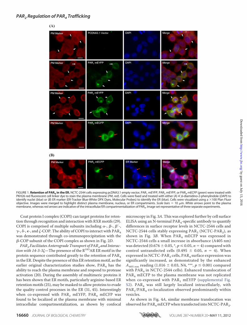
Intracellular Retention of PAR4 in the EndoplasmicReticulum—Tomonitor the expression level and localization ofPAR4, the receptor was tagged at the C terminus with a mono-meric variant form of enhanced cyan fluorescent protein(mECFP) and transiently expressed in keratinocyte-derivedNCTC-2544 cells. These cells provided an idealmodel for theseinvestigations, due to the lack of endogenous PAR expression(44). The localization of PAR4 was initially monitored usingfluorescence microscopy of NCTC-2544 cells transientlyexpressing PAR4 mECFP (Fig. 1A). In comparison to cellsexpressing either PAR1mEYFP or PAR2mEYFP, PAR4mECFPwas largely retained inside the cell with only weak membranelocalization observed. Further microscopy in cells treated withan ER tracker dye (Fig. 1B) highlighted that PAR4 mECFP waspredominantly retained in the ER.Presence of a Functional Arginine-based ER Retention Motif
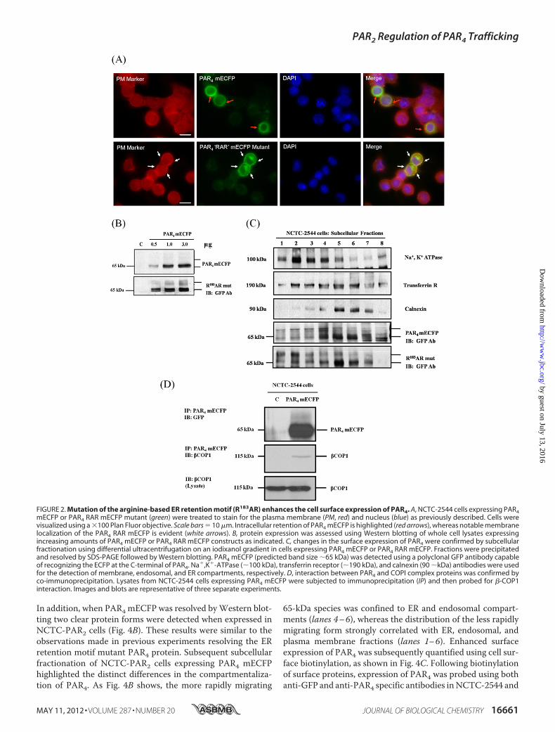
within PAR4—Analysis of the protein sequence for PAR4 iden-tified two potential arginine-based (RXR) ER retention motifslocated within the intracellular loop-2 of the receptor (supple-mental Fig. S1). Alignment of the primary sequences for all PARfamily members found that these motifs were unique to PAR4.The contribution of these motifs in controlling the cellularlocalization of PAR4was assessed by removing the arginine res-idues by alanine substitution (RXR 3 AXA). Of the possiblemotifs investigated, only mutation of the R183AR to A183AAresulted in a loss of ER retention and allowed PAR4 to translo-cate to the plasma membrane (Fig. 2A). Receptor expressionlevelswere determined byWestern blotting (Fig. 2B). Followingexpression of PAR4 mECFP the appearance of a protein band,resolving around 65 kDa, was observed. This correspondedwellwith the predictedmolecular mass of PAR4mECFP (38 kDa forPAR4 combined with 27 kDa for themECFP). As Fig. 2B shows,as the expression of the R183AR mutant increased, the appear-ance of multiple protein forms was observed, a doublet resolv-ing around 65 kDa and a slightly larger species resolvingbetween 70 and 80 kDa. Subcellular fractionation of cellsexpressing either PAR4 mECFP or the R183ARmutant was car-ried out to separate plasma membrane, endosomal, and ERcompartments followed byWestern blot (Fig. 2C). The 65-kDaprotein species observed in cells expressing PAR4 mECFP ormutant receptor reached maximal levels in ER and endosomalfractions (lanes 4–7), co-locating with calnexin and transferrinmarkers, respectively. These experiments identified that thehigher molecular mass species observed in cells expressing theR183ARmutant reflected receptors located in the plasmamem-brane and endosome compartments (lanes 1–4) as shownusing Na�,K�-ATPase and transferrin receptor markers,respectively.
PAR2 Regulation of PAR4 Trafficking
MAY 11, 2012 • VOLUME 287 • NUMBER 20 JOURNAL OF BIOLOGICAL CHEMISTRY 16659
by guest on July 13, 2016http://w
ww
.jbc.org/D
ownloaded from
Coat protein I complex (COPI) can target proteins for reten-tion through recognition and interaction with RXRmotifs (29).COPI is comprised of multiple subunits including �-, �-, ��-,�-, �-, �-, and �-COP. The ability of COPI to interact with PAR4was demonstrated through co-immunoprecipitation with the�-COP subunit of the COPI complex as shown in Fig. 2D.PAR2 Facilitates Anterograde Transport of PAR4 and Interac-
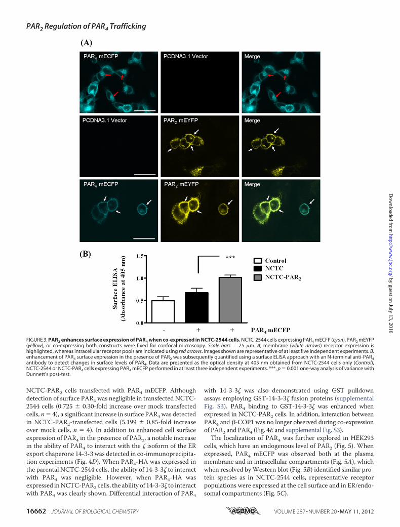
tion with 14-3-3�—The presence of the R183AR ERmotif in theprotein sequence contributed greatly to the retention of PAR4in the ER.Despite the presence of this ER retentionmotif, as theearlier original characterization studies show, PAR4 has theability to reach the plasma membrane and respond to proteaseactivation (20). During the assembly of multimeric proteins ithas been shown that ER motifs, particularly arginine-based ERretentionmotifs (25), may bemasked to allow proteins to evadethe quality control processes in the ER (31, 45). Interestinglywhen co-expressed with PAR2 mEYFP, PAR4 mECFP wasfound to be localized at the plasma membrane with minimalintracellular compartmentalization, as shown by confocal
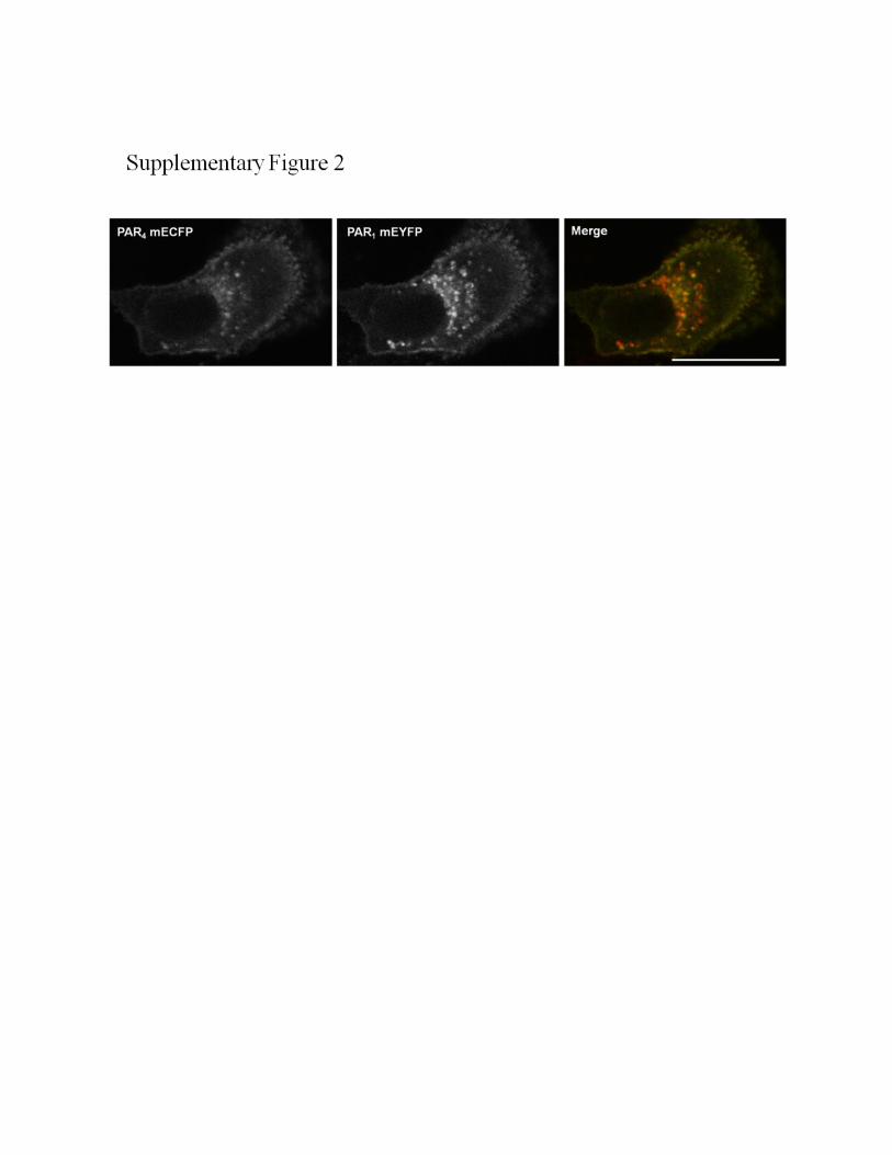
microscopy in Fig. 3A. This was explored further by cell surfaceELISA using an N-terminal PAR4-specific antibody to quantifydifferences in surface receptor levels in NCTC-2544 cells andNCTC-2544 cells stably expressing PAR2 (NCTC-PAR2), asshown in Fig. 3B. When PAR4 mECFP was expressed inNCTC-2544 cells a small increase in absorbance (A405 nm)was detected (0.676 � 0.05, *, p � 0.05, n� 4) compared withcontrol untransfected cells (0.495 � 0.05, n � 4). Whenexpressed in NCTC-PAR2 cells, PAR4 surface expression wassignificantly increased, as demonstrated by the enhancedA405 nm reading (1.016 � 0.03, N4, ***, p � 0.001 comparedwith PAR4 in NCTC-2544 cells). Enhanced translocation ofPAR4 mECFP to the plasma membrane was not replicatedwhen co-expressed with PAR1 mEYFP (supplemental Fig.S2). PAR4 was still largely localized intracellularly, withPAR1/PAR4 co-localization observed predominantly withinvesicles.As shown in Fig. 4A, similar membrane translocation was
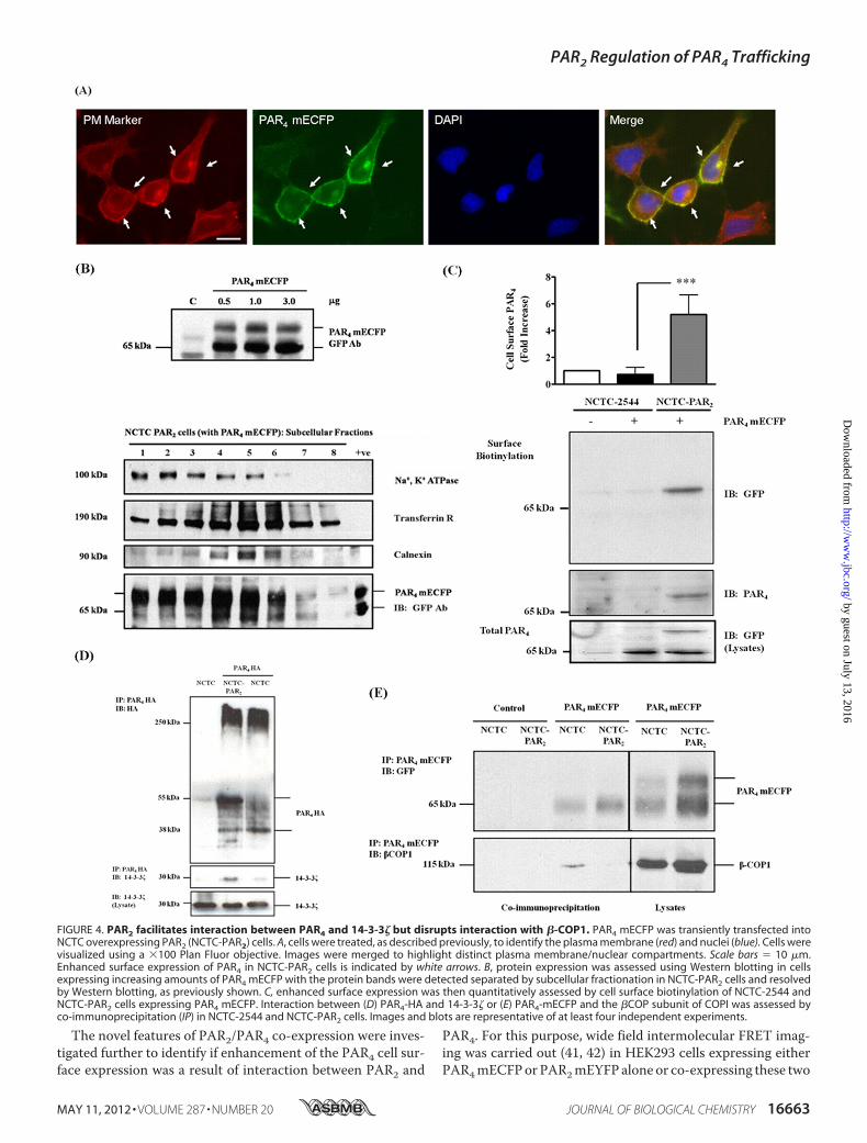
observed for PAR4mECFPwhen transfected intoNCTC-PAR2.
FIGURE 1. Retention of PAR4 in the ER. NCTC-2544 cells expressing pcDNA3.1 empty vector, PAR1 mEYFP, PAR2 mEYFP, or PAR4 mECFP (green) were treated withPKH26 red fluorescent cell linker dye to stain the plasma membrane (PM, red). Cells were fixed and treated with either (A) 4�,6-diamidino-2-phenylindole (DAPI) toidentify nuclei (blue) or (B) ER marker (ER-Tracker Blue-White DPX Dyes, Molecular Probes) to identify the ER (blue). Cells were visualized using a �100 Plan Fluorobjective. Images were merged to highlight distinct plasma membrane, nucleus, or ER compartments. Scale bars � 10 �m. White arrows point to the plasmamembrane, whereas red arrows are indicative of the intracellular/ER compartmentalization of PAR4. Image set representative of three separate experiments.
PAR2 Regulation of PAR4 Trafficking
16660 JOURNAL OF BIOLOGICAL CHEMISTRY VOLUME 287 • NUMBER 20 • MAY 11, 2012
by guest on July 13, 2016http://w
ww
.jbc.org/D
ownloaded from
In addition, when PAR4 mECFP was resolved byWestern blot-ting two clear protein forms were detected when expressed inNCTC-PAR2 cells (Fig. 4B). These results were similar to theobservations made in previous experiments resolving the ERretention motif mutant PAR4 protein. Subsequent subcellularfractionation of NCTC-PAR2 cells expressing PAR4 mECFPhighlighted the distinct differences in the compartmentaliza-tion of PAR4. As Fig. 4B shows, the more rapidly migrating
65-kDa species was confined to ER and endosomal compart-ments (lanes 4–6), whereas the distribution of the less rapidlymigrating form strongly correlated with ER, endosomal, andplasma membrane fractions (lanes 1–6). Enhanced surfaceexpression of PAR4 was subsequently quantified using cell sur-face biotinylation, as shown in Fig. 4C. Following biotinylationof surface proteins, expression of PAR4 was probed using bothanti-GFP and anti-PAR4 specific antibodies inNCTC-2544 and
FIGURE 2. Mutation of the arginine-based ER retention motif (R183AR) enhances the cell surface expression of PAR4. A, NCTC-2544 cells expressing PAR4mECFP or PAR4 RAR mECFP mutant (green) were treated to stain for the plasma membrane (PM, red) and nucleus (blue) as previously described. Cells werevisualized using a �100 Plan Fluor objective. Scale bars � 10 �m. Intracellular retention of PAR4 mECFP is highlighted (red arrows), whereas notable membranelocalization of the PAR4 RAR mECFP is evident (white arrows). B, protein expression was assessed using Western blotting of whole cell lysates expressingincreasing amounts of PAR4 mECFP or PAR4 RAR mECFP constructs as indicated. C, changes in the surface expression of PAR4 were confirmed by subcellularfractionation using differential ultracentrifugation on an iodixanol gradient in cells expressing PAR4 mECFP or PAR4 RAR mECFP. Fractions were precipitatedand resolved by SDS-PAGE followed by Western blotting. PAR4 mECFP (predicted band size �65 kDa) was detected using a polyclonal GFP antibody capableof recognizing the ECFP at the C-terminal of PAR4. Na�,K�-ATPase (�100 kDa), transferrin receptor (�190 kDa), and calnexin (90 �kDa) antibodies were usedfor the detection of membrane, endosomal, and ER compartments, respectively. D, interaction between PAR4 and COPI complex proteins was confirmed byco-immunoprecipitation. Lysates from NCTC-2544 cells expressing PAR4 mECFP were subjected to immunoprecipitation (IP) and then probed for �-COP1interaction. Images and blots are representative of three separate experiments.
PAR2 Regulation of PAR4 Trafficking
MAY 11, 2012 • VOLUME 287 • NUMBER 20 JOURNAL OF BIOLOGICAL CHEMISTRY 16661
by guest on July 13, 2016http://w
ww
.jbc.org/D
ownloaded from
NCTC-PAR2 cells transfected with PAR4 mECFP. Althoughdetection of surface PAR4 was negligible in transfected NCTC-2544 cells (0.725 � 0.30-fold increase over mock transfectedcells, n� 4), a significant increase in surface PAR4was detectedin NCTC-PAR2-transfected cells (5.199 � 0.85-fold increaseover mock cells, n � 4). In addition to enhanced cell surfaceexpression of PAR4 in the presence of PAR2, a notable increasein the ability of PAR4 to interact with the � isoform of the ERexport chaperone 14-3-3 was detected in co-immunoprecipita-tion experiments (Fig. 4D). When PAR4-HA was expressed inthe parental NCTC-2544 cells, the ability of 14-3-3� to interactwith PAR4 was negligible. However, when PAR4-HA wasexpressed inNCTC-PAR2 cells, the ability of 14-3-3� to interactwith PAR4 was clearly shown. Differential interaction of PAR4
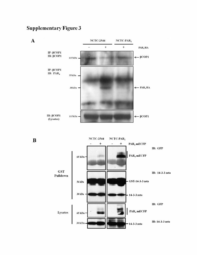
with 14-3-3� was also demonstrated using GST pulldownassays employing GST-14-3-3� fusion proteins (supplementalFig. S3). PAR4 binding to GST-14-3-3� was enhanced whenexpressed in NCTC-PAR2 cells. In addition, interaction betweenPAR4 and �-COP1 was no longer observed during co-expressionof PAR2 and PAR4 (Fig. 4E and supplemental Fig. S3).The localization of PAR4 was further explored in HEK293
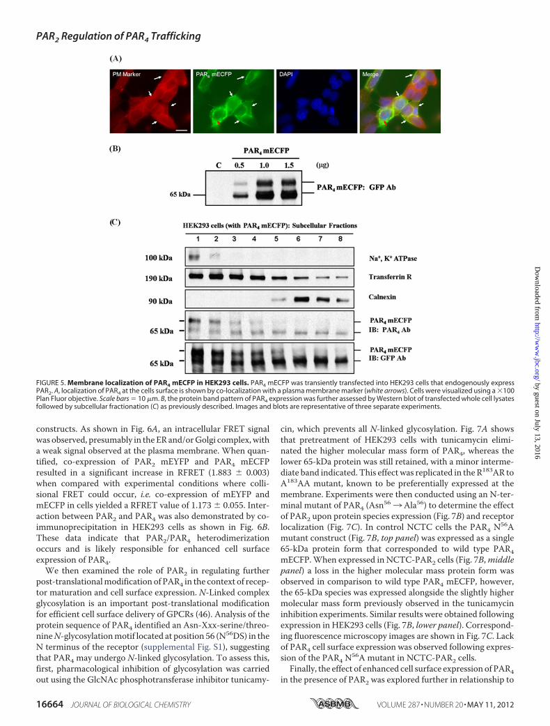
cells, which have an endogenous level of PAR2 (Fig. 5). Whenexpressed, PAR4 mECFP was observed both at the plasmamembrane and in intracellular compartments (Fig. 5A), whichwhen resolved byWestern blot (Fig. 5B) identified similar pro-tein species as in NCTC-2544 cells, representative receptorpopulations were expressed at the cell surface and in ER/endo-somal compartments (Fig. 5C).
FIGURE 3. PAR2 enhances surface expression of PAR4 when co-expressed in NCTC-2544 cells. NCTC-2544 cells expressing PAR4 mECFP (cyan), PAR2 mEYFP(yellow), or co-expressing both constructs were fixed for confocal microscopy. Scale bars � 25 �m. A, membrane (white arrows) receptor expression ishighlighted, whereas intracellular receptor pools are indicated using red arrows. Images shown are representative of at least five independent experiments. B,enhancement of PAR4 surface expression in the presence of PAR2 was subsequently quantified using a surface ELISA approach with an N-terminal anti-PAR4antibody to detect changes in surface levels of PAR4. Data are presented as the optical density at 405 nm obtained from NCTC-2544 cells only (Control),NCTC-2544 or NCTC-PAR4 cells expressing PAR4 mECFP performed in at least three independent experiments. ***, p � 0.001 one-way analysis of variance withDunnett’s post-test.
PAR2 Regulation of PAR4 Trafficking
16662 JOURNAL OF BIOLOGICAL CHEMISTRY VOLUME 287 • NUMBER 20 • MAY 11, 2012
by guest on July 13, 2016http://w
ww
.jbc.org/D
ownloaded from
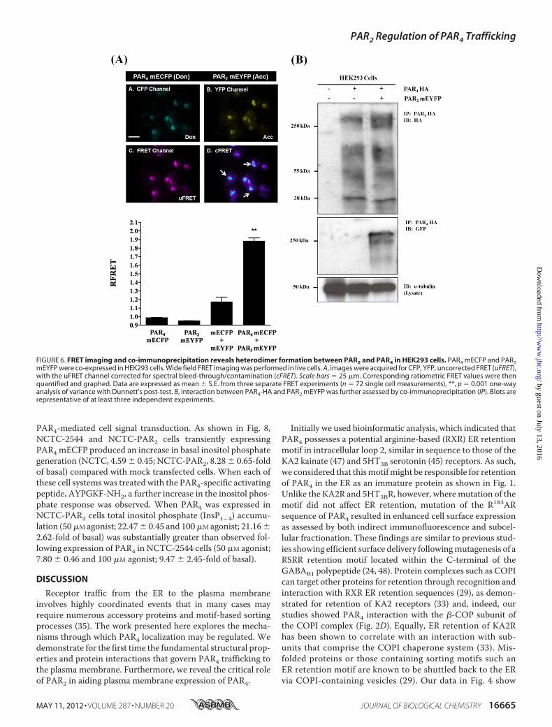
The novel features of PAR2/PAR4 co-expression were inves-tigated further to identify if enhancement of the PAR4 cell sur-face expression was a result of interaction between PAR2 and
PAR4. For this purpose, wide field intermolecular FRET imag-ing was carried out (41, 42) in HEK293 cells expressing eitherPAR4mECFPor PAR2mEYFP alone or co-expressing these two
FIGURE 4. PAR2 facilitates interaction between PAR4 and 14-3-3� but disrupts interaction with �-COP1. PAR4 mECFP was transiently transfected intoNCTC overexpressing PAR2 (NCTC-PAR2) cells. A, cells were treated, as described previously, to identify the plasma membrane (red) and nuclei (blue). Cells werevisualized using a �100 Plan Fluor objective. Images were merged to highlight distinct plasma membrane/nuclear compartments. Scale bars � 10 �m.Enhanced surface expression of PAR4 in NCTC-PAR2 cells is indicated by white arrows. B, protein expression was assessed using Western blotting in cellsexpressing increasing amounts of PAR4 mECFP with the protein bands were detected separated by subcellular fractionation in NCTC-PAR2 cells and resolvedby Western blotting, as previously shown. C, enhanced surface expression was then quantitatively assessed by cell surface biotinylation of NCTC-2544 andNCTC-PAR2 cells expressing PAR4 mECFP. Interaction between (D) PAR4-HA and 14-3-3� or (E) PAR4-mECFP and the �COP subunit of COPI was assessed byco-immunoprecipitation (IP) in NCTC-2544 and NCTC-PAR2 cells. Images and blots are representative of at least four independent experiments.
PAR2 Regulation of PAR4 Trafficking
MAY 11, 2012 • VOLUME 287 • NUMBER 20 JOURNAL OF BIOLOGICAL CHEMISTRY 16663
by guest on July 13, 2016http://w
ww
.jbc.org/D
ownloaded from
constructs. As shown in Fig. 6A, an intracellular FRET signalwas observed, presumably in the ER and/orGolgi complex,witha weak signal observed at the plasma membrane. When quan-tified, co-expression of PAR2 mEYFP and PAR4 mECFPresulted in a significant increase in RFRET (1.883 � 0.003)when compared with experimental conditions where colli-sional FRET could occur, i.e. co-expression of mEYFP andmECFP in cells yielded a RFRET value of 1.173 � 0.055. Inter-action between PAR2 and PAR4 was also demonstrated by co-immunoprecipitation in HEK293 cells as shown in Fig. 6B.These data indicate that PAR2/PAR4 heterodimerizationoccurs and is likely responsible for enhanced cell surfaceexpression of PAR4.We then examined the role of PAR2 in regulating further
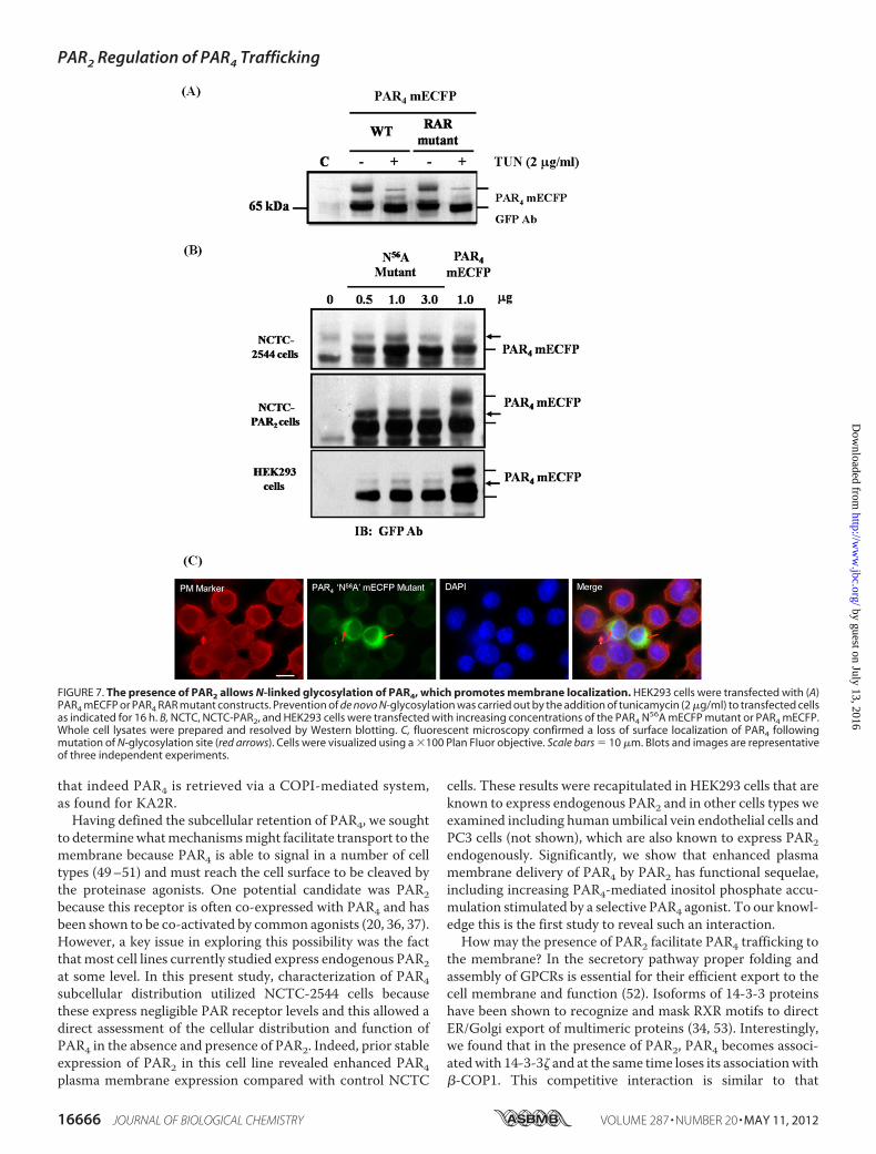
post-translationalmodification of PAR4 in the context of recep-tor maturation and cell surface expression. N-Linked complexglycosylation is an important post-translational modificationfor efficient cell surface delivery of GPCRs (46). Analysis of theprotein sequence of PAR4 identified an Asn-Xxx-serine/threo-nineN-glycosylationmotif located at position 56 (N56DS) in theN terminus of the receptor (supplemental Fig. S1), suggestingthat PAR4 may undergo N-linked glycosylation. To assess this,first, pharmacological inhibition of glycosylation was carriedout using the GlcNAc phosphotransferase inhibitor tunicamy-
cin, which prevents all N-linked glycosylation. Fig. 7A showsthat pretreatment of HEK293 cells with tunicamycin elimi-nated the higher molecular mass form of PAR4, whereas thelower 65-kDa protein was still retained, with a minor interme-diate band indicated. This effect was replicated in the R183AR toA183AA mutant, known to be preferentially expressed at themembrane. Experiments were then conducted using an N-ter-minal mutant of PAR4 (Asn563 Ala56) to determine the effectof PAR2 upon protein species expression (Fig. 7B) and receptorlocalization (Fig. 7C). In control NCTC cells the PAR4 N56Amutant construct (Fig. 7B, top panel) was expressed as a single65-kDa protein form that corresponded to wild type PAR4mECFP.When expressed in NCTC-PAR2 cells (Fig. 7B,middlepanel) a loss in the higher molecular mass protein form wasobserved in comparison to wild type PAR4 mECFP, however,the 65-kDa species was expressed alongside the slightly highermolecular mass form previously observed in the tunicamycininhibition experiments. Similar results were obtained followingexpression in HEK293 cells (Fig. 7B, lower panel). Correspond-ing fluorescence microscopy images are shown in Fig. 7C. Lackof PAR4 cell surface expression was observed following expres-sion of the PAR4 N56A mutant in NCTC-PAR2 cells.
Finally, the effect of enhanced cell surface expression of PAR4in the presence of PAR2 was explored further in relationship to
FIGURE 5. Membrane localization of PAR4 mECFP in HEK293 cells. PAR4 mECFP was transiently transfected into HEK293 cells that endogenously expressPAR2. A, localization of PAR4 at the cells surface is shown by co-localization with a plasma membrane marker (white arrows). Cells were visualized using a �100Plan Fluor objective. Scale bars � 10 �m. B, the protein band pattern of PAR4 expression was further assessed by Western blot of transfected whole cell lysatesfollowed by subcellular fractionation (C) as previously described. Images and blots are representative of three separate experiments.
PAR2 Regulation of PAR4 Trafficking
16664 JOURNAL OF BIOLOGICAL CHEMISTRY VOLUME 287 • NUMBER 20 • MAY 11, 2012
by guest on July 13, 2016http://w
ww
.jbc.org/D
ownloaded from
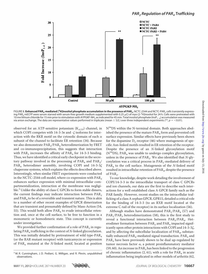
PAR4-mediated cell signal transduction. As shown in Fig. 8,NCTC-2544 and NCTC-PAR2 cells transiently expressingPAR4 mECFP produced an increase in basal inositol phosphategeneration (NCTC, 4.59� 0.45; NCTC-PAR2, 8.28� 0.65-foldof basal) compared with mock transfected cells. When each ofthese cell systems was treated with the PAR4-specific activatingpeptide, AYPGKF-NH2, a further increase in the inositol phos-phate response was observed. When PAR4 was expressed inNCTC-PAR2 cells total inositol phosphate (InsP1–4) accumu-lation (50�M agonist; 22.47� 0.45 and 100�M agonist; 21.16�2.62-fold of basal) was substantially greater than observed fol-lowing expression of PAR4 in NCTC-2544 cells (50 �M agonist;7.80 � 0.46 and 100 �M agonist; 9.47 � 2.45-fold of basal).
DISCUSSION
Receptor traffic from the ER to the plasma membraneinvolves highly coordinated events that in many cases mayrequire numerous accessory proteins and motif-based sortingprocesses (35). The work presented here explores the mecha-nisms through which PAR4 localization may be regulated. Wedemonstrate for the first time the fundamental structural prop-erties and protein interactions that govern PAR4 trafficking tothe plasma membrane. Furthermore, we reveal the critical roleof PAR2 in aiding plasma membrane expression of PAR4.
Initially we used bioinformatic analysis, which indicated thatPAR4 possesses a potential arginine-based (RXR) ER retentionmotif in intracellular loop 2, similar in sequence to those of theKA2 kainate (47) and 5HT3B serotonin (45) receptors. As such,we considered that thismotifmight be responsible for retentionof PAR4 in the ER as an immature protein as shown in Fig. 1.Unlike the KA2R and 5HT3BR, however, wheremutation of themotif did not affect ER retention, mutation of the R183ARsequence of PAR4 resulted in enhanced cell surface expressionas assessed by both indirect immunofluorescence and subcel-lular fractionation. These findings are similar to previous stud-ies showing efficient surface delivery followingmutagenesis of aRSRR retention motif located within the C-terminal of theGABAB1 polypeptide (24, 48). Protein complexes such as COPIcan target other proteins for retention through recognition andinteraction with RXR ER retention sequences (29), as demon-strated for retention of KA2 receptors (33) and, indeed, ourstudies showed PAR4 interaction with the �-COP subunit ofthe COPI complex (Fig. 2D). Equally, ER retention of KA2Rhas been shown to correlate with an interaction with sub-units that comprise the COPI chaperone system (33). Mis-folded proteins or those containing sorting motifs such anER retention motif are known to be shuttled back to the ERvia COPI-containing vesicles (29). Our data in Fig. 4 show
FIGURE 6. FRET imaging and co-immunoprecipitation reveals heterodimer formation between PAR2 and PAR4 in HEK293 cells. PAR4 mECFP and PAR2mEYFP were co-expressed in HEK293 cells. Wide field FRET imaging was performed in live cells. A, images were acquired for CFP, YFP, uncorrected FRET (uFRET),with the uFRET channel corrected for spectral bleed-through/contamination (cFRET). Scale bars � 25 �m. Corresponding ratiometric FRET values were thenquantified and graphed. Data are expressed as mean � S.E. from three separate FRET experiments (n � 72 single cell measurements), **, p � 0.001 one-wayanalysis of variance with Dunnett’s post-test. B, interaction between PAR4-HA and PAR2 mEYFP was further assessed by co-immunoprecipitation (IP). Blots arerepresentative of at least three independent experiments.
PAR2 Regulation of PAR4 Trafficking
MAY 11, 2012 • VOLUME 287 • NUMBER 20 JOURNAL OF BIOLOGICAL CHEMISTRY 16665
by guest on July 13, 2016http://w
ww
.jbc.org/D
ownloaded from
that indeed PAR4 is retrieved via a COPI-mediated system,as found for KA2R.Having defined the subcellular retention of PAR4, we sought
to determinewhatmechanismsmight facilitate transport to themembrane because PAR4 is able to signal in a number of celltypes (49–51) and must reach the cell surface to be cleaved bythe proteinase agonists. One potential candidate was PAR2because this receptor is often co-expressed with PAR4 and hasbeen shown to be co-activated by common agonists (20, 36, 37).However, a key issue in exploring this possibility was the factthatmost cell lines currently studied express endogenous PAR2at some level. In this present study, characterization of PAR4subcellular distribution utilized NCTC-2544 cells becausethese express negligible PAR receptor levels and this allowed adirect assessment of the cellular distribution and function ofPAR4 in the absence and presence of PAR2. Indeed, prior stableexpression of PAR2 in this cell line revealed enhanced PAR4plasma membrane expression compared with control NCTC
cells. These results were recapitulated in HEK293 cells that areknown to express endogenous PAR2 and in other cells types weexamined including human umbilical vein endothelial cells andPC3 cells (not shown), which are also known to express PAR2endogenously. Significantly, we show that enhanced plasmamembrane delivery of PAR4 by PAR2 has functional sequelae,including increasing PAR4-mediated inositol phosphate accu-mulation stimulated by a selective PAR4 agonist. To our knowl-edge this is the first study to reveal such an interaction.Howmay the presence of PAR2 facilitate PAR4 trafficking to
the membrane? In the secretory pathway proper folding andassembly of GPCRs is essential for their efficient export to thecell membrane and function (52). Isoforms of 14-3-3 proteinshave been shown to recognize and mask RXR motifs to directER/Golgi export of multimeric proteins (34, 53). Interestingly,we found that in the presence of PAR2, PAR4 becomes associ-atedwith 14-3-3� and at the same time loses its associationwith�-COP1. This competitive interaction is similar to that
FIGURE 7. The presence of PAR2 allows N-linked glycosylation of PAR4, which promotes membrane localization. HEK293 cells were transfected with (A)PAR4 mECFP or PAR4 RAR mutant constructs. Prevention of de novo N-glycosylation was carried out by the addition of tunicamycin (2 �g/ml) to transfected cellsas indicated for 16 h. B, NCTC, NCTC-PAR2, and HEK293 cells were transfected with increasing concentrations of the PAR4 N56A mECFP mutant or PAR4 mECFP.Whole cell lysates were prepared and resolved by Western blotting. C, fluorescent microscopy confirmed a loss of surface localization of PAR4 followingmutation of N-glycosylation site (red arrows). Cells were visualized using a �100 Plan Fluor objective. Scale bars � 10 �m. Blots and images are representativeof three independent experiments.
PAR2 Regulation of PAR4 Trafficking
16666 JOURNAL OF BIOLOGICAL CHEMISTRY VOLUME 287 • NUMBER 20 • MAY 11, 2012
by guest on July 13, 2016http://w
ww
.jbc.org/D
ownloaded from
observed for an ATP-sensitive potassium (KATP) channel, inwhich COPI competes with 14-3-3� and -� isoforms for inter-action with the RKR motif on the cytosolic domain of each �subunit of the channel to facilitate ER retention (34). Becausewe also demonstrate PAR2/PAR4 heterodimerization by FRETand co-immunoprecipitation, this suggests that interactionwith PAR2 increases the affinity of PAR4 for 14-3-3 binding.Thus, we have identified a critical early checkpoint in the secre-tory pathway involved in the processing of PAR4 and PAR2/PAR4 heterodimer assembly, involving COPI and 14-3-3�chaperone systems, which explains the effects described above.Interestingly, when similar FRET experiments were conductedin the NCTC-2544 cell model, where co-expression with PAR2enhances surface expression with minimal intracellular com-partmentalization, interaction at the membrane was negligi-ble.3 Unlike the ability of class C GPCRs to form stable dimers,the current findings may indicate interaction between PAR2and PAR4 to be of a reversible and transient nature. This is akinto a number of other recent examples of GPCR dimerizationthat are transient and presumably defined by Mass-Action (54,55). This would both allow PAR4 to evade intracellular reten-tion and, once at the cell surface, to be free to function in amonomeric or homodimeric state. This concept is currentlyunder investigation.We provided further confirmation of a role of PAR2 in regu-
lating PAR4 trafficking in the context ofN-linked glycosylation.This was initially detailed by pretreatment of wild type-PAR4(or the RAR mutant receptor) with tunicamycin or expressionof PAR4 mutated at the N-linked motif, located at position
N56DS within the N-terminal domain. Both approaches abol-ished the presence of the mature PAR4 form and prevented cellsurface expression. Similar effects have previously been shownfor the dopamine D5 receptor (46) where mutagenesis of spe-cific Asn-linkedmotifs resulted in ER retention of the receptor.Despite the presence of an N-linked glycosylation motif(N56DS), PAR4 was unable to undergo complex glycosylation,unless in the presence of PAR2. We also identified that N-gly-cosylation was a critical process in PAR2-mediated delivery ofPAR4 to the cell surface. Mutagenesis of the N-linked motifresulted in intracellular retention of PAR4, despite the presenceof PAR2.To our knowledge, despite work detailing the involvement of
COPI/14-3-3 in the intracellular transport of class C GPCRsand ion channels, our data are the first to describe such inter-actions for a well established class A GPCR family such as thePAR family. However, recent studies on the intracellular traf-ficking of a class A orphanGPCR,GPR15, detailed a critical rolefor the binding of 14-3-3 (to an RXR motif located at theextreme C-tail of the receptor) in its surface localization (56).Although studies have demonstrated PAR1/PAR4 (57) and
PAR1/PAR3 heterodimerization (58), this is the first study toreveal a functional interaction between PAR2/PAR4. Het-erodimer formation between PAR2 and PAR4 impacted signif-icantly upon other protein interactions with COPI and 14-3-3�,and by affecting the subcellular localization of PAR4, substan-tially enhanced PAR4-mediated signal transduction. PAR2 andPAR4 have been previously shown to be dual up-regulated bytumor necrosis factor �, a potent proinflammatory mediator(59, 60). An increase in PAR2 has been linked to the progressionof chronic inflammation (2, 61), with a role for PAR4 in acuteinflammation being implicated in other models of arthritis (62,
3 M. R. Cunningham, J. D. Pediani, G. Milligan, and R. Plevin, unpublishedobservations.
FIGURE 8. Enhanced PAR4-mediated [3H]inositol phosphate accumulation in the presence of PAR2. NCTC-2544 and NCTC-PAR2 cells transiently express-ing PAR4 mECFP were serum starved with serum-free growth medium supplemented with 0.25 �Ci of myo-[2-3H]inositol for 24 h. Cells were pretreated with10 mM lithium chloride for 15 min prior to stimulation with AYPGKF-NH2 as indicated for 45 min. Total inositol phosphate (InsP1– 4) accumulation was measuredvia anion exchange. The data are representative values performed in triplicate (mean � S.E.) over three independent experiments (**, p � �0.01).
PAR2 Regulation of PAR4 Trafficking
MAY 11, 2012 • VOLUME 287 • NUMBER 20 JOURNAL OF BIOLOGICAL CHEMISTRY 16667
by guest on July 13, 2016http://w
ww
.jbc.org/D
ownloaded from
63). Co-expression of these receptors in these pathophysiolog-ical environments, where tumor necrosis factor � is abundant,may be pivotal to the progression of a PAR2/PAR4-mediatedproinflammatory response. In this study we identified a novelheterodimer partnership formed between PAR2/PAR4, whichallows PAR4 to bypass COPI-dependent retrograde transportand exit the ER to undergo post-translational modification tobe delivered to the plasmamembrane as amature glycoprotein.These findings may be important in the understanding of theroles of each receptor in the context of inflammation.
REFERENCES1. Russell, F. A., and McDougall, J. J. (2009) Proteinase-activated receptor
(PAR) involvement in mediating arthritis pain and inflammation. In-flamm. Res. 58, 119–126
2. McIntosh, K. A., Plevin, R., Ferrell, W. R., and Lockhart, J. C. (2007) Thetherapeutic potential of proteinase-activated receptors in arthritis. Curr.Opin. Pharmacol. 7, 334–338
3. Macfarlane, S. R., Seatter, M. J., Kanke, T., Hunter, G. D., and Plevin, R.(2001) Proteinase-activated receptors. Pharmacol. Rev. 53, 245–282
4. Vu, T. K., Hung, D. T., Wheaton, V. I., and Coughlin, S. R. (1991) Molec-ular cloning of a functional thrombin receptor reveals a novel proteolyticmechanism of receptor activation. Cell 64, 1057–1068
5. Vu, T. K.,Wheaton, V. I., Hung, D. T., Charo, I., and Coughlin, S. R. (1991)Domains specifying thrombin-receptor interaction. Nature 353,674–677
6. Bouton,M.C., Jandrot-Perrus,M.,Moog, S., Cazenave, J. P., Guillin,M.C.,and Lanza, F. (1995) Thrombin interaction with a recombinant N-termi-nal extracellular domain of the thrombin receptor in an acellular system.Biochem. J. 305, 635–641
7. Ishii, K., Gerszten, R., Zheng, Y.W.,Welsh, J. B., Turck, C.W., andCough-lin, S. R. (1995) Determinants of thrombin receptor cleavage. Receptordomains involved, specificity, and role of the P3 aspartate. J. Biol. Chem.270, 16435–16440
8. Hoxie, J. A., Ahuja,M., Belmonte, E., Pizarro, S., Parton, R., and Brass, L. F.(1993) Internalization and recycling of activated thrombin receptors.J. Biol. Chem. 268, 13756–13763
9. Shapiro, M. J., Trejo, J., Zeng, D., and Coughlin, S. R. (1996) Role of thethrombin receptor’s cytoplasmic tail in intracellular trafficking. Distinctdeterminants for agonist-triggered versus tonic internalization and intra-cellular localization. J. Biol. Chem. 271, 32874–32880
10. Tiruppathi, C., Yan, W., Sandoval, R., Naqvi, T., Pronin, A. N., Benovic,J. L., andMalik, A. B. (2000)Gprotein-coupled receptor kinase-5 regulatesthrombin-activated signaling in endothelial cells. Proc. Natl. Acad. Sci.U.S.A. 97, 7440–7445
11. Paing, M. M., Stutts, A. B., Kohout, T. A., Lefkowitz, R. J., and Trejo, J.(2002) �-Arrestins regulate protease-activated receptor-1 desensitizationbut not internalization orDown-regulation. J. Biol. Chem. 277, 1292–1300
12. Paing, M.M., Johnston, C. A., Siderovski, D. P., and Trejo, J. (2006) Clath-rin adaptor AP2 regulates thrombin receptor constitutive internalizationand endothelial cell resensitization.Mol. Cell. Biol. 26, 3231–3242
13. Wolfe, B. L., and Trejo, J. (2007) Clathrin-dependent mechanisms of Gprotein-coupled receptor endocytosis. Traffic 8, 462–470
14. Gandhi, P. S., Chen, Z., Appelbaum, E., Zapata, F., and Di Cera, E. (2011)Structural basis of thrombin-protease-activated receptor interactions.IUBMB Life 63, 375–382
15. Böhm, S. K., Khitin, L. M., Grady, E. F., Aponte, G., Payan, D. G., andBunnett, N. W. (1996) Mechanisms of desensitization and resensitizationof proteinase-activated receptor-2. J. Biol. Chem. 271, 22003–22016
16. Roosterman, D., Schmidlin, F., and Bunnett, N. (2003) Rab5a and rab11amediate agonist-induced trafficking of protease-activated receptor 2.Am. J. Physiol. Cell Physiol 284, C1319–1329
17. Stalheim, L., Ding, Y., Gullapalli, A., Paing, M. M., Wolfe, B. L., Morris,D. R., and Trejo, J. (2005) Multiple independent functions of arrestins inthe regulation of protease-activated receptor-2 signaling and trafficking.Mol. Pharmacol. 67, 78–87
18. Luo, W., Wang, Y., and Reiser, G. (2007) p24A, a type I transmembraneprotein, controls ARF1-dependent resensitization of protease-activatedreceptor-2 by influence on receptor trafficking. J. Biol. Chem. 282,30246–30255
19. Luo, W., Wang, Y., and Reiser, G. (2011) Proteinase-activated receptors,nucleotide P2Y receptors, and�-opioid receptor-1B are under the controlof the type I transmembrane proteins p23 and p24A in post-Golgi traffick-ing. J. Neurochem. 117, 71–81
20. Xu,W. F., Andersen, H.,Whitmore, T. E., Presnell, S. R., Yee, D. P., Ching,A., Gilbert, T., Davie, E. W., and Foster, D. C. (1998) Cloning and charac-terization of human protease-activated receptor 4. Proc. Natl. Acad. Sci.U.S.A. 95, 6642–6646
21. Kahn, M. L., Zheng, Y. W., Huang, W., Bigornia, V., Zeng, D., Moff, S.,Farese, R. V., Jr., Tam, C., and Coughlin, S. R. (1998) A dual thrombinreceptor system for platelet activation. Nature 394, 690–694
22. Shapiro, M. J., Weiss, E. J., Faruqi, T. R., and Coughlin, S. R. (2000) Pro-tease-activated receptors 1 and 4 are shut off with distinct kinetics afteractivation by thrombin. J. Biol. Chem. 275, 25216–25221
23. Sabri, A., Guo, J., Elouardighi, H., Darrow, A. L., Andrade-Gordon, P., andSteinberg, S. F. (2003) Mechanisms of protease-activated receptor-4 ac-tions in cardiomyocytes. Role of Src tyrosine kinase. J. Biol. Chem. 278,11714–11720
24. Gassmann, M., Haller, C., Stoll, Y., Abdel Aziz, S., Biermann, B.,Mosbacher, J., Kaupmann, K., and Bettler, B. (2005) The RXR-type endo-plasmic reticulum-retention/retrieval signal of GABAB1 requires distantspacing from the membrane to function.Mol. Pharmacol. 68, 137–144
25. Michelsen, K., Yuan, H., and Schwappach, B. (2005) Hide and run. Argi-nine-based endoplasmic reticulum-sorting motifs in the assembly of het-eromultimeric membrane proteins. EMBO Rep. 6, 717–722
26. Barlowe, C. (2000) Traffic COPs of the early secretory pathway. Traffic 1,371–377
27. Barlowe, C., d’Enfert, C., and Schekman, R. (1993) Purification and char-acterization of SAR1p, a smallGTP-binding protein required for transportvesicle formation from the endoplasmic reticulum. J. Biol. Chem. 268,873–879
28. Barlowe,C.,Orci, L., Yeung, T.,Hosobuchi,M.,Hamamoto, S., Salama,N.,Rexach, M. F., Ravazzola, M., Amherdt, M., and Schekman, R. (1994)COPII, a membrane coat formed by Sec proteins that drive vesicle bud-ding from the endoplasmic reticulum. Cell 77, 895–907
29. Aridor, M., Bannykh, S. I., Rowe, T., and Balch, W. E. (1995) Sequentialcoupling between COPII and COPI vesicle coats in endoplasmic reticu-lum to Golgi transport. J. Cell Biol. 131, 875–893
30. Zerangue, N., Schwappach, B., Jan, Y. N., and Jan, L. Y. (1999) A new ERtrafficking signal regulates the subunit stoichiometry of plasma mem-brane K(ATP) channels. Neuron 22, 537–548
31. Margeta-Mitrovic, M., Jan, Y. N., and Jan, L. (2000) A trafficking check-point controlsGABA(B) receptor heterodimerization.Neuron 27, 97–106
32. Ren, Z., Riley, N. J., Garcia, E. P., Sanders, J. M., Swanson, G. T., andMarshall, J. (2003) Multiple trafficking signals regulate kainate receptorKA2 subunit surface expression. J. Neurosci. 23, 6608–6616
33. Vivithanaporn, P., Yan, S., and Swanson, G. T. (2006) Intracellular traf-ficking of KA2 kainate receptors mediated by interactions with coatomerprotein complex I (COPI) and 14-3-3 chaperone systems. J. Biol. Chem.281, 15475–15484
34. Yuan, H., Michelsen, K., and Schwappach, B. (2003) 14-3-3 dimers probethe assembly status of multimeric membrane proteins. Curr. Biol. 13,638–646
35. Duvernay, M. T., Filipeanu, C. M., and Wu, G. (2005) The regulatorymechanisms of export trafficking of G protein-coupled receptors. CellSignal. 17, 1457–1465
36. Sambrano, G. R., Huang,W., Faruqi, T., Mahrus, S., Craik, C., and Cough-lin, S. R. (2000) Cathepsin G activates protease-activated receptor-4 inhuman platelets. J. Biol. Chem. 275, 6819–6823
37. Cottrell, G. S., Amadesi, S., Grady, E. F., andBunnett,N.W. (2004)TrypsinIV, a novel agonist of protease-activated receptors 2 and 4. J. Biol. Chem.279, 13532–13539
38. Zacharias, D. A., Violin, J. D., Newton, A. C., and Tsien, R. Y. (2002)Partitioning of lipid-modifiedmonomeric GFPs intomembranemicrodo-
PAR2 Regulation of PAR4 Trafficking
16668 JOURNAL OF BIOLOGICAL CHEMISTRY VOLUME 287 • NUMBER 20 • MAY 11, 2012
by guest on July 13, 2016http://w
ww
.jbc.org/D
ownloaded from
mains of live cells. Science 296, 913–91639. Plevin, R., Kellock, N. A., Wakelam, M. J., andWadsworth, R. (1994) Reg-
ulation by hypoxia of endothelin-1-stimulated phospholipase D activity insheep pulmonary artery cultured smooth muscle cells. Br. J. Pharmacol.112, 311–315
40. Proctor, K. M., Miller, S. C., Bryant, N. J., and Gould, G. W. (2006) Syn-taxin 16 controls the intracellular sequestration of GLUT4 in 3T3-L1 adi-pocytes. Biochem. Biophys. Res. Commun. 347, 433–438
41. Wilson, S., Wilkinson, G., and Milligan, G. (2005) The CXCR1 andCXCR2 receptors form constitutive homo- and heterodimers selectivelyand with equal apparent affinities. J. Biol. Chem. 280, 28663–28674
42. Ellis, J., Pediani, J. D., Canals, M., Milasta, S., and Milligan, G. (2006)Orexin-1 receptor-cannabinoid CB1 receptor heterodimerization resultsin both ligand-dependent and -independent coordinated alterations ofreceptor localization and function. J. Biol. Chem. 281, 38812–38824
43. Lopez-Gimenez, J. F., Canals, M., Pediani, J. D., and Milligan, G. (2007)The �1b-adrenoceptor exists as a higher-order oligomer. Effective oligo-merization is required for receptormaturation, surface delivery, and func-tion.Mol. Pharmacol. 71, 1015–1029
44. Kanke, T., Macfarlane, S. R., Seatter, M. J., Davenport, E., Paul, A., McK-enzie, R. C., and Plevin, R. (2001) Proteinase-activated receptor-2-medi-ated activation of stress-activated protein kinases and inhibitory B ki-nases in NCTC 2544 keratinocytes. J. Biol. Chem. 276, 31657–31666
45. Boyd, G. W., Doward, A. I., Kirkness, E. F., Millar, N. S., and Connolly,C. N. (2003) Cell surface expression of 5-hydroxytryptamine type 3 recep-tors is controlled by an endoplasmic reticulum retention signal. J. Biol.Chem. 278, 27681–27687
46. Karpa, K. D., Lidow, M. S., Pickering, M. T., Levenson, R., and Bergson, C.(1999) N-Linked glycosylation is required for plasma membrane localiza-tion of D5, but not D1, dopamine receptors in transfected mammaliancells.Mol. Pharmacol. 56, 1071–1078
47. Nasu-Nishimura, Y., Hurtado, D., Braud, S., Tang, T. T., Isaac, J. T., andRoche, K.W. (2006) Identification of an endoplasmic reticulum-retentionmotif in an intracellular loop of the kainate receptor subunit KA2. J. Neu-rosci. 26, 7014–7021
48. Pagano, A., Rovelli, G., Mosbacher, J., Lohmann, T., Duthey, B., Stauffer,D., Ristig, D., Schuler, V., Meigel, I., Lampert, C., Stein, T., Prezeau, L.,Blahos, J., Pin, J., Froestl,W., Kuhn, R., Heid, J., Kaupmann, K., and Bettler,B. (2001) C-terminal interaction is essential for surface trafficking but notfor heteromeric assembly of GABA(b) receptors. J. Neurosci. 21,1189–1202
49. Wu, C. C., Wu, S. Y., Liao, C. Y., Teng, C. M., Wu, Y. C., and Kuo, S. C.(2010) The roles andmechanisms of PAR4 and P2Y12/phosphatidylinosi-tol 3-kinase pathway in maintaining thrombin-induced platelet aggrega-tion. Br. J. Pharmacol. 161, 643–658
50. Ando, S.,Otani,H., Yagi, Y., Kawai, K., Araki,H., Fukuhara, S., and Inagaki,C. (2007) Proteinase-activated receptor 4 stimulation-induced epithelial-
mesenchymal transition in alveolar epithelial cells. Respir. Res. 8, 3151. Hirano, K., Nomoto, N., Hirano, M., Momota, F., Hanada, A., and Ka-
naide, H. (2007) Distinct Ca2� requirement for NO production betweenproteinase-activated receptor 1 and 4 (PAR1 and PAR4) in vascular endo-thelial cells. J. Pharmacol. Exp. Ther. 322, 668–677
52. Lippincott-Schwartz, J., Roberts, T. H., and Hirschberg, K. (2000) Secre-tory protein trafficking and organelle dynamics in living cells. Annu. Rev.Cell Dev. Biol. 16, 557–589
53. Shikano, S., Coblitz, B., Wu, M., and Li, M. (2006) 14–3-3 proteins. Reg-ulation of endoplasmic reticulum localization and surface expression ofmembrane proteins. Trends Cell Biol. 16, 370–375
54. Hern, J. A., Baig, A. H., Mashanov, G. I., Birdsall, B., Corrie, J. E., Lazareno,S.,Molloy, J. E., and Birdsall, N. J. (2010) Formation and dissociation ofM1muscarinic receptor dimers seen by total internal reflection fluorescenceimaging of single molecules. Proc. Natl. Acad. Sci. U.S.A. 107, 2693–2698
55. Fonseca, J. M., and Lambert, N. A. (2009) Instability of a class a G protein-coupled receptor oligomer interface.Mol. Pharmacol. 75, 1296–1299
56. Okamoto, Y., and Shikano, S. (2011) Phosphorylation-dependent C-ter-minal binding of 14-3-3 proteins promotes cell surface expression of HIVco-receptor GPR15. J. Biol. Chem. 286, 7171–7181
57. Leger, A. J., Jacques, S. L., Badar, J., Kaneider, N. C., Derian, C. K., An-drade-Gordon, P., Covic, L., and Kuliopulos, A. (2006) Blocking the pro-tease-activated receptor 1–4 heterodimer in platelet-mediated thrombo-sis. Circulation 113, 1244–1254
58. McLaughlin, J. N., Patterson, M. M., and Malik, A. B. (2007) Protease-activated receptor-3 (PAR3) regulates PAR1 signaling by receptordimerization. Proc. Natl. Acad. Sci. U.S.A. 104, 5662–5667
59. Hamilton, J. R., Frauman, A. G., andCocks, T.M. (2001) Increased expres-sion of protease-activated receptor-2 (PAR2) and PAR4 in human coro-nary artery by inflammatory stimuli unveils endothelium-dependent re-laxations to PAR2 and PAR4 agonists. Circ. Res. 89, 92–98
60. Ritchie, E., Saka, M., Mackenzie, C., Drummond, R., Wheeler-Jones, C.,Kanke, T., and Plevin, R. (2007) Cytokine up-regulation of proteinase-activated-receptors 2 and 4 expression mediated by p38 MAP kinase andinhibitory B kinase � in human endothelial cells. Br. J. Pharmacol. 150,1044–1054
61. Ferrell, W. R., Lockhart, J. C., Kelso, E. B., Dunning, L., Plevin, R., Meek,S. E., Smith, A. J., Hunter, G. D., McLean, J. S., McGarry, F., Ramage, R.,Jiang, L., Kanke, T., and Kawagoe, J. (2003) Essential role for proteinase-activated receptor-2 in arthritis. J. Clin. Invest. 111, 35–41
62. McDougall, J. J., Zhang, C., Cellars, L., Joubert, E., Dixon, C. M., andVergnolle, N. (2009) Triggering of proteinase-activated receptor 4 leads tojoint pain and inflammation in mice. Arthritis Rheum. 60, 728–737
63. Russell, F. A., Veldhoen, V. E., Tchitchkan, D., andMcDougall, J. J. (2010)Proteinase-activated receptor-4 (PAR4) activation leads to sensitization ofrat joint primary afferents via a bradykinin B2 receptor-dependent mech-anism. J. Neurophysiol. 103, 155–163
PAR2 Regulation of PAR4 Trafficking
MAY 11, 2012 • VOLUME 287 • NUMBER 20 JOURNAL OF BIOLOGICAL CHEMISTRY 16669
by guest on July 13, 2016http://w
ww
.jbc.org/D
ownloaded from
SUPPLEMENTAL FIGURE LEGENDS
FIGURE S1. The presence of a functional arginine-based ER retention motif within intracellular
loop-2 of PAR4. The presence of arginine-based ER retention/retrieval motifs was assessed in the
protein sequences of human PAR1, PAR2, PAR3 and PAR4. A protein sequence alignment was carried out
using CLUSTAL W software (www.ebi.ac.uk/clustalw/). The putative sites for proteolytic cleavage have
been underlined and highlighted in blue. Of the PAR family, only the PAR4 sequence contains RxR
motifs (shown in red). The N-linked glycosylation site on the N-terminal of PAR4 is also highlighted
(pink).
FIGURE S2. Co-expression of PAR1 mEYFP and PAR4 mECFP does not enhance surface
localization of PAR4. PAR4 mECFP and PAR1 mEYFP were co-expressed transiently in NCTC-2544
cells and visualized by confocal microscopy following fixation. Scale bars = 25µm. Images shown are
representative of at least three experiments.
FIGURE S3. Reciprocal blots showing interaction between βCOP and PAR4 by co-
immmunoprecipitation and 14-3-3-zeta and PAR4 by GST pulldown. PAR4-HA was expressed in
both NCTC-2544 and NCTC-PAR2 cells. (A) Interaction with βCOP subunit of COPI was assessed by
co-immunoprecipitation using a specific anti-βCOP antibody for immunoprecipitation of endogenous
βCOP and probing for PAR4 interaction using anti-HA antibody. (B) NCTC-2544 or NCTC-PAR2 cell
lysates expressing PAR4 mECFP were incubated with a purified GST-14-3-3 zeta fusion protein. Using a
GST pulldown assay and Western blotting, interaction between PAR4 and 14-3-3 zeta in NCTC-PAR2
cells was confirmed using specific antibodies as shown. Blots shown are representative of at least three
experiments.
SUPPLEMENTAL FIGURES
and Robin PlevinAlexandra E. Cooke, Mary Nilsson, Gwyn W. Gould, Stuart Mundell, Graeme Milligan
Margaret R. Cunningham, Kathryn A. McIntosh, John D. Pediani, Joris Robben,)4of Proteinase-activated Receptor 4 (PAR
) in Membrane Trafficking2Novel Role for Proteinase-activated Receptor 2 (PAR
doi: 10.1074/jbc.M111.315911 originally published online March 12, 20122012, 287:16656-16669.J. Biol. Chem.
10.1074/jbc.M111.315911Access the most updated version of this article at doi:
Alerts:
When a correction for this article is posted•
When this article is cited•
to choose from all of JBC's e-mail alertsClick here
Supplemental material:
http://www.jbc.org/content/suppl/2012/03/12/M111.315911.DC1.html
http://www.jbc.org/content/287/20/16656.full.html#ref-list-1
This article cites 63 references, 39 of which can be accessed free at
by guest on July 13, 2016http://w
ww
.jbc.org/D
ownloaded from


