Embed Size (px)
Citation preview

© 2014 Ribeiro et al. This work is published by Dove Medical Press Limited, and licensed under Creative Commons Attribution – Non Commercial (unported, v3.0) License. The full terms of the License are available at http://creativecommons.org/licenses/by-nc/3.0/. Non-commercial uses of the work are permitted without any further
permission from Dove Medical Press Limited, provided the work is properly attributed. Permissions beyond the scope of the License are administered by Dove Medical Press Limited. Information on how to request permission may be found at: http://www.dovepress.com/permissions.php
International Journal of Nanomedicine 2014:9 877–890
International Journal of Nanomedicine Dovepress
submit your manuscript | www.dovepress.com
Dovepress 877
O r I g I N a l r e s e a r c h
open access to scientific and medical research
Open access Full Text article
http://dx.doi.org/10.2147/IJN.S55678
Novel targeting using nanoparticles: an approach to the development of an effective anti-leishmanial drug-delivery system
Tatiana g ribeiro1
Miguel a chávez-Fumagalli2
Diogo g Valadares3
Juçara r França1
lívia B rodrigues1
Mariana c Duarte2
Paula s lage2
Pedro h r andrade4
Daniela P lage4
leonardo V arruda5,6
Daniel r abánades5,6
lourena e costa2
Vivian T Martins3
carlos aP Tavares3
rachel O castilho1,7,*eduardo aF coelho2,4,*andré ag Faraco1,7,*1Programa de Pós-graduação em ciências Farmacêuticas, Faculdade de Farmácia, Universidade Federal de Minas gerais, Belo horizonte, Minas gerais, Brazil; 2Programa de Pós-graduação em ciências da saúde: Infectologia e Medicina Tropical, Faculdade de Medicina, Universidade Federal de Minas gerais, Belo horizonte, Minas gerais, Brazil; 3Departamento de Bioquímica e Imunologia, Instituto de ciências Biológicas, Universidade Federal de Minas gerais, Belo horizonte, Minas gerais, Brazil; 4Departamento de Patologia clínica, cOlTec, Universidade Federal de Minas gerais, Belo horizonte, Minas gerais, Brazil; 5Programa de Pós-graduação em Patologia humana, Universidade Federal da Bahia, salvador, Bahia, Brazil; 6centro de Pesquisas gonçalo Moniz (cPqgM), Fundação Oswaldo cruz (FIOcrUZ), salvador, Bahia, Brazil; 7Departamento de Produtos Farmacêuticos, Faculdade de Farmácia, Universidade Federal de Minas gerais, Belo horizonte, Minas gerais, Brazil
*These authors contributed equally to this work
Abstract: The study reported here aimed to develop an optimized nanoparticle delivery sys-
tem for amphotericin B (AmpB) using a polyelectrolyte complexation technique. For this, two
oppositely charged polymers presenting anti-leishmanial activity – chitosan (Cs) and chondroitin
sulfate (ChS) – were used: Cs as a positively charged polymer and ChS as a negatively charged
polymer. The chitosan (NQ) nanoparticles, chitosan-chondroitin sulfate (NQC) nanoparticles,
and chitosan-chondroitin sulfate-amphotericin B (NQC-AmpB) nanoparticles presented a
mean particle size of 79, 104, and 136 nm, respectively; and a polydispersity index of 0.2. The
measured zeta potential of the nanoparticles indicated a positive charge in their surface, while
scanning and transmission electron microscopy revealed spherical nanoparticles with a smooth
surface. Attenuated total reflectance-Fourier transform infrared spectroscopy analysis showed
an electrostatic interaction between the polymers, whereas the release profile of AmpB from
the NQC-AmpB nanoparticles showed a controlled release. In addition, the Cs; ChS; and NQ,
NQC, and NQC-AmpB nanoparticles proved to be effective against promastigotes of Leishmania
amazonensis and Leishmania chagasi, with a synergistic effect observed between Cs and ChS.
Moreover, the applied NQ, NQC, and NQC-AmpB compounds demonstrated low toxicity in
murine macrophages, as well as null hemolytic activity in type O+ human red blood cells. Pure
AmpB demonstrated high toxicity in the macrophages. The results show that cells infected with
L. amazonensis and later treated with Cs, ChS, NQ, NQC, NQC-AmpB nanoparticles, or pure
AmpB presented with a significant reduction in parasite number in the order of 24%, 31%, 55%,
66%, 90%, and 89%, respectively. The data presented indicate that the engineered NQC-AmpB
nanoparticles could potentially be used as an alternative therapy to treat leishmaniasis, mainly
due its low toxicity to mammals’ cells.
Keywords: chitosan, chondroitin, amphotericin B, nanoparticles, Leishmania spp.
IntroductionLeishmaniasis is a disease with a wide spectrum of clinical manifestations caused by
different species of protozoa belonging to the Leishmania genus.1 The disease has high
rates of morbidity and mortality throughout the world. There are 350 million people
in 98 countries at risk of contracting the disease,2 and approximately 700,000 to 1.2
million cases of tegumentary leishmaniasis, and 200,000 to 400,000 cases of visceral
leishmaniasis are registered annually worldwide.3
The parenteral administration of pentavalent antimony compounds continues
to be the first treatment of choice; however, the occurrence of side effects, such as
anorexia, myalgias, arthralgias, chemical pancreatitis, leucopenia, and cardiotoxicity,
is an important problem reported by patients.4,5 Amphotericin B (AmpB), a second-
line drug, is a highly hydrophobic antifungal product with effective anti-leishmanial
correspondence: eduardo aF coelho laboratório de Biotecnologia aplicada ao estudo das leishmanioses, Departamento de Patologia clínica, cOlTec, Universidade Federal de Minas gerais, avenida antônio carlos, 6627, 31 270-901, Belo horizonte, Minas gerais, Brazil Tel/Fax +55 31 3409 4983 email [email protected]

International Journal of Nanomedicine 2014:9submit your manuscript | www.dovepress.com
Dovepress
Dovepress
878
ribeiro et al
activity, but its clinical use is limited by high toxicity.6,7 To
improve the therapeutic index of AmpB and to reduce its
cytotoxicity, lipid-based formulations have been developed
for parenteral administration, such as AmBisome® (Gilead
Sciences, Inc., Foster City, CA, USA), Amphocil® (Kadmon
Pharmaceuticals, New York, NY, USA), and Abelcet®
(Sigma-Tau Pharmaceuticals, Inc., Gaithersburg, MD, USA).
The World Health Organization has recommended the use of
liposomal AmpB (L-AmpB) based on its high efficacy and
safety.2,8 Despite improvements in therapeutic indexes for
these lipid formulations, their use still remains limited due
mainly to their high cost.9
Therefore, the development of new drug-delivery systems
to treat leishmaniasis could be considered relevant.10 In this
regard, a wide variety of techniques is available to produce
polymeric nanoparticles, including solvent evaporation,
interfacial polymerization, and emulsion polymerization
methods.11 In relation to delivery systems using AmpB,
de Carvalho et al developed a system containing AmpB
encapsulated in poly(lactic-co-glycolic acid) and dimercap-
tosuccinic acid nanoparticles,12 Asthana et al formulated
nanometric AmpB-encapsulated chitosan (Cs) nanopar-
ticles using a polymeric deposition technique mediated by
nanoemulsion template fabrication,13 Shao et al designed
polymeric micelles using a formulation of 1,2-distearoyl-
glycerol-3- phosphoethanolamine-N-[methoxy(polyethylene
glycol)-2000]-based micelles loaded with AmpB,14 and Yang
et al formulated polymeric micelles prepared from a series of
poly(ethylene glycol)-poly(lactide) co-polymers with various
polylactide chain lengths for AmpB.15 Unfortunately, such
examples frequently require the use of organic solvents or
heat, which are undesirable steps and that may affect the
integrity of the macrolide substances.11,16
Polyelectrolyte complexes (PECs) are formed by inter-
actions between two macromolecules bearing oppositely
charged groups. The encapsulation of a drug during the
formation of PECs has shown great promise for use as
drug-delivery carriers.17 Recently, PECs prepared from
natural macromolecules have attracted attention for use in
the development of drug-delivery systems, such as the com-
plex between Cs and heparin, which have been formulated
as vaccine adjuvants.18 Some studies have investigated the
drug-delivery systems based on Cs-ChS complexes, such as
Huang et al,19 Ganza-Gonzáles et al,20 and Sui et al,21 who
have reported the use of Cs-ChS-based microcapsules for
controlled release by 5-fluorouracyl, heparin, and metoclo-
pramide, respectively, in their studies. Yeh et al character-
ized protein-loaded Cs-ChS nanoparticles using the PEC
technique,22 while Hu et al studied the influence of charge
on fluorescein isothiocyanate (FITC)-bovine serum albumin-
loaded Cs-ChS nanoparticles upon cell uptake in human
Caco-2 cell monolayers.23 Tsai et al produced doxorubicin-
encapsulated Cs-ChS nanoparticles that were applied against
cancer cells.24 However, to our knowledge, our study marks
the first time the construction of a Cs-ChS based system
for AmpB delivery has been developed for the treatment of
leishmaniasis.
“Cs,” b-(1,4)-2-amino-2-deoxy-D-glucan, prepared by
deacetylation from chitin, presents many interesting proper-
ties, including biocompatibility, biodegradability, mucoad-
hesivity, and bioactivity.25 Cs has an intrinsic pKa near 6.5
with a maximum of one positive charge per residue.26 “ChS”
is a glycosaminoglycan present in the extracellular matrix of
cartilage that is used in the treatment of osteoarthritis and
demonstrates an anti-inflammatory activity.27–29 ChS is also
a component of the dermal layer of the US Food and Drug
Administration-approved skin substitute,30 commonly used
to enhance re-epithelialization without causing scarring.31,32
Moreover, ChS can be degraded by colonic microflora, which
can be degraded by colonic microflora.32,33
The purpose of the study presented here was to develop
a nanoparticle system containing Cs, ChS, and AmpB, and
to evaluate its anti-leishmanial activity against in vitro
promastigote and in vivo amastigote forms of Leishmania
amazonensis and Leishmania chagasi. Studies were also
performed to its minimum inhibitory concentration (half
maximal inhibitory concentration [CC50]), as well as to
verify the efficacy of pure Cs and ChS, as well as chitosan
(NQ), chitosan-chondroitin sulfate (NQC), and chitosan-
chondroitin sulfate-amphotericin B (NQC-AmpB) nano-
particles in the treatment of infected macrophages, in an
attempt to develop an effective AmpB delivery system that
would decrease the toxicity of this drug in mammals’ cells,
as well as produce an alternative therapeutic option to treat
leishmaniasis.
Materials and methodsPreparation of NQ, NQc, and NQc-ampB nanoparticlesAll nanoparticles used in this study were prepared according
to the PEC technique.22 To prepare the NQ nanoparticles,
Cs (5 mg, MW 60,000–120,000 g/mol, [Sigma-Aldrich,
St Louis, MO, USA]) was dissolved in 2% (v/v) acetic acid
in a sodium triphosphate (TPP) buffer (1 mg/mL; Sigma-
Aldrich), under magnetic stirring (Color Squid IKAMAG®
White, IKA Works, Inc., Wilmington, NC, USA) at room
temperature. The mixture was sonicated (Ultra Cleaner –
1400A, Unique, São Paulo, Brazil) for 30 minutes and the

International Journal of Nanomedicine 2014:9 submit your manuscript | www.dovepress.com
Dovepress
Dovepress
879
approach to an effective anti-leishmanial drug-delivery system
pH adjusted to 6.0–6.5. The nanoparticles were purified using
Amicon® Ultra-15 filters with PLHK Ultracel-PL Membrane
(100 kDa; Millipore Corporation, MA, USA), followed by
centrifugation at 9,000 × g for 20 minutes at 20°C.34,35
To prepare the NQC nanoparticles, 5 mg of Cs was
dissolved in 2 mL of 2% acetic acid (v/v). ChS (MW
50,000 g/mol; Henrifarma, São Paulo, Brazil) was dissolved
in purified water to obtain solutions with concentrations
2.5, 5.0, 10.0, 25.0, and 50 mg/mL (w/v); with the Cs/ChS
solutions reaching theoretical ratios of 1:1, 1:2, 1:5, 1:10,
and 1:20 (w/w), respectively, at pH 5.0. The nanoparticles
were spontaneously formed by incorporating 2 mL of the
different concentrations of ChS, as described, and 1 mL of
TPP buffer in purified water (1 mg/mL) before sonicating
for 30 minutes (pH 6.0–6.5). Next, the nanoparticles were
purified using Amicon filters, followed by centrifugation at
9,000 x g for 20 minutes at 20°C.22
To prepare the NQC-AmpB nanoparticles, AmpB
(586 µg/mL; Cristália Produtos Químicos Farmacêuticos
Ltda, São Paulo, Brazil) was dissolved in 2% acetic acid
(v/v), pH 2.0, followed by the incorporation of Cs into this
solution. The subsequent methodology was the same as that
used to prepare the NQC nanoparticles.
Physicochemical characterization of nanoparticlesPhoton correlation spectroscopy (Pcs) and zeta potential analysisThis analytical procedure allows the determination of the
mean lengths of the nanoparticles (NQ, NQC, NQC-AmpB)
and of the polydispersity index (PI), which is a dimension
measure of the broadness of the particle-size distribution. A
Zetasizer Nano Zs (Malvern Instruments Ltd, Malvern, UK)
apparatus was used for this purpose, after adequate dilution in
ultra-pure Milli-Q water. The zeta potential was determined
by laser Doppler anemometry using the same equipment.
The samples were analyzed following a 1:1,000 dilution in
1 mM of NaCl, and a conductivity of approximately 120±20
S/cm2 to maintain a constant ionic strength.
scanning electron microscopy (seM) and transmission electron microscopy (TeM)The size and morphology of the nanoparticles were viewed
using SEM (FEI Quanta 200 F, FEI Company, Hillsboro,
OR, USA), and TEM (Tecnai G2-12 Spirit BioTwin,
FEI Company). SEM samples were mounted on a carbon
adhesive tab and sputter coated (10–15 nm) with gold
palladium (60/40 alloy). The images were examined using
SEM at 15 kV. TEM samples were placed on a carbon-coated
200 mesh copper specimen grid (Agar Scientific Ltd, Essex,
UK). In this case, one drop of nanoparticle solution was
deposited on the grid, and stained with 2% phosphotungstic
acid using the glow-discharged technique for 1.5 minutes.
The grids were allowed to dry for 2 days at room tem-
perature. The images were examined using a TEM at
120 kV.22,34,36
Determination of drug entrapment efficiency and AmpB loadingThe encapsulation efficiency (%EE) of AmpB was evaluated
by determining the difference between the concentration of
AmpB before preparing the nanoparticles (AmpBt) and the
non-incorporated AmpB (AmpBu). The AmpB that remained
insoluble was removed from the formulation by centrifugation.
The precipitate was diluted in a methanol:dimethyl sulfoxide
(DMSO) solution (9:1, v/v) to determine the concentration
of non-incorporated AmpB. The influence of the drug con-
centration on the external aqueous phase of the formulations
was considered non-significant due to the poor aqueous
solubility of AmpB.37,38 The amount of AmpB was measured
by ultraviolet–visible (UV-Vis) spectroscopy after adequate
dilution. The EE% and loading of AmpB were calculated by
the following equations:
EE t u
t
(%) = ×Massof AmpB -mass of AmpB
massof AmpB100 (1)
AmpB loadingw/w t u(% ) = ×
Massof AmpB massof AmpB
Massof polymers
−1000
(2)
UV-Vis spectrophotometric assayThe concentration of AmpB and the release studies were
determined by UV-Vis spectrophotometric assay.39 The
values of absorbances were evaluated at λ 406 nm (UV
160 UV-Vis Spectrophotometer, Shimadzu Corporation,
Kyoto, Japan) using a quartz cuvette. Two calibration
curves were obtained – the first for the concentration of
AmpB in the formulations, which was determined using
a methanol:DMSO solution (9:1, v/v), and the second to
evaluate AmpB in the release studies, which was performed
using a methanol:phosphate-buffered saline (PBS) solution
(40:60% v/v), pH 7.4, both with a linearity ranging from
5 to 50 µg/mL.
The first analytical curve for AmpB performed to study
the %EE and AmpB loading presented a regression equa-
tion of y = 0.093x + 0.0585 and linearity (r2) of 0.9951.
The analytical curve of AmpB prepared in methanol:PBS

International Journal of Nanomedicine 2014:9submit your manuscript | www.dovepress.com
Dovepress
Dovepress
880
ribeiro et al
to evaluate the release presented a regression equation of
y = 0.0909x + 0.0835, and linearity (r2) of 0.9951.
release of ampBThe release of AmpB from the NQC-AmpB formulation and
an AmpB solution was studied by the dialysis method, while
the concentration of the product was estimated by the UV-
Vis method. Briefly, 100 µL of NQC-AmpB nanoparticles,
equivalent to 1 mg of AmpB, was resuspended in 2 mL
of methanol/DMSO (9:1, v/v) in a dialysis bag (MWCO
1,052 Da; Sigma-Aldrich), and dialyzed in 100 mL of
PBS:methanol (60:40%, v/v). The mixture was continuously
stirred at room temperature with a small magnetic bar to
ensure homogeneity. Samples (1 mL) were collected at 1 hour
and 2, 3, 4, 6, 8, 12, 24, and 48 hours after, and washed with
a fresh solution of PBS:methanol (60:40%, v/v).39,40
UV-Vis spectral studyUV-Vis spectra of pure AmpB and the NQC-AmpB formu-
lation were obtained using a UV-Vis spectrophotometer.
Briefly, the NQC-AmpB formulation was dispersed in deion-
ized water to reach a AmpB concentration of 10 µg/mL, then
it was scanned in the range 300–500 nm using plain nanopar-
ticles as blank, and compared with the spectrum of a solution
containing AmpB (10 µg/mL), diluted in methanol.
Biological activity of nanoparticlesParasites and miceL. amazonensis (strain IFLA/BR/1967/PH-8) and L. chagasi
(strain MHOM/BR/1970/BH46) were used in this study.
The parasites were grown at 24°C in Schneider’s medium
(Sigma-Aldrich), supplemented with 10% heat-inactivated
fetal bovine serum (Sigma-Aldrich), 20 mM L-glutamine,
200 U/mL penicillin, and 100 µg/mL streptomycin, pH 7.4.41
Murine macrophages were collected from the peritoneal
cavities of female BALB/c mice (8 weeks old) purchased
of Institute of Biological Sciences (ICB) from Federal
University of Minas Gerais (UFMG). The UFMG Animal
Use Committee approved the experimental protocol (code
number 182/2012).
anti-leishmanial activity and cytotoxicity evaluationThe anti-leishmanial activity was assessed using station-
ary promastigotes of L. amazonensis and L. chagasi
(1 x 10⁶ of each species) in the presence of individual concen-
trations (0.8–100.0 µg/mL) of Cs; ChS; and NQ, NQC, and
NQC-AmpB nanoparticles in 96-well culture plates (Corning
Life Sciences, Corning, NY, USA), for 48 hours at 24°C. Pure
AmpB (0.1–10.0 µg/mL) was used as a positive control. The
cytotoxicity was assessed by cultivating murine macrophages
(1 × 10⁶ cells) in the presence of individual concentrations
(0.8–100.0 µg/mL) of Cs; ChS; and NQ, NQC, and NQC-
AmpB nanoparticles in 96-well culture plates (Corning Life
Sciences), for 48 hours at 24°C. AmpB (0.1–10.0 µg/mL) was
also used as a toxicity control. Cell viability was assessed
by measuring the cleavage of 2 mg/mL of 3-(4.5- dimeth
ylthiazol-2-yl)-2.5-diphenyl tetrazolium bromide (MTT;
Sigma-Aldrich). Absorbances were measured by using a
multi-well scanning spectrophotometer (LAB-660, Labtrade
of Brazil, São Paulo, Brazil), at a wavelength of 570 nm.
Results are expressed as the mean percentage reduction of
parasites or macrophages compared with non-treated control
wells, and are represented by 50% inhibitory concentration
for Leishmania (IC50
) and macrophages (CC50
).42 In addition,
the selectivity index was calculated by determining the ratio
between the CC50
and IC50
values.
hemolytic activityThe hemolytic activity was investigated by incubating the
products (0.8–100.0 µg/mL) with a 5% O+ human red blood
cell suspension for 1 hour at 37°C. AmpB (1–100 µg/mL)
was used as a control. The erythrocyte suspension was cen-
trifuged (1,000 × g for 5 minutes), and cell lysis was deter-
mined spectrophotometrically (540 nm), as described.43 The
absence (negative control) or presence of hemolysis (positive
control) was determined by replacing the tested substance
with an equal volume of PBS or distilled water, respectively.
The results were determined by the percentage of hemolysis
compared with the results obtained using the negative and
positive controls.44 The results were expressed as the mean
percentage reduction in O+ human red blood cells compared
with non-treated control wells, and represented by the 50%
hemolytic concentration (RBC50
).
Treatment of infected macrophagesMurine macrophages (5 × 10⁵ cells) were plated on round
glass coverslips inside the wells of a 24-well culture
plate (Nunc™ Culture-Treated Multidish, Thermo Fisher
Scientific, Waltham, MA, USA) in a Roswell Park Memorial
Institute (RPMI) 1640 medium supplemented with 20% fetal
bovine serum, 2 mM L-glutamine, 200 U/mL penicillin,
and 100 µg/mL streptomycin, at pH 7.4. After 24 hours of
incubation at 37°C in 5% CO2, stationary promastigotes of
L. amazonensis were added to the wells (5 × 10⁶ cells), and
the cultures were incubated for 24 hours at 37°C in 5% CO2.
A titration curve was performed prior to this to determine

International Journal of Nanomedicine 2014:9 submit your manuscript | www.dovepress.com
Dovepress
Dovepress
881
approach to an effective anti-leishmanial drug-delivery system
the minimum incubation time required for the parasites to
infect the macrophages to the maximum level of infection
(data not shown). Next, free parasites were removed by
extensive washing with RPMI 1640 medium, and the infected
macrophages were quantified and treated by 48 hours with
either pure Cs and ChS, as well as NQ,... NQ NQC (at 25,
50, and 100 µg/mL, each), or NQC-AmpB nanoparticles or
AmpB (0.04, 0.08, and 0.80 µg/mL, each), for 48 hours at
24°C in 5% CO2. Then, cells were washed in RPMI 1640 and
incubated with 4% paraformaldehyde for 15 minutes, after
which time they were treated with 70% ethanol in an ice-bath
for 4 hours, before again being washed three times with sterile
PBS. The inhibition of Leishmania intra-macrophage viabil-
ity (in percentage) was determined by counting 200 cells in
triplicate.45 Results shown in this study are representative
of three independent experiments, performed in triplicate,
which presented similar results.
Intracellular localization of the nanoparticlesPreparation of FITc-labeled nanoparticlesThe nanoparticles were labeled with FITC fluorophore
(Sigma-Aldrich) to perform fluorescence microscopy.
First, Cs was conjugated with the fluorophore, and this
was followed by the preparation of different nanoparticles.
The FITC-labeled Cs was based on the reaction between
the isothiocyanate group of FITC and the primary amino
group of Cs.46 Dehydrated methanol (100 mL) containing
2 mg/mL of FITC was added to 1% (w/v) Cs/hydrochloride
in 0.1 M acetic acid. After 3 hours of incubation at room
temperature and in the dark, the FITC-labeled Cs was pre-
cipitated in 0.2 M NaOH and separated from non-reacted
FITC by ultrafiltration at 10,000 × g for 30 minutes, after
which time the product was decanted using a freeze-drying
system.47 The nanoparticles were used immediately upon
preparation.
co-localization studies of macrophages infected with L. chagasi pX63NeO-mcherry and treated with FITc-labeled nanoparticlesTo analyze the intracellular distribution and the biological
activity of the NQ, NQC, and NQC-AmpB nanoparticles into
the macrophages, L. chagasi episomal pX63NEO-mCherry
strain (MHOM/BR/BA262) was used. This strain, which
naturally presents a red color when evaluated by fluorescence,
was kindly provided by Dr Manoel Barral Netto (Fiocruz,
Salvador, Brazil).
Macrophages (5 × 10⁵) were plated in a 24-well microplate
with sterile coverslip glass and infected with parasites
(1:10 ratio) for 4 hours, washed twice, and incubated for 24
hours at 24°C. After incubation, infected cells were incubated
with FITC-conjugated nanoparticles (FITC-NQ, FITC-NQC,
and FITC-NQC-AmpB) at a concentration of 5 µg/mL for
2 hours at 37°C in 5% CO2. To determine the fluorescence
background of the nanoparticles, non-labeled nanoparticles
(NQ, NQC, and NQC-AmpB) were used as controls. To
confirm that the infected cells were in fact macrophages,
these were incubated with a monoclonal anti-F4/80-Pecy.5
antibody for 20 minutes at 37°C in 5% CO2. The fluorescence
parameters used were: FITC: λ exc 485 nm and λ emi 535
nm; mCherry: λ exc 560 nm λ emi 635 nm; anti-F4/80-Pecy.5
antibody: λ exc 485 nm and λ emi 780 nm. The overlay of
the FITC-labeled nanoparticles and L. infantum chagasi-
mCherry (Lic-mCherry) cells was determined by the presence
of yellow/orange fluorescence. All images were taken with a
confocal laser-scanning microscope (Zeiss Axiovert 200 M,
Carl Zeiss, Oberkochen, Germany) equipped with imaging
software (LSM 5 Image Browser, Carl Zeiss).47
statistical analysesStatistical analyses were performed using GraphPad Prism™
(version 5.0; GraphPad Software, Inc., La Jolla, CA, USA).
Results are representative of three independent experiments,
performed in triplicate, which presented similar values.
UV-Vis spectrophotometric assay was determined by linear
regression and the r2 value indicates the measure of good-
ness-of-fit for linear regression. The Pearson’s normality
test was used to determine the normality of the data. Since
the results were found to be normally distributed, one-way
analysis of variance, followed by Tukey’s test was also used.
Differences were considered significant when P,0.05. The
IC50
, CC50
, and RBC50
curves were determined by applying
the sigmoidal regression of the logarithm dose-response
results.
ResultsPhysicochemical characterization of prepared nanoparticlesFigure 1 summarizes the preparation of the nanoparticles.
The optimized NQ nanoparticles were obtained with a
size of 79±1 nm and a PI of ,0.2 (Figure 2 and Table 1).
The observation and light scattering results showed that
the pH effect was important. It could be observed that the
formation of Cs and ChS nanoparticles occurred when
the pH ranged between 4.0 and 5.0.

International Journal of Nanomedicine 2014:9submit your manuscript | www.dovepress.com
Dovepress
Dovepress
882
ribeiro et al
The effect of the polymer ratio on the production of NQC
nanoparticles was studied using a fixed amount of Cs. The
size of NQC and NQC-AmpB nanoparticles depended on
the ratio between Cs and ChS of each formulation (Figure 2
and Table 1). The NQC nanoparticles ranged in size from
104±3 nm (1:1) to 135±9 nm (1:20). When the AmpB was
added, the nanoparticle size was increased (NQC-AmpB 1:1,
136±11 nm). All nanoparticles were obtained with a relatively
narrow size distribution – that is, PI #0.2 – and were analyzed
and considered positively charged (range +8.4 to +30.2 mV).
The morphology of the nanoparticles was examined by SEM
and TEM, and the results showed that the individual particles
were of uniform spherical shape with a smooth surface, and
were uniformly distributed throughout all formed product
(Figure 2).
The entrapment efficiency of AmpB ranged between
89% and 92%, while the loading of this product ranged
from 1% to 11% (w/w; Figure 3A). The release profiles
of AmpB through synthetic membrane from different for-
mulations are showed in Figure 3B. The AmpB content in
the release medium was evaluated spectrophotometrically.
The percentage of AmpB released from NQC-AmpB 1:1,
NQC-AmpB 1:2, NQC-AmpB 1:5, NQC-AmpB 1:10,
and NQC-AmpB 1:20 nanoparticles after 48 hours in
a methanol/DMSO solution (9:1, v/v) was 57%±6%,
58%±3%, 61%±6%, 68%±2%, and 59%±1%, respectively.
Therefore, the AmpB release from solution used as control
was significantly higher than that obtained from the other
formulations.
The UV spectrum of AmpB diluted in methanol exists
like a monomer, presenting four different absorption bands
of decreasing intensity at 405, 385, 365, and 344 nm. As
aggregation occurs and the self-associated form is also
emerged the small band at 344 undergoes a blue shift to
about 333, increasing in intensity, and the other three bands
undergo shifts to about 368, 390, and 420 nm, respectively,
as well as reducing in intensity, indicating the appearance
of the self-aggregated form. A comparison of UV spectra of
AmpB diluted in methanol and of NQC-AmpB formulation
is shown in Figure 3C. The changes in the spectrum clearly
indicate the presence of the self-associated form of AmpB
in the NQC-AmpB formulation.
Attenuated total reflectance-Fourier transform infrared
spectroscopy (ATR-FTIR) was applied to probe chemi-
cal reactions or interactions among different substances.
Figure 4 shows the ATR-FTIR spectra of Cs, ChS, NQC,
NQC-AmpB nanoparticles, and pure AmpB. Using only Cs,
two characteristic absorption bands at 1,634 and 1,539 cm−1
were detected, which were attributed to amide I (C=O) and
amine (N–H) vibration overlapping the amide II vibration,
respectively. The overlapped wide absorption band around
3,267 cm−1 was due to the stretching vibration of the O−H
bonded to the N−H in Cs. The characterization peaks in
the ChS spectrum were: 3,356 cm−1, which could be attrib-
uted to the −OH, −NH2, and −CH stretching; 1,645 cm−1,
attributed to the C=O of the amide I band stretching;
a shoulder in 1,609 cm−1, attributed to N−H deformation;
a band in 1,315 cm−1, attributed to S=O stretching; and a
band in 1,062 cm−1, attributed to C–SO3
− deformation. The
overlapped wide absorption band around 3,356 cm−1 was
Chitosan
Chitosan
Chondroitin
TPP
TPP
Chitosan
Chondroitin TPP
AmpB
NQ
NQC
NQC-AmpB
A
B
C
Figure 1 Preparation of nanoparticles. Diagram demonstrating the modeling of the nanoparticles: (A) NQ, formulated by Pec using cs and TPP; (B) NQc, formulated by Pec using cs, chs, and TPP; and (C) NQc-ampB, formulated by Pec using cs, chs, TPP, and ampB.Abbreviations: ampB, amphotericin B; cs, chitosan; chs, chondroitin sulfate; NQ, chitosan nanoparticle; NQc, chitosan-chondroitin sulfate nanoparticle; NQc-ampB, amphotericin B-chitosan-chondroitin sulfate nanoparticle; Pec, polyelectrolyte complexation; TPP, sodium triphosphate.

International Journal of Nanomedicine 2014:9 submit your manuscript | www.dovepress.com
Dovepress
Dovepress
883
approach to an effective anti-leishmanial drug-delivery system
due to the stretching vibration of the O–H bonded to the
N–H in ChS.
In the NQC nanoparticles, the 1,634 cm−1 peak of the
amide I band shifted to 1,640 cm−1, whereas the 1,539 cm−1
peak (N–H deformation) shifted to 1,557 cm−1, and the
1,315 cm−1 peak shifted to 1,262 cm−1 (S=O stretching), due
to the electrostatic interactions and hydrogen bonds that
probably occurred between these groups, when Cs and ChS
were used in the formulation. The intensity and width of
the bands of the sulfate group of the ChS spectra decreased
in intensity in NQC and NQC-AmpB spectra, showing the
interactions between the two polymers. In the NQC-AmpB
spectra, no significant difference could be observed when
compared with the NQC spectra.
Biological characterization of the nanoparticlesThe in vitro anti-leishmanial activity of the pure Cs and ChS, as
well as NQ, NQC, and NQC-AmpB nanoparticles was evalu-
ated using the stationary promastigotes of L. amazonensis and
L. chagasi. Parasites were incubated with the products for 48
hours at 24°C. The Cs and ChS presented IC50
values of 73±5
and 66±1 µg/mL, respectively, for L. amazonensis; and of 67±1
and 71±10 µg/mL, respectively, for L. chagasi (Table 2). The
NQ and NQC nanoparticles presented IC50
values of 52±2 and
44±2 µg/mL, respectively, for L. amazonensis; and of 46±6
and 39±1 µg/mL, respectively, for L. chagasi. In the evaluation
of the anti-leishmanial activity of the AmpB loaded in NQC-
AmpB, the IC50
values were 1±0 and 0.1±0 µg/mL against L.
amazonensis and L. chagasi, respectively. Using pure AmpB
the IC50
values were 0.1±0 and 0.1±0 µg/mL against L. ama-
zonensis and L. chagasi, respectively.
The cytotoxicity of Cs, ChS, NQ, NQC, and NQC-AmpB
nanoparticles was also investigated. The assays revealed
no significant toxicity in mammals’ cells using the highest
concentration (100 µg/mL) of Cs; ChS; and NQ, NQC, and
NQC-AmpB nanoparticles (Table 2). Using pure AmpB, the
CC50
was 1±0 µg/mL; however, when AmpB was loaded in
the NQC nanoparticles, a nearly ten-fold decrease in cyto-
toxicity was observed (9±0 µg/mL).
To study the safety of the nanoparticles for systemic appli-
cation in patients, the toxicity of the products was assessed in
human O+ human red blood cells (Table 2). No tested products
were found to significantly damage the O+ human red blood
cells. A RBC50
of 12±3 µg/mL was found for AmpB, and
of 240±33 µg/mL for the NQC-AmpB nanoparticles. Thus,
this formulation was significantly – nearly ten-fold – less
hemolytic than pure AmpB.
An evaluation of the capacity of the Cs; ChS; and NQ,
NQC, and NQC-AmpB nanoparticles in treating macrophages
A
B
C
25
20
Nu
mb
er (
%)
15
10
5
01 10 100
Size (r.nm)
1,000
D
E
F
15
Inte
nsi
ty (
%)
10
5
01 10 100
Size (r.nm)
1,000
G
H
I
12
Inte
nsi
ty (
%)
8
10
2
4
6
01 10 100
Size (r.nm)1,000
Mag500,000 x
HV5.0 kV
WD16.0 mm
Spot3.0
HFW0.54 µm
300 nm 300 nm 500 nm
200.0 nmCentro DE microscopia UFMG
Mag6000,000 x
HV15.0 kV
SigSE
WD12.7 mm
Spot3.0
HFW0.45 µm
200.0 nmCentro DE microscopia UFMG
Mag300,000 x
HV15.0 kV
SigSE
WD16.6 mm
Spot3.0
HFW0.45 µm
200.0 nmCentro DE microscopia UFMG
SigSE
Figure 2 size, polydispersity index (PI), and morphology of the engineered nanoparticles: (A) particle-size distribution of NQs determined by photon correlation spectroscopy (Pcs), (B) scanning electron microscopy (seM) image of an NQ, (C) transmission electron microscopy (TeM) image of NQs, (D) particle-size distribution of NQcs by Pcs, (E) seM image of an NQc, (F) TeM image of NQcs, (G) particle-size distribution of NQc-ampB by Pcs, (H) seM image of an NQc-ampB, and (I) TeM image of NQc-ampB.Abbreviations: NQ, chitosan nanoparticle; NQc, chitosan-chondroitin sulfate nanoparticle; NQc-ampB, chitosan-chondroitin sulfate-amphotericin B nanoparticle; TPP, sodium triphosphate.

International Journal of Nanomedicine 2014:9submit your manuscript | www.dovepress.com
Dovepress
Dovepress
884
ribeiro et al
0
20
40
60
80
100
100
75
50
25
00 5 10 15 20 25 30 35 40 45 50
1.000
ABS
0.000
300.0 350.0 400.0 450.0 500.0 nm
1:1 1:2 1:5 1:10
1:20
0
5
10
15
NQC-AmpB 1:1NQC-AmpB 1:2NQC-AmpB 1:5NQC-AmpB 1:10NQC-AmpB 1:20AmpB solution
NQC-AmpB 1:1NQC-AmpB 1:2NQC-AmpB 1:5NQC-AmpB 1:10NQC-AmpB 1:20
EE (%)AmpB loading (% w/w)
Proportion between chitosan and chondroitin inNQC-AmpB
% A
mp
B lo
adin
g (%
w/w
)
A
B
C
Time (hour)
Rel
ease
(%
)
AmpB solution
*
*
*
*
%E
E
Figure 3 Physicochemical characterization of the engineered nanoparticles. (A) Encapsulation efficiency (%EE) and drug loading (% w/w) of different ratios of cs:chs into NQc-ampB. Bars indicate the mean ± standard deviation of each evaluated formulation (n=3, per group). (B) Release profile of AmpB from NQC-ampB in different ratios of cs:chs and ampB. (C) study of aggregation state of ampB/ultraviolet spectrum in a methanol solution, and its loading into the NQcs. One-way analysis of variance followed by Tukey’s test were used, and a probability of 5% was considered significant using GraphPad Prism.Notes: *Significant difference in a 1:1 ratio between cs and chs in the NQc-ampB formulation when compared with the other formulations (P,0.05).Abbreviations: ampB, amphotericin B; cs, chitosan; chs, chondroitin sulfate; NQ, chitosan nanoparticle; NQc, chitosan-chondroitin sulfate nanoparticle; NQc-ampB, chitosan-chondroitin sulfate-amphotericin B nanoparticle.
Tab
le 1
Phy
sico
chem
ical
cha
ract
eriz
atio
n of
chi
tosa
n na
nopa
rtic
les
(NQ
s),
chito
san-
chon
droi
tin s
ulfa
te n
anop
artic
les
(NQ
cs)
, an
d ch
itosa
n-ch
ondr
oitin
sul
fate
-am
phot
eric
in B
na
nopa
rtic
les
(NQ
c-a
mpB
)
Par
amet
era
NQ
NQ
CN
QC
-Am
pB
1:1d
1:2e
1:5f
1:10
g1:
20h
1:1
1:2
1:5
1:10
1:20
Part
icle
siz
e
(nm
) (P
I)b
79.0
0±1.
00
(0.2
)10
4.00
±3.0
0 (0
.2)
113.
00±1
0.00
i (0
.2)
124.
00±8
.00i
(0.2
)13
2.00
±5.0
0i (0
.2)
135.
00±9
.00i
(0.2
)13
6.00
±11.
00
(0.2
)15
5.00
±5.0
0j (0
.2)
163.
00±8
.00j
(0.2
)18
0.00
±4.0
0j (0
.2)
198.
00±1
3.00
j (0
.2)
Zet
a po
tent
ial
(mV
)30
.00±
1.00
21.0
0±6.
0018
.00±
4.00
i14
.00±
3.00
i15
.00±
6.00
i10
.00±
5.00
i20
.00±
8.00
18.0
0±5.
00j
15.0
0±4.
00j
14.0
0±7.
00j
8.0
0±3.
00j
%EE
c–
––
––
–90
.00±
1.00
91±1
92±1
90.0
0±1.
0091
.00±
1.00
am
pB lo
adin
g
(% w
/w)
––
––
––
11.0
0±1.
007.
00±1
.00
3.69
±0.0
22.
00±0
.00
1.00
±0.0
0
Not
es: a V
alue
s re
pres
ent
the
mea
n ±
stan
dard
dev
iatio
n fo
r at
leas
t th
ree
diffe
rent
bat
ches
of e
ach
nano
part
icle
pre
para
tion;
b PI,
poly
disp
ersi
ty in
dex
at p
h 6
.0–6
.5; c %
EE, e
ncap
sula
tion
effic
ienc
y; d r
atio
bet
wee
n c
s:c
hs (
1:1)
; e rat
io
betw
een
cs:
chs
(1:
2); f ra
tio b
etw
een
cs:
chs
(1:
5); g r
atio
bet
wee
n c
s:c
hs (
1:10
); h r
atio
bet
wee
n c
s:c
hs (
1:20
); i si
gnifi
cant
diff
eren
ce b
etw
een
the
NQ
C 1
:1 fo
rmul
atio
n an
d th
e ot
her
form
ulat
ions
; j sign
ifica
nt d
iffer
ence
bet
wee
n th
e N
Qc
-am
pB 1
:1 fo
rmul
atio
n an
d th
e ot
her
form
ulat
ions
.A
bbre
viat
ions
: am
pB, a
mph
oter
icin
B; c
s, c
hito
san;
chs
, cho
ndro
itin
sulfa
te.
infected with L. amazonensis was also performed. For this,
cells were infected with promastigotes of L. amazonensis (10
parasites per 1 macrophage), and later treated with 25, 50, and
100 µg/mL of Cs; ChS; NQ or NQC nanoparticles, or with
0.04, 0.08, and 0.8 µg/mL of NQC-AmpB nanoparticles or
pure AmpB, for 48 hours at 24°C and 5% CO2. The results
showed that infected and non-treated macrophages presented
a degree of infection of 87%±5%, and an average number of

International Journal of Nanomedicine 2014:9 submit your manuscript | www.dovepress.com
Dovepress
Dovepress
885
approach to an effective anti-leishmanial drug-delivery system
amastigotes per macrophage of 8±1. When mammals’ cells
were treated with Cs; ChS; and NQ and NQC nanoparticles
(100 µg/mL, each), the degree of infection was 66%±6%,
60%±4%, 39%±2%, and 29%±2%, respectively, and the
average number of amastigotes per macrophage was 3.7±0.3,
3.1±0.2, 2.1±0.3, and 2±0.2, respectively. Using NQC-AmpB
nanoparticle or pure AmpB to treat infected cells (0.8 µg/mL,
each), the degree of infection was 8%±0.5% and 9%±1%,
respectively; and the average number of amastigotes per
macrophage was 0.2±0.1 and 0.3±0.1, respectively. In this
context, macrophages infected and later treated with pure
Cs and ChS, as well as NQ, NQC, or NQC-AmpB nanopar-
ticles; or pure AmpB presented reductions in the parasite
load in the order of 24%, 31%, 55%, 66%, 90%, and 89%,
respectively (Table 3).
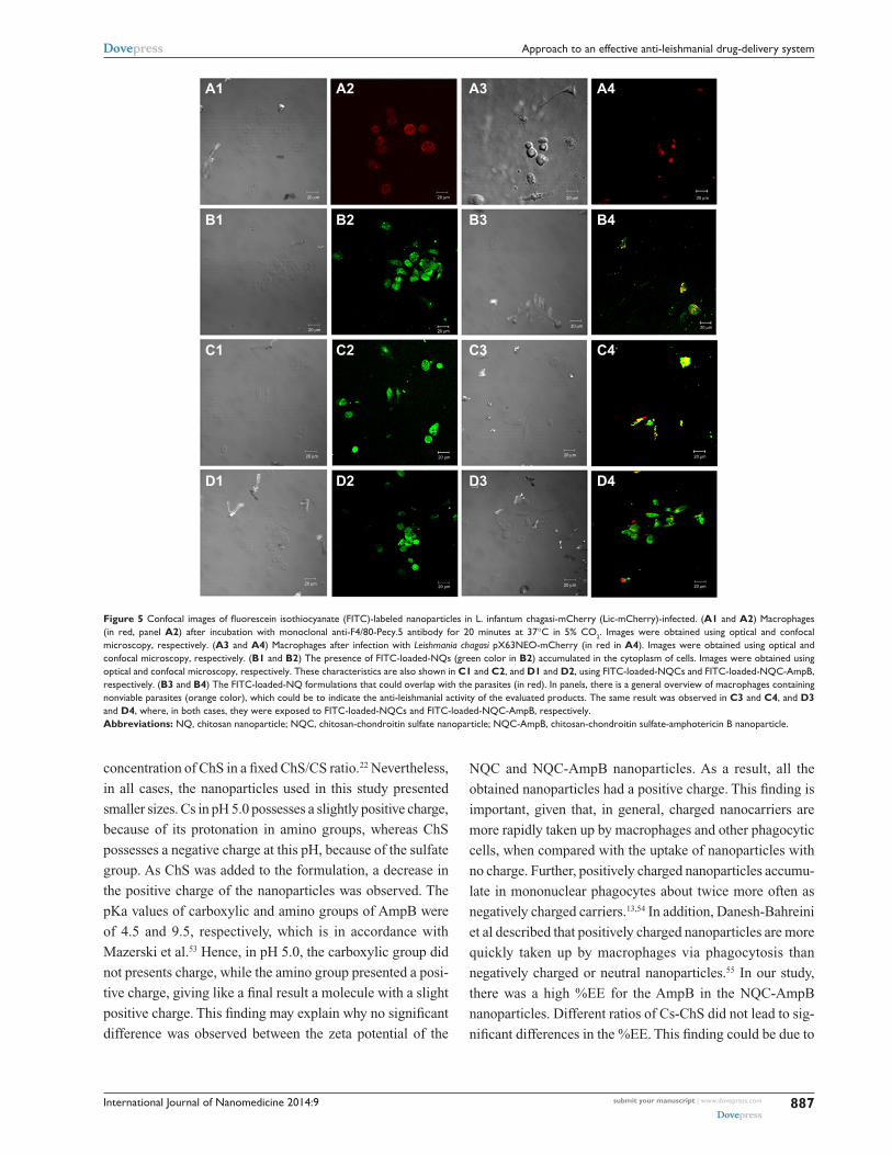
To determine the intracellular location of the nanopar-
ticles in the infected and treated macrophages, confocal
microscopy was performed. In the results, it was possible to
observe a cytoplasmatic localization of the tested NQ, NQC,
and NQC-AmpB formulations (Figure 5). Macrophages
infected with pX63NEO-mCherry L. chagasi were incubated
with the FITC-labeled nanoparticles, to analyze whether the
intracellular allocation of these formulations could, in fact,
overlap with the parasite localization. All the formulations
showed intracellular co-localization with the parasites, indi-
cating that these products could indeed reach the parasito-
phorous vacuoles. In addition, non-viable parasites were also
visualized in the experiments, indicating the anti-leishmanial
efficacy of the evaluated nanoparticles.
DiscussionThe current treatments for leishmaniasis have been consid-
ered unsatisfactory mainly due to the high toxicity of the
products and the growing resistance of the parasites to the
drugs.49 In this context, the purpose of the study reported
here was to find new formulations based on engineering
nanoparticles to carry a known drug to treat leishmaniasis,
AmpB. This product is an effective anti-leishmanial agent,
but is highly toxic to mammals’ cells. In this study, AmpB
was associated with two anti-leishmanial compounds,
ChS, a substance described here for the first time as having
3,356
3,267
1,6451,315
1,062
1,262
1,557
1,640
1634539
1,559
1,2371,644
4,000,0 3,600 3,200 2,800 2,400 2,000 1,800 1,600 1,400 1,200 1,000 650,0
% T
cm−1
A
B
C
D
E
800
Figure 4 Attenuated total reflectance-Fourier-transform infrared spectroscopy spectra of (A) chitosan, (B) chondroitin sulfate, (C) chitosan-chondroitin sulfate nanoparticles, (D) chitosan-chondroitin sulfate-amphotericin B nanoparticles, and (E) pure amphotericin B.
Table 2 anti-leishmanial activity (Ic50), cytotoxicity (cc50), hemolytic activity (rBc50), and selectivity index (sI) of chitosan (cs), chondroitin (chs), chitosan nanoparticles (NQs), chitosan-chondrotin nanoparticles (NQcs), amphotericin B-chitosan-chondroitin nanoparticles (NQc-ampB), and pure amphotericin B (ampB)
Compound/ formulation
Biological activity
Leishmania amazonensis (IC50
e)Leishmania chagasi (IC50)
Murine macrophages (CC50
f)SIh L. amazonensis
SI L. chagasi
Red blood cells (RBC50
g)
cs 73±5d 67±1d 1,559±10d 22 23 432±21d
chs 66±1d 71±10d 1,349±142d 20 19 697±106d
NQ 52±2a,d 46±6ª,d 831±13a,d 16 18 238±26a,d
NQc 44±2b,c,d 39±1b,c,d 1,189±67b,d 27 31 317±4b,d
NQc-ampB 1±0 0.1±0.1 9±0d 99 81 240±33d
ampB 0.1±0 0.1±0 1±0.2 8 9 12±3d
Notes: results are expressed as mean ± standard deviation of the groups. aSignificant difference between Cs and NQs (P,0.05); bsignificant difference between Cs and NQcs (P,0.05); csignificant difference between ChS and NQC (P,0.05); dsignificant difference between AmpB and other compounds (P,0.05); eIc50, concentration needed to inhibit 50% of the parasites’ viability; fcc50, concentration needed to inhibit 50% of the macrophages’ viability; grBc50, concentration needed to lysis 50% of the O+ human red blood cells; hsI, selectivity index, calculated by the ratio between cc50 and Ic50.

International Journal of Nanomedicine 2014:9submit your manuscript | www.dovepress.com
Dovepress
Dovepress
886
ribeiro et al
anti-leishmanial activity, and Cs, which has been previously
proven to have an anti-leishmanial effect.50 This formulation
was physicochemically characterized and assessed by its
anti-leishmanial activity against stationary promastigotes
of L. amazonensis and L. chagasi. Studies were extended
to establish its IC50
value, as well as its effects on the intra-
macrophage Leishmania, and its cytotoxic effect on murine
macrophages and O+ human red blood cells.
In our study, NQC-AmpB nanoparticles were prepared
and a physicochemical characterization was also performed
to characterize their stability. In addition, the biological
activity was also evaluated and this formulation proved to
be a notable targeted drug-delivery system to be used in the
treatment of disease. In addition to anti-leishmanial activity,
NQC-AmpB showed low toxicity to mammals’ cells and good
performance in treating infected macrophages.
To compose a PEC by combining Cs and ChS, both
polymers should carry opposite charges, which occur at a
pH range within their own pKa values. Cs possesses a pKa
of near 6.5, with a maximum of one charge per residue.26 The
apparent dissociation constant of ChS has been estimated
to be from 4.4 to 4.5.51 Therefore, to formulate an effective
nanoparticle combining Cs and ChS, it is necessary to cre-
ate an electrostatic interaction between Cs cations and ChS
anions, in which the positively charged amino groups of Cs
interact with the negatively charged sulfate groups of ChS.
Thus, the optimal pH necessary to achieve a stable PEC with
the highest yield is a pH of approximately 5.0, as observed
in our study, and also described by Chen et al.52 The main
advantage of this methodology is that it allows for the pro-
duction of AmpB-loaded nanoparticles without the use of
organic solvents or heating.
The NQ nanoparticles were also prepared by a PEC tech-
nique using Cs and TPP.34,35 This formulation was prepared
to analyze the level of efficacy of Cs against Leishmania
spp. and the synergistic biological effect of Cs and ChS in a
nanoparticle system (NQC). The formulations were optimized
and the nanoparticles were prepared to be smaller than 200
nm, of homogeneous size, and have a PI ,0.2. The addition
of the sonication step optimized the size of the formulations
(,200 nm), as described by Yeh et al.22 When ChS and AmpB
were added in our study, there was an increase in particle size,
as shown in Figure 2 and Table 1. Yeh et al also hypothesized
that there is a linear relationship between the particle size and
Table 3 Percentage of infected macrophages and parasite burden after treatment with the evaluated formulations
Compound/formulation
Concentration (μg/mL)
Percentage of infected macrophages after treatmenta
Number of amastigotes per macrophage after treatmentb
Reduction of internalized parasites (%)
cs 100.00 66±6 4±0 24
50.00 68±6 4±1 21
25.00 72±5 4±1 17
chs 100.00 60±4 3±0 31
50.00 62±2 3±1 29
25.00 70±5 4±1 20
NQ 100.00 39±2 2±0 55
50.00 53±4 3±1 29
25.00 70±1 4±2 19
NQc 100.00 29±2 2±0 66
50.00 44±3 3±1 49
25.00 67±3 4±1 23
NQc-ampB 0.80 9±1 0 90
0.08 29±3 2±1 66
0.04 44±7 3±1 49
ampB 0.80 9±1 0 89
0.08 34±5 2±1 61
0.04 58±9 3±1 34
Notes: Macrophages were plated on round glass coverslips in 24-well culture plates and infected with promastigotes of Leishmania amazonensis (10 parasites per 1 macrophage). Free parasites were removed by extensive washing, and infected macrophages were treated with 25, 50, and 100 µg/ml of cs, chs, NQ, NQc, or with 0.04, 0.08, and 0.8 µg/ml of NQc-ampB and pure ampB, for 48 hours at 24°C and 5% CO2. The percentage of infected macrophages, the average number of amastigotes per macrophage after treatment, and the average reduction in percentage of internalized parasites were determined by counting 200 cell coverslips, in triplicate, as compared with the non-treated controls. results are expressed as medium ± standard deviation of the percentages of the infected macrophages and by reduction of the internalized parasites in the treated cultures. Abbreviations: ampB, amphotericin B; cs, chitosan; chs, chondroitin; NQ, chitosan nanoparticle; NQc, chitosan-chondroitin nanoparticle; NQc-ampB, amphotericin B-chitosan-chondroitin nanoparticle.
International Journal of Nanomedicine 2014:9 submit your manuscript | www.dovepress.com
Dovepress
Dovepress
887
approach to an effective anti-leishmanial drug-delivery system
concentration of ChS in a fixed ChS/CS ratio.22 Nevertheless,
in all cases, the nanoparticles used in this study presented
smaller sizes. Cs in pH 5.0 possesses a slightly positive charge,
because of its protonation in amino groups, whereas ChS
possesses a negative charge at this pH, because of the sulfate
group. As ChS was added to the formulation, a decrease in
the positive charge of the nanoparticles was observed. The
pKa values of carboxylic and amino groups of AmpB were
of 4.5 and 9.5, respectively, which is in accordance with
Mazerski et al.53 Hence, in pH 5.0, the carboxylic group did
not presents charge, while the amino group presented a posi-
tive charge, giving like a final result a molecule with a slight
positive charge. This finding may explain why no significant
difference was observed between the zeta potential of the
NQC and NQC-AmpB nanoparticles. As a result, all the
obtained nanoparticles had a positive charge. This finding is
important, given that, in general, charged nanocarriers are
more rapidly taken up by macrophages and other phagocytic
cells, when compared with the uptake of nanoparticles with
no charge. Further, positively charged nanoparticles accumu-
late in mononuclear phagocytes about twice more often as
negatively charged carriers.13,54 In addition, Danesh-Bahreini
et al described that positively charged nanoparticles are more
quickly taken up by macrophages via phagocytosis than
negatively charged or neutral nanoparticles.55 In our study,
there was a high %EE for the AmpB in the NQC-AmpB
nanoparticles. Different ratios of Cs-ChS did not lead to sig-
nificant differences in the %EE. This finding could be due to
A1 A2 A3 A4
B1 B2 B3 B4
C1 C2 C3 C4
D1 D2 D3 D4
20 µm 20 µm 20 µm 20 µm
20 µm20 µm20 µm20 µm
20 µm 20 µm 20 µm 20 µm
20 µm20 µm20 µm20 µm
Figure 5 Confocal images of fluorescein isothiocyanate (FITC)-labeled nanoparticles in l. infantum chagasi-mcherry (lic-mcherry)-infected. (A1 and A2) Macrophages (in red, panel A2) after incubation with monoclonal anti-F4/80-Pecy.5 antibody for 20 minutes at 37°C in 5% CO2. Images were obtained using optical and confocal microscopy, respectively. (A3 and A4) Macrophages after infection with Leishmania chagasi pX63NeO-mcherry (in red in A4). Images were obtained using optical and confocal microscopy, respectively. (B1 and B2) The presence of FITc-loaded-NQs (green color in B2) accumulated in the cytoplasm of cells. Images were obtained using optical and confocal microscopy, respectively. These characteristics are also shown in C1 and C2, and D1 and D2, using FITc-loaded-NQcs and FITc-loaded-NQc-ampB, respectively. (B3 and B4) The FITc-loaded-NQ formulations that could overlap with the parasites (in red). In panels, there is a general overview of macrophages containing nonviable parasites (orange color), which could be to indicate the anti-leishmanial activity of the evaluated products. The same result was observed in C3 and C4, and D3 and D4, where, in both cases, they were exposed to FITc-loaded-NQcs and FITc-loaded-NQc-ampB, respectively.Abbreviations: NQ, chitosan nanoparticle; NQc, chitosan-chondroitin sulfate nanoparticle; NQc-ampB, chitosan-chondroitin sulfate-amphotericin B nanoparticle.

International Journal of Nanomedicine 2014:9submit your manuscript | www.dovepress.com
Dovepress
Dovepress
888
ribeiro et al
the concentration of AmpB in the nanoparticles, which was
saturated in a 1:1 ratio. In contrast, at pH 5.0, the amino group
in Cs possesses a positive charge that interacts electrostatically
with the sulfate group of ChS, which possesses a negative
charge, and the carboxylic group of AmpB, which possesses
a negative charge, interacting electrostatically with a positive
charge from the amino group of Cs.
It was also observed that TPP had an important function
in the stability of the NQC nanoparticles, in that the use of
TPP made the formulation stable for 3 weeks, while, without
this compound, the formulation was not stable for a week
(data not shown). A possible reason for this occurrence is
that the TPP formed a highly cross-linked network structure
in the nanoparticles. The limiting %EE factor is the solubil-
ity of AmpB in acidic media. In this study, a pH of nearly
2.0 was used with a 2% acetic acid solution to dilute the
AmpB in aqueous media.41,42 So as to be successfully incor-
porated into the system, AmpB should be first solubilized,
thus, in this study, a pH of 2.0 in aqueous media was used.
This strategy was employed because a net charge on the
molecule of AmpB, obtained in acid aqueous media, could
increase the solubility in two ways: first, it could decrease
the dimerization and association constants by ten-fold or
more and, second, it could increase the threshold of degree
of aggregation for which oligomers are soluble in water. For
these reasons, Mazerski et al postulated that the presence of
a net charge in the molecules is the main factor that induces
the solubility of polyenes.53 In our study, an AmpB solution
with the concentration of 586 µg/mL was found to be the
maximum solubility obtained for this product, so this single
dose was used with different proportions of Cs and ChS to
analyze the %EE and AmpB loading. The NQC-AmpB 1:1
formulation was found to have the smallest particle size,
highest charge (positive zeta potential), and the best %EE
and AmpB loading profile.
ATR-FTIR analysis demonstrated that the sulfate group
of ChS was linked with the ammonium group of Cs. No new
absorption bands on the NQC-AmpB could be observed,
which might mean that no obvious chemical reaction had
occurred between AmpB and the delivery system. In a
controlled drug-delivery system, an active product is incor-
porated into a polymeric network structure in such way that
the drug will be released from the material in a slow and pre-
defined manner.56,57 Depending on the drug delivery system
and the application route, the release time may last from a few
hours to several years.56 The lower release values obtained for
the NQC-AmpB nanoparticles compared with pure AmpB
were most likely to be due to strong interaction between
the AmpB and the Cs/ChS polymers. With this profile, it
could be suggested that the reduced toxicity of AmpB in the
NQC-AmpB formulation could also be attributed to the slow
release of the drug incorporated in the nanoparticles inside
the macrophages, favoring its anti-leishmanial activity for
a longer time.
UV-Vis spectral analysis is useful for deducing aggrega-
tion of AmpB. UV-Vis spectral data led us to deduce that
AmpB was present in NQC-AmpB nanoparticles in its
monomeric form, and the lower intensity could be due to the
small amount of available AmpB. Only this product has been
reported to cause a change in the UV-Vis spectrum, with a
very broad peak appearing around 329 nm and decreased
intensities at 405, 364, and 382 nm, when the monomeric
state is converted to the aggregate state.58 A close associa-
tion is believed to exist between AmpB’s state of aggregation
and its toxicity.58 The mechanism of action proposed for the
toxic effect of this drug is derived from its interaction with
sterols in bilayer membranes, such as cell walls, causing
either the formation of pores in the membrane, leading to
cellular destruction or to the inhibition of membrane repair.
Monomeric AmpB associated with the sterols in fungal cell
membranes, whereas self-associated AmpB could also form
pores in cholesterol-containing membranes, leading to the
drug’s high toxicity to host cells.59
In the evaluation of anti-leishmanial activity, it was
observed that Cs presented better anti-leishmanial activity in
relation to the evaluated nanoparticles systems. A synergistic
interaction between Cs and ChS was also observed against
L. amazonensis and L. chagasi. The activity of NQC-AmpB
nanoparticles was similar in comparison to the results visu-
alized using pure AmpB; however, the cytotoxicity of the
NQC-AmpB was approximately ten-fold lower than that of
pure AmpB. In this context, the selectivity index of AmpB
was increased ten-fold when added to the proposed delivery
system in this study. In addition, pure Cs and ChS, as well as
NQ and NQC nanoparticles presented no significant toxicity
for macrophages. Van de Ven et al60 and Corware et al61 also
obtained blank nanoparticles without significant cytotoxicity
in macrophages, but when they were loaded with AmpB, the
evaluated systems demonstrated an increase in their toxicity.
Finally, hemolytic activity was also determined as a cytotoxic-
ity parameter, and it was observed that none of the nanopar-
ticle formulations resulted in significant hemolysis.
ConclusionThe formulations tested in this study were found to have the
desired characteristics of effective drug-delivery systems for use

International Journal of Nanomedicine 2014:9 submit your manuscript | www.dovepress.com
Dovepress
Dovepress
889
approach to an effective anti-leishmanial drug-delivery system
in the treatment of leishmaniasis. The formulations were able
to reach the parasitic compartment in the infected cells, which
is relevant mainly because a lower drug concentration will be
required for administration in patients, which may, in turn, pre-
vent the several toxic effects of the drugs that are often observed
when conventional products are daily used at high doses.
As far as we are aware, the present study is the first
to date to describe a new drug-delivery system based on
NQC nanoparticles loaded with AmpB to treat leishmaniasis.
This formulation, in addition to demonstrating effective anti-
leishmanial activity, and a high capacity to treat infected
macrophages, presented none of the toxic effects in mice or
human cells that are normally observed when pure AmpB is
administered. Thus, we could suggest that the NQC-AmpB
formulation should be evaluated in future studies on in vivo
anti-leishmanial activity, in an attempt to find new chemo-
therapeutic alternatives for the treatment of leishmaniasis.
AcknowledgmentsThe authors would like to thank Dr Manuel Soto (Centro de
Biología Molecular Severo Ochoa [CSIC-UAM], Universidad
Autónoma de Madrid, Spain) for scientific assistance. This
work was supported by grants from Pró-Reitoria de Pesquisa
from UFMG (Edital 03/2013), Instituto Nacional de Ciência
e Tecnologia em Nano-biofarmacêutica (INCT-Nanobiofar),
FAPEMIG (CBB-APQ-02364-08 and CBB-APQ-00496-11),
and CNPq (APQ-472090/2011-9 and APQ-482976/2012-8).
Miguel A Chávez- Fumagalli is a grant recipient of PNPD/
CAPES. Eduardo AF Coelho is a grant recipient of CNPq.
DisclosureThe authors report no conflicts of interest in this work.
References1. Desjeux P. Leishmaniasis: current situation and new perspectives. Comp
Immunol Microbiol Infect Dis. 2004;27(5):305–318.2. World Health Organization (WHO). Control of the Leishmaniases:
Report of a Meeting of the 399 WHO Expert Committee on the Control of Leishmaniases, Geneva, 22–26 March 2010. WHO 400 Technical Report Series 949. Geneva: WHO; 2010. Available from: whqlibdoc.who.int/trs/WHO_TRS_949_eng.pdf. Accessed December 18, 2013.
3. Alvar J, Vélez ID, Bern C, et al; WHO Leishmaniasis Control Team. Leishmaniasis worldwide and global estimates of its incidence. PloS One. 2012;7(5):e35671.
4. Croft SL, Coombs GH. Leishmaniasis – current chemotherapy and recent advances in the search for novel drugs. Trends Parasitol. 2003;19(11): 502–508.
5. Grevelink SA, Lerner EA. Leishmaniasis. J Am Acad Dermatol. 1996; 34(2):257–272.
6. Annaloro C, Olivares C, Usardi P, et al. Retrospective evaluation of amphotericin B deoxycholate toxicity in a single centre series of hae-matopoietic stem cell transplantation recipients. J Antimicrob Chemother. 2009;63(3):625–626.
7. Denning DW. Therapeutic outcome of invasive aspergillosis. Clin Infect Dis. 1996;23(3):608–615.
8. Bern C, Adler-Moore J, Berenguer J, et al. Liposomal amphotericin B for the treatment of visceral leishmaniasis. Clin Infect Dis. 2006;43(7): 917–924.
9. Egger SS, Meier S, Leu C, et al. Drug interactions and adverse events associated with antimycotic drugs used for invasive aspergil-losis in hematopoietic SCT. Bone Marrow Transplant. 2009;45(7): 1197–1203.
10. Italia JL, Sharp A, Carter KC, Warn P, Kumar MN. Peroral amphot-ericin B polymer nanoparticles lead to comparable or superior in vivo antifungal activity to that of intravenous Ambisome® or Fungizone™. PLoS One. 2011;6(10):e25744.
11. Quintanar-Guerrero D, Allémann E, Fessi H, Doelker E. Preparation tech-niques and mechanisms of formation of biodegradable nanoparticles from preformed polymers. Drug Dev Ind Pharm. 1998;24(12):1113–1128.
12. de Carvalho RF, Ribeiro IF, Miranda-Vilela AL, et al. Leishmanicidal activity of amphotericin B encapsulated in PLGA-DMSA nanoparticles to treat cutaneous leishmaniasis in C57BL/6 mice. Exp Parasitol. 2013;135(2):217–222.
13. Asthana S, Jaiswal AK, Gupta PK, Pawar VK, Dube A, Chourasia MK. Immunoadjuvant chemotherapy of visceral leishmaniasis in hamsters using amphotericin B-encapsulated nanoemulsion template-based chitosan nanocapsules. Antimicrob Agents Chemother. 2013;57(4): 1714–1722.
14. Shao K, Huang R, Li J, et al. Angiopep-2 modified PE-PEG based polymeric micelles for amphotericin B delivery targeted to the brain. J Control Release. 2010;147(1):118–126.
15. Yang ZL, Li XR, Yang KW, Liu Y. Amphotericin B-loaded poly(ethylene glycol)-poly(lactide) micelles: preparation, freeze-drying, and in vitro release. J Biomed Mater Res A. 2008;85(2):539–546.
16. Kreuter J. Nanoparticles. In: Swarbrick J, Boylan JC, editors. Encyclopedia of Pharmaceutical Technology. New York, NY: Marcel Dekker; 1988:165–190.
17. Janes KA, Fresneau MP, Marazuela A, Fabra A, Alonso MJ. Chitosan nanoparticles as delivery systems for doxorubicin. J Control Release. 2001;73(2–3):255–267.
18. St John AL, Chan CY, Staats HF, Leong KW, Abraham SN. Synthetic mast-cell granules as adjuvants to promote and polarize immunity in lymph nodes. Nat Mater. 2012;11(3):250–257.
19. Huang L, Sui W, Wang Y, Jiao Q. Preparation of chitosan/chondroitin sulfate complex microcapsules and application in controlled release of 5-fluorouracil. Carbohydr Polym. 2010;80(1):168–173.
20. Ganza-González A, Anguiano-Igea S, Otero-Espinar FJ, Blanco Méndez J. Chitosan and chondroitin microspheres for oral- administration controlled release of metoclopramide. Eur J Pharm Biopharm. 1999;48(2):149–155.
21. Sui W, Huang L, Wang J, Bo Q. Preparation and properties of chi-tosan chondroitin sulfate complex microcapsules. Colloids Surf B Biointerfaces. 2008;65(1):69–73.
22. Yeh MK, Cheng KM, Hu CS, Huang YC, Young JJ. Novel protein-loaded chondroitin sulfate-chitosan nanoparticles: preparation and characterization. Acta Biomater. 2011;7(10):3804–3812.
23. Hu CS, Chiang CH, Hong PD, Yeh MK. Influence of charge on FITC-BSA-loaded chondroitin sulfate-chitosan nanoparticles upon cell uptake in human Caco-2 cell monolayers. Int J Nanomedicine. 2012;7: 4861–4872.
24. Tsai HY, Chiu CC, Lin PC, Chen SH, Huang SJ, Wang LF. Antitumor efficacy of doxorubicin released from crosslinked nanoparticulate chondroitin sulfate/chitosan polyelectrolyte complexes. Macromol Biosci. 2011;11(5):680–688.
25. Bhattarai N, Gunn J, Zhang M. Chitosan-based hydrogels for controlled, localized drug delivery. Adv Drug Deliv Rev. 2010;62(1):83–99.
26. Thongngam M, McClements DJ. Influence of pH, ionic strength, and temperature on self-association and interactions of sodium dodecyl-sulfate in the absence and presence of chitosan. Langmuir. 2005;21(1):79–86.

International Journal of Nanomedicine
Publish your work in this journal
Submit your manuscript here: http://www.dovepress.com/international-journal-of-nanomedicine-journal
The International Journal of Nanomedicine is an international, peer-reviewed journal focusing on the application of nanotechnology in diagnostics, therapeutics, and drug delivery systems throughout the biomedical field. This journal is indexed on PubMed Central, MedLine, CAS, SciSearch®, Current Contents®/Clinical Medicine,
Journal Citation Reports/Science Edition, EMBase, Scopus and the Elsevier Bibliographic databases. The manuscript management system is completely online and includes a very quick and fair peer-review system, which is all easy to use. Visit http://www.dovepress.com/ testimonials.php to read real quotes from published authors.
International Journal of Nanomedicine 2014:9submit your manuscript | www.dovepress.com
Dovepress
Dovepress
Dovepress
890
ribeiro et al
27. Iovu M, Dumais G, du Souich P. Anti-inflammatory activity of chon-droitin sulfate. Osteoarthritis Cartilage. 2008;16 Suppl 3:S14–S18.
28. Ronca F, Palmieri L, Panicucci P, Ronca G. Anti-inflammatory activity of chondroitin sulfate. Osteoarthritis Cartilage. 1998;6 Suppl A:14–21.
29. Ronca F, Palmieri L, Panicucci P, Ronca G. Anti-inflammatory activity of chondroitin sulfate. Osteoarthritis Cartigale. 1998;6(A):14–21.
30. Kirker KR, Luo Y, Morris SE, Shelby J, Prestwich GD. Glycosaminoglycan hydrogels as supplemental wound dressings for donor sites. J Burn Care Rehab. 2004;25(3):276–286.
31. Kirker KR, Luo Y, Nielson JH, Shelby J, Prestwich GD. Glycosaminoglycan hydrogel f ilms as bio-interactive dressings for wound healing. Biomaterials. 2002;23(17):3661–3671.
32. Wang LF, Wang JM, Chiang YL. Insolubilization of sodium chondroitin sulfate by forming a semi-interpenetrating polymer network with acrylic acid: a potential carrier for colon-specific drug delivery. J Appl Polym Sci. 2002;85(1):114–122.
33. Sintov A, Di-Capua N, Rubinstein A. Cross-linked chondroitin sulphate: characterization for drug delivery purposes. Biomaterials. 1995;16(6):473–478.
34. Costa DF, Franca JR, Ribeiro TG, Kaplan MA, Faraco AA, Castilho RO. Development and characterization of polymeric nanopar-ticles as Barbatimão (Stryphnodendron obovatum) standardized fraction carrier. Adv Biosci Biotechnol. 2012;4(1):89–92.
35. Calvo P, Remuñán-López C, Vila-Jato JL, Alonso MJ. Novel hydro-philic chitosan-polyethylene oxide nanoparticles as protein carriers. J Appl Polym Sci. 1997;63(1):125–132.
36. Grenha A, Gomes ME, Rodrigues M, et al. Development of new chitosan/carrageenan nanoparticles for drug delivery applications. J Biomed Mater Res A. 2010;92(4):1265–1272.
37. Müller RH, Schmidt S, Buttle I, Akkar A, Schmitt J, Brömer S. SolEmuls-novel technology for the formulation of iv emulsions with poorly soluble drugs. Int J Pharm. 2004;269(2):293–302.
38. Moreno MA, Frutos P, Ballesteros MP. Lyophilized lecithin based oil-water microemulsions as a new and low toxic delivery system for amphotericin B. Pharm Res. 2001;18(3):344–351.
39. Santos CM, Oliveira RB, Arantes VT, et al. Amphotericin B-loaded nanocarriers for topical treatment of cutaneous leishmaniasis: development, characterization, and in vitro skin permeation studies. J Biomed Nanotechnol. 2012;8(2):322–329.
40. Nahar M, Jain NK. Preparation, characterization and evaluation of targeting potential of amphotericin B-loaded engineered PLGA nanoparticles. Pharm Res. 2009;26(12):2588–2598.
41. Coelho EA, Tavares CA, Carvalho FA, et al. Immune responses induced by the Leishmania (Leishmania) donovani A2 antigen, but not by the LACK antigen, are protective against experimental Leishmania (Leishmania) amazonensis infection. Infect Immun. 2003;71(7):3988–3994.
42. Valadares DG, Duarte MC, Oliveira JS, et al. Leishmanicidal activity of the Agaricus blazei Murill in different Leishmania species. Parasitol Int. 2011;60(4):357–363.
43. Fumarola L, Spinelli R, Brandonisio O. In vitro assays for evaluation of drug activity against Leishmania spp. Res Microbiol. 2004;155(4): 224–230.
44. Löfgren SE, Miletti LC, Steindel M, Bachère E, Barracco MA. Trypanocidal and leishmanicidal activities of different antimicro-bial peptides (AMPs) isolated from aquatic animals. Exp Parasitol. 2008;118(2):197–202.
45. Jung SH, Lim DH, Jung SH, et al. Amphotericin B-entrapping lipid nanoparticles and their in vitro and in vivo characteristics. Eur J Pharm Sci. 2009;37(3–4):313–320.
46. Onishi H, Machida Y. Biodegradation and distribution of water-soluble chitosan in mice. Biomaterials. 1999;20(2):175–182.
47. Huang M, Ma Z, Khor E, Lim LY. Uptake of FITC-chitosan nanoparticles by A549 cells. Pharm Res. 2002;19(10):1488–1494.
48. Adams ML, Kwon GS. Relative aggregation state and hemolytic activity of amphotericin B encapsulated by poly(ethylene oxide)-block-poly(N-hexyl-L-aspartamide)-acyl conjugate micelles: effects of acyl chain length. J Control Release. 2003;87(1–3):23–32.
49. Tasdemir D, Kaiser M, Brun R, et al. Antitrypanosomal and anti-leishmanial activities of flavonoids and their analogues: in vitro, in vivo, structure-activity relationship, and quantitative structure- activity relationship studies. Antimicrob Agents Chemother. 2006;50(4): 1352–1364.
50. Pujals G, Suñé-Negre JM, Pérez P, et al. In vitro evaluation of the effectiveness and cytotoxicity of meglumine antimoniate microspheres produced by spray drying against Leishmania infantum. Parasitol Res. 2008;102(6):1243–1247.
51. Nakajima A, Shinoda K. Complex formation between oppositely charged polysaccharides. J Colloid Interface Sci. 1976;55(1):126–132.
52. Chen WB, Wang LF, Chen JS, Fan SY. Characterization of polyelec-trolyte complexes between chondroitin sulfate and chitosan in the solid state. J Biomed Mater Res A. 2005;75(1):128–137.
53. Mazerski J, Grzybowska J, Borowski E. Influence of net charge on the aggregation and solubility behaviour of amphotericin B and its derivatives in aqueous media. Eur Biophys J. 1990;18(3):159–164.
54. Batrakova EV, Gendelman HE, Kabanov AV. Cell-mediated drug delivery. Expert Opin Drug Deliv. 2011;8(4):415–433.
55. Danesh-Bahreini MA, Shokri J, Samiei A, Kamali-Sarvestani E, Barzegar-Jalali M, Mohammadi-Samani S. Nanovaccine for leishmaniasis: preparation of chitosan nanoparticles containing Leishmania superoxide dismutase and evaluation of its immunogenicity in BALB/c mice. Int J Nanomedicine. 2011;6:835–842.
56. Wise DL, editor. Handbook of Pharmaceutical Controlled Release Tech-nology. New York, NY: Marcel Dekker, 2000; Chap 9: pp 183–210.
57. Jogani V, Jinturkar K, Vyas T, Misra A. Recent patents review on intranasal administration for CNS drug delivery. Recent Pat Drug Deliv Formul. 2008;2(1):25–40.
58. Legrand P, Romero EA, Cohen BE, Bolard J. Effects of aggregation and solvent on the toxicity of amphotericin B to human erythrocytes. Antimicrob Agents Chemother. 1992;36(11):2518–2522.
59. Darole PS, Hegde DD, Nair HA. Formulation and evaluation of micro-emulsion based delivery system for amphotericin B. AAPS Pharm Sci Tech. 2008;9(1):122–128.
60. Van de Ven H, Paulussen C, Feijens PB, et al. PLGA nanoparticles and nanosuspensions with amphotericin B: Potent in vitro and in vivo alter-natives to Fungizone and AmBisome. J Control Release. 2012;161(3): 795–803.
61. Corware K, Harris D, Teo I, et al. Accelerated healing of cutane-ous leishmaniasis in non-healing BALB/c mice using water soluble amphotericin B-polymethacrylic acid. Biomaterials. 2011;2(31): 8029–8039.


