Embed Size (px)
Citation preview

NTPDase1/CD39 and aberrant purinergicsignalling in the pathogenesis of COPD
Zsofia Lazar1,2,7, Nina Müllner1,7, Monica Lucattelli3,7, Cemil Korcan Ayata1,7,Sanja Cicko1, Gennady G. Yegutkin4, Giovanna De Cunto3, Tobias Müller1,Anja Meyer1, Madelon Hossfeld1, Stephan Sorichter1, Ildiko Horvath2,Christian J. Virchow5, Simon C. Robson6, Giuseppe Lungarella3 and Marco Idzko1
Affiliations: 1Department of Pulmonology, University Hospital Freiburg, Freiburg, Germany. 2Department ofPulmonology, Semmelweis University, Budapest, Hungary. 3Department of Life Sciences, University of Siena,Siena, Italy. 4MediCity Research Laboratory, University of Turku, Turku, Finland. 5Department of Pulmonology,University Hospital Rostock, Rostock, Germany. 6Division of Gastroenterology, Department of Medicine, BethIsrael Deaconess Medical Center, Harvard Medical School, Boston, MA, USA. 7These authors contributed equally.
Correspondence: Marco Idzko, Department of Pulmonary Medicine, University Hospital Freiburg, Killianstraße5, 79106-Freiburg, Germany. E-mail: [email protected]
ABSTRACT Purinergic receptor activation via extracellular ATP is involved in the pathogenesis ofchronic obstructive pulmonary disease (COPD). Nucleoside triphosphate diphosphohydrolase-1/CD39hydrolyses extracellular ATP and modulates P2 receptor signalling.
We aimed to investigate the expression and function of CD39 in the pathogenesis of cigarette smoke-induced lung inflammation in patients and preclinical mouse models. CD39 expression and solubleATPase activity were quantified in sputum and bronchoalveolar lavage fluid (BALF) cells in nonsmokers,smokers and COPD patients or mice with cigarette smoke-induced lung inflammation. In mice,pulmonary ATP and cytokine concentrations, inflammation and emphysema were analysed in the presenceor absence of CD39.
Following acute cigarette smoke exposure CD39 was upregulated in BALF cells in smokers with furtherincreases in COPD patients. Acute cigarette smoke exposure induced CD39 upregulation in murine lungsand BALF cells, and ATP degradation was accelerated in airway fluids. CD39 inhibition and deficiency ledto augmented lung inflammation; treatment with ATPase during cigarette smoke exposure preventedemphysema.
Pulmonary CD39 expression and activity are increased in COPD. CD39 deficiency leads to enhancedemphysema in mice, while external administration of a functional CD39 analogue partially rescues thephenotype. The compensatory upregulation of pulmonary CD39 might serve as a protective mechanism incigarette smoke-induced lung damage.
@ERSpublicationsThe upregulation of pulmonary ATPase is a protective mechanism in cigarette smoke-inducedlung inflammation http://ow.ly/S5YcC
Copyright ©ERS 2015
This article has supplementary material available from erj.ersjournals.com
Received: Nov 21 2014 | Accepted after revision: Aug 05 2015
Support statement: M. Idzko is a recipient of grants from the German Research Foundation (Deutsche Forschungsgemeinschaft(DFG), grant ID 7/4-1) and the Boehringer Ingelheim Foundation. Z. Lazar was a recipient of the European Respiratory Societylong-term research fellowship (grant ID 50-2012). Funding information for this article has been deposited with FundRef.
Conflict of interest: None declared.
Eur Respir J 2015; In press | DOI: 10.1183/13993003.02144-2014 1
ORIGINAL ARTICLEIN PRESS | CORRECTED PROOF
. Published on November 5, 2015 as doi: 10.1183/13993003.02144-2014ERJ Express
Copyright 2015 by the European Respiratory Society.

IntroductionChronic obstructive pulmonary disease (COPD) is a leading cause of morbidity and mortality worldwide[1]. It is associated with the specific inflammation of small airways, which results in airway obstruction,parenchymal destruction and the development of emphysema. Although inhalation of cigarette smoke isthe main risk factor for the development of COPD, the precise mechanisms initiating and perpetuating thedisease and the underlying inflammation are poorly understood.
An increasing body of evidence supports the involvement of endogenous nucleotides, which activate P2receptor subtypes, in neutrophilic airway inflammation [2–5]. More specifically, increased pulmonary levels ofATP can be found in mice and human subjects following exposure to cigarette smoke, and also in patientswith COPD. Importantly, ATP concentration in the bronchoalveolar lavage fluid (BALF) of patients correlateswith neutrophil counts and airflow limitation [3]. In the mouse model, inhibition of specific P2 receptorsubtypes has been shown to decrease cigarette smoke-induced lung inflammation and emphysema [6, 7].
The levels of nucleotides in mammalian tissues are tightly controlled by the nucleoside triphosphatediphosphohydrolases (NTPDases) 1–8 and other ectonucleotidases. CD39/NTPDase1/ecto-apyrase is thedominant purinergic ecto-enzyme in the immune and vascular barrier in the lung, and it is also a key regulatorof the functionality of immune cells [8]. It regulates ATP-mediated P2 receptor signalling by hydrolysing ATP/ADP to AMP, which is then further metabolised by CD73/ecto-5′-nucleotidase to adenosine [9].
Interestingly, besides its general ecto-enzymatic function, CD39 might directly modulate the function ofimmune cells such as neutrophils, macrophages or dendritic cells [10, 11]. Consequently, an importantrole of CD39 in inflammatory disorders, including acute respiratory distress syndrome, hyperoxic acutelung injury, colitis and autoimmune diabetes has been demonstrated [12, 13]. Recently, we have shownthat CD39 is involved in the pathogenesis of allergic airway inflammation by modulating dendritic cellfunction [14]. However, the role and regulation of CD39 in the development and maintenance of cigarettesmoke-induced lung inflammation and emphysema is unknown.
The aim of this study was to investigate, for the first time, the expression and functional impact of CD39 onthe pathogenesis of COPD. For this purpose we measured the expression of CD39 and ATPase in sputumand/or BALF of nonsmokers, smokers, ex-smokers and patients with COPD, as well as in BALF and lungtissue of mice with cigarette smoke-induced lung inflammation. In order to elucidate the functional relevanceof these findings, the effect of CD39 inhibition/deficiency on the development of cigarette smoke-inducedlung inflammation and lung emphysema was analysed in a translational mouse model. Finally, to support thepotential therapeutic implications of these findings, cigarette smoke-exposed mice were treated with afunctional analogue of CD39 and the degree of lung inflammation and lung emphysema was determined.
MethodsCollection of BALF and sputum from control subjects and patients with COPDBALF was collected from control subjects and ex-smoking COPD patients as part of a previous study andprocessed as described previously [3]. A subset of smokers underwent bronchoscopy and bronchoalveolarlavage after a 4-h period of smoking deprivation (no smoke exposure) and immediately after a 4-h periodduring which eight cigarettes were smoked (acute smoke exposure), with a 2-week interval in between.Induced or spontaneous sputum was collected from control subjects, as previously reported [15]. Patientswere ex-smokers (abstinence >6 months prior to recruitment) and had known COPD which had beenestablished previously by a respiratory specialist and were in stable condition. Spirometric classificationwas performed according to RABE et al. [1]. Detailed information on eligibility criteria, patientcharacteristics and the procedure is given in the online supplementary material.
The study was conducted in line with the recommendations of the Declaration of Helsinki and wasapproved by the local ethics committee of University Medical Centre (Freiburg, Germany). All participantsgave written informed consent.
MiceC57BL/6 mice (aged 6–8 weeks) were purchased from Charles River (Calco, Italy) or were bred at theanimal facilities at the University Hospital Freiburg. Cd39-deficient mice (Cd39−/−) on a C57BL/6background were generated as previously described [14]. All experiments were performed according toinstitutional guidelines of the animal ethics committees of the Italian or German governments.
Animal models of acute and chronic cigarette smoke-induced lung inflammationThe induction of acute and chronic smoke-associated lung inflammation was performed as previouslydescribed [6, 7, 16]. In some experiments, intratracheal or intraperitoneal treatment was performed prior
2 DOI: 10.1183/13993003.02144-2014
COPD | Z. LAZAR ET AL.

to smoke exposure. BALF and lungs were collected and processed as previously described [3, 7].Additional details are provided in the online supplementary material.
Enzyme histochemistry on murine lungsFor localisation of ATPase activity, the lead phosphate method was applied [17], which is based on theprecipitation of lead by free phosphate generated from ATPase activity. The intensity of lead staining wasquantified using ImageJ v1.46r software (National Institutes of Health, Bethesda, MD, USA). Further detailsare available in the online supplementary material.
ATP and cytokine measurements in BALF and sputum supernatantCell-free human sputum and murine BALF supernatants were incubated for 30 min at room temperaturewith 100 mM and 100 nM ATP, respectively. ATPase activity is shown as percentage of ATP hydrolysed(100 − percentage ATP recovered). ATP concentration was determined as previously described [3, 18].Cytokine concentrations in BALF were measured using commercially available ELISA kits (R&D Systems,Minneapolis, MN, USA).
Real-time PCRTotal RNA was isolated using QIAzol (Qiagen, Hilden, Germany); complementary DNA synthesis wasperformed using a First Strand cDNA synthesis kit (ThermoScientific, Bremen, Germany). Quantitative PCRwas performed using a LightCycler 480 (Roche, Mannheim, Germany) using a fast-blue+UNG kit (Eurogentec,Cologne, Germany). ß2M and GAPDH were used as reference genes. Gene expression assay design and analysiswere performed as previously described [19]; primer/probe sequences are available upon request.
Statistical analysisSputum data were not normally distributed (D’Agostino–Pearson test) and were analysed usingnonparametric tests (Kruskal–Wallis with Dunn’s post hoc test, Spearman correlation) and reported asmedian (interquartile range). CD39 expression data in sputum were logarithmically transformed to yieldnormal distribution. Otherwise, a one-sample t-test, unpaired t-test or ANOVA with post hoc Bonferronitest were applied and data are shown as mean±SEM (GraphPad Prism 5; GraphPad Software, San Diego,CA, USA). Differences were considered significant if p<0.05.
ResultsCD39 expression of airway inflammatory cells is modulated by smoking and increased in COPDTo analyse the effects of smoking on CD39 expression and activity in the airways, BALF leukocytescollected in a previously published cohort of nonsmokers, smokers and patients with COPD were studied[3]; subject characteristics are shown in table 1. We observed a tendency to increased expression of CD39on BALF leukocytes from patients compared to smokers and nonsmokers (p=0.09; fig. 1a). Previously, wereported that in human smokers BALF ATP levels were strongly increased after acute smoke exposurescompared to a period of smoking cessation [3]. Thus we questioned whether CD39 expression in BALFcells might also be influenced. As shown in figure 1b, a 4-h period of smoking induced a strongupregulation of CD39 expression in BALF cells of human smokers (n=8).
TABLE 1 Subject characteristics in the bronchoalveolar lavage fluid (BALF) study
Control subjects COPD patients
Nonsmoking Smoking
Subjects 7 10 8Male/female 2/6 2/8 0/8Age years 42±7 37±7 55±5**,#
Pack-years NA 10±4 35±8#
GOLD severity I/II/III NA NA 2/4/2Leukocytes ×106 cells·100 mL-1 BALF 9.4±6.6 13.7±10.7 31.9±16.3FEV1 % predicted 102±8 104±10 70±15
Data are presented as n or mean±SD. ANOVA with Bonferroni post hoc test or t-test were used. COPD: chronicobstructive pulmonary disease; GOLD: Global Initiative for Chronic Obstructive Lung Disease; FEV1: forcedexpiratory volume in 1 s; NA: not applicable. **: p<0.01 versus nonsmoking control; #: p<0.01 versus currentlysmoking control.
DOI: 10.1183/13993003.02144-2014 3
COPD | Z. LAZAR ET AL.

To support these findings in a broader cohort, sputa were collected from control subjects and ex-smokingpatients with COPD. Patient characteristics and sputum variables are presented in table 2. As shown in fig. 1c,CD39 expression of total sputum cells was increased in current smokers compared to non- and ex-smokingcontrol subjects. In fact, CD39 expression was higher in ex-smoking patients with COPD (n=26) than incontrol subjects. Furthermore, the activity of ATPase in cell-free sputum supernatant was enhanced inpatients (n=23) compared to nonsmokers, ex-smokers and current smokers (fig. 1d). Of note, ATPase activitywas increased in patients with severe/very severe COPD compared to subjects with mild/moderate disease(p=0.03, data not shown), and showed a negative correlation with FEV1 % predicted (p=0.01, r=−0.52),which suggests that more severe airflow limitation is associated with the accelerated activity of airway ATPase.However, no relationship was observed between CD39 expression and disease severity or FEV1 (data notpresented). Interestingly, in patients, ATPase activity showed a significant positive correlation to leukocytecount·g−1 sputum (p<0.01, r=0.57), sputum neutrophil percentages (fig. 1e) and neutrophil counts (fig. 1f),but not to the counts or percentages of macrophages, eosinophils and lymphocytes (data not shown).
CD39
exp
ressio
n
(ne
t to
β2M
)
0.1
0.2
0.3
0.4
0.5
0.6a)
Never-
smokers
Smokers COPD
CD39
exp
ressio
n
(ne
t to
β2M
)
0.0
0.6
1.2c)
Non
smokers
Ex-
smokers
Current
smokers
**,#
*,#
Ex-smoking
COPD
Ind
ex CD
39 e
xpre
ssio
n
aft
er
no
sm
oke
exp
osu
re
0
1
2
34
6
8
10b)
No smoke
exposure
Acute smoke
exposure
**
AT
Pa
se
acti
vity
%
0
40
8080
90
100e)
60 70 80
Sputum neutrophils %
90 100
r=0.47* AT
Pa
se
acti
vity
%
0
40
8080
90
100f)
10 100 1000 10 000
Sputum neutrophil count ×104 cells·g–1
r=0.59**
AT
Pa
se
acti
vity
%
0
25
50
75
100d)
Non
smokers
Ex-
smokers
Current
smokers
**,¶,+
Ex-smoking
COPD
FIGURE 1 Cellular CD39 expression and soluble ATPase activity in airway inflammatory cells of nonsmokers,ex-smokers, current smokers and patients with chronic obstructive pulmonary disease (COPD). a) Expression ofCD39 in total bronchoalveolar lavage fluid (BALF) cells isolated from never-smoking controls, smokers andpatients with COPD was measured using quantitative (q)PCR; p=0.09. b) Effect of acute cigarette smokeinhalation on CD39 expression in BALF leukocytes of human smokers; **: p<0.01. c) Expression of CD39 in totalsputum cells isolated from controls and ex-smoking patients with COPD was quantified using qPCR. Data arepresented as mean±SEM. *: p<0.05; **: p<0.01 nonsmoker versus current smoker, nonsmoker versus ex-smokingCOPD; #: p<0.05 ex-smoker versus current smoker, ex-smoker versus ex-smoking COPD. d) Freshly preparedcell-free sputum supernatant was incubated with 100 mM ATP for 30 min at room temperature, then ATPaseactivity was measured in a bioluminescent assay. Data are presented as mean±SEM. **: p<0.01 nonsmokerversus ex-smoking COPD; ¶: p<0.01 ex-smoker versus ex-smoking COPD, +: p<0.05 current smoker versusex-smoking COPD. e, f ) Correlation of ATPase activity in sputum supernatant to e) percentages and f) counts ofsputum neutrophils in ex-smoking patients with COPD. *: p<0.05; **: p<0.01.
4 DOI: 10.1183/13993003.02144-2014
COPD | Z. LAZAR ET AL.

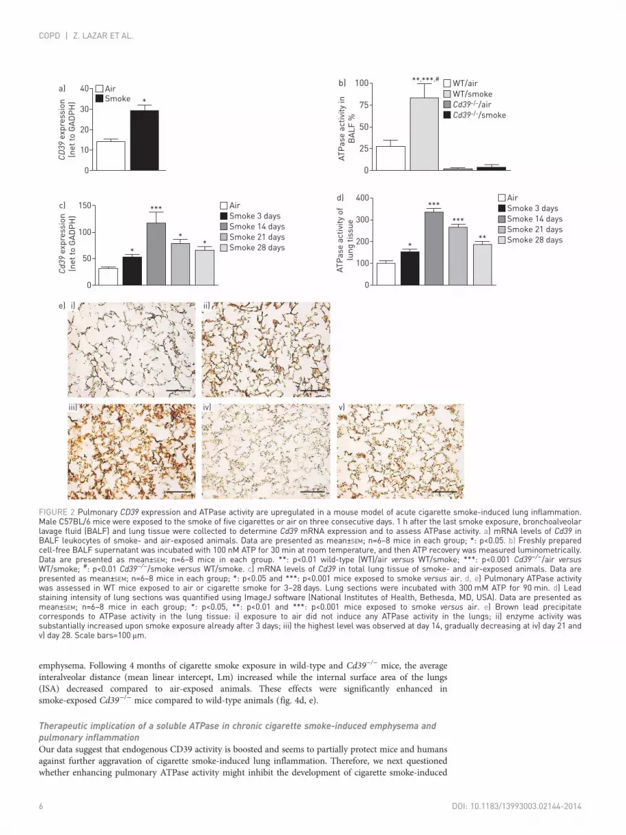
CD39 expression and pulmonary ATPase activity are enhanced in a mouse model of cigarettesmoke-induced lung inflammationTo corroborate the human findings, Cd39 expression and ATP hydrolysing activity were studied in BALFcells and supernatants in a mouse model for cigarette smoke-induced lung inflammation. Similarly to thehuman data, we observed an increase in Cd39 expression and soluble ATPase activity in BALF cells andsupernatant of wild-type mice exposed to the smoke of five cigarettes for three consecutive days comparedto air-exposed controls (fig. 2a, b).
Besides inflammatory cells, pulmonary ATP concentration is also controlled by CD39 strongly expressedon airway epithelial cells [14]. Therefore, we further assessed the expression and pulmonary activity ofmembrane-bound ATPase in lung tissue of mice exposed to cigarette smoke for up to 28 days. Indeed,cigarette smoke exposure led to the strong upregulation of Cd39 mRNA (fig. 2c) as well as pulmonaryATPase activity in the lung tissue compared to air-exposed mice (fig. 2d, e). Interestingly, the highestATPase activity was observed after 14 days of cigarette smoke exposure; however, pulmonary ATPaseactivity remained elevated on days 21 and 28. No ATPase activity was observed in lungs of CD39-deficientanimals (online supplementary fig. S1).
Inhibition of the ectonucleotidase CD39 aggravates cigarette smoke-induced acute lunginflammationTo evaluate the functional relevance of increased Cd39 expression due to smoke-induced lung inflammation,mice were treated intratracheally with the ectonucleotidase inhibitor ARL-67156 (100 mM) or vehicle 30 minbefore each smoke exposure. Compared to vehicle treatment ATP concentration was elevated in BALF ofanimals treated with ARL-67156, which was accompanied by an increase in macrophage and neutrophilcounts and an elevation in the concentrations of interleukin (IL)-6, keratinocyte-derived chemokine (KC)and macrophage inflammatory protein (MIP)-2 in the BALF (fig. 3a–c).
CD39 deficiency aggravates acute and chronic cigarette smoke-induced lung inflammationAs a further proof of concept, we also exposed Cd39−/− mice to cigarette smoke for 3 days (five cigarettesper day). Again, CD39 deficiency was associated with an increase in BALF ATP levels, numbers ofneutrophils and alveolar macrophages, as well as higher concentrations of IL-6, KC and MIP-2 comparedto wild-type mice (fig. 4a–c).
In order to study the natural history and the pathological consequences leading to COPD, Cd39−/− micewere additionally examined in a model of chronic cigarette smoke induced-lung inflammation and lung
TABLE 2 Subject characteristics in the sputum study
Control subjects COPD patients
Nonsmokers Smokers Ex-smokers
Subjects 10 8 9 28Male/female 4/6 4/4 6/3 19/9Age years 30±6 48±9** 49±10** 65±8**,##,
Pack-years NA 47±10 21±5## 46±19
GOLD severity I/II/III/IV NA NA NA 4/8/5/11FVC L 4.69±1.08 4.15±0.70 4.73±1.50 2.87±1.00**,#,
FVC % pred 102±7 105±14 108±18 82±23*,#,
FEV1 L 3.90±0.89 3.46±0.81 3.90±1.10 1.54±0.83**,##,
FEV1 % pred 100±10 104±11 111±15 50±23**,##,
FEV1/FVC 0.83±0.08 0.83±0.07 0.83±0.05 0.51±0.14**,##,
Leukocytes ×106 cells·g-1 sputum 2.98 (1.30–4.65) 1.34 (0.67–2.87) 2.81 (1.43–4.39) 4.33 (2.76–11.35)##
Macrophages % 40.9 (34.4–51.5) 33.2 (23.2–52.9) 57.7 (32.9–78.1) 13.1 (4.2–28.0)**,#, 
Neutrophils % 57.9 (45.5–62.3) 64.7 (46.3–74.9) 41.8 (21.4–66.5) 85.9 (70.3–94.5)**,#,
Eosinophils % 0 (0–0.1) 1.3 (0.2–1.5)* 0.3 (0–0.7) 0.5 (0.3–1.7)*Lymphocytes % 1.2 (0.6–2.6) 0.8 (0.3–1.0) 0.2 (0–0.8) ** 0.5 (0.3–0.8)**
Data are presented as n, mean±SD for normally distributed data and median (interquartile range) for non-normally distributed data.Post-bronchodilator (10 min after the inhalation of 400 μg salbutamol from a metered-dose inhaler) spirometric values are presented. ANOVAwith Bonferroni post hoc test, Kruskal–Wallis with Dunn’s post hoc test and the Fisher exact test were used. COPD: chronic obstructivepulmonary disease; GOLD: Global Initiative for Chronic Obstructive Lung Disease; FVC: forced vital capacity; % pred: % predicted; FEV1: forcedexpiratory volume in 1 s; NA: not applicable. *: p<0.05 and **: p<0.01 versus nonsmoking control; #: p<0.05 and ##: p<0.01 versus currentlysmoking control; ¶: p<0.01 versus ex-smoking control.
DOI: 10.1183/13993003.02144-2014 5
COPD | Z. LAZAR ET AL.
emphysema. Following 4 months of cigarette smoke exposure in wild-type and Cd39−/− mice, the averageinteralveolar distance (mean linear intercept, Lm) increased while the internal surface area of the lungs(ISA) decreased compared to air-exposed animals. These effects were significantly enhanced insmoke-exposed Cd39−/− mice compared to wild-type animals (fig. 4d, e).
Therapeutic implication of a soluble ATPase in chronic cigarette smoke-induced emphysema andpulmonary inflammationOur data suggest that endogenous CD39 activity is boosted and seems to partially protect mice and humansagainst further aggravation of cigarette smoke-induced lung inflammation. Therefore, we next questionedwhether enhancing pulmonary ATPase activity might inhibit the development of cigarette smoke-induced
CD39
exp
ressio
n
(ne
t to
GA
DP
H)
0
10
20
30
40 AirSmoke *
a)
AT
Pa
se
acti
vity
in
BA
LF
%
0
25
50
75
100 **,***,#b)
e)
WT/air
WT/smoke
Cd39-/-/air
Cd39-/-/smoke
Cd39
exp
ressio
n
(ne
t to
GA
DP
H)
0
50
100
150
*
***c)
**
Air
Smoke 3 days
Smoke 14 days
Smoke 21 days
Smoke 28 days
AT
Pa
se
acti
vity
of
lun
g t
issu
e
0
100
200
300
400
*
***d)
***
**
Air
Smoke 3 days
Smoke 14 days
Smoke 21 days
Smoke 28 days
i) ii)
iii) iv) v)
FIGURE 2 Pulmonary CD39 expression and ATPase activity are upregulated in a mouse model of acute cigarette smoke-induced lung inflammation.Male C57BL/6 mice were exposed to the smoke of five cigarettes or air on three consecutive days. 1 h after the last smoke exposure, bronchoalveolarlavage fluid (BALF) and lung tissue were collected to determine Cd39 mRNA expression and to assess ATPase activity. a) mRNA levels of Cd39 inBALF leukocytes of smoke- and air-exposed animals. Data are presented as mean±SEM; n=6–8 mice in each group; *: p<0.05. b) Freshly preparedcell-free BALF supernatant was incubated with 100 nM ATP for 30 min at room temperature, and then ATP recovery was measured luminometrically.Data are presented as mean±SEM; n=6–8 mice in each group. **: p<0.01 wild-type (WT)/air versus WT/smoke; ***: p<0.001 Cd39−/−/air versusWT/smoke; #: p<0.01 Cd39−/−/smoke versus WT/smoke. c) mRNA levels of Cd39 in total lung tissue of smoke- and air-exposed animals. Data arepresented as mean±SEM; n=6–8 mice in each group; *: p<0.05 and ***: p<0.001 mice exposed to smoke versus air. d, e) Pulmonary ATPase activitywas assessed in WT mice exposed to air or cigarette smoke for 3–28 days. Lung sections were incubated with 300 mM ATP for 90 min. d) Leadstaining intensity of lung sections was quantified using ImageJ software (National Institutes of Health, Bethesda, MD, USA). Data are presented asmean±SEM; n=6–8 mice in each group; *: p<0.05, **: p<0.01 and ***: p<0.001 mice exposed to smoke versus air. e) Brown lead precipitatecorresponds to ATPase activity in the lung tissue: i) exposure to air did not induce any ATPase activity in the lungs; ii) enzyme activity wassubstantially increased upon smoke exposure already after 3 days; iii) the highest level was observed at day 14, gradually decreasing at iv) day 21 andv) day 28. Scale bars=100 μm.
6 DOI: 10.1183/13993003.02144-2014
COPD | Z. LAZAR ET AL.

lung emphysema. To this end, wild-type mice were exposed to cigarette smoke for 4 months (threecigarettes per day, 5 days a week). Afterwards mice received cigarette smoke for an additional 3 monthswith concomitant apyrase (grade VI, 200 mL 4 IU·mL−1 intraperitoneally) or vehicle (once daily, 5 days perweek). Similarly to the function of CD39, apyrase hydrolyses nucleoside triphosphates and diphosphatesincluding ATP and ADP. Lungs were isolated to analyse mean linear intercept and the internal surface areaof the lungs. As shown in table 3, administration of apyrase significantly decreased cigarette smoke-inducedlung emphysema. Apyrase treatment also suppressed pulmonary inflammation as shown by the decreasedBALF neutrophil and macrophage cell counts (fig. 5).
AT
P n
mo
l·m
L–
1
0
200
400
600
800
*
*,# Vehicle/air
Vehicle/smoke
a)
ARL/smoke
Ce
lls n
0
52 500
Macrophages Neutrophils
105 000
157 500
*
*,#c)
**,#
Cyt
ok
ine
s p
g·m
L–
1
0
120
240b)
KC
*
*,#
**,#
*
*,#
MIP-2 IL-6
FIGURE 3 Inhibition of CD39 aggravates acute cigarette smoke-induced lung inflammation. Male C57BL/6 micewere exposed to air or the smoke of five cigarettes on three consecutive days. 30 min before smoke exposure,animals received an intratracheal injection of either vehicle or the CD39-inhibitor ARL 67156. 1 h after the lastsmoke exposure animals were killed and a) ATP concentrations; b) cytokine levels; and c) total with differentialcell counts were analysed in bronchoalveolar lavage fluid. Data are presented as mean±SEM; n=5 mice in eachgroup. KC: keratinocyte-derived chemokine; MIP: macrophage inflammatory protein; IL: interleukin. *: p<0.05vehicle/smoke or ARL/smoke versus vehicle/air; #: p<0.05 ARL/smoke versus vehicle/smoke.
AT
P n
mo
l·m
L–
1
0
800
#
*
*,#
1600
2400a)
Lm
µm
0
8
# *,#
16
24d)
ISA
cm
2
0
428
**,#850
1275e)
Ce
lls n
0
160 000
Macrophages Neutrophils
*
*,#
320 000
480 000b)
* *,#
WT/air
CD39-/-/air
WT/smoke
CD39-/-/smoke
Cyt
ok
ine
s p
g·m
L–
1
0
100
KC MIP-2
*
*,#
200
300c)
**,#
IL-6
*
*,#
FIGURE 4 Cd39−/− mice display a more severe phenotype of acute and chronic cigarette smoke-induced lung inflammation and emphysema. a–c) MaleCd39−/− mice and wild-type (WT) animals were either exposed to room air or smoke of five cigarettes on each of three consecutive days. 1 h after thelast smoke exposure animals were killed and bronchoalveolar lavage fluid was analysed for a) ATP concentration; b) the number and distribution ofcells; and c) cytokine content. Data are presented as mean±SEM; n=5 mice in each group. d, e) Age-matched male Cd39−/− or WT mice were exposed toair or smoke of three cigarettes 5 days a week for four consecutive months. Animals were sacrificed and the d) mean linear intercept (Lm) ande) internal surface area of the lungs (ISA) were measured (n=8 per group). KC: keratinocyte-derived chemokine; MIP: macrophage inflammatoryprotein; IL: interleukin. *: p<0.05 air versus cigarette smoke exposure; #: p<0.05 smoke exposure of Cd39−/− versus WT animals.
DOI: 10.1183/13993003.02144-2014 7
COPD | Z. LAZAR ET AL.

DiscussionCompelling evidence implicates an important role of the pulmonary ATP–P2 receptor axis in the pathogenesisof cigarette smoke-induced lung inflammation and emphysema in mice and men [3, 5, 7]. Extracellularpulmonary ATP levels are tightly regulated by the ectonucleotidase CD39/NTPDase1. Interestingly, CD39deficiency has been linked to both decreased (e.g. allergic airway inflammation) and increased airwayinflammation (such as in LPS-induced acute lung injury) [14, 20]. Here we demonstrate that expressions ofcellular CD39 (on BALF and sputum cells) as well as the activity of the soluble ectonucleotidase are elevated inthe airways of patients with COPD. Increased CD39 activity/expression is maintained in patients with COPDeven after smoking cessation. By using a translation mouse model, we can show that CD39 deficiency/inhibitionis associated with the aggravation of cigarette smoke-induced lung inflammation and emphysema, whiletreatment of animals with exogenous CD39 rescues cigarette smoke-induced lung emphysema. Thus, ourfindings suggest that CD39 in the lungs has been upregulated, probably in a direct compensatory manner todampen neutrophilic airway inflammation in COPD.
We previously demonstrated that cigarette smoke-induced lung inflammation in mice and humans isassociated with an increase in pulmonary ATP levels, which correlates with disease severity and airwayneutrophilia in COPD [3, 6]. It has also been shown that CD39 activity is decreased in tissuecompartments due to oxidative stress or pro-inflammatory cytokines such as tumour necrosis factor-α[12, 13, 21]. Indeed, allergic airway inflammation is associated with a decrease in pulmonary CD39expression [14]. Surprisingly, we found an increase in CD39 expression of inflammatory cells and solubleATPase activity in the airways of smokers and patients with COPD. Similar observations were alsoreported in platelets after acute smoke exposure [22, 23]. However, in platelets an opposing effect of acutecigarette smoke stimulation on ATP hydrolysing capacity has also been noted [24]. In our translationalmouse model, cigarette smoke exposure led to upregulation of CD39 in the lung tissue. Interestingly, asimilar upregulation was previously reported in a mouse model of lipopolysaccharide (LPS)-induced acutelung injury (ALI) [20]. However, contradicting our observations, KRATZER et al. [25] reported that CD39expression was significantly downregulated in lung tissue in rats exposed to second-hand smoke, and inlung tissue from COPD patients. The difference in animal models (direct versus second-hand smoke) and
TABLE 3 Effect of a soluble ectonucleotidase on the progression of cigarette smoke-inducedemphysema
Mean linearintercept mm
Internal surfacearea of the lungs cm2
Air 4 months 37.6±1.1 1223±85Smoke 4 months 40.2±1.4* 1063±64*Air 7 months 38.9±1.9 1194±92Smoke 7 months + vehicle 43.4±1.4# 995±18#
Smoke 7 months + apyrase 40.0±1.3+ 1161±61+
Data are presented as mean±SEM. Air: exposure to room air; smoke: exposure to cigarette smoke; apyrase/vehicle: treatment with apyrase (200 mL 4 IU·mL−1 intraperitoneally) or vehicle in the last 3 months ofcigarette smoke exposure. *: p<0.05 air 4 months versus smoke 4 months; #: p<0.05 air 7 months versussmoke 7 months + vehicle; +: p<0.05 smoke 7 months + apyrase versus smoke 7 months + vehicle.
Ce
lls i
n B
AL
F n
0
50 000
Macrophages Neutrophils
#
*100 000
150 000
200 000
#*
Air 7 months/vehicle 3 months
Smoke 7 months/vehicle 3 months
Smoke 7 months/apyrase 3 months
FIGURE 5 Effect of a solubleectonucleotidase on cigarettesmoke-induced chronic pulmonaryinflammation. Male wild-type animalswere either exposed to room air orcigarette smoke for 7 months withapyrase treatment (200 mL 4 IU·mL−1
intreperitoneally) or vehicle in the last3 months of cigarette smokeexposure. Bronchoalveolar lavagefluid (BALF) was analysed for thenumber of macrophages andneutrophils. Data are presented asmean±SEM. n=6 per group. *: p<0.05smoke 7 months + apyrase versussmoke 7 months + vehicle; #: p<0.05air 7 months versus smoke 7 months+ vehicle.
8 DOI: 10.1183/13993003.02144-2014
COPD | Z. LAZAR ET AL.

the compartmentalisation of the induction in CD39 activity in COPD patients (inflammatory cells versuslung tissue) might explain these seemingly contradictory findings.
Neutrophils play an important role both in ALI and cigarette smoke-induced lung inflammation, andbinding of ATP to the P2Y2 receptor is responsible for neutrophil activation and recruitment to the side ofinflammation [26]. Of note, it has been shown that CD39 regulates neutrophil recruitment [11] andLPS-induced IL-8 production [5, 12]. Interestingly, we also observed a correlation between CD39 activityand airway neutrophilia in COPD. Besides its essential role in activation of neutrophils, the ATP–P2receptor axis also contributes to other fundamental pro-inflammatory cell functions, such as cell migrationand/or mediator production (e.g. matrix metalloproteinase-9, reactive oxygen species, tissue inhibitor ofmetalloproteinase-1, IL-1β and IL-6) by macrophages, dendritic cells and airway epithelial cells [3, 6, 27],known to be involved in the pathogenesis of COPD. The termination of ATP signalling by CD39 might befollowed by the generation of extracellular adenosine, and consequently P1 receptor signalling, as studiedby exhaled breath condensate measurements in COPD [28], which in many cases dampens acuteinflammation and lung injury [5]. In summary, considering the crucial role of the ATP–P2 receptorpathway in cigarette smoke lung inflammation, our results allow speculation that CD39 activity might beupregulated as a consequence of smoke-induced inflammation to modulate ATP-induced lung damage.
This hypothesis is supported by our finding that pulmonary application of the CD39 inhibitor ARL-67156 inwild-type mice significantly increases pulmonary ATP levels and aggravates acute cigarette smoke-inducedlung inflammation, as determined by the increase in neutrophil and macrophage counts as well as by elevatedlevels of pro-inflammatory cytokines (IL-6, KC and MIP-2) in BALF. Accordingly, Cd39−/− mice displayenhanced acute cigarette smoke-induced lung inflammation. These observations are in line with previousreports demonstrating that CD39 deficiency aggravates pulmonary neutrophilia and lung damage in ALI [20],while ARL-67156 treatment also intensifies allergic bronchospasm in guinea-pigs [29]. In addition, CD39 hasbeen shown to regulate the ATP–P2X7 receptor-mediated leukocyte migration/recruitment and IL-1βproduction by macrophages [30, 31]. Finally the ATP–CD39 axis is also involved in regulating cigarettesmoke extract-induced production of IL-8/-6 by airway epithelia in vitro (authors’ unpublished observation).
It is noteworthy that other soluble enzymes apart from CD39, namely pyrophosphatase/phosphodiesterase-1 and adenylate kinase-1 are also involved in the metabolism of circulating ATP andADP in serum [32, 33]. However, we did not observe any substantive reconstitution of pulmonary ATPmetabolism in cell-free BALF supernatants from CD39-deficient animals exposed to cigarette smoke or air(fig. 2b), but the function of membrane-bound enzymes in the lungs should still be explored. Nevertheless,our data suggest a major role for soluble CD39/NTPDase1 in the regulation of pulmonary ATP levels incigarette smoke-induced lung inflammation.
Thus we provide evidence that smoke exposure leads to compensatory upregulation of cellular and solubleCD39 to alleviate cigarette smoke-induced lung inflammation. This observation is further supported byour histological findings that CD39 deficiency is accompanied by more severe emphysema in the model ofchronic smoke exposure. In a similar manner to airway inflammation, a more severe form of colitis andaggravated hypoxic/ischaemic injury were reported in the CD39-deficient mouse [34, 35].
Finally, we demonstrate that apyrase, a functional analogue of CD39, limits the development of lungemphysema in mice with established cigarette smoke-associated lung inflammation. A similar beneficialeffect of exogenous ATPase treatment has been reported for other inflammatory conditions, such asallergic airway inflammation and bronchospasm [18, 29], graft versus host disease [36] or colitis [37].Despite all the limitations of such animal models, these data further strengthen our assumption that CD39plays a protective role in cigarette smoke-associated lung inflammation and in COPD.
Our study also has limitations. NTPDase1/CD39 is expressed in human epithelial cells, playing a role inthe regulation of leukocyte infiltration [38]. In the human study we measured CD39 expression andATPase activity in sputum and BALF inflammatory cells, but not in airway epithelium. However, in themurine model we clearly show that ATPase activity in lung tissue is heightened after smoke exposure,which suggests that airway epithelium is involved in the regulation of airway ATP in smoking controlsubjects. This needs further clarification by ex vivo studies on human bronchial epithelial cell cultures. Wealso acknowledge that the COPD patients were older than the control subjects. Nonetheless, no correlationbetween age and CD39 expression was observed in normal circulating leukocytes [39]. Lastly, apyrase, asoluble ATPase, was administered intraperitoneally in the murine model of chronic smoke exposure;therefore, besides pulmonary function, its protective role on the development of emphysema may alsoreflect apyrase activities in the systemic circulation.
In summary, we demonstrate that CD39 functionality is enhanced in cigarette smoke-induced airwayinflammation in humans and mice and may play a compensatory role in this setting, as null mice suffermore severe injury. More importantly, treatment with soluble CD39 ameliorates the development of
DOI: 10.1183/13993003.02144-2014 9
COPD | Z. LAZAR ET AL.

cigarette smoke-induced emphysema. Our findings further underline the significance of P2 receptorsignalling in the pathomechanism of COPD, which can be modulated beneficially by therapeutic agentstargeting ATP hydrolysis.
AcknowledgementsThe authors would like to thank Jonas Schupp, Wolfram Meschede, Marcio Jose Dias Amorim and Dorota Rombach(Department of Pulmonary Medicine, University Hospital Freiburg, Freiburg, Germany) for their assistance with patientrecruitment.
References1 Rabe KF, Hurd S, Anzueto A, et al. Global strategy for the diagnosis, management, and prevention of chronic
obstructive pulmonary disease: GOLD executive summary. Am J Respir Crit Care Med 2007; 176: 532–555.2 Lázár Z, Vass G, Huszár E, et al. Exhaled breath condensate: adenosine, ATP and other purines. In: Horvath I and
de Jongste JC, eds. Exhaled Biomarkers. Eur Respir Mon 2010; 49: 183–195.3 Lommatzsch M, Cicko S, Müller T, et al. Extracellular adenosine triphosphate and chronic obstructive pulmonary
disease. Am J Respir Crit Care Med 2010; 181: 928–934.4 Esther CR Jr, Alexis NE, Clas ML, et al. Extracellular purines are biomarkers of neutrophilic airway inflammation.
Eur Respir J 2008; 31: 949–956.5 Idzko M, Ferrari D, Eltzschig HK. Nucleotide signalling during inflammation. Nature 2014; 509: 310–317.6 Cicko S, Lucattelli M, Müller T, et al. Purinergic receptor inhibition prevents the development of smoke-induced
lung injury and emphysema. J Immunol 2010; 185: 688–697.7 Lucattelli M, Cicko S, Müller T, et al. P2X7 receptor signaling in the pathogenesis of smoke-induced lung
inflammation and emphysema. Am J Respir Cell Mol Biol 2011; 44: 423–429.8 Robson SC, Sévigny J, Zimmermann H. The E-NTPDase family of ectonucleotidases: structure function
relationships and pathophysiological significance. Purinergic Signal 2006; 2: 409–430.9 Picher M. Mechanisms regulating airway nucleotides. Subcell Biochem 2011; 55: 17–49.10 Mizumoto N, Kumamoto T, Robson SC, et al. CD39 is the dominant Langerhans cell-associated ecto-NTPDase:
modulatory roles in inflammation and immune responsiveness. Nat Med 2002; 8: 358–365.11 Corriden R, Chen Y, Inoue Y, et al. Ecto-nucleoside triphosphate diphosphohydrolase 1 (E-NTPDase1/CD39)
regulates neutrophil chemotaxis by hydrolyzing released ATP to adenosine. J Biol Chem 2008; 283: 28480–28486.12 Eltzschig HK, Sitkovsky MV, Robson SC. Purinergic signaling during inflammation. N Engl J Med 2012; 367:
2322–2333.13 Antonioli L, Pacher P, Vizi ES, et al. CD39 and CD73 in immunity and inflammation. Trends Mol Med 2013; 19:
355–367.14 Idzko M, K Ayata C, Müller T, et al. Attenuated allergic airway inflammation in Cd39 null mice. Allergy 2013; 68:
472–480.15 Djukanović R, Sterk PJ, Fahy JV, et al. Standardised methodology of sputum induction and processing. Eur Respir
J 2002; 37: Suppl. p1s–2s.16 Cavarra E, Lucattelli M, Gambelli F, et al. Human SLPI inactivation after cigarette smoke exposure in a new in
vivo model of pulmonary oxidative stress. Am J Physiol Lung Cell Mol Physiol 2001; 281: L412–L417.17 Langer D, Hammer K, Koszalka P, et al. Distribution of ectonucleotidases in the rodent brain revisited. Cell Tissue
Res 2008; 334: 199–217.18 Idzko M, Hammad H, van Nimwegen M, et al. Extracellular ATP triggers and maintains asthmatic airway
inflammation by activating dendritic cells. Nat Med 2007; 13: 913–919.19 Ayata CK, Ganal SC, Hockenjos B, et al. Purinergic P2Y2 receptors promote neutrophil infiltration and hepatocyte
death in mice with acute liver injury. Gastroenterology 2012; 143: 1620–1629.20 Reutershan J, Vollmer I, Stark S, et al. Adenosine and inflammation: CD39 and CD73 are critical mediators in
LPS-induced PMN trafficking into the lungs. FASEB J 2009; 23: 473–482.21 Vlaar AP, van Son WJ, Bakker WW. Histochemical detection of ischemia-like alterations induced in kidney tissue
in vitro – different sensitivity to oxidant stress of glomerular ENTPD1 versus E5NT. Nephron Physiol 2009; 111:p1–p8.
22 Thomé GR, Mazzanti CM, Ahmed M, et al. Activity of ectonucleotidases and adenosine deaminase in ratsexposed to cigarette smoke. Inhal Toxicol 2009; 21: 906–912.
23 dos Santos Jaques JA, Ruchel JB, Schlemmer KB, et al. Effects of curcumin on the activities of the enzymes thathydrolyse adenine nucleotides in platelets from cigarette smoke-exposed rats. Cell Biochem Funct 2011; 29:630–635.
24 Togna AR, Latina V, Orlando R, et al. Cigarette smoke inhibits adenine nucleotide hydrolysis by human platelets.Platelets 2008; 19: 537–542.
25 Kratzer A, Salys J, Sévigny J, et al. Second hand smoke exposure impairs CD39 expression and function in thelung. Eur Respir J 2012; 40: Suppl. 56, P1429.
26 Chen Y, Corriden R, Inoue Y, et al. ATP release guides neutrophil chemotaxis via P2Y2 and A3 receptors. Science2006; 314: 1792–1795.
27 Myrtek D, Müller T, Geyer V, et al. Activation of human alveolar macrophages via P2 receptors: coupling tointracellular Ca2+ increases and cytokine secretion. J Immunol 2008; 181: 2181–2188.
28 Esther CR Jr, Lazaar AL, Bordonali E, et al. Elevated airway purines in COPD. Chest 2011; 140: 954–960.29 Chávez J, Vargas MH, Rebollar-Ayala DC, et al. Inhibition of extracellular nucleotides hydrolysis intensifies the
allergic bronchospasm. A novel protective role of ectonucleotidases. Allergy 2013; 68: 462–471.30 Hyman MC, Petrovic-Djergovic D, Visovatti SH, et al. Self-regulation of inflammatory cell trafficking in mice by
the leukocyte surface apyrase CD39. J Clin Invest 2009; 119: 1136–1149.31 Lévesque SA, Kukulski F, Enjyoji K, et al. NTPDase1 governs P2X7-dependent functions in murine macrophages.
Eur J Immunol 2010; 40: 1473–1485.32 Yegutkin GG, Samburski SS, Mortensen SP, et al. Intravascular ADP and soluble nucleotidases contribute to acute
prothrombotic state during vigorous exercise in humans. J Physiol 2007; 579: 553–564.
10 DOI: 10.1183/13993003.02144-2014
COPD | Z. LAZAR ET AL.

33 Yegutkin GG, Wieringa B, Robson SC, et al. Metabolism of circulating ADP in the bloodstream is mediated viaintegrated actions of soluble adenylate kinase-1 and NTPDase1/CD39 activities. FASEB J 2012; 26: 3875–3883.
34 Friedman DJ, Künzli BM, A-Rahim YI, et al. CD39 deletion exacerbates experimental murine colitis and humanpolymorphisms increase susceptibility to inflammatory bowel disease. Proc Natl Acad Sci USA 2009; 106:16788–16793.
35 Eltzschig HK, Köhler D, Eckle T, et al. Central role of Sp1-regulated CD39 in hypoxia/ischemia protection. Blood2009; 113: 224–232.
36 Wilhelm K, Ganesan J, Müller T, et al. Graft-versus-host disease is enhanced by extracellular ATP activatingP2X7R. Nat Med 2010; 16: 1434–1438.
37 Atarashi K, Nishimura J, Shima T, et al. ATP drives lamina propria T(H)17 cell differentiation. Nature 2008; 455:808–812.
38 Fausther M, Pelletier J, Ribeiro CM, et al. Cystic fibrosis remodels the regulation of purinergic signaling byNTPDase1 (CD39) and NTPDase3. Am J Physiol Lung Cell Mol Physiol 2010; 298: L804–L818.
39 Pulte ED, Broekman MJ, Olson KE, et al. CD39/NTPDase-1 activity and expression in normal leukocytes.Thromb Res 2007; 121: 309–317.
DOI: 10.1183/13993003.02144-2014 11
COPD | Z. LAZAR ET AL.


