Embed Size (px)
Citation preview

RESEARCH ARTICLE
Nuclear �-Catenin Promotes Non-NeuralEctoderm and Posterior Cell Fates inAmphioxus EmbryosLinda Z. Holland,* Kristen A. Panfilio,† Roger Chastain, Michael Schubert,‡ and Nicholas D. Holland
In vertebrate development, Wnt/�-catenin signaling has an early role in specification of dorsal/anterioridentity and a late one in posterior specification. To understand the evolution of these roles, we cloned�-catenin from the invertebrate chordate amphioxus. The exon/intron organization of �-catenin is highlyconserved between amphioxus and other animals including a cnidarian, but not Drosophila. Indevelopment, amphioxus �-catenin is concentrated in all nuclei from the 16-cell stage until the onset ofgastrulation when it becomes undetectable in presumptive mesendoderm. Li�, which up-regulates Wnt/�-catenin signaling, had no detectable effect on axial patterning when applied before the late blastula stage,suggesting that a role for �-catenin in specification of dorsal/anterior identity may be a vertebrateinnovation. From the mid-gastrula through the neurula stage, the highest levels of nuclear �-catenin arearound the blastopore. In the early neurula, �-catenin is down-regulated in the neural plate, but remainshigh in adjacent non-neural ectoderm. Embryos treated with Li� at the late blastula stage are markedlyposteriorized and lack a neural plate. These results suggest that in amphioxus, as in vertebrates, down-regulation of Wnt/�-catenin signaling in the neural plate is necessary for maintenance of the neuroectodermand that a major evolutionarily conserved role of Wnt/�-catenin signaling is to specify posterior identityand pattern the anterior/posterior axis. Developmental Dynamics 233:1430–1443, 2005.© 2005 Wiley-Liss, Inc.
Key words: amphioxus; lithium; �-catenin; axial patterning; Wnt; brachyury; deuterostome evolution; chordateevolution
Received 17 March 2005; Revised 20 April 2005; Accepted 23 April 2005
INTRODUCTION
�-catenin functions in cell adhesion asa component of adherens junctionsand is a key part of the canonical Wnt-signaling pathway. Signaling by Wnts1, 3, and 8 results in translocation of�-catenin from the cytoplasm to thenucleus where it partners with theDNA-binding protein TCF/LEF to reg-ulate transcription of down-stream
targets (reviewed in Huelsken andBirchmeier, 2001; Pandur et al.,2002). In animals as phylogeneticallydistant as cnidarians, sea urchins,and vertebrates, nuclear �-catenin islocalized to cells at the site of gastru-lation where it functions in specifica-tion of axial polarity and/or mesen-doderm (Wikramanayake et al., 2003).For example, in the sea urchin blas-
tula, nuclear �-catenin becomes local-ized to vegetal cells, becoming re-stricted at the onset of gastrulation toa ring of cells around the blastopore(Logan et al., 1999). Similarly, afterthe mid-blastula transition in Xeno-pus, �-catenin becomes localized tonuclei in the marginal zone where theWnt/�-catenin pathway interacts withother posteriorly expressed genes
Marine Biology Research Division, Scripps Institution of Oceanography, University of California San Diego, La Jolla, CaliforniaGrant sponsor: NSF; Grant numbers: IBN00-78599, IOB04-16292.†Kristen A. Panfilio’s present address is University Museum of Zoology, Downing Street, Cambridge, CB2 3EJ UK.‡Michael Schubert’s present address is Ecole Normale Superieure de Lyon, 46 Allee d’Italie, 69364 Lyon Cedex 07, France.*Correspondence to: Linda Z. Holland, Marine Biology Research Division, Scripps Institution of Oceanography, University ofCalifornia San Diego, La Jolla, CA 92093-0202. E-mail: [email protected]
DOI 10.1002/dvdy.20473Published online 22 June 2005 in Wiley InterScience (www.interscience.wiley.com).
DEVELOPMENTAL DYNAMICS 233:1430–1443, 2005
© 2005 Wiley-Liss, Inc.

such as brachyury and caudal in spec-ification of posterior identity (Dorskyet al., 2002; Schohl and Fagotto, 2002,2003). Subsequently, this posteriorWnt/�-catenin center mediates ante-rior/posterior patterning of the nervecord and ventral and lateral meso-derm (Kiecker and Niehrs, 2001;Schiomi et al., 2003). In addition tothese late roles in axial patterning, invertebrates, nuclear �-catenin has anearly role in specification of dorsal/an-terior identity and establishing thedorso/ventral axis (Moon and Kimel-man, 1998; Kelly et al., 2000). Thisearly role, in which maternal �-cate-nin, is concentrated in dorsal nuclei,may be Wnt-independent. However,at the gastrula and neurula stages,Wnt/�-catenin signaling is suppressedanteriorly, thus allowing proper spec-ification of anterior tissues such as thebrain and heart (Nordstrom et al.,2002; Saneyoshi et al., 2002; Lagutinet al., 2003). Such an early role forWnt/�-catenin in specification of dor-sal/anterior structures has not beendescribed outside the vertebrates.
The classic tool in understandingthe roles of �-catenin in axial pattern-ing during embryogenesis is Li�,which upregulates Wnt/�-catenin sig-naling by blocking Gsk3�, thus releas-ing �-catenin from a complex withGsk3� and other proteins and allow-ing its translocation to the nucleus(Hedgepeth et al., 1997). Applicationof Li� to embryos typically disruptsaxial patterning. However, the pheno-type varies depending on the organ-ism and the concentration and time ofLi� application. Continuous exposureof cnidarian embryos to moderate con-centrations (10–40 mM) results inelongated embryos with excessendoderm that lack a pharynx or ten-tacles (Wikramanayake et al., 2003).Similarly treated sea urchin embryosalso produce excess endoderm and of-ten exogastrulate (Cameron and Da-vidson, 1997). For vertebrate em-bryos, Li� is typically applied as a 10–20-min pulse of a relatively highconcentration (e.g., 300 mM). Such apulse applied before the mid-blastulatransition in frogs or zebrafish or atthe early streak stage in the chick dor-sal/anteriorizes embryos (Yamaguchiand Shinagawa, 1989; Stachel et al.,1993; Roeser et al., 1999). However,when applied after the mid-blastula
transition, Li� posteriorizes embryos(Yamaguchi and Shinagawa, 1989;Stachel et al., 1993). Manipulating ex-pression of other components of theWnt/�-catenin pathway has estab-lished that a shift from dorso/ventralto anterior/posterior patterning occursat the midblastula transition in Xeno-pus (Kinoshita and Asashima, 1995;Schneider et al., 1996; Hamilton et al.,2001; Kiecker and Niehrs, 2001).
To gain insight into the evolution ofthe molecular mechanisms involved inaxial patterning, we have begun aninvestigation of Wnt/�-catenin signal-ing in amphioxus, the closest livinginvertebrate relative of the verte-brates. Early development of thesmall, relatively non-yolky eggs re-sembles that of other invertebratessuch as echinoderms with regularcleavage and gastrulation by simpleinvagination (Zhang et al., 1997). Incontrast, later development of am-phioxus embryos is more like that ofvertebrates with formation of a noto-chord, dorsal, hollow nerve cord,paraxial muscles, and gill slits. More-over, amphioxus embryos, like thoseof vertebrates, elongate from a poste-rior tail bud (Schubert et al., 2001).Not surprisingly, the genetic pro-grams involved in specification anddifferentiation of these structures aremuch the same in amphioxus and ver-tebrates. Thus, the neuroectoderm ofthe early amphioxus and vertebrategastrula is marked by the concomitantdown-regulation of dll (Xdll-2 in Xeno-pus) and up-regulation of the earlyneural plate marker Sox1/2/3 (Dirk-sen et al., 1994; N.D. Holland et al.,1996; L.Z. Holland et al., 2000b), fol-lowed by the down-regulation ofBMP2/4 (Panopoulou et al., 1998).
One advantage of amphioxus em-bryos is the histological simplicity ofgastrulation with little movement ofcells over the lips of the blastopore(Zhang et al., 1997). Thus, the blas-topore, which forms around the equa-tor of the late blastula as invaginationbegins, is always approximately pos-terior. The anterior pole is markedfrom the late blastula by expression ofFoxQ2 (Yu et al., 2003) and is offset byabout 20–30° from the animal pole(Conklin, 1932). At the mid-gastrulastage, the posterior pole coincideswith the dorsal blastopore lip, shiftingto the center of the blastopore as it
closes. Posterior markers (e.g., Notch,brachyury and several Wnt genes) areexpressed around the blastopore(P.W.H. Holland et al., 1995; L.Z. Hol-land et al., 2001; Zhang et al., 1997;L.Z. Holland, 2002). The first Wntgene to be expressed is Wnt-8, whichturns on at the late blastula stagethroughout the mesendoderm, moststrongly in a ring around the futureblastopore at the ectoderm/mesen-doderm boundary (Schubert et al.,2000a; Yasui et al., 2001). Most of theother amphioxus Wnts (includingWnts1 and 3, which also preferentiallysignal through �-catenin) become ex-pressed around the blastopore duringthe gastrula and neurula stages (Hol-land et al., 2000a; Schubert et al.,2001). This suggests that Wnt/�-cate-nin signaling may be involved in an-terior/posterior patterning in am-phioxus as it is in sea urchins andvertebrates. To investigate the roles ofWnt/�-catenin in amphioxus develop-ment, we determined the distributionof �-catenin in amphioxus eggs andembryos and up-regulated Wnt/�-catenin signaling with Li�. Our re-sults support a late role for Wnt/�-cate-nin in anterior/posterior patterning ofthe amphioxus gastrula and neurula.They do not suggest a role for Wnt/�-catenin signaling in axial patterningof amphioxus embryos before the lateblastula stage.
RESULTS
Intron/Exon Organization
�-catenin proteins typically have 12armadillo/�-catenin repeat sequencesof 40–42 amino acids each, an n-ter-minal GSK-3 phosphorylation site andn- and c-terminal transactivation do-mains (Schneider et al., 2003). Struc-tural conservation of the protein hasled to the suggestion that there was asingle �-catenin gene in the last com-mon ancestor of metazoans that, like�-catenins in modern metazoans, hadboth cell adhesion and signalling func-tions (Schneider et al., 2003). None-theless, in spite of structural conser-vation of the protein, a comparison ofintron/exon organization of �-cateninin human and Drosophila showed thatnone of the intron positions wereshared between the two organisms(Nollet et al., 1996). To determine ifthis lack of conservation of the
AMPHIOXUS �-CATENIN 1431

Fig. 1.
Fig. 2.
1432 HOLLAND ET AL.

genomic structure is general amongmetazoans, we extended the compari-son to include �-catenins from the cni-darian Nematostella vectensis, the seaurchin Strongylocentrotus purpura-tus, and amphioxus Branchiostomafloridae. The number of introns in thecoding region of �-catenin varies from5 in Drosophila to a high of 17 in am-phioxus. Human �-catenin has 13 in-trons in the coding region, sea urchin�-catenin has 16, while the cnidariangene has 10. Homologies of intron po-sitions in and near the armadillo/�-catenin repeats can easily be deter-mined due to the high level ofsequence identity among species.However, because of sequence diver-gence, they are less readily deter-mined at the 3� and 5� ends of thecoding region. As would be expected,the intron positions are highly con-served among the three deuterostome�-catenins, with 11 shared betweenhuman and amphioxus, 10 betweenhuman and sea urchin, and 12 be-tween amphioxus and sea urchin (Fig.1). Although only one intron is sharedbetween Drosophila �-catenin andthose of amphioxus and sea urchin,there are eight shared between�-catenins of one or more deuteros-tomes and that of the cnidarian. Theseresults suggest that �-catenin has notonly been highly evolutionarily con-served at the protein level but that the�-catenin gene in the common ances-tor of cnidarians and bilaterians hadat least 8 introns, which have beenconserved in the deuterostome lin-
eage. In contrast, Drosophila �-cate-nin gene has lost numerous introns.
The Distribution of�-catenin in EarlyAmphioxus Embryos
The distribution of nuclear �-cateningenerally reveals the cells in whichthe Wnt/�-catenin pathway is operat-ing. Therefore, to investigate the rolesof this pathway in amphioxus devel-opment, we determined the pattern ofaccumulation of nuclear �-cateninprotein in eggs and embryos throughthe late neurula and up-regulated
Wnt/�-catenin with Li�
both beforeand after the mid-blastula stage. Inthe unfertilized amphioxus egg, whichis arrested at second meiotic meta-phase, maternal �-catenin is distrib-uted relatively uniformly within thecytoplasm (Fig. 2A; see also Fig. 6A).However, by the two-cell stage (1 hrpost-fertilization), it becomes prefer-entially localized to the animal pole,which is close to the future anteriorpole (Conklin, 1932; Fig. 2B; dia-grammed in Fig. 6). �-catenin remainsstrictly cytoplasmic at the 4-cell (Fig.2C) and 8-cell stages (Fig. 2D; see alsoFig. 6A). In some embryos, there is a
Fig. 3. Electron micrograph of an unstained section through the ectoderm of an amphioxusneurula at the same stage as in Figure 2 immunostained for �-catenin. The dense deposits areNi�- enhanced DAB and show �-catenin concentrated at the inner periphery of the nuclei.Magnification � 10,000�.
Fig. 1. Intron positions (triangles) in �-catenins from human, Homo sapiens (H.s.), amphioxus, Branchiostoma floridae (B.f.), sea urchin, Strongylo-centrotus purpuratus (S.p.), fruitfly Drosophila melanogaster (D.m.), and cnidarian Nematostella vectensis (N. v.). The number of amino acids in eachprotein is indicated on the right. All the �-catenins have 12 conserved armadillo/�-catenin repeats (numbered).Fig. 2. Distribution of �-catenin protein in normal amphioxus embryos. Scale bars � 50 �m. A: Unfertilized egg with polar body (arrow) at animal pole.B: First cleavage with deeper furrow (top) marking animal pole. The polar bodies have been lost during labelling. C: A 4-cell stage. �-catenin isundetectable in the nuclei. D: An 8-cell embryo (dissociated blastomeres). E: A 16-cell stage with �-catenin in all nuclei. F: A 32-cell stage withinterphase nuclei. G: A 32-cell stage with �-catenin associated with the mitotic spindle. H: Early blastula with �-catenin in nuclei and associated withcell boundaries. I: Late blastula with nuclear �-catenin (less conspicuous in mitotic cells). J: Animal pole view of early gastrula showing nuclear�-catenin in all nuclei of the animal hemisphere (less conspicuous in mitotic cells). K: Vegetal pole view of the embryo in J. The nuclei of thepresumptive mesendoderm have lost �-catenin. L: Mid-gastrula in side view with blastopore at right. Nuclear �-catenin is reduced in anteriorectoderm. M: Mid-gastrula in blastopore view with highest level of nuclear �-catenin in circumblastoporal ectoderm. N: Early neurula in dorsal view.�-catenin is down-regulated in the neural plate. Highest levels of nuclear �-catenin are around the blastopore (at right) (arrowhead shows level ofcross-section in P). O: Sagittal section of the neurula in N. Nuclear �-catenin is absent from the mesendoderm and neural plate, concentrated inectodermal cells around the blastopore and elsewhere in the ectoderm largely restricted to the inner nuclear border. P: Cross-section of early neurulathrough level of arrowhead in N. The ectoderm bordering the neural plate (arrow) has high levels of nuclear �-catenin and is beginning to migrate overthe neural plate. Q: Mid-neurula in dorsal view with neural plate mostly overgrown by non-neural ectoderm. The leading-edge cells have lost nuclear�-catenin. R: Side view of mid-neurula. Nuclear �-catenin is largely limited to the anterior and posterior ectoderm. S: Higher magnification of theanterior end of the neurula in R showing �-catenin in ectodermal nuclei. T: Sagittal section of mid-neurula. Nuclear �-catenin is most conspicuous inthe anterior ectoderm (arrow) and at the ventral lip of the blastopore (arrowhead). U: Surface view of anterior end of a mid-neurula. Scatteredectodermal cells have conspicuous nuclear �-catenin. V: Late neurula with little nuclear �-catenin except in the extreme anterior and posteriorectoderm.
AMPHIOXUS �-CATENIN 1433
suggestion of some asymmetry in thedistribution of cytoplasmic �-cateninat the 4-cell stage, but by the 8-cellstage, no such differences are appar-ent (Fig. 2D).
At the 16-cell stage, �-catenin accu-mulates in the nuclei of all blas-tomeres (Fig. 2E; see also Fig. 6A). Inline with the even distribution of cyto-plasmic �-catenin at the 8-cell stage,there is no apparent difference amongthe blastomeres in the concentrationof nuclear �-catenin. Throughout theblastula stage, �-catenin remains uni-formly localized to the nuclei of all thecells (Fig. 2F–I; see Fig. 6A). Scatteredcells in Figure 2I that appear to haveless nuclear �-catenin are, in fact, be-ginning to undergo mitosis and, con-sequently, have slightly more diffuse�-catenin in the nucleus. Even so,some �-catenin stays associated withthe mitotic spindle (Fig. 2G). �-cate-nin also becomes concentrated at thecell boundaries at the early blastulastage as the blastomeres become moretightly adherent to one another (Fig.2G,H). At the onset of gastrulation (4hr after insemination), the nuclei ofcells in the animal half of the embryo
(presumptive ectoderm), retain nu-clear �-catenin; however, it largelydisappears both from the nuclei andcytoplasm of cells in the vegetal half,the presumptive mesendoderm (Fig.2J,K; see also Fig. 6A).
To determine if nuclear �-catenin isinvolved in axial patterning in am-phioxus embryos, we exposed embryosto seawater with Li
�
partly or com-pletely substituted for Na� [10–486mM Li� (5–100% of the Na� in normalsea water)] for varying durations dur-ing cleavage and blastula stages. Theconditions were chosen to correspondto those typically used to upregulateWnt/�-catenin signaling in both seaurchins (lower concentrations, longerduration) and Xenopus (higher con-centrations, shorter duration). Con-tinuous exposure to 10 mM Li� fromthe 32–64 cell stage had no obviouseffect on development. At 20 mM Li�,embryos appeared normal throughthe neurula stage; however, by theearly larval stage (36 hr) althoughotherwise normal, most lacked themouth and first gill slit. Continuousexposure to higher concentrations ofLi� inhibited hatching and resulted in
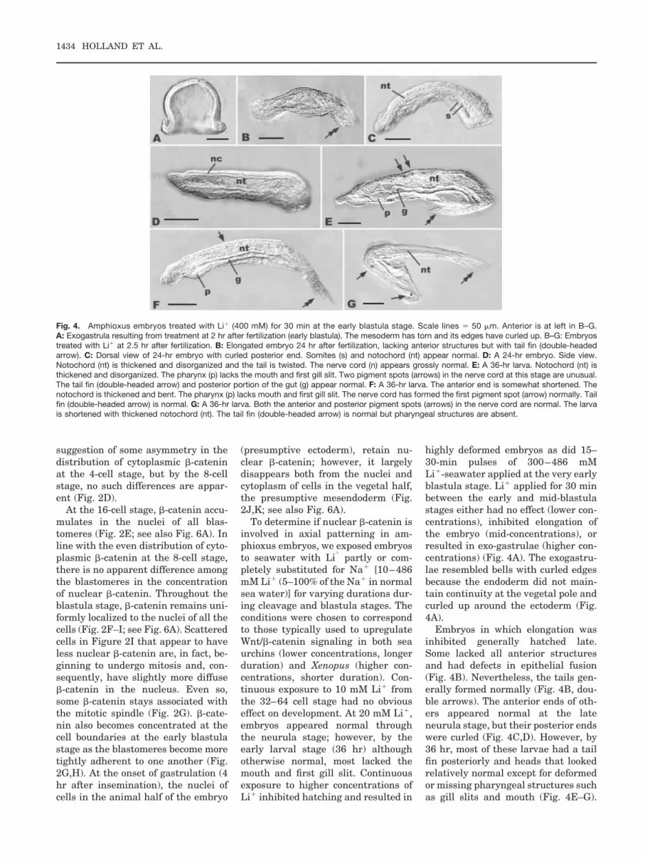
highly deformed embryos as did 15–30-min pulses of 300–486 mMLi�-seawater applied at the very earlyblastula stage. Li� applied for 30 minbetween the early and mid-blastulastages either had no effect (lower con-centrations), inhibited elongation ofthe embryo (mid-concentrations), orresulted in exo-gastrulae (higher con-centrations) (Fig. 4A). The exogastru-lae resembled bells with curled edgesbecause the endoderm did not main-tain continuity at the vegetal pole andcurled up around the ectoderm (Fig.4A).
Embryos in which elongation wasinhibited generally hatched late.Some lacked all anterior structuresand had defects in epithelial fusion(Fig. 4B). Nevertheless, the tails gen-erally formed normally (Fig. 4B, dou-ble arrows). The anterior ends of oth-ers appeared normal at the lateneurula stage, but their posterior endswere curled (Fig. 4C,D). However, by36 hr, most of these larvae had a tailfin posteriorly and heads that lookedrelatively normal except for deformedor missing pharyngeal structures suchas gill slits and mouth (Fig. 4E–G).
Fig. 4. Amphioxus embryos treated with Li� (400 mM) for 30 min at the early blastula stage. Scale lines � 50 �m. Anterior is at left in B–G.A: Exogastrula resulting from treatment at 2 hr after fertilization (early blastula). The mesoderm has torn and its edges have curled up. B–G: Embryostreated with Li� at 2.5 hr after fertilization. B: Elongated embryo 24 hr after fertilization, lacking anterior structures but with tail fin (double-headedarrow). C: Dorsal view of 24-hr embryo with curled posterior end. Somites (s) and notochord (nt) appear normal. D: A 24-hr embryo. Side view.Notochord (nt) is thickened and disorganized and the tail is twisted. The nerve cord (n) appears grossly normal. E: A 36-hr larva. Notochord (nt) isthickened and disorganized. The pharynx (p) lacks the mouth and first gill slit. Two pigment spots (arrows) in the nerve cord at this stage are unusual.The tail fin (double-headed arrow) and posterior portion of the gut (g) appear normal. F: A 36-hr larva. The anterior end is somewhat shortened. Thenotochord is thickened and bent. The pharynx (p) lacks mouth and first gill slit. The nerve cord has formed the first pigment spot (arrow) normally. Tailfin (double-headed arrow) is normal. G: A 36-hr larva. Both the anterior and posterior pigment spots (arrows) in the nerve cord are normal. The larvais shortened with thickened notochord (nt). The tail fin (double-headed arrow) is normal but pharyngeal structures are absent.
1434 HOLLAND ET AL.

Nevertheless, the trunks of these lar-vae were generally bent and twistedwith notochords that were oftenthicker than normal with disorientedcells (the amphioxus notochord con-sists mainly of muscle cells [Flood,1975]). Posterior truncations wererare. These results show that the ma-jor effects of Li� applied before themid-blastula stage are either preven-tion of mesendoderm invagination or,if the mesendoderm does invaginate,inhibition of anterior/posterior axiselongation. Thus, down-regulation of�-catenin in the mesendoderm ap-pears to be required for gastrulationand/or specification of the mesen-doderm or the ectoderm/mesendodermboundary.
At Gastrulation, a Wnt/�-Catenin Signaling CenterAround the BlastoporeConfers Posterior Identity
During the mid-late gastrula stage,nuclear �-catenin decreases in ecto-dermal cells near the animal pole, butremains highly concentrated in ecto-dermal cells around the blastopore(Fig. 2L,M). Throughout the gastrulastage, �-catenin remains undetectablein either nuclei or cytoplasm in themesendoderm (Figs. 2L, 6A). As neu-rulation begins, �-catenin becomesundetectable in both the cytoplasmand nuclei of all the neural plate cells(Figs. 2N,O, 6A), suggesting thatdown-regulation of �-catenin may benecessary for formation of the neuralplate and/or maintenance of the neu-roectoderm. In the early to mid-neu-rula, �-catenin remains localized tonuclei of the non-neural ectoderm. Inmost of these cells, �-catenin becomesrestricted to the inner periphery of thenuclear envelope, as shown by elec-tron microscopy (Figs. 2O, 3). This isprobably indicative of export of �-cate-nin from the nucleus to the cytoplasm,where it is degraded (Henderson,2000; Wieschens and Fagotto, 2000).However, the level of nuclear �-cate-nin remains high in two regions:around the blastopore, suggestingthat there is a posterior Wnt/�-cate-nin signaling center, and in non-neu-ral ectoderm immediately adjacentthe neural plate (Figs. 2N–Q, 6A). Be-fore the neural plate rounds up, thenon-neural ectoderm detaches from
the periphery of the neural plate andmigrates over the open neural plate,where it fuses in the dorsal midline.This sharp cut-off of nuclear �-cateninmay be involved in specification of theneuroectoderm/non-neuroectodermboundary. At the mid-neurula stage,the only cells with nuclear �-cateninare at the ventral, posterior side of thenearly-closed blastopore, in the ven-tral, anterior end of the embryo (Fig.2S,T) and scattered in the ectoderm(Fig. 2U), suggesting that Wnt/�-cate-nin signaling is involved in specifyingthe identity of some ectodermal cells.By the late neurula, nuclear �-cateninis concentrated only in the ectodermat the extreme anterior and posteriorends of the embryo (Fig. 2V).
To determine whether localizationof �-catenin to nuclei around the blas-topore promotes posterior develop-ment, we transferred embryos to sea-water with 85–100% of the Na�
substituted by Li� (400–484 mM Li�)during the 30 min just prior to theonset of gastrulation. Embryos werereturned to normal seawater beforethey began to gastrulate. At the high-est concentrations of Li�, develop-ment arrests at the gastrula stage. Atthe lower concentrations, gastrulationinitially appears normal (Figs. 5A,6B). However, the blastopore does notclose and the neural plate does notform. Instead, the gastrula flattensand elongates on a line drawn be-tween the posterior pole and the oppo-site lip of the blastopore. Then,whereas normal embryos elongatewith the lips of the blastopore at theposterior pole, in Li�-treated em-bryos, only one side of the ectodermelongates (compare Figs. 2L and 5A).Embryos do not elongate from the lipof the blastopore, but slightly lateralto it (Figs. 5C–I, 6B). The elongatingside is probably ventral/posterior, al-though we cannot rule out the possi-bility that it is dorsal/posterior, sincethe polar bodies are not retained dur-ing labelling and since these embryosnever express neuroectodermal mark-ers (see below). The opposite side ofthe Li�-treated embryos does notelongate substantially. Sometimes thearchenteron disappears as the mesen-doderm apparently pulls out of thearchenteron (Fig. 5G). At other times,a small archenteron persists (Figs.5H–J, 6B). In some embryos, the re-
mains of the blastocoel contain a looseassortment of cells and extracellularmaterial (Fig. 5I,L). In others, there issome differentiation of mesodermaltissues such as somites and a partialnotochord (Fig. 5M,O).
Not surprisingly, Li� alters the dis-tribution of �-catenin. At the gastrulastage, the level of cytoplasmic �-cate-nin remains relatively high, and boththe mesendoderm and ectoderm, espe-cially on the posterior/ventral side ofblastopore, retain nuclear �-catenin(Figs. 5A,B, 6B). Ectodermal cells atthe animal pole initially have less nu-clear �-catenin than those around theblastopore (Fig. 5F), but as develop-ment proceeds, the concentration of�-catenin increases in all the ectoder-mal nuclei (Fig. 5H,I). Nevertheless,in spite of previous Li� treatment,�-catenin eventually became unde-tectable in both the cytoplasm and nu-clei of the mesendoderm (Figs. 5F,G,I,6B). By 15 hr of development, whenthe embryos would normally be at themid-neurula stage, the Li�-treatedembryos are over 400 �m long com-pared to about 150 �m for normal em-bryos (compare Figs. 5K to 2Q). Thissuggests a role for Wnt/�-catenin sig-naling in tissue elongation. It is nota-ble that in cells near the center ofthese embryos, �-catenin was local-ized to the inner border of the nuclearenvelope (Fig. 5L). However, at theextremities of the embryo, particu-larly in the one that is growing out, itis distributed throughout the nucleus.
The posterior identity of the embry-onic outgrowth is shown by hybridiza-tion of Li�-treated embryos withprobes for posteriorly expressedgenes. Brachyury (AmBra2), Wnt5,Wnt1, and Wnt11, which are normallyexpressed around the lips of the blas-topore (P.W.H. Holland et al., 1995;L.Z. Holland et al., 2000a; Schubert etal., 2000b, 2001), are all expressed atthe tip of the projection (Fig. 5M–P).In an embryo with some differentia-tion of mesodermal tissues, Ambra2 isalso expressed along the midline onone side of the embryo in cells thatprobably have notochord identity.However, in embryos lacking meso-dermal structures, the normal ante-rior domains of expression for Amphi-Wnt11 are missing (Fig. 5O). Inaddition, these embryos fail to expressan anterior marker (FoxQ2) (Yu et al.,
AMPHIOXUS �-CATENIN 1435
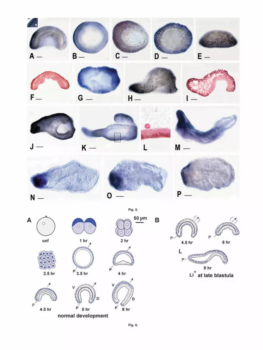
Fig. 5.
Fig. 6.

2003), showing that the anterior endof the Li�-treated embryo lacks ante-rior identity. Moreover, localization of�-catenin to the nuclei of all the ecto-dermal cells (Fig. 5H,I) and the lack ofexpression of the early neural platemarker Sox1/2/3 show that all of theectoderm is specified as non-neural.These results demonstrate that treat-ment of embryos with a high concen-tration of Li� for 30 min before gastru-lation strongly posteriorizes amphioxusembryos. Anterior identity is lost andthe dorsal ectoderm does not becomespecified as neural plate. We concludethat Wnt/�-catenin signaling confersposterior identity and that down-reg-ulation of �-catenin is essential forspecification and/or maintenance ofanterior and neuroectodermal identi-ties.
DISCUSSION
Evolution of Intron/ExonOrganization in �-Catenin
The lack of any shared intron posi-tions between Drosophila and human�-catenins led to the suggestion thateither the gene in the common ances-tor completely lacked introns or thatthere was a combination of intron in-sertion and loss during evolution thaterased the original intron/exon orga-nization (Nollet et al., 1996). Ouranalysis suggests that neither sce-nario is accurate. The high degree ofconservation of intron positions be-tween �-catenins of the cnidarian Ne-matostella and the deuterostomes ar-gues for an ancestral bilaterian�-catenin gene with at least eight in-
trons. In the deuterostome lineage,only a few introns have been gained.One, that shared among �-catenins ofDrosophila and the two invertebratedeuterostomes, may have evolved denovo in the ancestral bilaterian, al-though a loss in Nematostella cannotbe ruled out. Similarly, four intronsshared between the invertebrate deu-terostomes that are not present in Ne-matostella or Drosophila may havebeen gained after the cnidarian/deu-terostome split, while the two intronpositions common only to amphioxusand humans may have arisen de novoin their common ancestor. Loss ap-pears to be equally uncommon in deu-terostomes. Only one intron has evi-dently been lost from sea urchin andamphioxus �-catenin and only threein the human. In contrast, in the Dro-sophila lineage there has been a largeloss of introns. None of the eight in-trons shared between the cnidarianand the deuterostome �-catenins ispresent in Drosophila. The only con-served intron position in Drosophila�-catenin is one that is shared withsea urchin and amphioxus. This mas-sive loss of introns has been observedin a number of other genes not only inDrosophila but in other ecdysozoansas well (Roy and Gilbert, 2005). Ourresults suggest that this loss occurredafter the divergence of protostomesfrom the cnidarian and deuterostomelineages. However, where in the pro-tostome lineage this loss of introns oc-curred is not certain, since genome se-quences of basal ecdysozoans such asonychophorans and of lophotrochozo-
ans such as annelids and molluscs arenot yet available.
The Distribution of �-Catenin During EarlyDevelopment Is NotConserved AmongDeuterostomes
The distribution of �-catenin after fer-tilization and during cleavage stagesdiffers considerably amongst the deu-terostomes, suggesting that the rolesof this gene in early development arenot evolutionarily conserved. In am-phioxus, cytoplasmic �-catenin be-comes concentrated at the animal poleat the first cleavage, suggesting a pos-sible role in maintaining animal/vege-tal polarity during early cleavage, butby the 8-cell stage, distribution is ap-parently uniform. The pattern of cyto-plasmic �-catenin in a second speciesof amphioxus, Branchiostoma belcheri,is the same as in B. floridae throughthe two-cell stage (Yasui et al., 2002).However, these authors described apersistent asymmetry of cytoplasmic�-catenin until the mid-gastrula, thelatest stage examined. Although thiscould reflect a species difference, itmay be due to differences in affinity ofthe antibodies used in the two studiesfor the phosphorylated versus thenon-phosphorylated forms of �-cate-nin or to the lack of affinity purifica-tion of the antiserum used for B.belcheri, which on Western blots ap-pears to recognize some additionalproteins (Yasui et al., 2002; Oda et al.,2003). Indeed, in the B. belcheri studycompared to ours, there was a much
Fig. 5. Amphioxus embryos treated with Li� for 30 min at the late-blastula stage (3.5 hr after fertilization). Scale lines � 50 �m. A–L show distributionof �-catenin. A: Five-hr gastrula. Nuclear �-catenin concentrated in posterior blastoporal lip. Inset: Higher magnification of a section of the ectodermin A showing strong nuclear label. Arrowhead shows border between ectoderm and mesendoderm. Scale � 5 �m. B: Blastopore view of gastrula atsame stage as in A. �-catenin concentrated in ectodermal nuclei on one side of embryo with posterior identity. C: A 5.5-hr gastrula. Blastopore-viewof a slightly later stage than in A. Nuclear �-catenin concentrated at the posterior and lateral sides of the blastopore. D: Six-hr gastrula. Blastoporeview of an embryo at stage similar to that in C. E: Side view of embryo in D. All ectodermal cells have nuclear �-catenin. F: Longitudinal section ofembryo in E. Nuclear �-catenin most prominent in ectoderm around the blastopore. G: Seven-hr embryo. Blastopore view. �-catenin is concentratedin the ectodermal nuclei. H: Ten-hr embryo. All ectodermal nuclei have �-catenin, most concentrated in the posterior extension. I: Longitudinal sectionof embryo in H. The endoderm lacks nuclear �-catenin. A loose aggregate of cells occurs between the endoderm and ectoderm. J: twelve-hr embryo.Blastopore at right. Posterior extension has a high concentration of �-catenin. K: Fifteen-hr embryo, which is 400 �m long. The rectangle indicatessection in L. L: Section indicated by the rectangle in K. �-catenin concentrated at the inner edges of nuclei. Inside the embryo is an amorphousaggregate of extracellular material including some cells. M: Fifteen-hr embryo. In situ hybridization for brachyury. This embryo has structuresresembling somites. Brachyury heavily expressed in the posterior extension and more weakly in the somites and midline of longitudinal axis. N:Twelve-hr embryo. in situ hybridization for Wnt5. Wnt5 strongly expressed at posterior tip. O: Twelve-hr embryo. In situ hybridization for Wnt1. Wnt1strongly expressed at posterior tip. This embryo has some internal structure resembling somites. P: Twelve-hr embryo hybridized for Wnt11. Wnt11expressed at posterior tip.Fig. 6. Diagram of distribution of �-catenin in (A) normal amphioxus embryos and (B) embryos treated with Li� at the late blastula. Arrows indicatethe anterior/posterior axis. The first polar body is shown for the unfertilized egg. Both polar bodies are generally lost from embryos duringantibody-labelling. Nuclei are shown as small circles. The intensity of color is proportional to the intensity of staining for �-catenin.
AMPHIOXUS �-CATENIN 1437

higher level of cytoplasmic label,which tended to mask the nuclear la-bel (Yasui et al., 2002). In sea urchins,the distribution of cytoplasmic �-cate-nin is also uniform at the 8-cell stage,although a few embryos appear tohave higher levels of cytoplasmic�-catenin in vegetal blastomeres atthe 16-cell stage (Logan et al., 1999).In contrast to both sea urchins andamphioxus, in Xenopus, cytoplasmic�-catenin, together with other compo-nents of the canonical Wnt-signalingpathway and microtubules, is local-ized to the future dorsal side of theembryo by the cortical rotation.
In spite of differences in cytoplasmicdistribution of maternal �-catenin,nuclear localization of �-catenin oc-curs in all three of the above deuter-ostomes after the 4th cleavage (16-cellstage), although in tunicates, it occursone cleavage later (Imai et al., 2000).Not surprisingly, nuclear �-catenin ismost prominent in the cells with thehighest concentration of cytoplasmic�-catenin at the 8-cell stage. Thus, insea urchins, �-catenin becomes prefer-entially localized to vegetal nuclei (mi-cromeres and macromeres), where itfunctions in specification of the vege-tal plate and formation of mesend-oderm (Wikramanayake et al., 1998;Logan et al., 1999; reviewed in Brand-horst and Klein, 2002). Nuclear�-catenin induces secondary mesen-chyme by signaling through Notch,which in turn becomes concentrated atthe boundary between secondary mes-enchyme and presumptive endoderm(Sherwood and McClay, 1997; Sweetet al., 1999; McClay et al., 2000). Incontrast, in amphioxus, there is uni-form nuclear localization of �-cateninin all cells from the 16-cell stagethrough the late blastula/early gas-trula, which correlates with the evi-dent absence of either a dorsalizing orvegetalizing effect of Li� when appliedbefore the mid-blastula stage. While anegative result may be due to incor-rect timing or concentration of Li�,this result together with the lack ofpreferential localization of �-cateninto dorsal or vegetal nuclei suggeststhat in amphioxus, neither dorso/ven-tral polarity nor mesendodermal iden-tity is established by signaling via�-catenin. In fact, the sharp reductionof both cytoplasmic and nuclear�-catenin in presumptive mesen-
doderm suggests that, in contrastwith sea urchins, absence of �-cateninsignaling may be necessary for mesen-doderm specification in amphioxus.The down-regulation of �-catenin inpresumptive mesendoderm at the lateblastula stage correlates with up-reg-ulation of the Wnt/�-catenin inhibitorDickkopf1/2/4 in the same cells (ourunpublished data), suggesting that, asin vertebrates (Gonzalez-Sancho etal., 2005), induction of Dickkopf down-regulates the Wnt/�-catenin pathway.In ascidian tunicates, as in sea ur-chins, �-catenin accumulates in nucleiof vegetal blastomeres, specifyingthem as endoderm (Imai et al., 2000;Satou et al., 2001), although it doesnot seem to function in patterning ei-ther the dorso/ventral or anterior/pos-terior axis. Instead, endodermal cellswith nuclear �-catenin induce the no-tochord, which can be respecified asendoderm by application of Li� (Yo-shida et al., 1998; Imai et al., 2001). InXenopus, unlike other deuterostomes,�-catenin becomes preferentially in-corporated at the 16-cell stage intodorsal nuclei where it functions inspecification of dorsal identity and ac-tivation of organizer genes (Schneideret al., 1996; Larabell et al., 1997;Miller et al., 1999; Kelly et al., 2000;Schohl and Fagotto, 2002; Xanthos etal., 2002). Blockage of signaling via�-catenin in early embryos preventsformation of dorsal/anterior struc-tures (Heasman et al., 2000), whileupregulation of �-catenin signalingwith Li� has the opposite effect. Thisrole of nuclear �-catenin in dorsal/an-terior specification in Xenopus doesnot appear to be comparable to therole of �-catenin signaling in oral/ab-oral axis patterning in sea urchins,which is evidently mediated by local-ized �-catenin in the vegetal plate(Wikramanayake and Klein, 1997;Brandhorst and Klein, 2002).
A Posterior Wnt/�-CateninSignaling Center Around theBlastopore Is ConservedAmong Deuterostomes andActs in Patterning theAnterior/Posterior Axis
Although early roles of nuclear �-cate-nin in deuterostomes appear to bequite divergent, later roles in pattern-
ing along the anterior/posterior (ani-mal/vegetal) axis are evolutionarilyconserved except in the tunicates, inwhich development is considerablymodified in association with determi-nate cleavage. In all the other deuter-ostomes studied including amphioxus,a Wnt/�-catenin signaling center de-velops around the blastopore. It ischaracterized by elevated levels of nu-clear �-catenin as well as Notch andbrachyury and functions in specifica-tion of posterior identity. At the onsetof gastrulation in amphioxus, cells atthe mesendoderm/ectoderm boundaryhave high levels of nuclear �-cateninand express Brachyury and Wnt8(Zhang et al., 1997; Holland et al.,2000a; Schubert et al., 2001; Yasui etal., 2001). By the mid-gastrula, Wnt1and then Notch, Wnt3 and Wnt5 alsoturn on around the blastopore (P.W.H.Holland et al., 1995; L.Z. Holland etal., 2001; Schubert et al., 2001). Sim-ilarly, in the late sea urchin blastula,primary mesenchyme and veg2 cellslose nuclear �-catenin leaving a highconcentration in a ring of endodermcells at the endoderm/ectodermboundary (Logan et al., 1999). Wnt-8and Wnt-1 are both expressed in veg-etal cells: Wnt-8 in the vegetal plateand Wnt-1 in cells adjacent the arch-enteron (Wikramanayake et al., 1998;Ferkowicz and Raff, 2001). Notch,which controls the localization of nu-clear �-catenin in this region, andbrachyury are also concentrated incells at the endoderm/ectodermboundary (Sherwood and McClay,1997, 1999, 2001; Gross and McClay,2001; reviewed in Brandhorst andKlein 2002).
Posterior Wnt/�-catenin signaling isalso present throughout the verte-brates. Wnt/�-catenin signaling isconcentrated at the posterior end ofthe zebrafish gastrula, and in themouse and chick, in the primitivestreak and node (Dorsky et al., 2002;Mohamed et al., 2004; Schmidt et al.,2004). In the late Xenopus blastula,there is a ring of high levels of nuclear�-catenin around the marginal zone(the future mesoderm) that later local-izes to the dorsal and ventral lips ofthe blastopore (Schohl and Fagotto,2002). The patterns of XWnt-8,brachyury (Xbra) and XWnt-11 aresimilar to those of nuclear �-catenin(Smith and Harland, 1991; Christian
1438 HOLLAND ET AL.

and Moon, 1993; Lemaire and Gur-don, 1994). During the neurula stage,expression of XWnt-8 ceases, whileXWnt-3a and caudal also turn onaround the blastopore (Epstein et al.,1997; McGrew et al., 1997; Beck andSlack, 1998, 1999; Ikeya and Takada,2001; Lickert and Kemler, 2002).Brachyury, directly regulated by Wnt/�-catenin, stays turned on posteriorlyduring the neurula and tailbud stages(Gont et al., 1993; Yamaguchi et al.,1999; Arnold et al., 2000; Vonica andGumbiner, 2002). Components ofNotch-signaling (XNotch and X-Delta-1) are also expressed in poste-rior tissues from gastrulation on-wards (Beck and Slack, 1998).
Manipulation of Wnt/�-catenin sig-naling shows that in amphioxus, seaurchins, and vertebrates, Wnt/�-cate-nin signaling functions in conferringposterior identity to tissues aroundthe blastopore and in patterning theanterior/posterior axis (reviewed inHuelsken and Birchmeier, 2001; Kiec-ker and Niehrs, 2001; Brandhorst andKlein, 2002). For amphioxus, we haveshown that a pulse of Li� at the lateblastula stage slows the loss of nu-clear �-catenin from the mesen-doderm and increases its concentra-tion in posterior tissues, which elon-gate greatly and express the posteriormarkers Wnt1, Wnt5, Wnt11,brachyury. The failure of �-catenin toremain expressed in the mesend-oderm after Li� treatment is probablydue to the concomitant upregulationof Dickkopf 1/2/4 in these cells (ourunpublished data), which, after re-moval of the Li�, down-regulates�-catenin signaling. Enhanced degra-dation of �-catenin by Dickkopf-1 onwithdrawal of Li� has similarly beenobserved in human mesenchymalstem cells in culture (Gregory et al.,2005). Li�-treated amphioxus em-bryos also lose anterior identity asshown by the lack of expression ofFoxQ, an exclusive marker of the an-teriormost ectoderm in normal em-bryos (Yu et al., 2003). In addition,these embryos lose neural plate iden-tity as shown by lack of expression ofthe neural plate marker Sox1/2/3 andcontinued expression of �-catenin inthe dorsal ectoderm. The extremeelongation of amphioxus embryostreated with Li� at the late blastula isreminiscent of Xenopus animal caps
treated with Li� or overexpressingXWnt8, �-catenin, or XWnt-11, whichpreferentially signals through theWnt/JNK pathway (Tada and Smith,2000; Kuhl et al., 2001; Tada et al.,2002), and could be due to the induc-tion of convergent extension move-ments. Conversely, Wnts preferen-tially signaling through the Wnt/Ca�� pathway inhibit convergentextension (Kuhl et al., 2001; Choi andHan, 2002). In amphioxus, Wnt(s) sig-naling though all three pathways—the Wnt/�-catenin (Wnt-1), Wnt/Ca��
(Wnt-5), and Wnt/JNK (Wnt-11)—areexpressed around the blastopore ofnormal embryos and at the elongatingtip of the Li�-treated ones. Thus, it islikely that convergent-extension is in-volved in elongation of both Li�-treated and normal embryos.
In sea urchins, nuclear Wnt/�-cate-nin signaling from vegetal cells actstogether with Notch and brachyury topattern the animal/vegetal axis andcontrol the position of the ectoderm/endoderm boundary (Emily-Fenouil,et al., 1998; Wikramanayake et al.,1998; Huang et al., 2000; Vonica et al.,2000; Howard et al., 2001). Up-regu-lating Wnt/�-catenin signaling withLi� or injection of dominant/negativeforms of GSK3-� vegetalizes sea ur-chin embryos, reducing the number ofanimal cells, increasing the propor-tion of endoderm, and shifting expres-sion patterns of several genes includ-ing Brachyury toward the animal pole(Ghiglione et al., 1993; Cameron andDavidson, 1997; Emily-Fenouil et al.,1998; Gross and McClay, 2001). Block-ing Wnt/�-catenin signaling has theopposite effect, inhibiting formation ofendoderm (Logan et al., 1999; Huanget al., 2000) and blocking brachyuryexpression (Gross and McClay, 2001).
In vertebrates, Wnt/�-catening sig-naling is also essential for proper an-terior/posterior patterning. Mouseembryos deficient in �-catenin expressneither the anterior markers Hex andOtx nor the posterior markerbrachyury (Huelsken et al., 2000). InXenopus, the effects of Li�-applicationat the late blastula (Fredieu et al.,1997) are remarkably like those ofcomparable experiments in am-phioxus. Forebrain and midbrainmarkers are not expressed, the arch-enteron does not form, and blastoporeclosure is delayed or incomplete
(Fredieu et al., 1997). Experimentalevidence shows that the Wnt/�-cate-nin and Notch pathways togetherwith brachyury and caudal constitutea posterior signaling center that spec-ifies and maintains posterior identity(Beck and Slack, 1998, 1999; reviewedin Gamse and Sive, 2000; Kiecker andNiehrs, 2001). After the mid-blastulatransition, up-regulating �-cateninsignaling (i.e., injection of XWnt-8,Li�, �-catenin overexpression) inhib-its dorsal-anterior development, re-sulting in embryos with a shorteneddorsal axis, no heads or notochords,and enlarged somites (Yamaguchi andShinagawa, 1989; Cui et al., 1995; Ki-noshita and Asashima, 1995; Fredieuet al., 1997; Kao and Elinson, 1998;Domingos et al., 2001; Hamilton et al.,2001). Gene markers of ventral andlateral mesoderm are concomitantlyup-regulated (Hamilton et al., 2001).In addition, over-expression ofXWnt-3a together with active Notchinduces ectopic tails in animal cap ex-plants grafted onto the neural plate(Beck and Slack, 1998, 1999), whileover-expression of Disheveled, a com-ponent of both the Notch and Wnt-signaling pathways, posteriorizesneural tissue and activates posteriormarkers such as brachyury (Itoh andSokol, 1997). Conversely, blockingWnt/�-catenin or Notch signaling(e.g., injection of a dominant-negativeXWnt-8, antisense Wnt-8 morpholinooligonucleotide) expands anterior neu-ral fates and/or causes posterior de-fects (Takada et al., 1994; McGrew etal., 1997; Beck and Slack, 2002; Erteret al., 2001).
CONCLUSIONS
Our results suggest four main rolesfor Wnt/�-catenin signaling in earlydevelopment of amphioxus: first, information and/or maintenance of theectoderm/mesendoderm boundary andin specification of posterior identity;second, in formation of the neuroecto-derm/non-neuroectoderm boundary;third, in tissue elongation; and fourth,in specification of cell identity in theectoderm. In amphioxus, we found noevidence that nuclear �-catenin par-ticipates in establishing the dorso/ventral axis. Such a role for localized�-catenin in dorsal nuclei in verte-brates may represent a co-option of
AMPHIOXUS �-CATENIN 1439

the pathway as a result of the evolu-tion of yolky eggs, and precocious for-mation of ventral mesoderm (Kozmiket al., 2001). In amphioxus, the onlyconsistent effects of upregulation ofWnt/�-catenin signaling before themid-blastula stage are to inhibit de-velopment of pharyngeal structuresand to impair elongation of the em-bryo. Inhibition of elongation suggeststhat Wnt/�-catenin signaling may op-pose convergent/extension in am-phioxus as it does in other organisms.What is evolutionarily conserved inmost deuterostomes is a role for Wnt/�-catenin signaling in cells around theblastopore in establishing the ecto-derm/mesendoderm boundary andgiving the cells posterior identity. Inamphioxus and vertebrates, this sig-naling center is involved in patterningalong the anterior/posterior axis andin forming part of the tail organizer.
EXPERIMENTALPROCEDURES
Embryo Culture and Li�
Administration
Adult amphioxus, Branchiostomafloridae, were collected during sum-mer in Old Tampa Bay, Florida. Eggsand sperm were obtained by electricalstimulation, and embryos and larvaewere raised in laboratory cultures(Holland and Holland, 1993b). The sa-linity of Old Tampa Bay fluctuatesslowly according to the amount of localrainfall, and the animals adapt. Con-sequently, the concentration of Li� re-quired to obtain a particular morphol-ogy depends on the tonicity of theseawater to which the animals areadapted. We adjusted the tonicity ofLi� seawater, to that of the seawaterat the collection site by mixing full-strength Li� seawater (484 mM LiCl,29 mM MgSO4, 27 mM MgCl2, 10 mMKCl, 2.4 mM NaHCO3, 10 mM CaCl2)with distilled water. To obtain a par-ticular concentration of Li�, Li�-sea-water was mixed with seawater fromthe collection site. This procedureavoids effects due to altered tonicity.
Cloning of �-Catenin cDNAand Genomic Analysis
A 1,340-bp partial cDNA clone includ-ing about 60% of the coding region of
amphioxus �-catenin was obtained byPCR with primers 5�-GACCGCAGCT-GGCGTCTGGCG-3�and 5�-GCAG-GTTTACAATATGATAAGAC-3� andcloned into the pCR 2.1 vector (In-vitrogen, Inc., Carlsbad, CA). Thisclone was used to design nested prim-ers that were paired with a vector-specific primer to amplify the entirecDNA as overlapping 3� and 5� clonesfrom a gastrula through neurulacDNA library in pBluescript sk. Gene-specific primer sequences are as fol-lows: Forward primers 5�-TGCG-GATCCCAGGCCCTTGGTCAGCAC-CTGTCCCAC-3�and 5�-TGCGGATC-CCCCCGTCTGGTCCAGAACTGC-CTCTGG-3�. Reverse primers 5�-TAGGAATTCCGCGCCCGTTGCAG-GTCAGGTTCGACAG-3� and 5�-TAGGAATTCCgGCAGCGCACGT-CACAATGTTGATGTC-3�. The com-plete amphioxus �-catenin sequence isdeposited in GenBank as accessionnumber DQ013259. Intron/exon splicesites of amphioxus and cnidarian�-catenins were determined by com-paring available cDNA sequences tosequences in the trace archives ofGenBank. Because the cDNA se-quence for the cnidarian Nematostellavectensis in GenBank (accession no.AF08421) is incomplete, the 5� end ofthe �-catenin of N. vectensis and theintro/exon splice sites were deter-mined from a blast search of the N.vectensis sequences in the trace ar-chives of GenBank with the nucleotidesequence of amphioxus �-catenin. Nu-cleotide conservation between the�-catenin sequences of B. floridae andN. vectensis is surprisingly high (75–80% identity). For the intron/exon or-ganization of �-catenin from the seaurchin Strongylocentrotus purpura-tus, the �-catenin cDNA sequencefrom another sea urchin Lytechinuspictus (accession no. U34814) wasblasted to the genome sequences fromS. purpuratus available at http://sugp.caltech.edu.
Antibody Labelling and InSitu Hybridization
For antibody labelling of embryos, anaffinity-purified, polyclonal antibodygenerated against the amino-terminal173 amino acids of �-catenin of the seaurchin Lytechinus variegatus wasused (Miller and McClay, 1997). The
amino acid sequences of amphioxusand L. variegatus �-catenin are 93%identical over the c-terminal half ofthis region, and 30% identical over theamino-terminal half. This antibody isknown to recognize �-catenin in tuni-cates (Imai et al., 2000), even thoughthe comparable percentages of identi-ties with L. variegatus �-catenin areonly 60 and 17%. The antibody wasused at a dilution of 1:500. Embryosfor antibody labelling and in situ hy-bridization were fixed in 4% parafor-maldehyde in 0.1 M MOPS, 0.5 MNaCl, 1 mM EGTA, 2 mM MgSO4, pH7.4 (L.Z. Holland et al., 1996), andstored in either 100% methanol or70% ethanol. There was no differencein antibody-labelling between thesestorage methods. The protocol for an-tibody labelling is as previously pub-lished (Holland and Holland, 1993a).Staining was with nickel-enhanceddiamino-benzidine (DAB). For trans-mission electron microscopy, anti-body-labelled embryos in Spurr’s resinwere fine-sectioned and observed inan electron microscope without stain-ing with either uranyl acetate or leadcitrate. The nickel-enhanced DABproduct is electron-dense.
Methods for in situ hybridizationare in L. Z. Holland et al. (1996). Ri-boprobes were synthesized from full-length clones of AmBra1 (X91903),AmphiWnt1 (AF06194), AmphiWnt5(AF361014), AmphiWnt3 (AF3610132),AmphiWnt8 (AF190470), AmphiFoxQ(AY163864), and AmphiSox1/2/3(AF271787).
ACKNOWLEDGMENTSWe thank D. R. McClay, Duke Univer-sity, Chapel Hill, NC, for his kind giftof the �-catenin antibody. John Law-rence, University of South Florida,Tampa, FL, generously provided labo-ratory space during the amphioxusbreeding season.
REFERENCES
Arnold SJ, Stappert J, Bauer A, Kispert A,Herrmann BG, Kemler R. 2000.Brachyury is a target gene of the Wnt/�-catenin signaling pathway. Mech Dev 91:249–258.
Beck CW, Slack JMW. 1998. Analysis ofthe developing Xenopus tail bud revealsseparate phases of gene expression dur-ing determination and outgrowth. MechDev 72:41–52.
1440 HOLLAND ET AL.

Beck CW, Slack JMW. 1999. A Develop-mental pathway controlling outgrowth ofthe Xenopus tail bud. Development 126:1611–1620.
Beck CW, Slack JM. 2002. Notch is re-quired for outgrowth of the Xenopus tailbud. Int J Dev Biol 46:255–258
Brandhorst BP, Klein WH. 2002. Molecu-lar patterning along the sea urchin ani-mal-vegetal axis. Int Rev Cytol 213:183–232.
Cameron RA, Davidson EH. 1997. LiClperturbs ectodermal veg1 lineage alloca-tions in Strongylocentrutus purpuratusembryos. Dev Biol 187:236–239.
Choi S-C, Han J-K. 2002. Xenopus Cdc42regulates convergent extension move-ments during gastrulation through Wnt/Ca2� signaling pathway. Dev Biol 244:342–357.
Christian JL, Moon RT. 1993. Interactionsbetween XWnt-8 and Spemann orga-nizer signaling pathways generate dor-soventral pattern in the embryonic me-soderm of Xenopus. Genes Dev 7:13–28.
Conklin EG. 1932. The embryology of am-phioxus. J Morph 54:69–151.
Cui Y, Brown JD, Moon RT, Christian JL.1995. XWnt-8b: A maternally expressedXenopus Wnt gene with a potential rolein establishing the dorsoventral axis. De-velopment 121:2177–2186.
Dirksen M.-L, Morasso MI, Sargent TD,Jamrich M. 1994. Differential expressionof a Distal-less homeobox gene Xdll-2 inectodermal cell lineages. Mech Dev 46:63–70.
Domingos PM, Itasaki N, Jones, CM, Mer-curio S, Sargent MG, Smith JC, Krum-lauf R. 2001. The Wnt/�-catenin path-way posteriorizes neural tissue inXenopus by an indirect mechanism re-quiring FGF signalling. Dev Biol 235:148–160.
Dorsky RI, Sheldahl LC, Moon RT. 2002. Atransgenic Lef1/�-catenin-dependent re-porter is expressed in spatially restricteddomains throughout zebrafish develop-ment. Dev Biol 241:229–237.
Emily-Fenouil F, Ghiglione C, Lhomond G,Lepage T, Gache C. 1998. GSK3�/shaggymediates patterning along the animal-vegetal axis of the sea urchin embryo.Development 125:2489–2498.
Epstein M, Pillemer G, Yelin R, YisraeliJK, Fainsod A. 1997. Patterning of theembryo along the anterior-posterior axis:The role of the caudal genes. Develop-ment 124:3805–3814.
Erter CE, Wilm TP, Basler N, Wright CVE,Solnica-Krezel L. 2001. Wnt8 is requiredin lateral mesendodermal precursors forneural posteriorization in vivo. Develop-ment 128:3571–3583.
Ferkowicz MJ, Raff RA. 2001. Wnt geneexpression in sea urchin Development:Heterochronies associated with the evo-lution of developmental mode. Evol Dev3:24–33.
Flood PR. 1975. The fine structure of thenotochord of amphioxus. Symp Zool SocLondon 36:81–104.
Fredieu JR, Cui Y, Maier D, Danilchik MV,Christian JL. 1997. XWnt-8 and lithium
can act upon either dorsal mesodermalor neuroectodermal cells to cause a lossof forebrain in Xenopus embryos. DevBiol 186:100–114.
Gamse J, Sive H. 2000. Vertebrate antero-posterior patterning: The Xenopusneurectoderm as a paradigm. BioEssays22: 976-986.
Ghiglione C, Lhomond G, Lepage T, GacheC. 1993. Cell-autonomous expressionand position-dependent repression byLi� of two zygotic genes during sea ur-chin early development. EMBO J 12:87–96.
Gont LK, Steinbeisser H, Blumberg B, DeRobertis EM. 1993. Tail formation as acontinuation of gastrulation: The multi-ple cell populations of the Xenopus tail-bud derive from the late blastopore lip.Development 119:991–1004.
Gonzalez-Sancho JM, Aguilera O, GarciaJM, Pendas-Franco N, Pena C, Cal S,Garcia de Herreros A, Bonilla F, MunozA. 2005. The wnt antagonist DICK-KOPF-1 gene is a downstream target of�-catenin/TCF and is downregulated inhuman colon cancer. Oncogene 24:1098–1103.
Gregory CA, Perry AS, Reyes E, Conley A,Gunn WG, Prockop DJ. 2005. Dkk-1-de-rived synthetic peptides and lithiumchloride for the control and recovery ofadult stem cells from bone marrow.J Biol Chem. 280:2309–2323
Gross JM, McClay DR. 2001. The role ofBrachyury (T) during gastrulation move-ments in the sea urchin Lytechinus var-iegatus. Dev Biol 239:132–147.
Hamilton FS, Wheeler GN, Hoppler S.2001. Difference in XTcf-3 dependencyaccounts for change in response to�-catenin-mediated Wnt signalling inXenopusblastula.Development128:2063–2073.
Heasman J, Kofron M, Wylie C. 2000.�-catenin signaling activity dissected inthe early Xenopus embryo: a novel anti-sense approach. Dev Biol 222:124–134.
Hedgepeth CM, Conrad LJ, Zhang J,Huang H -C, Lee VMY, Klein PS. 1997.Activation of the Wnt signaling pathway:a molecular mechanism for lithium ac-tion. Dev Biol 185:82–91.
Henderson BR. 2000. Nuclear-cytoplasmicshuttling of APC regulates beta-cateninsubcellular localization and turnover.Nature Cell Biol 2:653–660.
Holland LZ. 2002. Heads or tails? Am-phioxus and the evolution of anterior-posterior patterning in deuterostomes.Dev Biol 241:209–228.
Holland LZ, Holland PWH, Holland ND.1996. Revealing homologies betweenbody parts of distantly related animalsby in situ hybridization to developmentalgenes: amphioxus versus vertebrates. InFerraris JD, Palumbi SR, editors. Molec-ular zoology, advances, strategies andprotocols. New York: Wiley-Liss, Inc. p267–282.
Holland LZ, Holland ND, Schubert M,2000a. Developmental expression of Am-phiWnt1, an amphioxus gene in the
Wnt1/wingless subfamily. Dev GenesEvol 210:522–524.
Holland LZ, Schubert M, Holland ND,Neuman T. 2000b. Evolutionary conser-vation of the presumptive neural platemarkers AmphiSox1/2/3 and Am-phiNeurogenin in the invertebrate chor-date amphioxus. Dev Biol 226:8–33.
Holland LZ, Abi Rached L, Tamme R,Kortschak D, Holland ND, Inoko H, Shi-ina T, Burgtorf C, Lardelli M. 2001.Characterization and developmental ex-pression of the amphioxus homolog ofNotch (AmphiNotch): Evolutionary con-servation of multiple expression domainsin amphioxus and vertebrates. Dev Biol232:493–507.
Holland ND, Holland LZ. 1993a. Seroto-nin-containing cells in the nervous sys-tem and other tissues during ontogeny ofa lancelet, Branchiostoma floridae. ActaZool Stockh 74:195–204.
Holland ND, Holland LZ. 1993b. Embryosand larvae of invertebrate deuteros-tomes. In: Stern C, Holland PWH, edi-tors. Essential developmental biology: anexperimental approach. Oxford: I.R.L.Press. p 21–32.
Holland ND, Panganiban G, Henyey EL,Holland LZ. 1996. Sequence and devel-opmental expression of AmphiDll, anamphioxus Distal-less gene transcribedin the ectoderm, epidermis and nervoussystem: Insights into evolution of crani-ate forebrain and neural crest. Develop-ment 122:2911–2920.
Holland PWH, Koschorz B, Holland LZ,Herrmann BG. 1995. Conservation ofBrachyury (T) genes in amphioxus andvertebrates: Developmental and evolu-tionary implications. Development 121:4283–4291.
Howard EW, Newman LA, Oleksyn DW,Angerer RC, Angerer LM. 2001. SpKrl: Adirect target of beta-catenin regulationrequired for endoderm differentiation insea urchin embryos. Development 128:365–375.
Huang L, Li X, El-Hodiri HM, Dayal S,Wikramanayake AH, Klein WH. 2000.Involvement of Tcf/Lef in establishingcell types along the animal-vegetal axisof sea urchins. Dev Genes Evol 210:73–81.
Huelsken J, Birchmeier W. 2001. New as-pects of Wnt signaling pathways inhigher vertebrates. Curr Opin Gen Dev11:547–553.
Huelsken J, Vogel R, Brinkmann V, Erd-mann B, Birchmeier C, Birchmeier W.2000. Requirement for beta-catenin inanterior-posterior axis formation inmice. J Cell Biol 148:567–578.
Imai K, Takada N, Satoh N, Satou Y. 2000.�-catenin mediates the specification ofendoderm cells in ascidian embryos. De-velopment 127:3009–3020.
Ikeya M, Takada S. 2001. Wnt-3a is re-quired for somite specification along theanteroposterior axis of the mouse em-bryo and for regulation of cdx-1 expres-sion. Mech Dev 103:27–33.
Itoh, Sokol SY. 1997. Graded amounts ofXenopus dishevelled specify discrete an-
AMPHIOXUS �-CATENIN 1441

teroposterior cell fates in prospective ec-toderm. Mech Dev 61:113–125.
Kao KR, Elinson RP. 1998. The legacy oflithium effects on development. Biol CellParis 90:585–590.
Kelly C, Chin AJ, Leatherman JL, Kozlow-ski DJ, Weinbert ES. 2000. Maternallycontrolled �-catenin-mediated signalingis required for organizer formation in thezebrafish. Development 127:3899–3911.
Kiecker C, Niehrs C. 2001. A morphogengradient of Wnt/�-catenin signalling reg-ulates anteriorposterior neural pattern-ing in Xenopus. Development 128:4189–4201.
Kinoshita K, Asashima M. 1995. Effect ofactivin and lithium on isolated Xenopusanimal blastomeres and response alter-ation at the midblastula transition. De-velopment 121:1581–1589.
Kozmik Z, Holland LZ, Schubert M, LacalliTC, Kreslova J, Vlcek C, Holland ND.2001. Characterization of amphioxusAmphiVent, an evolutionarily conservedmarker for chordate ventral mesoderm.Genesis 29:172–179.
Kuhl M, Geis K, Sheldahl LC, Pukrop T,Moon RT, Wedlich D. 2001. Antagonisticregulation of convergent extension move-ments in Xenopus by Wnt/�-catenin andWnt/Ca�� signaling. Mech Dev 106:61–76.
Lagutin OV, Zhu CC, Kobayashi D, Topc-zewski J, Shimamura K, Puelles L, Rus-sell HRC, McKinnon PJ, Solnica-KrezelL, Oliver G. 2003. Six3 repression of Wntsignaling in the anterior neuroectodermis essential for vertebrate forebrain de-velopment. Genes Dev 17:368–379.
Larabell CA, Torres M, Rowning BA,YostC, Miller JR, Wu M, Kimelman D, MoonRT. 1997. Establishment of the dorso-ventral axis in Xenopus embryos is pre-saged by early asymmetries in �-cateninthat are modulated by the Wnt signalingpathway. J Cell Biol 136:1123–1136.
Lemaire P, Gurdon JB. 1994. Vertebrateembryonic inductions. BioEssays 16:617–620.
Lickert H, Kemler R. 2002. Functionalanalysis of cis-regulatory elements con-trolling initiation and maintenance ofearly Cdx1 gene expression in themouse. Dev Dyn 225:216–220.
Logan CY, Miller JR, Ferkowicz MJ, Mc-Clay DR. 1999. Nuclear �-catenin is re-quired to specify vegetal cell fates in thesea urchin embryo. Development 129:345–357.
McClay DR, Peterson RE, Range RC, Win-ter-Vann AM, Ferkowicz MJ. 2000. A mi-cromere induction signal is activated by�-catenin and acts through Notch to ini-tiate specification of secondary mesen-chyme cells in the sea urchin embryo.Development 127:5113–5122.
McGrew LL, Hoppler S, Moon RT. 1997.Wnt and FGF pathways cooperativelypattern anteroposterior neural ectodermin Xenopus. Mech Dev 69:105–114.
Miller JR, McClay DR. 1997. Characteriza-tion of the role of cadherin in regulatingcell adhesion during sea urchin develop-ment. Dev Biol 192:323–339.
Miller JR, Rowning BA, Larabell CA,Yang-Snyder JA, Bates RL, Moon RT.1999. Establishment of the dorsal-ven-tral axis in Xenopus embryos coincideswith the dorsal enrichment of Dishev-elled that is dependent on cortical rota-tion. J Cell Biol 146:427–437.
Mohamed OA, Clarke HJ, Dufort D. 2004.�-catenin signaling marks the prospec-tive site of primitive streak formation inthe mouse embryo. Dev Dyn 231:416–424.
Moon RT, Kimelman D. 1998. From corti-cal rotation to organizer gene expression:toward a molecular explanation of axisspecification in Xenopus. BioEssays 20:536–545.
Nollet F, Berx G, Molemans F, van Roy F.1996. Genomic organization of the hu-man �-catenin gene (CTNNB1). Genom-ics 32:413–424.
Nordstrom U, Jessell TM, Edlund T. 2002.Progressive induction of caudal neuralcharacter by graded Wnt signaling. Na-ture Neurosci 5:525–532.
Oda H, Akiyama-Oda Y, Zhang S 2003.Two classic cadherin-related moleculeswith no cadherin extracellular repeats inthe cephalochordate amphioxus: distinctadhesive specificities and possible in-volvement in the development of multi-cell-layered structures. J Cell Sci 117:2757–2767.
Pandur P, Maurus D, Kuhl M. 2002. In-creasingly complex: New players enterthe Wnt signaling network. BioEssays24:881–884.
Panopoulou GD, Clark MD, Holland LZ,Lehrach H, Holland ND. 1998. Am-phiBMP2/4, an amphioxus bone morpho-genetic protein closely related to Dro-sophila decapentaplegic and vertebrateBMP2 and BMP4: Insights into evolu-tion of dorsoventral axis specification.Dev Dyn 213:130–139.
Roeser T, Stein S, Kessel M. 1999. Nuclearbeta-catenin and the development of bi-lateral symmetry in normal and LiCl-exposed chick embryos. Development126:2955–2965
Roy SW, Gilbert W. 2005. Complex earlygenes. Proc Natl Acad Sci USA 102:1986–1991.
Saneyoshi T, Kume S, Amasaki Y, Miko-shiba K. 2002. The Wnt/calcium path-way activates NF-AT and promotes ven-tral cell fate in Xenopus embryos. Nature417:295–299.
Satou Y, Imai KS, Satoh N. 2001. Earlyembryonic expression of a LIM-ho-meobox gene Cs-lhx3 is downstream ofbeta-catenin and responsible for theendoderm differentiation in Ciona savig-nyi embryos. Development 128:3559–3570.
Schiomi K, Uchida H, Keino-Masu K, MasuM. 2003. Ccd1, a novel protein with aDIX domain, is a positive regulator inthe Wnt signaling during zebrafish neu-ral patterning. Curr Biol 13:73–77.
Schmidt M, Patterson M, Farrell E, Mun-sterberg. 2004. Dynamic expression ofLef/Tcf family members and �-cateninduring chick gastrulation, neurulation,
and early limb development. Dev Dyn229:703–707.
Schneider S, Steinbeisser H, Warga RM,Hausen P. 1996. �-catenin translocationinto nuclei demarcates the dorsalizingcenters in frog and fish embryos. MechDev 57:191–198.
Schneider SQ, Finnerty JR, MartindaleMQ. 2003. Protein evolution: structure-function relationships of the oncogenebeta-catenin in the evolution of multicel-lular animals. J Exp Zool B Mol DevEvol. 295:25–44.
Schohl A, Fagotto F. 2002. beta-catenin,MAPK and Smad signaling during earlyXenopus development. Development 129:37–52.
Schohl A, Fagotto F. 2003. A role for ma-ternal beta-catenin in early mesoderminduction in Xenopus. EMBO J. 22:3303–3313.
Schubert M, Holland LZ, Panopoulou GD,Lehrach H, Holland ND. 2000a. Charac-terization of amphioxus AmphiWnt8: in-sights into the evolution of patterning ofthe embryonic dorsoventral axis. EvolDev 2:85–92.
Schubert M, Holland LZ, Holland ND.2000b. Characterization of an am-phioxus Wnt gene, AmphiWnt11, withpossible roles in myogenesis and tail out-growth. Genesis 27:1–5.
Schubert M, Holland LZ, Stokes MD, Hol-land ND. 2001. Three amphioxus Wntgenes (AmphiWnt3, AmphiWnt5, andAmphiWnt6) associated with the tailbud: The evolution of somitogenesis inchordates. Dev Biol 240:262–273.
Sherwood DR, McClay DR. 1997. Identifi-cation and localization of a sea urchinNotch homologue: insights into vegetalplate regionalization and Notch receptorregulation. Development 124:3363–3374.
Sherwood DR, McClay DR. 1999. LvNotchsignaling mediates secondary mesen-chyme specification in the sea urchin em-bryo. Development 126:1703–1713.
Sherwood DR, McClay DR. 2001. LvNotchsignaling plays a dual role in regulatingthe position of the ectoderm-endodermboundary in the sea urchin embryo. De-velopment 128:221–2232.
Smith WC, Harland RM. 1991. InjectedXWnt-8 RNA acts early in Xenopus em-bryos to promote formation of a vegetaldorsalizing center. Cell 67:753–65.
Stachel SE, Grunwald DJ, Myers PZ. 1993.Lithium perturbation and goosecoid ex-pression identify a dorsal specificationpathway in the pregastrula zebrafish.Development 117:1261–1274.
Sweet HC, Hodor PG, Ettensohn CA. 1999.The role of micromere signaling in Notchactivation and mesoderm specificationduring sea urchin embryogenesis. Devel-opment 126:5255–5265.
Tada M, Smith JC. 2000. XWnt11 is a tar-get of Xenopus Brachyury: Regulation ofgastrulation movements via dishevelled,but not through the canonical Wnt path-way. Development 127:2227–2238.
Tada M, Concha ML, Heisenberg C-P.2002. Non-canonical Wnt signalling and
1442 HOLLAND ET AL.

regulation of gastrulation movements.Semin Cell Dev Biol 13:251–260.
Takada S, Stark KL, Shea MJ, VassilevaG, McMahon JA, McMahon AP. 1994.Wnt-3a regulates somite and tailbud for-mation in the mouse embryo. Genes Dev8:174–189.
Vonica A, Gumbiner BM. 2002. ZygoticWnt activity is required for Brachyuryexpression in the early Xenopus laevisembryo. Dev Biol 250:112–127.
Vonica A, Weng W, Gumbiner BM, VenutiJM. 2000. TCF is the nuclear effector ofthe �-catenin signal that patterns thesea urchin animal-vegetal axis. Dev Biol217:230–243.
Wiechens N, Fagotto F. 2000. CRM1-andRan-independent nuclear export of�-catenin. Curr Biol 11:18–27.
Wikramanayake AH, Klein WH. 1997.Multiple signaling events specify ecto-derm and pattern the oral-aboral axis inthe sea urchin embryo. Development 124:13–20.
Wikramanayake AH, Huang L, Klein WH.1998. �-catenin is essential for pattern-
ing the maternally specified animal-veg-etal axis in the sea urchin embryo. DevBiol 95:9343–9348.
Wikramanayake AH, Hong M, Lee PN,Pang K, Byrum CA, Bince JM, Xu R,Martindale MQ. 2003. An ancient rolefor nuclear �-catenin in the evolution ofaxial polarity and germ layer segrega-tion. Nature 426:446–450.
Xanthos JB, Kofron M, Tao Q, Schaible K,Wylie C, Heasman J. 2002. The roles ofthree signaling pathways in the forma-tion and function of the Spemann orga-nizer. Development 129:4027–4043.
Yamaguchi Y, Shinagawa A. 1989. Markedalteration at midblastula transition inthe effect of lithium on formation of thelarval body pattern of Xenopus laevis.Dev Growth Differ 31:5313–541.
Yamaguchi TP, Takada S, Yoshikawa Y,Wu N, McMahon AP. 1999. T(Brachyury) is a direct target of Wnt3aduring paraxial mesoderm specification.Genes Dev 13:3185–3190.
Yasui K, Saiga H, Wang Y, Zhang PJ,Semba I. 2001. Early expressed genes
showing a dichotomous developing pat-tern in the lancelet embryo. Dev GrowthDiffer 43:185–194.
Yasui K, Li G, Wang Y, Saiga H, Zhang P,Aizawa S. 2002. �-catenin in early devel-opment of the lancelet embryo indicatesspecific determination of embryonic po-larity. Dev Growth Differ 44:467–475.
Yoshida S, Marikawa Y, Satoh N. 1998.Regulation of the trunk-tail patterningin the ascidian embryo: a possible inter-action of cascades between lithium/�-catenin and localized maternal factorpem. Dev Biol 202:264–279.
Yu J-K, Holland ND, Holland LZ. 2003.AmphiFoxQ2, a novel winged helix/fork-head gene, exclusively marks the ante-rior end of the amphioxus embryo. DevGenes Evol 213:102–105.
Zhang S-C, Holland ND, Holland LZ. 1997.Topographic changes in nascent andearly mesoderm in amphioxus embryostudies by DiI labeling and by in situhybridization for a Brachyury gene. DevGenes Evol 206:532–535.
AMPHIOXUS �-CATENIN 1443


