Embed Size (px)
Citation preview

This is a repository copy of Objective shade matching, communication, and reproduction by combining dental photography and numeric shade quantification.
White Rose Research Online URL for this paper:https://eprints.whiterose.ac.uk/164099/
Version: Accepted Version
Article:
Hein, S, Modrić, D, Westland, S orcid.org/0000-0003-3480-4755 et al. (1 more author) (2020) Objective shade matching, communication, and reproduction by combining dental photography and numeric shade quantification. Journal of Esthetic and Restorative Dentistry. ISSN 1496-4155
https://doi.org/10.1111/jerd.12641
© 2020 Wiley Periodicals LLC. This is the peer reviewed version of the following article: Hein, S, Modrić, D, Westland, S, Tomeček, M. Objective shade matching, communication, and reproduction by combining dental photography and numeric shade quantification. J Esthet Restor Dent. 2020; 1– 11. https://doi.org/10.1111/jerd.12641, which has been published in final form at https://doi.org/10.1111/jerd.12641. This article may be used for non-commercial purposes in accordance with Wiley Terms and Conditions for Use of Self-Archived Versions. Uploaded in accordance with the publisher's self-archiving policy.
[email protected]://eprints.whiterose.ac.uk/
Reuse
Items deposited in White Rose Research Online are protected by copyright, with all rights reserved unless indicated otherwise. They may be downloaded and/or printed for private study, or other acts as permitted by national copyright laws. The publisher or other rights holders may allow further reproduction and re-use of the full text version. This is indicated by the licence information on the White Rose Research Online record for the item.
Takedown
If you consider content in White Rose Research Online to be in breach of UK law, please notify us by emailing [email protected] including the URL of the record and the reason for the withdrawal request.

Authors’ PostPrint accepted for Journal of Esthetic and Restorative Dentistry
1
Shade matching without shade guides: using the eLAB system for objective shade communication S Hein, D Modrić, M Tomeček & S Westland

Authors’ PostPrint accepted for Journal of Esthetic and Restorative Dentistry
2
Clinical Article
Shade matching without shade guides: using the eLAB system for objective shade communication
Abstract Objective: The subject of this case report is the application of a newly developed workflow for objective shade communication sans visual shade assessment or the use of shade guides. Clinical Considerations: Clinical complications stemming from issues relating to esthetic integration can present a burden on the restorative team, often resulting in strenuous relationships among its members. This is particularly true for the daunting task of reliable shade matching of a maxillary single central restoration. The faithful imitation of the optical appearance of dental hard tissues with direct- and indirect restorations has been at the center of interest in a great number of publications from the realm of esthetic dentistry over the past forty years. Suggestions put forward have ranged from visual shade assessment in various forms to the introduction of instrumental shade measurement and finally to a combination of both, either with or without the use of dental photography. The present report describes a new approach to objective shade communication, by transcending the role of dental photography from its purely descriptive purpose to the level of quantification, thus abandoning the use of the established shading regimes and replacing them with a patient personal shade recipe based on the CIELAB color space instead.
Conclusions:
Objective shade communication is possible with the eLAB system by combining numeric shade quantification with dental photography. However, the skill and experience of a well-trained dental ceramist are still essential prerequisites for achieving consistent shade matching. Clinical Significance: The eLAB system presents a viable alternative to the traditional approach to shade communication and shade matching in dentistry.
KEYWORDS shade matching, shade communication, dental ceramics, natural tooth color space, coverage error, dehydration

Authors’ PostPrint accepted for Journal of Esthetic and Restorative Dentistry
3
1 INTRODUCTION Reliable shade matching of indirect restorations with natural dentition remains to be a formidable challenge, even for the most experienced restorative team. Redo’s are unfortunately common1 and costly2, providing frequent cause for friction among its members. The precise circumstances leading to shade mismatches are complex and easily underestimated. The actual process of shade matching can be distilled down to two main procedural elements, each harboring its own specific pitfalls.
- Shade selection and communication - Shade matching using available materials and techniques
Shade selection and communication In daily practice, shade selection and shade communication is still largely carried out with the use of shade guides in combination with visual shade assessment3. The well-known pitfalls of this approach include the inadequate range of available shades, their illogical distribution as well as inconsistencies among different clinicians using shade guides for visual shade assessment4. Although improvements have been attempted with the introduction of newer and more advanced shade guide systems, most notably the Vita 3D Master shade guide (Vita Zahnfabrik, Bad Säckingen, Germany), widespread adoption in dentistry has been limited5. Moreover, there doesn’t appear to be a uniform standard for tooth colors in dentistry, leading to inconsistent shading regimes among different manufacturers for materials intended for direct6,7,8 as well as for indirect use9,10,11,12,13 (Figure 1).
FIGURE 1 A Vita Classical A3 shade tab (left) compared to A3 dentin ceramic samples (d=1.00 mm) from various manufacturers. From left to right; Vita VM09, IPS e.max ceram, Norizake CZR, Creation Zi-CT, GC LiSi,
HeraCeram Zirkonia 750
Shade matching using available materials and techniques The actual process of shade matching is largely empirical and highly dependent on the dental ceramist’s level of skill and experience. Often unbeknown to many ceramists,

Authors’ PostPrint accepted for Journal of Esthetic and Restorative Dentistry
4
multiple variations of only two essential layering techniques are in common use, namely that of Yamamoto14 and that of Geller15. The precise selection process from a large range of individually shaded ceramic powders is usually based on the evaluation of images, custom drawings of shade maps and above all, personal preference and experience 16 (Figure 2). The complexity of this process usually imposes a considerable level of uncertainty regarding the predictability of shade matching in clinical reality17. The following section describe a clinical report and illustrates how the eLAB system (a standardized protocol for dental photography and colorimetry) can help to overcome some of the aforementioned problems.
FIGURE 2 The precise selection process from a large range of individually shaded ceramic powders for the purpose of shade matching is usually based on the evaluation of images, custom drawings of shade maps and
above all, personal preference and experience
2 CLINICAL REPORT A 35-year-old male patient presented in a private dental office in Zagreb, Croatia, complaining about the color of his maxillary left central incisor. The clinician opted for full crown preparation to accommodate an all-ceramic restoration. 2.1 Theoretical Background 2.1.1 Using a color space instead of shades tabs The Commission Internationale de l’Éclairage (CIE) adopted a new color space in 1976 with the official terminology CIE (1976) L*a*b*, simply abbreviated as CIELAB. The realization that equal chromaticity differences of colors of varying lightness would not yield equal visual differences, led to the desire for a more perceptually uniform color space than the previous tristimulus or chromaticity color spaces18. The CIELAB color space is an opponent-type system where the variables L*, a* and b* represent lightness, redness-greenness and yellow-blueness, respectively19. The main aim of the development of the CIELAB color space, however, was to provide a uniform practice for the quantification of color differences, which cannot be easily done with the aforementioned color spaces20. This feature makes it

Authors’ PostPrint accepted for Journal of Esthetic and Restorative Dentistry
5
suitable for dental research. A Pubmed search using the keywords “CIELAB” and “Dentistry” returned 384 results over the last 20 years that include both words, demonstrating the popularity of CIELAB among dental researchers. Due to its objectiveness, logic and the ability to express color differences numerically, CIELAB forms the foundation for the eLAB system thus replacing the use of stock shade guides. 2.1.2 Expressing color difference relative to clinical context It is acknowledged that although CIELAB is approximately visual uniform, it is not in fact a totally visual uniform color space. Gradual improvement of color difference equations resulted in CIEDE2000 (abbreviated ∆E00), a color difference formula that can more reliably predict perceived color differences. Note, however, that the CIEDE2000 equation is still based on CIELAB color space. The definition of numerical thresholds for visual perception is dependent on the exact industry and application. They may differ somewhat between them21. Paravina et al.22 have determined a ranking scale which relates measured ∆E00 color differences to clinical relevance and this approach has been adopted by the eLAB system (Figure 3).
FIGURE 3 The definition of numerical thresholds for visual perception is dependent on the exact industry and application. The 50/50 perceptibility thresholds (PT) and acceptability thresholds (AT) defined by Paravina et al
serve as a practical compass for shade evaluation under clinically realistic conditions. This ranking scale has been implemented in the eLAB system.
2.2 Practical steps and considerations 2.2.1 Photographic Protocol The eLAB system is centered around a standardized protocol for dental photography. Disciplined adherence its guidelines and sequences is essential for ensuring accurate and repeatable results. 2.2.2 Dehydration

Authors’ PostPrint accepted for Journal of Esthetic and Restorative Dentistry
6
The opacity of enamel increases due to dehydration, making teeth appear whiter than during their normal state of hydration23. From experience, this effect provides the most common cause for complication during esthetic integration. Relatively few studies are available which have examined the effects of dehydration on tooth color, and these have deployed various measurement regimes and equipment24,25,26,27. Both, Burki et al. and Suliman et al. reported color changes after 10 minutes of exposure to air that exceed the clinical 50/50 acceptability threshold (∆E00 3.84 (sd 0.16) vs. ∆E00 4.88 (sd 2.48)). Suliman et
al. also found a mean color difference of ∆E00 3.94 (sd 2.62) after only one minute of exposure to air. However, when colorimetric results are obtained for the purpose of evaluation, it is essential to make sure that like is being compared to like. For the data under consideration, settings for the chosen illuminants, standard observers and illumination geometries must be the same, otherwise no meaningful comparison is possible and thus differences between results may merely demonstrate differences in measurements conditions28. In recognition of this circumstance and in order to provide practical advice to the clinician, dehydration data was obtained for the maxillary centrals of 14 volunteers (6 females and 8 males) between the ages of 20 and 34. Baseline measurements were obtained from 0 – 40 min in 2 min intervals using the eLAB system. The results showed a mean color difference of ∆E00 1.01 (sd 0.66) after 2 min of exposure to air (Figure 4). Based on this data, standard advice has been issued to stay below the 2 min dehydration threshold for measuring the target tooth color when using the eLAB system. This step is best carried out right at the beginning of the treatment. 2.2.3 Use of cross polarization The use of cross polarization presents a convenient method to record the appearance of teeth without surface reflections (gloss) to ensure accurate color measurement29. The most straightforward approach to achieving adequate cross polarization is with the use of a ring flash, paired with the appropriate cross polarization filter (Emulation, Freiburg, Germany) (Figure 4). The use of a lateral flash is also possible, but care must be taken to ensure correct alignment between the polarizers covering the flash which must be in a perfectly perpendicular orientation to the analyzer filter placed over the lens (Figure 5). The most important implication of the use of cross polarization is the extraction of a single beam of polarized light from a burst of unpolarized light and that the rest of the light is wasted30. To account for this effect, it is of paramount importance to switch off E-TTL and to use the manual mode instead, with flash intensity set to maximum (1:1) on all channels in order to prevent severely underexposed images (Figure 7). 2.2.4 Use of white-balance card The data from the image sensor of a modern DSLR camera is typically modelled as naturally being linear. The white balance and color correction are often, though not always, linear operations upon them. That is, the white-balanced/color corrected RGB data vector at each pixel location can be seen as a linear combination (via matrix multiplication) of the raw RGB vector at the same pixel. This type of linearization is automatically carried out by the eLAB_prime software (Emulation, Freiburg, Germany) using a dedicated grey reference card (Emulation, Freiburg, Germany) using a fully linear RAW processing pipeline to mitigate the

Authors’ PostPrint accepted for Journal of Esthetic and Restorative Dentistry
7
influence from cross polarization on color temperature and above all, to synchronize DSLR cameras of different models and manufacturers reliably (Figure 8)31.
FIGURE 4 Dehydration provides the most common cause for complication during esthetic integration. CIELAB data was obtained for the maxillary centrals of 14 volunteers (6 females and 8 males) between the ages of 20 and 34. Baseline measurements were obtained from 0 – 40 min in 2 min intervals using the eLAB system. The
results showed a mean color difference of ∆E00 1.01 (sd 0.66) after 2 min of exposure to air. Error handles indicate the 95% CI interval.
FIGURE 5 The use of cross polarization presents a convenient method to record the appearance of teeth
without surface reflections (gloss) to ensure accurate color measurement. The most straightforward approach to achieving adequate cross polarization is with the use of a ring flash, paired with the appropriate cross
polarization filter

Authors’ PostPrint accepted for Journal of Esthetic and Restorative Dentistry
8
FIGURE 6 The use of a lateral flash is also possible if attention is paid to the correct alignment between the two polarizing filters covering the lens and the flash
FIGURE 7 For cross polarized photography it is of paramount importance to switch off E-TTL and to use the manual mode instead, with flash intensity set to maximum (1:1) on all channels in order to prevent severely
underexposed images
FIGURE 8 A dedicated grey reference card is used to mitigate the influence from cross polarization on color temperature and above all, to synchronize DSLR cameras of different models and manufacturers reliably. It is equipped with a hair cross style aiming circle to aid with correct positioning (a), a millimeter scale (b) and the
width of the white_balance card corresponds to the average intercanine distance of the Caucasian adult to aid in finding the correct working distance (c). The known reflectance values are noted on the grey reference card
for manual processing in proprietary software like Adobe Lightroom or Photoshop

Authors’ PostPrint accepted for Journal of Esthetic and Restorative Dentistry
9
2.2.5 Exposure and aperture settings Photo colorimetric quantification with the eLAB system is sensitive to strict adherence to fixed parameters in order to provide a uniform guidance for the measurement of tooth color using CIELAB. This leaves little room for personal preference or interpretation. The correct camera settings for exposure time is 1/125 sec and for the aperture f22 while image quality must be set to RAW at all times, regardless of the model and make of DSLR camera used (Figure 9). The associated software is capable of synchronizing over 100 different DSLR camera models with each other to a high degree of intercomparability (∆E00 ~1.00) but only if the original RAW data (i.e. Nikon Electronic Format (NEF) or Canon Raw (CR2)) is available. Compression formats like JPEG or proprietary formats like DNG (Adobe) are not supported.
FIGURE 9 Photo colorimetric quantification with the eLAB system is sensitive to strict adherence to fixed parameters in order to provide a uniform guidance for the measurement of tooth color using CIELAB. The
correct camera settings for exposure time is 1/125 sec and for the aperture f22 while image quality must be set to RAW at all times, regardless of the model and make of DSLR camera used
2.2.6 Finding the correct ISO prior to first use The only variable which requires individual definition is the correct value for the ISO. This is best determined prior to clinical implication of the eLAB system, through a simple exposure series starting with an ISO value of 100 and incremental increase towards a maximum value of ISO 400. The resulting RAW images are then batch-imported into the eLAB_prime software to determine which ISO value yields the ideal exposure for the given combination of DSLR body, macro lens and type of flash. This is easily done by referencing the exposure indicator in the top left corner of each image. A warning triangle indicates the use of incorrect settings or when severe under- or over exposure has been detected (Figure 10). 2.2.7 Shooting position and distance During image acquisition the optical axis should be normal to the vertical plane of the white-balance card which in turn should be positioned just below the incisal edges of the maxillary centrals and roughly parallel to the labial plane of the maxillary anteriors (Figure 11). It is a common error to choose a too remote working distance. The shooting distance should

Authors’ PostPrint accepted for Journal of Esthetic and Restorative Dentistry
10
roughly equal a canine-to-canine reproduction ratio. Using the view finder, care should be taken that the white-balance card runs through the middle of the frame, covering the entire lower half, while the upper half is reserved for the teeth to be quantified (Figure 12).
FIGURE 10 Prior to first use and for initial set-up it is necessary to find the correct ISO value for the chosen combination of DSLR camera, lens and type of flash. This is best determined through a simple exposure series starting with an ISO value of 100 and incremental increase towards a maximum value of ISO 400. The resulting RAW images are then batch-imported into the eLAB_prime software to determine which ISO value yields the ideal exposure by referencing the exposure indicator in the top left corner of each image. A warning triangle
indicates the use of incorrect settings or when severe under- or over exposure has been detected
FIGURE 11 During image acquisition the optical axis should be normal to the vertical plane of the white_balance card which in turn should be positioned just below the incisal edges of the maxillary centrals and roughly parallel to the labial plane of the maxillary anteriors
2.2.8 Digital work-flow Once the cross polarized image has been taken in accordance with the photographic protocol, it can be imported into the eLAB_prime application for convenient automatic calibration. The desired target shade is obtained through image analysis using artificial intelligence algorithms to determine the distribution of the most common tooth color with the help of statistical modeling. This process is carried out for two areas of the target tooth,

Authors’ PostPrint accepted for Journal of Esthetic and Restorative Dentistry
11
the middle and lower third as well as for the incisal third (Figure 13). The same process is applied when it is desired to quantify the actual substrate color instead of selecting one of 8 Natural Die Material shades (Ivoclar Vivadent, Amherst, N.Y) (Figure 14).
FIGURE 12 The shooting distance should roughly equal a canine-to-canine reproduction ratio. Using the view finder, care should be taken that the white_balance card runs through the middle of the frame, covering the
entire lower half, while the upper half is reserved for the teeth to be quantified
FIGURE 13 The desired target shade is obtained through image analysis using artificial intelligence algorithms
for quantification of the most common tooth color with the help of statistical modeling. This process is carried out for two areas of the target tooth, the middle and lower third as well as for the incisal third and mixing recipes can be generated for a variety of popular ceramic systems and framework materials including all
ceramic and metal ceramic systems or even for feldspathic veneers
2.2.9 Obtaining a patient personal mixing recipe The main strength of the eLAB system lies in its ability to provide individual shade matching recipes defined by the chromatic coordinates from an RGB image and under consideration of the substrate color as well as the amount of available space. This unique approach presents a departure from traditional shading regimes and also from its constraints and limitations. A photometrically acquired database of only few standard-shaded dentin
Authors’ PostPrint accepted for Journal of Esthetic and Restorative Dentistry
12
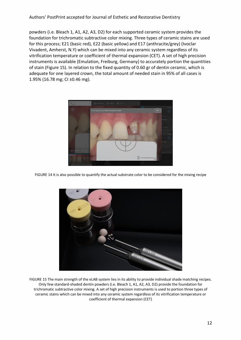
powders (i.e. Bleach 1, A1, A2, A3, D2) for each supported ceramic system provides the foundation for trichromatic subtractive color mixing. Three types of ceramic stains are used for this process; E21 (basic red), E22 (basic yellow) and E17 (anthracite/grey) (Ivoclar Vivadent, Amherst, N.Y) which can be mixed into any ceramic system regardless of its vitrification temperature or coefficient of thermal expansion (CET). A set of high precision instruments is available (Emulation, Freiburg, Germany) to accurately portion the quantities of stain (Figure 15). In relation to the fixed quantity of 0.60 gr of dentin ceramic, which is adequate for one layered crown, the total amount of needed stain in 95% of all cases is 1.95% (16.78 mg; CI ±0.46 mg).
FIGURE 14 It is also possible to quantify the actual substrate color to be considered for the mixing recipe
FIGURE 15 The main strength of the eLAB system lies in its ability to provide individual shade matching recipes.
Only few standard-shaded dentin powders (i.e. Bleach 1, A1, A2, A3, D2) provide the foundation for trichromatic subtractive color mixing. A set of high precision instruments is used to portion three types of ceramic stains which can be mixed into any ceramic system regardless of its vitrification temperature or
coefficient of thermal expansion (CET)

Authors’ PostPrint accepted for Journal of Esthetic and Restorative Dentistry
13
2.2.10 Layering strategy The main purpose of the eLAB protocol is to provide the aforementioned patient personal mixing recipe based on numeric quantification instead of relying on visual shade assessment. It is not designed to provide a complete layering map or to replace the skill and experience of a well-trained ceramist. Instead, the eLAB protocol is intended to compliment these attributes and to guide the ceramist towards a shade match that lies well within the threshold of clinical acceptability. As mentioned previously, many variations of only two archetypal types of layering strategy are in frequent use, either one of which is suitable for the eLAB protocol (Figure 16).
FIGURE 16 The main purpose of the eLAB protocl is to guide the ceramist towards a shade match that lies well within the threshold of clinical acceptability by combining quantification with traditional skills
FIGURE 17 One of the most powerful features of the eLAB_prime application is its ability to perform a semi-automated digital try-in at any stage of the manufacturing process. This not only serves the purpose of
obtaining a qualitative impression of the visual color match but also to utilize the advantages of the CIELAB color space.

Authors’ PostPrint accepted for Journal of Esthetic and Restorative Dentistry
14
FIGURE 18 The use of the ranking scale by Paravina et al makes it easy to determine the color difference for evaluation a few weeks later, showing a near excellent clinical color match
FIGURE 19a & 19b The overall visual appearance was also esthetically pleasing despite some challenges like a congenically missing maxillary lateral

Authors’ PostPrint accepted for Journal of Esthetic and Restorative Dentistry
15
2.2.11 Digital try-in One of the most powerful features of the eLAB_prime application is its ability to perform a semi-automated digital try-in at any stage of the manufacturing process (i.e. at the bisque bake or semi glaze stage). This not only serves the purpose of obtaining a qualitative impression of the visual color match but also to utilize the advantages of the CIELAB color space. The use of the ranking scale by Paravina et al. makes it easy to determine the color difference for evaluation (Figure 17). Practical corrections can thus be carried out via the targeted application of stains. 2.2.12 Integration Upon integration, the eLAB system provides the advantage of objective shade evaluation and communication for adjustments if needed. The case presented here did not require any adjustments and could be cemented during the try-in appointment. Precise shade evaluation was carried out a few weeks later showing a near excellent clinical color match (Figure 18). The overall visual appearance was also esthetically pleasing despite some challenges like a congenically missing maxillary lateral (Figure 19).
3 DISCUSSION Today, colorimetric measurements can be carried out with digital cameras on diffusely scattering objects with uneven geometries or complicated shapes and structures. This task would otherwise present a tedious and costly challenge to overcome with photo spectrometers32. This case report addresses a new approach to objective shade communication and shade reproduction in dentistry, based on numeric quantification obtained from standardized RGB images, and the formulation of a patient personal shade recipe using trichromatic subtractive color mixing laws33, thus abandoning the use of visual assessment and shade guides entirely. This approach is currently enjoying increasing popularity for its ease of use, reliability as well as for its practically oriented features like its imaging ability or the digital try-in. The measurement of heavy light scatterers like teeth presents a particular challenge due to a phenomenon referred to as “edge loss”. It describes a loss of light due to its emergence outside of the detector field which is hence excluded from quantification. To avoid this, measurements are best carried out from an appropriate distance which does not interfere with the illumination34. This particular requirement is met by the eLAB protocol. More recently however, advanced research has been conducted in the area of Biophotonics for the purpose of determining the radiative transport through different types of diffusely scattering media like tissue35 or dentin36. This has revealed a complex relationship between four optical parameters. They include the refractive index of the medium under consideration as well as the scattering and absorption coefficients and the scattering phase function which represents the scattering angular distribution37. Monte Carlo simulations are used to solve the radiative transport equation (RTE) for diffusely scattering media under consideration38, to predict their appearance under any incident light condition and viewing angle. This approach renders the evaluation of the optical appearance of natural teeth based exclusively on the concept of color (i.e. absorption and scattering), rather one-

Authors’ PostPrint accepted for Journal of Esthetic and Restorative Dentistry
16
dimensional because important components describing the radiative transport are simply ignored. Thus, a limitation of any currently available color quantification system intended for the use in dentistry, including the eLAB system, is that measured values for CIEDE color difference between a restoration and a natural tooth may suggest an imperceptible shade match, which yet contradicts dynamic visual observation under the given incident light conditions in the dental office or elsewhere. The origins for these complications are to be found in fundamental differences in material properties between dental hard tissues and dental ceramics. Especially discrepancies of the scattering phase function, which is largely isotropic in the case of dental materials39, can lead to considerable differences in radiative transport compared to dental hard tissue. This needs to be recognized by general practitioners and especially by prosthodontists who tend to be hypercritical when it comes to evaluating the quality of shade matches clinically40. Instead, the threshold recommendations for perceptibility and acceptability by Paravina et al serve as a practical compass for shade evaluation under the given limitations and the eLAB system can help to determine these parameters objectively using the advantages of the CIELAB color space. Despite the aforementioned limitations, the eLAB system and other systems based on shade quantification may at last pave the way for becoming the new standard for best practice for objective shade communication in dentistry.
4 CONCLUSION The eLAB system provides a systematic approach to shade matching in dentistry based on numeric quantification in combination with traditional skills.

Authors’ PostPrint accepted for Journal of Esthetic and Restorative Dentistry
17
REFERENCES
1 Duane Douglas R , Steinhauer TJ, Wee AG. Intraoral determination of the tolerance of dentists for perceptibility and acceptability of shade mismatch. J Prosthet Dent., 2007; 4:200-8. 2 Corcodel N, Zenthöfer A, Setz J, et al. Estimating Costs for Shade Matching and Shade Corrections of Fixed Partial Dentures for Dental Technicians in Germany: A Pilot Investigation. Acta Odontol Scand., 2011; 5:319-20. 3 Pecho Yataco O.E., Ghinea R.I., Della Bona A. Color Management and Communication in Dentistry. In: Della Bona A. (eds) Color and Appearance in Dentistry. Springer, Cham. 2020. 4Westland S, Luo W, Ellwood R, et al. Color Assessment in Dentistry. Annals of the BMVA., 2007; 4:1-10. 5 Vichi A, Louca C, Corciolani G. Color related to ceramic and zirconia restorations: A review. Dent Mat., 2011; 27:97-108. 6 Browning WD, Contreras-Bulnes R. Bracket MG, et al. Color Differences: Polymerized Composite and Corresponding Vitapan Classical Shade Tab. J Dent., 2009; 37:E34-9. 7 Carney MN, Johnston WM. Appearance Differences Between Lots and Brands of Similar Shade Designations of Dental Composite Resins. J Esthet Restor Dent., 2017; 29:E6-E14. 8 Lee YK, Yu B, Seung-Hun L, et al. Shade compatibility of esthetic restorative materials - A Review. Dent Mat., 2010; 26:1119-1126. sthet Restor Dent., 2017; 29:E6-E14. 9 Lee YK, Yu B, Seung-Hun L, et al. Shade compatibility of esthetic restorative materials - A Review. Dent Mat., 2010; 26:1119-1126. 10 Barghi N, Pedreror JA, Bosch RR. J Prosthet Dent., 1985; 5:625-7. 11 Fazi G, Vichi A, Corciolani G, et al. Spectrophotometric Evaluation of Color Match to VITA Classical Shade Guide of Four Different Veneering Porcelain Systems for Metal Ceramic Restorations. Am J Dent., 2009; 1:19-22. 12 Vichi A, Fazi G, Carrabba M, et al. Spectrophotometric Evaluation of Color Match of Three Different Porcelain Systems for All-Ceramic Zirconia-Based Restorations. Am J Dent., 2012; 4:191-4. 13 Groh CL, O’Brien WJ, Boenke KM. Differences in Color Between Fired Porcelain and Shade Guides. Int J Prosthodont., 1992; 6:510-4. 14 Yamamoto M. Metal-Ceramics. Chicago: Quintessence; 1985; 305-344. 15 Geller W. Dark and Shaded Zones - One of the Important Aspects of the W. Geller Creative Color Technic. Quintessenz Zahntech., 1982; 4:467-73. 16 Hayashi N. A Challenge to Natural Teeth – Colors & Beyond. The International Journal of Dental Technology., 2008; 2:149-168. 17 Ghulman MA, Awad MA. Color Variation Between Matched and Fabricated Shades of Different Ceramics. J Prosthodont., 2013; 6:472-7. 18 McDonald R. Acceptability and Perceptibility Decisions Using the CMC Color Difference Formula. Textile Chemist & Colorist, 1988; 6: 31-36. 19 Berns RS. Billmeyer and Saltzmann’s Principles of Color Technology. 3rd ed. New York (NY): Wiley; 2000. 20 Fairchild MD. Color Appearance models. 3rd ed. New York (NY): Wiley; 2013.

Authors’ PostPrint accepted for Journal of Esthetic and Restorative Dentistry
18
21 Choudhury AKR. Colour-difference assessment. In: Principles of color appearance and measurement. Volume 2: Visual Measurement of Colour, Colour Comparison and Management. Woodhead Publishing publications, Cambridge, U.K; 2015. 22 Paravina RD, Pérez MM, Ghinea R. Acceptability and perceptibility thresholds in dentistry: A comprehensive review of clinical and research applications. J Esthet Restor Dent. 2019;1–10. 23 Brodbelt RH, O’Brien WJ, Fan PL et al. Translucency of Human Dental Enamel. J Dent Res., 1981; 10:1749-53. 24 Russel MD, Gulfraz M, Moss BW. In vivo measurement of colour changes in natural teeth. J Oral Rehabil., 2000; 9:786-92. 25 Du RX, Li MY, Ma JF. Effect of Dehydration Time on Tooth Colour Measurement in vitro. The Chinese Journal of Dental Research., 2012; 15:37-39. 26 Burki Z, Watkins S, Wilson R, et al. A Randomised Controlled Trial to Investigate the Effects of Dehydration on Tooth Colour. J Dent., 2013; 3:250-7. 27 Suliman S, Sulaiman T, Olafsson V, et al. Effect of Time on Tooth Dehydration and Rehydration. J Esthet Restor Dent., 2019; 2:118-123. 28 Hunt RWG, Pointer MR., Precision and Accuracy in Colorimetry. In: Measuring Colour. 4th ed.:Wiley; 2011. 29 Gordon P, Wander P. Specialised equipment for dental photography. Br Dent J., 1987; 9:346-59. 30 Lipson A, Lipson SG, Lipson H. Production of polarized light. In: Optical Physics. 4th ed.: Cambridge University Press; 2011. 31 Hein S, Zangl M. The use of a standardized gray reference card in dental photography to correct the effects of five commonly used diffusers on the color of 40 extracted human teeth. Int J Esthet Dent., 2016; 2:246-59. 32 Hunt RWG, Pointer MR., Colorimetry with Digital Cameras. In: Measuring Colour. 4th ed.:Wiley; 2011. 33 Hein S, Tapia J, Bazos P. eLABor_aid: A New Approach to Digital Shade Management. Int J Esthet Dent., 2017; 2:186-202. 34 Bosch JJ, Coops JC. Tooth color and reflectance as related to light scattering and enamel hardness. J Dent Res., 1995; 74:374–380. 35 Liemert A, Reitzle D, Kienle A. Analytical Solutions of the Radiative Transport Equation for Turbid and Fluorescent Layered Media. Sci Rep., 2017; 1:3819 36 Kienle A, Foschum F, Hohmann A. Light Propagation in Structural Anisotropic Media in the Steady-State and Time Domains. Phys Med Bio., 2013; 17:6205-23. 37 M. del M. Pérez Gómez et al. Color Science and Its Application in Dentistry. In: Color and Appearance in Dentistry. 1st ed.: Springer International Publishing; 2020. 38 Ramella-Roman J, Prahl S, Jacques S. Three Monte Carlo Programs of Polarized Light Transport Into Scattering Media: Part I., Opt Express., 2005; 12:4429-38. 39 Fernández-Oliveras A, Rubiño M, Perez MM. Scattering anisotropy measurements in dental tissues and biomaterials. J. Europ. Opt. Soc. Rap. Public., 2012; 7:12016-2. 40 Al-Wahadni A et al. Shade-match perception of porcelain-fused-to-metal restorations: A comparison between dentist and patient. Dent Assoc., 2002; 9:1220-5.


