Embed Size (px)
Citation preview
Journal of Biomedical Informatics xxx (2009) xxx–xxx
ARTICLE IN PRESS
Contents lists available at ScienceDirect
Journal of Biomedical Informatics
journal homepage: www.elsevier .com/locate /y jb in
Cognitive processes as integrative component for developing expertdecision-making systems: A workflow centered framework
Ashis Jalote-Parmar a,*, Petra Badke-Schaub a, Wajid Ali b, Eigil Samset b
a Faculty of Industrial Design and Engineering, Delft University of Technology, Landbergstraat 15, 2628 CE Delft, The Netherlandsb University of Oslo, The Interventional Centre, Rikshospitalet, Sognsvannsveien 20, 0027 Oslo, Norway
a r t i c l e i n f o
Article history:Received 3 December 2008Available online xxxx
Keywords:Complex workspaceWorkflowExpert decision-making systemInformation visualizationSituation awarenessMinimally invasive surgeryMultidisciplinary team
1532-0464/$ - see front matter � 2009 Elsevier Inc. Adoi:10.1016/j.jbi.2009.07.001
* Corresponding author. Fax: +31 15 2787179.E-mail addresses: [email protected], a.jalote
Parmar).
Please cite this article in press as: Jalote-Parmarworkflow centered framework. J Biomed Inform
a b s t r a c t
The development of expert decision-making systems, which improve task performance and reduce errorswithin an intra-operative clinical workspace, is critically dependent on two main aspects: (a) Analyzingthe clinical requirements and cognitive processes within the workflow and (b) providing an optimal con-text for accurate situation awareness through effective intra-operative information visualization. Thispaper presents a workflow centered framework and its theoretical underpinnings to design expert deci-sion-making systems. The framework integrates knowledge of the clinical workflow based on therequirements within the clinical workspace. Furthermore, it builds upon and integrates the theory of sit-uation awareness into system design to improve decision-making. As an application example, this frame-work has been used to design an intra-operative visualization system (IVS), which provides imageguidance to the clinicians to perform minimally invasive procedure. An evaluative study, comparingthe traditional ultrasound guided procedure with the new developed IVS, has been conducted with expertintervention radiologists and medical students. The results reveal significant evidence for improved deci-sion-making when using the IVS. Therefore, it can be stated that this study demonstrates the benefits ofintegrating knowledge of cognitive processes into system development to support clinical decision-mak-ing and hence improvement of task performance and prevention of errors.
� 2009 Elsevier Inc. All rights reserved.
1. Introduction
To develop expert systems, that provide appropriate decision-making support for the clinician at the right time [1–3], it is re-quired to ensure that the information content and presentation iscorresponding to the clinicians’ information processing activities.Significant research in the area of medical informatics points tothe importance of understanding cognitive processes to supporthuman centered development of expert decision-making systemsfor complex workspaces [4–9]. Cognitive research investigates psy-chological processes during cognitive activities such as problemsolving and decision-making. Empirical studies illustrate the bene-fits of including cognitive theories into system design to developinformation systems, which lead to safer working environmentsand prevention of errors. Recent examples of such web based sys-tems in the clinical workspace are computer based patient recordsystems [10], knowledge management systems for bio-medicalengineering [11], computer based training systems in pathology[7], and anesthesiology training [9].
ll rights reserved.
[email protected] (A. Jalote-
A et al. Cognitive processes as(2009), doi:10.1016/j.jbi.2009
The introduction of new clinical techniques such as minimallyinvasive surgeries (MIS) has led to several technological innova-tions in the operation theatre [12]. However, inadequate informa-tion transparency, limited access, and poor visualization, compelthe clinicians to rely on advancements in medical imaging technol-ogy, which promise to improve task visualization and navigationduring interventions. These limitations in MIS are constantly givingrise to new research and development of activities in the area ofexpert decision-support systems. Such expert systems are provid-ing real-time image guidance and task automation [12,13] whilethe clinician is performing the task (intra-operatively). The theo-retical assumption is, that the expert systems should improve deci-sion-making in dynamic workspaces by enhancing situationawareness of critical information related to the clinical workflow[14,15]. The term clinical workflow is defined as the clinical problemsolving process which is determined by the task boundaries, in terms ofpossibilities and limitations, within the clinical workspace in the threephases: before (pre-operative), during (intra-operative) and after(post-operative) [16].
Reviewing the literature on recent technological development,it is obvious that aiming at the design of expert systems and pur-suing a human centered approach involves major deficiencies withrespect to the following issues:
integrative component for developing expert decision-making systems: A.07.001
2 A. Jalote-Parmar et al. / Journal of Biomedical Informatics xxx (2009) xxx–xxx
ARTICLE IN PRESS
(1) The current trends in the development of decision-supportsystems are mainly focused on applying workflow technology asa mean to optimize processes in the clinical workspace such as,component interaction [17] and imaging data automation[18,19]. However, the critical issue concerning the developmentof expert systems is not to automate clinical tasks by using work-flow technology but to develop technologies that assist the clinicalworkflow. Consequently, technologies that are being developed arestill driven by technological workflow rather than clinicalworkflow.
(2) The development of visualization support tools such as aug-mented reality [20], pre-operative planning [13] and fusion imag-ing [12] are often centered around introducing new technologies inthe clinical workspace [3,16]. There is a rare evidence that suffi-cient understanding of clinical requirements is integrated in theearly technology development phase [21]. As a consequence solu-tions are often more influenced by the latest technological trendsrather than required by the clinicians [5]. The introduction of suchtechnology may even lead to an increase in cognitive load ratherthan decreasing it, resulting in low performance and clinical errors.For example, the use of augmented reality head mounted displays(AHMD) [22] in the intra-operative clinical workspace seems to in-crease the cognitive load on the clinician. The problem with AHMDis that they require adapting to two ways of visualization at thesame time; one which is the visualization provided via AHMDand the other during the clinical task itself. Although, the sametechnology may have it’s benefits if the technology is integratedin the planning stage rather than the intra-operative stage.
To avoid a technology push into the clinical workspace, and de-velop solutions that support decision-making, requires a workflowcentered development. The paper addresses these requirementsfocusing on the following two research questions:
� How can the knowledge of the clinical workflow be included intothe system development cycle to provide a foundation for designingexpert decision-making systems?
� To what extent do expert systems, developed on the knowledge ofthe clinical workflow, aid in decision-making and improving theperformance of the clinicians by preventing errors? In this paper,the term clinician is referred for the expert such as, surgeonsand intervention radiologists who perform the MIS procedures.
The existing ISO [23] standardized human centered develop-ment cycle outlines the standard phases of product development.However, this cycle lacks in incorporating or suggesting theoreticalunderpinning necessary to tackle developmental issues in complexwork domains. Especially in the development of expert systems forclinical workspace, where complexity is determined by a lack oftransparency, unpredictability of events [24] and low tolerancefor errors [25]. Here the development of expert systems, whichprovide real-time image guidance to clinicians, requires the knowl-edge of expert decision-making in naturalistic decision-makingenvironments [26]. To support system development this knowl-edge must be investigated and incorporated in various stages ofthe development cycle. Recent research has also illustrated a meth-odology to integrate theories from cognitive science such as, dis-tributed cognition into the human centered design cycle fordesigning web based knowledge management systems [11].
This paper presents a workflow centered framework, which as-sists in developing expert systems for complex workspaces. Theframework integrates a previously developed workflow integrationmatrix [16] into the development cycle to assess the requirementswithin the clinical workflow. The framework builds upon the the-ory of situation awareness, which outlines three cognitive pro-cesses as basic elements of decision-making: perception,comprehension and action plan. These processes are integrated
Please cite this article in press as: Jalote-Parmar A et al. Cognitive processes asworkflow centered framework. J Biomed Inform (2009), doi:10.1016/j.jbi.2009
into design and evaluation of the system to improve informationvisualization as the primary basis for supporting situation aware-ness. As an example, this paper illustrates how this frameworkcan be applied in order to develop an expert decision-making sys-tem guiding minimally invasive procedures.
The organization of the paper is as follows: Section 2 explainsthe application of the development of the workflow centeredframework, which has been applied to build up an intra-operativevisualization system (IVS). Section 3 describes the design of the IVSprototype developed to provide image guidance for a selected MIS:radiofrequency ablation (RFA). Section 4 describes the experimen-tal setup of the evaluative study that compares the performance ofexpert intervention radiologists and medical students while exe-cuting RFA using the two systems: IVS and the conventional ultra-sound guided intervention. Section 5 describes the results of theevaluation study. The paper concludes with guidelines for thedevelopment of expert decision-making systems and with somecomments on future implications of the framework.
2. A workflow centered development framework
In this section, we describe the application of a workflow cen-tered framework by designing an intra-operative visualization sys-tem (IVS). IVS is an expert decision-making system, which providesreal-time (intra-operative) image guidance to the clinicians whileperforming a minimally invasive procedure called radiofrequencyablation (RFA). The workflow centered development frameworkwas generated to aid the development of expert decision-makingsystems for the multidisciplinary European Union project ARIS*ER(Augmenting Reality in Surgery) [27]. IVS has been developed to-gether in collaboration with a multidisciplinary team including cli-nicians, technology developers, and a HCI designer.
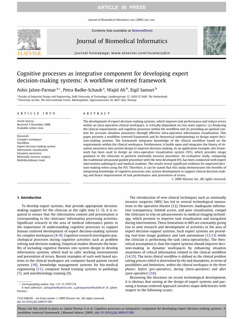
The framework integrates cognitive processes in differentphases of the human centered development cycle, which are: spec-ify context of use, analyze requirements, design prototype andevaluate prototype of the system (see Fig. 1). This development cy-cle is followed by assessing the requirements within the clinicalworkspace and integrating the knowledge of the clinical workflow.Furthermore, the framework is built upon the theory of situationawareness [14], which is regarded as the theoretical backbone forimproving information visualization in system design. Taking intoaccount the complexity of the developmental process the involve-ment of a multidisciplinary team is needed. Therefore, this frame-work also considers the issue of sharing the requirements andknowledge of the surgical and developmental processes within adevelopment team. The following section explains the applicationof the framework to develop IVS.
2.1. Phase 1: Specify context of use
In the initial phase of the development of any tool or expert sys-tem it is necessary to identify the user and specify the context inwhich the system will be used. As a development case an upcom-ing minimal invasive surgery (MIS) called, radiofrequency ablation(RFA) was selected. This selection was made by conducting inter-views (n = 10) with surgeons and intervention radiologists per-forming RFA. These interviews were conducted at nationalhospitals in Norway and The Netherlands. RFA involves the useof radiofrequency needle to ablate cancerous tumors. Surgeons orintervention radiologists mainly perform RFA either laproscopical-ly or percutaneously. Percutaneous approach of RFA was selectedfor developing an intra-operative visualization system. In this ap-proach the RF needle is inserted through the patient skin to ablatethe tumor in the liver. The key findings from the interviews aresummarized below:
integrative component for developing expert decision-making systems: A.07.001
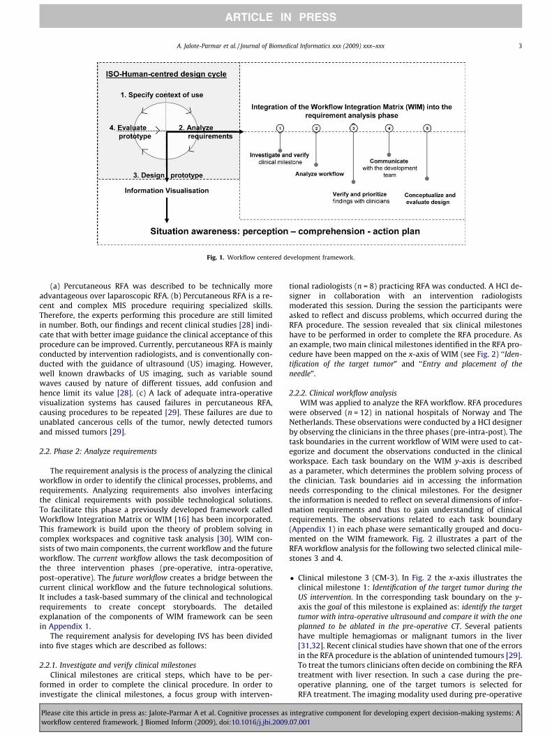
Fig. 1. Workflow centered development framework.
A. Jalote-Parmar et al. / Journal of Biomedical Informatics xxx (2009) xxx–xxx 3
ARTICLE IN PRESS
(a) Percutaneous RFA was described to be technically moreadvantageous over laparoscopic RFA. (b) Percutaneous RFA is a re-cent and complex MIS procedure requiring specialized skills.Therefore, the experts performing this procedure are still limitedin number. Both, our findings and recent clinical studies [28] indi-cate that with better image guidance the clinical acceptance of thisprocedure can be improved. Currently, percutaneous RFA is mainlyconducted by intervention radiologists, and is conventionally con-ducted with the guidance of ultrasound (US) imaging. However,well known drawbacks of US imaging, such as variable soundwaves caused by nature of different tissues, add confusion andhence limit its value [28]. (c) A lack of adequate intra-operativevisualization systems has caused failures in percutaneous RFA,causing procedures to be repeated [29]. These failures are due tounablated cancerous cells of the tumor, newly detected tumorsand missed tumors [29].
2.2. Phase 2: Analyze requirements
The requirement analysis is the process of analyzing the clinicalworkflow in order to identify the clinical processes, problems, andrequirements. Analyzing requirements also involves interfacingthe clinical requirements with possible technological solutions.To facilitate this phase a previously developed framework calledWorkflow Integration Matrix or WIM [16] has been incorporated.This framework is build upon the theory of problem solving incomplex workspaces and cognitive task analysis [30]. WIM con-sists of two main components, the current workflow and the futureworkflow. The current workflow allows the task decomposition ofthe three intervention phases (pre-operative, intra-operative,post-operative). The future workflow creates a bridge between thecurrent clinical workflow and the future technological solutions.It includes a task-based summary of the clinical and technologicalrequirements to create concept storyboards. The detailedexplanation of the components of WIM framework can be seenin Appendix 1.
The requirement analysis for developing IVS has been dividedinto five stages which are described as follows:
2.2.1. Investigate and verify clinical milestonesClinical milestones are critical steps, which have to be per-
formed in order to complete the clinical procedure. In order toinvestigate the clinical milestones, a focus group with interven-
Please cite this article in press as: Jalote-Parmar A et al. Cognitive processes asworkflow centered framework. J Biomed Inform (2009), doi:10.1016/j.jbi.2009
tional radiologists (n = 8) practicing RFA was conducted. A HCI de-signer in collaboration with an intervention radiologistsmoderated this session. During the session the participants wereasked to reflect and discuss problems, which occurred during theRFA procedure. The session revealed that six clinical milestoneshave to be performed in order to complete the RFA procedure. Asan example, two main clinical milestones identified in the RFA pro-cedure have been mapped on the x-axis of WIM (see Fig. 2) ‘‘Iden-tification of the target tumor” and ‘‘Entry and placement of theneedle”.
2.2.2. Clinical workflow analysisWIM was applied to analyze the RFA workflow. RFA procedures
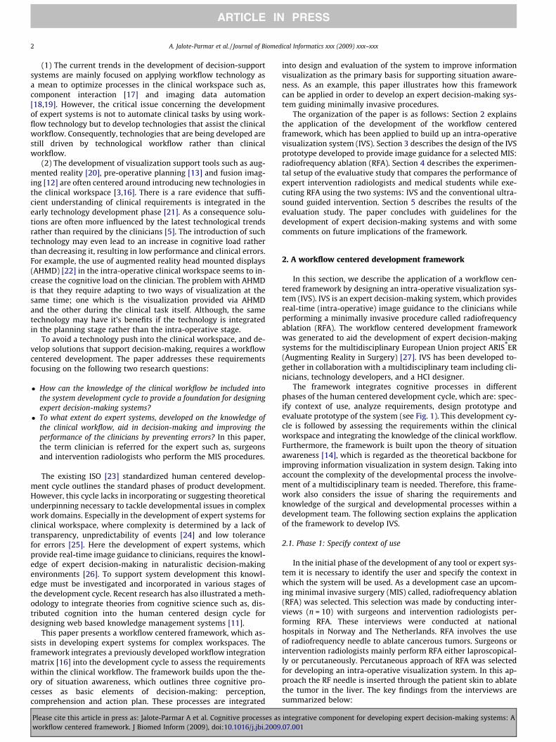
were observed (n = 12) in national hospitals of Norway and TheNetherlands. These observations were conducted by a HCI designerby observing the clinicians in the three phases (pre-intra-post). Thetask boundaries in the current workflow of WIM were used to cat-egorize and document the observations conducted in the clinicalworkspace. Each task boundary on the WIM y-axis is describedas a parameter, which determines the problem solving process ofthe clinician. Task boundaries aid in accessing the informationneeds corresponding to the clinical milestones. For the designerthe information is needed to reflect on several dimensions of infor-mation requirements and thus to gain understanding of clinicalrequirements. The observations related to each task boundary(Appendix 1) in each phase were semantically grouped and docu-mented on the WIM framework. Fig. 2 illustrates a part of theRFA workflow analysis for the following two selected clinical mile-stones 3 and 4.
� Clinical milestone 3 (CM-3). In Fig. 2 the x-axis illustrates theclinical milestone 1: Identification of the target tumor during theUS intervention. In the corresponding task boundary on the y-axis the goal of this milestone is explained as: identify the targettumor with intra-operative ultrasound and compare it with the oneplanned to be ablated in the pre-operative CT. Several patientshave multiple hemagiomas or malignant tumors in the liver[31,32]. Recent clinical studies have shown that one of the errorsin the RFA procedure is the ablation of unintended tumours [29].To treat the tumors clinicians often decide on combining the RFAtreatment with liver resection. In such a case during the pre-operative planning, one of the target tumors is selected forRFA treatment. The imaging modality used during pre-operative
integrative component for developing expert decision-making systems: A.07.001
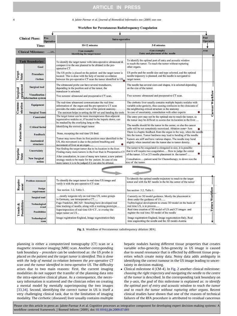
Fig. 2. Workflow of Percutaneous radiofrequency ablation (RFA).
4 A. Jalote-Parmar et al. / Journal of Biomedical Informatics xxx (2009) xxx–xxx
ARTICLE IN PRESS
planning is either a computerized tomography (CT) scan or amagnetic resonance imaging (MRI) scan. Another correspondingtask boundary – procedure can be understood as: the US probe isplaced on the patient and the target tumor is identified. This is donewith the help of mental co-relation between the pre-operative CTscan and the tumor identified in intra-operative US. The difficultyarises due to two main reasons: First, the current imagingmodalities do not support the transfer of the planning data intothe intra-operative clinical phase. As a consequence, the neces-sary information is scattered and the clinician relies on creatinga mental model by mentally superimposing the two images[33,34]. Second, identifying the correct tumor in US is itself avery challenging clinical task, due to the limitation of imagingmodality. The cirrhotic (diseased) liver usually contains multiple
Please cite this article in press as: Jalote-Parmar A et al. Cognitive processes asworkflow centered framework. J Biomed Inform (2009), doi:10.1016/j.jbi.2009
hepatic nodules having different tissue properties that createsvariable echo-genecity. Echo-genecity in US image is causeddue to sound resonance that is effected by different tissue prop-erties which create noisy data. Noisy data adds ambiguity inidentifying the correct tumour in the US image leading to uncer-tainty in decision-making.
� Clinical milestone 4 (CM-4). In Fig. 2 another clinical milestone:choosing the right trajectory and navigating the needle to the centreof the tumor is described. In the corresponding task boundary onthe y-axis, the goal of this milestone is explained as: to identifythe optimal port of entry and acoustic window to reach the tumorand to reach the tumor without rupturing other organs. Recentclinical studies have shown that one of the reasons of technicalfailures of the RFA procedure is attributed to residual cancerous
integrative component for developing expert decision-making systems: A.07.001
A. Jalote-Parmar et al. / Journal of Biomedical Informatics xxx (2009) xxx–xxx 5
ARTICLE IN PRESS
cells [28,29]. In the corresponding task boundary on the y-axis,the procedure of this milestone is explained as: First, The USprobe, the RF needle size and type is selected and, second the opti-mal needle trajectory is planned and the needle is navigated to tar-get tumor. With the guidance of US image, the clinician placesthe RFA needle into the center of the tumor. Before hitting thecenter of the tumor, the clinicians consider multiple levels ofclinical constraints before locating the right entry point and nav-igating path. The task boundary critical factor on the y-axis isunderstood as: If the trajectory is not chosen correctly, the needledoes not hit the tumor in the center, causing unablated cancer cells.The ablation zone is normally taken as 5 cm. The maximum tumorsize selected for ablation is 3 cm, in order to leave a safety margin of1 cm around it. The difficulty arises due to the fact that US imagegenerates a 2D data, while the task of hitting the tumor is a spa-tial task. Additionally, while navigating the needle the informa-tion about critical anatomical structures in the part of needlenavigation is not displayed in US. This missing information leadsto uncertainty in performing clinical tasks.
2.2.3. Verify and prioritize findings with the target user groupResults of the clinical workflow analysis serve as the basis for
the development of the expert system. Each observation or clinicalproblem identified may not be considered critical by the clinicianfor developing a technological solution. It is therefore, importantto get the documentation of the clinical procedures and require-ments verified by the clinicians after conducting the analysis.These requirements are summarized as problem statements andclinical requirements in the future workflow of WIM. An interven-tion radiologist verified and prioritized the requirements corre-sponding to each clinical milestone that can be seen as dotsplaced in the selected cells of WIM (see Fig. 2).
2.2.4. Communicate within a multidisciplinary development teamResults of the clinical workflow analysis need to be communi-
cated within the multidisciplinary development team. This wasdone by using WIM as a communication platform during focusgroup sessions. Innovative ideas, current clinical trends and possi-ble solutions (possibilities) discussed during the sessions, weredocumented in the future workflow component in WIM (see Fig. 2).
2.2.5. Conceptualize and evaluate the designFinally, WIM can be used to provide an overview of the assessed
requirements and clinical procedures of the current clinical work-flow and possible technological solutions in the future workflow.Based on the requirements several alternative concepts have beendeveloped by generating storyboards in the multidisciplinary team.The storyboards depicting IVS design were iterated with the imag-ing technologists and the intervention radiologists by consideringthe clinical and technological bottlenecks. Based on current techni-cal feasibility and clinical viability IVS prototype was developed.
The following sections will further explain the last two phases ofthe human centered design cycle: design and evaluation of the IVS.
3. Designing an intra-operative visualization system (IVS)
For developing an expert decision-making system two require-ments are of major importance, the core technological develop-ment on the one hand and the information visualization on theother. Information visualization can be understood as real-timeinformation provided to the clinician to assist in performing clini-cal tasks and decision-making. This information can originate fromvarious sources within the clinical workspace such as planninginformation based on pre-operative data, real-time imaging feed-
Please cite this article in press as: Jalote-Parmar A et al. Cognitive processes asworkflow centered framework. J Biomed Inform (2009), doi:10.1016/j.jbi.2009
back from the patient body and real-time video feedback of the ro-botic control system.
The IVS prototype was developed to provide information visual-ization during RFA to support the above mentioned two clinicalmilestones. The information visualization has been providedthrough real-time image fusion between ultrasound (US) and com-puterized tomography (CT). These two imaging modalities wereselected for image fusion because these formats were routinelyused by the intervention radiologist to perform the RFA procedure.The technology required to develop real-time image fusion is stillunder development [35]. Based on the current technical feasibilitythe IVS prototype was developed.
3.1. Theoretical framework: Theory of situation awareness
Similar to other complex workspaces such as aviation industry,the development of expert systems in the clinical workspace isdependent on the knowledge of factors that influence expert deci-sion-making in complex naturalistic environments [26]. Theoreti-cal concepts which provide an explanation on how to improveinformational support of critical factors related to clinical tasksmay assist in the development of better decision-making systems[15]. In this regard, the theory of situation awareness was consid-ered relevant [36,37]. This theory had been used to design systemsmainly in aviation such as, fighter aircrafts [38] and for pilot cock-pits [39]. Situation awareness within complex domains involvesbeing aware of what is happening across many aspects of the workenvironment. For example, while performing MIS the cliniciansmust be adequately aware of their location inside the patient body,and the location of critical organs in the path of the clinical tasks.
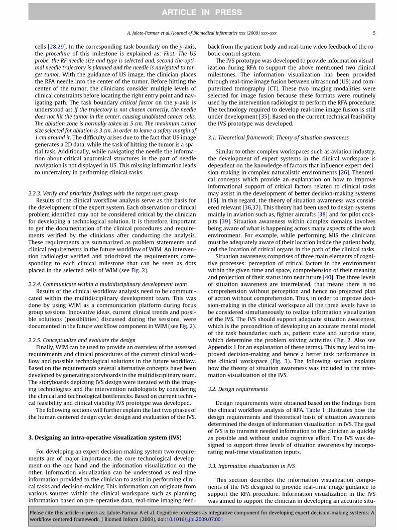
Situation awareness comprises of three main elements of cogni-tive processes: perception of critical factors in the environmentwithin the given time and space, comprehension of their meaningand projection of their status into near future [40]. The three levelsof situation awareness are interrelated, that means there is nocomprehension without perception and hence no projected planof action without comprehension. Thus, in order to improve deci-sion-making in the clinical workspace all the three levels have tobe considered simultaneously to realize information visualizationof the IVS. The IVS should support adequate situation awareness,which is the precondition of developing an accurate mental modelof the task boundaries such as, patient state and surprise state,which determine the problem solving activities (Fig. 2. Also seeAppendix 1 for an explanation of these terms). This may lead to im-proved decision-making and hence a better task performance inthe clinical workspace (Fig. 3). The following section explainshow the theory of situation awareness was included in the infor-mation visualization of the IVS.
3.2. Design requirements
Design requirements were obtained based on the findings fromthe clinical workflow analysis of RFA. Table 1 illustrates how thedesign requirements and theoretical basis of situation awarenessdetermined the design of information visualization in IVS. The goalof IVS is to transmit needed information to the clinician as quicklyas possible and without undue cognitive effort. The IVS was de-signed to support three levels of situation awareness by incorpo-rating real-time visualization inputs.
3.3. Information visualization in IVS
This section describes the information visualization compo-nents of the IVS designed to provide real-time image guidance tosupport the RFA procedure. Information visualization in the IVSwas aimed to support the clinician in developing an accurate situ-
integrative component for developing expert decision-making systems: A.07.001
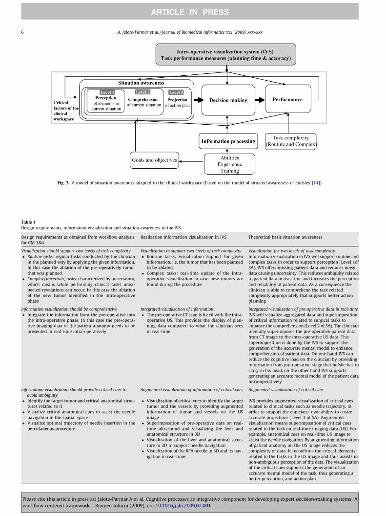
Fig. 3. A model of situation awareness adapted to the clinical workspace (based on the model of situated awareness of Endsley [14]).
Table 1Design requirements, information visualization and situation awareness in the IVS.
Design requirements as obtained from workflow analysisfor CM 3&4
Realization Information visualization in IVS Theoretical basis situation awareness
Visualization should support two levels of task complexity Visualization to support two levels of task complexity Visualization for two levels of task complexity� Routine tasks: regular tasks conducted by the clinician
in the planned way by applying the given information.In this case the ablation of the pre-operatively tumorthat was planned
� Complex (uncertain) tasks: characterized by uncertainty,which means while performing clinical tasks unex-pected revelations can occur. In this case the ablationof the new tumor identified in the intra-operativephase
� Routine tasks: visualization support for giveninformation, i.e. the tumor that has been plannedto be ablated
� Complex tasks: real-time update of the intra-operative visualization in case new tumors arefound during the procedure
Information visualization in IVS will support routine andcomplex tasks in order to support perception (Level 1ofSA). IVS offers missing patient data and reduces noisydata causing uncertainty. This reduces ambiguity relatedto patient data in real-time and increases the perceptionand reliability of patient data. As a consequence theclinician is able to comprehend the task relatedcomplexity appropriately that supports better actionplanning
Information visualization should be comprehensive Integrated visualization of information Integrated visualization of pre-operative data in real-time� Integrate the information from the pre-operative into
the intra-operative phase. In this case the pre-opera-tive imaging data of the patient anatomy needs to bepresented in real-time intra-operatively
� The pre-operative CT scan is fused with the intra-operative US. This provides the display of plan-ning data compared to what the clinician seesin real-time
IVS will visualize aggregated data and superimpositionof critical information related to surgical tasks toenhance the comprehension (Level 2 of SA). The clinicianmentally superimposes the pre-operative patient datafrom CT image to the intra-operative US data. Thissuperimposition is done by the IVS to support thegeneration of the accurate mental model to enhancecomprehension of patient data. On one hand IVS canreduce the cognitive load on the clinician by providinginformation from pre-operative stage that he/she has tocarry in his head, on the other hand IVS supportsgenerating an accurate mental model of the patient dataintra-operatively
Information visualization should provide critical cues toavoid ambiguity
Augmented visualization of information of critical cues Augmented visualization of critical cues
� Identify the target tumor and critical anatomical struc-tures related to it
� Visualize critical anatomical cues to assist the needlenavigation in the spatial space
� Visualize optimal trajectory of needle insertion in thepercutaneous procedure
� Visualization of critical cues to identify the targettumor and the vessels by providing augmentedinformation of tumor and vessels on the USimage
� Superimposition of pre-operative data on real-time ultrasound and visualizing the liver andanatomical structure in 3D
� Visualization of the liver and anatomical struc-ture in 3D to support needle navigation
� Visualization of the RFA needle in 3D and its nav-igation in real-time
IVS provides augmented visualization of critical cuesrelated to clinical tasks such as needle trajectory, inorder to support the clinicians’ own ability to createaccurate projections (Level 3 of SA). Augmentedvisualization means superimposition of critical cuesrelated to the task on real-time imaging data (US). Forexample, anatomical cues on real-time US image toassist the needle navigation. By augmenting informationof patient anatomy on the US image reduces thecomplexity of data. It reconfirms the critical elementsrelated to the tasks in the US image and thus assists innon-ambiguous perception of the data. The visualizationof the critical cues supports the generation of anaccurate mental model of the task, thus generating abetter perception, and action plan.
6 A. Jalote-Parmar et al. / Journal of Biomedical Informatics xxx (2009) xxx–xxx
ARTICLE IN PRESS
Please cite this article in press as: Jalote-Parmar A et al. Cognitive processes as integrative component for developing expert decision-making systems: Aworkflow centered framework. J Biomed Inform (2009), doi:10.1016/j.jbi.2009.07.001
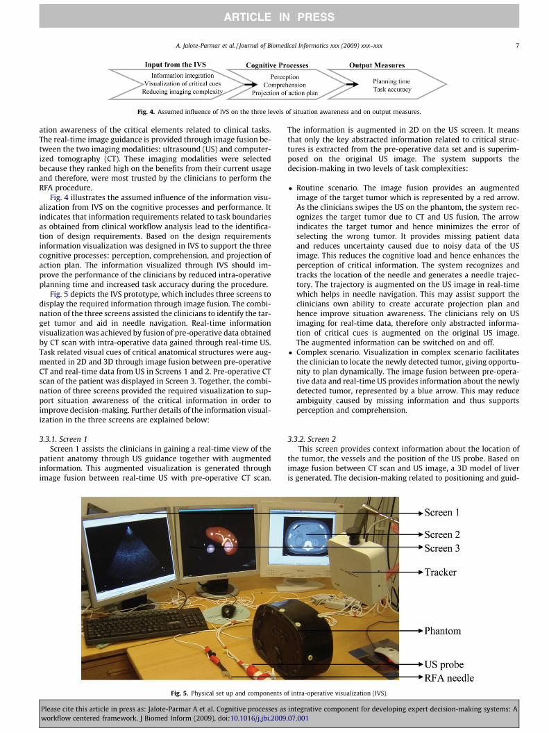
Fig. 4. Assumed influence of IVS on the three levels of situation awareness and on output measures.
A. Jalote-Parmar et al. / Journal of Biomedical Informatics xxx (2009) xxx–xxx 7
ARTICLE IN PRESS
ation awareness of the critical elements related to clinical tasks.The real-time image guidance is provided through image fusion be-tween the two imaging modalities: ultrasound (US) and computer-ized tomography (CT). These imaging modalities were selectedbecause they ranked high on the benefits from their current usageand therefore, were most trusted by the clinicians to perform theRFA procedure.
Fig. 4 illustrates the assumed influence of the information visu-alization from IVS on the cognitive processes and performance. Itindicates that information requirements related to task boundariesas obtained from clinical workflow analysis lead to the identifica-tion of design requirements. Based on the design requirementsinformation visualization was designed in IVS to support the threecognitive processes: perception, comprehension, and projection ofaction plan. The information visualized through IVS should im-prove the performance of the clinicians by reduced intra-operativeplanning time and increased task accuracy during the procedure.
Fig. 5 depicts the IVS prototype, which includes three screens todisplay the required information through image fusion. The combi-nation of the three screens assisted the clinicians to identify the tar-get tumor and aid in needle navigation. Real-time informationvisualization was achieved by fusion of pre-operative data obtainedby CT scan with intra-operative data gained through real-time US.Task related visual cues of critical anatomical structures were aug-mented in 2D and 3D through image fusion between pre-operativeCT and real-time data from US in Screens 1 and 2. Pre-operative CTscan of the patient was displayed in Screen 3. Together, the combi-nation of three screens provided the required visualization to sup-port situation awareness of the critical information in order toimprove decision-making. Further details of the information visual-ization in the three screens are explained below:
3.3.1. Screen 1Screen 1 assists the clinicians in gaining a real-time view of the
patient anatomy through US guidance together with augmentedinformation. This augmented visualization is generated throughimage fusion between real-time US with pre-operative CT scan.
Fig. 5. Physical set up and components o
Please cite this article in press as: Jalote-Parmar A et al. Cognitive processes asworkflow centered framework. J Biomed Inform (2009), doi:10.1016/j.jbi.2009
The information is augmented in 2D on the US screen. It meansthat only the key abstracted information related to critical struc-tures is extracted from the pre-operative data set and is superim-posed on the original US image. The system supports thedecision-making in two levels of task complexities:
� Routine scenario. The image fusion provides an augmentedimage of the target tumor which is represented by a red arrow.As the clinicians swipes the US on the phantom, the system rec-ognizes the target tumor due to CT and US fusion. The arrowindicates the target tumor and hence minimizes the error ofselecting the wrong tumor. It provides missing patient dataand reduces uncertainty caused due to noisy data of the USimage. This reduces the cognitive load and hence enhances theperception of critical information. The system recognizes andtracks the location of the needle and generates a needle trajec-tory. The trajectory is augmented on the US image in real-timewhich helps in needle navigation. This may assist support theclinicians own ability to create accurate projection plan andhence improve situation awareness. The clinicians rely on USimaging for real-time data, therefore only abstracted informa-tion of critical cues is augmented on the original US image.The augmented information can be switched on and off.
� Complex scenario. Visualization in complex scenario facilitatesthe clinician to locate the newly detected tumor, giving opportu-nity to plan dynamically. The image fusion between pre-opera-tive data and real-time US provides information about the newlydetected tumor, represented by a blue arrow. This may reduceambiguity caused by missing information and thus supportsperception and comprehension.
3.3.2. Screen 2This screen provides context information about the location of
the tumor, the vessels and the position of the US probe. Based onimage fusion between CT scan and US image, a 3D model of liveris generated. The decision-making related to positioning and guid-
f intra-operative visualization (IVS).
integrative component for developing expert decision-making systems: A.07.001
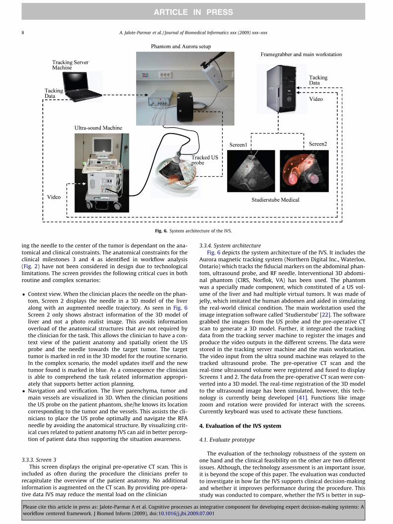
Fig. 6. System architecture of the IVS.
8 A. Jalote-Parmar et al. / Journal of Biomedical Informatics xxx (2009) xxx–xxx
ARTICLE IN PRESS
ing the needle to the center of the tumor is dependant on the ana-tomical and clinical constraints. The anatomical constraints for theclinical milestones 3 and 4 as identified in workflow analysis(Fig. 2) have not been considered in design due to technologicallimitations. The screen provides the following critical cues in bothroutine and complex scenarios:
� Context view. When the clinician places the needle on the phan-tom, Screen 2 displays the needle in a 3D model of the liveralong with an augmented needle trajectory. As seen in Fig. 6Screen 2 only shows abstract information of the 3D model ofliver and not a photo realist image. This avoids informationoverload of the anatomical structures that are not required bythe clinician for the task. This allows the clinician to have a con-text view of the patient anatomy and spatially orient the USprobe and the needle towards the target tumor. The targettumor is marked in red in the 3D model for the routine scenario.In the complex scenario, the model updates itself and the newtumor found is marked in blue. As a consequence the clinicianis able to comprehend the task related information appropri-ately that supports better action planning.
� Navigation and verification. The liver parenchyma, tumor andmain vessels are visualized in 3D. When the clinician positionsthe US probe on the patient phantom, she/he knows its locationcorresponding to the tumor and the vessels. This assists the cli-nicians to place the US probe optimally and navigate the RFAneedle by avoiding the anatomical structure. By visualizing crit-ical cues related to patient anatomy IVS can aid in better percep-tion of patient data thus supporting the situation awareness.
3.3.3. Screen 3This screen displays the original pre-operative CT scan. This is
included as often during the procedure the clinicians prefer torecapitulate the overview of the patient anatomy. No additionalinformation is augmented on the CT scan. By providing pre-opera-tive data IVS may reduce the mental load on the clinician
Please cite this article in press as: Jalote-Parmar A et al. Cognitive processes asworkflow centered framework. J Biomed Inform (2009), doi:10.1016/j.jbi.2009
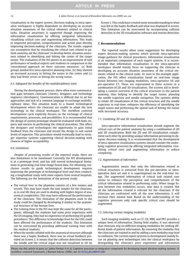
3.3.4. System architectureFig. 6 depicts the system architecture of the IVS. It includes the
Aurora magnetic tracking system (Northern Digital Inc., Waterloo,Ontario) which tracks the fiducial markers on the abdominal phan-tom, ultrasound probe, and RF needle. Interventional 3D abdomi-nal phantom (CIRS, Norflok, VA) has been used. The phantomwas a specially made component, which constituted of a US vol-ume of the liver and had multiple virtual tumors. It was made ofjelly, which imitated the human abdomen and aided in simulatingthe real-world clinical condition. The main workstation used theimage integration software called ‘Studierstube’ [22]. The softwaregrabbed the images from the US probe and the pre-operative CTscan to generate a 3D model. Further, it integrated the trackingdata from the tracking server machine to register the images andproduce the video outputs in the different screens. The data werestored in the tracking server machine and the main workstation.The video input from the ultra sound machine was relayed to thetracked ultrasound probe. The pre-operative CT scan and thereal-time ultrasound volume were registered and fused to displayScreens 1 and 2. The data from the pre-operative CT scan were con-verted into a 3D model. The real-time registration of the 3D modelto the ultrasound image has been simulated, however, this tech-nology is currently being developed [41]. Functions like imagezoom and rotation were provided for interact with the screens.Currently keyboard was used to activate these functions.
4. Evaluation of the IVS system
4.1. Evaluate prototype
The evaluation of the technology robustness of the system onone hand and the clinical feasibility on the other are two differentissues. Although, the technology assessment is an important issue,it is beyond the scope of this paper. The evaluation was conductedto investigate in how far the IVS supports clinical decision-makingand whether it improves performance during the procedure. Thisstudy was conducted to compare, whether the IVS is better in sup-
integrative component for developing expert decision-making systems: A.07.001
A. Jalote-Parmar et al. / Journal of Biomedical Informatics xxx (2009) xxx–xxx 9
ARTICLE IN PRESS
porting the decision-making and performance of expert interven-tion radiologists and medical students in comparison to the con-ventional ultrasound guided (USG). This is the final phase of theclinical development cycle which includes the evaluation of theIVS prototype.
4.2. Participants
Eight expert intervention radiologists, who were practicing RFAor biopsy procedures, were selected as participants. These expertswere associated with the Rikshospitalet and Radium HospitaletOslo, Norway and had 8–20 years of experience in interventionradiology. It is important to mention that RFA has been recentlyintroduced in the medical field for treating cancer of liver tumors.Therefore, the ratio of experts practicing this procedure is limited.Eight experts were the maximum number of experts available forthe study in Oslo. In addition, (n = 8) final year medical studentsof the Rikshospitalet Oslo also participated in the study. All the se-lected student participants were required to have primary knowl-edge and understanding of CT scans and working with ultrasoundsystem. This was a difficult selection to make, as the usage of boththe imaging modalities is not a part of the standard educationcurriculum of the final year medical students. Only by personalinterest, the medical students learned the usage of imagingmodalities. The students had no previous training on performingRFA procedures.
4.3. Experimental set up
Each participant was given an hour of training time on the IVSand the US. Although, 1 h is limited time to get acquainted withIVS it was the maximum time that was available with the expertsand the students. In case of students, half an hour more was keptfor the training as most of them were new to performing interven-tions by using the US. In the training period, the participants had toperform several tasks of hitting the centre of a tumor by using boththe systems. For the final task, each participant was given twotasks of hitting the center of the tumor again by using both sys-tems. The usage of the system was alternated between the partic-ipants. First, four experts and four students were asked to performthe tasks using the US and then the IVS. This situation was reversedfor the next group of participants. Two levels of task complexityroutine and complex were selected. These can be further under-stood as:
� Routine scenario: This task required the participants to ablate thetumor that was selected for ablation during the pre-operativeplanning stage. To simulate this clinical scenario in the experi-ment, one of the tumors was highlighted on the CT of theabdominal phantom. The participants were required to ablatethe selected tumor by using the two different systems.
� Complex scenario: This task required the participants to ablate thetumor which was newly detected while conducting intra-opera-tive US. This newly detected tumour was not visible in the pre-operative CT, thus causing uncertainty in the originally plannedclinical action. To simulate this complex scenario in the experi-ment, a tumor existing in the abdominal phantom was hiddenin the CT scan. The hidden tumor was visible to the participantsonly while conducting the intra-operative US. The participantswere expected to dynamically plan the RFA procedure.
4.4. Measurement
The following data were assessed to compare the output of theparticipants:
Please cite this article in press as: Jalote-Parmar A et al. Cognitive processes asworkflow centered framework. J Biomed Inform (2009), doi:10.1016/j.jbi.2009
4.4.1. Performance measuresThe two main criteria selected for measuring task performance
are: intra-operative planning time to execute the task and the taskaccuracy in hitting the center of the tumor with the RFA needle.Statistical analysis was performed using Wilcoxon signed-rankedtest.
� Intra-operative task planning time. The planning time has beenmeasured as the time taken by the participants to plan the pro-cedure intra-operatively. This is measured as the time takenafter explaining the task to the participant till the time he/sheis ready to execute the task. Time to perceive and comprehendinformation towards deciding on an action plan is an importantcriterion influencing the task performance. It is a well estab-lished fact that the integration of information is an importantcognitive strategy to reduce information overload [42]. It isassumed that the visualization of integrated information byIVS reduces the mental load and thus the intra-operative plan-ning time.
� Task accuracy. Clinical findings state that the major cause ofclinical errors performing the RFA procedure is either causedby the wrong tumor hit, or not hitting in the center of the tumorcausing unablated cancer cells [29]. In addition to informationintegration, the visualization of critical cues is essential to sup-port decision-making. Therefore, it is assumed that by providingthe critical cues related to the patient’s anatomy will assist theclinicians to identify and hit the right tumor in the center. As aconsequence, it will improve the task accuracy and thus the clin-ical viability of the RFA procedure. The accuracy of hitting thecenter of the tumor was measured as the distance betweenthe points of needle insertion by the participant and the mathe-matical center of the tumor.
� Hitting the wrong tumor. The participant’s accuracy of hittingthe correct target tumor during the task was measured. Thewrong tumor hit was measured by the distance between theneedle hit and the center of the target tumor.
4.4.2. Evaluation by the participantsSubjective measures were integrated in conjunction with per-
formance data to gain a true understanding of self reported evalu-ations [42]. Follow up questions were asked to each participantimmediately after the study. A questionnaire with a 5 point Likertscale was used to evaluate the subjective opinion. The participantswere asked to rank the visualization support and the ‘felt’ situationawareness obtained through both of the systems.
5. Results
The results from the evaluative study were analyzed in terms ofperformance related to intra-operative planning time and taskaccuracy are explained below:
5.1. Performance measures
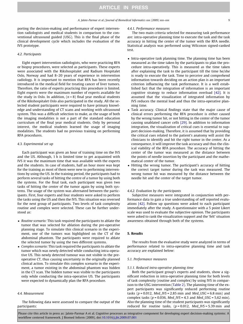
5.1.1. Reduced intra-operative planning timeBoth the participant group’s experts and students, show a sig-
nificant reduction in intra-operative planning time for both levelsof task complexity (routine and complex) by using IVS in compar-ison to the USG intervention (Table 2). The planning time of the ex-pert participants was significantly reduced performing routinetasks (p = 0.012, Med_IVS = 2.85 min and Med_USG = 6.8 min) andcomplex tasks (p = 0.036, Med_IVS = 4.3 and Med_USG = 5,62 min).Also the planning time of the student participants was significantlyreduced for routine tasks, (p = 0.012, Med_IVS = 5.39 min and
integrative component for developing expert decision-making systems: A.07.001
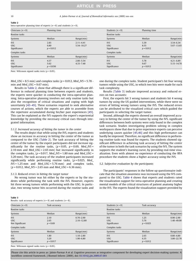
Table 2Intra-operative planning time of experts (n = 8) and students (n = 8).
Clinicians (n = 8) Planning time Students (n = 8) Planning time
Routine tasks Routine tasks
Systems Median Range(min) Systems Median Range(min)
IVS 2.85 2.11–4.41 IVS 5.39 3.54–6.34USG 6.80 5.54–10.27 USG 8.55 5.67–13.65Significance p = 0.012 Significance p = 0.012
Complex tasks Complex tasks
Systems Median Range(min) Systems Median Range(min)
IVS 4.37 2.80–5.54 IVS 5.78 4.21–6.89USG 5.62 4.33–7.40 USG 9.67 5.15–14.70Significance p = 0.036 Significance p = 0.012
Note: Wilcoxon signed ranks tests (p < 0.05).
10 A. Jalote-Parmar et al. / Journal of Biomedical Informatics xxx (2009) xxx–xxx
ARTICLE IN PRESS
Med_USG = 8.5 min) and complex tasks (p = 0.012, Med_IVS = 5.78 -min and Med_USG = 9.67 min).
Results in Table 2, show that although there is a significant dif-ference in reduced planning time between experts and students,the experts were quicker in conducting the intra-operative plan-ning. Intra-operative planning involves not only routine tasks butalso the recognition of critical situations and coping with highuncertainty [43–45]. These scenarios required to seek alternativecourses of actions, which the experts are able to assemble fromthe repertoire accumulated during his/her past experiences [45].This can be explained as the IVS supports the expert’s experientialknowledge by providing the necessary critical cues through inte-grated information.
5.1.2. Increased accuracy of hitting the tumor in the centerThe results depict that while using the IVS, experts and students
show an increase in accuracy in hitting the center of the tumor ascompared to the USG (Table 2). The task accuracy of hitting thecenter of the tumor by the expert participants did not increase sig-nificantly for the routine tasks, (p < 0.05, p = 0.69, Med_IVS =2.10 mm and Med_USG = 2.65 mm) but increased significantly inthe complex tasks, (p = 0.017, Med_IVS = 1.80 mm and Med_USG =3.20 mm). The task accuracy of the student participants increasedsignificantly while performing routine tasks, (p = 0.025, Med_IVS = 1.25 mm and Med_USG = 5.76 mm) and complex tasks,(p = 0.012, Med_IVS = 2.65 mm and Med_USG = 6.36 mm).
5.1.3. Reduced errors in hitting the target tumorNo wrong tumor was hit either by the experts or by the stu-
dents while performing the task with the IVS. However, expertshit three wrong tumors while performing with the USG. In partic-ular, two wrong tumor hits occurred during the routine tasks and
Table 3Results: task accuracy of experts (n = 8) and students (n = 8).
Clinicians (n = 8) Task accuracy
Routine tasks
Systems Median Range(mm)
IVS 2.10 0.70–2.90USG 2.65 2.10–12.80Not Significant p = 0.69
Complex tasks
Systems Median Range(mm)
IVS 1.80 1.10–2.80USG 3.20 1.90–6.80Significance p = 0.017
Note: Wilcoxon signed ranks tests (p < 0.05).
Please cite this article in press as: Jalote-Parmar A et al. Cognitive processes asworkflow centered framework. J Biomed Inform (2009), doi:10.1016/j.jbi.2009
one during the complex tasks. Student participant’s hit four wrongtumors while using the USG, in which two hits were made for eachtask complexity.
Results (Table 3) indicate improved accuracy and reduced er-rors on two accounts:
First, the experts hit 3 wrong tumors and students hit 4 wrongtumors by using the US guided interventions, while there were noerrors of hitting wrong tumors using the IVS. The reduced errorscan be attributed to the visualized critical cues which guided theparticipants in selecting the target tumor.
Second, although the experts showed an overall improved accu-racy in hitting the center of the tumor by using the IVS, significantdifferences between both systems were only found in the complextask scenario. Studies investigating problem solving in complexworkspaces show that due to prior experience experts can perceiveunderlying causes quicker [45,46] and this high performance canhardly be improved. Therefore, no significant difference in perform-ing routine tasks was found for experts. The students showed sig-nificant difference in achieving task accuracy of hitting the centerof the tumor in both the task scenarios by using the IVS. The systemsupports the student’s learning curve, by providing real-time visu-alization. Even with almost no experience of conducting the RFAprocedure the students show a higher accuracy using the IVS.
5.2. Subjective evaluation by the participants
The participants’ responses in the follow up questionnaire indi-cate that the situation awareness was increased using the IVS com-pared to the USG. Table 4 shows that experts and students ratedthe visualization support for intra-operative planning and creatingmental models of the critical structures of patient anatomy higherin the IVS. The experts found the visualization support provided by
Students (n = 8) Task accuracy
Routine tasks
Systems Median Range(mm)
IVS 1.25 0.90–2.90USG 5.76 1.40–49.00Significance p = 0.025
Complex tasks
Systems Median Range(mm)
IVS 2.65 0.80–4.80USG 6.35 3.80–22.78Significance p = 0.012
integrative component for developing expert decision-making systems: A.07.001
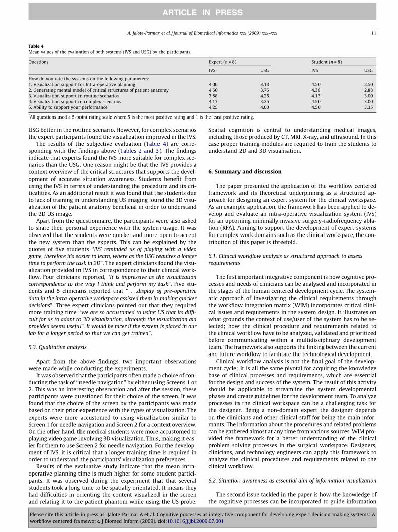
Table 4Mean values of the evaluation of both systems (IVS and USG) by the participants.
Questions Expert (n = 8) Student (n = 8)
IVS USG IVS USG
How do you rate the systems on the following parameters:1. Visualization support for Intra-operative planning 4.00 3.13 4.50 2.502. Generating mental model of critical structures of patient anatomy 4.50 3.75 4.38 2.883. Visualization support in routine scenarios 3.88 4.25 4.13 3.004. Visualization support in complex scenarios 4.13 3.25 4.50 3.005. Ability to support your performance 4.25 4.00 4.50 3.35
*All questions used a 5-point rating scale where 5 is the most positive rating and 1 is the least positive rating.
A. Jalote-Parmar et al. / Journal of Biomedical Informatics xxx (2009) xxx–xxx 11
ARTICLE IN PRESS
USG better in the routine scenario. However, for complex scenariosthe expert participants found the visualization improved in the IVS.
The results of the subjective evaluation (Table 4) are corre-sponding with the findings above (Tables 2 and 3). The findingsindicate that experts found the IVS more suitable for complex sce-narios than the USG. One reason might be that the IVS provides acontext overview of the critical structures that supports the devel-opment of accurate situation awareness. Students benefit fromusing the IVS in terms of understanding the procedure and its cri-ticalities. As an additional result it was found that the students dueto lack of training in understanding US imaging found the 3D visu-alization of the patient anatomy beneficial in order to understandthe 2D US image.
Apart from the questionnaire, the participants were also askedto share their personal experience with the system usage. It wasobserved that the students were quicker and more open to acceptthe new system than the experts. This can be explained by thequotes of five students ‘‘IVS reminded us of playing with a videogame, therefore it’s easier to learn, where as the USG requires a longertime to perform the task in 2D”. The expert clinicians found the visu-alization provided in IVS in correspondence to their clinical work-flow. Four clinicians reported, ‘‘It is impressive as the visualizationcorrespondence to the way I think and perform my task”. Five stu-dents and 5 clinicians reported that ‘‘ . . .display of pre-operativedata in the intra-operative workspace assisted them in making quickerdecisions”. Three expert clinicians pointed out that they requiredmore training time ‘‘we are so accustomed to using US that its diffi-cult for us to adapt to 3D visualization, although the visualization aidprovided seems useful”. It would be nicer if the system is placed in ourlab for a longer period so that we can get trained”.
5.3. Qualitative analysis
Apart from the above findings, two important observationswere made while conducting the experiments.
It was observed that the participants often made a choice of con-ducting the task of ‘‘needle navigation” by either using Screens 1 or2. This was an interesting observation and after the session, theseparticipants were questioned for their choice of the screen. It wasfound that the choice of the screen by the participants was madebased on their prior experience with the types of visualization. Theexperts were more accustomed to using visualization similar toScreen 1 for needle navigation and Screen 2 for a context overview.On the other hand, the medical students were more accustomed toplaying video game involving 3D visualization. Thus, making it eas-ier for them to use Screen 2 for needle navigation. For the develop-ment of IVS, it is critical that a longer training time is required inorder to understand the participants’ visualization preferences.
Results of the evaluative study indicate that the mean intra-operative planning time is much higher for some student partici-pants. It was observed during the experiment that that severalstudents took a long time to be spatially orientated. It means theyhad difficulties in orienting the content visualized in the screenand relating it to the patient phantom while using the US probe.
Please cite this article in press as: Jalote-Parmar A et al. Cognitive processes asworkflow centered framework. J Biomed Inform (2009), doi:10.1016/j.jbi.2009
Spatial cognition is central to understanding medical images,including those produced by CT, MRI, X-ray, and ultrasound. In thiscase proper training modules are required to train the students tounderstand 2D and 3D visualisation.
6. Summary and discussion
The paper presented the application of the workflow centeredframework and its theoretical underpinning as a structured ap-proach for designing an expert system for the clinical workspace.As an example application, the framework has been applied to de-velop and evaluate an intra-operative visualization system (IVS)for an upcoming minimally invasive surgery-radiofrequency abla-tion (RFA). Aiming to support the development of expert systemsfor complex work domains such as the clinical workspace, the con-tribution of this paper is threefold.
6.1. Clinical workflow analysis as structured approach to assessrequirements
The first important integrative component is how cognitive pro-cesses and needs of clinicians can be analysed and incorporated inthe stages of the human centered development cycle. The system-atic approach of investigating the clinical requirements throughthe workflow integration matrix (WIM) incorporates critical clini-cal issues and requirements in the system design. It illustrates onwhat grounds the context of use/user of the system has to be se-lected; how the clinical procedure and requirements related tothe clinical workflow have to be analyzed, validated and prioritizedbefore communicating within a multidisciplinary developmentteam. The framework also supports the linking between the currentand future workflow to facilitate the technological development.
Clinical workflow analysis is not the final goal of the develop-ment cycle; it is all the same pivotal for acquiring the knowledgebase of clinical processes and requirements, which are essentialfor the design and success of the system. The result of this activityshould be applicable to streamline the system developmentalphases and create guidelines for the development team. To analyzeprocesses in the clinical workspace can be a challenging task forthe designer. Being a non-domain expert the designer dependson the clinicians and other clinical staff for being the main infor-mants. The information about the procedures and related problemscan be gathered almost at any time from various sources. WIM pro-vided the framework for a better understanding of the clinicalproblem solving processes in the surgical workspace. Designers,clinicians, and technology engineers can apply this framework toanalyze the clinical procedures and requirements related to theclinical workflow.
6.2. Situation awareness as essential aim of information visualization
The second issue tackled in the paper is how the knowledge ofthe cognitive processes can be incorporated to guide information
integrative component for developing expert decision-making systems: A.07.001
12 A. Jalote-Parmar et al. / Journal of Biomedical Informatics xxx (2009) xxx–xxx
ARTICLE IN PRESS
visualization in the expert system. Decision-making in intra-oper-ative workspaces is highly dependant on developing an accuratesituation awareness of the critical elements related to clinicaltasks. Situation awareness is supported though improving theinformation visualization by offering integrated information,visualizing critical cues and augmenting information. This leadsto improved perception, comprehension and action plan therebyimproving decision-making of the clinicians. The results supportour assumption that by visualizing the critical cues related to pa-tient anatomy aid the clinicians’ to develop better situation aware-ness related to identifying and hitting the center of the targettumor. The evaluation of the IVS points to an improvement of taskperformance of medical experts and students in comparison to theconventional approach on three outcome measures: (a) Bothgroups needed less intra-operative planning time, (b) they showedan increased accuracy in hitting the tumor in the center and (c)they had fewer errors in hitting the wrong tumor.
6.3. Safeguard the benefits of the multidisciplinary team
During the development process, there often exist communica-tion gaps between clinicians (clients), designers and technologyengineers (developers). Communication is difficult due to ad hocapproaches and lack of a common language to exchange multidis-ciplinary ideas. This situation leads to a biased technologicaldevelopment where the clinicians are unable to adapt the newtechnology to the clinical workspace. The workflow centereddevelopment cycle explicitly focuses on the exchange of clinicalrequirements, processes, and possibilities. It is recommended thatthe design of system prototype should be evaluated with both, ex-perts and novices in performing the clinical procedure. This offersthe designers and technologists the opportunity to gain initialfeedback from the clinicians and iterate the design to suit variedlevels of expertise. This procedure would eventually lead to stron-ger expertise systems supporting clinical decision-making withchances of higher acceptability.
6.4. Limitations
Despite of promising results of the reported study, there arealso limitations to be mentioned. Currently the IVS developmentis at a prototype level, and has still several technological limita-tions in generating real-time image fusion data. For obtaining con-clusive results to guide technological development requiresimproving the prototype at technological level and then conduct-ing a longitudinal study with more experts from several hospitals.The following are the limitations of the present study:
� The virtual liver in the phantom consists of a few tumors andvessels. This may have made the task simpler for the clinicians,as in real life they are used to dealing with higher degree of ana-tomical complexities. This may have influenced the performanceof the clinicians. This limitation of the phantom used in thestudy could be changed by developing it similar to the anatom-ical structure of the human liver.
� The training time in the IVS was kept the same for all the partic-ipants. Although the students had some prior experience withthe US imaging, they had no experience of performing US guidedprocedures. This difference in knowledge level for the USG couldhave affected the performance of the students. This limitationcan be overcomed by providing additional training time withthe medical students.
� When the needle collided with the anatomical structure althoughthere was a haptic feedback, there was no visual feedback pro-vided on Screen 2. It means that the collision detection betweenthe needle and the critical organ was not visualized in 3D on
Please cite this article in press as: Jalote-Parmar A et al. Cognitive processes asworkflow centered framework. J Biomed Inform (2009), doi:10.1016/j.jbi.2009
Screen 1. This could have created some misunderstanding in whatwas felt in the haptic feedback and what was displayed in screen.This limitation can be overcomed by incorporating collisiondetection in the 3D visualization software and motion detection.
7. Recommendations
The reported results allow some suggestions for developingexpert decision-making systems which provide intra-operativeimage-guidance for clinical procedures. Information visualizationis an important component of such expert systems. It is recom-mended that information visualization in the intra-operativeworkspace should mainly focus on supporting the clinician indeveloping an accurate situation awareness of the critical ele-ments related to the clinical tasks. As seen in the example appli-cation, the IVS offers visualization based on real-time imagefusion between two imaging modalities, intra-operative US andpre-operative CT. These are represented in three screens in acombination of 2D and 3D visualization. The screens aid in devel-oping a context overview of the critical structures in the patientanatomy, thus helping to identify the target tumor, planningthe needle trajectory and needle navigation. The systems abilityto rotate 3D visualization of the critical structure and the needletrajectory in real-time, enhances the efficiency of identifying thetarget tumor and performing the spatial task of hitting the tumorwith increased accuracy.
7.1. Combining 2D and 3D visualization
Intra-operative information visualization should augment thecritical cues of the patient anatomy by using a combination of 2Dand 3D visualization. Both the 2D and 3D visualization comple-ment each other by providing unique information pertaining to dy-namic decision-making. It is recommended that the developmentof intra-operative visualization systems should consider the under-lying cognitive processes by offering integrated information, visu-alizing critical cues and augmenting information to reducecomplexity.
7.2. Augmentation of information
Augmentation means that only the information related tocritical structures is extracted from the pre-operative or intra-operative data set and it is superimposed on the real-time im-age. The augmented information of critical task related cuesseems to enhance the perception and comprehension of thecritical information related to performing tasks. When image fu-sion between two modalities occurs, new data is created. Notall the information created is relevant for the clinicians. If theclinicians are confronted with all the new information, it willincrease their mental load. Based on the understanding of thecognitive processes only task specific critical cues should beaugmented.
7.3. Selecting various imaging modalities
Each imaging modality such as CT, US, MRI, and PET provides aunique level of information. During field studies, it was observedthat clinicians rely on different imaging modalities for seeking dif-ferent kinds of patient information. By removing the modality thatthe clinicians are trained in and by adding a new modality may leadto confusion. The current expert systems in development mainly fo-cus on planting new technologies into the clinical workspace bydisregarding the clinician’s prior experience and information
integrative component for developing expert decision-making systems: A.07.001
A. Jalote-Parmar et al. / Journal of Biomedical Informatics xxx (2009) xxx–xxx 13
ARTICLE IN PRESS
dependencies on the existing modalities. In case of RFA, by remov-ing the US modality and by just providing the clinicians with fusionimaging will take away critical information for conducting the pro-cedure. Therefore, a detailed understanding of the critical informa-tion provided by each imaging modality is required beforegenerating fusion imaging.
7.4. Information integration
By visualizing integrated information (through image fusion)from the pre-operative to the intra-operative phase, IVS supportsthe comprehension of interrelated information that allows aquicker intra-operative planning. It also decreases mental load,as the participants were not forced to rely on his/her memory.Current visualization systems in development for RFA are mainlyfocused on the pre-operative planning phase, or only on the in-tra-operative phase, and have not yet researched on integratingthe information of both phases. It is recommended that for future
Please cite this article in press as: Jalote-Parmar A et al. Cognitive processes asworkflow centered framework. J Biomed Inform (2009), doi:10.1016/j.jbi.2009
development of intra-operative systems the requirements in thethree phases of the clinical workflow (pre-intra-post) need tobe investigated and integrated. This would not only assist the cli-nicians in improving the overall efficiency of the procedure butalso the technologists towards optimizing the softwaredevelopment.
Acknowledgments
The authors thank Mr. Petter Risholm for his technical sup-port in setting up the phantom study and Mr. Vikram Parmarfor his critical comments on the paper. The authors thank themedical team of the Rikshospitalet and Radium Hospitalet, Oslofor their valuable time and clinical inputs. This research is partof a European Union project called Augmented reality in sur-gery (ARIS*ER) and is funded by the 6th framework programmefor research under the Marie Curie Actions for Clinician Re-sources and Mobility.
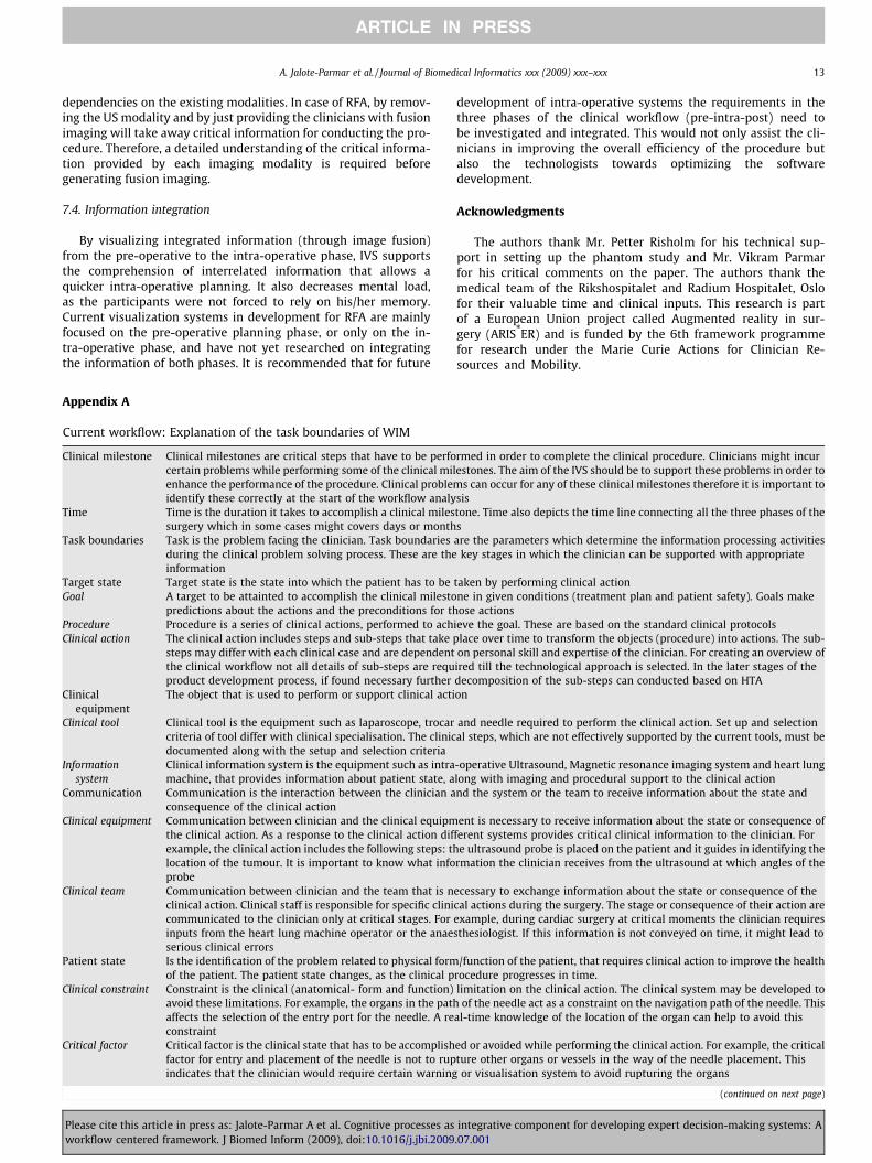
Appendix A
Current workflow: Explanation of the task boundaries of WIM
Clinical milestone
Clinical milestones are critical steps that have to be performed in order to complete the clinical procedure. Clinicians might incurcertain problems while performing some of the clinical milestones. The aim of the IVS should be to support these problems in order toenhance the performance of the procedure. Clinical problems can occur for any of these clinical milestones therefore it is important toidentify these correctly at the start of the workflow analysisTime
Time is the duration it takes to accomplish a clinical milestone. Time also depicts the time line connecting all the three phases of thesurgery which in some cases might covers days or monthsTask boundaries
Task is the problem facing the clinician. Task boundaries are the parameters which determine the information processing activitiesduring the clinical problem solving process. These are the key stages in which the clinician can be supported with appropriateinformationTarget state
Target state is the state into which the patient has to be taken by performing clinical action Goal A target to be attainted to accomplish the clinical milestone in given conditions (treatment plan and patient safety). Goals makepredictions about the actions and the preconditions for those actions
Procedure Procedure is a series of clinical actions, performed to achieve the goal. These are based on the standard clinical protocols Clinical action The clinical action includes steps and sub-steps that take place over time to transform the objects (procedure) into actions. The sub-steps may differ with each clinical case and are dependent on personal skill and expertise of the clinician. For creating an overview ofthe clinical workflow not all details of sub-steps are required till the technological approach is selected. In the later stages of theproduct development process, if found necessary further decomposition of the sub-steps can conducted based on HTA
Clinicalequipment
The object that is used to perform or support clinical action
Clinical tool
Clinical tool is the equipment such as laparoscope, trocar and needle required to perform the clinical action. Set up and selectioncriteria of tool differ with clinical specialisation. The clinical steps, which are not effectively supported by the current tools, must bedocumented along with the setup and selection criteriaInformationsystem
Clinical information system is the equipment such as intra-operative Ultrasound, Magnetic resonance imaging system and heart lungmachine, that provides information about patient state, along with imaging and procedural support to the clinical action
Communication
Communication is the interaction between the clinician and the system or the team to receive information about the state andconsequence of the clinical actionClinical equipment
Communication between clinician and the clinical equipment is necessary to receive information about the state or consequence ofthe clinical action. As a response to the clinical action different systems provides critical clinical information to the clinician. Forexample, the clinical action includes the following steps: the ultrasound probe is placed on the patient and it guides in identifying thelocation of the tumour. It is important to know what information the clinician receives from the ultrasound at which angles of theprobeClinical team
Communication between clinician and the team that is necessary to exchange information about the state or consequence of theclinical action. Clinical staff is responsible for specific clinical actions during the surgery. The stage or consequence of their action arecommunicated to the clinician only at critical stages. For example, during cardiac surgery at critical moments the clinician requiresinputs from the heart lung machine operator or the anaesthesiologist. If this information is not conveyed on time, it might lead toserious clinical errorsPatient state
Is the identification of the problem related to physical form/function of the patient, that requires clinical action to improve the healthof the patient. The patient state changes, as the clinical procedure progresses in time.Clinical constraint
Constraint is the clinical (anatomical- form and function) limitation on the clinical action. The clinical system may be developed toavoid these limitations. For example, the organs in the path of the needle act as a constraint on the navigation path of the needle. Thisaffects the selection of the entry port for the needle. A real-time knowledge of the location of the organ can help to avoid thisconstraintCritical factor
Critical factor is the clinical state that has to be accomplished or avoided while performing the clinical action. For example, the criticalfactor for entry and placement of the needle is not to rupture other organs or vessels in the way of the needle placement. Thisindicates that the clinician would require certain warning or visualisation system to avoid rupturing the organs(continued on next page)
integrative component for developing expert decision-making systems: A.07.001
14 A. Jalote-Parmar et al. / Journal of Biomedical Informatics xxx (2009) xxx–xxx
ARTICLE IN PRESS
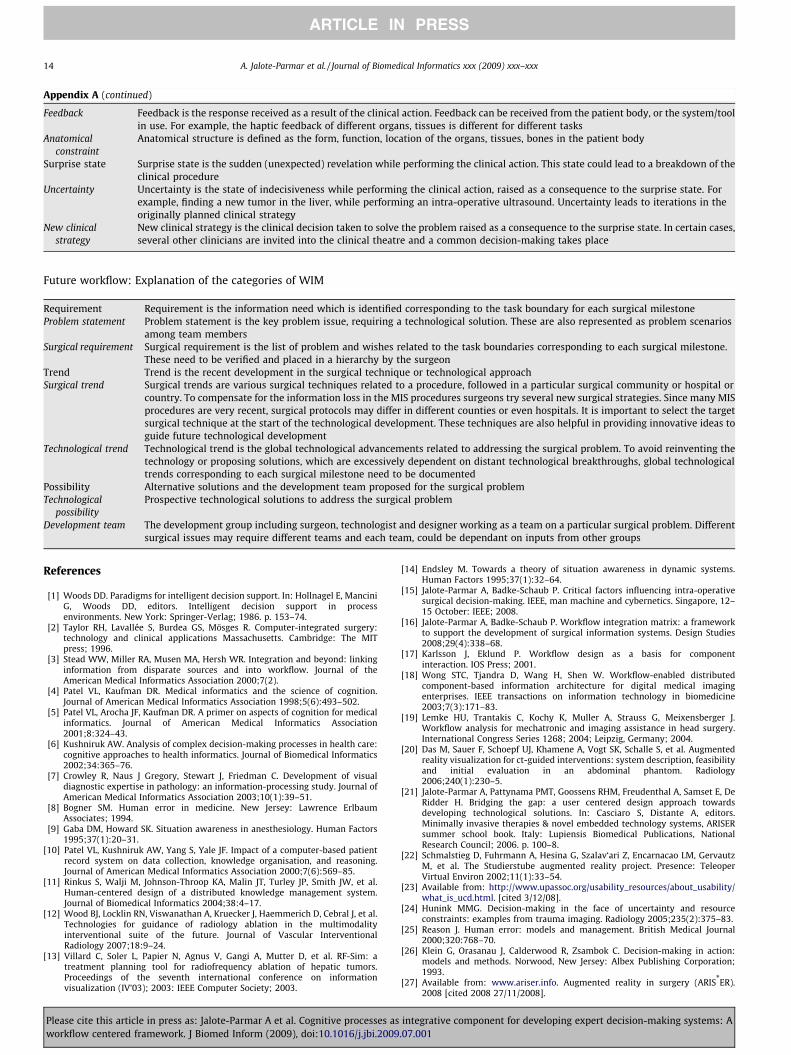
Appendix A (continued)
Feedback
Please cite this articworkflow centered f
Feedback is the response received as a result of the clinical action. Feedback can be received from the patient body, or the system/toolin use. For example, the haptic feedback of different organs, tissues is different for different tasks
Anatomicalconstraint
Anatomical structure is defined as the form, function, location of the organs, tissues, bones in the patient body
Surprise state
Surprise state is the sudden (unexpected) revelation while performing the clinical action. This state could lead to a breakdown of theclinical procedureUncertainty
Uncertainty is the state of indecisiveness while performing the clinical action, raised as a consequence to the surprise state. Forexample, finding a new tumor in the liver, while performing an intra-operative ultrasound. Uncertainty leads to iterations in theoriginally planned clinical strategyNew clinicalstrategy
New clinical strategy is the clinical decision taken to solve the problem raised as a consequence to the surprise state. In certain cases,several other clinicians are invited into the clinical theatre and a common decision-making takes place
Future workflow: Explanation of the categories of WIM
Requirement
ler
Requirement is the information need which is identified corresponding to the task boundary for each surgical milestone
Problem statement Problem statement is the key problem issue, requiring a technological solution. These are also represented as problem scenariosamong team members
Surgical requirement Surgical requirement is the list of problem and wishes related to the task boundaries corresponding to each surgical milestone.These need to be verified and placed in a hierarchy by the surgeon
Trend Trend is the recent development in the surgical technique or technological approach Surgical trend Surgical trends are various surgical techniques related to a procedure, followed in a particular surgical community or hospital orcountry. To compensate for the information loss in the MIS procedures surgeons try several new surgical strategies. Since many MISprocedures are very recent, surgical protocols may differ in different counties or even hospitals. It is important to select the targetsurgical technique at the start of the technological development. These techniques are also helpful in providing innovative ideas toguide future technological development
Technological trend
Technological trend is the global technological advancements related to addressing the surgical problem. To avoid reinventing thetechnology or proposing solutions, which are excessively dependent on distant technological breakthroughs, global technologicaltrends corresponding to each surgical milestone need to be documentedPossibility
Alternative solutions and the development team proposed for the surgical problem Technologicalpossibility
Prospective technological solutions to address the surgical problemDevelopment team
The development group including surgeon, technologist and designer working as a team on a particular surgical problem. Differentsurgical issues may require different teams and each team, could be dependant on inputs from other groupsReferences
[1] Woods DD. Paradigms for intelligent decision support. In: Hollnagel E, ManciniG, Woods DD, editors. Intelligent decision support in processenvironments. New York: Springer-Verlag; 1986. p. 153–74.
[2] Taylor RH, Lavallée S, Burdea GS, Mösges R. Computer-integrated surgery:technology and clinical applications Massachusetts. Cambridge: The MITpress; 1996.
[3] Stead WW, Miller RA, Musen MA, Hersh WR. Integration and beyond: linkinginformation from disparate sources and into workflow. Journal of theAmerican Medical Informatics Association 2000;7(2).
[4] Patel VL, Kaufman DR. Medical informatics and the science of cognition.Journal of American Medical Informatics Association 1998;5(6):493–502.
[5] Patel VL, Arocha JF, Kaufman DR. A primer on aspects of cognition for medicalinformatics. Journal of American Medical Informatics Association2001;8:324–43.
[6] Kushniruk AW. Analysis of complex decision-making processes in health care:cognitive approaches to health informatics. Journal of Biomedical Informatics2002;34:365–76.
[7] Crowley R, Naus J Gregory, Stewart J, Friedman C. Development of visualdiagnostic expertise in pathology: an information-processing study. Journal ofAmerican Medical Informatics Association 2003;10(1):39–51.
[8] Bogner SM. Human error in medicine. New Jersey: Lawrence ErlbaumAssociates; 1994.
[9] Gaba DM, Howard SK. Situation awareness in anesthesiology. Human Factors1995;37(1):20–31.
[10] Patel VL, Kushniruk AW, Yang S, Yale JF. Impact of a computer-based patientrecord system on data collection, knowledge organisation, and reasoning.Journal of American Medical Informatics Association 2000;7(6):569–85.
[11] Rinkus S, Walji M, Johnson-Throop KA, Malin JT, Turley JP, Smith JW, et al.Human-centered design of a distributed knowledge management system.Journal of Biomedical Informatics 2004;38:4–17.
[12] Wood BJ, Locklin RN, Viswanathan A, Kruecker J, Haemmerich D, Cebral J, et al.Technologies for guidance of radiology ablation in the multimodalityinterventional suite of the future. Journal of Vascular InterventionalRadiology 2007;18:9–24.
[13] Villard C, Soler L, Papier N, Agnus V, Gangi A, Mutter D, et al. RF-Sim: atreatment planning tool for radiofrequency ablation of hepatic tumors.Proceedings of the seventh international conference on informationvisualization (IV’03); 2003: IEEE Computer Society; 2003.
in press as: Jalote-Parmar A et al. Cognitive processes asamework. J Biomed Inform (2009), doi:10.1016/j.jbi.2009
[14] Endsley M. Towards a theory of situation awareness in dynamic systems.Human Factors 1995;37(1):32–64.
[15] Jalote-Parmar A, Badke-Schaub P. Critical factors influencing intra-operativesurgical decision-making. IEEE, man machine and cybernetics. Singapore, 12–15 October: IEEE; 2008.
[16] Jalote-Parmar A, Badke-Schaub P. Workflow integration matrix: a frameworkto support the development of surgical information systems. Design Studies2008;29(4):338–68.
[17] Karlsson J, Eklund P. Workflow design as a basis for componentinteraction. IOS Press; 2001.
[18] Wong STC, Tjandra D, Wang H, Shen W. Workflow-enabled distributedcomponent-based information architecture for digital medical imagingenterprises. IEEE transactions on information technology in biomedicine2003;7(3):171–83.
[19] Lemke HU, Trantakis C, Kochy K, Muller A, Strauss G, Meixensberger J.Workflow analysis for mechatronic and imaging assistance in head surgery.International Congress Series 1268; 2004; Leipzig, Germany; 2004.
[20] Das M, Sauer F, Schoepf UJ, Khamene A, Vogt SK, Schalle S, et al. Augmentedreality visualization for ct-guided interventions: system description, feasibilityand initial evaluation in an abdominal phantom. Radiology2006;240(1):230–5.
[21] Jalote-Parmar A, Pattynama PMT, Goossens RHM, Freudenthal A, Samset E, DeRidder H. Bridging the gap: a user centered design approach towardsdeveloping technological solutions. In: Casciaro S, Distante A, editors.Minimally invasive therapies & novel embedded technology systems, ARISERsummer school book. Italy: Lupiensis Biomedical Publications, NationalResearch Council; 2006. p. 100–8.
[22] Schmalstieg D, Fuhrmann A, Hesina G, Szalav‘ari Z, Encarnacao LM, GervautzM, et al. The Studierstube augmented reality project. Presence: TeleoperVirtual Environ 2002;11(1):33–54.
[23] Available from: http://www.upassoc.org/usability_resources/about_usability/what_is_ucd.html. [cited 3/12/08].
[24] Hunink MMG. Decision-making in the face of uncertainty and resourceconstraints: examples from trauma imaging. Radiology 2005;235(2):375–83.
[25] Reason J. Human error: models and management. British Medical Journal2000;320:768–70.
[26] Klein G, Orasanau J, Calderwood R, Zsambok C. Decision-making in action:models and methods. Norwood, New Jersey: Albex Publishing Corporation;1993.
[27] Available from: www.ariser.info. Augmented reality in surgery (ARIS*ER).2008 [cited 2008 27/11/2008].
integrative component for developing expert decision-making systems: A.07.001

A. Jalote-Parmar et al. / Journal of Biomedical Informatics xxx (2009) xxx–xxx 15
ARTICLE IN PRESS
[28] Solbiati L, Goldberg SN, Ierace T, Dellanice M, Livraghi T, Gazelle GS. Radio-frequency ablation of hepatic metastases: post procedural assessment with a USmicrobubble contrast agent-early experience. Radiology 1999;211(3):643–9.
[29] Rhim H, Kim YS, Choi D, Lim D, Lim HK, Park K. Percutaneous radiofrequencyablation of hepatocellular carcinoma: analysis of 80 patients treated with twoconsecutive sessions. European Radiology 2008;18(7):1442–8.
[30] Rasmussen J. Information processing and human-machine interaction. NewYork: Elsevier; 1986.
[31] Solbiati L, Tonoloni M, Lerace T. Guidance of percutaneous tumor ablation. In:Lencioni R, editor. Enhancing the role of ultrasound with contrastagents. Springer; 2006.
[32] Curley SA, Izzo F, Delrio P, Ellis LM, Granchi J, Vallone P, et al. Radiofrequencyablation of unrespectable primary and metastatic hepatic malignancies:results in 123 patients. Annals of Surgery 1999;230(1):1–8.
[33] Edwards JC, Sardoski M, Burdenski T. Reported used of mental imagery in thepractice of physicians. Imagination Cognition and Personality 2005;24(1):41–9.
[34] Leplat J. Task complexity in work situations. In: Goodstein LP, Andersen HB,Olsen SE, editors. Tasks, errors and mental models. London: Taylor & Francis;1988.
[35] Kleemann M, Hildebrand P, Birth M, Bruch HP. Laparoscopic ultrasoundnavigation in liver surgery: technical aspects and accuracy. Surgical Endoscopy2006;20:726–9.
[36] Klein G. Recognition-primed decisions. In: Rouse WB, editor. Advances inman–machine systems research. Greenwich: JAI Press, Inc.; 1989. p. 47–92.
[37] Drefus SE. Formal models vs. human situational understanding: Inherentlimitations on the modeling of business expertise. Berkeley: OperationsResearch Center, University of California; 1981.
Please cite this article in press as: Jalote-Parmar A et al. Cognitive processes asworkflow centered framework. J Biomed Inform (2009), doi:10.1016/j.jbi.2009
[38] Endsley M, Kiris EO. Information presentation for expert systems in futurefighter aircraft. International Journal of Aviation Psychology1994;4(4):333–48.
[39] Endsley M, et al., Situation awareness information requirements forcommercial airline pilots (ICAT-98-1). Cambridge, MA: MassachusettsInstitute of Technology International Center for Air Transportation; 1998.
[40] Endsley M. Measurement of situation awareness in dynamic systems. HumanFactors 1995;37(1):65–84.
[41] Milko S, Samset E, Kadir T. Segmentation of the liver in ultrasound: a dynamictexture approach. International Journal of Computer Assisted Radiology andSurgery 2008;3(1–2):143–50.
[42] Endsley M, Bolte B, Jones D. Designing for situation awareness – an approachto user – centered design. New York: Taylor & Francis; 2003.
[43] Orasanu J, Connolly T. The reinvention of decision-making. In: Klein G,Orasanau J, Calderwood R, Zsambok C, editors. Decision-making in action:models and methods. New Jersey: Albex Publishing Corporation; 1993. p.3–20.
[44] Badke-Schaub P, Frankenberger E. Analysis of design projects. Design Studies1999;20(5):465–80;Elstein AS, Shuman LS, Sprafka SA. Medical problem solving: an analysis ofclinical reasoning Cambridge. Mass: Harvard University Press; 1978.
[45] Rouse W, Valusek J. Evolutionary design of systems to support decision-making. In: Klein G, Orasanau J, Calderwood R, Zsambok C, editors. Decision-making in action: models and methods. New Jersey: Albex PublishingCorporation; 1993. p. 270–86.
[46] Dreyfus H, Dreyfus S, editors. Mind over machine. New York: The Free Press;1986.
integrative component for developing expert decision-making systems: A.07.001


