Embed Size (px)
Citation preview
The Official Publication of
Perinatal Medicine Foundation
Turkish Perinatology Society
Turkish Society of Ultrasound in Obstetrics and Gynecology
ISSN 1305–3124
PE
R
I NA T A L J O U R
NA
L
PE
R
I N A T A L J O U R
NA
L
PERINATALJOURNALw w w . p e r i n a t a l j o u r n a l . c o m
Volume 19 | Issue 1 | April 2011

PERINATAL JOURNALVolume 19 / Issue 1 / April 2011
On behalf of the Perinatal Medicine Foundation
Prof. Dr. Cihat fien
Managing Editor
Prof. Dr. Murat Yayla
The Official publication of Perinatal Medicine Foundation,Turkish Perinatology Society and
Turkish Society of Ultrasound in Obstetrics and Gynecology
Advisory Board
Published three times a year • Publication local periodical
Correspondence: Rumeli Caddesi 47/606, Niflantafl› 34371 ‹stanbulPhone: (0212) 224 68 49 • Fax: (0212) 296 01 50
e-mail: [email protected]
www.perinataljournal.com
Deomed Publishing • Ac›badem Cad. ‹smail Hakk› Bey Sok. Pehlivan ‹fl Merkezi No: 7 Kat: 1 Kad›köy 34718 ‹stanbulPhone: (0216) 414 83 43 (Pbx) Fax: (0216) 414 83 42 www.deomed.com
Press: Birmat Matbaac›l›k Phone: (0212) 629 05 59 (April 2011)
Arif AkflitFigen AksoyTayfun AlperSadet ArsanHediye ArslanOlufl ApiSebahat Atar GürelTahsin Ayano¤luAhmet BaschatNazif Ba¤r›aç›kGökhan BayhanYeflim BayturTugan BefleNur DaniflmendFuat Demirk›ranÖzgür DerenGönül DinçMelahat Dönmez
Yakup ErataAli ErgünKubilay ErtanBilgin GürateflMetin Gülmezo¤luArif GüngörenMelih GüvenAyfle Kafkasl›Ömer KandemirHakan Kan›tÖmer K›lavuzSelahattin Kumru As›m KurjakNilgün Kültürsay R›za Madazl› Ercüment MüngenLütfü Öndero¤luAbdurrahman Önen
Soner ÖnerSemih ÖzerenOkan Özkaya Y›ld›z PerkHaluk SaymanYunus SöyletMekin SezikTurgay fienerMete Tan›rAlper Tanr›verdiEbru Tar›mAyd›n TekayNeslihan TekinBeyhan TüysüzSeyfettn Uluda¤Ahmet Yal›nkaya
EDITORS
Cihat fien, Murat Yayla
ISSN 1305-3124
Instructions for the Authors
Coverage
The manuscripts should be prepared for one of the following article cate-gories which are peer-reviewed:
• Clinical Research Article• Experimental Study• Case Report• Technical Note• Letter to the Editor
In addition, the journal includes article categories which do not require apeer review process but are prepared by the Editorial Board or consist ofinvited articles, titled as:
• Editorial• Viewpoint Article • Review Article • Abstracts• Announcements• Erratum
Manuscript Evaluation
All submissions to Perinatal Journal must be original, unpublished, and notunder the review of any other publication. This is recorded by the systemautomatically with the IP number, the date and time of submission. Onbehalf of all authors the corresponding author should state that all authorsare responsible for the manuscripts. The name, date, and place of the rele-vant meeting should be stated if the submission is a work that was previ-ously presented in a scientific meeting.
Following the initial review, manuscripts which have been accepted forconsideration are reviewed by at least two reviewers. The Editors of the jour-nal decide to accept or reject the manuscript considering the comments ofthe reviewers. They are authorized to reject or revise the manuscript, to sug-gest required corrections and changes upon the comments and suggestionsof reviewers, and/or to correct or condense the text by permission of the cor-responding author. They have also the right to reject a manuscript afterauthors’ revision. Author(s) should provide additional relevant data, docu-ments, or information upon the editorial request if necessary.
Ethical Issues
All manuscripts presenting data obtained from studies involving human sub-jects must include a statement that the written informed consent of the par-ticipants was obtained and that the study was approved by an institutionalethics board or an equivalent body. This institutional approval should besubmitted with the manuscript. Authors of case reports must submit thewritten informed consent of the subject(s) of the report or of the patient’slegal representatives for the publication of the manuscript. All studies shouldbe carried out in accordance with the World Medical Association Declarationof Helsinki, covering the latest revision date. Patient confidentiality must beprotected according to the universally accepted guidelines and rules.Manuscripts reporting the results of experimental studies on animals mustinclude a statement that the study protocol was approved by the animalethics committee of the institution and that the study was conducted inaccordance with the internationally accepted guidelines, including theUniversal Declaration of Animal Rights, European Convention for theProtection of Vertebrate Animals Used for Experimental and Other ScientificPurposes, Principles of Laboratory Animal Science, and the Handbook forthe Care and Utilization of Laboratory Animals. The authors are stronglyrequested to send the approval of the ethics committee together with themanuscript. In addition, manuscripts on human and animal studies shoulddescribe procedures indicating the steps taken to eliminate pain and suffer-ing.
The authors should also disclose all issues concerning financial relation-ship, conflict of interest, and competing interest that may potentially influ-ence the results of the research or scientific judgment. All financial contri-butions or sponsorship, financial relations, and areas of conflict of interest
should be clearly explained in the cover letter to the Editor-in-Chief at thetime of submission, with full assurance that any related document will besubmitted to the journal when requested. For the details of journal's"Conflict of Interest Policy" please read the PDF document which includes"Conflicts of Interest Disclosure Statement".
Perinatal Journal follows the ethics flowcharts developed by theCommittee on Publication Ethics (COPE) for dealing with cases of possiblescientific misconduct and breach of publication ethics. For detailed informa-tion please visit www.publicationethics.org.
Manuscript Preparation
In addition to the rules listed below, manuscripts to be published in PerinatalJournal should be in compliance with the Uniform Requirements forManuscripts Submitted to Biomedical Journals published by InternationalCommittee of Medical Journal Editors (ICMJE) of which latest version is avail-able at www.icmje.org.
Authors are requested to ensure that their manuscript follows theappropriate guidelines such as CONSORT for randomized controlled trials,STROBE for observational studies, STARD for diagnostic accuracy studies,and PRISMA for systematic reviews and meta-analyses, for the study designand reporting if applicable.
Authorship and Length of Texts
The author(s) must declare that they were involved in at least 3 of the 5stages of the study stated in the “Acknowledgement of Authorship andTransfer of Copyright Agreement” as “designing the study”, “collecting thedata”, “analyzing the data”, “writing the manuscript” and “confirming theaccuracy of the data and the analyses”. Those who do not fulfill this pre-requisite should not be stated as an author.
Original research articles base on clinical or experimental studies. Themain text should not exceed 2500 words (max. 16 pages) and there shouldbe a maximum 6 authors
Case reports should illustrate interesting cases including their treat-ment options. The main text should not exceed 2000 words (max. 8 pages)and there should be a maximum 5 authors.
Viewpoint articles: Only by invitation and should be no more than2000 words long (max. 8 pages).
Review articles: Only by invitation and should be no more than 4000-5000 words long (max. 20 pages).
Technical notes aims to present a newly diagnostic or therapeuticmethod. They should not exceed 2000 words (max. 8 pages) and include amaximum of 10 references.
Letters to the Editor should be no more than 500 words long (max.2 pages) and include a maximum of 10 references.
Sections in the Manuscripts
Manuscripts should be designed in the following order: title page, abstract,main text, references, and tables, with each typeset on a separate page:
Page 1 - Title pagePage 2 - Abstract and key wordsPage 3 and next - Main textNext Page - ReferencesNext Page - Table heading and tables (each table should be placed in
separate pages)Next Page - Figure legends and figures (each figure should be placed
in separate pages)Last Page - Appendices (patient forms, surveys etc.)
Title pageThis page should only include the title of the manuscript, which should becarefully chosen to better reflect the contents of the study. No anusualabbreviations should be used in the title of the manuscript. A short title asrunning heading not exceeding 40 characters should be given which isdesired to appear on top part of continuing pages when journal is pub-lished.
Abstract page
Abstracts should not contain any abbreviation and references. They shouldbe prepared under following designs.
— Abstracts of research articles should be max. 250 words andstructured in four paragraphs using the following subtitles: Objective,Methods, Results, and Conclusion. Following the abstract, each abstractpage should include max. 5 key words separated with comma and writtenin lower cases.
— Abstracts of case reports should be max. 125 words and struc-tured in three paragraphs using the following subtitles: Objective, Case,Conclusion. Following the abstract, each abstract page should include max.3 key words separated with comma and written in lower cases.
— Abstracts of review articles should be max. 300 words and pre-sented not structured in one paragraph. Following the abstract, eachabstract page should include max. 5 key words separated with comma andwritten in lower cases.
— Abstracts of technical notes should be max. 125 words and struc-tured in three paragraphs using the following subtitles: Objective,Technique, Conclusion. Following the abstract, each abstract page shouldinclude max. 3 key words separated with comma and written in lowercases.
Main text:
The sections in main text are defined according to the manuscript type.
— In research articles, main text should consist of sections titled as"Introduction, Methods, Results, Discussion and Conclusion". Each titlemay have subtitles. The categories of subtitles should be clearly defined.
The Introduction section should include a brief summary of the base ofthe work and clearly states the purpose of the study.
The Methods section should contain a detailed description of thematerial, the study design and clinical and laboratory tests, and statisticalmethods used. A statement regarding the ethical issues should also begiven in this section.
The Results section should provide the main findings of the study. Datashould be concisely presented, preferably in tables or graphs.
The Discussion section should mainly rely on the results derived fromthe study, with relevant citations from the most recent literature.
The Conclusion section should briefly and claearly present the conclu-sions derived from the results of the study. It should be in compliance withthe aim of the work and and point out its application in clinical practice.
— In Case Reports, main text should be divided with the titles"Introduction, Case(s), Discussion". Reported case(s) should be introducedclearly including the case story, and the results of laboratory tests should begiven in table format as far as possible.
— The text of the reviews articles should follow the "Introduction"and be organized under subtitles which should clearly define the text'scontext categorization. The Reviews are expected to include wide survey-ing of literature and reflect the author's personal experiences as far aspossible.
— The text of the technical note type of articles should be dividedinto "Introduction, Technic, Discussion". The presented technic should bedefined briefly under the related title, and include illustrations or figures assoon as possible.
— Letters to the Editor should not have titled sections. If there is acitation about a formerly published article within the text, reference(s)should be provided.
References
References used in the text should be directly related to the topic, as recentas possible and in enough numbers. They should be numbered in squarebrackets in the order in which they are mentioned in the text includingTables and Figures. Citation order should be checked carefully.
Only published articles or articles in press can be used in references.Unpublished data including conference papers or personal communicationsshould not be used. Papers published in only electronic journals or in the
preprint or online first issues of the electronic versions of conventional peri-odicals should be absolutely presented with DOI (digital object identifier)numbers.
Journal titles should be abbreviated according to the Index Medicus. Allauthors if six or fewer should be listed; otherwise, the first six and “et al.”should be written.
Direct use of references is strongly recommended and the authors maybe asked to provide the first and last pages of certain references. Publicationof the manuscript will be suspended until this request is fulfilled by theauthor(s).
The style and punctuation should follow the formats outlined below:
— Standard journal article: Hammerman C, Bin-Nun A, Kaplan M.Managing the patent ductus arteriosus in the premature neonate: a newlook at what we thought we knew. Semin Perinatol 2012;36:130-8.
— Article published in an only electronic journal: Lee J, Romero R,Xu Y, Kim JS, Topping V, Yoo W, et al. A signature of maternal anti-fetalrejection in spontaneous preterm birth: chronic chorioamnionitis, anti-human leukocyte antigen antibodies, and C4d. PLoS ONE 2011;6:e16806.doi:10.1371/ journal.pone.0011846.
— Book: Jones KL. Practical perinatology. New York: Springer; 1990.p. 112-9.
— Chapter in a book: Sibai BM, Frangieh AY. Eclampsia. In: GleicherN, editors. Principles and practice of medical therapy in pregnancy. 3rd ed.New York: Appleton&Lange; 1998. p. 1022-7.
Figures and tables
All illustrations (photographs, graphics, and drawings) accompanying themanuscript should be referred to as “figure”. All figures should be num-bered consecutively and mentioned in the text. Figure legends should beadded at the end of the text as a separate section. Each figure should beprepared as a separate digital file in “jpeg” format, with a minimum 300dpi or better resolution. All illustrations should be original. Illustrations pub-lished elsewhere should be submitted with the written permission of theoriginal copyright holder. For recognizable photographs of human subjects,written permission signed by the patient or his/her legal representativeshould be submitted; otherwise, patient names or eyes must be blocked outto prevent identification. Microscopic photographs should include informa-tion on staining and magnification.
Each table should be prepared on a separate page with table headingon top of the table. Table heading should be added to the main text file ona separate page when a table is submitted as a supplementary file.
Submission
For a swift peer review, Perinatal Journal operates a web-based submission,peer review and manuscript tracking system. Authors are required to sub-mit their articles online. Details of how to submit online can be found atwww.perinataljournal.com.
Submission Checklist
The following list will be useful during the final check of a manuscriptbefore submission:
1. Manuscript length (max. 4000 words for research articles)
2. Number of authors (max. 6 authors for research articles)
3. Title page (no anusual abbreviations)
4. Abstracts (max. 250 words for research articles)
5. Key words (max. 5 keys for research articles)
6. Main text (subtitles)
7. References (listed according to the rules of ICMJE)
8. Figures and tables (numbering; legends and headings; copyrightinfo/permission)
9. Cover letter
10. Acknowledgement of Authorship and Transfer of CopyrightAgreement (undersigned by all authors)
11. Conflicts of Interest Disclosure Statement (if necessary)

Announcement

Announcement
Perinatal JournalVolume 19 / Issue 1 / April 2011
Contents
Multicentric Multiple Pregnancy Study IV: Mortality in Twins -Spontaneuos Versus Artificial Reproductive Techniques
Murat Yayla, Rahime Nida Ergin, Yeflim Baytur
The Role of Ultrasound in Fırst Trimester Pregnancy in Prediction of Miscarriages
Fatma Çetin Pelit, Hatice Y›lmaz, Necdet Süer
Neonatal Outcomes of Pregnancy with Intrahepatic CholestasisAbdullah Kurt, Ayfle Ecevit, Burcu K›sa, Deniz Anuk ‹nce, Aylin Tarcan, Filiz Bilgin Yan›k
Long Term Effects Of Cesarean Births On Unintended Pregnancy Rates and Obstetric Expenditures
Kahraman Ülker, ‹smail Temur, Abdülaziz Gül
Selective Fetoreduction: Report of Two Cases Muhammet Erdal Sak, Mehmet S›dd›k Evsen, Hatice Ender Soydinç, Sibel Sak, Ahmet Yal›nkaya
The Diagnosis and Management of Ornithine Transcarbamylase Deficiency in Pregnancy: A Case Report
Orkun Çetin, Cihat fien, Begüm Aydo¤an, Seyfettin Uluda¤, ‹pek Dokurel Çetin, Hakan Erenel
Progressive Fetal Diaphragnatic Hernia: A Case Report Ercüment Müngen, Ali Babacan, ‹smet Gün
Spontaneous Resolution of Intra-Abdominal Cyst in a Fetus with Normal Karyotype
M. Murat Naki, Olufl Api, Hasniye Ac›o¤lu, Müge Emeksiz,Aybala Ak›l, Orhan Ünal
The Conclusion Report of 13th National Perinatology Congress Ayfle Kafkasl›, Alper Tanr›verdi, Yefllim Baytur, Özlem Pata, Ertan Adal›, Hakan Camuzcuo¤lu, Arif Güngören, ‹lker Ar›kan
Research Articles
Case Reports
Presentation
1
6
10
15
20
23
28
32
35
Abstract
Objective: Both spontaneous and ART twin pregnancies have maternal and fetal risks. In this multi-centered cross-sectional studywe aimed to determine prognostic differences between spontaneous and ART twin pregnancies in some centers of our country.
Methods: Demographic data of women delivered between the period of 2003 and 2004, including mean maternal age, parity, fetaland perinatal mortality, gestational week at delivery, mode of delivery and maternal morbidity, fetal or newborn’s weight and sexwere determined from the questionnaire forms and data obtained from 10 obstetrics centers from different parts of Turkey, includ-ing university and Health Ministry training hospitals.
Results: The number of twins among the births in ten centers participated in the study was 818. The percentage of spontaneoustwins was 24% whereas in 76% ART was applied. There was no difference in the mean age of mothers between the groups. Meanbirth week of spontaneous twins was 35.03±2.58 weeks and mean birth week of ART twins was 34.31±3.51 weeks. The ratio ofcaeserean section in ART and spontaneous twins were 78.6% and 68.8% respectively. While late second trimester mortality was highin ART group and third trimester mortality was high in spontaneous group, perinatal mortaliy was similar between two groups.
Conclusion: In our study there was no significant difference in the perinatal mortaliy between ART and spontaneous twins.
Keywords: Twin pregnancy, mortality, artificial reproductive techniques, spontaneous.
Çok merkezli ço¤ul gebelik çal›flmas› IV – Spontan ikizlerdeki mortalitenin yard›mla üreme
teknikleriyle gebe kalanlardaki ikizler ile karfl›laflt›r›lmas›
Amaç: Hem spontan, hem de yard›mla üreme teknikleri (YÜT) sonras› ortaya ç›kan ço¤ul gebelikler maternal ve fetal riskleriberaberinde getirmektedir. Çok merkezli kesitsel olan bu çal›flmam›zda, ülkemizin baz› merkezlerindeki spontan ve YÜT ikiz gebelik-leri aras›ndaki prognoz farkl›l›¤›n› araflt›rmay› amaçlad›k.
Yöntem: Bu çal›flma 2003-2004 y›llar› içinde Türkiye’nin de¤iflik bölgelerinde Üniversite ve E¤itim Araflt›rma Hastanesi bünyesinde yeralan 10 ayr› Kad›n Hastal›klar› ve Do¤um Merkezine gönderilen anket ve klinik bilgi formlar› ile yap›ld›. Ankette anne yafl›, gebelik vedo¤um say›lar›, gebe kal›fl flekli, do¤um haftas› ve flekli, yenido¤an a¤›rl›¤›, cinsiyeti ve mortalitesi, maternal mortalite-morbidite para-metreleri sorguland›.
Bulgular: Çal›flmaya kat›lan 10 merkezdeki toplam ikiz do¤um say›s› 818’dir. Bu ço¤ul gebeliklerin %24’ü spontan olup %76’s›naYÜT uyguland›. Gruplar aras›ndaki anne yafl› ortalamalar› aras›nda fark saptanmad›. YÜT gebeliklerinde ortalama do¤um haftas›34.31±3.51 hafta iken, spontan gebelerde 35.03±2.58 hafta idi. YÜT ve spontan gebeliklerdeki sezaryen oranlar› s›ras›yla %78.6 ve%68.8 olarak saptand›. Geç ikinci trimester kay›plar› YÜT grubunda, üçüncü trimester kay›plar› ise spontan grupta daha fazlagözlenirken, perinatal mortalite her iki grupta benzer bulundu.
Sonuç: Çal›flmam›zda ikiz gebeliklerde gebeli¤in YÜT ya da spontan olmas› mortalite aç›s›ndan anlaml› farkl›l›k yaratmamaktad›r.
Anahtar Sözcükler: ‹kiz gebelik, mortalite, yard›mc› üreme tekni¤i, spontan.
Multicentric Multiple Pregnancy Study IV:Mortality in Twins - Spontaneuos Versus
Artificial Reproductive Techniques
Murat Yayla1, Rahime Nida Ergin1, Yeflim Baytur2
1International Hospital Kad›n Do¤um Klini¤i, ‹stanbul, Türkiye2Celal Bayar Üniversitesi T›p Fakültesi Kad›n Hastal›klar› ve Do¤um Anabilim Dal›, Manisa, Türkiye
Correspondence: Rahime Nida Ergin, Defne Apt No: 140 D: 5 Fenerbahçe Ba¤dat Caddesi, ‹stanbul, Türkiye
e-mail: [email protected]
Perinatal Journal 2011;19(1):1-5
e-Adress: http://www.perinataljournal.com/20110191001doi:10.2399/prn.11.0191001
Introduction Progress in the artificial reproductive tech-
niques (ART) is the cause of the increase of theratio of multiple pregnancies at the present. Thesuccess of centers using induction of ovulation, invitro fertilization ( IVF) or intracytoplasmic sperminjection (ICSI) is measured by the ratio of preg-nancy, meanwhile, the real measure, the healthyfetus at term (favorably singleton) is sometimesignored. Because of the reduced pregnancy ratesby the conservative approaches in the favor ofSingleton pregnancies and the psychological stressbrought by failed pregnancy after a long andexpensive treatment processes and to prevent thedecrease in the success of the AssistedReproductive Technique (ART) centers, implemen-tation of aggressive therapies which may resultmultiple pregnancies are preferred. However, bothspontaneous and ART multiple pregnancies areaccompanied by maternal and fetal risks.[1-4]
Although the increased ratio of multiple pregnan-cies in our country has been mentioned in manyscientific meetings, no comprehensive nationalstudy has been planned to determine the factorsrelated to the increase of multiple pregnancies andmaternal/fetal mortality and morbidity related tothese multiple pregnancies. The comprehensivemulti-centered studies related on this subject havebeen published by the present study group.[5,6] Theaim of this cross-sectional multi-center study wasto reveal the mortality ratios in the spontaneousand ART twins in various Obstetrics andGynecology clinics in our country.
MethodsDemographic data of women delivered
between the period of 2003 and 2004, includingmean maternal age, parity, fetal and perinatal mor-tality, gestational week at delivery, mode of deliv-ery and maternal morbidity, fetal or newborn’sweight and sex were determined from the ques-tionnaire forms and data obtained from 10 obstet-rics centers from different parts of Turkey, includ-ing university and Health Ministry training hospi-tals. Some unreported data in the survey were re-questioned and the missing ones were completed.Ovulation induction, ICSI and IVF pregnancieshave been accepted as assisted reproductive tech-
niques. The term “stillbirth” has been defined forthe death of the fetus, who (or any of his twins) isat least 400 grams of weight or who completed 20th gestational week, before birth or no respirationafter birth or no heart beat. The term “early neona-tal death” has been defined for neonatal deathswithin the first 7 days after birth. The data of spon-taneous and ART twin pregnancies were comparedusing statistical tests of chi-square, Fisher's exactand Student's t-test. SPSS for Windows version 14.0(SPSS Inc, Chicago, IL, ABD) was used for statisti-cal analyses. The value of p <0.05 was accepted assignificant.
ResultsThe total number of births was 43.258 between
2003 and 2004 in ten centers which participated inthe study. The total numbers of twins were 818(18.9/1000) and the total numbers of triplets was42 (0.97/1000). The data of type of pregnancy wasavailable in 265 twins and 24.15% of them werespontaneous and in 75.85% of them ART wasapplied. The demographics of both spontaneousand ART twins are given in the table 1. There wasno significant difference in the mean maternal age.The only significant difference was higher numberof previous gestations in the spontaneous twinpregnancies as expected. Mean birth weeks were34.31 ± 3.51weeks in ART twins and 35.03 ± 2.58weeks in spontaneous twins. The perinatal mortal-ity ratio in the twin pregnancies was 106.9/1000and the mortality ratio and survival ratios accord-ing to the type of pregnancy are given in the Table2. The chance of being alive of both fetuses was10% higher in ART twins though not statisticallysignificant. Concerning the type of delivery, theratio of death of at least one fetus was 7.7% andthe ratio of live birth of both fetuses was 92.3% incaesarean section. These ratios were 42.1% and57.9% in vaginal deliveries. The mortality ratios inbirths with caesarean section and vaginal deliver-ies according to the type of pregnancy are given inthe Table 3. When the spontaneous and ART twinswere compared according to the mean weights oflive and dead fetuses, mean weights of ART twinswere lesser in all groups. But this difference wasfound to be statistically significant only in thegroup where both fetuses were dead (Table 4).Survival ratios were lower in fetuses with weight of
Yayla M, Ergin RN, Baytur Y. Multicentric Multiple Pregnancy Study IV: 2
Perinatal Journal 2011;19(1):1-5 3
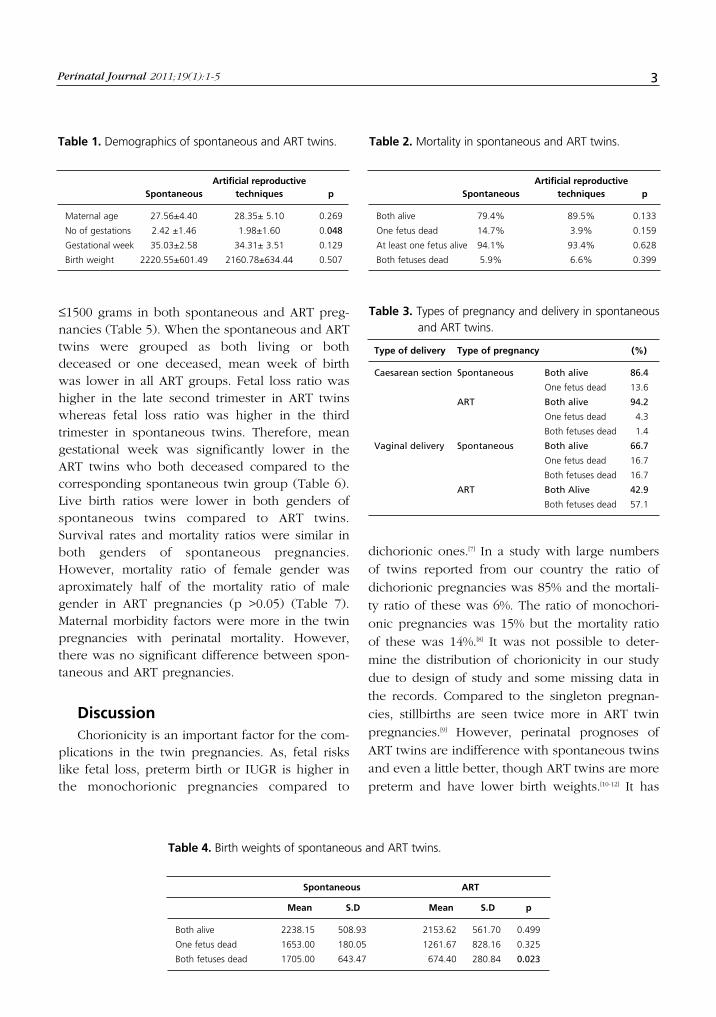
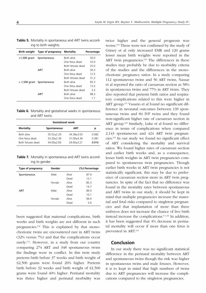
≤1500 grams in both spontaneous and ART preg-nancies (Table 5). When the spontaneous and ARTtwins were grouped as both living or bothdeceased or one deceased, mean week of birthwas lower in all ART groups. Fetal loss ratio washigher in the late second trimester in ART twinswhereas fetal loss ratio was higher in the thirdtrimester in spontaneous twins. Therefore, meangestational week was significantly lower in theART twins who both deceased compared to thecorresponding spontaneous twin group (Table 6).Live birth ratios were lower in both genders ofspontaneous twins compared to ART twins.Survival rates and mortality ratios were similar inboth genders of spontaneous pregnancies.However, mortality ratio of female gender wasaproximately half of the mortality ratio of malegender in ART pregnancies (p >0.05) (Table 7).Maternal morbidity factors were more in the twinpregnancies with perinatal mortality. However,there was no significant difference between spon-taneous and ART pregnancies.
DiscussionChorionicity is an important factor for the com-
plications in the twin pregnancies. As, fetal riskslike fetal loss, preterm birth or IUGR is higher inthe monochorionic pregnancies compared to
dichorionic ones.[7] In a study with large numbersof twins reported from our country the ratio ofdichorionic pregnancies was 85% and the mortali-ty ratio of these was 6%. The ratio of monochori-onic pregnancies was 15% but the mortality ratioof these was 14%.[8] It was not possible to deter-mine the distribution of chorionicity in our studydue to design of study and some missing data inthe records. Compared to the singleton pregnan-cies, stillbirths are seen twice more in ART twinpregnancies.[9] However, perinatal prognoses ofART twins are indifference with spontaneous twinsand even a little better, though ART twins are morepreterm and have lower birth weights.[10-12] It has
Artificial reproductiveSpontaneous techniques p
Maternal age 27.56±4.40 28.35± 5.10 0.269
No of gestations 2.42 ±1.46 1.98±1.60 0.048
Gestational week 35.03±2.58 34.31± 3.51 0.129
Birth weight 2220.55±601.49 2160.78±634.44 0.507
Table 1. Demographics of spontaneous and ART twins.
Artificial reproductiveSpontaneous techniques p
Both alive 79.4% 89.5% 0.133
One fetus dead 14.7% 3.9% 0.159
At least one fetus alive 94.1% 93.4% 0.628
Both fetuses dead 5.9% 6.6% 0.399
Table 2. Mortality in spontaneous and ART twins.
Type of delivery Type of pregnancy (%)
Caesarean section Spontaneous Both alive 86.4
One fetus dead 13.6
ART Both alive 94.2
One fetus dead 4.3
Both fetuses dead 1.4
Vaginal delivery Spontaneous Both alive 66.7
One fetus dead 16.7
Both fetuses dead 16.7
ART Both Alive 42.9
Both fetuses dead 57.1
Table 3. Types of pregnancy and delivery in spontaneousand ART twins.
Spontaneous ART
Mean S.D Mean S.D p
Both alive 2238.15 508.93 2153.62 561.70 0.499
One fetus dead 1653.00 180.05 1261.67 828.16 0.325
Both fetuses dead 1705.00 643.47 674.40 280.84 0.023
Table 4. Birth weights of spontaneous and ART twins.
been suggested that maternal complications, birthweeks and birth weights are not different in suchpregnancies.[13] This is explained by that mono-chorionic twins are encountered rare in ART twins(32% versus 7%) and that the complications occurrarely.[14]. However, in a study from our countrycomparing 274 ART and 348 spontaneous twinsthe findings were in conflict. In this twin studypreterm birth before 37 weeks and birth weight of≤2,500 grams were found 20% higher. Pretermbirth before 32 weeks and birth weight of ≤1,500grams were found 40% higher. Perinatal mortalitywas thrice higher and perinatal morbidity was
twice higher and the general prognosis wasworse.[15] These were not confirmed by the study ofGüney et al only increased EMR and 120 gramslesser mean birth weights were reported in theART twin pregnancies.[16] The differences in thesestudies may probably be due to morbidity criteriaof the studies and the differences in the mono-chorionic pregnancy ratios. In a study comparing112 spontaneous twins and 56 ART twins, Nassaret al reported the ratio of caesarean section as 58%in spontaneous twins and 77% in ART twins. Theyalso reported that preterm birth ratios and respira-tory complications related to this were higher inART group.[17] Vasario et al found no significant dif-ference in neonatal outcomes between 139 spon-taneous twins and 84 IVF twins and they foundnon-significant higher rate of caesarean section inART group.[18] Similarly, Luke et al found no differ-ence in terms of complications when compared2,143 spontaneous and 424 ART twin pregnan-cies.[19] In our study we found no significant effectof ART considering the mortality and survivalratios. We found higher rates of caesarean sectionand earlier birth weeks and, as a consequence,lesser birth weights in ART twin pregnancies com-pared to spontaneous twin pregnancies. Thoughearlier birth weeks in ART twin pregnancies is notstatistically significant, this may be due to prefer-ence of caesarean section more in ART twin preg-nancies. In spite of the fact that no difference wasfound in the mortality rates between spontaneousand ART twins in our study, it should be kept inmind that multiple pregnancies increase the mater-nal and fetal risks compared to singleton pregnan-cies and that implantation of more than threeembryos does not increase the chance of live birthinstead increase the complications.[1-4,20] In addition,it has been suggested that 4% decrease in perina-tal mortality will occur if more than one fetus isprevented in ART.[21]
ConclusionIn our study there was no significant statistical
difference in the perinatal mortality between ARTand spontaneous twins though the risk was higherin spontaneous twins and male fetuses. However,it is to kept in mind that high numbers of twinsdue to ART pregnancies will increase the compli-cations compared to the singleton pregnancies.
Yayla M, Ergin RN, Baytur Y. Multicentric Multiple Pregnancy Study IV: 4
Birth weight Type of pregnancy Mortality Percentage
<1,500 gram Spontaneous Both alive 50.0
One fetus dead 25.0
Both fetuses dead 25.0
ART Both alive 56.3
One fetus dead 12.5
Both fetuses dead 31,3
> 1,500 gram Spontaneous Both alive 83.3
One fetus dead 13.3
Both fetuses dead 3.3
ART Both alive 98.3
One fetus dead 1.7
Tablo 5. Mortality in spontaneous and ART twins accord-ing to birth weights.
Gestational week
Mortality Spontaneous ART p
Both alive 35.52±2.29 34.38±3.03 0.082
One fetus dead 33.20±2.39 31.00±4.36 0.381
Both fetuses dead 34.00±2.83 24.60±3.21 0.016
Table 6. Mortality and gestational weeks in spontaneousand ART twins.
Type of pregnancy Gender (%) Percentage
Spontaneous Male Alive 87.9
Dead 12.1
Female Alive 85.3
Dead 14.7
ART Male Alive 89.9
Dead 10.1
Female Alive 94.4
Dead 5.6
Table 7. Mortality in spontaneous and ART twins accord-ing to gender.
References1. Chan FY. Obstetrics implication of multiple gestation. Aust
N Z J Obstet Gynaecol 2006;46(Supp1):3-13.
2. Lee MY, Cleary-Goldman J, D’Alton ME. Multiple gestationsand late preterm (near term) deliveries. Semin Perinatol2006;30:103-12..
3. Huang CT, Au HK, Chien LW, Chang CW, Chien YY, TzengCR. Twin pregnancy outcome among cases of spontaneousconceptions, intrauterin insemination, and invitro fertiliza-tion/intracytoplasmic sperm injection. Fertil Steril2006;86:1017-9.
4. Reddy UM, Wapner RJ, Rebor RW, Tosca RJ. Infertility,assisted reproductive technology and adverse pregnancyoutcomes: executive summary of a National Institute ofChild Health and Human Development workshop. ObstetGynecol 2007;109:967-77.
5. Yayla M, Baytur Y. Çok merkezli ço¤ul gebelik çal›flmas› I:Epidemiyoloji. Perinatoloji Dergisi 2008;16:1-8.
6. Yayla M, Baytur Y. Multicentric multiple pregnany study II:Perinatal mortality in twins. Perinatal Journal 2009;17:8-17.
7. Sebire NJ, Snijders RJM, Hughes K, Sepulveda W,Nicolaides KH. The hidden mortality of monochorionictwin pregnancies. Br J Obstet Gynaecol 1997;104:1203- 7.
8. Y›ld›r›m G, Gül A, Aslan H, Erol O, Güngördük K, CeylanY. ‹kiz gebeliklerde koryonisitenin neonatal ve maternalsonuçlara etkisi. Türk Jinekoloji ve Obstetrik Derne¤i Dergisi2007;4:178- 83.
9. Pinborg A, Loft A, Nyboe Andersen A. Neonatal outcome ina Danish National Cohort of 8602 children born after invitro fertilization or intracytoplasmic sperm injection: therole of twin pregnancy. Acta Obstet Gynecol Scand2004;83:1071-8.
10. McDonald S, Murphy K, Beyene J, Ohlsson A. Perinatal out-comes of in vitro fertilization twins: a systematic review andmeta-analyses. Am J Obstet Gynecol 2005; 193:141-52.
11. Pinborg A. IVF/ICSI twin pregnancies: risks and prevention.Hum Reprod Update 2005;11:575-93.
12. Fitzsimmons BP, Bebbington MW, Fluker MR. Perinatal andneonatal outcomes in multiple gestations: assisted repro-duction versus spontaneous conception. Am J ObstetGynecol 1998;179:1162-7.
13. Pinborg A, Loft A, Rasmussen S, Schmidt L, Langhoff_RoosJ, Greisen G, Andersen AN. Neonatal outcome in a Danishnational cohort of 3438 IVF/ICSI and 10,362 non-IVF/ICSItwins born between 1995 and 2000. Hum Reprod2004;19:435-41.
14. Mukopadya N, Arulkumaran S. Reproductive outcomesafter in-vitro fertilization. Curr Opin Obstet Gynecol2007;19:113-9.
15. Saygan-Karamürsel B, Tekflam O, Aksu T, Yurdakök M,Öndero¤lu L. Perinatal outcomes of spontaneous twinscompared with twins conceived through intracytoplasmicsperm injection. J Perinat Med 2006;34:132-8.
16. Güney M, Oral B, Mungan T, Özbaflar D. Antepartum, intra-partum and perinatal outcome of twin pregnancies after invitro fertilization. J Turkish-German Gynecol Assoc2006:7;115-9.
17. Nassar AH, Usta IM, Rechdan JB, Harb TS, Adra AM, Abu-Musa AA. Pregnancy outcome in spontaneous twins versustwins who were conceived through in vitro fertilization. AmJ Obstet Gynecol 2003;189:513-8.
18. Vasario E, Borgarello V, Bossotti C, Libanori E, Biolcati M,Arduino S, et al. IVF twins have similar obstetric and neona-tal outcome as spontaneously conceived twins: a prospec-tive follow-up study. Reprod Biomed Online 2010;21:422-8.
19. Luke B, Brown MB, Nugent C, Gonzalez-Quintero VH,Witter FR, Newman RB. Risk factors for adverse outcomesin spontaneous versus assisted conception twin pregnan-cies. Fertil Steril 2004;81:315-9.
20. El-Toukhy T, Khalaf Y, Braude P. IVF results: optimize notmaximize. Am J Obstet Gynecol 2006;194:322-31.
21. Oakley L, Doyle P. Predicting the impact of in vitro fertili-sation and other forms of assisted conception on perinataland infant mortality in England and Wales: examining therole of multiplicity. BJOG 2006;113:738-41.
Perinatal Journal 2011;19(1):1-5 5
Introduction Despite the developments in modern medicine,
reasons of abortions are usually not known. Themost important reason of abortions is chromosomalanomalies. Also infections, teratogenic medicinesand radiation are among the most important aborti-on reasons. The rate of abortion after implantationperiod is about 30-40%.[1] Even under the best con-
ditions, 10-15% of all pregnancies result with abor-
tion. The purpose of this study is to detect the ca-
ses which have high abortion risk by checking ges-
tational sac length, CRL, yolk sac diameter and
morphology, embryonic heart rate and desidual re-
action thickness in women who are at their 6th and
7th gestational week according to CRL measured by
TV USG.
Perinatal Journal 2011;19(1):6-9
Abstract
Objective: This study was aimed to predict the abortion by using transvaginal ultrasonography.
Methods: 86 pregnant women at 6th-7th weeks of gestation according to the crown-rump lenght were looked through gestasta-tional sac dimension, fetal heart rate, yolc sac diameter-morphology, decidual reaction dimensin. Pregnant women were followed upto 12th weeks of gestation. Data of normal pregnancy and pregnancy resulting with abortion were prospectively compared by usingStatistical Package for Social Sciences for Windows 15.0 software.
Results: Six (%6.97) of 86 patient were resulted with abortion. Mean gestastational sac dimension, fetal heart rate, yolc sac diame-ter-morphology, decidual reaction dimensin were respectively 16.58±6.66, 4.69±1.89, 112.5±38.43, 7.48±0.71 in pregnancy result-ing with abortion.
Conclusion: To determine the prognosis of pregnanacy gestastational sac dimension, fetal heart rate, yolc sac diameter-morpholo-gy are effective but decidual reaction dimensin is not.
Keywords: Abortion, transvaginal ultrasound.
‹lk trimester gebelik kay›plar›n› öngörmede ultrasonografinin yeri
Amaç: Bu çal›flmada transvajinal ultrasonografi kullan›m› ile abortuslar›n öngörülmesi amaçland›.
Yöntem: Bafl-popo mesafesine göre 6 ve 7 haftal›k 86 gebede gestasyonel kese boyutuna, fetal kalp h›z›na, yolk sac boyutuna vekalitesine desidual reaksiyon kal›nl›¤›na bak›ld›. Gebeler 12. haftaya kadar takip edildi. Abort yapan olgular›n sonuçlar› prospektifolarak Statistical Package for Social Sciences for Windows 15.0 program› kullan›larak abortus yapmayan olgularla karfl›laflt›r›ld›.
Bulgular: Seksenalt› olgunun 6’s› (%6.97) abort ile sonuçland›. Abort yapan olgular›n ortalama gestasyonel kese çap› 16.58± 6.66, yolksac boyutu 4.69±1.89, embriyo kalp at›m h›z› 112.5±38.43, desidual reaksiyon kal›nl›¤› 7.48±0.71 olarak bulundu.
Sonuç: Yolk sac boyutu ve morfolojisinin, embriyo kalp at›m h›z›n›n, gestasyonel kese boyutunun gebelik prognozunu belirlemedeetkin oldu¤u bulunmufl ancak desidual reaksiyon kal›nl›¤›n›n etkisiz oldu¤u saptanm›flt›r.
Anahtar Sözcükler: Abortus, transvajinal ultrasonografi.
The Role of Ultrasound in First TrimesterPregnancy in Prediction of Miscarriages
Fatma Çetin Pelit, Hatice Y›lmaz, Necdet Süer
1S.B. Göztepe E¤itim ve Araflt›rma Hastanesi Kad›n Hastal›klar› ve Do¤um Anabilim Dal›, ‹stanbul, Türkiye
Correspondence: Fatma Çetin Pelit, Mustafa Mazhar Bey Cad. Ifl›k Ap. No:38 D:9 Selamiçeflme Göztepe, ‹stanbul, Türkiye
e-mail: [email protected]
e-Adress: http://www.perinataljournal.com/20110191002doi:10.2399/prn.11.0191002
Perinatal Journal 2011;19(1):6-9 7
MethodBy taking ethical board approval no 54/C on
10.02.09, the study was performed on 95 pregnantswho were between 18 and 40 years old and at their6th and 7th gestational weeks according to CRLmeasurements by TV USG and applied to theMerdivenköy Gynecology and Obstetrics Clinic ofS.B. Göztepe Training and Research Hospital inbetween 01.07.2009 and 01.04.2010. Those who hadchronic disease, abortion history, uterine anomaly(such as myoma, uterus septus or endometrialpolyp) that would cause abortion, multiple preg-nancy having babies with anomaly or genetic dis-ease history and those who had abortion risk at firstfollow-up were excluded from the study.
The study was initiated by including 95 casesbetween 18 and 40 years old who met the study cri-teria by taking their approvals. However, 3 casesterminated their pregnancy voluntarily. Six caseswere not available. Therefore, these cases were alsoexcluded and the study was carried on with 86cases.
The ages, last menstrual period dates, gravida,parity and abortion numbers of pregnants wererecorded. Pregnants at their 6th and 7th weeksaccording to their CRLs measured by TV USG wereincluded into the study. The evaluation, the exami-nation and the follow-up were done by the samephysician every time via Logic A5 ultrasonographydevice.
After pregnants taken into the study urinated,they were taken to transvaginal ultrasonographicexamination in dorsolitomoty position. It waschecked whether gestational sac was regular in thepregnants who were included into the study. Caseswith irregular gestational sac or subchorionic hem-orrhage were excluded from the study. Then, ante-rior-posterior and longitudinal diameters were mea-sured at sagittal plane in order to determine the ges-tational sac size. Transverse diameter of gestationalsac was measured at coronal plane. The average ofthese three values was taken. Ultrasonographic ageaccording to average gestational sac diameter wasrecorded. Yolk sac morphology was examinedmeticulously and it was checked whether it wasregular and included calcification. Transverse diam-eter of yolk sac was measured inwardly on sagittalplane and its length was recorded as millimeter.
Hyperechogenic halo around gestational sac wasmeasured through the widest point for desidualreaction thickness and recorded. Then, embryonicheart rate was evaluated. Cases without hearth ratewere excluded from the study. Embryonic heart ratewas calculated by M-mode ultrasonography andrecorded. Data were recorded and pregnantsincluded into the study were again called on for re-check at the end of 12th week. Parameters of casesthat had abortion (Table 1) in this period were com-pared with the cases that were pregnant. Data of thestudy were collected at Excel 2000 and StatisticalPackage for Social Sciences (SPSS) for Windows15.0 was used for statistical analyses. While studydata were being evaluated, definitive statisticalmethods (average, standard deviation and frequen-cy) were used as well as Chi-Square test for non-parametric data in inter-group and intra-group com-parisons. Results were evaluated by 95% confidenceinterval and p<0.05 significancy level.
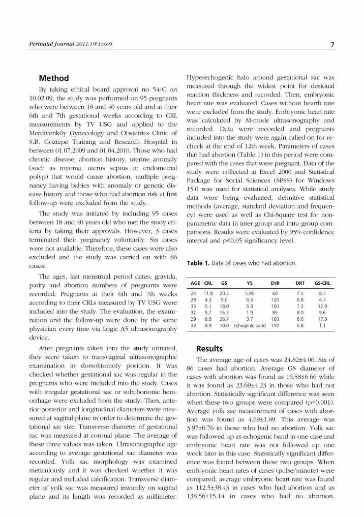
ResultsThe average age of cases was 24.82±4.06. Six of
86 cases had abortion. Average GS diameter ofcases with abortion was found as 16.58±6.66 whileit was found as 23.69±4.23 in those who had notabortion. Statistically significant difference was seenwhen these two groups were compared (p=0.001).Average yolk sac measurement of cases with abor-tion was found as 4.69±1.89. This average was3.97±0.76 in those who had no abortion. Yolk sacwas followed up as echogenic band in one case andembryonic heart rate was not followed up oneweek later in this case. Statistically significant differ-ence was found between these two groups. Whenembryonic heart rates of cases (pulse/minute) werecompared, average embryonic heart rate was foundas 112.5±38.43 in cases who had abortion and as138.56±15.14 in cases who had no abortion.
AGE CRL GS YS EHR DRT GS-CRL
24 11.8 20.5 5.95 60 7.5 8.728 4.3 9.0 6.6 120 6.8 4.735 5.1 18.0 5.3 100 7.2 12.932 5.7 15.3 1.9 85 8.0 9.629 8.8 26.7 3.7 160 8.6 17.933 8.9 10.0 Echogenic band 150 6.8 1.1
Table 1. Data of cases who had abortion.
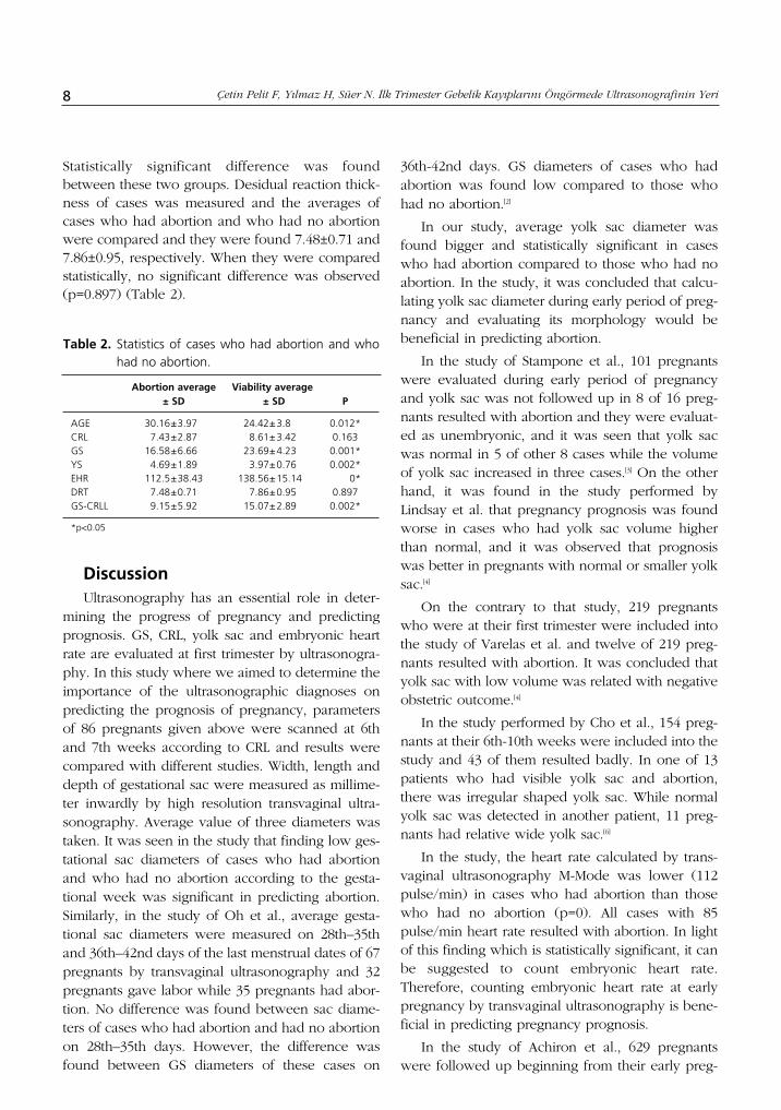
Statistically significant difference was foundbetween these two groups. Desidual reaction thick-ness of cases was measured and the averages ofcases who had abortion and who had no abortionwere compared and they were found 7.48±0.71 and7.86±0.95, respectively. When they were comparedstatistically, no significant difference was observed(p=0.897) (Table 2).
DiscussionUltrasonography has an essential role in deter-
mining the progress of pregnancy and predictingprognosis. GS, CRL, yolk sac and embryonic heartrate are evaluated at first trimester by ultrasonogra-phy. In this study where we aimed to determine theimportance of the ultrasonographic diagnoses onpredicting the prognosis of pregnancy, parametersof 86 pregnants given above were scanned at 6thand 7th weeks according to CRL and results werecompared with different studies. Width, length anddepth of gestational sac were measured as millime-ter inwardly by high resolution transvaginal ultra-sonography. Average value of three diameters wastaken. It was seen in the study that finding low ges-tational sac diameters of cases who had abortionand who had no abortion according to the gesta-tional week was significant in predicting abortion.Similarly, in the study of Oh et al., average gesta-tional sac diameters were measured on 28th–35thand 36th–42nd days of the last menstrual dates of 67pregnants by transvaginal ultrasonography and 32pregnants gave labor while 35 pregnants had abor-tion. No difference was found between sac diame-ters of cases who had abortion and had no abortionon 28th–35th days. However, the difference wasfound between GS diameters of these cases on
36th-42nd days. GS diameters of cases who hadabortion was found low compared to those whohad no abortion.[2]
In our study, average yolk sac diameter wasfound bigger and statistically significant in caseswho had abortion compared to those who had noabortion. In the study, it was concluded that calcu-lating yolk sac diameter during early period of preg-nancy and evaluating its morphology would bebeneficial in predicting abortion.
In the study of Stampone et al., 101 pregnantswere evaluated during early period of pregnancyand yolk sac was not followed up in 8 of 16 preg-nants resulted with abortion and they were evaluat-ed as unembryonic, and it was seen that yolk sacwas normal in 5 of other 8 cases while the volumeof yolk sac increased in three cases.[3] On the otherhand, it was found in the study performed byLindsay et al. that pregnancy prognosis was foundworse in cases who had yolk sac volume higherthan normal, and it was observed that prognosiswas better in pregnants with normal or smaller yolksac.[4]
On the contrary to that study, 219 pregnantswho were at their first trimester were included intothe study of Varelas et al. and twelve of 219 preg-nants resulted with abortion. It was concluded thatyolk sac with low volume was related with negativeobstetric outcome.[4]
In the study performed by Cho et al., 154 preg-nants at their 6th-10th weeks were included into thestudy and 43 of them resulted badly. In one of 13patients who had visible yolk sac and abortion,there was irregular shaped yolk sac. While normalyolk sac was detected in another patient, 11 preg-nants had relative wide yolk sac.[6]
In the study, the heart rate calculated by trans-vaginal ultrasonography M-Mode was lower (112pulse/min) in cases who had abortion than thosewho had no abortion (p=0). All cases with 85pulse/min heart rate resulted with abortion. In lightof this finding which is statistically significant, it canbe suggested to count embryonic heart rate.Therefore, counting embryonic heart rate at earlypregnancy by transvaginal ultrasonography is bene-ficial in predicting pregnancy prognosis.
In the study of Achiron et al., 629 pregnantswere followed up beginning from their early preg-
Çetin Pelit F, Y›lmaz H, Süer N. ‹lk Trimester Gebelik Kay›plar›n› Öngörmede Ultrasonografinin Yeri8
Abortion average Viability average± SD ± SD P
AGE 30.16±3.97 24.42±3.8 0.012*CRL 7.43±2.87 8.61±3.42 0.163GS 16.58±6.66 23.69±4.23 0.001*YS 4.69±1.89 3.97±0.76 0.002*EHR 112.5±38.43 138.56±15.14 0*DRT 7.48±0.71 7.86±0.95 0.897GS-CRLL 9.15±5.92 15.07±2.89 0.002*
*p<0.05
Table 2. Statistics of cases who had abortion and whohad no abortion.
nancy period and 580 of them were kept in thestudy until to the end of 13th weeks and 23 of 580pregnancies resulted with abortion. Average heartrate in 8 of the cases resulted with abortion wassimilar to the embryonic heart rate in pregnantswho had no abortion. It was seen that average heartrate in 15 pregnants was out of 95% confidenceinterval.[7] Doubilet et al. evaluated 1,185 singlepregnancy cases in their study. All cases wereexamined by transvaginal ultrasonography at aver-agely week 6.2 and embryonic heart rates of caseswere counted. Average heart rate was found as 110pulse/min. The embryonic heart rates of all caseswere counted again by transvaginal ultrasonogra-phy at averagely week.[8] Average heart rate wasfound as 159 pulse/min. In 122 cases who had abor-tion, the heart rate was <110 pulse/min.[3]
Schats et al. counted heart rates of 47 cases withIVF pregnancy by transvaginal ultrasonography andthe heart rate was observed at 25th day in all cases.[8]
In the study performed by Thedor et al., 2,164pregnants were evaluated by transvaginal ultra-sonography at their 6th-8th weeks and their heartrate was counted. Average heart rate was found as125±15 pulse/min. in cases who had no abortionwhile it was found as 85 pulse/min. in cases whohad abortion. They found relation between abortionand having 85 pulse/min.[9] The heart rates of twocases who had abortion were found below 85pulse/min. in this study. In the study, desidual reac-tion thicknesses of cases who had abortion andwho had no abortion were compared and no sig-nificant difference was found statistically.
Bajo et al. measured trophoblastic thickness onembryonic implantation region in women whowere between 5th and 12th gestational week and itwas found that trophoblastic thickness was 3 mmmore according to the gestational age in 15% ofcases.[10]
ConclusionIt was concluded that calculating yolk sac
diameter and its morphology, gestational sac diam-eter and embryonic heart rate at 6th and 7th ges-tational weeks was significant in predicting gesta-tional prognosis and it was found that the desidualreaction thickness was insignificant in predictinggestational prognosis. It was also found thatembryonic heart rate below 85, small gestationalsac according to gestational week, yolk sac diam-eter above 6 mm or the existence of calcificationin yolk sac had a relation with bad prognosis.
References1. Edwards RG. Physiological and molecular aspects of human
implantation. Hum Reprod 1995;10(Suppl 2):1-13.
2. Oh J, Wright G, Coulam C. Gestational sac diameter in veryearly pregnancy as predictor of fetal outcome. UltrasoundObstet Gynecol 2002;20:267-9.
3. Stampone C, Nicotra M, Muttinelli C, Cosmi V. Transvaginalsonography of yolk sac in normal and abnormal pregnan-cy. J Clin Ultrasound 1996;24:3-9.
4. Lindsay DJ, Lovett IS, Lyons EA. Yolk sac diameter andshape at endovaginal US: predictors of pregnancy outcomein the first trimester. Radiology 1992;183:115-8.
5. Varelas FK, Prapas MN. Yolk sac size and embryonic heartrate as prognostic factors of first trimester pregnancy out-come. Eur J Obstet Gynecol Reprod Biol 2008;138:10-3.
6. Cho FN, Chen SN, Ta› MH, Yang TL. The quality and sizeof yolk sac in early pregnancy loss. Aust N Z J ObstetGynaecol 2006;46:413-8.
7. Achiron R, Tadmor O, Mashiach S. Heart rate as a predic-tor of first trimester spontaneous abortion after ultrasoundproven viability. Obstet Gynecol 1991;78:330-4.
8. Schats R, Jansen JA, Wladimiroff JW. Embryonic heart activ-ity; appearance and development in earlyhuman pregnan-cy. Br J Obstetrics Gynecology 1990;97:989-94.
9. Theodor S, Dimitrios E, Alexander S, George Z. Embryonicheart rate in early pregnancy. J Clin Ultrasound 1997;26:33-6.
10. Bajo J, Moreno-Calvo FJ, Martinez-Cortes L. Is trophoblasticthickness at the embryonic implantation site a new sign ofnegative evolution in first trimester pregnancy. Hum Reprod2000;15:1629-30.
Perinatal Journal 2011;19(1):6-9 9
10 Perinatal Journal 2011;19(1):10-14
Abstract
Objective: Intrahepatic cholestasis of pregnancy is a clinical syndrome, characterized by maternal pruritus and biochemical cholesta-sis. Epidemiologic surveys show significant regional differences in the incidence of intrahepatic cholestasis of pregnancy. In this study,we evaluated the frequency of intrahepatic cholestasis of pregnancy and perinatal/neonatal outcomes in our hospital.
Methods: Twenty patients with intrahepatic cholestasis of pregnancy and their 22 newborn babies (3 twins), and one intrauterineexitus with unknown etiology were included in this retrospective analysis. Ursodeoxycholic acid was given to 50%of intrahepaticcholestasis of pregnancy patients. Fifteen (65.2%) of the newborn babies were ≤37 weeks. Eight (34.7%) of newborns had transientrespiratory support, 2 with continuous positive airway prssure. None of the babies had diagnosed respiratory distress syndrome, thusnone of them required surfactant. One case of intrauterine exitus has occured with unknown etiology.
Results: We conducted a retrospective study about frequency and perinatal outcome of pregnancies complicated by intrahepaticcholestasis of pregnancy between June 2007-August 2010 at Baskent University Ankara Hospital. Perinatal/neonatal outcomes wereretrospectively studied by medical chart review.
Conclusion: In our study intrahepatic cholestasis of pregnancy was present in 1.4% of pregnancies, that was similar to the Europeanpopulation. Increased risk of preterm delivery and respiratory distress syndrome were reported in literature, in our study group, noneof the babies had RDS or other serious neonatal complications of preterm labor despite high rate of preterm birth rate and ratherhigh levels of maternal serum bile acid levels.
Keywords: Intrahepatic cholestasis, newborn, pregnancy, perinatal outcomes.
‹ntrahepatik kolestazl› gebelerin neonatal sonuçlar›
Amaç: Gebelik kolestaz›, maternal kafl›nt› ve biyokimyasal kolestaz ile karakterize klinik bir sendromdur. Epidemiyolojik çal›flmalardagebelik kolestaz›n›n insidans› bölgesel farkl›l›klar göstermektedir. Bu çal›flmada gebelik kolestazl› hastalar›m›z›n s›kl›¤› ve perinatal/ne-onatal sonuçlar›n›n belirlenmesi amaçland›.
Yöntem: Çal›flma Baflkent Üniversitesi’inde Ankara Hastanesi Haziran 2007-A¤›stos 2010 tarihleri aras›nda takip edilen kolestazl› ge-belerin ve yenido¤anlar›n›n dosyalar›n›n retrospektif olarak taranmas› ile yap›ld›.
Bulgular: Yirmi kolestazl› gebenin 22 bebe¤i (3’ü ikiz) çal›flmaya dahil edildi. Ursodeoksikolik asit kolestazl› gebelerin yar›s›na verildi.On befl (%65.2) bebek ≤37 hafta alt›ndayd›. Sekiz (%34.7) yenido¤ana geçici solunum deste¤i sa¤land› (F‹O2<%30), bunlardan 2 be-be¤e yenido¤an›n geçici takipnesi nedeniyle sürekli pozitif bas›nç uygulamas› yap›ld›. Hiçbir yenido¤ana respiratuvar distres sendro-mu tan›s› konulmad› ve bu nedenle sürfaktan tedavisi verilmedi. Bir fetus intrauterin eksitus idi ancak ölüm nedeni belirlenemedi.
Sonuç: Çal›flmam›zda kolestazl› gebelerin s›kl›¤›, Avrupa toplumuna benzer flekilde %1.4 olarak belirlendi. Literatürde prematüre do-¤um ve respiratuvar distres sendromunun kolestazl› gebelerde artt›¤› bildirilirse de bizim çal›flmam›zda hiçbir bebekte respiratuvar dis-tres sendromunu görülmedi. Yüksek maternal serum safra asitleri ve prematüre do¤uma ra¤men yenido¤anlarda di¤er ciddi neona-tal komplikasyonlar gözlenmedi.
Anahtar Sözcükler: ‹ntrahepatik kolestazis, yenido¤an, gebelik, perinatal/neonatal sonuçlar.
Neonatal Outcomes of Pregnancy with Intrahepatic Cholestasis
Abdullah Kurt1, Ayfle Ecevit1, Burcu K›sa2, Deniz Anuk ‹nce1, Aylin Tarcan1, Filiz Bilgin Yan›k2
1Baflkent Üniversitesi T›p Fakültesi Neonatoloji Bilim Dal›, Ankara, Türkiye2Baflkent Üniversitesi T›p Fakültesi Kad›n Hastal›klar› ve Do¤um Anabilim Dal›, Ankara, Türkiye
Correspondence: Abdullah Kurt, Fevzi Çakmak Cad. 11. Sok. No: 45 Bahçelievler, Ankara, Türkiye
e-mail: [email protected]
e-Adress: http://www.perinataljournal.com/20110191003doi:10.2399/prn.11.0191003
Perinatal Journal 2011;19(1):10-14 11
Introduction Intrahepatic cholestasis of pregnancy (ICP) is a
pregnancy specific liver disease characterized bymaternal pruritus without any skin rash, and bio-chemical cholestasis with abnormal liver functionsin the absence of other liver diseases, occuring inthe late second trimester or third trimester and per-sisting until delivery. The etiology of ICP is compli-cated and poorly understood.[1-5] This syndrome mayresult in spontaneous preterm delivery, meconium-stained amniotic fluid, fetal distress and intrauterinefetal death, although thought to be benign for moth-ers.[6] Clinical studies clearly show that it may lead topreterm delivery in up to 19-60%, fetal distress in27-33%, and fetal loss in 0.2-4.1% of patients.[1-3
Respiratory distress syndrome (RDS) was reportedin some of the infants born from mothers with ICP.[7]
Epidemiologic surveys show significant regional dif-ferences in the incidence of ICP; it varies from 0.1to 1.5% of pregnancies in Europe and 1 to 5% inChina.[4] There are also differences due to ethnicity.In comparison with the overall prevalence of ICPwhich is 0.7%, the white population has a lowoccurrence of 0.62%, while this number is 1.46% inthe Pakistanian population and 1.24% in the Indianpopulation.[8] It is found to be 5.6% in a primarilyLatina Los Angeles population, much higher thanthe prevalence previously reported in the UnitedStates.[9] The frequency of ICP in Turkey has notbeen reported so far. In this study, we evaluated thefrequency of ICP and perinatal/neonatal outcome inour hospital.
MethodsThe study was performed retrospectively on 20
pregnancies complicated by ICP between June 2007and August 2010, at Baskent University AnkaraHospital, a tertiary care maternity center. The diag-nosis of ICP is based on (i) development of pruritusand cholestasis during the second or third trimesterof pregnancy, and (ii) elevated TBA ≥11 μmol/L(and elevated serum transaminases but not neces-sarily). (iii) Spontaneous relief of signs and symp-toms after delivery and (iv) absence of other dis-eases that cause pruritus and jaundice.[10,11]
Ultrasonography of the abdomen and serologicalscan of viral hepatitis were performed to excludeother causes of liver diseases in all patients beforeenrollment. Liver biopsy is not necessary for the
diagnosis and histopathology is not diagnostic.[10,11]
Patients with chronic liver diseases, skin diseases,allergic disorders, symptomatic cholelithiasis, andongoing viral infections affecting the liver (hepatitisA, B and C virus, cytomegalovirus, herpes simplexvirus, and epstein-barr virus) severe preeclampsiaaffecting liver functions and acute fatty liver of preg-nancy, were excluded. Statistical analysis was per-formed with SPSS for Windows 17.0 (SPSS Inc.,Chicago, IL, USA) and p <0.05 was considered sta-tistically significant. The results are expressed asmean ± SD. The difference between two sampleswas calculated using the Mann-Whitney U test andChi-Square test.
ResultsTwenty patients with ICP and their 22 newborn
babies (3 twins), and 1 intrauterine exitus withunknown etiology were included in this retrospec-tive analysis. Thirteen of babies were female(56.5%) and 10 of them were male (43.5%). Meanage of mothers with ICP was 31.4±4.5 years. ICPwas diagnosed at 32.6±3.8 weeks of gestation. Themain symptom of patients was pruritus (96%).Abnormal liver function tests and epigastric painwere present in 5% of the cases. Initial serum bileacid levels were 41.3±81.7 μmol/L. Liver and bileduct ultrasound findings were normal in 30% ofpatients. Bile stones, hemangioma of liver, intra-hepatic minimal dilatation of bile ducts and inspis-sated bile were revealed in, 5, 2, 1 and 1 of thepatients respectively. Ursodeoxycholic acid wasbeen given to 50% of ICP patients. Cesarean sectionrate was 70% (Table 1). Antenatal corticosteroid wasgiven to 15% of women with ICP for the preventionof RDS in preterm infants. Neonatal outcome: Meangestational age at delivery was 36.4±1.7 (33-39)weeks, 15 (65.2%) of the newborn babies were ≤37.Mean birth weigth was 2900±536 g. Mean Apgarscores were 8.3±0.7 (7-9) and 9.5±0.5 (9-10) at 1and 5 minutes respectively, excluding the case ofintrauterine exitus. Eight (34.7%) of newborns hadtransient respiratory support [6 newborns with nasalhigh flow oxygen (FiO2 <30%), 2 with continuouspositive airway prssure (CPAP) because of transienttachypnea of newbrn (TTN)]. All of the newbornswere fed with breast milk. Indirect hyperbilirubine-mia, TTN, patent ductus arteriosus (PDA) and uri-nary tract infection were present in 9 (39.1%), 4
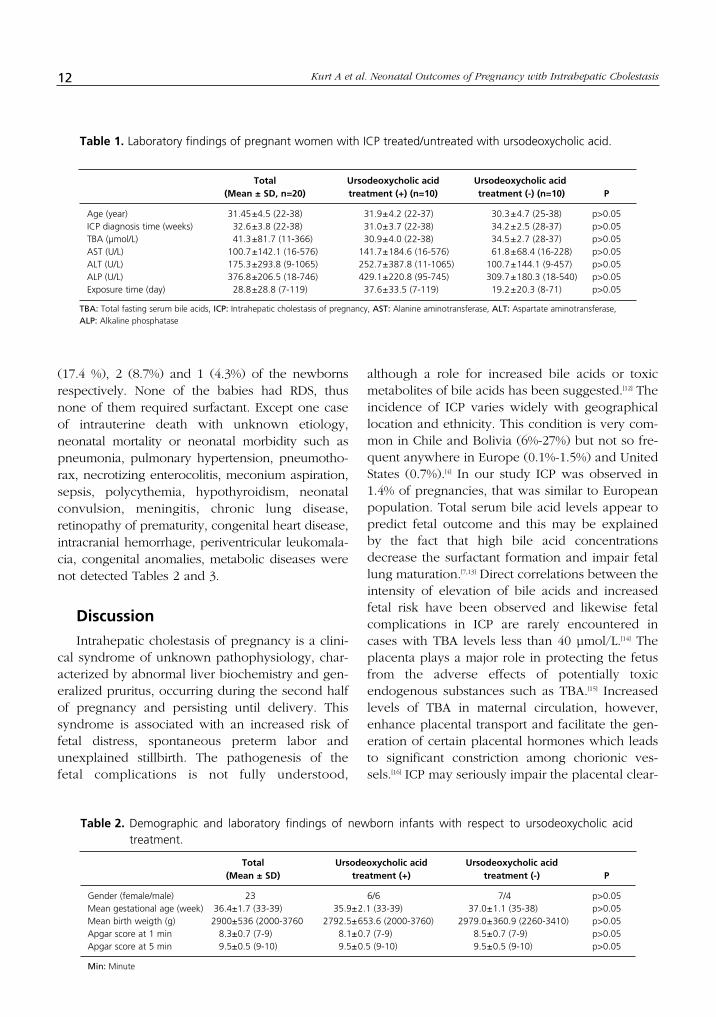
(17.4 %), 2 (8.7%) and 1 (4.3%) of the newbornsrespectively. None of the babies had RDS, thusnone of them required surfactant. Except one caseof intrauterine death with unknown etiology,neonatal mortality or neonatal morbidity such aspneumonia, pulmonary hypertension, pneumotho-rax, necrotizing enterocolitis, meconium aspiration,sepsis, polycythemia, hypothyroidism, neonatalconvulsion, meningitis, chronic lung disease,retinopathy of prematurity, congenital heart disease,intracranial hemorrhage, periventricular leukomala-cia, congenital anomalies, metabolic diseases werenot detected Tables 2 and 3.
DiscussionIntrahepatic cholestasis of pregnancy is a clini-
cal syndrome of unknown pathophysiology, char-acterized by abnormal liver biochemistry and gen-eralized pruritus, occurring during the second halfof pregnancy and persisting until delivery. Thissyndrome is associated with an increased risk offetal distress, spontaneous preterm labor andunexplained stillbirth. The pathogenesis of thefetal complications is not fully understood,
although a role for increased bile acids or toxicmetabolites of bile acids has been suggested.[12] Theincidence of ICP varies widely with geographicallocation and ethnicity. This condition is very com-mon in Chile and Bolivia (6%-27%) but not so fre-quent anywhere in Europe (0.1%-1.5%) and UnitedStates (0.7%).[4] In our study ICP was observed in1.4% of pregnancies, that was similar to Europeanpopulation. Total serum bile acid levels appear topredict fetal outcome and this may be explainedby the fact that high bile acid concentrationsdecrease the surfactant formation and impair fetallung maturation.[7,13] Direct correlations between theintensity of elevation of bile acids and increasedfetal risk have been observed and likewise fetalcomplications in ICP are rarely encountered incases with TBA levels less than 40 μmol/L.[14] Theplacenta plays a major role in protecting the fetusfrom the adverse effects of potentially toxicendogenous substances such as TBA.[15] Increasedlevels of TBA in maternal circulation, however,enhance placental transport and facilitate the gen-eration of certain placental hormones which leadsto significant constriction among chorionic ves-sels.[16] ICP may seriously impair the placental clear-
Kurt A et al. Neonatal Outcomes of Pregnancy with Intrahepatic Cholestasis12
Total Ursodeoxycholic acid Ursodeoxycholic acid (Mean ± SD, n=20) treatment (+) (n=10) treatment (-) (n=10) P
Age (year) 31.45±4.5 (22-38) 31.9±4.2 (22-37) 30.3±4.7 (25-38) p>0.05ICP diagnosis time (weeks) 32.6±3.8 (22-38) 31.0±3.7 (22-38) 34.2±2.5 (28-37) p>0.05TBA (μmol/L) 41.3±81.7 (11-366) 30.9±4.0 (22-38) 34.5±2.7 (28-37) p>0.05AST (U/L) 100.7±142.1 (16-576) 141.7±184.6 (16-576) 61.8±68.4 (16-228) p>0.05ALT (U/L) 175.3±293.8 (9-1065) 252.7±387.8 (11-1065) 100.7±144.1 (9-457) p>0.05ALP (U/L) 376.8±206.5 (18-746) 429.1±220.8 (95-745) 309.7±180.3 (18-540) p>0.05Exposure time (day) 28.8±28.8 (7-119) 37.6±33.5 (7-119) 19.2±20.3 (8-71) p>0.05
TBA: Total fasting serum bile acids, ICP: Intrahepatic cholestasis of pregnancy, AST: Alanine aminotransferase, ALT: Aspartate aminotransferase, ALP: Alkaline phosphatase
Table 1. Laboratory findings of pregnant women with ICP treated/untreated with ursodeoxycholic acid.
Total Ursodeoxycholic acid Ursodeoxycholic acid (Mean ± SD) treatment (+) treatment (-) P
Gender (female/male) 23 6/6 7/4 p>0.05Mean gestational age (week) 36.4±1.7 (33-39) 35.9±2.1 (33-39) 37.0±1.1 (35-38) p>0.05Mean birth weigth (g) 2900±536 (2000-3760 2792.5±653.6 (2000-3760) 2979.0±360.9 (2260-3410) p>0.05Apgar score at 1 min 8.3±0.7 (7-9) 8.1±0.7 (7-9) 8.5±0.7 (7-9) p>0.05Apgar score at 5 min 9.5±0.5 (9-10) 9.5±0.5 (9-10) 9.5±0.5 (9-10) p>0.05
Min: Minute
Table 2. Demographic and laboratory findings of newborn infants with respect to ursodeoxycholic acidtreatment.
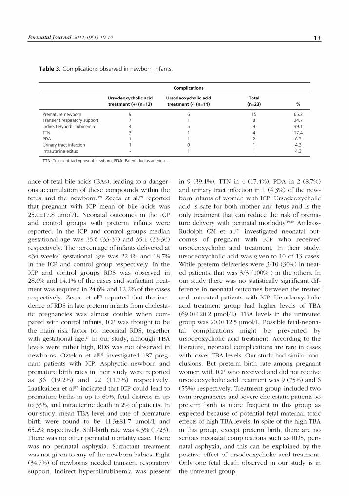
ance of fetal bile acids (BAs), leading to a danger-ous accumulation of these compounds within thefetus and the newborn.[17] Zecca et al.[7] reportedthat pregnant with ICP mean of bile acids was25.0±17.8 μmol/L. Neonatal outcomes in the ICPand control groups with preterm infants werereported. In the ICP and control groups mediangestational age was 35.6 (33-37) and 35.1 (33-36)respectively. The percentage of infants delivered at<34 weeks’ gestational age was 22.4% and 18.7%in the ICP and control group respectively. In theICP and control groups RDS was observed in28.6% and 14.1% of the cases and surfactant treat-ment was required in 24.6% and 12.2% of the casesrespectively. Zecca et al[7] reported that the inci-dence of RDS in late preterm infants from cholesta-tic pregnancies was almost double when com-pared with control infants, ICP was thought to bethe main risk factor for neonatal RDS, togetherwith gestational age.[7] In our study, although TBAlevels were rather high, RDS was not observed innewborns. Oztekin et al[18] investigated 187 preg-nant patients with ICP. Asphyctic newborn andpremature birth rates in their study were reportedas 36 (19.2%) and 22 (11.7%) respectively.Laatikainen et al[17] indicated that ICP could lead topremature births in up to 60%, fetal distress in upto 33%, and intrauterine death in 2% of patients. Inour study, mean TBA level and rate of prematurebirth were found to be 41.3±81.7 μmol/L and65.2% respectively. Still-birth rate was 4.3% (1/23).There was no other perinatal mortality case. Therewas no perinatal asphyxia. Surfactant treatmentwas not given to any of the newborn babies. Eight(34.7%) of newborns needed transient respiratorysupport. Indirect hyperbilirubinemia was present
in 9 (39.1%), TTN in 4 (17.4%), PDA in 2 (8.7%)and urinary tract infection in 1 (4.3%) of the new-born infants of women with ICP. Ursodeoxycholicacid is safe for both mother and fetus and is theonly treatment that can reduce the risk of prema-ture delivery with perinatal morbidity[19,20] Ambros-Rudolph CM et al.[21] investigated neonatal out-comes of pregnant with ICP who receivedursodeoxycholic acid treatment. In their study,ursodeoxycholic acid was given to 10 of 13 cases.While preterm deliveries were 3/10 (30%) in treat-ed patients, that was 3/3 (100% ) in the others. Inour study there was no statistically significant dif-ference in neonatal outcomes between the treatedand untreated patients with ICP. Ursodeoxycholicacid treatment group had higher levels of TBA(69.0±120.2 μmol/L). TBA levels in the untreatedgroup was 20.0±12.5 μmol/L. Possible fetal-neona-tal complications might be prevented byursodeoxycholic acid treatment. According to theliterature, neonatal complications are rare in caseswith lower TBA levels. Our study had similar con-clusions. But preterm birth rate among pregnantwomen with ICP who received and did not receiveursodeoxycholic acid treatment was 9 (75%) and 6(55%) respectively. Treatment group included twotwin pregnancies and severe cholestatic patients sopreterm birth is more frequent in this group asexpected because of potential fetal-maternal toxiceffects of high TBA levels. In spite of the high TBAin this group, except preterm birth, there are noserious neonatal complications such as RDS, peri-natal asphyxia, and this can be explained by thepositive effect of ursodeoxycholic acid treatment.Only one fetal death observed in our study is inthe untreated group.
Perinatal Journal 2011;19(1):10-14 13
Complications
Ursodeoxycholic acid Ursodeoxycholic acid Totaltreatment (+) (n=12) treatment (-) (n=11) (n=23) %
Premature newborn 9 6 15 65.2Transient respiratory support 7 1 8 34.7Indirect Hyperbilirubinemia 4 5 9 39.1TTN 3 1 4 17.4PDA 1 1 2 8.7Urinary tract infection 1 0 1 4.3Intrauterine exitus - 1 1 4.3
TTN: Transient tachypnea of newborn, PDA: Patent ductus arteriosus
Table 3. Complications observed in newborn infants.
ConclusionICP carries risks for mothers and their newborn
infants. But appropriate diagnosis, timely treatmentand delivery of the pregnant women with ICP mayeliminate the risks in the newborn infants duringthe perinatal and postnatal period.
References1. Lammert F, Marschall HU, Glantz A, Maternl S. Intrahepatic
cholestasis of pregnancy: molecular pathogenesis, diagno-sis and management. J Hepatol 2000; 33:1012-21.
2. Pusl T, Beuers U. Intrahepatic cholestasis of pregnancy.Orphanet J Rare Dis 2007;2:26.
3. Wang XD, Peng B, Yao Q. Perinatal outcomes of intrahep-atic cholestasis of pregnancy: analysis of 1210 cases.Zhonghua Yi Xue Za Zhi 2006;86:446-9.
4. Ai Y, Liu SY, Yao Q. Clinical characteristics of 1241 casesof intrahepatic cholestasis of pregnancy. Zhonghua Fu ChaKe Za Zhi 2004;39:217-20.
5. Geenes V, Williamson C. Intrahepatic cholestasis of preg-nancy. World J Gastroenterol 2009;15:2049-66.
6. Beuers U, Pusl T. Intrahepatic cholestasis of pregnancy: aheterogeneous group of pregnancy related disorders?.Hepatology 2006;43:647-9.
7. Zecca E, Luca D, Marras M, Caruso A, Bernardini T,Romagnoli C. Intrahepatic cholestasis of pregnancy andneonatal respiratory distress syndrome. Pediatrics2006;117:1669-72.
8. Abedin P, Weaver JB, Egginton E. Intrahepatic cholestasisof pregnancy: prevalence and ethnic distribution. EthnHealth 1999;4:35-7.
9. Lee RH, Goodwin TM, Green spoon J, Incerpi M. Theprevalence of intrahepatic cholestasis of pregnancy in a pri-marily Latina Los Angeles population. J Perinatol2006;26:527-32.
10. Knox TA, Olans LB. Liver disease in pregnancy. N Engl JMed 1996;335:569-76.
11. Rolfes DB, Ishak KG. Liver disease in pregnancy.Histopathology 1986;10:555-70.
12. Germain AM, Carvajal JA, Glasinovic JC, Kato CS,Williamson C. Intrahepatic cholestasis of pregnancy: anintriguing pregnancyspecific disorder. J Soc Gynecol Investig2002;9:10-14.
13. Zecca E, De Luca D, Barbato G, Marras M, Tiberi E,Romagnoli C. Predicting respiratory distress syndrome inneonates from mothers with intrahepatic cholestasis ofpregnancy. Early Hum Dev 2007;84:337-41.
14. Glantz A, Marschall HU, Mattsson L. Intrahepatic cholesta-sis of pregnancy: relationship between bile acids levels andfetal complication rates. Hepatology 2004;40:467-74.
15. Marin JJ, Macias RI, Serrano MA. The hepatobiliary-likeexcretory function of the placenta. A review. Placenta2003;24:431-8.
16. Meng LJ, Reyes H, Palma J, Hernandez J, Ribalta J, SjovallJ. Progesterone Metabolism in Normal Human Pregnancyand in Patients with Intrahepatic Cholestasis of Pregnancy.In: Reyes HB, Leuschner U, Arias IM (Eds). Pregnancy sexhormones and the liver. New York: Kluwer; 1996. p. 91-100.
17. Laatikainen TJ. Fetal bile acid levels in pregnancies com-plicated by maternal intrahepatic cholestasis. Am J ObstetGynecol 1975;122:852-6.
18. Oztekin D, Aydal I, Oztekin O, Okcu S, Borekci R, Tinar S.Predicting fetal asphyxia in intrahepatic cholestasis of preg-nancy. Arch Gynecol Obstet 2009;280:975-9.
19. Zapata R, Sandoval L, Palma J, Hernández I, Ribalta J, ReyesH, et al. Ursodeoxycholic acid in the treatment of intrahep-atic cholestasis of pregnancy. A 12-year experience. LiverInt 2005;25:548-54.
20. Williamson C, Hems LM, Goulis DG, Walker I, Chambers J,Donaldson O, et al. Clinical outcome in a series of cases ofobstetric cholestasis identified via a patient support group.BJOG 2004;111:676-81.
21. Ambros-Rudolph CM, Glatz M, Trauner M, Kerl H,Müllegger RR. The importance of serum bile acid levelanalysis and treatment with ursodeoxycholic acid in intra-hepatic cholestasis of pregnancy: a case series From CentralEurope. Arch Dermatol 2007;143:757-62.
Kurt A et al. Neonatal Outcomes of Pregnancy with Intrahepatic Cholestasis14
Abstract
Objective: To study the long-term effects of the cesarean and vaginal births on unintended pregnancy rates and the obstetric costs.
Methods: Parous women (n=501) were grouped into two in accordance to the mode of their first two births as: vaginal birth groupwith two subsequent vaginal births and cesarean birth group with at least one cesarean birth. Turkish Republic Social SecurityInstitution payment values in 2010 were used as the cost standards. Means of parity, ectopic pregnancy, spontaneous abortion andvoluntary abortion were used to calculate the life time obstetrics expenditure of the groups. The calculated values were divided toeach group’s mean reproductive time to find out the expenditure per reproductive year. In statistical analysis Student’s t test or analy-sis of variance tests were used. A p value of ≤0.05 was considered statistically significant.
Results: Vaginal group had significantly higher gravidity, parity and voluntary abortion rates (p<0.05). The means of the age of theparticipating women, spontaneous abortions and ectopic pregnancies were similar in both groups (p>0.05). The total number ofpregnancies, births, spontaneous abortions, voluntary abortions and offspring decreased significantly in each decade (p<0.05).Hospital births significantly increased in each decade, reaching from 48.29% in 70s to 88.08% in 1990s. The obstetric expenditurefor a single woman in a reproductive year time was found 71.92 Turkish Liras in vaginal and 53.41 Turkish Liras in cesarean birthgroups.
Conclusion: Cesarean birth decreases the total obstetric costs in long term, particularly in areas with high fertility rates.
Keywords: Cesarean section, cesarean section rate, mode of birth, cost control, obstetrics economics, elective surgical procedure.
Sezaryen do¤umlar›n istenmeyen gebelik oran› ve obstetrik harcamalar üzerine uzun dönem etkileri
Amaç: Sezaryen ve vajinal do¤umlar›n istenmeyen gebelik oran› ve obstetrik harcamalar üzerine uzun dönem etkilerini araflt›rmak.
Yöntem: Daha önceden do¤um yapm›fl kad›nlar (n=501) do¤um yöntemine göre iki gruba ayr›ld›lar: ‹lk iki gebeli¤ini vajinal do¤u-ranlar vajinal do¤um grubu ve ilk iki gebelikten en az birini sezaryen ile do¤uranlar sezaryen do¤um grubunu oluflturdu. Maliyet stan-dartlar› için Türkiye Cumhuriyeti Sosyal Güvenlik Kurumu 2010 ödeme de¤erleri kullan›ld›. Gruplar›n ömür boyu obstetrik giderlerinihesaplamak için, ortalama do¤um, d›fl gebelik, kendili¤inden ve istemli düflük oranlar› kullan›ld›. Hesaplanan de¤erler her grubun or-talama tüketilmifl üreme y›llar›na bölünerek, üreme y›l› bafl›na gider hesaplamas› yap›ld›. ‹statistiksel analizde Student T testi ve var-yans analizi (ANOVA) testleri kullan›ld›. P de¤erinin ≤0.05 olmas› istatistiksel olarak anlaml› kabul edildi.
Bulgular: Vajinal grubun gebelik, do¤um ve istemli düflük oranlar› belirgin olarak daha yüksek bulundu (p<0.05). Ortalama anne ya-fl›, kendili¤inden düflük ve d›fl gebelik say›lar› ise her iki grupta benzerdi (p>0.05). Her 10 y›ll›k dönemde ortalama gebelik, do¤um,kendili¤inden düflük, istemli düflük ve sahip olunan çocuk say›s› belirgin olarak azalm›flt›r (p<0.05). Hastane do¤um oranlar› da her 10y›lda belirgin olarak artm›flt›r. 1970’de %48.29 olan hastane do¤umu oran›, 1990’larda %88.08’e ulaflm›flt›r. Üreme y›l› bafl›na obs-tetrik gider, bir kad›n için vajinal do¤um grubunda 71.92 Türk Liras› ve sezaryen do¤um grubunda 53.41 Türk Liras› bulundu.
Sonuç: Özellikle do¤urganl›¤›n yüksek h›zda oldu¤u bölgelerde, sezaryen do¤um obstetrik giderleri uzun dönemde azalt›r. Ama uzundönem giderlere göre cerrahi karar vermek do¤ru olmayacakt›r. Unutulmamal›d›r ki, sa¤l›k politikalar›ndaki de¤ifliklikler bu gider he-saplamalar›n› tamamen de¤ifltirebilir.
Anahtar Sözcükler: Sezaryen do¤um, sezaryen do¤um oran›, do¤um yöntemi, maliyet kontrolü, obstetrik ekonomi, elektif cerrahi ifllem.
Long-Term Effects of Cesarean Births onUnintended Pregnancy Rates and Obstetric
Expenditures
Kahraman Ülker, ‹smail Temur, Abdülaziz Gül
Kafkas Üniversitesi T›p Fakültesi Kad›n Hastal›klar› ve Do¤um Anabilim Dal›, Kars, Türkiye
Correspondence: Kahraman Ülker, Kafkas Üniversitesi T›p Fakültesi Kad›n Hastal›klar› ve Do¤um Anabilim Dal›, Kars, Türkiye
e-mail: [email protected]
Perinatal Journal 2011;19(1):15-19
e-Adress: http://www.perinataljournal.com/20110191004doi:10.2399/prn.11.0191004
IntroductionCesarean birth rates continuously rise in
Turkey, like many other countries.[1-6] The increaseis generally associated with the women’s request,physicians’ choice, and previous cesarean birth,low level skill of primary care providers, injudi-cious use of uterotonics, changing demographics,altered clinical practice, and an increasing aware-ness of traumatic childbirth amongst the public.[7-10]
There are contraries in opinions about the rise incesarean rates, particularly in cases of maternalrequest without any clinical indication for thesurgery.[11,12] One of the contraries is the cost of theprocedures. The insurance companies are unwill-ing to pay for the cesarean births without anacceptable surgical indication, because the cesare-an birth prices are generally more expensive thanvaginal birth prices. However, the long-term costsare not well studied. Some researchers comparedthe costs of each vaginal and cesarean birthaccording to the expenditure made by the hospitaland the patient;[13] others compared the cost ofeach procedure with total perinatal expenditures.[14]
In our study we planned to compare the long-termcosts of cesarean birth (CB) and vaginal birth (VB).Because the mode of birth may affect future preg-nancy rates and the gestational outcomes, we usedthe parity and abortion rates to calculate awoman’s mean life-time obstetrics expenditure.We hypothesized that, by decreasing the futurebirth and abortion rates, having at least one of thefirst two births by cesarean section would decreasethe total obstetric expenditures spent for a woman.
MethodsThe study was approved by the local
Institutional review board of our university hospi-tal and performed between January and October2010 within our obstetrics and gynecology depart-ment. All patients gave informed consent beforeparticipating in the study. To study the long-termeffects of the mode of delivery, parous womenstarted to give births before 2000 (n=501) weredivided into two groups in accordance to the modeof their first two births as: vaginal birth group(VBG) with two subsequent vaginal births andcesarean birth group with at least one cesareanbirth. A questionnaire was distributed to the partic-
ipants to obtain demographic information such asmaternal age, gravidity, parity, spontaneous abor-tion, induced abortion, ectopic pregnancy, numberof offspring and their age. A nurse capable ofspeaking several local dialects helped the partici-pants to fill in the questionnaires and providedassistance in cases of confusion, misunderstandingand illiteracy. After collecting the data, one of theauthors checked the questionnaire form for theaccuracy of the obtained data. Unreliable andincomplete data caused exclusion. Inconsistent anddiscordant data between variables like maternalage, first child’s age and birth dates caused exclu-sions. Means of age, gravidity, parity, spontaneousand voluntary abortion, ectopic pregnancy, num-ber of offspring with their age and gravidity-livingoffspring difference were calculated for eachgroup. According to the reproductive years, thegroups were further analyzed for giving the firstbirth between 1970-1980, 1980-1990, and 1990-2000. The decision for termination of a pregnancymay be influenced by the actual family size. Mostof the time, the unintended pregnancies are termi-nated if the family has reached the desired numberof offspring. To eliminate the effect of family size,women having two or more offspring were includ-ed. In addition, a woman’s age and reproductiveyear stage could influence the decision of preg-nancy termination. The range for reproductiveyears was defined as 15-49 years. 15 years weresubtracted from actual maternal age to calculatethe bygone reproductive years. In cases of womenolder than 49 (45-15=34), reproductive years wereconsidered as 34. The total cost calculation weused was not a real cost effectiveness calculation.It did not reflect the actual cost for each procedureat the time of the study. We did not calculate thereal market prices of the expenditures. We usedTurkish Republic Social Security Institution’s 2010payment values as the cost standards during thecalculation. For year 2010 the institute pays 450Turkish Liras (TL) for cesarean birth, 400 TL forvaginal birth, 455 TL for the management ofectopic pregnancy and 200 TL for the managementof spontaneous or voluntary abortion. In our studythe total cost (TC) of the mode of birth (MB) foreach woman was calculated by using the formula:TCMB = (Mean number of births according to theMB x cost of the MB) + (Mean count of sponta-neous abortion according to the MB x cost of spon-
Ülker K, Temur ‹, Gül A. Long-Term Effects of Cesarean Births on Unintended Pregnancy Rates and Obstetric Expenditures16

Perinatal Journal 2011;19(1):15-19 17
taneous abortion treatment)+ (Mean count of vol-untary abortion according to the MB x cost of vol-untary abortion treatment)+ (Mean count of ectopicpregnancy according to the MB x cost of ectopicpregnancy treatment). To eliminate the effect ofthe maternal age and the stage of the reproductiveyears, the total costs of the groups were divided tothe mean reproductive bygone times of their owngroup. The calculation represented the total obstet-ric expenditure for a reproductive year of a singlewoman. By using SPSS for Windows 16.0 packageprogram the data were collected and analyzed.Vaginal birth and cesarean birth groups were com-pared using the independent sample Student t test.Demographical data was compared by 10 yearintervals from 1970 to 2000 using analysis of vari-ance test (ANOVA). A p value of <0.05 was con-sidered statistically significant.
ResultsThe demographical findings and their compari-
son results according to the mode of birth are sum-marized in Table 1. The mean numbers of preg-nancies, births and voluntary abortions were signif-icantly higher in VBG (p<0.05). The means of theage of the participating women, spontaneous abor-tions and ectopic pregnancies were similar in both
groups (p>0.05). The demographical findings andtheir comparison according to the years by ten yearintervals are summarized in Table 2. The total num-ber of pregnancies, births, spontaneous abortions,voluntary abortions and offspring decreased signifi-cantly in each decade (p<0.05). The percentage ofhospital births significantly increased in eachdecade, reaching from 48.29% in 70s to 88.08% in1990s. The total obstetric cost of a woman wasfound 1,940.55 Turkish Liras (TL) in VBG and1,332.85 Turkish Liras in CBG. Although the mater-nal ages in both groups were not significantly dif-ferent from each other, to eliminate the effect of thematernal age and the stage of the reproductiveyears, the total costs of the groups were divided tothe mean bygone reproductive years of their owngroup. The mean bygone reproductive years were26.98±6.82 years in vaginal birth and 24.95±6.82years in cesarean birth groups and did not show sig-nificant difference (p>0.05). After dividing the costsby each group’s own mean bygone reproductiveyear, the obstetric cost of a reproductive year of awoman was calculated. The yearly obstetric cost fora woman was found 71.92 Turkish Liras in VBG and53.41 Turkish Liras in CBG. Reproductive failurewas defined as the rates of the pregnancies that didnot result in a living offspring. The difference of liv-ing offspring from the total pregnancies divided by
Age* (years) Gravidity# Parity# Miscarriage# Induced Ectopicabortion# pregnancy#
Vaginal birth group (n= 458 ) 42.72±7.78 5 4 0 0 0
Cesarean birth group (n= 43) 40.42±7.53 3 2 0 0 0
p 0.06 0.00 0.00 0.61 0.00 0.11
Table 1. Comparison of vaginal birth and cesarean birth groups data are presented as mean± standard deviation* or median.#
1970-1980 1980-1990 1990-2000 p(n= 105) (n= 181) (n= 215)
Age of the women at the timeof the study* 50.66±3.56 45.97±4.34 35.651±5.41 0.00Gravidity# 7 5 4 0.00Parity# 5 4 3 0.00Miscarriages# 0 0 0 0.00Induced abortion# 1 1 0 0.00Ectopic pregnancy# 0 0 0 0.05Offspring number# 4 3 3 0.00Hospital Births (%)* 48.29±46.51 79.02±34.27 88.08±29.23 0.00
Table 2. Demographic changes of the 501 women by 10 year intervals. Data are presentedas mean ± standard deviation* or median.#
the total number of pregnancies gave the failurerates for each group. The reproductive failure ratewas 0.27±0.23 in the VBG and 0.25±0.21 in theCBG. Although the reproductive failure rate wasfound slightly higher in the VBG, the difference wasnot significant (p>0.05).
DiscussionIn developing and overpopulated countries
health resources are generally used in public hos-pitals instead of primary and preventive sectors.[13,15]
From this point of view, the high fertility rates,because of the inadequate primary family planningservices, further increases the costs. The realobstetric costs differ between countries, cities andhospitals. The differences in health policies, pro-duction and providing costs of the medical andnonmedical outgoings, staff wages, and the lifestandards of the environment, poverty and afford-ability affect the costs of birth modes.[16-22] Two dif-ferent hospitals, even in the same region of a city,may have different incomes and expenses. Thequality of medical and non-medical services maybe different. We think that it is irrational to com-pare the costs of the vaginal and cesarean births byusing the amount of the money spent for each pro-cedure in different conditions. Our study wasbased on the effect of cesarean births on the preg-nancy related events, like the number of births andabortions. From this point of view, differences inthe production and providing costs of the medicaland non-medical outgoings, staff wages and etc.do not change the power of this effect, and in thelong term, cesarean births decrease the totalexpenditure of obstetric costs. Unintended preg-nancies constitute half of all pregnancies (49%) inthe United States. Moreover, 52% (approximatelyone third of all) of them result in an unintendedbirth.[23] In 2001 6.7 million women used the publicfamily planning services in the United States. Theexpense per woman was $ 188 ($ 1.26 billion intotal).[24] This amount is huge and unaffordable formost of the countries. In addition it could not pro-vide a full contraceptive service for the wholecommunity. In our study cesarean birth decreasedthe gravidity, parity and voluntary abortion rates,which resulted in a decrease in the unintendedpregnancy and birth rates. Tubal sterilization orintra-operative intra-uterine device insertion duringthe same operation may facilitate the acceptance
of contraceptive use. The rate of tubal sterilizationduring cesarean births is higher than interval ster-ilization.[25] The decreases in both total fertility andinduced abortion rates can be explained by thewomen’s more decided attitudes against unintend-ed pregnancy. The hospital, operation and anes-thesia experience and fear, the physician’s orienta-tion, the observed increased complication rates inthe repeated operations, difficulty in baby care andthe delay in returning to the job may be the caus-es of the decided attitude against the unintendedpregnancies. Hospital births may also decrease theunintended pregnancy rates. Many women mayhave the first and only chance to benefit from ahealth and family planning service at the time oftheir births. Although, the hospital birth counts ofthe vaginal births increases continuously, thecesarean births are always in hospitals. In addition,the repeat cesarean births with their potential com-plications are always taken more seriously by thephysicians and the pregnant women. The risk ofthe complications of all the obstetrical and gyne-cological operations increases after each repeatedcesarean section, which may orient the physiciansto give stricter advices and orders about the unin-tended pregnancies. The decrease in total fertilityand induced abortions rates after cesarean birthsshould be further studied. In this manner, the roleof intra-partum contraception, hospital births, thewomen’s educational and socio-economic status,physician’s orientation, health politics and the sci-entific advances in health management need to bedetermined by multi-center studies. Many womenrequest for cesarean births. We think that after giv-ing an informed consent, those women should berespected and promoted for their decisions.Whatever the causes are, cesarean section decreas-es the rates of pregnancy, births and voluntaryabortions. This effect should also be considered inthe comparison of the risks of cesarean and vagi-nal births.
ConclusionIf one of the first two births is a cesarean birth,
the pregnancy, birth and voluntary abortion ratesdecrease. Moreover, the obstetric costs alsodecrease in long term. In cases of maternal requestcesarean births may be acceptable, particularly inthe regions with high fertility rates and poor familyplanning services. However, the decrease in the
Ülker K, Temur ‹, Gül A. Long-Term Effects of Cesarean Births on Unintended Pregnancy Rates and Obstetric Expenditures18
long-term expenditure, pregnancy, birth and volun-tary abortions is not a surgical indication. Our datamay possibly influence the surgical indication in thefuture. However, this influence may be acceptableonly after the prospective studies which show equalsafety for the cesarean births and the vaginal births.It should be kept in mind that changes in healthpolitics and efficient contraception against the unin-tended pregnancies can totally change these expen-diture calculations.
References1. Center for disease control and prevention. Births:
Preliminary data for 2005..http://www.cdc.gov/nchs/ prod-ucts/pubs/pubd/hestats/prelimbirths05/prelimbirths05.htmMay›s 31, 2006.
2. Coskun A, Kotsu B, Ercan O, K›ran H, Guven MA, K›ran G.Kahramanmarafl il merkezinde 2004 ve 2006 y›llar›ndakido¤umlar›n karfl›laflt›r›lmas›. Journal of Turkish Obstetricand Gynecology Society 2007;4:168-72.
3. Meikle SF, Steiner CA, Zhang J, Lawrence WL. A nationalestimate of the elective primary cesarean birth rate. ObstetGynecol 2005;105:751–6.
4. Lin HC, Xirasagar S. Institutional factors in cesarean birthrates: policy and research implications. Obstet Gynecol2004;103:128-13.
5. Özkaya O. Birth rates and cesarean indications at SüleymanDemirel University Obstetrics and Gynecology Clinicthrough a 5-year period. SDÜ Tip Fak Derg 2005;12:36-9.
6. Guney M, Uzun E, Oral B, Sar›kan I, Bayhan G, Mungan T.Cesarean section rates and indications at our clinic between2001 and 2005. Türkiye Klinikleri J Gynecol Obst2006;3:249-54.
7. Yaflar O, fiahin FK, Coflar E, Koken GN, Çevrio¤lu AS. Birthmethod choices of primiparous women and the factorswhich have an effect on these choices. Türkiye Klinikleri JGynecol Obst 2007;17:414-20.
8. Shamshad. Factors leading to increased cesarean sectionrate. Gomal Journal of Medical 2008;6:1-5.
9. Farrell SA, Basket TF, Farrell KD. The choice of electivecesarean birth in obstetrics: a voluntary survey of Canadianhealth care professionals. Int Urogynecol J 2005;16:378-83.
10. Dietz HP. Elective cesarean section- the right choice forwhom? Current Women’s Health Review 2005;1:85-8.
11. Bewley S, Cockburn J. II. The unfacts of ‘request’ caesare-an section. BJOG 2002;109:597-605.
12. Hannah M. Planned elective cesarean section: A reasonablechoice for some women? CMAJ 2004;170:813-4.
13. Khan A, Zaman S. Costs of vaginal birth and Caesarean sec-tion at a tertiary level public hospital in Islamabad,Pakistan. BMC Pregnancy Childbirth 2010;10:2; DOI (e-adres): http://www.ncbi.nlm.nih.gov/pmc/articles/PMC2826286/ (bask›da).
14. Palencia R, Gafni A, Hannah ME, Ross S, Willan AR,Hewson S et al. The costs of planned cesarean versusplanned vaginal birth in the Term Breech Trial. CMAJ2006;174(8);DOI (e-adres): http://www.cmaj.ca/cgi/ con-tent/full/174/8/1109 (bask›da).
15. Tabish SA, Mustaffa A, Rangrez RA. Hospital accountingbased cost studies: Indian experience. J Academy ofHospital Administration 2005;13:1-6.
16. Hendrix MJ, Evers SM, Basten MC, Nijhuis JG, Severens JL.Cost analysis of the Dutch obstetric system: low-risk nulli-parous women preferring home or short-stay hospital birth--a prospective non-randomised controlled study. BMCHealth Serv Res 2009; 9:211;DOI (e-adres):http://www.ncbi.nlm.nih.gov/pmc/articles/PMC2784768/?tool=pubmed (bask›da).
17. Vander Plaetse B, Hlatiwayo G, Van Eygen L, Meessen B,Criel B. Costs and revenue of health care in a ruralZimbabwean district. Health Policy Plan 2005;20:243-51.
18. Minh HV, Giang KB, Huong DL, Huong le T, Huong NT,Giang PN, Hoat LN, Wright P. Costing of clinical services inrural district hospitals in northern Vietnam. Int J HealthPlann Manage 2010;25:63-73.
19. Adam T, Evans DB, Murray CJ. Econometric estimation ofcountry-specific hospital costs. Cost Eff Resour Alloc2003;1:3; DOI (e-adres): http://www.resource-allocation.com/content/1/1/3 (bask›da).
20. Nahar S, Costello A. The hidden cost of 'free' maternity carein Dhaka, Bangladesh. Health Policy Plan 1998;13:417-22.
21. Lagarde M, Palmer N. The impact of user fees on health ser-vice utilization in low- and middle-income countries: howstrong is the evidence? Bull World Health Organ2008;86:839-48.
22. Olukoga A. Unit costs of inpatient days in district hospitalsin South Africa. Singapore Med J 2007;48:143-7.
23. Frost, J, Darroch, J. Contraceptive Use and UnintendedPregnancy. Glob. libr. women's med.,(ISSN: 1756-2228)2008; DOI 10.3843/GLOWM.10379.
Perinatal Journal 2011;19(1):15-19 19
Introduction It was found in multiple pregnancies that the
incidence of fetal structural and chromosomal
anomalies is higher as well as bad maternal and
fetal obstetric outcomes.[1] In recent years, the
chance of detecting fetus with anomaly during
early pregnancy has increased by the improvement
of antenatal follow-up.
The expectant approach in multiple pregnan-
cies where there is fetus with anomaly is to choose
either to end the pregnancy or to do selective
fetoreduction (SF).[2]
Selective fetoreduction is to apply fetocide on
the fetus with anomaly in multiple pregnancies. It
is aimed to decrease maternal morbidity, to pre-
vent early labor, to recover the prognosis of nor-
20 Perinatal Journal 2011;19(1):20-22
Abstract
Objective: The purpose of this study is to evaluate selective fetoreduction in two twin pregnancies which have one of abnormalities
Case: 28 and 34 years old two patients have 18 and 16 weeks gestation and one of abnormal sibling, respectively. Vertebral anom-aly and oligohydramnios were found in the first case, and the encephalocele was found in the second case. Transabdominal ultra-sound guided selective fetoreduction was performed in both anomaly fetuses, and no complications were occurred in early period.Preterm labor was occurred in both cases at 32 and 35 weeks of gestation. The first case was delivered vaginally, and the secondcase due to previous cesarean section was delivered abdominally. The babies were 1,700 g and 2,400, respectively. There was notfound a problem in infants in the first 3 months of postpartum period.
Conclusion: Due to potential risks of multiple pregnancies for mother and fetus, we think that selective fetoreduction is useful forthe anomalous one of sibling twin.
Keywords: Selective fetoreduction, twin pregnancy.
Selektif fetoredüksiyonun de¤erlendirilmesi: ‹ki olgu
Amaç: Bu çal›flman›n amac› ikizlerden birinin anomalili olmas› nedeniyle selektif fetoredüksiyon (SF) uygulanan iki olguyu de¤erlen-dirmektir.
Olgu: 28 ve 34 yafl›nda, s›ras›yla 18 ve 16 haftal›k, ikiz efllerinden biri anomalili iki olgu incelendi. ‹lk olguda vertebral anomali, oligo-hidramnios, ikinci olguda ise ensefalosel tespit edildi. Transabdominal ultrason eflli¤inde her iki anomalili fetüse selektif fetosit uygu-land› ve erken döneminde her hangi bir komplikasyon izlenmedi. Her iki olguda, gebeli¤in 32 ve 35 haftalar›nda erken do¤um eyle-mi geliflti. ‹lk olgu normal vaginal, ikinci ise eski sezaryen seksiyo nedeniyle sezaryen ile do¤umu gerçeklefltirildi. S›ras›yla 1,700 g ve2,400 g iki bebek do¤urtuldu. Postpartum 3 ayl›k dönemde yenido¤anlarda bir anomali izlenmedi.
Sonuç: Ço¤ul gebeliklerin anne ve fetüs için olas› riskleri nedeniyle, anomalili ikiz efline selektif fetoredüksiyon uygulanmas›n›n dahayararl› olaca¤› kanaatindeyiz.
Anahtar Sözcükler: Selektif fetoredüksiyon, ikiz gebelik.
Selective Fetoreduction: Report of Two Cases
Muhammet Erdal Sak1, Mehmet S›dd›k Evsen1, Hatice Ender Soydinç1, Sibel Sak2, Ahmet Yal›nkaya1
1Dicle Üniversitesi T›p Fakültesi Kad›n Hastal›klar› ve Do¤um Anabilim Dal›, Diyarbak›r, Türkiye2Diyarbak›r Kad›n Hastal›klar› ve Çocuk Hastanesi Kad›n Hastal›klar› ve Do¤um Bölümü, Diyarbak›r, Türkiye
Correspondence: Muhammet Erdal Sak, Dicle Üniversitesi T›p Fakültesi Kad›n Hastal›klar› ve Do¤um Anabilim Dal›, Diyarbak›r, Türkiye
e-mail: [email protected]
e-Adress: http://www.perinataljournal.com/20110191005doi:10.2399/prn.11.0191005
Perinatal Journal 2011;19(1):20-22 21
mal fetus and to terminate fetus with anomaly dueto socio-economical and psychological reasons.
Case Report
Case 1
Twenty-eight years old (G3P1) twin pregnantwas referred to our center since anomaly wasdetected in one of the fetuses. It was learnt that thepatient got pregnant spontaneously. In her ultra-sonographic examination, 18 weeks and 4 days oldnormal fetus without fetal anomaly and its 18weeks old twin fetus with oligohydramnios andvertebral angulation were detected. The motherwas informed about her gestational status and theprocess which can be done. By the approval of thefamily, selective fetocide was decided.Accompanied by transabdominal ultrasonography,it was entered into the fetal cardiac cavity by 22gauge 120 mm spinal needle and intracardiac 2.0cc 7.5% potassium chloride (KCl) was injected. Theprocess was terminated after 7 minutes of obser-vation in the fetus who developed cardiac arrest.No complication was observed during early peri-od. Preterm labor developed at 32nd week in thepatient who had no problem at her normal follow-up. 1,700 g baby girl was delivered by normalvaginal way. It was found in the examinationwhich was done three months after the deliverythat the baby was healthy.
Case 2
Thirty-four years old (G2P1) pregnant with oldsection was followed up as a twin pregnancydeveloping spontaneously and externally. Upondetecting anomaly in one of the fetuses, she wasreferred to our clinic. In her ultrasonographicexamination, 16 weeks old twin pregnancy wasdetected; while one of the fetuses was normal,other one had encephalocele. The family wasinformed about the fetuses and selective fetocidewas suggested. By the approval of the family, itwas entered into the fetal cardiac cavity by 22gauge 120 mm spinal needle accompanied bytransabdominal ultrasonography and intracardiac2.0 cc 7.5% KCl was injected. It was observed for7 minutes after cardiac arrest developed; no viabil-ity was seen and the patient was taken to the fol-low-up. The cesarean was applied at 35th gesta-
tional week due to preterm action and old sectionand a healthy 2,300 g baby boy was delivered. Itwas found in the examination which was donethree months after the delivery that the baby washealthy.
DiscussionThe administration is hard in twin pregnancies
where a fetus has anomaly, because it is possibleto affect normal fetus by the decision taken forfetus with anomaly. If it is a minor anomaly (cleftlip), the administration is not different than a nor-mal pregnancy; however, two fetuses should beconsidered in the administration of major congen-ital anomalies. Major congenital anomalies involvestructural or chromosomal anomalies causing fetalmorbidity and mortality increase.[2]
The termination of whole pregnancy causesproblem in multiple pregnancies especially inthose who want to have child or where it developsas a result of infertility treatment. Again, the fami-ly may meet long-term morbidity of fetus exposeto preterm labor by expectant approach and livingfetus with anomaly. Selective fetoreduction is awell accepted option and Eddleman et al.[3] pub-lished a series of 300 cases. In this series, 164 ofthem were twin, 32 of them were triplet and 4 ofthem were quadruplet. It was reported that preg-nancy loss was 4% and 84.4% of deliveries were at32nd gestational weeks and above when selectivetermination was applied. Evans et al.[4] found preg-nancy loss as 7.5% in their series of 402 cases.
Techniques described at second trimester forselective fetoreduction are exsanguinating fetalheart by puncture, calcium gluconate infusion bycardiac puncture, air embolization through umbili-cal vessels in company with fetoscopy and intracar-diac potassium chloride injection. Potassium chlo-ride injection was widely accepted in selective ter-mination process and multifetal pregnancy termina-tion process where fetuses were followed up nor-mally in multiple pregnancies by the verification ofefficiency and reliability of potassium chloride injec-tion.[5] Picking wrong fetus, technical incompetencyof the person who will perform the process, pre-mature rupture of membrane, infection and the lossof whole pregnancy are the undesired early periodcomplications of SF.
Sak ME et al. Selective Fetoreduction: Report of Two Cases22
ConclusionIn conclusion, SF is a well accepted process
since maternal and fetal risks brought by multiplepregnancy are decreased for normal fetus by ter-minating fetus with anomaly and it affects familiesin a positive way both emotionally and psycholog-ically. When SF is performed by experienced peo-ple, the chance of success is high while the risk ofpregnancy loss is low.
References1. Pat A, Smith M. Textbook of Fetal Abnormalities. Philadelphia:
Churchill Livingston-Elsevier: 2007. p. 405-26.
2. Chang YL, Chao AS, Cheng PJ, Chung CL, Chueh HY,
Chang SD, et al. Presence of a single fetal major anomaly
in a twin pregnancy does not increase the preterm rate.
Aust N Z J Obstet Gynaecol 2004;44:332-6.
3. Eddleman KA, Stone JL, Lynch L, Berkowitz RL. Selective
termination of anomalous fetuses in multifetal pregnancies:
Two hundred cases at a single center. Am J Obstet Gynecol
2002;187: 1168-72.
4. Evans MI, Goldberg JD, Horenstein J. Selective termination
for structural, chromosomal and mendelian anomalies:
international experience. Am J Obstet Gynecol 1999;181:893-
7.
5. Golbus MS, Cunningham N, Goldberg JD, Anderson R, Filly
R, Callen P. Selective termination of multiple gestations. Am
J Med Genet 1988;31:339-48.
Abstract
Objective: Ornithine transcarbamylase (OTC) deficiency is the most common urea cycle disorder. In our case, we discussed the fol-low up and the management of the OTC deficiency patient, diagnosed during pregnancy.
Case: 32-year-old patient, OTC deficiency was diagnosed during pregnancy which was resulted with missed abortion. In the nextpregnancy, the patient treated with phenyl butyrate, arginin ornitin lisin and carbamazepine. Coryon villus sampling (CVS) was donein the first trimester. There was not any mutation in the fetal gene locus. In the 39. gestational week, healthy female baby was deliv-ered by caesarean section.
Conclusion: OTC deficiency is a rare disease. To make the followup and the management of these patients during pregnancy, mayrequire knowledge and experience about complications. The treatment must be carried out with multidisciplinary approach. Thegenetic counseling should be given to the family about the prenatal diagnosis of OTC deficiency (CVS, amniosentesis).
Keywords: Ornithine transcarbamylase deficiency, pregnancy, prenatal diagnosis, multidisciplinary approach.
Gebelikte ornitin transkarbamilaz eksikli¤i tan›s› ve yönetimi: Olgu sunumu
Amaç: Ornitin transkarbamilaz (OTC) eksikli¤i, en s›k rastlanan üre döngüsü bozuklu¤udur. Olgumuzda; abortus ile sonlanan gebe-li¤i s›ras›nda OTC eksikli¤i tan›s› alan hastan›n, sonraki gebeli¤indeki takip ve yönetimini literatür bilgileri ›fl›¤›nda tart›flt›k.
Olgu: 32 yafl›nda hastaya, abortus ile sonuçlanan gebeli¤i s›ras›nda OTC eksikli¤i tan›s› konuldu. Takip eden gebeli¤inde; oral sod-yum fenilbutirat, arginin ornitin lizin ve karbamazepin tedavisi baflland›. Birinci trimesterde koryon villus örneklemesi yap›ld›. Fetal genlokusunda mutasyona rastlanmad›. 39. gebelik haftas›nda, sa¤l›kl› k›z bebek sezaryen ile do¤urtuldu.
Sonuç: OTC eksikli¤i nadir görülen bir hastal›kt›r. Bu hastalar›n gebelik süresince takip ve yönetimini yapmak, olabilecek komplikas-yonlar hakk›nda bilgi birikimini ve tecrübeyi gerektirmektedir. Tedavi multidisipliner yaklafl›mla gerçeklefltirilmelidir. OTC eksikli¤ininprenatal tan›s›n›n (CVS, amniyosentez) mümkün oldu¤u konusunda aileye gerekli genetik dan›flmanl›k verilmelidir.
Anahtar Sözcükler: Ornitin transkarbamilaz eksikli¤i, gebelik, prenatal tan›, multidisipliner yaklafl›m.
The Diagnosis and Management of OrnithineTranscarbamylase Deficiency in Pregnancy: A
Case Report
Orkun Çetin1, Cihat fien1, Begüm Aydo¤an1, Seyfettin Uluda¤1, ‹pek Dokurel Çetin2, Hakan Erenel1
1Cerrahpafla T›p Fakültesi Kad›n Hastal›klar› ve Do¤um Anabilim Dal›, ‹stanbul, Türkiye2Cerrahpafla T›p FakültesiÇocuk Sa¤l›¤› ve Hastal›klar› Anabilim Dal›, ‹stanbul Türkiye
Correspondence: Orkun Çetin, Cerrahpafla T›p Fakültesi Kad›n Hastal›kar› ve Do¤um Anabilim Dal›, Fatih, ‹stanbul, Türkiye
e-mail: [email protected]
Introduction Urea cycle disorders are seen once in every
30000 living delivery. All such disorders are auto-somal recessively inherited except transcarbamy-lase deficiency (OTC) displaying inheritance viaX.[1,2] OTC deficiency is the most frequent urea
cycle disorder.[1] Homozygote males are affected
more severely by the disease than heterozygote
females. Citruline level is distinctively low while
glutamine, glycine, and alanine levels are high.
Orotic acid levels in urine increase notably.
Enzymatic deficiency is shown by liver biopsy.
Perinatal Journal 2011;19(1):23-27
e-Adress: http://www.perinataljournal.com/20110191006doi:10.2399/prn.11.0191006
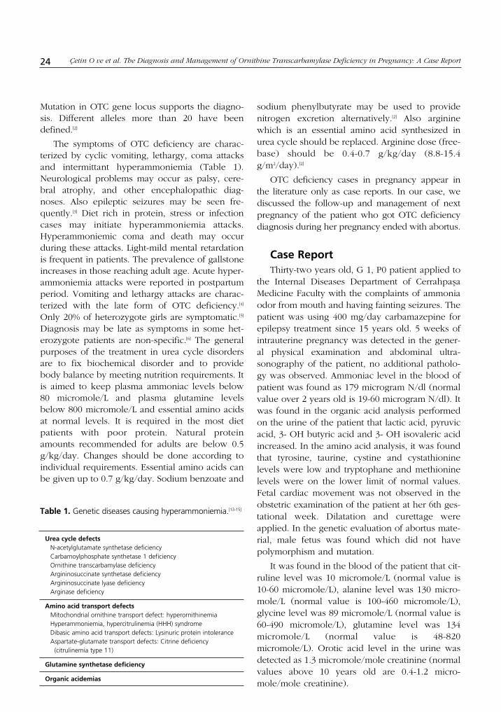
Mutation in OTC gene locus supports the diagno-sis. Different alleles more than 20 have beendefined.[2]
The symptoms of OTC deficiency are charac-terized by cyclic vomiting, lethargy, coma attacksand intermittant hyperammoniemia (Table 1).Neurological problems may occur as palsy, cere-bral atrophy, and other encephalopathic diag-noses. Also epileptic seizures may be seen fre-quently.[3] Diet rich in protein, stress or infectioncases may initiate hyperammoniemia attacks.Hyperammoniemic coma and death may occurduring these attacks. Light-mild mental retardationis frequent in patients. The prevalence of gallstoneincreases in those reaching adult age. Acute hyper-ammoniemia attacks were reported in postpartumperiod. Vomiting and lethargy attacks are charac-terized with the late form of OTC deficiency.[4]
Only 20% of heterozygote girls are symptomatic.[5]
Diagnosis may be late as symptoms in some het-erozygote patients are non-specific.[6] The generalpurposes of the treatment in urea cycle disordersare to fix biochemical disorder and to providebody balance by meeting nutrition requirements. Itis aimed to keep plasma ammoniac levels below80 micromole/L and plasma glutamine levelsbelow 800 micromole/L and essential amino acidsat normal levels. It is required in the most dietpatients with poor protein. Natural proteinamounts recommended for adults are below 0.5g/kg/day. Changes should be done according toindividual requirements. Essential amino acids canbe given up to 0.7 g/kg/day. Sodium benzoate and
sodium phenylbutyrate may be used to providenitrogen excretion alternatively.[2] Also argininewhich is an essential amino acid synthesized inurea cycle should be replaced. Arginine dose (free-base) should be 0.4-0.7 g/kg/day (8.8-15.4g/m2/day).[2]
OTC deficiency cases in pregnancy appear inthe literature only as case reports. In our case, wediscussed the follow-up and management of nextpregnancy of the patient who got OTC deficiencydiagnosis during her pregnancy ended with abortus.
Case ReportThirty-two years old, G 1, P0 patient applied to
the Internal Diseases Department of CerrahpaflaMedicine Faculty with the complaints of ammoniaodor from mouth and having fainting seizures. Thepatient was using 400 mg/day carbamazepine forepilepsy treatment since 15 years old. 5 weeks ofintrauterine pregnancy was detected in the gener-al physical examination and abdominal ultra-sonography of the patient, no additional patholo-gy was observed. Ammoniac level in the blood ofpatient was found as 179 microgram N/dl (normalvalue over 2 years old is 19-60 microgram N/dl). Itwas found in the organic acid analysis performedon the urine of the patient that lactic acid, pyruvicacid, 3- OH butyric acid and 3- OH isovaleric acidincreased. In the amino acid analysis, it was foundthat tyrosine, taurine, cystine and cystathioninelevels were low and tryptophane and methioninelevels were on the lower limit of normal values.Fetal cardiac movement was not observed in theobstetric examination of the patient at her 6th ges-tational week. Dilatation and curettage wereapplied. In the genetic evaluation of abortus mate-rial, male fetus was found which did not havepolymorphism and mutation.
It was found in the blood of the patient that cit-ruline level was 10 micromole/L (normal value is10-60 micromole/L), alanine level was 130 micro-mole/L (normal value is 100-460 micromole/L),glycine level was 89 micromole/L (normal value is60-490 micromole/L), glutamine level was 134micromole/L (normal value is 48-820micromole/L). Orotic acid level in the urine wasdetected as 1.3 micromole/mole creatinine (normalvalues above 10 years old are 0.4-1.2 micro-mole/mole creatinine).
Çetin O ve et al. The Diagnosis and Management of Ornithine Transcarbamylase Deficiency in Pregnancy: A Case Report24
Urea cycle defectsN-acetylglutamate synthetase deficiencyCarbamoylphosphate synthetase 1 deficiencyOrnithine transcarbamylase deficiencyArgininosuccinate synthetase deficiencyArgininosuccinate lyase deficiencyArginase deficiency
Amino acid transport defectsMitochondrial ornithine transport defect: hyperornithinemiaHyperammoniemia, hypercitrulinemia (HHH) syndromeDibasic amino acid transport defects: Lysinuric protein intoleranceAspartate-glutamate transport defects: Citrine deficiency
(citrulinemia type 11)
Glutamine synthetase deficiency
Organic acidemias
Table 1. Genetic diseases causing hyperammoniemia.[13-15]
Perinatal Journal 2011;19(1):23-27 25
Mutation was not observed in the genomicamplification process of the patient performed bypolymerase chain reaction (PCR). However, in theXp21.1 gene locus, K46R, lus3- 8A> T, Q 270R het-erozygote polymorphism was detected. In the lightof findings, the patient was diagnosed as genedefect of OTC heterozygote enzyme deficiency.
As the result of the diagnosis, 15.8 g/day (6 x2.500 mg) sodium phenylbutyrate, 6.600 mg/day(2x3.300 mg) arginine – ornithine – lysine and 400mg/day (2x200 mg) carbamazepine were given tothe patient.
Spontaneous single pregnancy was detected 5months after the treatment of the patient wasarranged. It was accepted suitable to perform thefollow-up of the pregnancy by multidisciplinaryapproach together with the Perinatology andInternal Metabolism Diseases. Upon the joint deci-sion, arginine – ornithine – lysine amino acid com-plex (AOL), sodium phenylbutyrate and carba-mazepine treatments were carried on during thepregnancy. Genetic consultation was provided tothe patient about the prognosis of urea cycle dis-order and the carbamazepine treatment duringpregnancy. The patient stated that she wanted pre-natal diagnosis, but she could not use the drug bytaking teratogenic effects of carbamazepine intoconsideration. Carbamazepine treatment wasceased on 8th gestational week and it was plannedto do a chorionic villus sampling (CVS) on 13thgestational week.
Nuchal thickness (NT) was measured as 1.8mm and nasal bone was screened on the firsttrimester scanning. A wave was positive on ductusvenosus and regurgitation was not detected on tri-cuspid valve.
Ammonia level was 157 microgram N/dl at 12thgestational week. Numerical anomaly was notobserved in the microscopic evaluation performedby GT 6 band technique of metaphase platesobtained by direct and long-term cultures of CVSsample. Chromosome establishment was found asnormal. No mutation was seen in OTC gene locus.
In the 22nd gestational week examination,bipariteal diameter (BPD) was measured as 52mm, head circumference (HC) as 195 mm, abdom-inal circumference (AC) as 177 mm, femur length(FL) as 35 mm, peak heart rate as 158 beat/min,and placenta anterior wall located nasal bone was
measured as 6.2 mm. Right uterine artery pulsatil-ity index (PI) was found as 0.96 and resistanceindex (RI) was 0.54, but no notch was observed.Left uterine artery PI was 0.76 and RI was 0.53 butno notch was observed. In the detailed ultra-sonography, fetal anatomy and development wasobserved appropriate to gestational week.Ammoniac level of the patient was 148 microgramN/dl. Fetal anatomy and development was foundas normal in the ultrasonography examination ather 28th gestational week. Estimated birth weightwas measured as 1,529 g. As the patient had oncetonic-clonic seizure in her history, she was con-sulted by Metabolism Diseases and carbamazepinewas initiated as 200 mg/day and clonazepam dropas 7.5 mg/day. Ammoniac level of the patient wasmeasured as 81 microgram N/dl.
By cesarean on her 39th gestational week, thepatient gave a 3,820 gr girl baby who hadcephalopelvic disproportion indication, 7 APGARscore at 1st minute and 9 APGAR score at 5thminute. Ammoniac level after delivery was foundas 116 microgram N/dl.
The patient was followed up at postpartumperiod in terms of clinical findings of hyperam-moniemia (anorexia, vomiting, lethargy, respirato-ry alkalosis, tremor, feebleness, ataxia, hypother-mia, epileptic attacks). 250 mg/kg sodium ben-zoate and sodium phenylacetate, and 500mg/kg/day (intravenous) arginine were kept avail-able in order to use on hyperammoniemia seizure.However, no symptom requiring emergency treat-ment appeared after cesarean.
Mother and baby did not require intense careafter delivery. Mother and baby were dischargedfrom the hospital on postoperative 4th day. Nomutation was found in OTC gene locus of thebaby via the genomic amplification process byPCR. The baby was taken into routine follow-up inHealthy Child Polyclinic.
DiscussionOTC deficiency is inherited via X and is the
most frequent urea cycle disorder. Its diagnosis isbased on inheritance via X, concentration of highserum ammonia, glutamine and alanine, and highorotidine existence within urea after taking allop-urinol.[7]
Mental deficiency is frequently seen in patientsand generalized tonic-clonic or focal seizures areseen in those with hyperammoniemia.[7] The levelof relative lyonization (random X chromosomeinactivation) of normal and abnormal OTC genes inhepatocytes determines the severity of disease.[2]
The diagnosis in our case was late until 32 yearsold since she had heterozygote mutation.Establishing certain diagnosis during pregnancy is arare case.
Citruline level of our case was found as 10micromole/L, alanine level as 130 micromole/L,glycine level as 89 micromole/L and glutaminelevel as 134 micromole/L. Low citruline level wasfound as compatible with the literature. However,normal values of alanine, glycine and glutaminelevels were not parallel to the literature.
Orotic acid level in urine was found as 1.3micromole/mole creatinine in our case. High orot-ic acid level in urine was compatible with the liter-ature. High orotic acid level in urine also togetherwith Carbamyl Phosphate Synthetase is a significantmarker in distinctive diagnosis.
The disease is observed as acute metabolicencephalopathy attacks in adult age group. Attacksgenerally appear as a result of metabolic stressessuch as infection, anesthesia, pregnancy, delivery.Triggering factor may not always be detected.When acute encephalopathy attacks (due to hyper-ammoniemia) are not treated, deteriorationadvances, patient is lost due to coma or patientcontinues to live with neurological sequel. Deathreason is frequently cerebral edema.[2] Cases withhyperammoniemia attack and atonia bleeding inpostnatal period were reported in the literature. Wedid not observe such a complication in antenataland postnatal follow-up of our case. Since postpar-tum 1st hour ammoniac value was 116 microgramN/dl, emergency treatment of hyperammoniemiawas not required (emergency treatment applica-tions are initiated when plasma ammoniac value isthree times higher than the normal value).
CVS was applied to our case as prenatal inva-sive diagnosis test. Numerical and structural anom-aly and mutation in OTC gene locus were notdetected when evaluating the sample.
AOL amino acid complex and sodium phenyl-butyrate treatment carried on during pregnancy ofour case. Though anti-epileptics were not useduntil 28th gestational week, no seizure occurred.
Upon tonic clonic epileptic seizure at 28th gesta-tional week, carbamazepine treatment was initiat-ed. The patient was followed up under anti-epilep-tic treatment in the rest of the pregnancy periodand no seizure occurred.
Consequently, OTC deficiency is a rare disease.Performing follow-up and management of thesepatients during pregnancy requires knowledge andexperience about possible complications. Patientsshould be protected as much as possible againstmetabolic stresses such as hunger, excessive pro-tein loading, infection etc. in order to prevent acutehyperammoniemia attack, and surgical applicationssuch as normal delivery and cesarean should befollowed up carefully. In such cases, protein intakeshould be lowered and more carbohydrate shouldbe given as a precaution. It should always be pre-pared against acute encephalopathy attack.Intravenous treatment (Sodium benzoate, sodiumphenylbutyrate, arginine) should be applied topatients who have vomiting and progressiveencephalopathy which can not tolerate oral intake.
As seen in our case, management of patientsshould be performed by multidisciplinary approach(Internal Endocrinology Department andGynecology and Obstetrics PerinatologyDepartment) and tertiary centers.
Required genetic consultancy should be provid-ed to family that prenatal diagnosis (CVS,Amniocentesis) of OTC deficiency is possible. Inthe first 24 hours, protein should be removed fromthe diet of prenatally diagnosed newborn. Also,female relatives of patient should be checked forheterozygote inheritance of OTC deficiency.Detection of inheritance helps the early diagnosisof women who have acute hyperammoniemiaattack risk. Early diagnosis of these people decreas-es complication possibility and provides prenataldiagnosis possibility in a pregnancy.
ConclusionOTC deficiency is a rare disease. Making the fol-
low-up and the management of these patients dur-ing pregnancy may require knowledge and experi-ence about complications. The treatment must becarried out with multidisciplinary approach. Thegenetic consultancy should be given to the familyabout the prenatal diagnosis of OTC deficiency(CVS, Amniocentesis).
Çetin O ve et al. The Diagnosis and Management of Ornithine Transcarbamylase Deficiency in Pregnancy: A Case Report26
References1. Oechsner M, Steen C, Sturenburg HJ, Kohlschutter A.
Hyperammonaemic encephalopathy after initiation of val-proate therapy in recognized ornithine transcarbamylasedeficiency. J Neurol Neurosurg Psychiatry 1998;64:680-2.
2. Neyzi O, Ertu¤rul T. Pediatri. ‹stanbul: Nobel T›p Kitabevi:2002. s. 673-8.
3. Msall M, Batshaw ML, Suss R, Brusilow SW, Mellits ED.Neurological outcome in children with inborn errors ofurea synthesis. Outcome of urea cycle enzymopathies. ArchDis Child 1984;310:1500-5.
4. Kennedy CR, Cogswell JJ. Late onset ornithine transcar-bamylase deficiency in males. Letter 1989;64:638.
5. Scaglia F, Zheng Q, O’Brien W, Henry J, Rosenberger J,Reeds P, Lee B. An integrated approach to the diagnosisand progpestive management of partial ornithine transcar-bamylase deficiency. Pediatrics 2002;109:150-2.
6. Ahrens MJ, Berry SA, Whitley CB, Markowitz DJ, Plante RJ,Tuchman M. Clinical and biochemical heterogeneity infemales of a large pedigree with ornithine transcarbamylasedeficiency due to the R141 Q mutation. Am J Med Genet1996;66:311-15.
7. Arn PH, Hauser ER, Thomas GH, Herman G, Hess D,Brusilow SW. Hyperammoneamia in women with a muta-tion at the ornithine transcarbamylase locus: a cause ofpostpartum coma. N Engl J Med 1990;322: 1652-5.
8. Brusilow SW, Horwich AL. Urea Cycle Enzymes. New York:McGraw-Hill; 2001. p. 1909-61..
9. Butterworth RF. Effects of hyperammonaemia on brainfunction. J Inherit Metab Dis 1998;21(Suppl 1):6–20.
10. Gropman AL, Summar M, Leonard JV. Neurological impli-cations of urea cycle disorders. J Inherit Metab Dis2007;30:865-9.
11. Nassogne MC, Héron B, Touati G, Rabier D, Saudubray JM.Urea cycle defects: management and outcome. J InheritMetab Dis 2005;28:407-14.
12. Enns GM, Berry SA, Berry GT, Rhead WJ, Brusilow SW,Hamosh A. Survival after treatment with phenylacetate andbenzoate for urea-cycle disorders. N Engl J Med2007;356:2282-92.
13. Leonard JV. Disorders of the Urea Cycle and RelatedEnzymes. Heidelberg: Springer; 2006. p. 263-72.
14. Saheki T, Kobayashi K, Iijima M. Adult-onset type 11 citrul-linaemia and idiopathic neonatal hepatitis caused by citrindeficiency: involvement of the aspartate glutamate carrierfor urea synthesis and maintenance of the urea cycle. MolGenet Metab 2004;81(Suppl 1):S20–S26.
15. Häberle J, Görg B, Rutsch F, Schmidt E, Toutain A, BenoistJF, Gelot A, Suc AL, Höhne W, Schliess F, Häussinger D,Koch HG. Congenital glutamine deficiency with glutaminesynthetase mutations. N Engl J Med 2005;353:1926-33.
Perinatal Journal 2011;19(1):23-27 27
28 Perinatal Journal 2011;19(1):28-31
Abstract
Objective: We herein aimed to present a case of progressive diaphragmatic hernia in which fetal gastric herniation occurred in theend of the second trimester and this case revealed no major sign except cardiac dextroposition in previous pregnancy weeks.
Case: Cardiac dextroposition was detected in fetal ultrasonography which was carried out in a woman with 16 week pregnancy forher first child. Irregular hyperechoic sites were observed in lower left part of fetal thorax. There was no finding to consider the intesti-nal peristaltism in this area. Fetal heart and the other fetal anatomy were evaluated and admitted as normal.Primarily,diaphragmat-ic hernia was considered. Information about the possible diagnosis and prognosis was given to the family. The same ultrasonographicfindings were also detected in the 19th pregnancy week. The patient did not accept the amniocentesis for the karyotype identifica-tion. When she was controlled at the 28th pregnancy week, it was observed that there was a gastric herniation supporting the diag-nosis of diaphragmatic hernia. Hernia reperation was made for the baby who was operated postpartum 3rd day following the birthby ceserean in 39th pregnancy week. The newborn was dependent on the ventilator for about 16 days and was discharged withrecovery in postoperative 30th day.
Conclusion: In fetal diaphragmatic hernia, a hypoechoic intestinal ans with peristaltism or the hypoechoic appearance of the stom-ach which is observed at the lateral side of the heart may not be always seen ultrasonographically. These findings may emerge inlater pregnancy weeks. Cardiac dextroposition may be the only cautionary major finding in early pregnancy weeks.
Keywords: Fetal diaphragmatic hernia, cardiac dextroposition, prenatal diagnosis.
Progresif fetal diafragmatik herni: Olgu sunumu
Amaç: Fetal mide herniasyonu ikinci trimesterin sonunda gerçekleflen ve daha önceki gebelik haftalar›nda kardiak dekstropozisyond›fl›nda major bir belirti vermeyen progresif bir diafragmatik herni olgusunu sunmay› amaçlad›k.
Olgu: Birinci çocu¤una 16 haftal›k gebede yap›lan fetal ultrasonografide kardiak dekstropozisyon saptand›. Fetal toraks sol alt loka-lizasyonunda düzensiz hiperekoik alanlar izlenmekteydi. Bu alanda barsak peristaltizmini düflündürecek bulgu mevcut de¤ildi. Fetalkalp ve di¤er fetal anatomi normal olarak de¤erlendirildi. Öncelikli olarak diafragmatik herni düflünüldü. Aileye olas› tan› ve prognozhakk›nda bilgilendirme yap›ld›. 19. gebelik haftas›nda da ayn› ultrasonografik bulgular saptand›. Karyotip tayini amaçl› amniosentezikabul etmeyen hasta daha sonra 28. gebelik haftas›nda baflvurdu¤unda diafragma hernisi tan›s›n› destekleyen mide herniasyonunungerçekleflti¤i izlendi. 39. gebelik haftas›nda sezaryen ile do¤umu takiben postpartum 3. gün opere edilen bebe¤e herni reparasyonuuyguland›. 16 gün ventilatörde kalan yenido¤an postoperatif 30. gün flifa ile taburcu edildi.
Sonuç: Fetal diafragmatik hernide ultrasonografik olarak her zaman kalbin lateralinde mideye ait hipoekoik görünüm veya peristal-tizmin izlendi¤i hipoekoik barsak anslar› görülmeyebilir ve bu bulgular daha geç gebelik haftalar›nda ortaya ç›kabilir. Kardiak dekstro-pozisyon erken gebelik haftalar›nda uyar›c› tek major bulgu olabilir.
Anahtar Sözcükler: Fetal diafragmatik herni, kardiak dekstropozisyon, prenatal tan›.
Progressive Fetal Diaphragmatic Hernia: A Case Report
Ercüment Müngen, Ali Babacan, ‹smet Gün
‹stanbul GATA Haydarpafla E¤itim Hastanesi Kad›n Hastal›klar› ve Do¤um Anabilim Dal›, ‹stanbul, Türkiye
Correspondence: ‹smet Gün, GATA Haydarpafla E¤itim Hastanesi, Üsküdar, ‹stanbul, Türkiye
e-mail: [email protected]
e-Adress: http://www.perinataljournal.com/20110191007doi:10.2399/prn.11.0191007
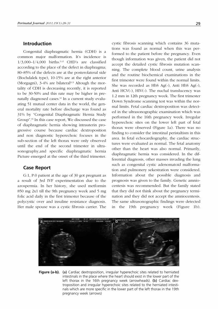
Perinatal Journal 2011;19(1):28-31 29
Figure (a-b). (a) Cardiac dextroposition, irregular hyperechoic sites related to herniatedintestinals in the place where the heart should exist in the lower part of theleft thorax in the 16th pregnancy week (arrowheads). (b) Cardiac dex-troposition and irregular hyperechoic sites related to the herniated intesti-nals which are more specific in the lower part of the left thorax in the 19thpregnancy week (arrows)
Introduction
Congenital diaphragmatic hernia (CDH) is acommon major malformation. It’s incidence is1/3,000–1/4,000 births.[1,2] CHD’s are classifiedaccording to the place of the defect in diaphragma;80–85% of the defects are at the posterolateral side(Bochdalek type), 10-15% are at the right anterior(Morgagni), 3-4% are bilateral[2,3] Altough the mor-tality of CDH is decreasing recently, it is reportedto be 30-50% and this rate may be higher in pre-natally diagnosed cases.[4] In a current study evalu-ating 51 mutual center data in the world, the gen-eral mortality rate before discharge was found as31% by “Congenital Diaphragmatic Hernia StudyGroup”.[5] In this case report, We discussed the caseof diaphragmatic hernia showing intrauterin pro-gressive course because cardiac dextropositionand non diagnostic hyperechoic focuses in thesub-section of the left thorax were only observeduntil the end of the second trimester in ultra-sonography,and specific diaphragmatic herniaPicture emerged at the onset of the third trimester.
Case Report
G:1, P:0 patient at the age of 30 got pregnant asa result of 3rd IVF experimentation due to theazospermia. In her history, she used metformin850 mg 2x1 till the 9th pregnancy week and 5 mgfolic acid daily in the first trimester because of thepolycystic over and insuline resistance diagnosis.Her male spouse was a cystic fibrosis carrier. The
cystic fibrosis scanning which contains 36 muta-tions was found as normal when this was per-formed to the patient before the pregnancy. Eventhough information was given, the patient did notaccept the detailed cystic fibrosis mutation scan-ning. The complete blood count, urine analysisand the routine biochemical examinations in thefirst trimester were found within the normal limits.She was recorded as HbS Ag(-), Anti HbS Ag(-),Anti HCV(-), HIV(-). The nuchal translucency was1.2 mm in 12th pregnancy week. The first trimesterDown Syndrome scanning test was within the nor-mal limits. Fetal cardiac dextroposition was detect-ed in the ultrasonographic examination which wasperformed in the 16th pregnancy week. Irregularhyperechoic sites on the lower left part of fetalthorax were observed (Figure 1a). There was nofinding to consider the intestinal peristaltism in thisarea. In fetal echocardiography, the cardiac struc-tures were evaluated as normal. The fetal anatomyother than the heart was also normal. Primarily,diaphragmatic hernia was considered. In the dif-ferential diagnosis, other masses invading the lungsuch as congenital cystic adenomatoid malforma-tion and pulmonery sekestration were considered.Information about the possible diagnosis andprognosis was given to the family. Genetic amnio-centesis was recommended. But the family statedthat they did not think about the pregnancy termi-nation and they did not accept the amniocentesis.The same ultrasonographic findings were detectedin the 19th pregnancy week (Figure 1b).
a b
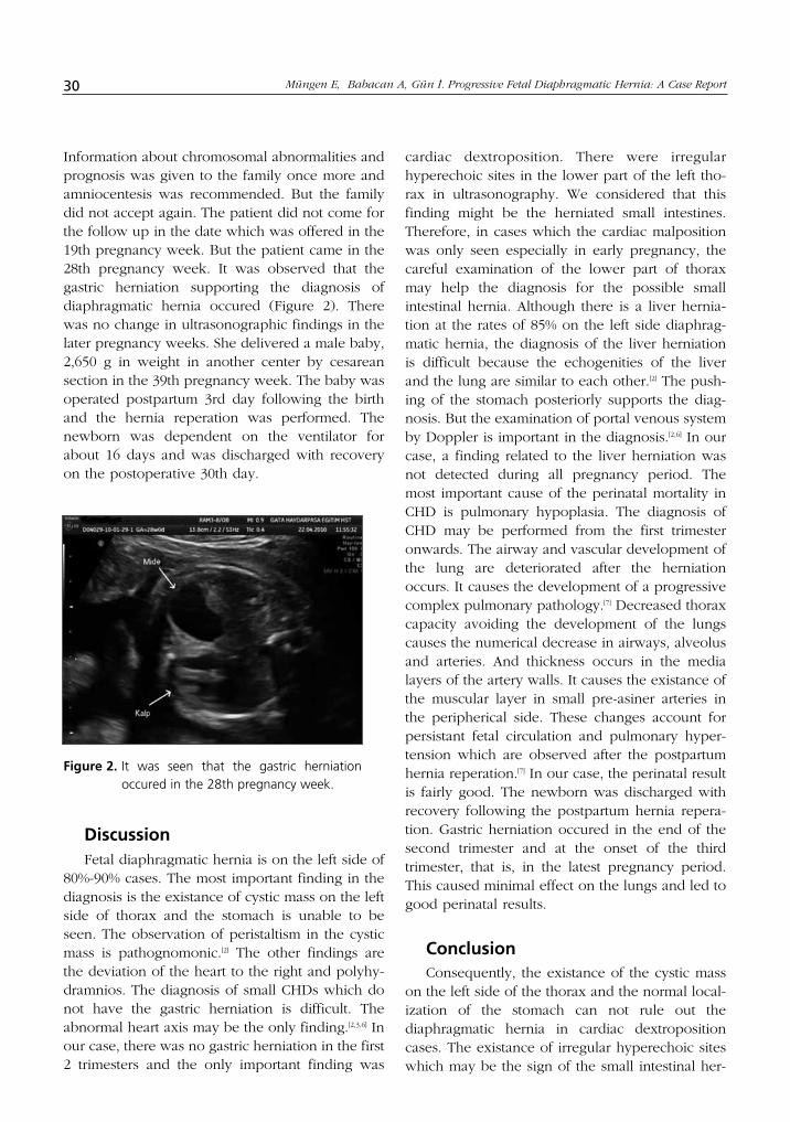
Müngen E, Babacan A, Gün ‹. Progressive Fetal Diaphragmatic Hernia: A Case Report30
Information about chromosomal abnormalities andprognosis was given to the family once more andamniocentesis was recommended. But the familydid not accept again. The patient did not come forthe follow up in the date which was offered in the19th pregnancy week. But the patient came in the28th pregnancy week. It was observed that thegastric herniation supporting the diagnosis ofdiaphragmatic hernia occured (Figure 2). Therewas no change in ultrasonographic findings in thelater pregnancy weeks. She delivered a male baby,2,650 g in weight in another center by cesareansection in the 39th pregnancy week. The baby wasoperated postpartum 3rd day following the birthand the hernia reperation was performed. Thenewborn was dependent on the ventilator forabout 16 days and was discharged with recoveryon the postoperative 30th day.
DiscussionFetal diaphragmatic hernia is on the left side of
80%-90% cases. The most important finding in thediagnosis is the existance of cystic mass on the leftside of thorax and the stomach is unable to beseen. The observation of peristaltism in the cysticmass is pathognomonic.[2] The other findings arethe deviation of the heart to the right and polyhy-dramnios. The diagnosis of small CHDs which donot have the gastric herniation is difficult. Theabnormal heart axis may be the only finding.[2,3,6] Inour case, there was no gastric herniation in the first2 trimesters and the only important finding was
cardiac dextroposition. There were irregularhyperechoic sites in the lower part of the left tho-rax in ultrasonography. We considered that thisfinding might be the herniated small intestines.Therefore, in cases which the cardiac malpositionwas only seen especially in early pregnancy, thecareful examination of the lower part of thoraxmay help the diagnosis for the possible smallintestinal hernia. Although there is a liver hernia-tion at the rates of 85% on the left side diaphrag-matic hernia, the diagnosis of the liver herniationis difficult because the echogenities of the liverand the lung are similar to each other.[2] The push-ing of the stomach posteriorly supports the diag-nosis. But the examination of portal venous systemby Doppler is important in the diagnosis.[2,6] In ourcase, a finding related to the liver herniation wasnot detected during all pregnancy period. Themost important cause of the perinatal mortality inCHD is pulmonary hypoplasia. The diagnosis ofCHD may be performed from the first trimesteronwards. The airway and vascular development ofthe lung are deteriorated after the herniationoccurs. It causes the development of a progressivecomplex pulmonary pathology.[7] Decreased thoraxcapacity avoiding the development of the lungscauses the numerical decrease in airways, alveolusand arteries. And thickness occurs in the medialayers of the artery walls. It causes the existance ofthe muscular layer in small pre-asiner arteries inthe peripherical side. These changes account forpersistant fetal circulation and pulmonary hyper-tension which are observed after the postpartumhernia reperation.[7] In our case, the perinatal resultis fairly good. The newborn was discharged withrecovery following the postpartum hernia repera-tion. Gastric herniation occured in the end of thesecond trimester and at the onset of the thirdtrimester, that is, in the latest pregnancy period.This caused minimal effect on the lungs and led togood perinatal results.
ConclusionConsequently, the existance of the cystic mass
on the left side of the thorax and the normal local-ization of the stomach can not rule out thediaphragmatic hernia in cardiac dextropositioncases. The existance of irregular hyperechoic siteswhich may be the sign of the small intestinal her-
Figure 2. It was seen that the gastric herniationoccured in the 28th pregnancy week.
niation in the lower part of the left thorax supportsthe diagnosis of diaphragmatic hernia in thesecases. In cases which produce the diagnostic diffi-culties and progress with only cardiac dextroposi-tion, the serial ultrasonographic follow ups areimportant during the pregnancy in order to detectthe organ herniations such as stomach and liver.
References1. Pober BR. Genetic aspects of human congenital diaphrag-
matic hernia. Clin Genet 2008;74:1-15.
2. Woodward PJ, Kennedy A, Sohaey R, Byrne JLB, Oh KY,Puchalski MD. Diagnostic Imaging: Obstetrics. 1st ed. Utah:Amirsys Inc; 2005. p 5 (Chest).
3. Paladini D, Volpe P. Ultrasound of Congenital FetalAnomalies. 1St ed. London-Informa Healthcare: 2007; p:187-92.
4. Skari H, Bjornland K, Haugen G, et al. Congenital diaphrag-matic hernia: a meta-analysis of mortality factors. J PediatrSurg 2000;35:1187-97.
5. Lally KP, Lally PA, Lasky RE, et al. Defect size determinessurvival in infants with congenital diaphragmatic hernia.Pediatrics 2007;120:e651-e657.
6. Goldstein RB.Ultrasound evaluation of the fetal thorax. In:Callen PW (Ed). Ultrasonography in Obstetrics andGynecology. 4th ed. Philadelphia: Saunders; 2000. p. 426-55.
7. Hajdu J, Papp Z. Ultrasound examination of the fetal tho-rax. In: Kurjak A, Chervenak FA (Eds). Textbook ofUltrasound in Obstetrics and Gynecology. 2nd ed. NewDelhi: Jaypee; 2008. p. 335-44.
Perinatal Journal 2011;19(1):28-31 31
Introduction
Fetal intra-abdominal cystic lesions, though notcommon, are increasingly referred for evaluationsince the advent of routine antenatal ultrasonogra-phy. They may arise virtually from any abdominalorgan; however, the urogenital and gastrointestinalsystems are the ones most commonly involved. We
report a spontaneously regressed intra-abdominalcyst in a fetus with normal karyotype.
Case Report
25-year-old primigravida was referred for rou-tine ultrasonographic examination at 19 weeks ofgestation. Ultrasonographic examination showed
32 Perinatal Journal 2011;19(1):32-34
Abstract
Objective: To report a case of an intraabdominal cyst of a 19 weeks’ fetus which was diagnosed in a 25-year-old primigravida whowas referred for routine ultrasonographic examination
Case: Bmode sonography revealed an anechoic, multilocular, septated cystic mass located lower right abdominal section, beneaththe liver whereas color Doppler examination revealed no remarkable blood flow. The 23 weeks’ scan showed that the cystic abdom-inal mass was spontaneously regressed.
Conclusion: We discuss the significance and differential diagnoses of the spontaneously regressed cyst in a fetus with normal kary-otype.
Keywords: Intra-abdominal cyst, fetal ultrasound, spontaneous regression, normal karyotype.
Normal karyotipli bir fetüste intra-abdominal kistik kitlenin spontan rezolüsyonu
Amaç: Rutin ultrasonografik de¤erlendirme için baflvuran 25 yafl›nda, primigravid bir gebenin 19 haftal›k fetüsünde tan› alan bir in-traabdominal kistik kitle olgusunu sunmak.
Olgu: 25 yafl›nda primigravid hasta, 19. gebelik haftas›nda rutin ultrasonografik inceleme için klini¤imize baflvurmufltur. Ultrasonog-rafik incelemede, sa¤ abdominal bölgede, karaci¤er komflulu¤unda yerleflmifl, düzenli s›n›rlar› olan, 32x39 mm boyutlar›nda anekoik,multiloküler, septal› kistik kitle saptanm›flt›r. Renkli Doppler incelemede lezyon içi kan ak›m›nda art›fl izlenmemifltir. Fetal büyüme veamniotik s›v› indeksi normaldi. Hastan›n iste¤i üzerine amniosentez yap›lm›fl ve sonucu normal gelmifltir. Yirmiüçüncü haftada yap›lanincelemede kistik abdominal kitlenin spontan olarak regrese oldu¤u görülmüfltür. Do¤um an›na kadar rekürrens saptanmam›flt›r.Otuzsekizinci haftada sezaryenle canl› k›z bebek do¤urtulmufltur. Bebe¤in do¤um sonras› abdominal ultrasonografik incelemesindepatolojik bulgu saptanmam›flt›r. Bmod ultrasonografide (USG) sa¤ alt abdominal bölgede, karaci¤erin alt›nda yerleflim gösteren, Renk-li Doppler USG’de belirgin kan ak›m› izlenmeyen, anekoik, multiloküler, septal› kistik kitle bulunmufltur. Yirmi üçüncü hafta USG’sin-de kistik abdominal kitlenin spontan regrese oldu¤u görülmüfltür.
Sonuç: Burada, normal karyotipe sahip fetüste spontan regresyon gösteren kistin önemini ve ay›r›c› tan›lar›n› tart›flt›k.
Anahtar Sözcükler: ‹ntra-abdominal kist, fetal ultrasonografi, spontan regresyon, normal karyotip.
Spontaneous Resolution of Intra-AbdominalCyst in a Fetus with Normal Karyotype
M. Murat Naki, Olufl Api, Hasniye Ac›o¤lu, Müge Emeksiz, Aybala Ak›l, Orhan Ünal
Dr. Lütfi K›rdar Kartal E¤itim ve Araflt›rma Hastanesi Kad›n Hastal›klar› ve Do¤um Anabilim Dal›, ‹stanbul, Türkiye
Correspondence: Olufl Api, Dr. Lütfi K›rdar Kartal E¤itim ve Araflt›rma Hastanesi, Kad›n Hastal›klar› ve Do¤um Anabilim Dal›, ‹stanbul, Türkiye
e-posta: [email protected]
e-Adress: http://www.perinataljournal.com/20110191008doi:10.2399/prn.11.0191008
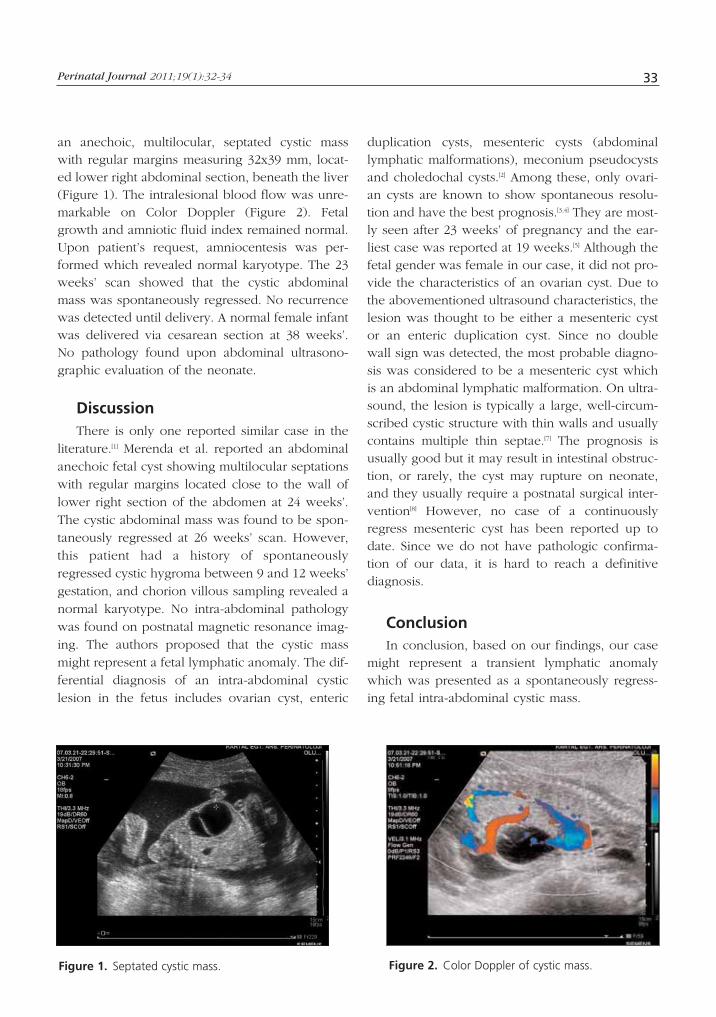
Perinatal Journal 2011;19(1):32-34 33
an anechoic, multilocular, septated cystic masswith regular margins measuring 32x39 mm, locat-ed lower right abdominal section, beneath the liver(Figure 1). The intralesional blood flow was unre-markable on Color Doppler (Figure 2). Fetalgrowth and amniotic fluid index remained normal.Upon patient’s request, amniocentesis was per-formed which revealed normal karyotype. The 23weeks’ scan showed that the cystic abdominalmass was spontaneously regressed. No recurrencewas detected until delivery. A normal female infantwas delivered via cesarean section at 38 weeks’.No pathology found upon abdominal ultrasono-graphic evaluation of the neonate.
DiscussionThere is only one reported similar case in the
literature.[1] Merenda et al. reported an abdominalanechoic fetal cyst showing multilocular septationswith regular margins located close to the wall oflower right section of the abdomen at 24 weeks’.The cystic abdominal mass was found to be spon-taneously regressed at 26 weeks’ scan. However,this patient had a history of spontaneouslyregressed cystic hygroma between 9 and 12 weeks’gestation, and chorion villous sampling revealed anormal karyotype. No intra-abdominal pathologywas found on postnatal magnetic resonance imag-ing. The authors proposed that the cystic massmight represent a fetal lymphatic anomaly. The dif-ferential diagnosis of an intra-abdominal cysticlesion in the fetus includes ovarian cyst, enteric
duplication cysts, mesenteric cysts (abdominallymphatic malformations), meconium pseudocystsand choledochal cysts.[2] Among these, only ovari-an cysts are known to show spontaneous resolu-tion and have the best prognosis.[3,4] They are most-ly seen after 23 weeks’ of pregnancy and the ear-liest case was reported at 19 weeks.[5] Although thefetal gender was female in our case, it did not pro-vide the characteristics of an ovarian cyst. Due tothe abovementioned ultrasound characteristics, thelesion was thought to be either a mesenteric cystor an enteric duplication cyst. Since no doublewall sign was detected, the most probable diagno-sis was considered to be a mesenteric cyst whichis an abdominal lymphatic malformation. On ultra-sound, the lesion is typically a large, well-circum-scribed cystic structure with thin walls and usuallycontains multiple thin septae.[7] The prognosis isusually good but it may result in intestinal obstruc-tion, or rarely, the cyst may rupture on neonate,and they usually require a postnatal surgical inter-vention[8] However, no case of a continuouslyregress mesenteric cyst has been reported up todate. Since we do not have pathologic confirma-tion of our data, it is hard to reach a definitivediagnosis.
ConclusionIn conclusion, based on our findings, our case
might represent a transient lymphatic anomalywhich was presented as a spontaneously regress-ing fetal intra-abdominal cystic mass.
Figure 1. Septated cystic mass. Figure 2. Color Doppler of cystic mass.

Naki MM, et al. Spontaneous Resolution of Intra-Abdominal Cyst in a Fetus with Normal Karyotype34
References1. Merenda A, Pisaturo ML, Laviscio P, Poppiti R, Nazzaro G.
Spontaneous resolution of cystic hygroma colli and intraab-dominal cyst in a fetus with normal karyotype. UltrasoundObstet Gynecol 2008;32:398-446.
2. Khong PL, Cheung SC, Leong LL, Ooi CG. Ultrasonographyof intra-abdominal cystic lesions in the newborn. ClinRadiol 2003;58:449-54.
3. Crombleholme TM, Craigo SD, Garmel S, D’Alton ME. Fetalovarian cyst decompression to prevent torsion. J PediatrSurg 1997;32:1447-9.
4. Giorlandino C, Rivosecchi M, Bilancioni E, Bagolan E,Zaccara A, Taramanni C, Vizzone A. Successful intrauterine
therapy of a large fetal ovarian cyst. Prenat Diagn1990;10:473-5.
5. Meizner I, Levy A, Katz M, Maresh AJ, Glezerman M. Fetalovarian cysts: prenatal ultrasonographic detection and post-natal evaluation and treatment. Am J Obstet Gynecol1991;164:874-8.
6. Konen O, Rathaus V, Dlugy E, Freud E, Kessler A, ShapiroM, Horev G. Childhood abdominal cystic lymphangioma.Pediatr Radiol 2002;32:88-94.
7. Lin JI, Fisher J, Caty MG. Newborn intraabdominal cysticlymphatic malformations. Semin Pediatr Surg 2000; 9:141-5.
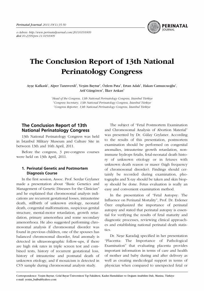
The Conclusion Report of 13th NationalPerinatology Congress
Ayfle Kafkasl›1, Alper Tanr›verdi2, Yeflim Baytur3, Özlem Pata3, Ertan Adal›3, Hakan Camuzcuo¤lu3,
Arif Güngören3, ‹lker Ar›kan3
1Head of the Congress, 13th National Perinatology Congress, ‹stanbul Türkiye2Congress Secretary, 13th National Perinatology Congress, ‹stanbul Türkiye3Congress Reporter, 13th National Perinatology Congress, ‹stanbul Türkiye
Correspondence: Yeflim Baytur, Celal Bayar Üniversitesi T›p Fakültesi, Kad›n Hastal›klar› ve Do¤um Anabilim Dal›, Manisa, Türkiye
e-mail: [email protected]
The Conclusion Report of 13th National Perinatology Congress 13th National Perinatology Congress was held
in ‹stanbul Military Museum and Culture Site inbetween 13th and 16th April, 2011.
Before the congress, 3 pre-congress courseswere held on 13th April, 2011.
1. Perinatal Genetic and Postmortem Diagnosis Course
In the first session, Assoc. Prof. Serdar Ceylanermade a presentation about “Basic Genetics andManagement of Genetic Diseases for the Clinician”and he explained that chromosomal analysis indi-cations are recurrent gestational losses, intrauterinedeath, stillbirth of unknown etiology, neonataldeath, congenital malformations, suspicious genitalstructure, mental-motor retardation, growth retar-dation, primary amenorrhea and some secondaryamenorrhoea. He also suggested performing chro-mosomal analysis if chromosomal disorder wasfound in previous children, one of the spouses hasbalanced chromosomal disorder, fetal anomaly isdetected in ultrasonographic follow-ups, if thereare high risk rates in triple screen test and com-bined tests, history of recurrent gestational loss,history of intrauterine and postnatal death ofunknown etiology, and if mosaicism is detected inCVS sample during chromosomal analysis study.
The subject of “Fetal Postmortem Examinationand Chromosomal Analysis of Abortion Material”was presented by Dr. Gülay Ceylaner. Accordingto the results of this presentation, postmortemexamination should be performed on congenitalanomalies, intrauterine growth retardation, non-immune hydrops fetalis, fetal-neonatal death histo-ry of unknown etiology or in fetuses withunknown death reason or maser (high frequencyof chromosomal disorder). Findings should cer-tainly be recorded during examination, pho-tographs and X-ray should be taken and skin biop-sy should be done. Fetus evaluation is really aneasy and convenient examination method.
In the presentation of “Fetal Autopsy: TheInfluence on Perinatal Mortality”, Prof. Dr. ErdenerÖzer emphasized the importance of perinatalautopsy and stated that perinatal autopsy is essen-tial for verifying the results of fetal maturity anddiagnostic processes, reviewing clinical approach-es and establishing national perinatal death statis-tics.
Dr. Nefle Karada¤ specified in her presentation“Placenta: The Importance of PathologicalExamination” that evaluating placenta providesimportant information in terms of care and healthof mother and baby during and after delivery aswell as creating medicolegal support in terms ofphysician when compared to unexpected fetal or
Perinatal Journal 2011;19(1):35-50
e-Adress: http://www.perinataljournal.com/20110191009doi:10.2399/prn.11.0191009
maternal results and she emphasized that evaluat-ing placenta is an inseparable part of autopsies forfetal and neonatal deaths.
2. Perinatal Ultrasonography Course
Assoc. Prof. Yeflim Baytur talked on “NormalSonographic Findings in Early Pregnancy” andemphasized the importance of determining gesta-tional age, viability and chorionicity within firstthree months. She stated that yolk sac had to beobserved at 5th week and fetal heart beat had tobe observed when the sac reached 25 mm. Sheemphasized that some of fetal anomalies such asanencephaly and omphalocele can be detectedand she also said that fetal anatomy should be paidattention. She indicated that week 14 should bewaited for heart screening and that anencephaly,omphalocele and Dandy-Walker can not be diag-nosed before 11th week.
“Fetal Anatomical Examination in 11-14 WeeksGestation” was presented by Prof. Dr. Yakup Erata.By the developed technique and the perinatologybeing a sub-branch, it was begun to do anatomicalevaluation in earlier weeks and therefore ultra-sonographic imaging became important.
“Fetal Chromosomal Anomaly Markers in 11-14Weeks Gestation” was explained by Prof. Dr.Turgay fiener. He stated that the major anomaliesamong sonographic identifiers that may causechromosomal anomalies were anatomical defectssuch as omphalocele, megacystitis, holoprosen-cephaly, and that the tests such as NT measure-ment, nasal bone, ductus venosus, and tricuspidregurgitation should be used for screening purpos-es. Though it is known that chromosomal anomalyrisk increases as maternal age increases, it wasremarked by him that it is possible to decreaseinvasive tests performed and to increase DownSyndrome diagnoses detected by various screeningstrategies. The importance of training was empha-sized and it was also stated that it should certainlybe certified for effective use of screening tests.
On the session of second trimester, Prof. Dr.Alper Tanr›verdi made a speech about “FetalAnatomical Examination in the Second Trimester”.It was stated that the examination was done fromthe point of fetal anatomy during ultrasonographyevaluation and it was important to question
patient’s history and to check some systems morecarefully accordingly and he emphasized theimportance of systematic evaluation and the use ofa check list. It was underlined that at least 20 min-utes should be spared for examination and he sug-gested the importance of carrying on physiciantraining and technical competency of the device.He also indicated that anomalies could be missedeven in the best clinics; skeletal system and KVSdefects were those mostly missed, and CNS andGUS anomalies were those easily detected. He alsoremarked following topics:
a. The evaluation of fetal biometry, placentaand amnion fluid, and the systematicscreening of fetal anatomy should be per-formed.
b. If necessary, Doppler ultrasonographyshould be included.
c. During systematic evaluation of the anatomy,orbitas on face, lips, nasopalatine, fetal pro-file, extremities, 4 cardiac chambers, majorvessel outlets, 3 vessel images, falx cerebrion central nervous system, ventricular sys-tem, posterior fossa, choroids plexuses,stomach, bladder, diaphragm and kidneysshould be checked.
d. Indications such as heart anomaly of Downsyndrome, duodenal atresia that may beseen at 2nd trimester should be paid atten-tion.
In the same session, Prof. Dr. Lütfü Öndero¤luemphasized in his presentation “Which FetalAnomalies Should Not Be Overlooked in theSecond Trimester?” that there is confusion aboutwho should perform the ultrasonographic evalua-tion due to the increase in medicolegal events andstated that there is 60% chance to detect in ultra-sonography and expectation of patient should notbe raised. He expressed that all those normally tobe checked between 18th and 22nd weeks couldbe possibly found in all books and internet, andeverybody who got ultrasonography training canperform standard and detailed ultrasonography.He noted that regular care of devices, reportingand image recording should be provided, andobese patients should be registered. He stated that83% of lethal anomalies were detected; however,we can detect 54% of lethal anomalies such as
Kafkasl› A et al. The Conclusion Report of 13th National Perinatology Congress36
Perinatoloji Dergisi 2011;19(1):35-50 37
hypoplastic heart and 90% of renal agenesia, butlethal musculoskeletal anomalies can be missed.He pointed out that the actual problem is in babieswith serious morbidity and chance of living. Heexpressed that neural tube defects, hydrocephaly,complex heart abnormalities, abdominal anteriorwall defects, diaphragmatic hernia, small intestineobstructions and bladder extrophies should bedetected (and also they can be missed).
Assoc. Prof. Dr. Ebru Tar›m presented the“Anomalies that can be detected during late preg-nancy period” and she stated that anomaly canbecome apparent at 3rd trimester (diaphragmatichernia) though it exists at 2nd trimester sinceanomaly appears lately and there is a pause in thedevelopment of anatomical structure (micro-cephalia). It was said that normal anatomic devel-opment continues during pregnancy and someanomalies such as central nervous system anom-alies may arise late. It was emphasized that cere-bellar vermis, ventriculomegaly, and moreechogen view around ventricle at intracranial hem-orrhages should be paid attention during thirdtrimester. She indicated that transvaginal ultra-sonography can be used in uncertain cases in SSSanomalies and that MR can be performed inbreech delivered babies. It was said that SSStumors and neuronal migration anomalies such aslissencephaly may appear lately. She mentionedabout findings developed related to CMV infectionand explained that coarctation of aorta, pulmonarystenosis, achondroplasia from skeletal dysplasia,ovarian cysts, sacrococcygeal teratoma, intestinalatresia, vesicoureteric reflux and multicystic dys-plastic kidney may be diagnosed lately. Sheemphasized that only biometry check is not suffi-cient on third trimester but also the anatomyshould be evaluated in terms of these anomalies.
The topic of “Sonography of the Placenta andits Adnexa” was narrated by Prof. Dr. As›m Kurjak.He expressed that placentation can be detectedbeginning from early gestational weeks byDoppler ultrasonography and that 3D Doppler ismore advantageous than 2D Doppler. He statedthat placental vessels can be imaged by “placentalvascular biopsy” and that spiral arteries and pla-cental vascular tree can be imaged by 3D, and 3Dindices may change according to the parity and
gestational week. He indicated that intervillousblood flow can be measured by 3D and it can beutilized in hypoxic cases. He also gave informationabout placental anomalies. It was talked about pla-cental tumors, chorioangioma and placentalinfarcts. Though placental calcifications are accept-ed as physiological, it was asked to pay attentionthat they may be the indication of infections atearly pregnancy. It was stated that there may behyperechoic image at 0th-48th hours in ablatio pla-centae, isoechoic image at 3rd-7th days, hypoe-choic image at 1st-2nd weeks and anechoic imageafter 2nd week. It was emphasized that umbilicalvessels blocks servical channel at vasa praevia, it isfrequent at velamentous insertion, and membranerupture and abnormal bleeding may occur if notdiagnosed. It was underlined that inferior uterinesegment is formed at 28th week at placenta prae-via diagnosis and therefore it should not be diag-nosed before this week. It was expressed thatsonoluscent areas behind placenta disappeared inplacenta adhesion abnormalities, there were pla-cental lacunas including turbulence and coloredand power Doppler may help the diagnosis. It wasclarified that Hyrtl anostomoses are important andthat single umbilical artery may be related withchromosomal anomalies and gestational complica-tions.
Prof. Dr. Cihat fien presented the topic of“Sonography as a Contributory Tool in MultiplePregnancy”. It was stated that dichorionic twinscan be followed up as singles and it is wrong toperform unnecessary elective cesarean. It wasmentioned that Down syndrome risk calculation isdifferent in twins compared to single pregnanciesand it was also said that it is not appropriate to usebiochemical tests. Checking for nuchal translucen-cy and nasal bone is appropriate and risk will becalculated for each fetus while these risks will varyaccording to chorionicity. It was stated that takingone sample in monochorionics if invasive processwill be performed; however, it may have rarely dif-ferent genetic structures in postzygotic abnormali-ties. Since preeclampsia risk increases in multiples,uterine artery PI should be checked in firsttrimester as well as measuring blood pressure. Inthis way, heavy preeclampsia that occurs before32nd week can be diagnosed at a rate of 90%.Miscarriage risk increases to 6% from 3% and early
labor risk decreases to 10% from 20% if embryoreduction is performed in triple pregnancies. Itshould be decided together with the family.Reduction is not performed in monochorionics,but cord coagulation should be performed. If thereis any case requiring termination, it should bedone at the earliest period.
In this course, after speakers presented theirpresentations, examples about normal fetal anato-my in point-of-care training were given by EbruTar›m, Özlem Pata, Yeflim Baytur, and MertihanKurto¤lu and information was given about fetalobstructive uropathy, neural tube defect andincreased NT on 3 patients at 1st, 2nd and 3rdtrimesters.
3. The Course of Obstetric Emergencies
It was expressed that the real reason of earlyperiod bleedings can not be frequently deter-mined, but the most important one is the differ-ence between ectopic pregnancy and abortus anda good history, vaginal examination, hemogramfollow-ups and ultrasonography gain importancein that case.
It was said that vaginal 70-90% success wasobtained by misoprostol applications (600-800microgram/day) in abortuses. Following opinionswere shared about placenta praevia: Placenta prae-via is a spectrum. Those with obstetric bleedingrisk should be determined by ultrasonography at20th-23rd weeks. Lifestyle should not be changedif cervical length is normal and placental coveringis below 15 mm at 20th-23rd weeks. Placenta prae-via–vasa praevia, placenta accrete–cesarean under-gone–maternal age, IVF/ICSI pregnancy are asso-ciated with each other. Normal delivery can beperformed under close follow up by placental tipat delivery – internal os <1.2 cm.
Placental decollement is seen generally at 3rdtrimester. In most of the cases, fetal distress accom-panies and its frequency is 0.3-1.6%. The diagnosisis clinical at ablation placentae. It is confirmed byshowing postnatal retroplacental hematoma.Clinical findings vary according to the decollementlevel and bleeding amount. Placenta praevia, dys-tocia and rupture should be eliminated at differen-tial diagnosis. Hemorrhage may not be observed indecollement cases ultrasonographically.
Ultrasonographic diagnosis criteria are pre-placen-tal accumulation under chorionic layers, gel-likemovement of chorionic layer by fetal activity,retroplacental accumulation, marginal hematoma,sub-chorionic hematoma, heterogen increased pla-cental thickness and intra-amniotic hematoma.
There is a clinical classification of placentaldecollement and it gets intensified from 0 to 3.Management of decollement in placenta includesappropriate maternal monitorization, fetal statusindication, performing differential diagnosis, trans-fusion of proper blood and blood products, andtermination of pregnancy at a suitable time.Coagulation tests should be performed and urineoutgoing should be kept under control. If patientis stable during management, she can deliver. Ifthere is fetal distress, then cesarean should be con-sidered. It should be remembered that maternaland fetal complications increase in decollementplacenta.
Can we prevent decollement placenta?
It should be prevented to drink alcohol andsmoke during pregnancy, antenatal controlsshould be performed, pregnants with hypertensionshould follow treatment recommendations andfolic acid should be taken.
Hypertension at Pregnancy was discussed onthe first session on 14th April, 2011.
Prof. Dr. R›za Madazl› emphasized in his pre-sentation “Etiology and Staging of Preeclampsia”that preeclampsia is a unique formation of humanplacenta and it is the cost of evolution. He said thatit causes 50,000 maternal deaths in the world. Hetalked about its common endothelial damage inthe etiology caused by insufficient trophoblasticinvasion. He expressed that the classificationshould be changed as early (<34 week) and late(>34 week). He highlighted that early preeclamp-sia is caused by placentation anomaly which haveserious risk on mother and fetus, and latepreeclampsia is caused by overreaction of motherfor pregnancy.
Assoc. Prof. Arif Güngören said in his presen-tation “Preeclampsia Prediction byUltrasonography” that there is resistance increasein uterine artery of preeclampsia patients againstcurrent and that early diastolic notch may develop;
Kafkasl› A et al. The Conclusion Report of 13th National Perinatology Congress38
but he also added that Doppler of uterine arteryhas a limited value for predicting preeclampsia,IUGR and perinatal death. He expressed that itsdecrease in uterine artery Pulsatility Index alone ortogether with notching will be successful for pre-dicting preeclampsia. It was said that there is 90%sensitivity when 1st Trimester PI and PP-13 areused together. He emphasized that it is more valu-able to use biochemical markers and USG identi-fiers together.
In “Hellp Syndrome: Practical Management”,the speaker Sanjay Gupte said that the situationdevelops 70% before delivery. He talked aboutendothelial damage caused by cytokines secretedby maternal cell immunity as a reaction againstpregnancy. He stated that the early diagnosis ofthe syndrome may give the best chance to motherand delivery should be performed when the situa-tion arises, no matter what happens. He highlight-ed that nausea or vomit developing at the secondhalf of pregnancy would cause a high level of sus-picion. He expressed that the state will regresswithin 48 hours in most of the cases, and it can bewaited for steroid for 48 hours by stabilizing casesdeveloped between 7th and 32nd weeks. He saidthat high dose of steroid use is not suggested insyndrome treatment and it does not change out-comes and also said that thrombocyte suspensionshould not be given when thrombocyte count isover 50,000. He emphasized that aggressive treat-ment should be performed in order to save the lifeof patient.
“Gestational Diabetes” was discussed in thenext session. Gestational diabetes is a diabetestype which begins during pregnancy or appearsduring pregnancy for the first time. 90% of diabetesobserved during pregnancy is gestational diabetesand its incidence is about 5-10%.
Even light hyperglycaemia affects progno-sis. Therefore, GD should be checked at pregnan-cy. The screening can be performed as two-phased(50 g OGTT – 100 OGTT) or one-phased (75OGTT).
At 75 g GTT, FBG (fasting blood glucose)threshold value is 95 mg/dL, while 1st hour thresh-old is used as 180 mg/dL and 2nd hour thresholdis used as 155 mg/dL.
Carpenter Causton values are mostly used at100 g OGTT. FBG, 1st, 2nd and 3rd hour thresholdvalues are accepted as 95, 180, 155, and 140respectively.
75 g GTT threshold values were decreased afterthe results of HAPO study were taken and FBG, 1sthour and 2nd hour values were determined as 92,180 and 153 respectively.
FBG should be checked at first examination inall pregnants and FBG should be below 92 mg/dL.In diabetic pregnants, maternal and fetal complica-tions increase. If blood glucose is above 200mg/dL in pregnant with nausea and vomit, weshould remember diabetic ketoacidosis.Management of diabetes should begin in precon-ceptional period. If there is type-1 diabetes andpatient currently uses any cholesterol loweringdrug and ACE inhibitors, they should be not beused and it should be begun to use Ca channelblockers and folic acid.
Renal function tests should be asked forpatients with Type-1 DM before pregnancy, eyeground examination should be performed andconsultancy should be given in terms of maternal-fetal risks.
Congenital malformation risks should be told tocase who applies for bad glycemic control at firsttrimester, and tight glycemic control should beprovided as not being hypoglycemic. During 20th-24th weeks at second trimester, it should be sug-gested to do detailed ultrasonography and fetalecho in terms of fetal anomalies.
At final trimester, patient should be called forweekly controls and NST and Biophysical Scoringshould be done twice in a week if necessary.
Complications are lesser in gestational DM, butcareful follow-up should not be forgotten. Itshould be paid attention to hypertension andpreeclampsia. In these cases, it is not necessary tocheck urine routinely for ketone but it can bechecked for diet control. It is suggested to do firsttrimester test at 11th-14th weeks, detailed ultra-sonography and fetal echo at 18th-22nd weeks,ultrasonography once at every four weeks after28th week and delivery as not exceeding 40thweek.
Perinatoloji Dergisi 2011;19(1):35-50 39
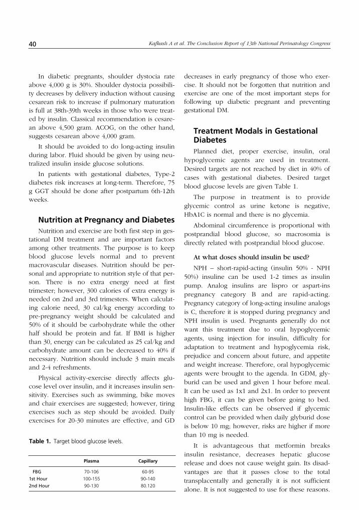
In diabetic pregnants, shoulder dystocia rateabove 4,000 g is 30%. Shoulder dystocia possibili-ty decreases by delivery induction without causingcesarean risk to increase if pulmonary maturationis full at 38th-39th weeks in those who were treat-ed by insulin. Classical recommendation is cesare-an above 4,500 gram. ACOG, on the other hand,suggests cesarean above 4,000 gram.
It should be avoided to do long-acting insulinduring labor. Fluid should be given by using neu-tralized insulin inside glucose solutions.
In patients with gestational diabetes, Type-2diabetes risk increases at long-term. Therefore, 75g GGT should be done after postpartum 6th-12thweeks.
Nutrition at Pregnancy and DiabetesNutrition and exercise are both first step in ges-
tational DM treatment and are important factorsamong other treatments. The purpose is to keepblood glucose levels normal and to preventmacrovascular diseases. Nutrition should be per-sonal and appropriate to nutrition style of that per-son. There is no extra energy need at firsttrimester; however, 300 calories of extra energy isneeded on 2nd and 3rd trimesters. When calculat-ing calorie need, 30 cal/kg energy according topre-pregnancy weight should be calculated and50% of it should be carbohydrate while the otherhalf should be protein and fat. If BMI is higherthan 30, energy can be calculated as 25 cal/kg andcarbohydrate amount can be decreased to 40% ifnecessary. Nutrition should include 3 main mealsand 2-4 refreshments.
Physical activity-exercise directly affects glu-cose level over insulin, and it increases insulin sen-sitivity. Exercises such as swimming, bike movesand chair exercises are suggested; however, tiringexercises such as step should be avoided. Dailyexercises for 20-30 minutes are effective, and GD
decreases in early pregnancy of those who exer-cise. It should not be forgotten that nutrition andexercise are one of the most important steps forfollowing up diabetic pregnant and preventinggestational DM.
Treatment Modals in GestationalDiabetesPlanned diet, proper exercise, insulin, oral
hypoglycemic agents are used in treatment.Desired targets are not reached by diet in 40% ofcases with gestational diabetes. Desired targetblood glucose levels are given Table 1.
The purpose in treatment is to provideglycemic control as urine ketone is negative,HbA1C is normal and there is no glycemia.
Abdominal circumference is proportional withpostprandial blood glucose, so macrosomia isdirectly related with postprandial blood glucose.
At what doses should insulin be used?
NPH – short-rapid-acting (insulin 50% - NPH50%) insuline can be used 1-2 times as insulinpump. Analog insulins are lispro or aspart-inspregnancy category B and are rapid-acting.Pregnancy category of long-acting insuline analogsis C, therefore it is stopped during pregnancy andNPH insulin is used. Pregnants generally do notwant this treatment due to oral hypoglycemicagents, using injection for insulin, difficulty foradaptation to treatment and hypoglycemia risk,prejudice and concern about future, and appetiteand weight increase. Therefore, oral hypoglycemicagents were brought to the agenda. In GDM, gly-burid can be used and given 1 hour before meal.It can be used as 1x1 and 2x1. In order to preventhigh FBG, it can be given before going to bed.Insulin-like effects can be observed if glycemiccontrol can be provided when daily glyburid doseis below 10 mg; however, risks are higher if morethan 10 mg is needed.
It is advantageous that metformin breaksinsulin resistance, decreases hepatic glucoserelease and does not cause weight gain. Its disad-vantages are that it passes close to the totaltransplacentally and generally it is not sufficientalone. It is not suggested to use for these reasons.
Kafkasl› A et al. The Conclusion Report of 13th National Perinatology Congress40
Plasma Capillary
FBG 70-106 60-95
1st Hour 100-155 90-140
2nd Hour 90-130 80.120
Table 1. Target blood glucose levels.
Treatment Manual• If FBG is higher than 95 or 1st hour postpran-
dial blood glucose is over 140, then insulinshould be used. If FBG is 95–110 mg/dl, and1st hour is 110–150, then treatment should beinitiated by NPH as 0.3-0.4 unit/kg.
• If FBG is 110mg/dl, and 1st hour is over 200,then treatment should be initiated by 0.7unit/kg insulin.
• Individual capillary blood glucose should bechecked for 4-7 times daily in 3 days in a week.
Preterm Labor Management inDiabetic PregnantIatrogenic preterm labor is frequently seen
especially in diabetes. Diabetes-caused IUGR,macrosomia or bad biophysical profile can causeearly labor requirement. Also bad maternal bloodglucose control, noncontrollable HT caused byvascular complications may require delivery to beperformed early. If blood glucose is high, thenpreterm labor risk is high, too. Preterm labor riskin those with good glycemic control is similar witha normal pregnant.
Fetal hyperinsulinism antagonizes cortisoleffect and Type-2 alveolus cell development isdelayed. Surfactant is decreased and newborntachypnea occurs. In cases where pregnancy willbe terminated before 38 weeks and 7 days, pul-monary maturation should be known for sure.Tests used in pulmonary maturation are L/S, PG,and Lamellar Body (37 thousand / microliter)counts. Sometimes lung can be RDS though it ismature according to L/S test. Therefore, PG shouldalso be checked.
Tocolytic agents + antenatal steroid are used inpreterm labor management. There are some risksabout antenatal steroid use, because maternalhyperglycemia may be observed. This also maycause fetal hyperglycemia and increases fetalinsulin. Fetal hyperinsulinemia delays pulmonarymaturation. Checking hourly capillary blood glu-cose and doing insulin infusion in diabetic preg-nant to whom steroid is applied are properapproaches.
Beta agonist can be given as a tocolytic agentto diabetic pregnant who has good glycemic con-
trol. Also, calcium channel blockers and oxytocinreceptor antagonists should be preferred.
Gestational period is closely related with bloodglucose level. Since antenatal steroid is applied,insulin infusion is the best effective method inblood glucose management.
In “Multiple Pregnancies” session, Prof. Dr.Murat Yayla gave information about“Epidemiology of Twin Pregnancies in Turkey”.The incidence of multiple pregnancy increasedafter 1980. The major reason is the improvementsin reproductive medicine. Its incidence at first peri-ods of pregnancies is indicated as 6-12%. The rateis observed as 2-3% in deliveries. (The increase ofabortus risk?). The incidence is over 30% in HTpregnancies. Vaginal delivery is suggested at ver-tex presentations in twin pregnancies while cesare-an is suggested non-vertex presentations. Maternalmortality increases approximately 3 times. It isrequired to do chorionicity determination by ultra-sonography at early period. It is suggested to dofollow-up in perinatology clinics for pregnanciesmore than two and monochorionic twins. Routinecervical sonography should be performed at 22nd-24th weeks and follow-ups of pregnants who haveearly labor risk should be suggested doing at peri-natology clinics. Less number of embryo transfermade by the new law decreased multiple preg-nancy rates 4 times; however, it did not changepregnancy rates. It should be remembered thatperinatal mortality and morbidity will increase inmultiple pregnancies.
Assoc. Prof. Okan Özkaya then talked about“Management in Preterm Twins”.
More than 50% of twin pregnancies give birthbefore 37th gestational week. Short cervix and fFnscreening by ultrasonography is useful for deter-mining preterm labor. There is no effective methodfor preventing preterm labor. Contrarily, prophy-lactic circlage may increase preterm labor.Hospitalization, bed rest and prophylactic proges-terone are not effective. The purpose in acutepreterm labor treatment is to perform antenatalsteroid application and to gain time for referringappropriate center.
The last session of the day was “Turkish-Georgian Joint Meeting”. In this meeting,“Cesarean on Demand” was presented by Assoc.
Perinatoloji Dergisi 2011;19(1):35-50 41
Prof. Mekin Sezik. He emphasized that the defini-tion of cesarean on demand is quite difficult. Hestated that the situation is complicated if it ispatient who demands cesarean as it may not be ahealthy decision. It should be discussed whetherpatient demands cesarean as her health is deterio-rated or it is possible that we call it as cesarean outof indication.
Who determines cesarean indications? Shouldphysician be determiner traditionally with hisknowledge and experience or should there beinstructions and regulations about it? In fact, thereare many factors affecting indication determiningprocess (ministry, SSI, societies, physician, NGOsetc.). Consequently, both patient-physician relationdecreases and technician role is given to physician.
As a reflection, “increase in cesarean rates”becomes an intricate situation caused by the com-bination of some “interrupted” social practices andvalue judgments through global-scaled excessivemedicalization and mechanization in obstetricpractice.
Solution offers are not as easy as thought, theyshould cover the change of some complicatedsocial value judgments. In fact, women are in thefocus of the debate; however, everyone exceptwomen participates to the debate.
What can be done to decrease cesarean rates?First of all, transparency should be provided.Pregnant should be informed clearly about howpregnant will be prepared for labor, where laborwill be performed, who will make do labor, andwho will also be there.
Another topic emphasized by the speaker is ifcesarean rates can be decreased. Support to laborby someone else except health personnel and peo-ple close to pregnant decreased cesarean ratesabout 2.5%. There should not be any places likepain room of delivery room in hospitals since suchplaces insulate and alienate pregnant. Lithotomyposition and routine episiotomy should be ques-tioned. Importance should be attached to patientprivacy.
The Ministry of Health established mother-friend hospital criteria. According to this, a closeperson chosen by candidate mother should staywith her during delivery; candidate mother should
receive physical and emotional support regularlyfrom an educated health employee, walking andmoving freedom should be provided for her; rou-tine lithotomy position should not be insisted andposition preferred by her should be provided;induction rate should be decreased ≤10% and epi-siotomy rate should be decreased ≤20% (target is≤5%). Unless required, it is not encouraged to useanalgesic and anesthetic materials.
Consequently, it was stated it could not beexpected to see a decrease in increasing cesareanrates without leaving male-dominated medical dis-course and interiorizing the privilege of “givingbirth” by women which is peculiar to women.
Dr. Nana Gvetadze spoke about“Homotoxicological Therapy of Chronic Feto-Placental Insufficiency and Fetal GrowthRetardation”. In this session, it was mentionedabout the use of homotoxic drugs in gestationalcomplications. The most important reasons forchoosing homotoxicology in fetal medicine arethat they do not have embryotoxic and teratogeniceffects, they do not cause allergy and do not haveany contraindications, they are more effective intreatment of chronic diseases and they are advan-tageous in terms of costs. These preparates areobtained by mixing various dilutions of certain nat-ural herbs. They can be used in especially growthretardation and placental insufficiency in terms offetal medicine.
On 15 April 2011, the second day of the con-gress, the first session was “High-RiskPregnancies”. The topic “From Pregnancy to theNeonatal Period” was presented by Prof. Dr.Kalbiye Yalaz. By findings at early period of high-risk pregnancies, it is not always possible to detectneurologic problems such as cerebral palsy andmental retardation that will appear on advancedages. Babies who are delivered on time and haveno risk factor (cerebral palsy 60%-70%) may haveneurological problems in the future. Intrauterine orlow body weight during delivery according to ges-tational age, small head circumference, intrauterinemovement shortage, low heart beats during preg-nancy, hypoglycemia during intrauterine and natalperiod, placenta dysfunction, and retardation atdevelopment steps are most important preliminaryfindings of cerebral palsy and mental retardation.
Kafkasl› A et al. The Conclusion Report of 13th National Perinatology Congress42
Prof. Dr. Neslihan Tekin explained the topic of“Prevention and Management of PerinatalAsphyxia”. Perinatal asphyxia may develop atintrauterine period, during labor, during deliveryor at early postnatal period. Recognizing riskycases and eliminating risk factors, referring riskypatients to perinatal centers, providing sufficientantenatal care, training health personnel to enablethem to evaluate and resuscitate newborns, andestablishing a safe transport for risky pregnantsand sick newborns are basic approaches to pre-vent perinatal asphyxia. Fetal heart rate, fetalblood flow rate and similar tests are important todetermine proper delivery time by evaluating fetuscondition. Hypothermia treatment applied as cool-ing head circumference or whole body of a babyabout 3-4 °C who born as asphyxiating is accept-ed as the latest treatment method since it decreas-es the rate of a series of metabolic event (whichwill lead to secondary neuron death), decreasesmortality and affects neurological prognosis posi-tively.
“Induction and Management of Labor” was pre-sented by Assoc. Prof. Dr. N. Ömer Kandemir.Today, obstetrics is an art in which it is required touse knowledge, skill and technology together. Itwas begun to use Bishop Score in 1960s for laborinduction and using prostaglandins marked a newera beginning from the mid-80s. Mechanical servi-cal dilatation is among invasive methods used onstubborn cases and it is generally used togetherwith prostaglandins and oxytocin.
Assoc. Prof. Dr. Serdar Yalvaç talked about“Fetal Hypoxia at Labor”. Nowadays, due tomedicolegal issues caused by perinatal asphyxia,obstetricians and gynecologists feel themselvesunder pressure when they are doing their jobs. Inorder to detect hypoxia on time, intrauterinegrowth retardation, hypertension, postterm preg-nancies, reduce in baby movements, breech pre-sentation, decrease in amniotic fluid and infectionsare those to be paid attention in the first place.Ultrasonographic evaluation, Doppler, cord bloodexamination, Non-stress test, counting baby move-ments are major methods used for predictingasphyxia. Although none of classical-modern fol-low-up methods leads to certain diagnosis for pre-dicting antepartum/intrapartum asphyxia, using
proper method on chosen cases may help toreduce hypoxic encephalopathy cases.
First speech on “Postpartum Bleedings” wasdone by Assoc. Prof. Dr. Umut Dilek with the pre-sentation of “PP Bleeding and Risk Factors”. Thespeaker made the definition of PP Bleeding as thebleeding more than 500 ml in vaginal deliveriesand more than 1,000 ml in cesarean deliveries. Hestated that it is first reason of maternal deaths inTurkey, it has a rate of 4-6% and about 125-140women die every year in the world for that reason.He expressed that its most frequent reason is ato-nia (70-90%) and that it is preventable clinical sit-uation with 50% chance. He talked about 4 T fac-tors in the etiology of PP Bleeding (Toe 70%,Tissue 10%, Trauma 20%, and Thrombin 1%). Heindicated that extended 3rd phase, preeclampsia,episiotomy, PP bleeding history, multiple pregnan-cy, arrest of descent, delivery induction, and oper-ative deliveries are risk factors. He emphasizedthat clinics mostly overlook and diagnosis isdelayed.
“Clinic and Hypovolemic Shocks in PPBleeding” was presented by Assoc. Prof. Dr.Selahattin Kumru and he classified the shockunder 4 topics: 1- 15% (900 ml) loss of blood (nosymptom), 2- 20-25% (1,200-1,500 ml) loss ofblood (tachycardia, tachypnea, orthostaticchanges), 3- 30-35% (1,800-2,100 ml) loss of blood(hypotension, distinctive tachycardia, cold moistskin), 4- 40% (>2,400 ml) loss of blood (oliguria-anuria, no peripheral pulse). He expressed thatactive management type in which loss of bloodwill diminish should be a routine approach by giv-ing uterotonics at third phase of delivery, remov-ing placenta by clamping cord. Consequently, lifecan be saved by recognizing risky cases, deter-mining bleeding amount, managing third phaseactively and with a good team work.
Assoc. Prof. Dr. Ahmet Yal›nkaya made aspeech on “Medical and Surgical Approaches in PPBleeding”. He stated that early breast-feeding, nip-ple massage, uterine massage and bimanual uter-ine compression are medical treatments in PPbleeding. He indicated that anti-shock treatmentshould be performed by doing blood and bloodproduct transfusion. He also mentioned that tam-ponade, compression suture techniques, artery lig-
Perinatoloji Dergisi 2011;19(1):35-50 43
ations and artery embolization are among theoptions as surgical techniques. He expressed that0.05% hysterectomy is required and he suggesteddoing it to save life. He stated that success rates ofthese methods are about 80-90%. As result, heemphasized that treatment should be done aggres-sively and effectively.
“Postpartum Bleeding: Preventive Measures ofMaternal Death” was presented by Prof. Dr. ÖzgürDeren. The speaker stated that uterotonic agentsshould be administered rapidly in postpartumbleeding and that the biggest problems are DIC(disseminated intravascular coagulation) andmyocardial ischemia developed due to delay. Hespecified that oxytocin should be preferred ratherthan oral misoprostol.
In “Screening Tests” session, the presentationon “First Trimester Screening on Aneuploidies”was made by Giovanni Monni. In the beginning ofthe presentation, Dr. Monni emphasized that manyscannings (gestational diabetes, preeclampsia risk,preterm labor risk etc.) together with chromosomalanomalies should be performed at first trimester.Fetal biometry (CRL, microcephaly etc.), function-al evaluation (decreased or increased heart beat,increased UA PI, pulsatility in umbilical vein), softmarkers (choroid plexus, caliectasis, echogenicfocus, hyperechogenic intestine etc.) and othermarkers which are popular these days (nasal bone,facial angle, ductal current, tricuspid regurgitation,mitral gap etc.) can be evaluated in the ultra-sonography used in the first trimester aneuploidyscreening.
The importance of detecting chromosomalanomaly especially in nuchal translucence mea-surement was emphasized and advantages and dis-advantages of semi-automatic NT measurement oflate years were mentioned.
Finally, it was stated at the end of this sessionthat scanning only maternal age does not have anymeaning; scanning first trimester is more effectivethan second trimester; consecutive scanning maywork in patients with intermediate risk; step-wisescanning would be more useful in patients withlow risk; additional markers would decrease therequirement for second trimester scanning, contin-gent scanning performed by checking nasal bone-tricuspid regurgitation-ductus venosus currentwould contribute to obtain best results.
“Fetal Heart Scanning” was presented by Prof.Dr. Cihat fien. The speaker began his speech byemphasizing the incidence rate of fetal cardiacanomalies among the society and he stated thatcardiac malformations constitute a major part ofneonatal deaths. It would be possible to keepmany newborns alive by doing fetal echo evalua-tion especially risky patients and performing deliv-ery in senior centers due to existing pathology. Heexpressed that checking only four chambers whenscreening fetal heart cause us to miss much patho-logical stuff, but it would be possible to catchmany existing pathologies by checking especiallymajor vessel outlets. Therefore, it was emphasizedthat gynecologists should certainly check majorvessel outlets when screening fetal heart and it wassaid that it would be useful for physicians to attendcourses held for their trainings related with thistopic.
Assoc. Prof. Dr. Ertan Adal› talked about “NewApproaches for Screening of Gestational Diabetes”.The concept of Gestational diabetes mellitus(GDM) is used for defining abnormal glucose tol-erance which is either first noticed during preg-nancy or appeared at various levels. However, thenumber of patients increases who previously havediabetes during pregnancy but not diagnosed yetdue to type-2 diabetes frequency throughout theworld. These patients have different risks thanthose of whom diabetes begin with pregnancy.Therefore, IADPSG (International Association ofDiabetes and Pregnancy Study Groups) offered achange in this terminology in 2010. According tothis, diabetes diagnosed during pregnancy is clas-sified into two groups as “gestational” and “overt-preexisting diabetes”.
In this terminology, overt diabetes diagnosisshould be established at first prenatal visit forwomen from risk group. Accordingly, if diagnosisof FBG is ≥126 mg/dl or of hemoglobin is ≥ 6.5%or randomly checked blood glucose is found as ≥mg/dl (later on, it should be confirmed by FBGand hemoglobin A1C levels), then pregnant isdiagnosed as overt diabetes.
According to the new terminology, gestationaldiabetes mellitus is diagnosed when FBG is ≥92mg/dl but <126 mg/dl at first prenatal visit or highabnormal values (FBG ≥ 92, 1st hour; ≥ 180, 2nd
Kafkasl› A et al. The Conclusion Report of 13th National Perinatology Congress44
hour ≥153) high by 75 g one phased test at 24th-28th gestational weeks. ADA 2011 also adopts thisapproach. However, ACOG 2001 recommenda-tions offer to do scanning on all pregnants by twophased test (first 50 g GCT, then 100 g OGTT toabnormal ones).
In the Manual of Diabetes Mellitus andDiagnosis, Treatment and Follow-up itsComplications published by The Society ofEndocrinology and Metabolism of Turkey in 2009,it is asked to do risk evaluation (GDM history, obe-sity, glycosuria, DM in first degree relative) at firstprenatal visit for GDM screening and to do screen-ing at next trimester if risk is negative. The manu-al recommended doing screening (50 g GCT) to allpregnants at their 24th-28th gestational weeks.
Another issue to emphasize is that there is noconsensus in the world on performing diagnosisand screening of gestational diabetes. The conclu-sion obtained from HAPO study is to apply 75 gone-phased screening and diagnosis test by usingnew diagnostic criteria. However, professionalorganizations should decide based on their localrisk factors.
In the “Ultrasonography” session, ThomasEverett made a speech about “New Studies aboutPreeclampsia”. He expressed that preeclampsiacan not be treated efficiently though it is an impor-tant health problem and that treatment approachesare very old. He emphasized that preeclampsia isnot only hypertension. He mentioned about stud-ies performed with newly developed drugs such as“Digoxin binding antibody fragments, relaxin,sildenafil, recombinant active protein C”.
In the same session, Prof. Dr. As›m Kurjak pre-sented “The 4d Assessment of Fetal Brain Functionin Preeclampsia”. He expressed that cerebral palsymostly (80%) develops related with intrauterinereasons, but its diagnoses appear at a later periodafter delivery and that the cerebral palsy risk ishigher in multiple pregnancies. He stated that eval-uating moves of fetus at intrauterine period pro-vides benefit in evaluating fetal brain functions.There are six different facial expressions thatshould be analyzed in ultrasonography: blinking,yawning, lip sucking, sticking tongue out, grimac-ing and swallowing. Head and head moves to beevaluated are retroflexion, rotation and anteflexion
of head, taking hand to head, eye, face and ear.KANET score system has been developed for scor-ing these moves in antenatal period. He, however,emphasized that more studies are required to eval-uate normal levels for evaluating brain functions.He said that positions of hand fingers and thumbstanding apart can be determined by intrauterine4D ultrasonography when evaluating brain func-tions at neonatal period by Amiel-Tisson.
Prof. Dr. Turgay fiener made a speech about“Ultrasonographic evaluation of placenta, umbili-cal cord and membranes”. He spoke of placentalanomalies. Placenta circumvallate is membranetwisting onto placenta and may be confused withadhesion and tapes. Placenta succentriata maycause complication since accessory lobe staysbehind after placenta is removed. This is why it isimportant to evaluate by ultrasonography and toknow it at delivery. Battledore (Racket) placenta ismore in twins and insertion is more marginal.Placenta membranacea is thinner than normal pla-centa and it covers whole uterus. Vessel structuresalso may be less. Chorioangiomas are tumorswhich have high vascular content and may causeintrauterine hydrops, growth retardation anddeath. Doppler helps diagnosis. Lipomas are thestructures which are more hypoechoic and do notinclude Doppler signal, and they do not haveeffect on prognosis very much. Placental cysts donot affect obstetric approach. Placental lacunas(ponds) are the structures which need more atten-tion clinically. They have motion inside.Thrombosed infarct areas seem hyperechogenic.Placenta praevia and vasa praevia can be definedby using Doppler and vaginal ultrasonography. Wemay meet placenta edema Rh incompatibility ininfections. The thickness is above 4 cm. He alsomentioned about evaluating molar pregnanciesand membranes.
Prof. Dr. Yakup Erata talked about “PerinatalDoppler”. He stated that Doppler can be used inpreeclampsia and IUGR prediction by using it as auterine artery screening test and that fetal Dopplercan be used in order to evaluate fetuses withIUGR. He said that preeclampsia may be predictedby applying uterine artery Doppler on high riskgroup and it would not be useful on low riskgroup. He expressed that it is not necessary to
Perinatoloji Dergisi 2011;19(1):35-50 45
apply Doppler on all pregnants and it should beused only in cases such as preeclampsia and IUGR.He explained that the non-existence of diastoleend blood flow in umbilical artery is normal,because diastole end blood flow is formed after15th week and it increases as pregnancy proceedswhile PI decreases. Depending on the MCA brainprotective effect, diastole blood flow increasesunder hypoxia case, and cerebro-umbilical rategoes below 1.08 accordingly. In such case, thedecision of giving birth can be given depending ondescendant aorta blood flow. If arterial Dopplerdiagnoses are corrupted, then venous Dopplershould be performed. If ductus venosus diagnosesare corrupted, then delivery should be performed.There is adverse flow which does not exist in duc-tus in inferior vena cava, and being deepening isthe diagnosis of hypoxia. Pulsation in umbilicalvein is the worst diagnosis which appears lately.
Assoc. Prof. Dr. Yeflim Baytur made a presen-tation entitled as “IUGR on Single Fetus in Twins:Diagnosis Management”. sIUGR in twins is a rarebut significant problem. Management in DC is notso different than singles. It should be acted accord-ing to the classification made depending on UAdiagnoses in MC twins, and delivery should beperformed at 34th week in fetuses with normal UAdiagnoses. In Type-2 and Type-3 MC sIUGR, themanagement varies according to family request,frequency of weight difference and gestationalweek. Cord occlusion should be considered in badprognoses that are diagnosed early.
On the third day of the congress (16th April,2011), first session was “IUGR and Preterm Labor”.The first speaker Prof. Dr. Lütfü Öndero¤lu pre-sented “IUGR and Preterm Labor: AntenatalStrategies”. Gestational complications expectedantenatally should be detected early, preventiveprocesses should be performed, and maternal andfetal health should be considered together. Whendoing this, all health personnel should worktogether and the awareness of families should beraised. The most frequent reasons for losing babyare preterm labor, IUGR and preeclampsia. Insteadof traditional monthly approach, screening tests at11th-14th week, anatomy and uterine artery, cervixevaluation by ultrasonography at 20th-22nd week,and growth follow-up at 32nd week should be
performed. Evaluation at 12th week will give us anidea about follow-up frequency at next period.Uterine artery evaluation should be performed athigh-risk group; if there is bilateral notch and resis-tance increase, then preeclampsia risk is high.Cervix measurement should be done twice in aweek at 14th-18th week and 18th-22nd week inhigh risk cases, and at 20th-22nd week in low riskcases. Mud-like structure around cervix orifice maybe related with preterm labor. When cervix is con-tracted, preterm labor prevention strategieschange. Progesterone may be used. If cervix isbelow 20 mm, tocolysis indication may occur.Circlage use should be discussed.
Prof. Dr. Serdar Ural mentioned about “ShortCervix: Diagnosis and Management” as the secondspeaker. The most important one among risk fac-tors for preterm labor is the contraction of cervix.Contraction in cervix is important especiallybetween 16th and 24th weeks. When cervix istaken as 3 cm, labor rate increases four times whileit increases 6 times when it is taken as 25 mm.Cervical length evaluation should be done bytransvaginal ultrasonography. If cervix is shorterthan 25 mm and there is no history of pretermlabor more than one, close follow-up can be rec-ommended; but if there are two pregnancy lossesor more, then circlage can be suggested. Steroidcan be applied after 24th week to short cervix ifthere are preterm labors more than two. This is thestandard approach in the USA. It was reported thatpregnancy might be elongated 15% when circlageis done if short cervix limit is taken as 15 mm. Themost important value of cervical length measure-ment is negative predictive value. When it is usedtogether with the fetal fibronectin, unnecessaryinterventions would be prevented. In the recentstudies, using vaginal progesterone gel beginningfrom 24th week when short cervix is between 10and 20 mm decreased the rate of delivery before33rd week about 45%. As a result of evaluatingbenefits of progesterone use together with otherstudies, it seems that it can be suggested to applyalone or with other treatments to groups whichhad losses at second trimester, had only shortcervix or had preterm labor history. Cervix mea-surement can also be suggested in terms ofdecreasing health expenses.
Kafkasl› A et al. The Conclusion Report of 13th National Perinatology Congress46
Jason Gardosi talked about “New Concepts inIUGR Diagnosis”. He stated that most of the deathsin the womb are related with IUGR and they alsocause neonatal deaths. It was said that cerebralpalsy risk increases in term IUGR. Making thesebabies intrauterinely with a non-functional placen-ta may increase this risk. It was emphasized thatthe physiological parameters affecting birth weightsuch as gender, maternal weight, height and raceetc. should be taken into consideration and usingcustomized growth cards are important. It was stat-ed that these cards have not been developed forTurkey yet; however, they can be done soon. Hesaid that SGA and IUGR distinction can be donebetter by customized cards. It was emphasized thatmore proper information can be obtained by usinggrowth curves in IUGR diagnosis, fundal height,serial ultrasonography measurements andDoppler. There is “selection bias” in randomizedstudies about the time for carrying out the labor.GRIT study was criticized and it was expressedthat patients who had IUGR beginning from 26thweek and patients who had IUGR beginning from29th week can not be same, also the results of pre-ferring to wait against delivery are evaluated in theshort period and the long-term status of thesebabies are not known. Patients rather in good con-dition during randomization were chosen butdelivery might be done in patients in bad condi-tion. Consequently, it was highlighted that theissue of delivery timing is critical and each patientshould be evaluated separately.
Alex Vidaeff talked about “Risks and Benefits ofAntenatal Corticosteroid Therapy Prior PretermIUGR". It was stated that antenatal steroid usedecreases RDS about 34% and neonatal deathabout 30%. It should be considered well whetherthe metabolic speed increased by corticosteroiduse on IUGR fetus would be harmful or not, espe-cially on hypoxic fetuses. There are studies report-ing that results of steroid use are better in IUGRbabies between 25th and 30th weeks as well asreporting useless and also ineffective in short termin acute IUGR babies. It was shown in some ani-mal experiments that genomic effect of steroidsincreased maturation and its nongenomic effectsuppressed liver and lung increase. This effect ofdexamethasone is much more compared tobetamethasone. Growth and CNS development are
suppressed in recurrent doses. Steroid endogeny isincreased in IUGR babies, and 11-beta hydroxys-teroid dehydrogenase is decreased in placenta.This enzyme is neuroprotective. It has a great rolein normal development of HPA axis. If this barrieris broken as in IUGR, the transformation of corti-sol into cortisone is decreased and hypertensionmay develop in progressive years. Vasoconstric-tion and decreased cardiac output are seen whenantenatal steroid is given to a healthy baby ofsheep, but vasodilation and increased cardiac out-put are seen when it is given to IUGR fetus.Antenatal steroid does not create great effect inhuman IUGR and babies with normal umbilicalartery Doppler; however, in IUGR fetuses withAEDF have effects similar to sheep and this may bedangerous in terms of fetus. 62% of babies withIUGR and AEDF who were applied antenatalsteroid had progress in Dopper diagnoses within24 hours. It is related with the removal of MCAdilation and disappearance of cerebral protectiveeffect. Though results are better in cases that havediastolic flow returned, acute worsening mayoccur at a rate of 40% in these cases. Brains ofIUGR babies may reach half of their neuronaldevelopment. Steroid use may cause this situationto be worsened much. Re-increase of reducedblood flow of IUGR fetus may cause oxidativedamage. Consequently, steroid should not beapplied without doing Doppler. If AEDF exists,fetus should be followed up closely for 3 days aftersteroid application, steroid may damage in worsecases; betamethasone should be preferred whennongenomic effects are less. In light of currentinformation, it can not be forbid to apply steroidon IUGR fetuses, however it should be paid atten-tion.
The final speaker of the session was ThomasEverett and he made a speech entitled as “PerinatalManagement in Extreme Preterm Labor”. He statedthat preterm labors between 23rd and 27th weeksare accepted as extreme preterm. He explainedthat deaths related with preterm labor in Englanddecreased and survival rates in births between 29thand 32nd weeks were close to term births by theimprovements in newborn care. He mentionedthat cerebral palsy rates were about 50% thoughsurvival rates increased in labors before 25th week.He expressed that preterm labors after 32nd week
Perinatoloji Dergisi 2011;19(1):35-50 47
are not a problem in developed countries. It wasemphasized that long term handicaps should betaken into consideration while efforts are made tokeep fetus alive in labors before 25th week.
In the session of “Neonatal Problems”, Prof. Dr.Neslihan Tekin made a presentation about“Prevention and Management of PerinatalAsphyxia”. Perinatal asphyxia can be defined ashypoxia, hypercapnia and acidosis development innewborn or fetus as a result of insufficient gasexchange. It typically occurs in intrapartumantepartum period. The most significant way toprevent or protect against perinatal asphyxia is todetermine risky cases beforehand, and to referpatients to advanced perinatal centers by usingrequired dispatch and transport mechanisms. Testsused to detect antenatal hypoxia are biophysicalscore, modified biophysical score, umbilical arteryDoppler, cardiotacography, scalp pH, and NIRS(near infrared spectroscopy). All organ systems,especially central nervous system, can be affectedby asphyxia.
In the efficient management of asphyxia, sub-structure should be prepared for required resusci-tation and stabilization. Various neuroprotectivetreatment methods are used for decreasing theeffect of asphyxia (hypothermia, magnesium,adenosine, vitamin C and E, indomethacin etc.).There are some recent promising developments inthe use of prophylactic barbiturate and giving ery-thropoietin.
Consequently, perinatal asphyxia maintains itsimportance in terms of early and late morbidityand mortality. It is important to determine com-pensation potential of fetus by antenatal tests.Supportive treatment is required after successfulresuscitation. First hours are significant for neuro-protective treatment. Nowadays, the most promis-ing results have still been obtained by hypother-mia; however, multi-centered studies are neededto obtain the effects of future periods.
“Neonatal problems in multiple pregnancies”was presented by Prof. Dr. Nilgün Kültürsay. Dueto the increase in reproductive techniques espe-cially in recent years, there has been an increase inprematurity due to multiple pregnancies and inmiscarriages. As known, fetal mortality is fourtimes higher in twins than singles and neonatal
mortality is 6 times higher. Also other neonatalcomplications are higher in multiple pregnancies.Cerebral palsy rate increases in death of twin ofmonochorionic baby in twin pregnancies, TTTS,and SGA cases in discordant twins. Especially themonochorionicity is an independent bad prognos-tic factor. Studies done to show that selective feto-cide decreases cerebral palsy risk are not reliabledue to small sample group. Especially reducingembryo number (transferred during ART – assistedreproductive techniques) to one was found effec-tive for having a living and healthy baby as well asreducing health expenses. It was found that 75% ofmultiple pregnancies in our country were obtainedby ART applications. In pregnancies obtained byART, increased antenatal complications are met aswell as increased maternal complications.
Consequently, the multiple pregnancies haveincreased risk in terms of neonatal mortality,preterm labor, miscarriage weight, cerebral palsyand neurocognitive disorders. Multiple pregnan-cies place physical, psychosocial and financial bur-dens on whole society, especially on mother-fatherand children. Our target should always be singlehealthy baby.
“Premature baby mortality and morbidity inTurkey and World” was presented by Prof. Dr.Asuman Çoban. Though newborn baby death ratesdecelerate, it is still not at a desired level. InTurkey, we lose 22,000 babies every year beforethey reach their first age. Most of the newborndeaths occur in low-income countries. 50% ofbabies are lost within first 24 hours. The mostimportant reason for newborn deaths is the pre-maturity (low birth weight), it is 13 times higherthan those born in risk term. Deaths in pretermlabors gradually decrease in especially developedcountries and the most significant reasons are tech-nological developments and increase in newborncare opportunities.
There may be neurodevelopmental sequelas inpreterms and difficulties in rough-thin motor func-tion. Also behavioral and emotional problems inthem are stated. Eye and hearing problems arehigh according to the terms. Besides, metabolicdisorders such as diabetes are frequently met.
In conclusion, it is important to cure prognosisin preterm babies. In order to do that, approaches
Kafkasl› A et al. The Conclusion Report of 13th National Perinatology Congress48
such as antenatal steroid in preterm labor, antibi-otics in EMR, K vitamin, neonatal sepsis treatment,clamping cordon lately, early breast feeding andsurfactant treatment gain importance.
Prof. Dr. Saadet Arsan presented “VeryPremature Births: Substantial PerinatalManagement for a Healthy Life”. Integratedapproach means to manage patients by perinatol-ogy team, newborn team and follow-up team andthe collective work of these teams. Perinatologicpart of the approach includes healthy pregnancyplanning, decrease of assisted multiple pregnan-cies, use of antenatal steroid and antibiotics, tocol-ysis, intrauterine transport and delivery type.Neonatological approach includes delivery roomstabilization, oxygen targets, surfactant treatment,respiration support and antibiotic treatment.Antenatal steroid treatment contributes lung devel-opment as well as neurological development.Antibiotic treatment at extended EMR is useful forelongating delivery. Tocolysis is used to gain timein antenatal steroid applications and transport.
In low premature, cesarean is protective at alow rate; however, patients should be evaluated interms of maternal morbidity.
It was found that autologous blood transfusion(late clamping of the cord) during delivery did notcause increase in blood pressure of newborns,increase in urine amount, decrease in transfusionneed, decrease in oxygen need and increase in therisk of polycythemia and hepatitis. Resuscitationand stabilization processes are significant in deliv-ery room. Providing improper oxygen to prema-ture babies would do more harm than good. CPAPapplications and non-invasive ventilation userather than mechanical ventilation should be pro-vided as respiratory support.
Maintaining body temperature, sufficient fluidreplacement, total parenteral nutrition and earlyfull enteral nutrition can be deemed as supportivecare.
Consequently, premature babies with low birthweights benefit from integrated prenatal approachand non-invasive care applications. In that way,bronchopulmonary dysplasia, necrotising entero-colitis and premature retinopathy are decreasedand the development is positively affected.
In the last session of the congress, “MedicolegalAspects” were spoken. Prof. Dr. Seyfettin Uluda¤made a speech about the topic of “What IsMalpractice? What Is A Complication?”. Performingirregular processes, not obeying requirement rule,not performing those which can be done by any-one or not preventing a preventable complicationcan be deemed as malpractice. Being unable topredict possible danger can also be deemed asmalpractice. In order to accept it as a malpractice,there should be damage due to medical process.Not performing things that should be done beforeoperation, not checking pathological results, notperforming things that should be done duringdelivery follow-up, forgetting gas compress etc.can be given as the examples of causing injury anddeath as a result of negligence and recklessness. Ina complication case, everything which should bedone as a standard is done, but unpredictableresults occur. It is physician’s responsibility tokeep all kinds of medical records. Also informedconsent should be taken. These consents shouldbe conformed to ethics and medical rules andshould include complications. Explaining whatscreening tests mean and not saying “everything isok” may protect physician. Overlooking presenta-tion, overlooking preeclampsia by not checkingtension, overlooking fetal asphyxia reasons andnot following up normal delivery process carefullyare the examples of malpractices made duringdelivery. Malpractice and complication may betogether in cases such as fetal macrosomia.Though macrosomia is frequent in shoulder dysto-cia, it also may be in babies with normal weight. Itis not possible to predict. If physicians feel them-selves competent in breech presentation, babyshould be about 2,000-3,000 gr, pure breech andhead is in flexion. If there are broken bones inpatients with these conditions, then it is complica-tion. If first baby in twins is breech, then cesareanshould be done. If vacuum and forceps are appliedunnecessarily, it can be deemed as malpractice;injuries in applications performed by proper indi-cation can be deemed as complication.
Nezih Varol presented the speech “BuildingPhysicians Strategy in Medicolegal Cases”. Thespeaker expressed that medicolegal problems gen-erally appear as a result of complaints and patientdissatisfaction. It should be established that who
Perinatoloji Dergisi 2011;19(1):35-50 49
will provide solution and who will meet financialliabilities when a problem appears. When patientis accepted to hospital, an admittance agreement isdone first. As physicians, we have employmentcontract with hospital and also a patient treatmentagreement. First of all, health legislation should besufficient. Turkish Criminal Code gives physicianthe right of intervening to patient, therefore, appli-cations done within standards are complications.Surveyors pay attention whether standards are fol-lowed or not. The frequent legal case examplesshoulder dystocia, bleedings, prenatal diagnosis,and organ injuries. This can be protective if partic-ipative treatment is done, in other words informedconsent is taken. The basic o health service is tocarry out a team work in compliance with lawsand legislations, by using initiative and alsoexpecting the same from other service units.
Parties are liable to prove their claims; therefore,records should be kept properly.
Prof. Dr. Alper Tanr›verdi made a speech about“Up-to-date Practice Standards”. He emphasizedthat it should be paid attention that standards haveto be applicable in everywhere when putting themforth. He mentioned manuals established by theMinistry of Health briefly. The Manual of “PrenatalCare Management” can be given as an example.Tests and examinations specified here should beperformed and patient should be referred to anupper step if there is something significant. Ifpatient is referred to an upper step, it should bechecked whether she went or not.Ultrasonographic examinations are recommendedbut they are not compulsory. Also, “LaborManagement Guide” can be used as a standard forlabor.
Kafkasl› A et al. The Conclusion Report of 13th National Perinatology Congress50