Embed Size (px)
Citation preview

Journal of Molecular and Cellular Cardiology 82 (2015) 136–152
Contents lists available at ScienceDirect
Journal of Molecular and Cellular Cardiology
j ourna l homepage: www.e lsev ie r .com/ locate /y jmcc
Review Article
Perspective: A dynamics-based classification of ventricular arrhythmias
James N. Weiss a,b,⁎, Alan Garfinkel a,c, Hrayr S. Karagueuzian a, Thao P. Nguyen a, Riccardo Olcese b,d,Peng-Sheng Chen e, Zhilin Qu a
a UCLA Cardiovascular Research Laboratory, Department of Medicine (Cardiology), UCLA and the David Geffen School of Medicine at UCLA, Los Angeles, CA, United Statesb Department of Physiology, UCLA and the David Geffen School of Medicine at UCLA, Los Angeles, CA, United Statesc Department of Integrative Biology and Physiology, UCLA and the David Geffen School of Medicine at UCLA, Los Angeles, CA, United Statesd Department of Anesthesiology, UCLA and the David Geffen School of Medicine at UCLA, Los Angeles, CA, United Statese Krannert Institute of Cardiology and Department of Medicine (Cardiology), Indiana University School of Medicine, Indianapolis, IN, United States
Abbreviations: AP, Action potential; APD, AP durationConduction velocity; DAD, Delayed afterdepolarization; EArent; IKr, Rapid component of the delayed rectifier K curreNa–Ca exchanger; PVC, Premature ventricular complex;Ventricularfibrillation; VT, Ventricular tachycardia.⁎ Corresponding author at: Division of Cardiology, 3645M
5777.E-mail address: [email protected] (J.N. Weiss).
http://dx.doi.org/10.1016/j.yjmcc.2015.02.0170022-2828/© 2015 Elsevier Ltd. All rights reserved.
a b s t r a c t
a r t i c l e i n f oArticle history:Received 9 December 2014Received in revised form 5 February 2015Accepted 20 February 2015Available online 11 March 2015
Keywords:Nonlinear dynamicsFibrillationReentryRepolarization reserveCa signalingSudden cardiac death
Despite key advances in the clinical management of life-threatening ventricular arrhythmias, culminating withthe development of implantable cardioverter-defibrillators and catheter ablation techniques, pharmacologic/bi-ologic therapeutics have lagged behind. The fundamental issue is that biological targets are molecular factors.Diseases, however, represent emergent properties at the scale of the organism that result from dynamic interac-tions between multiple constantly changing molecular factors. For a pharmacologic/biologic therapy to beeffective, it must target the dynamic processes that underlie the disease. Here we propose a classification ofventricular arrhythmias that is based on our current understanding of the dynamics occurring at the subcellular,cellular, tissue and organism scales, which cause arrhythmias by simultaneously generating arrhythmia triggersand exacerbating tissue vulnerability. The goal is to create a framework that systematically links these keydynamic factors together with fixed factors (structural and electrophysiological heterogeneity) synergisticallypromoting electrical dispersion and increased arrhythmia risk to molecular factors that can serve as biologicaltargets. We classify ventricular arrhythmias into three primary dynamic categories related generally to unstableCa cycling, reduced repolarization, and excess repolarization, respectively. The clinical syndromes, arrhythmiamechanisms, dynamic factors and what is known about their molecular counterparts are discussed. Based onthis framework, we propose a computational–experimental strategy for exploring the links between molecularfactors, fixed factors and dynamic factors that underlie life-threatening ventricular arrhythmias. The ultimateobjective is to facilitate drug development by creating an in silico platform to evaluate and predict comprehen-sively how molecular interventions affect not only a single targeted arrhythmia, but all primary arrhythmiadynamics categories as well as normal cardiac excitation–contraction coupling.
© 2015 Elsevier Ltd. All rights reserved.
Contents
1. Introduction . . . . . . . . . . . . . . . . . . . . . . . . . . . . . . . . . . . . . . . . . . . . . . . . . . . . . . . . . . . . . . 1372. Rationale . . . . . . . . . . . . . . . . . . . . . . . . . . . . . . . . . . . . . . . . . . . . . . . . . . . . . . . . . . . . . . . 1383. Dynamics-based classification of arrhythmias . . . . . . . . . . . . . . . . . . . . . . . . . . . . . . . . . . . . . . . . . . . . . . . 138
3.1. Arrhythmias related to unstable Ca cycling . . . . . . . . . . . . . . . . . . . . . . . . . . . . . . . . . . . . . . . . . . . . . 1393.1.1. Clinical settings . . . . . . . . . . . . . . . . . . . . . . . . . . . . . . . . . . . . . . . . . . . . . . . . . . . . . 1393.1.2. Arrhythmia mechanisms . . . . . . . . . . . . . . . . . . . . . . . . . . . . . . . . . . . . . . . . . . . . . . . . . 1393.1.3. Dynamic mechanisms . . . . . . . . . . . . . . . . . . . . . . . . . . . . . . . . . . . . . . . . . . . . . . . . . . 1393.1.4. Dynamic factors/targets . . . . . . . . . . . . . . . . . . . . . . . . . . . . . . . . . . . . . . . . . . . . . . . . . 1413.1.5. Molecular factors/biological targets . . . . . . . . . . . . . . . . . . . . . . . . . . . . . . . . . . . . . . . . . . . . 141
; BS, Brugada syndrome; CaMKII, Ca-calmodulin kinase II; CPVT, Catecholaminergic polymorphic ventricular tachycardia; CV,D, Early afterdepolarization; ERS, Early repolarization syndrome; ICD, Implantable cardioverter-defibrillator; ICa,L, L-type Ca cur-nt; IKs, Slow component of the delayed rectifier K current; INa, Na current; Ito, Transient outward K current; LQT, long QT; NCX,RyR, Ryanodine receptor; SERCA, Sarco-endoplasmic reticulum Ca ATPase; SQT, Short QT; SR, Sarcoplasmic reticulum; VF,
RL Bldg, David Geffen School of Medicine at UCLA, Los Angeles, CA 90095, United States. Tel.: +1 310 825-9029; fax: +1 310 206

137J.N. Weiss et al. / Journal of Molecular and Cellular Cardiology 82 (2015) 136–152
3.2. Arrhythmias related to reduced repolarization . . . . . . . . . . . . . . . . . . . . . . . . . . . . . . . . . . . . . . . . . . . 1423.2.1. Clinical settings . . . . . . . . . . . . . . . . . . . . . . . . . . . . . . . . . . . . . . . . . . . . . . . . . . . . . 1423.2.2. Arrhythmia mechanisms . . . . . . . . . . . . . . . . . . . . . . . . . . . . . . . . . . . . . . . . . . . . . . . . . 1423.2.3. Dynamical mechanisms . . . . . . . . . . . . . . . . . . . . . . . . . . . . . . . . . . . . . . . . . . . . . . . . . 1423.2.4. Dynamic factors/targets . . . . . . . . . . . . . . . . . . . . . . . . . . . . . . . . . . . . . . . . . . . . . . . . . 1443.2.5. Molecular factors/biological targets . . . . . . . . . . . . . . . . . . . . . . . . . . . . . . . . . . . . . . . . . . . . 144
3.3. Arrhythmias related to excess repolarization . . . . . . . . . . . . . . . . . . . . . . . . . . . . . . . . . . . . . . . . . . . . 1453.3.1. Clinical settings . . . . . . . . . . . . . . . . . . . . . . . . . . . . . . . . . . . . . . . . . . . . . . . . . . . . . 1453.3.2. Arrhythmia mechanisms . . . . . . . . . . . . . . . . . . . . . . . . . . . . . . . . . . . . . . . . . . . . . . . . . 1453.3.3. Dynamical mechanisms . . . . . . . . . . . . . . . . . . . . . . . . . . . . . . . . . . . . . . . . . . . . . . . . . 1453.3.4. Dynamic factors/targets . . . . . . . . . . . . . . . . . . . . . . . . . . . . . . . . . . . . . . . . . . . . . . . . . 1463.3.5. Molecular factors/biological targets . . . . . . . . . . . . . . . . . . . . . . . . . . . . . . . . . . . . . . . . . . . . 147
4. Linking molecular factors to dynamic and fixed factors . . . . . . . . . . . . . . . . . . . . . . . . . . . . . . . . . . . . . . . . . . . 1475. Developing an in silico drug-testing strategy . . . . . . . . . . . . . . . . . . . . . . . . . . . . . . . . . . . . . . . . . . . . . . . 1486. Application to supraventricular arrhythmias . . . . . . . . . . . . . . . . . . . . . . . . . . . . . . . . . . . . . . . . . . . . . . . 1497. Summary and challenges . . . . . . . . . . . . . . . . . . . . . . . . . . . . . . . . . . . . . . . . . . . . . . . . . . . . . . . . 150Funding sources . . . . . . . . . . . . . . . . . . . . . . . . . . . . . . . . . . . . . . . . . . . . . . . . . . . . . . . . . . . . . . . 150Disclosures . . . . . . . . . . . . . . . . . . . . . . . . . . . . . . . . . . . . . . . . . . . . . . . . . . . . . . . . . . . . . . . . . 150Acknowledgments . . . . . . . . . . . . . . . . . . . . . . . . . . . . . . . . . . . . . . . . . . . . . . . . . . . . . . . . . . . . . 150References . . . . . . . . . . . . . . . . . . . . . . . . . . . . . . . . . . . . . . . . . . . . . . . . . . . . . . . . . . . . . . . . . 150
1. Introduction
With the advent in the 1940s and 1950s of intracellular microelec-trode recordings and the development in subsequent decades of sophisti-cated arrhythmia mapping and imaging techniques, followed by theblossoming of molecular ion channel biophysics and computationalbiology, a wealth of information about arrhythmia mechanisms at themolecular, cellular, organ and organism scales has come to light [1–6].Based on this research, major advances in arrhythmia treatment haveoccurred over the past century, culminating with the development ofsurgical and catheter ablation techniques and implantable cardioverter-defibrillators (ICDs). Pharmacologic/biologic approaches to treat arrhyth-mias, however, have lagged behind. Well-designed clinical trials, suchas CAST [7] and SWORD [8], revealed that promising agents that wereeffective at suppressing one arrhythmia mechanism proved to beproarrhythmic by exacerbating other mechanisms. As a result, theenthusiasm of the pharmaceutical industry for antiarrhythmic drugdevelopment has been severely curtailed. Yet the need is clearly there.Ablation is curative for some arrhythmias, but others do not have discreteanatomical structures to target. Device therapy is also not ideal, since ICDsterminate arrhythmias effectively, but do not prevent them. Moreover,80% of patients who will die suddenly each year do not meet clinicalindications (i.e. low ejection fraction or prior documented arrhythmiahistory) for implanting an ICD [9]. Of the ones who do, only one of fiveICDs implantedwill actually deliver a life-saving shock due to the difficul-ty in predicting which patients are at the greatest risk [10].
Whyhas thedevelopment of effective antiarrhythmic drugs beenso frustrating? The generic challenge in controlling or curing anydisease pharmacologically is that drugs target molecules, but diseasesoccur at the organism scale, and the relationship between the behaviorof amolecule and the behavior of an organism is hardly straightforward.In the case of arrhythmias, electrophysiological properties that deter-mine the likelihood of an arrhythmia at the tissue level are not directlycontrolled in a straightforward way by single molecules. Rather, verycomplex nonlinear interactions betweenmanymolecules first integrateto produce the electrophysiological properties at the level of the cell,including the action potential (AP), Ca cycling features, automaticity,early (EAD) and delayed (DAD) afterdepolarizations, excitability andrefractoriness, etc. These cellular electrophysiological factors nextintegrate, again in complex nonlinear ways, to generate the tissue elec-trophysiological properties, including excitability characteristics,conduction properties, dispersion of refractoriness, electrical alternansand a variety of other factors impacting the initiation and maintenanceof cardiac arrhythmias. Meanwhile, other organ systems in the body are
constantly dynamically modulating these factors at the molecular,cellular and organ scales through autonomic tone, electrolyte balance,various endocrine functions, etc. Given the complexity of the system,predicting how modifying a single target molecule will impact thebehavior of the overall system is a daunting task. Despite this com-plexity, however, successful precedents exist. For example, betablockers targeting the beta adrenergic receptor, and drugs inhibitingangiotensin-converting enzyme (ACE) and antagonizing the hormonealdosterone have been proven to reduce the incidence of sudden cardi-ac death in large cohorts of patients with heart failure and ischemicheart disease [11]. Unfortunately, we have been much less successfulwith drugs targeting the ion channels that directly influence cardiacelectrophysiological properties, possibly because available pre-clinicalexperimental and clinical arrhythmia induction paradigms incomplete-ly recapitulate clinical arrhythmias.
Can this impasse bebroken? Inour view, tomake substantive progresswill require a comprehensive analysis linking the function of molecules,thematerial entities such as ion channels or receptors that canbe targetedby drugs or biologics, to electrophysiological factors that determinearrhythmia risk at the organism scale. This is an inherently multiscaleproblem which requires linking molecular factors first to electrophysio-logical and structural factors at the subcellular scale (e.g. Ca cycling prop-erties), then to factors at the cellular scale (e.g. action potentialproperties), next to factors at the tissue scale (e.g. conduction properties),and finally factors to the organism scale (e.g. autonomic and metabolicproperties) [6]. An advantage of the arrhythmia field, compared tomany other disease areas, is that we already have a fairly detailed under-standing of the electrophysiological and structural factors at the subcellu-lar, cellular, organ and organism levels that promote arrhythmias. Whatis lacking is an integrated framework for evaluating and predictinghow the behaviors of proteins at the molecular scale are linked tothe relevant electrophysiological and structural factors promotingarrhythmias at the subcellular/cellular/organ/organism scales. Toooften antiarrhythmic drug development strategies have focused onone arrhythmia mechanism without considering, in any systematicway, how the drug may impact other arrhythmia mechanisms. Forexample, the CAST study [7] evaluating Na channel blockers wasbased on the premise that suppressing premature ventricularcomplexes (PVCs), the triggers that initiate reentry, should decreasethe incidence of reentrant ventricular tachycardia (VT) and fibrilla-tion (VF). This was a very reasonable hypothesis, but failed to takeinto account the effects of Na channel blockers on the tissuesubstrate. We know now that Na channel blockers increase thevulnerability of the tissue to initiation of reentry. Thus, if the PVC

138 J.N. Weiss et al. / Journal of Molecular and Cellular Cardiology 82 (2015) 136–152
frequency is reduced by a factor of five, but vulnerability is concom-itantly increased by a factor of 10, the net effect is to double theincidence of reentrant arrhythmias. Similarly, the SWORD trial [8]evaluating the K channel blocker D-sotalol was based on the premisethat prolonging the refractory period would suppress reentrantarrhythmias by eliminating excitable gaps in reentry circuits, butfailed to take into account the pro-arrhythmic effects of K channelblockers on a different mechanism, namely Torsades de Pointes (TdP).
The goal of this perspective is to take stock of our current under-standing of the molecular, electrophysiological and structural factorsthat underlie various mechanisms of ventricular arrhythmias, andbegin to organize this information into a conceptual framework thatwill linkmolecular behaviors to the subcellular/cellular/organ/organismscale behaviors underlying ventricular arrhythmias. The ultimate objec-tive is to utilize this framework to evaluate systematically, throughcombined computational and experimental approaches, how potentialmolecular targets impact not just a single arrhythmia mechanism, butall major mechanisms that are clinically relevant to ventricular arrhyth-mias. Even if our understanding of arrhythmia dynamics is currentlyincomplete, the proposed framework will serve as a starting point thatcan be further developed to incorporate new information as it becomesavailable.
Fig. 1. A. The cause versus the mechanism of an arrhythmia. A molecular factor, such as agenetic mutation that alters a protein's function, can unequivocally be the cause of an ar-rhythmia, but does not directly define the mechanism. The mechanism of the arrhythmiais defined by the effects of themolecular factor on the dynamic factors and fixed factors ateach increasingly integrated scale. B. Fixed factors which increase electrical dispersiondecrease the threshold of dynamic instability atwhich arrhythmias occur. Dynamic factorscreating dynamic instability (dark shading) jointly increase the probability of triggers (leftoval) and tissue vulnerability (right oval), whose overlap defines the arrhythmia zone(red). Fixed factors that increase structural and electrophysiological heterogeneity actsynergistically with dynamic factors to promote triggers and increase vulnerability, wid-ening the arrhythmia zone.
2. Rationale
The rationale for developing a dynamics-based classification ofventricular arrhythmias is to link molecular factors to the dynamicfactors controlling arrhythmias at the supra-molecular scales.Molecularfactors are the material biological entities, e.g. druggable or genetically-modifiable proteins like ion channels, transporters, receptors, transcrip-tion factors or their post-translational modification sites, as well asnucleotides, lipids or carbohydrates, etc., that can be directly targetedto modify behavior in a desired fashion. Arrhythmias, however, arenotmolecules, but behaviors of an integrateddynamical system, namelythe excitable medium of cardiac tissue, whose behavior is governed bydynamic factors [12]. Dynamics refers to processes that change in timeand/or space. Dynamic factors refer to the functional electrophysiologi-cal properties that are not actual material entities, like molecules, butrepresent the aggregate behavior of many interacting material entities.For example, the cardiac AP is a phenomenological descriptor of thechange of voltage in a myocyte that arises from the aggregate behaviorof a large number of interacting ion channels/transporters. A drug doesnot interact directly with the AP to modify its properties. A drug canonly physically interact with another molecule, such as an ion chan-nel/transporter or signaling protein, whose interaction with othermolecules then influences the AP. Moreover, a given property of theAP, such as its refractory period, is not uniquely specified by a singlecombination of ion channel/transporter properties. There are literallybillions of combinations of ion channel/transporter properties that canproduce a nearly identical AP duration (APD) or refractory period [13,14].
In addition to molecular and dynamic factors, a third categoryalso plays a vital role in arrhythmogenesis, which we call fixedfactors. Fixed factors include structural and electrophysiologicaltissue heterogeneities, such as fibrosis, scars and other anatomicobstacles, transmural and apex-to-base AP gradients and electricalion channel remodeling (including gap junction remodeling). Thesepre-existing tissue heterogeneities create dispersion of refractori-ness and excitability, which play critical roles in arrhythmogenesis.In our view, however, fixed factors do not directly cause ventriculararrhythmias—rather, they increase the probability that dynamicfactors will initiate arrhythmias by generating triggers and exacer-bating tissue heterogeneity. In other words, the fixed factors anddynamic factors interact synergistically to lower the threshold ofdynamic instability required for arrhythmias to emerge.
Fig. 1A summarizes schematically the relationship betweenmolecu-lar factors and the dynamic factors and fixed factors that promotearrhythmias, and alsomakes an important distinction—that it is possibleto understand the cause of an arrhythmia without understanding themechanism. For example, a genetic mutation in an ion channel protein(channelopathy) can cause, unequivocally, a long QT syndrome. How-ever, identification of the abnormal protein does not, by itself, providedirect information about the mechanism (Fig. 1A). Rather, the mecha-nism is governed by dynamic factors and fixed factors that link the ab-normal protein function at the molecular scale to the arrhythmiasyndrome at the organism scale.
3. Dynamics-based classification of arrhythmias
Traditionally, arrhythmias are often classified by mechanism:reentry, triggered activity or automaticity. In the ventricles, reentrycan take several forms [6]: 1) anatomic reentry around an obstacle(such as a scar) manifesting as monomorphic VT; 2) functional reentrydue to a stable rotor (spiral/scroll wave reentry), also manifesting asmonomorphic VT; 3) reentry due to a meandering rotor manifestingas polymorphic VT or TdP; 4) multiple wavelet VF due to rotor (spiral/scroll wave) breakup; 5) mother rotor VF due to a fast stablerotor with peripheral wavebreaks (fibrillatory conduction block) insurrounding tissue. Triggered activity is caused by EADs or DADs,and typically manifests as PVCs or monomorphic or polymorphic VT.Automaticity is due to spontaneous diastolic depolarization, which is

139J.N. Weiss et al. / Journal of Molecular and Cellular Cardiology 82 (2015) 136–152
generally slower and manifests as PVCs and idioventricular rhythms.Ventricular arrhythmias can be nonsustained or sustained, purely focalor purely reentrant or involve a mixture of focal and reentrant mecha-nisms. In addition, reentry which is inherently nonsustained can beconstantly reinitiated by focal activations (mixed focal-reentrant VF)[15]. In this scenario, the rapid rates during reentry also promote addi-tional triggers (via Ca overload), creating a positive feedback loop thatsustains the arrhythmia.
The lessons from the CAST study [7] and SWORD trial [8] suggestthat antiarrhythmic strategies focused solely on suppressing triggers(CAST) or preventing reentry (SWORD) are not likely to be successful.Both factors must be taken into account concomitantly. PVCs aregenerally benign if the tissue substrate is not in a state vulnerable toreentry, i.e. if electrical dispersion is low. Conversely, PVCs only becomedangerous when electrical dispersion is high, enhancing the risk ofreentry. The highest risk of arrhythmias arises when dynamic factors,interacting with fixed factors, simultaneously promote the emergenceof triggers while enhancing electrical dispersion in the tissue (Fig. 1B).
Accordingly, the goal of a dynamics-based classification of ventricu-lar arrhythmias is to place the primary focus on the trigger–tissuesubstrate interactions—that is, to base the classification on the condi-tions in which the underlying dynamics promote the emergence oftriggers and electrical dispersion simultaneously. As illustrated inFig. 1B, the basic premises are as follows: 1) Arrhythmias arise acutelyfrom interactions between a trigger and a vulnerable tissue substrate,made vulnerable by a combination of dynamic and fixed factorspromoting electrical dispersion. 2) The interactions between triggersand substrate vulnerability are dynamic and interdependent, bothacutely and chronically. 3) The episodic nature of arrhythmias indicatesthat fixed factors causing electrical dispersion, such as anatomical struc-tures, scars, fibrosis, etc., do not directly cause arrhythmias, but rathersensitize the heart to arrhythmia episodes by lowering the thresholdof dynamic instability required for arrhythmias to emerge.
Based on these premises, Table 1 presents a proposed dynamics-based classification of ventricular arrhythmias summarizing how thedynamic factors at the cellular, tissue and organism scales relate toarrhythmia risk at the organ scale. In this classification, the majorclinically relevant ventricular arrhythmias fall into three broad catego-ries, in which the primary dynamic instability promoting arrhythmiasis related to 1) unstable Ca cycling, 2) reduced repolarization, or 3) ex-cess repolarization. Each category specifies the relevant rare andcommon clinical diseases, arrhythmia mechanisms, cellular and tissuedynamics and the key dynamic factors controlling them that can serve
Table 1Dynamics-based classification of cardiac arrhythmias. Clinical settings, arrhythmia mechanismunder exploration as potential biological targets are summarized for each category. Not shownfactors to promote arrhythmias. Abbreviations: AP, action potential; APD, action potential duratCV, conduction velocity; DAD, delayed afterdepolarization; EAD; early afterdepolarization; ERS,transient outward K current; LQTS, long QT syndrome; PVT, polymorphic ventricular tachycarpointes.
Dynamics category Unstable Ca cycling
Clinical settingrarecommon
• CPVT• HF & ischemia
Arrhythmia mechanisms • DAD-mediated VT• Multiple wavelet VF• Mother rotor VF
Dynamic instability • SDA• Ca waves/DADs• Dispersion of excitability
Dynamic factors/targets • Steep SR Ca release–loadrelationship
• Steep APD restitution• Broad CV restitution
Molecular factors/potentialbiological targets
• RyR modification?
as targets to prevent arrhythmias. The final row in Table 1 suggests po-tentialmolecular factors, still largely speculative at this point, thatmightbe targeted biologically to regulate the key dynamic factors/targets.Each category is discussed in detail below.
3.1. Arrhythmias related to unstable Ca cycling
3.1.1. Clinical settingsThe prototype in this arrhythmia dynamics category is CPVT [16,17],
in whichmutations to the ryanodine receptor (RyR) and/or its associat-ed sarcoplasmic reticulum (SR) proteins, such as calsequestrin, causeRyRs to become overly sensitive to Ca, especially when SR Ca storesare potentiated by catecholamine stimulation. Ca cycling instabilitiesare also an important component promoting arrhythmias in heartfailure, in which RyR Ca sensitivity is increased by chronic adrenergicand Ca-calmodulin kinase II (CaMKII) activation, in addition to otherchanges [18,19]. Acute ischemia also alters Ca cycling by direct andindirect effects of high energy phosphate depletion and metaboliteaccumulation on the function of ion channels/transporters in the sarco-lemma and SR.
3.1.2. Arrhythmia mechanismsThe major triggers are DADs, which can induce focal ventricular
tachycardia (monomorphic, polymorphic or bidirectional VT) dueto DAD-mediated triggered activity [20,21], or initiate reentrydegenerating to multiple wavelet or mother rotor VF. Substratevulnerability is also increased by spatially discordant APD and Caalternans causing dispersion of refractoriness, and subthresholdDADs causing dispersion of excitability.
3.1.3. Dynamic mechanismsAltered Ca cycling under these conditions makes ventricular
myocytes susceptible to spontaneous SR Ca release from regions ofthe subcellular Ca release unit network, which propagates throughthe network regeneratively as a Ca wave. The resulting elevation inintracellular Ca is sensed by Ca-sensitive inward currents, primarilythe Na-Ca exchange current (INCX). When Ca waves occur duringdiastole, the activation of depolarizing inward current causes DADs,which, if they reach the threshold for Na current (INa) activation,can elicit triggered activity from the myocyte. For DADs to elicit aPVC or triggered activity in tissue, however, a large number ofmyocytes (up to hundreds of thousands in well-coupled three-dimensional (3D) tissue [22]) must develop Ca waves synchronously
s, the relevant dynamic instabilities, dynamic factors/targets and several molecular factorshere are fixed factors that also increase electrical dispersion synergistically with dynamicion; BS, Brugada syndrome; CPVT, catecholaminergic polymorphic ventricular tachycardia;early repolarization syndrome; HF, heart failure; ICa,L, L-type Ca current; INa, Na current; Ito,dia; SDA, spatially discordant APD alternans; SQTS, short QT syndrome; TdP, Torsade de
Reduced repolarization Excess repolarization
• LQTS• HF
• BS, ERS, SQTS• Ischemia
• TdP & PVT• Mixed reentrant-focal VF
• Multiple wavelet VF• Mother rotor VF
• EADs• Chaos synchronization• Biexcitability
• AP domes• Chaos synchronization• Phase 2 reentry
• Steep APD (EAD) restitution • Steep APD (AP dome)restitution
• Late INa blockade?• Late ICa,L blockade?
• Ito blockade?

140 J.N. Weiss et al. / Journal of Molecular and Cellular Cardiology 82 (2015) 136–152
to overcome the source–sink (loading) effect of adjacent myocyteswithout Ca waves. The mechanisms by which large numbers ofmyocytes synchronously develop Ca waves sufficient to induce a DADin tissue is still under investigation [23,24]. In one-dimensional (1D)tissue, however, the number of myocytes required falls dramatically,into double digits [22]. This may be one reason why Purkinje fibers,which are quasi-1D cables, are more susceptible to DADs than 3Dventricular tissue, although electrophysiological differences such as alower density of inward rectifier K channels also contributes. Otherfactors affecting source–sink relationships in tissue can also markedlyreduce the number of myocytes required for a DAD-mediated PVC toemerge. For example, fibrosis has the effect of electrically insulatingstrands of myocytes from one another, effectively converting 3D tissueinto a network of interconnected 1D cables [25,26]. When DADs doovercome the source–sink mismatch in tissue, PVCs and focal ventricu-lar tachycardia (VT) can result. Bidirectional VT, a hallmark of CPVT [17]and Ca-overload induced by digitalis toxicity [27,28], has beenexplained by reciprocating bigeminy in which DAD-triggered APs aris-ing from different regions of the His-Purkinje system trigger eachother in a ping-pong fashion [29].
In addition to PVCs triggered by DADs, unstable Ca cycling cansimultaneously dynamically increase substrate vulnerability by two
Fig. 2. Trigger–substrate interactions initiating reentry. A. Spatially discordant alternans: steep(CV) restitution causes APD alternate on a short-long (blue-red) pattern in one region and long-(yellow). The pattern in beat #1 reverses in beat #2. In beat #2, a DAD arising from the short A(solid lines), initiating reentry. B. EAD chaos synchronization: steep EAD restitution causes EAchaos synchronization to generate EAD islands (red regions), separated by regions without EADnario in which a triggered PVC (★) arising from an EAD island blocks superiorly (dashed line)reentry. Beat #2 illustrates a scenario inwhich the triggered PVC arising from an EAD island enclines). C. AP dome chaos synchronization: steep AP dome restitution causes AP domes to appeachronization to generate APdome islands (red regions), separated by regionswith very short APto-beat. Beat #1 illustrates a scenario inwhich an AP dome (★) propagating out of an island bloblocked region to induce phase 2 reentry. Beat #2 illustrates a scenario in which the propagatinduction block (dashed line) and reentry (solid lines).
mechanisms: promoting dispersion of excitability, and promotingdispersion of refractoriness. In the first case, regions of tissue in whichDADs do not reach the threshold to trigger an AP can neverthelessdepolarize the resting membrane voltage sufficiently to depress excit-ability by inactivating Na channels. This can lead to regional conductionblock of impulses arising from adjacent regions with supra-thresholdDADs, thereby initiating reentry [30,31]. In the second case, the rapidrates during DAD-induced triggered activity can also promote Catransient alternans (manifesting clinically as pulsus alternans in heartfailure) which secondarily causes APD to alternate, equivalent to Twave or repolarization alternans (for review see [32]). APD alternans,when coupled to conduction velocity (CV) restitution, results in spatial-ly discordant APD alternans [33,34]. During spatially discordant APDalternans, APD alternates in a long-short pattern in some regions buta short-long pattern in adjacent regions, producing a steep APD gradi-ent between the two regions (Fig. 2A). This makes the tissue highlyvulnerable to initiation of reentry [35], since a DAD-mediated PVCarising from the regionwith short APD encounters a gradient of increas-ing refractoriness as it propagates into the region with long APD. Ifthis APD gradient is too steep, the impulse will block and propagatearound the longAPD region until it regains excitability, and then reenterthis region retrogradely to induce reentry, as illustrated in Fig. 2A (beat
SR Ca release relationship and/or steep APD restitution coupled with conduction velocityshort (red-blue) in another region, separated by a nodal linewhere APD does not alternatePD region triggers a PVC (★) which blocks superiorly (dashed line) but conducts laterallyDs to occur chaotically from beat-to-beat. In tissue, electrotonic coupling causes regionals (blue regions), whose position and size vary from beat-to-beat. Beat #1 illustrates a sce-but conducts inferiorly (solid line), subsequently reentering the blocked region to induceounters another EAD island, resulting in conduction block (dashed line) and reentry (solidr chaotically from beat-to-beat. In tissue, electrotonic coupling causes regional chaos syn-D due to all-or-none repolarization (blue regions),whose position and size vary frombeat-cks inferiorly (dashed line) but conducts laterally (solid line), subsequently reentering theg AP dome (*) encounters another AP dome region that is still refractory, resulting in con-

141J.N. Weiss et al. / Journal of Molecular and Cellular Cardiology 82 (2015) 136–152
#2). Once reentry is induced, the arrhythmia can either self-terminateby colliding with a border, or degenerate into multiple wavelet ormother rotor VF.
Spatially discordant APD alternans is also a mechanism by whichextremely rapid pacing (N300 beats per min) induces VF in completelynormal hearts [32,35]. Normally, such fast heart rates are not physiolog-ically achievable, so that VF arising directly from sinus tachycardia innormal hearts is extremely uncommon. However, VF is almost univer-sally inducible by very rapid external pacing in normal hearts. Inheart failure and acute ischemia, the threshold required to inducespatially discordant alternans decreases to much slower heart rates, asevidenced clinically by pulsus alternans and frank or microvolt T wavealternans, both of which portend an increased risk of sudden cardiacdeath [32,36–38]
3.1.4. Dynamic factors/targetsThe relationship between SR Ca release and SR Ca load (Fig. 2A)
plays a key role in the genesis of both Ca waves causing DADs and Caalternans leading to spatially discordant APD alternans (for review see[39]). The SR Ca release–load relationship in cardiac myocytes isnormally very steep [40,41], such that as the SR Ca load increases, theprobability that spontaneous openings of RyRs in one region of amyocyte will trigger a regenerative Ca-induced Ca release wave thatpropagates throughout the myocyte to cause a DAD also increases. Asteep SR Ca release–load relationship also promotes Ca alternans [42].Dynamically, the SR Ca release–load relationship can be thought of asan input–output relationship with a high gain. If a large amount of SRCa is released on one beat, the SR is depleted to a greater extent andmay not be able to refill to the same level before the next beat arrives.The lower SR Ca then results in a smaller release, causing less SR deple-tion, which allows the SR to refill to a higher level before the next beat,causing a large release, and so forth, generating an alternating large–small–large–small release pattern. This mechanism of Ca alternans iscalled SR load-dependent Ca alternans. In addition, Ca alternans canresult from an SR load-independent mechanism due to RyR refractori-ness [43]. The consequence of Ca alternans is APD alternans, since theCa transient amplitude affects Ca-sensitive currents regulating APD,including the L-type Ca current (ICa,L), the slow component of thedelayed rectifier K current (IKs) and INCX. This relationship is referredto as Ca-voltage coupling [44], and can be either positive (a large Catransient prolongs APD) or negative (a large Ca transient shortensAPD), depending on whether the net effect on the various Ca-sensitivecurrents is inward or outward. Finally, Ca alternans in cardiac myocytescan become spatially discordant at the subcellular levels, such thatone region of the myocyte exhibits a large-small pattern of Ca release,whereas another region exhibits a small-large pattern [45,46].This tends to attenuate APD alternans, since the whole cell Ca transientshows less beat-to-beat variationwhen subcellular regions are alternat-ing out-of-phase with each other.
In addition to Ca alternans, APD alternans can be caused by steepAPD restitution [32,47]. APD restitution refers to the dependence ofAPD on the previous diastolic interval (DI). Normally, when heart rateincreases, APD also shortens in order to preserve diastolic filling timeand coronary blood flow. This relationship, plotted graphically inFig. 2A, also represents an input–output relationship. At a constantheart rate, the cycle length equals the sum of the APD and DI, so that ifthe DI is short, then the subsequent APDwill be short. That will prolongthe DI before the next beat arrives, causing a long APD. This long APDmeans a shorter DI, so the next beat will have a shorter APD, and soforth, in an alternating long-short-long-short pattern. Dynamical analy-sis reveals that the slope of the APD restitution curve is a critical deter-minant of whether the APD alternans will gradually disappear (slopeb1) or increase (slope N1) to cause persistent APD alternans(or block). In reality, the situation is somewhat more complex, becausealthough the previous DI has a powerful influence of the subsequentAPD, it is not the sole determinant. The other determinants are
collectively referred to as memory effects, so that the onset of APDalternans often does not correlate precisely with APD restitution slopeof N1 [48]. Nevertheless, other factors being equal, flattening APDrestitution slope will suppress APD alternans and vice versa.
A third dynamic factor, CV restitution, plays a critical role inwhetherAPD alternans becomes spatially discordant [33,34,49]. CV restitution isthe dependence of CV on the previous DI (Fig. 2A). At very short DI, in-complete recovery from inactivation of INa causes CV to slow. Dynami-cally, this acts once again as an input–output relationship for a wavepropagating through tissue. At very short DI, the wavefront slows, in-creasing its DI with respect to the wave in front of it. As the DI becomeslonger, however, the APD prolongs, which shortens the DI of the wavebehind it, initiating a chain reaction that causes the APD (and CV) to os-cillate as the wave moves through the tissue, resulting in spatially dis-cordant APD alternans. In this situation, APD alternans drivesoscillations in the waveback, whereas CV restitution drives oscillationsin the wavefront, together creating head-tail interactions that, if unsta-ble and self-amplifying, can lead towavebreak. In promoting spatial dis-cordance, the steepness of CV restitution is not as important as therange of DI over which the CV varies. Therefore we refer to “broad,”rather than “steep,” CV restitution as promoting spatially discordantalternans. Premature beats arising from a different location in the tissuecan convert spatially concordant APD alternans to spatially discordantalternans [34]. Fixed factors, such as pre-existingAPD gradients or tissuefibrosis, can markedly reduce the threshold of dynamic instability re-quired for spatially discordant alternans to develop [50].
There are alsomechanisms that generate APD alternans at normal orslow heart rates in the settings of reduced or excess repolarization re-serve [51], but these do not typically generate spatially discordantalternans because the rate is too slow to engage CV restitution. Otherdynamic mechanisms promote electrical dispersion in these settings,as discussed later.
3.1.5. Molecular factors/biological targetsThe strategy to identify molecular factors as biological targets to
suppress DADs and spatially discordant APD alternans requires amultiscale analysis. First, how molecular factors relate to the dynamicfactors and fixed factors promoting Ca waves and Ca alternans at thesubcellular levelmust be defined. Next, how these subcellular behaviorsrelate to DADs and APD alternans at the cellular level must be defined.Finally, how these cellular behaviors relate to DAD-mediated PVCs andspatially discordant APD alternans at the tissue level must be defined.In addition, to qualify as biological targets, molecular factors mustmeet two equally important criteria—that they do not impair normalexcitation–contraction (EC) coupling, and do not exacerbate thedynamic factors promoting arrhythmias in the other twomajor arrhyth-mia dynamics categories.
At the subcellular level, the task is to identify molecular factorsregulating the dynamic factors promoting Ca waves and Ca alternans.The theoretical analysis of Ca cycling has revealed three key dynamicfactors underlying the emergence of Cawaves and Ca alternans: randomactivation, refractoriness and recruitment of Ca release units in theirsubcellular network (the 3 Rs theory) [39,52]. The regulation of the 3Rs by molecular factors, however, is very complex, since Ca cyclingproteins not only affect the 3 Rs directly, but also indirectly by alteringSR Ca load. Nevertheless, the theory provides a comprehensive compu-tational framework to systematically identify properties of Ca cyclingproteins that can be targeted to suppress Ca waves and Ca alternans,for example by altering RyR properties or upregulating SERCA activity,without adversely impacting normal Ca cycling and excitation–contrac-tion coupling.
At the cellular level, an important dynamic factor for DAD formationis the sensitivity of diastolic membrane voltage to spontaneous SR Carelease, referred to as the diastolic Ca-voltage coupling gain [53].For example, when a Ca wave stimulates INCX to generate a DAD in amyocyte, the amplitude of the DAD depends on the resting K

Fig. 3. The L-type Ca window current and EADs. A. A rabbit ventricular myocyte was ex-posed to hypokalemia (2.7 mM) to induce EADs, after which the endogenous L-type Cacurrent (ICa,L)was pharmacologically blocked and replacedwith a virtual ICa,L using thedy-namic clamp technique, which reconstituted the EADs (blue trace). B. Shifting the ICa,Lhalf-maximal activation voltage from−1 mV (blue curve) to +4 mV (black curve) to re-duce the overlap between the steady state activation (SS Act) and inactivation (SS Inact)curves defining the ICa,L window current region (blue-hatched area) suppressed EADsand restored APD to normal (black trace in A). C. Summary results from three myocytes,showing that APD was restored from N2 s (with EADs present in 80% of action potentials)to b0.5 s (with 0% EADs present). (Adapted from Madhvani et al. [93], with permission.)
142 J.N. Weiss et al. / Journal of Molecular and Cellular Cardiology 82 (2015) 136–152
conductance, mainly determined by the inward rectifier K current IK1,relative to the amplitude of INCX. When resting K conductance is low,the same INCXwill generate amuch larger DAD thanwhen resting K con-ductance is high, facilitating DAD-mediated triggered activity. In heartfailure, IK1 is down-regulated by approximately 50% while INCX is typi-cally doubled [54], important factors contributing to the increased inci-dence of DAD-mediated triggered activity in failinghearts. Therefore, IK1is an importantmolecular factor that directly regulates a dynamic factor(diastolic Ca-voltage coupling gain) that controls DAD amplitude. Thiscould be a potential biological target, with the caveat that its effectson normal EC coupling and other dynamic factors in all three arrhyth-mia categories must be shown to be neutral or anti-arrhythmic.
Another key dynamic factor at the cellular level is APD restitutionslope, which, if steep (N1), promotes APD alternans and spiral wavebreakup initiating multiple wavelet VF [55]. A multitude of molecularfactors affect APD restitution slope, but one of the most influentialdeterminants is ICa,L , specifically its kinetics of recovery from inactiva-tion. By reducing the influence of ICa,L on APD, Ca channel blockadeflattens APD restitution slope, thereby suppressing voltage-driven APDalternans [56], and also inhibits DADs by reducing cellular Ca loading.However, the degree of Ca channel blockade required to producethese effects also significantly depresses EC coupling, making Cachannel blockade impractical as an antiarrhythmic strategy. Moreover,although Ca channel blockade suppresses voltage-drivenAPD alternans,it can promote Ca-driven APD alternans [52,57], which is pro-arrhythmic.These adverse effects of Ca channel blockade arise largely from suppress-ing the early peak of ICa,L which is critical for normal EC coupling. Howev-er, modifying the kinetics of inactivation and recovery from inactivationof ICa,L alone, without suppressing the early peak current, can flattenAPD restitution slope without greatly impacting the whole cell Ca tran-sient amplitude [58]. Thus, modification of ICa,L kinetics could be a poten-tial molecular factor to target, assuming it does not adversely affectdynamic factors in the other arrhythmia categories.
At the tissue level, an important dynamic factor promoting spatiallydiscordant APD alternans is broad CV restitution [33,34]. The mainmolecular factor controlling CV restitution is recovery of inactivationof INa. Speeding INa recovery from inactivation reduces the range of DIover which CV varies, and is a potential molecular factor that couldserve as a biological target. Conversely, slowing INa recovery frominactivation broadens CV restitution, which promotes spatially discor-dant APD alternans. Na channel blockers typically slow INa recoveryfrom inactivation [59], and the increased propensity to develop spatiallydiscordant APD alternans may have contributed to the excess mortalityin patients treated with Na channel blockers in the CAST study [7],despite the N80% reduction in PVCs. Chronic and acute ischemia alsoslow INa recovery from inactivation [59–61], potentiating spatiallydiscordant APD alternans.
Fixed factors causing electrical dispersion can also be biologicaltargets. Fibrosis plays a key role in both promoting triggers and creatinga vulnerable tissue substrate [26]. The source–sink relationship is muchmore favorable for DADs (and EADs) to emerge and cause triggeredactivity in moderately fibrotic tissue than in normal tissue [22]. Fibrosisalso promotes spatially discordant APD alternans [50] and slow conduc-tion, facilitating reentry. Molecular interventions that reverse fibrosiscompletely could have powerful antiarrhythmic actions. A caveat, how-ever, is that if such agents only partially reverse fibrosis, such thatregions of severe fibrosis were converted to moderate fibrosis, the neteffect could be proarrhythmic. This is because impulses that cannotpropagate into or out of severely fibrotic tissue may regain the abilityto propagate slowly through moderately fibrotic regions, unmaskingsources for triggers and creating new slow conduction pathways forreentry.
The above are only a few conceptual examples illustrating howmolecular factors can be related to subcellular, cellular and organ scaledynamic and fixed factors promoting DADs and spatially discordantAPD alternans. Their plausibility as biological targets, however, depends
on systematically demonstrating that they do not interfere with normalEC coupling and do not exacerbate the dynamic factors controlling theother arrhythmia categories.
3.2. Arrhythmias related to reduced repolarization
3.2.1. Clinical settingsThe prototype clinical settings for this dynamical category of
arrhythmias are the congenital and acquired LQT syndromes, in whichrepolarization reserve is reduced due to either decreased outward orincreased inward currents during the AP plateau [62,63]. Heart failureis also associated with reduced repolarization reserve as a result ofelectrical remodeling [19,64], predisposing the failing heart to this cate-gory of arrhythmias in addition to arrhythmias in the unstable Cacycling category discussed above in Section 3.1.
3.2.2. Arrhythmia mechanismsIn this category, EAD-mediated PVCs and triggered activity are the
major triggers which can initiate TdP, polymorphic VT, and/or mixedfocal-reentrant VF.
3.2.3. Dynamical mechanismsWhen repolarization reserve is reduced, myocytes become suscepti-
ble to EADs [65,66]. At the cellular level, EADs often exhibit chaoticand bistable behaviors [15,67]. At the tissue level, these factors lead toEAD-mediated arrhythmias arising from regional chaos synchronizationand biexcitability. The relevant subcellular, cellular and tissue levelmechanisms are discussed below.

143J.N. Weiss et al. / Journal of Molecular and Cellular Cardiology 82 (2015) 136–152
At the subcellular level, spontaneous SR Ca release due to amix of Cawaves and ICa,L-triggered Ca release before repolarization is completecan stimulate inward INCX, thereby slowing and/or transiently reversingrepolarization [68–70]. However, as voltage depolarizes and/or the Caelevation subsides, inward INCX decreases and cannot by itself triggeran AP at the cellular level. A regenerative current is required for thelatter. This most commonly involves reactivation of ICa,L, although ifthe takeoff potential of the EAD is sufficiently low, as in a phase-3EAD, INa reactivation can also be involved [71].
At the cellular level, the major factor promoting EADs of sufficientamplitude to trigger an AP is ICa,L [72]. The dynamics of the process isnow reasonably well-understood (for review see [66]). APD prolonga-tion by itself is not sufficient to induce EADs, since if the membranevoltage remains above 0 mV, there is no possibility of reactivating ICa,L.When the membrane voltage falls below 0 mV, however, the windowregion of ICa,L is engaged, such that L-type Ca channels are neither fullyactivated nor fully inactivated (Fig. 3B). Depending on the relativerates atwhich L-type Ca channels recover from inactivation and K chan-nels deactivate as the AP repolarizes into this voltage window, themembrane voltage may begin to oscillate as a result of a Hopf bifurca-tion [73]. A Hopf bifurcation is a generic cause of oscillations in systemscontaining positive and negative feedback loopswhich are coupledwitha time delay (other examples include the cell cycle and sinus nodepacemaking). In the case of EADs, the positive feedback element is thereactivation of ICa,L, such that as voltage increases during the EADupstroke, ICa,L is further activated, causing more rapid depolarization,and so forth. The negative feedback elements are the inactivation ofICa,L and the activation of IKs (or other outward currents) as the mem-brane depolarizes during the EAD upstroke, which causes the cell torepolarize. The time constants of inactivation/recovery from inactiva-tion of ICa,L and activation/deactivation of IKs must be in a properlybalanced range for oscillations to develop. A characteristic feature ofHopf bifurcation dynamics is that the oscillations grow in amplitudeover time, eventually repolarizing the voltage to a negative enoughlevel to allow full blown reactivation of ICa,L to elicit triggered activity.Eventually, the oscillations amplify to a level that engages anothertype of bifurcation, called a homoclinic bifurcation, which terminatesthe oscillations and triggered activity, leading to full repolarization[73]. This Hopf-homoclinic bifurcation scenario is similar to that under-lying bursting behavior in neurons [74,75]. In the case of EADs, theHopf-homoclinic bifurcation dynamics do not result solely from theICa,L–IKs interaction independently of other ionic currents. Rather, all ofthe ionic currents influencing repolarization must be balanced in acertain range for the EAD dynamics to emerge. The key requirement isfor the rate of repolarization to be delayed as the plateau voltage fallsbelow 0 mV. This is the reason why triangular-shaped APs are moresusceptible to EADs than long square-shaped APs in which voltageremains above 0 mV [76]. It also explains why Ito, despite being an out-ward K current, can promote EADs by holding the early plateau voltageat a lower level, which delays the activation of IKs and thereby reducesrepolarization reserve during the late phase of the plateau [77]. Fibro-blast coupling to myocytes can have a similar effect [78]. Thus, repolar-ization reserve evolves dynamically during the AP plateau phase, and isimpacted by all ionic currents during the plateau, even though theactive dynamics underlying the oscillations are primarily mediated bythe ICa,L–IKs interaction. An analogy can be drawn to pacemaking byventricular myocytes—IK1 normally stabilizes the resting voltage ofmyocytes, preventing automaticity from emerging. If IK1 is suppressed,however, the myocyte develops spontaneous diastolic depolarizationby allowing other conductances, such as If, to depolarize the restingpotential to the activation threshold. Similar to IK1, most of the ionicconductances during the AP are passively summating to determinewhether or not the background is permissive for the active currents togenerate the voltage oscillations. Therefore, repolarization reserve isnot a static factor that is either high or low, but a key dynamic factorlinking alterations of many different outward and inward ionic currents
to EAD susceptibility. This accounts for the wide variety of geneticcauses of LQT syndromes (currently LQT1–LQT13) as well as drugsinducing acquired LQT syndrome. Various signaling pathways (adrener-gic, Ca-calmodulin kinase) and electrolyte disturbances (hypokalemia,hypomagnesemia) can also modify channel properties to makemyocytes more vulnerable to EADs.
Whereas the Hopf bifurcation initiates the voltage oscillationsduring the EAD, a homoclinic bifurcation terminates the oscillationsand causes repolarization [73]. The homoclinic bifurcation mechanismis important because its dynamical signature is chaos. That is, the termi-nation of voltage oscillations during the EAD is a chaotic process, suchthat at a constant heart rate, the pattern of EADswill vary unpredictably,with some beats exhibiting one or many EADs, while other beatsrepolarize without EADs. The chaotic behavior of EAD termination hasimportant consequences at the tissue level, referred to as regionalchaos synchronization [15]. The principle is straightforward. Whentwo myocytes are coupled electrotonically via gap junctions, theycannot exhibit markedly different AP waveforms, since any voltagedifference between the cells causes electrotonic current to flow throughgap junctions (by Ohm's law), which hyperpolarizes the moredepolarized cell, and conversely depolarizes the more hyperpolarizedcell. Thus, if one myocyte is set to have an EAD but its neighbor is not,one or the other will predominate, and either both myocytes will havean EAD or bothwill repolarize.When this situation is extended to cardi-ac tissue level, in which thousands of myocytes are electrotonicallycoupled to each other, democracy rules. In regions in which a largemajority of myocytes are poised to have an EAD (EAD+ myocytes),they will force all of the myocytes in that region will exhibit an EAD.However, in regions in which less than the critical majority are poisedto have an EAD (EAD− myocytes), all of the cells will repolarize. Thechaotic behavior of EADs therefore can tip the balance in either direc-tion, so that some regions fall below the critical threshold of EAD+myocytes, while other regions are above the threshold. The result isthe formation of islands of myocytes all exhibiting EADs, betweenwhich are interposed regions without EADs (Fig. 2B). The size of theEAD islands (referred to in dynamics as their characteristic lengthscale) depends on the violence of the chaos (tending to create voltagedifferences) relative to the strength of electronic coupling (tending tosmooth voltage differences) [15,66]. On each beat, the pattern changesunpredictably (compare beat #1 and beat #2, Fig. 2B). The result ismarked dispersion of refractoriness whose pattern changes on eachbeat.Moreover, if one of the EAD islands reaches the threshold to triggeranAPwhichpropagates unidirectionally (due to the irregular contour ofthe EAD island) into adjacent repolarized tissue, the impulse can poten-tially reenter the EAD region after it repolarizes and thereby inducereentry (illustrated in beat #1, Fig. 2B). Alternatively, the triggered APcan propagate out of the EAD island until it encounters an adjacentEAD island which has not yet repolarized. If the APD gradient is steepenough, wavebreak can result, initiating reentry (illustrated in beat #2in Fig. 2B). The resulting arrhythmia involves a mixture of EAD-mediated triggered activity and reentry, manifesting as polymorphicVT which can quickly degenerate into mixed focal-reentrant VF.
Reentry induced by EAD-mediated arrhythmias also can exhibit anunusual property. It has been known for decades that under someconditions of reduced repolarization reserve (such as hypokalemia),myocytes can exhibit two stable resting membrane potentials, onenear −80 mV and the second at a more depolarized voltage around−50 mV [79], a phenomenon known in dynamics as bistability. Whenthe combination of EAD-mediated triggers and electrical dispersioncreated by EAD islands leads to initiation of reentry, the resulting rotorscan arise from either resting potential [67,80]. If the rotor arises fromthe more polarized resting potential, the rotor propagates primarilyvia INa (with ICa,L activated secondarily). However, if the rotor arisesfrom the depolarized resting potential, at which INa is inactivated, ICa,Lmediates propagation. We call this condition biexcitability, such thateither INa- or ICa,L-mediated rotors, or mixtures of the two, can coexist

144 J.N. Weiss et al. / Journal of Molecular and Cellular Cardiology 82 (2015) 136–152
in the same tissue. Whereas INa-mediated rotors have a very rapid rate,typically around 600 beats per min, ICa,L-mediated rotors are muchslower, in the range of 150–300 beats per min. ICa,L-mediated rotorsalso tend to meander, causing the QRS morphology to vary. Thesefeatures of ICa,L-mediated rotors match both the typical rates andvarying QRS morphologies of TdP, in contrast to INa-mediated rotorswhich are much faster and produce the high frequency activationmore characteristic of VF. We have proposed that as ischemia developsduring TdP or polymorphic VT, the activation of ATP-sensitive K chan-nels increases repolarization reserve and converts ICa,L-mediated rotorsto INa-mediated rotors, causing degeneration of TdP and polymorphicVT into multiple wavelet or mother rotor VF [81].
It is important to note that the EAD-mediated arrhythmias describedabove can all originate purely from dynamic mechanisms, in theabsence of any fixed factors, such as pre-existing structural or electro-physiological tissue heterogeneities [66]. This can be demonstrated incomputer simulations performed in completely homogeneous isotropiccardiac tissue. Fixed factors creating electrical dispersion, however,generally lower the threshold of dynamic instability required to gener-ate these behaviors. Thus, real cardiac tissue, which always containspre-existing heterogeneities, is inherently more sensitive to the emer-gence of these dynamic instabilities, and becomes even more sensitivewhen disease processes exacerbate the degree of heterogeneity furtheras a result of electrical and structural remodeling. For example, phase 3EADs caused by tissue heterogeneities have been shown to initiatereentrant arrhythmias [82].
3.2.4. Dynamic factors/targetsAt the subcellular level, Ca cycling abnormalities that promote spon-
taneous SR Ca release and Ca waves during diastole can also generatespontaneous Ca release during systole, contributing to the formation ofEADs by stimulating inward INCX. This may be an important factor inphase-3 EADs, since inward INCX amplitude is enhanced at negative volt-ages. Thus, dynamical targets designed to suppress spontaneous Carelease and DADs based on the 3R theory described in Section 3.1 [39,52] may potentially have efficacy against EAD-mediated arrhythmias.
At the cellular level, the main dynamic factor regulating EAD behav-iors can be observed from the APD restitution curve (Fig. 2B), whichexhibits a very steep region corresponding to the sudden APD prolonga-tionwhen EADs appear (Fig. 2B) due to theHopf-homoclinic bifurcationmechanism. As noted earlier, from a dynamics standpoint APD restitu-tion is an input–output relationship, since the preceding DI determinesthe APD for the next beat, but the APD of the next beat determines thenext DI, and so forth. The slope is the gain of this input–output function,so that steeply-sloped regions act as amplifiers of small differences,creating instability. In LQT syndromes, one manifestation of this insta-bility is APD (and T wave) alternans [83], which can occur when heartrate is such that the DI falls in the steeply-sloped region of the APDrestitution curve [51]. These DIs may correspond to slow or normalheart rates in LQT syndromes. Since CV restitution (which occurs atvery short DI) is not typically engaged, APD alternans is not likely tobecome spatially discordant at the tissue level. Instead, electrical disper-sion at the tissue level is generated when the EAD behavior becomeschaotic and the chaos synchronization mechanism comes into play, asdescribed above [15,66]. The chaotic EAD behavior is also predictedfrom the APD restitution curve, since not only does the region of theHopf-homoclinic bifurcation have a very steep slope, but a portion ofthis region is negatively-sloped (Fig. 2B). The latter is a well-recognized generator of higher order periodicities and chaotic behavior,as can be readily demonstrated by a cobweb diagram [15]. Eliminatingthis steeply-sloped region in the APD restitution curve is predicted notonly to prevent EADs and EAD chaos at the cellular level, but also toprevent EAD chaos synchronization at the tissue level. This is a powerfuldynamic factor to target, since it simultaneously prevents EADs fromgenerating PVCs as triggers and from creating marked dispersion ofrefractoriness by forming EAD islands.
3.2.5. Molecular factors/biological targetsMolecular factors, if appropriately identified, can be targeted to sup-
press subcellular, cellular, or tissue scale dynamic factors promoting theinstability creating EADs. At the subcellular scale, Cawaves play a facilita-tory role in EAD generation. Thus, molecular factors targeting the 3Rs asdynamic factors at the subcellular scale may potentially have favorableeffects at suppressing EAD- and as well as DAD-mediated arrhythmias.
At the cellular scale, a promising dynamic target for EADs is the steepAPD restitution curve, reflecting the underlying Hopf-homoclinic bifur-cation sequence that generates chaotic behavior of EADs. This is a potenttarget since it generates not only EADs at the cellular scale, but is also al-lows chaos synchronization to generate both triggers and a vulnerablesubstrate at the tissue scale. What molecular factors control the Hopf-homoclinic bifurcations? There are many potential candidates, since,as noted above, all ionic currents during the AP plateau are involved insetting the conditions for the Hopf-homoclinic bifurcation to occurand generate chaotic EADs. Most of these currents play relativelypassive background roles at reducing overall repolarization reserve,whereas the key active currents generating the oscillatory dynamicsare ICa,L and IKs, and in some cases, INa [84]. Two examples, one fromthe passive group (late INa) and one from the active group (ICa,L), arediscussed below.
Late INa: One of the common congenital LQT syndromes, LQT3, iscaused by mutations that interfere with inactivation of INa, such that asmall component of residual inward current, called late INa (typicallyonly 1–2% of the peak current, remains active during the AP plateau[85]. This increase in inward current relative to the normal outward Kcurrents during the AP plateau reduces repolarization reserve, settingthe stage for EADs to occur. The importance of late INa, however, is notrestricted to LQT3. Late INa is also induced by signaling pathwayswhich modify Na channel function, notably CaMKII activation [86] andphosphoinositol 3 kinase (PI3K) inhibition [87]. CaMKII signaling isactivated in heart failure by both Ca overload and oxidative stress [18].Moreover, late INa poses an increased Na load on the heart, whichcontributes to the rise in intracellular [Na] during heart failure, second-arily potentiating Ca overload by inhibiting forward INCX [88]. The Caoverload causes further activation of CaMKII, initiating a positive feed-back cycle. PI3K signaling has recently been shown to be inhibited bya variety of IKr blocking drugs known to cause acquired LQT syndrome,such as dofetilide [87]. These observations have stimulated considerableinterest in drugs that selectively block late INa, without depressing theearly peak INa, such as the antianginal agent ranolazine and newerderivatives. Unlike other Na channel blockers which block both peakand late INa, ranolazine does not appear to increasemortality in patientswith coronary artery disease or heart failure [89], and in experimentalstudies has been shown to potently suppress EAD-mediated arrhyth-mias [90,91]. The reason may be that drugs selectively blocking lateINa do not simultaneously exacerbate arrhythmia risk by potentiatingspatially discordant APD alternans (via broadening CV restitution) andconduction block like drugs blocking peak INa.
ICa,L: ICa,L is a molecular factor that plays a key dynamic role in EADgeneration. ICa,L normally has a substantial late component, averaging10–20% of peak ICa,L. Genetic mutations that interfere with voltage- orCa-induced inactivation of ICa,L and thereby increase this late compo-nent of inward current during the AP plateau cause clinical LQTsyndromes [92]. CaMKII activation and drugs such as BayK4688 whichreduce repolarization reserve by increasing both peak and late ICa,Lalso are potent generators of EADs and EAD-mediated arrhythmias[18]. The major factor promoting the EAD upstroke is reactivationof ICa,L, which is determined by the properties of ICa,L in the windowcurrent range from approximately −40 to 0 mV [72]. In this voltagerange, ICa,L is neither fully activated nor inactivated, and generates asustained current that regeneratively increases from −40 to−20 mV,afterwhich it peaks and then falls off to the late ICa,L pedestal componentabove 0 mV. Fig. 3B illustrates the quasi-steady state ICa,L windowconductance, measured 300 ms after a voltage clamp to different

145J.N. Weiss et al. / Journal of Molecular and Cellular Cardiology 82 (2015) 136–152
membrane potentials. As the voltage during the AP plateau repolarizesinto this voltage range, the ICa,L window current grows larger as chan-nels recover from inactivation. The rate at which the current increasesis governed by the time constant of recovery from inactivation. Sincethe rate of repolarization is normally too rapid for much recovery tooccur, ICa,L remains too small to reverse repolarization. If the rate ofrepolarization has been slowed, because of reduced outward K currentslike IKr or IKs or increased inward currents like late INa or late ICa,L, thenthe recovery of ICa,L may be adequate to reverse repolarization and initi-ate an EAD. The amplitude of the EAD depends on how much ICa,L isavailable to be reactivated, such that the lower the take-off potentialof the EAD, the larger its amplitude. In this scenario, both the size andthe kinetics of the ICa,L window region play major roles. EADs can bepowerfully suppressed by manipulating the size of the ICa,L windowcurrent region in a manner that does not suppress the peak ICa,L criticalfor regulating Ca release and normal EC coupling. Fig. 3 illustrates theeffects of a modest 5 mV depolarizing shift in the voltage dependenceof the steady state activation to reduce the overlap between thesecurves and make the window current region smaller. This interventionhad nomajor effect on the peak ICa,L, Ca transient amplitude or APD, butpotently suppressed EADs induced by either oxidative stress (H2O2) ormodest hypokalemia [93]. Hyperpolarizing the steady state inactivationcurve by 5 mV to reduce the window current region had comparableeffects, as did reducing the late ICa,L non-activating pedestal current. Incontrast, altering other ICa,L parameters, such as time constants of activa-tion, inactivation and recovery from inactivation or the slopes of steadystate activation and inactivation curves had less reliable effects (OlceseR, unpublished observations). These studies demonstrate that it ispossible, in principle, to modify the biophysical properties of ICa,L tosuppress EADs, while preserving peak ICa,L and normal EC coupling.This raises the possibility that a drug that selectively blocks late ICa,L,analogous to the manner in which ranolazine selectively blocks lateINa, could have powerful antiarrhythmic effects at suppressing EAD-mediated arrhythmias, either alone or in combination with a drug thatblocks late INa. Identifying compounds that selectively block late ICa,Lseems plausible, given the overall structural similarities between gatingof Na and Ca channels and the fact that selective late INa blockers such asranolazine already exist and are in clinical use. The biophysical proper-ties of ICa,L could potentially also be modified by altering the subunitcomposition of L-type Ca channels. Cardiac L-type Ca channelcomplexes are comprised of pore-formingαl subunit as well as accesso-ryα2−δ andβ subunits. Theβ subunits have a large number of isoformsexpressed differentially in various tissues. In heart, the predominantisoform is the β2a subunit, which confers a relatively large non-inactivating pedestal component to ICa,L, but other β subunits such asβ3 have different effects on inactivation which may potentially reducethe size of the ICa,L window current region [94]. This raises the possibilityof a gene therapy approach to replace the native β subunits with β3.
At the tissue level, the dynamical factors that regulate the spatial scaleat which EAD islands form during regional chaos synchronization alsocan be potentially targeted by molecular factors. As noted above, thecharacteristic length scale at which these islands form is determined bythe violence of the chaos generating EADs relative to the smoothing effectof electrotonic coupling [15]. The latter is determined by gap junctioncoupling between adjacent myocytes. If gap junction coupling is verystrong, the characteristic size of the EAD islands increases, and if the char-acteristic size exceeds the physical size of the ventricles, then multipleislands cannot form, reducing both the dispersion of refractoriness andfrequency of EAD-triggered activity in the tissue. Thus, drugs thatincrease gap junction conductance could have antiarrhythmic properties.Similarly, molecular interventions that prevent gap junction remodelingor fibrosis (which effectively reduces myocyte-myocyte coupling byinterposing fibrous strands between myocytes) in diseased hearts couldhave beneficial effects by this mechanism.
We have used the above examples to illustrate how molecularfactors might be targeted to suppress the dynamical factors causing
EAD-mediated arrhythmias, but these represent only a few of myriadpossibilities. Many other potential interventions targeting variousbiophysical properties of different ion channels/transporters, their traf-ficking and regulation by various signaling pathways are also possible,once the proper framework for systematically evaluating their dynami-cal effects has been established. As with DAD-mediated arrhythmias,fixed factors can also be biological targets for EAD-mediated arrhyth-mias. Fibrosis again plays a key role in both promoting triggers andcreating a vulnerable tissue substrate [26]. The source–sink relationshipis muchmore favorable for EADs to emerge and cause triggered activityin moderately fibrotic tissue than in normal tissue [22,95]. Fibrosis alsopromotes slow conduction and conduction block, facilitating initiationof reentry by EAD-mediated PVCs.
Even if pharmacologic or gene therapy-based approaches to pre-vent EAD-mediated arrhythmias can be identified, they must notinterfere with normal EC coupling if they are to be clinically useful.For example, currently available Ca channel blockers which blockboth peak and late ICa,L are very effective at preventing EAD-mediated arrhythmias as well as arrhythmias related to Ca cyclinginstabilities in isolated hearts. However, the doses required marked-ly impair EC coupling, which limits their usefulness clinically.Second, the intervention must not increase susceptibility to ventric-ular arrhythmias in the other dynamic categories. These issues haveyet to be fully evaluated for the molecular factors, such as late INa
blockers or the ICa,L-modifying interventions, discussed above.
3.3. Arrhythmias related to excess repolarization
3.3.1. Clinical settingsThe prototype clinical settings for this dynamical category of
arrhythmias are Brugada Syndrome, Early Repolarization Syndrome,and Short QT Syndromes [96,97]. In these cases, excess repolarizationreserve is classically caused by decreased inward current (Na or Cachannel loss-of-function mutations) or increased outward current (Kcurrent gain-of-function mutations) affecting the AP plateau. Acutemyocardial ischemia is also associated with excess repolarizationreserve as a result of activation of the ATP-sensitive K current (IKATP)and decreased INa and ICa,L, which, together with accompanying Cacycling instabilities, to promote arrhythmias.
3.3.2. Arrhythmia mechanismsExcess repolarization causes all-or-none repolarization which can
promote phase 2 reentry [98], resulting in reentrant VT and/or multiplewavelet or mother rotor VF. Phase-3 EADs arising from a short APD canalso occur in the settings of SQT syndromes and acute ischemia.
3.3.3. Dynamical mechanismsAt the cellular level, themain dynamic factor regulating arrhythmias
in this category manifests itself in the APD restitution curve (Fig. 2C)[99], which exhibits a very steep region corresponding to the all-or-none early repolarization of the AP. All-or-none repolarization is afeature arising primarily in epicardial ventricular myocytes with aprominent early notch in the AP before the plateau fully develops, calleda spike-and-domemorphology, as seen in the traces in Fig. 2C. The earlyrepolarization phase (phase 1) is caused by Ito, which is largest in theright ventricle epicardium. In Fig. 2C, APD is relatively normal at shortDI because Ito is small, having not yet recovered from inactivation. Asthe DI becomes longer, Ito has more time to recover and progressivelyincreases. If Ito has a large enough density and increases sufficiently,inward Na and Ca currents are no longer able to sustain the AP plateau,and the AP suddenly repolarizes, causing all-or-none repolarization.Note that just before reaching the DI at which APD suddenly shortens,APD prolongs due to an accentuation of the AP dome. The upstroke ofthe accentuated AP dome in the setting of excessive repolarizationreserve plays the same role as the upstroke of an EAD in the setting ofreduced repolarization reserve. If the upstroke is rapid enough, the AP

146 J.N. Weiss et al. / Journal of Molecular and Cellular Cardiology 82 (2015) 136–152
dome can propagate into adjacent regions of tissue with shorter APDthat have already repolarized and recovered excitability. When Itodensity is heterogeneous in different regions of the epicardium, a stim-ulated beat which propagates into a region with high Ito density candevelop early repolarization and very short APD, whereas an adjacentregions with slightly less Ito density can develop an AP with a largedome that then propagates unidirectionally into the very short APDregion, generating a closely-coupled PVC. The PVC can then propagatethrough the short APD region until the region with the AP domerepolarizes, and then reenter this region to induce phase 2 reentry(Fig. 2C). Alternatively, the PVC may encounter an adjacent regionwith longer APD, causing wavebreak and initiation of reentry (Fig. 2C).
Heterogeneous Ito distribution, however, may not be required forphase 2 reentry. In the simulation shown in Fig. 2C, the tissue iscompletely homogeneous. This highlights the second consequence ofthe steeply-sloped region of APD restitution curve associated with all-or-none repolarization. As noted earlier, the APD restitution curvereflects an input–output relationship, in which the preceding DI deter-mines the APD for the next beat, but the APD of the next beat deter-mines the next DI, and so forth. When a pacing rate is such that the DIfalls in the steeply-sloped region, very small changes in DI cause largechanges in APD (i.e. APs with and without domes), which then feedback to cause large changes in DI. Thus, at a constant heart rate, someAPs will have prominent AP domes while others will exhibit veryshort APs without domes. Depending on the range of DIs visited, thiscan produce APD alternans or higher order periodicities, similar toEAD-mediated APD behavior associated with the EAD restitution curvein Fig. 2B. The AP domes can also appear irregularly in a chaotic pat-tern at some heart rates. The dynamic mechanisms underlying thechaotic pattern of AP domes are still under investigation. However,because chaos is present, the same scenario of regional chaos syn-chronization described for EADs in the setting of reduced repolarization(Section 3.2) also applies to AP domes in the setting of excessrepolarization. In regions in which a critical percentage of myocytesare poised to have an APwith a dome, all of themyocytes in that regionwill be forced by electrotonic coupling to exhibit an AP with a dome.However, in regions below that critical percentage, all of the myocyteswill be forced to have a short AP without a dome. The chaotic behaviorof the AP dome is critical, because some regions will fall below thepercentage of myocytes required for an AP dome, while other regionsfall above this threshold. The result is the formation of islands ofmyocytes all exhibiting AP domes, separated by regions with veryshort APD due to all-or-none repolarization (Fig. 2C). Similar to EADislands, the size of the AP dome regions depends on the violence ofthe chaos (tending to create voltage differences) versus the strengthof electronic coupling (tending to smooth voltage differences). Oneach beat, the pattern changes (Fig. 2C). The result is markeddynamically-generated dispersion of refractoriness.
When the AP dome within an island becomes large enough, thedome may propagate into adjacent repolarized tissue, decrementallyat first, and then regeneratively for larger domes. When the AP domepropagates decrementally, the local electrogramwill exhibit late poten-tials following the initial voltage deflection. In patients with BS, theselate potentials have been interpreted by some investigators to representslow conduction due to localized fibrosis in the right ventricular epicar-dium. This interpretation has led to an alternative “depolarization”hypothesis that slow conduction is the most important factor promot-ing initiation of reentry [100], rather than the “repolarization hypothe-sis” of phase 2 reentry [97]. Ablating these sites has been shown toprevent arrhythmias in some of these patients. Imaging techniquesdo not currently have sufficient resolution to detect regions of fibrosis,which may be microscopic rather than macroscopic, in these patients.Therefore, validation of this alternative hypothesis will requireimprovements in imaging techniques or histological analysis at autopsy.One possibility that might explain both observations is that areaswhichchronically repolarize early would also begin to relax shortly after
excitation and then be stretched by the surrounding tissue with normalAPD that is still contracting. The systolic stretching might activatesignaling pathways involved in electrical and structural remodeling,promoting fibrosis and slow conduction in these areas, as has beendocumented for electrical remodeling at late-activated sites in pacingstudies [101]. In this case, phase 2 reentry and slow conduction mightinteract synergistically to promote arrhythmias, explaining whyablation of these regions can be therapeutically effective in somepatients with BS. At the present time, however, this scenario is purelyspeculative.
When the AP dome becomes large enough to propagate regene-ratively, the consequence is a PVC with a short coupling intervalthat may initiate phase 2 reentry by the following scenario [97]. Asthe AP propagates unidirectionally into the adjacent repolarizedtissue, it can reenter the AP dome region after it has repolarized toinduce reentry (illustrated in beat #1, Fig. 2C). Alternatively, thetriggered AP can propagate unidirectionally until it encounters anadjacent AP dome island which has not yet repolarized. If the APDgradient is steep enough, wavebreak can result, initiating reentry(illustrated in beat #2 in Fig. 2C). Because real cardiac tissue isheterogeneous, further experimental studies are required to deter-mine whether the AP dome islands are primarily due to pre-existingheterogeneous Ito distribution in ventricular epicardium (in whichcase the islands should be stationary from one beat to the next) ordue to chaos synchronization of AP domes (in which case the islandshould change location from beat to beat). This is currently an area ofactive investigation.
Whenwavebreak due to phase 2 reentry occurs and initiates a rotor,the wavefront propagates via INa and therefore is very rapid (in the10 Hz range). The arrhythmia is likely to manifest as a closely-coupledPVC initiating brief polymorphic VT which then either spontaneouslyterminates (if the core of the rotor drifts to a tissue boundary and extin-guishes), or degenerates into multiple wavelet or mother rotor VFdepending on the tissue dynamics. This corresponds to the classicpattern of idiopathic VF initiated by a closely-coupled PVC observedclinically in BS and SQT syndromes as well as acute ischemia.
Thus, the same process of regional chaos synchronization thatunderlies EAD-mediated arrhythmias is likely to be important in thesetting of excess repolarization. The main difference is that withreduced repolarization, the chaotic behavior of EADs creates islandswith long APD (due to EADs) next to regions with normal APD(Fig. 2B), whereas with excess repolarization, the chaotic behavior ofall-or-none repolarization creates islands with normal APD (due to APdomes) next to regions with short APD (Fig. 2C). Both scenarios resultin marked dispersion of refractoriness.
Finally, excess repolarization can also result in late phase 3 EADs by amechanism that does not necessarily involve Ito-induced all-or-nonerepolarization. Late phase 3 EADs have been observed when the dura-tion of the Ca transient markedly outlasts the duration of the AP. TheCa transient duration is mostly determined by the rate of reuptake ofcytoplasmic Ca back into the SR by the SERCA pump. If the AP is shortenough to fully repolarize while cytoplasmic Ca remains elevated,inward INCX can delay full repolarization, allowing INa and/or ICa,L torecover from inactivation sufficiently to trigger another AP. Thisphenomenon was first observed in atrial pulmonary vein myocytes[102], but has been observed in ventricular tissue as well [103]. Wheth-er these EADs exhibit chaotic behavior has not been determined, andtherefore the role of chaos synchronization in promoting arrhythmiasby this mechanism is uncertain and will require further experimentaland modeling studies.
3.3.4. Dynamic factors/targetsSimilar to EADs, the main dynamic factor regulating all-or-none
repolarization at the cellular level is embodied in the steeply-slopedregion of the APD restitution curve (Fig. 2C). This region correspondsto the DI at which Ito becomes large enough to overcome inward
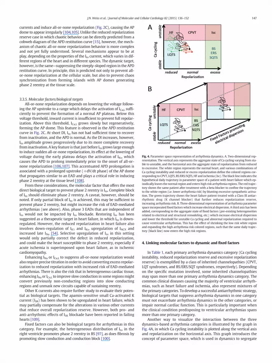
147J.N. Weiss et al. / Journal of Molecular and Cellular Cardiology 82 (2015) 136–152
currents and induce all-or-none repolarization (Fig. 2C), causing the APdome to appear irregularly [104,105]. Unlike the reduced repolarizationreserve case in which chaotic behavior can be directly predicted from acobweb diagram of the APD restitution curve [15], however, the mech-anism of chaotic all-or-none repolarization behavior is more complexand not yet fully understood. Several mechanisms appear to be atplay, depending on the properties of the Ito current, which varies in dif-ferent regions of the heart and in different species. The dynamic target,however, is the same—suppressing the steeply-sloped region in the APDrestitution curve. In principle, this is predicted not only to prevent all-or-none repolarization at the cellular scale, but also to prevent chaossynchronization from forming islands with AP domes generatingphase 2 reentry at the tissue scale.
Fig. 4. Parameter space representation of arrhythmia dynamics. A. Two-dimensional rep-resentation. The vertical axis represents the aggregate state of Ca cycling varying from sta-ble to unstable, and the horizontal axis the aggregate state of repolarization from reducedto excessive. The white region represents the normal heart, and various combinations ofCa cycling instability and reduced or excess repolarization define the colored regions cor-responding to CPVT, LQTS, BS/ERS/SQTS, HF and ischemia (Isc). The black line indicates thehypothetical daily trajectory in parameter space of a patient with heart failure which ep-isodically leaves thenormal region and enters high risk arrhythmia regions. The red trajec-tory shows the same patient after treatment with a beta blocker to confine the trajectoryto the white region (i.e. lower arrhythmia risk) by blunting excessive sympathetic activa-tion. The green trajectory shows the heart failure patient treated with a Class III antiar-rhythmic drug (K channel blocker) that further reduces repolarization reserve,increasing arrhythmia risk. B. Three-dimensional representation of arrhythmia parameterspace incorporated fixed factors which increase electrical dispersion. A third axis has beenadded, corresponding to the aggregate state of fixed factors (pre-existing heterogeneitiesrelated to electrical and structural remodeling, etc.) which increase electrical dispersionand lower the threshold for unstable Ca cycling and abnormal repolarization required tocause ventricular arrhythmias. This has the effect of shrinking the low risk white regionand expanding the high arrhythmia risk colored regions, such that the same daily trajec-tory (black line) now enters the high risk regions.
3.3.5. Molecular factors/biological targetsAll-or-none repolarization depends on lowering the voltage follow-
ing the AP upstroke to a range which delays the activation of ICa,L suffi-ciently to prevent the formation of a normal AP plateau. Below thisvoltage threshold, inward current is insufficient to prevent full repolar-ization. Above this threshold, ICa,L grows slowly but regeneratively,forming the AP dome. This feature is observed in the APD restitutioncurve in Fig. 2C. At short DI, Ito has not had sufficient time to recoverfrom inactivation, and the APD is normal. As the DI increases, however,Ito amplitude grows progressively due to its more complete recoveryfrom inactivation. A key feature is that just before Ito grows large enoughto induce sudden all-or-none repolarization, its effect at the lowering ofvoltage during the early plateau delays the activation of IKs, whichcauses the APD to prolong immediately prior to the onset of all-or-none repolarization [106,107]. This accentuated APD prolongation isassociated with a prolonged upstroke (+dV/dt phase) of the AP domethat propagates similar to an EAD and plays a critical role in inducingphase 2 reentry at the tissue level [108].
From these considerations, the molecular factor that offers the mostdirect biological target to prevent phase 2 reentry is Ito. Complete blockof Ito should eliminate phase 2 reentry. A caveat, however, should benoted. If only partial block of Ito is achieved, this may be sufficient toprevent phase 2 reentry, but might increase the risk of EAD-mediatedarrhythmias (see above). Also, phase 3 EADs that are independent ofIto would not be impacted by Ito blockade. Restoring Ito has beensuggested as a therapeutic target in heart failure, in which Ito is down-regulated. However, the reduced repolarization in heart failure alsoinvolves down-regulation of IK1 and IKs, upregulation of INCX andincreased late INa [54]. Selective upregulation of Ito in this settingwould only partially correct the defect in reduced repolarization,and could make the heart susceptible to phase 2 reentry, especially ifacute ischemia is superimposed upon heart failure, as in ischemiccardiomyopathy.
Enhancing INa or ICa,L to suppress all-or-none repolarization wouldalso require precise titration in order to avoid converting excess repolar-ization to reduced repolarization with increased risk of EAD-mediatedarrhythmias. There is also the risk that in heterogeneous cardiac tissue,enhancing INa or ICa,L to improve slow conduction in some regionsmightconvert previously non-conducting regions into slow conductingregions and unmask new circuits capable of sustaining reentry.
Other K currents also require further study to evaluate their poten-tial as biological targets. The apamin-sensitive small Ca-activated Kcurrent (ISK) has been shown to be upregulated in heart failure, whichmay partially compensate for the alterations in various other currentsthat reduce overall repolarization reserve. However, both pro- andanti-arrhythmic effects of ISK blockade have been reported in failinghearts [109].
Fixed factors can also be biological targets for arrhythmias in thiscategory. For example, the heterogeneous distribution of Ito in theright ventricle promotes phase 2 reentry in BS [97], as does fibrosis bypromoting slow conduction and conduction block [100].
4. Linking molecular factors to dynamic and fixed factors
In Table 1, each primary arrhythmia dynamics category (Ca cyclinginstability, reduced repolarization reserve and excessive repolarizationreserve) is exemplified by a class of inherited channelopathies (CPVT,LQT syndromes, and BS/ERS/SQT syndromes, respectively). Dependingon the specific mutation involved, some inherited channelopathiesmay span more than one primary arrhythmia dynamics category. Thecommon clinical diseases causing the majority of ventricular arrhyth-mias, such as heart failure and ischemia, also represent mixtures ofthe primary categories. To develop a successful antiarrhythmic strategy,biological targets that suppress arrhythmia dynamics in one categorymust not exacerbate arrhythmia dynamics in the other categories, orimpair normal cardiac function. This is particularly important whenthe clinical condition predisposing to ventricular arrhythmias spansmore than one primary category.
Another way to visualize the interaction between the threedynamics-based arrhythmia categories is illustrated by the graph inFig. 4A, in which Ca cycling instability is plotted along the vertical axisand repolarization on the horizontal axis. This graph introduces theconcept of parameter space, which is used in dynamics to segregate

148 J.N. Weiss et al. / Journal of Molecular and Cellular Cardiology 82 (2015) 136–152
different behaviors into distinct regions of parameter combinations,where each relevant parameter is represented by a separate axis. Anycombination of molecular factors which collectively produce a particu-lar level (parameter value) of repolarization and a particular level(parameter value) of Ca cycling instability can be represented by apoint (x,y) on this two-dimensional graph. Normal cardiac functionfalls in the central region in which both repolarization reserve and Cacycling are in the physiologically normal ranges. Classic CPVT occupiesthe region in which Ca cycling is unstable, but repolarization is normal.Classic LQT syndromes fall in the region with stable Ca cycling butreduced repolarization. Classic BS/ERS/SQT syndromes fall in the regionwith stable Ca cycling but excess repolarization. Heart failure and acuteischemia exhibit features of both unstable Ca cycling and abnormalrepolarization, and so occupy their own distinct regions in this parame-ter space (upper left and upper right, respectively). The hypotheticalboundaries between these different parameter regions are indicatedby dashed lines as a simplification, because the boundaries are morelikely to be nonuniform gradients rather than distinct straight lines.
Graphically, the arrhythmia risk profile of a patient can be represent-ed as a trajectory in this parameter space. Over a 24 h period, levels ofrepolarization and Ca cycling instability vary continuously in responseto the daily activities of living such as exercise, emotional stress, eatingand sleeping, and changing environmental conditions such as tempera-ture, electrolyte balance, drugs, etc. At a given point in time, eachparameter combination can be represented by a point on the graph,defining a trajectory that wanders through this parameter space duringthe course of the day. The trajectory varies from day to day dependingon the environmental factors encountered, but generally remainswithin a bounded region in this parameter space. Thus, in a healthyperson, the trajectory stays within the normal region, whereas in aperson with heart disease, the trajectory can wander into other regionsin which the probability of a potentially lethal ventricular arrhythmiaincreases.
For example, in a heart failure patient inwhich electrical remodelingand post-translational signaling have reduced repolarization anddestabilized Ca cycling, the trajectory may wander from the normalregion to visit the upper right region in response to changing environ-mental conditions such as stress or exercise or hypokalemia, placingthem at increased risk for a lethal ventricular arrhythmia (black trace).Treatmentwith a beta blocker confines the trajectory to amore restrict-ed region by eliminating parameter combinations associated high levelsof sympathetic activation, thereby reducing the time spent in dangerousparameter space regions, as illustrated in Fig. 4A (red trace). In contrast,treatment of the same patient with a Class III antiarrhythmic drug suchas the IKr-blocker D-sotalol would further reduce repolarization, shift thetrajectory to the left and increase the time spent in dangerous regions(green trace). As another example, the trajectory of a patient withischemia heart disease might visit the upper right region during ananginal episode, which could also be potentially avoided by effectiveantianginal therapy with a beta blocker.
The therapeutic goal of antiarrhythmic therapy, then, is to prevent apatient's trajectory from crossing into these high risk regions of param-eter space. Beta blockers accomplish this by reducing the dynamicresponsiveness to sympathetic activation, whereas other drugs mayaccomplish this by altering different molecular factors that restore nor-mal repolarization and/or Ca cycling stability. Pro-arrhythmic effectsoccur when an intervention that restricts entry into one dangerousregion inadvertently facilitates entry into another dangerous region.
To incorporate fixed factors that increase electrical dispersion, suchas pre-existing structural and electrophysiological heterogeneity, intothe conceptual framework, we can add a third axis to reflect the aggre-gate level of structural and electrical heterogeneity throughoutthe tissue (Fig. 4B). In our view, fixed factors increasing electrical disper-sion do not directly cause arrhythmias. Rather, they sensitize theheart to arrhythmias by reducing the level of dynamic instabilityrequired to initiate arrhythmias. For example, tissue fibrosis (structural
remodeling) markedly alters the source–sink relationship to facilitatethe emergence of triggers such as EADs and DADs, as well as promoteslow conduction by reducing gap junction coupling (for review see[26]). However, the process that generates the trigger that initiatesreentry in the region of slow conduction is a dynamic process. Thus,the general effect of fixed factors is to expand the arrhythmogenicregions in parameter space so that they encroach upon and shrink thenormal region, as illustrated in Fig. 4B. In other words, fixed factorsincreasing electrical dispersion make the tissue more vulnerable to theemergence of triggers and initiation of reentry by dynamic factors.Thus, the daily trajectory through this parameter space in a patientwith a structurally and electrically normally heart may never wanderoutside of the normal region, whereas the same daily trajectory in apatient with a structurally and electrically abnormal heart may episod-ically visit the high risk arrhythmogenic regions, as illustrated by theblack traces in Fig. 4B.
Within this conceptual framework, there are three general strategiesto reduce arrhythmia risk. The first is to expand the normal region sothat the patient's trajectory does not wander outside of the normalregion during the course of the day. For example, in the case of a dam-aged heart in which fixed factors increasing electrical dispersion havelowered the threshold of dynamic instability required to inducearrhythmias, reversing pre-existing structural and electrical heteroge-neities (i.e. moving back to the normal region along the third axis inFig. 4B) will restore the low-risk region to its previously larger normalrange. The same daily trajectory may then no longer enter the highrisk arrhythmia regions. Interventions which reverse fibrosis wouldfall into this class. For example, the anti-fibrotic effects of ACE inhibitorsmight contribute to their protective effects against sudden cardiac deathin heart failure by this mechanism.
The second general strategy is to restrict the patient's daily trajectoryin parameter space, so that the trajectory remains within the boundariesof the low risk region. For example, beta blockers and antianginal agentsfall into this class, as illustrated by the red trajectory in Fig. 4A. The thirdstrategy is to reposition the patient's trajectory so that it is more centrallylocated in the low risk region. In this case, the same level of excursion ofthe trajectorymay no longer cross into the high risk regions. For example,in a patient with LQT3 syndrome caused by defective INa inactivation, alate INa blockerwouldmove their trajectory into the low risk region by in-creasing repolarization from the reduced into the normal range.
5. Developing an in silico drug-testing strategy
How does the dynamic state in the low dimensional parameterspace illustrated in Fig. 4 relate to the actual molecular state of a pa-tient? The molecular state of a patient can also be displayed on agraph, in which each molecular factor, such as the state of each ionchannel and transporter, ion concentration, drug concentration, meta-bolic state, etc., is represented along a separate axis (Fig. 5). These mo-lecular parameters also change continuously over the course of eachday, tracing a trajectory through this very high dimensional parameterspace with thousands of dimensions instead of just two. The challenge,then, is to create amap that links the high-dimensional parameter spaceto the low-dimensional parameter space—that is, to define which com-binations ofmolecular parameter values correspond towhich aggregatedynamic parameter combinations of repolarization state and Ca cyclinginstability. If this can be achieved, then in principle one can predict sys-tematically how a molecular intervention will impact all of the relevantdynamic factors, in all three categories of arrhythmia dynamics. A mo-lecular factor could be profiled to define its potential antiarrhythmicand proarrhythmic actions, as well as its effects on normal excitation–contraction coupling.
Is this a wild fantasy, or a realistic possibility? Already, the U.S.Federal Drug Administration is exploring the modeling-basedapproaches for preclinical assessment of drug cardiotoxicity [110]. Ascomputational power has increased and computational approaches to

Fig. 5. Linking dynamic factors to molecular factors. The aggregate dynamic parameters in the low dimensional parameter space defining arrhythmia risk (left) are ultimately determinedby the interactions betweenmolecular factors in amuch higher dimensional parameter space (right) inwhich eachmolecular factor (e.g. properties of an ion channel) is represented by itsown axis. The ultimate goal is to create amap that links the high dimensional space to the low dimensional space, allowingmolecular factors to be evaluated as biological targets to lowerarrhythmia risk.
149J.N. Weiss et al. / Journal of Molecular and Cellular Cardiology 82 (2015) 136–152
explore high dimensional parameter spaces have becomemore sophis-ticated [13,14], in silico drug testing to link drug effects at the molecularlevel to arrhythmogenic cellular and tissue properties has becomeincreasingly feasible [111,112]. In addition, dimension reduction tech-niques have been developed to condense high dimensional informationsystems into more tractable low dimensional representations for arange of ‘big data’ applications [113]. One approach might be to createa panel of detailed multi-scale models that integrate behaviors at thesubcellular, cellular and tissue levels to represent the different behav-iors in each arrhythmia dynamics category. For example, a thousandor more detailed models with different molecular parameter settingscould be generated to represent each of the six normal and arrhythmo-genic parameter regions in Fig. 4A spanning a range of organism-scalephysiological conditions (autonomic tone, electrolyte values, etc.). Toscreen a molecular factor as a potential biological target in silico, themolecular intervention could be implemented in each of these models,and the effects on the dynamic factors regulating arrhythmias in eachcategory assessed.
This approach has several conceptual advantages. First, it automati-cally provides a balanced evaluation of both anti- and pro-arrhythmiceffects of a molecular intervention across the full spectrum of arrhyth-mia dynamics categories as well as normal cardiac function. Second,the effects of a molecular intervention are assessed on populations ofmodels [13,14], emulating the genetic and environmental diversityencountered in human populations, rather than on a single model ineach arrhythmia dynamics category. Third, in addition to human cardiacelectrophysiology, complementary populations of models can be gener-ated for nonhuman species to facilitate direct comparison and valida-tion of in silico predictions with experimental findings in nonhumanarrhythmia models.
How would this work in practice? The in silico approach could beused in two complementary ways. First, to facilitate drug design, theapproach can be used to predict theoretically the ideal modificationsto a molecular factor, such as the properties of an ion channel that willsuppress arrhythmias. For example, there aremany possible biophysicalmanipulations to reduce the ICa,L window current towards the goalof suppressing EAD-mediated arrhythmias. These include shiftingthe voltage dependence of activation or inactivation, altering kineticparameters, and reducing the non-inactivating pedestal component.Implementing these altered parameters in the population of modelscan be used to identify ideal parameter combinations that are most ef-fective at suppressing EAD-mediated arrhythmias without disturbingnormal EC coupling, exacerbating Ca cycling instability to a level thatpromotes DADs and/or alternans, or altering repolarization reserve in
a manner promoting all-or-none repolarization and phase 2 reentry.In a complementary fashion, the approach could be used to facilitatedrug screening. Imagine that a small molecule drug screen identifies acompound with novel effects on the biophysical properties of an ionchannel. For example, the compound might selectively block the lateICa,L without affecting the peak amplitude (analogous to ranolazine forlate INa). The detailed biophysical properties could be implemented inthe population of models to predict whether the altered ICa,L propertieswould be effective at suppressing EAD-mediated arrhythmias withoutcreating the other adverse effects. Thus, as it is refined, the in silicoapproach could serve as a drug-development and screening platformboth to predict ideal antiarrhythmic drug properties, and to evaluatedrugs whose biophysical properties have already been experimentallycharacterized, prior to moving forward to costly animal models forfurther testing.
6. Application to supraventricular arrhythmias
Many supraventricular arrhythmias can now be effectively andpermanently cured by catheter ablation techniques, including atrioven-tricular reentrant tachycardia, atrioventricular nodal reentrant tachy-cardia, atrial tachycardia and atrial flutter. The most challenging,however, remains atrial fibrillation (AF). AF mechanisms are stillcontroversial, in part because of the greater structural and electrophys-iological complexity of the atria and its associated structures includingthe sinus and atrioventricular nodes, venae cavae, pulmonary veinsand valvular annuli. AF is also the most common sustained arrhythmia,affecting 5–10% of the population over the age of 70 [114] and account-ing for a quarter of cerebrovascular accidents (strokes) in the elderly.Under the most favorable conditions (paroxysmal atrial fibrillation instructurally normal hearts), the current success rate of ablation isaround 70%, and under 50% for long-standing persistent AF in patientswith enlarged atria or heart failure [115]. Repeat ablations are oftennecessary, and each procedure carries a 5% risk of significant complica-tions [116]. Thus, as with ventricular arrhythmias, development ofeffective pharmacologic/biologic therapy for AF is highly desirable.
Although the dynamics-based classification in Table 1 focuses onventricular arrhythmias, the same approach can be adapted to supra-ventricular arrhythmias and AF, once the key dynamic mechanismshave been appropriately defined. There is no reason to believe that thedynamic mechanisms outlined in Table 1 are not also applicable to theatria. DADs, DAD-mediated trigger activity [117] and spatially discor-dant alternans [118] have been described in atrial tissue, as have EADsand EAD-mediated triggered activity [102]. In addition, evidence of

150 J.N. Weiss et al. / Journal of Molecular and Cellular Cardiology 82 (2015) 136–152
both multiple wavelet and mother rotor fibrillation have been present-ed in atria (for review see [119]). Excessive repolarization reservecaused by gain-of-function K channel mutations has also been associat-ed with familial AF [96]. Direct experimental evidence for chaossynchronization, biexcitability and phase-2 reentry in atrial tissue, how-ever, has yet to be obtained.
Is AF driven by fundamentally differentmechanisms thanVF?UnlikeVF, which is lethal if not immediately defibrillated, atrial fibrillation isusually tolerated hemodynamically, so that its dynamical mechanismsmay evolve over time as electrical and structural remodeling develop.For example, paroxysmal AF in normal sized atria may initially be relat-ed to triggers emanating from the pulmonary veins and/or other venousstructures, caused by localized focal activity or microreentry which ini-tiates nonsustained multiple wavelet or mother rotor fibrillation. Thiswould account for the reasonably high success rate of ablation at thisstage. However, as AF becomes persistent and long-lasting, the underly-ing electrical and structural remodeling may alter the tissue substratesufficiently to allow multiple wavelet or mother rotor fibrillation tocontinue indefinitely [120], so that isolation of triggers is no longereffective. Enlarged atria due to valvular disease or diastolic ventriculardysfunction may also fall into the latter category due to electrical andstructural remodeling induced by chronic hemodynamic stress.
7. Summary and challenges
Our goal has been to illustrate how molecular factors interact withdynamic and fixed factors at the subcellular, cellular and tissue andorganism scales to cause ventricular arrhythmias. Achieving a thoroughunderstanding of these interactions is critical for developing novel effec-tive pharmacologic/biologic therapies for cardiac arrhythmias. Pharma-cologic/biologic targets are molecular factors, but arrhythmias aredetermined by dynamic factors at the subcellular, cellular, tissue andorganism scales interacting with fixed factors increasing electricaldispersion at the tissue scale. The highest risk of arrhythmias ariseswhen dynamic factors, interacting with fixed factors, simultaneouslypromote the emergence of triggers while enhancing electrical disper-sion within the tissue. The proposed dynamics-based classificationoutlined in Table 1 represents an attempt to synthesizewhat is current-ly known about these relationships. Accordingly, we have classifiedventricular arrhythmias into three major dynamic categories relatingto unstable Ca cycling, reduced repolarization and excess repolarization.Each of these primary categories is represented by a group of inheritedchannelopathies (CPVT, LQT syndrome and BS/ERS/SQT syndrome,respectively). In addition, the more common causes of ventriculararrhythmias, heart failure and ischemia, represent mixtures of theprimary categories. We have summarized the current state-of-knowledge about the dynamic factors operating at the subcellular,cellular, tissue and organism scales relevant to each arrhythmia dynam-ics category, and provided illustrative examples of how these dynamicfactors can be related to molecular factors.
To illustrate conceptually how these arrhythmia dynamics mightapply to a patient at increased risk of ventricular arrhythmias, we pre-sented the parameter space concept (Fig. 4). We envision the patientas wandering through this parameter space during the course of theirdaily activities, defining a trajectory. The trajectory of a healthy personwith a normal heart does not wander outside of the normal region,whereas the trajectory of a patient with congenital or acquired heartdisease crosses into the abnormal regions, placing them at increasedrisk of a ventricular arrhythmia. In this representation, the goal of phar-macologic/biologic antiarrhythmic therapy is to prevent a patient'strajectory from wandering outside of the normal region (Fig. 4A).In principle, this can be accomplished by one of three strategies:1) expanding the normal region by reversing fixed factors that makethe heart more sensitive to dynamic instability (e.g. reversing fibrosisin heart failure); 2) reducing the extent towhich the trajectorywanders(e.g. beta blocker therapy in ischemia); or 3) re-centering the trajectory
in the normal region (e.g. late INa blocker drugs to correct the defect inLQT3 syndrome). The great challenge will be to create an accuratemap that links the low dimensional parameter space of dynamic factorsto the high dimensional parameter space of molecular factors, usingcomputational methods and dimensional reduction analytics (Fig. 5).If successful, this endeavor has the potential to provide a systematicapproach to evaluate and predict in silico how molecular factors, suchas altered biophysical properties of an ion channel, affect not only thetargeted arrhythmia category, but all of the other relevant arrhythmiacategories as well as normal cardiac EC coupling. Efficacy and safetywill be addressed simultaneously. May the journey begin!
Funding sources
This work was supported by NIH/NHLBI grants P01 HL078931, R01HL114437, and the Laubisch and Kawata endowments.
Disclosures
None
Acknowledgments
We are grateful to Alex Baher, M.D., and Michael Liu, B.S. for theirhelp in preparing the figures.
References
[1] Hoffman BF, Rosen MR. Cellular mechanisms for cardiac arrhythmias. Circ Res1981;49:1–15.
[2] Rosen MR. Mechanisms for arrhythmias. Am J Cardiol 1988;61:2A–8A.[3] Jalife J. Ventricular fibrillation: mechanisms of initiation and maintenance. Ann Rev
Physiol 2000;62:25–50.[4] Keating MT, Sanguinetti MC. Molecular and cellular mechanisms of cardiac
arrhythmias. Cell 2001;104:569–80.[5] Rubart M, Zipes DP. Mechanisms of sudden cardiac death. J Clin Invest 2005;115:
2305–15.[6] Qu Z, Weiss JN. Mechanisms of ventricular arrhythmias: from molecular fluctua-
tions to electrical turbulence. Ann Rev Physiol 2015;77 [in press].[7] Echt DS, Liebson PR, Mitchell LB, Peters RW, Obias-Manno D, Barker AH, et al. Mor-
tality and morbidity in patients receiving encainide, flecainide, or placebo. TheCardiac Arrhythmia Suppression Trial. N Engl J Med 1991;324:781–8.
[8] Waldo AL, CammAJ, deRuyter H, Friedman PL, MacNeil DJ, Pauls JF, et al. Effect of D-sotalol on mortality in patients with left ventricular dysfunction after recent andremote myocardial infarction. The SWORD Investigators. Survival With Oral D-Sotalol. Lancet 1996;348:7–12.
[9] Myerburg RJ, Mitrani R, Interian Jr A, Castellanos A. Interpretation of outcomes ofantiarrhythmic clinical trials: design features and population impact. Circulation1998;97:1514–21.
[10] Bardy GH, Lee KL, Mark DB, Poole JE, Packer DL, Boineau R, et al. Amiodarone or animplantable cardioverter-defibrillator for congestive heart failure. N Engl J Med2005;352:225–37.
[11] Fonarow GC, Yancy CW, Hernandez AF, Peterson ED, Spertus JA, Heidenreich PA.Potential impact of optimal implementation of evidence-based heart failure thera-pies on mortality. Am Heart J 2011;161:1024–30.
[12] Qu Z, Hu G, Garfinkel A, Weiss JN. Nonlinear and stochastic dynamics in the heart.Phys Rep 2014;543:61–162.
[13] Sarkar AX, Sobie EA. Regression analysis for constraining free parameters in elec-trophysiological models of cardiac cells. PLoS Comput Biol 2010;6:e1000914.
[14] Britton OJ, Bueno-Orovio A, Van Ammel K, Lu HR, Towart R, Gallacher DJ, et al.Experimentally calibrated population of models predicts and explains intersubjectvariability in cardiac cellular electrophysiology. Proc Natl Acad Sci U S A 2013;110:E2098–105.
[15] Sato D, Xie LH, Sovari AA, Tran DX, Morita N, Xie F, et al. Synchronization of chaoticearly afterdepolarizations in the genesis of cardiac arrhythmias. Proc Natl Acad SciU S A 2009;106:2983–8.
[16] Gyorke S. Molecular basis of catecholaminergic polymorphic ventricular tachycar-dia. Heart Rhythm 2009;6:123–9.
[17] Mohamed U, Napolitano C, Priori SG. Molecular and electrophysiological basesof catecholaminergic polymorphic ventricular tachycardia. J CardiovascElectrophysiol 2007;18:791–7.
[18] Swaminathan PD, Purohit A, Hund TJ, Anderson ME. Calmodulin-dependentprotein kinase II: linking heart failure and arrhythmias. Circ Res 2012;110:1661–77.
[19] Tomaselli GF, Zipes DP. What causes sudden death in heart failure? Circ Res 2004;95:754–63.

151J.N. Weiss et al. / Journal of Molecular and Cellular Cardiology 82 (2015) 136–152
[20] CerroneM, Noujaim SF, Tolkacheva EG, Talkachou A, O'Connell R, Berenfeld O, et al.Arrhythmogenic mechanisms in a mouse model of catecholaminergic polymorphicventricular tachycardia. Circ Res 2007;101:1039–48.
[21] Katra RP, Laurita KR. Cellular mechanism of calcium-mediated triggered activity inthe heart. Circ Res 2005;96:535–42.
[22] Xie Y, Sato D, Garfinkel A, Qu Z, Weiss JN. So little source, so much sink: require-ments for afterdepolarizations to propagate in tissue. Biophys J 2010;99:1408–15.
[23] Fujiwara K, Tanaka H, Mani H, Nakagami T, Takamatsu T. Burst emergence of intra-cellular Ca waves evokes arrhythmogenic oscillatory depolarization via the Na-Caexchanger: simultaneous confocal recording ofmembrane potential and intracellu-lar Ca in the heart. Circ Res 2008;103:509–18.
[24] Wasserstrom JA, Shiferaw Y, Chen W, Ramakrishna S, Patel H, Kelly JE, et al. Vari-ability in timing of spontaneous calcium release in the intact rat heart is deter-mined by the time course of sarcoplasmic reticulum calcium load. Circ Res 2010;107:1117–26.
[25] de Jong S, van Veen TAB, van Rijen HVM, de Bakker JMT. Fibrosis and cardiacarrhythmias. J Cardiovasc Pharmacol 2011;57:630–810.
[26] Nguyen TP, Qu Z, Weiss JN. Cardiac fibrosis and arrhythmogenesis: the road torepair is paved with perils. J Mol Cell Cardiol 2014;70:83–91.
[27] Kummer JL, Nair R, Krishnan SC. Images in cardiovascular medicine. Bidirectionalventricular tachycardia caused by digitalis toxicity. Circulation 2006;113:e156–7.
[28] Valent S, Kelly P. Images in clinical medicine. Digoxin-induced bidirectionalventricular tachycardia. N Engl J Med 1997;336:550.
[29] Baher AA, Uy M, Xie F, Garfinkel A, Qu Z, Weiss JN. Bidirectional ventricular tachy-cardia: ping pong in the His-Purkinje system. Heart Rhythm 2011;8:599–605.
[30] de Lange E, Garfinkel A, Weiss JN, Qu Z. Delayed afterdepolarizations provide thetrigger and substrate for reentrant arrhythmias in cardiac tissue: a modelingstudy. Heart Rhythm 2011;8:S325.
[31] Rosen MR, Wit AL, Hoffman BF. Electrophysiology and pharmacology of cardiac ar-rhythmias. IV. Cardiac antiarrhythmic and toxic effects of digitalis. Am Heart J1975;89:391–9.
[32] Weiss JN, Karma A, Shiferaw Y, Chen PS, Garfinkel A, Qu Z. From pulsus topulseless: the saga of cardiac alternans. Circ Res 2006;98:1244–53.
[33] Qu Z, Garfinkel A, Chen PS, Weiss JN. Mechanisms of discordant alternans andinduction of reentry in simulated cardiac tissue. Circulation 2000;102:1664–70.
[34] WatanabeMA, Fenton FH, Evans SJ, Hastings HM, Karma A. Mechanisms for discor-dant alternans. J Cardiovasc Electrophysiol 2001;12:196–206.
[35] Pastore JM, Girouard SD, Laurita KR, Akar FG, RosenbaumDS. Mechanism linking T-wave alternans to the genesis of cardiac fibrillation. Circulation 1999;99:1385–94.
[36] Rosenbaum DS, Jackson LE, Smith JM, Garan H, Ruskin JN, Cohen RJ. Electricalalternans and vulnerability to ventricular arrhythmias. N Engl J Med 1994;330:235–41.
[37] Wilson LD, Jeyaraj D, Wan X, Hoeker GS, Said TH, Gittinger M, et al. Heart failureenhances susceptibility to arrhythmogenic cardiac alternans. Heart Rhythm 2009;6:251–9.
[38] Wasserstrom JA, Sharma R, Kapur S, Kelly JE, Kadish AH, Balke CW, et al. multipledefects in intracellular calcium cycling in whole failing rat heart. Circulation:heart failure 2009;2:223–32.
[39] Weiss JN, Nivala M, Garfinkel A, Qu Z. Alternans and arrhythmias: from cell toheart. Circ Res 2011;108:98–112.
[40] Bassani JW, Yuan W, Bers DM. Fractional SR Ca release is regulated by trigger Caand SR Ca content in cardiac myocytes. Am J Physiol 1995;268:C1313–9.
[41] Shannon TR, Ginsburg KS, Bers DM. Potentiation of fractional sarcoplasmic reticu-lum calcium release by total and free intra-sarcoplasmic reticulum calcium concen-tration. Biophys J 2000;78:334–43.
[42] Diaz ME, O'Neill SC, Eisner DA. Sarcoplasmic reticulum calcium content fluctuationis the key to cardiac alternans. Circ Res 2004;94:650–6.
[43] Picht E, Desantiago J, Blatter LA, Bers DM. Cardiac alternans do not rely on diastolicsarcoplasmic reticulum calcium content fluctuations. Circ Res 2006;99:740–8.
[44] Shiferaw Y, Sato D, Karma A. Coupled dynamics of voltage and calcium in pacedcardiac cells. Phys Rev E Stat Nonlin Soft Matter Phys 2005;71:021903.
[45] DiazME, Eisner DA, O'Neill SC. Depressed ryanodine receptor activity increases var-iability and duration of the systolic Ca transient in rat ventricular myocytes. CircRes 2002;91:585–93.
[46] Gaeta SA, Bub G, Abbott GW, Christini DJ. Dynamical mechanism for subcellularalternans in cardiac myocytes. Circ Res 2009;105:335–42.
[47] Nolasco JB, Dahlen RW. A graphic method for the study of alternation in cardiacaction potentials. J Appl Physiol 1968;25:191–6.
[48] Tolkacheva EG, Schaeffer DG, Gauthier DJ, Krassowska W. Condition for alternansand stability of the 1:1 response pattern in a "memory" model of paced cardiacdynamics. Phys Rev E Stat Nonlin Soft Matter Phys 2003;67:031904.
[49] Cao JM, Qu Z, Kim YH,Wu TJ, Garfinkel A, Weiss JN, et al. Spatiotemporal heteroge-neity in the induction of ventricular fibrillation by rapid pacing: importance ofcardiac restitution properties. Circ Res 1999;84:1318–31.
[50] Qu Z, Karagueuzian HS, Garfinkel A, Weiss JN. Effects of Na channel and cell cou-pling abnormalities on vulnerability to reentry: a simulation study. Am J PhysiolHeart Circ Physiol 2004;286:H1310–21.
[51] Qu Z, Xie Y, Garfinkel A, Weiss JN. T-wave Alternans and arrhythmogenesis incardiac diseases. Front Physiol 2010;1.
[52] Qu Z, Nivala M, Weiss JN. Calcium alternans in cardiac myocytes: order from disor-der. J Mol Cell Cardiol 2013;58:100–9.
[53] Maruyama M, Joung B, Tang L, Shinohara T, On YK, Han S, et al. Diastolic intracellu-lar calcium-membrane voltage coupling gain and postshock arrhythmias: role ofpurkinje fibers and triggered activity. Circ Res 2010;106:399–408.
[54] Pogwizd SM, Schlotthauer K, Li L, Yuan W, Bers DM. Arrhythmogenesis andcontractile dysfunction in heart failure: roles of sodium-calcium exchange, inward
rectifier potassium current, and residual beta-adrenergic responsiveness. Circ Res2001;88:1159–67.
[55] Karma A. Spiral breakup in model equations of action potential propagation incardiac tissue. Phys Rev Lett 1993;71:1103–6.
[56] Saitoh H, Bailey JC, Surawicz B. Alternans of action potential duration after abruptshortening of cycle length: differences between dog Purkinje and ventricularmuscle fibers. Circ Res 1988;62:1027–40.
[57] Rovetti R, Cui X, Garfinkel A, Weiss JN, Qu Z. Spark-induced sparks as a mechanismof intracellular calcium alternans in cardiac myocytes. Circ Res 2010;106:1582–91.
[58] Mahajan A, Sato D, Shiferaw Y, Baher A, Xie L-H, Peralta R, et al. Modifying L-typecalcium current kinetics: consequences for cardiac excitation and arrhythmiadynamics. Biophys J 2008;94:411–23.
[59] Pu J, Balser JR, Boyden PA. Lidocaine action on Na currents in ventricular myocytesfrom the epicardial border zone of the infarcted heart. Circ Res 1998;83:431–40.
[60] Joyner RW, Ramza BM, Osaka T, Tan RC. Cellular mechanisms of delayed recoveryof excitability in ventricular tissue. Am J Physiol 1991;260:H225–33.
[61] Pu J, Boyden PA. Alterations of Na currents inmyocytes from epicardial border zoneof the infarcted heart. A possible ionic mechanism for reduced excitability andpostrepolarization refractoriness. Circ Res 1997;81:110–9.
[62] Roden DM. Taking the "idio" out of "idiosyncratic": predicting Torsades de Pointes.Pacing Clin Electrophysiol 1998;21:1029–34.
[63] Roden DM. Long QT, syndrome: reduced repolarization reserve and the geneticlink. J Int Med 2006;259:59–69.
[64] Nattel S, Maguy A, Le Bouter S, Yeh Y-H. Arrhythmogenic ion-channel remodelingin the heart: heart failure, myocardial infarction, and atrial fibrillation. Physiol Rev2007;87:425–56.
[65] January CT, Moscucci A. Cellular mechanisms of early afterdepolarizations. Ann N YAcad Sci 1992;644:23–32.
[66] Weiss JN, Garfinkel A, Karagueuzian HS, Chen PS, Qu Z. Early afterdepolarizationsand cardiac arrhythmias. Heart Rhythm 2010;7:1891–9.
[67] Chang MG, Sato D, de Lange E, Lee JH, Karagueuzian HS, Garfinkel A, et al. Bi-stablewave propagation and early afterdepolarization-mediated cardiac arrhythmias.Heart Rhythm 2012;9:115–22.
[68] Choi BR, Burton F, Salama G. Cytosolic Ca triggers early afterdepolarizations andTorsade de Pointes in rabbit hearts with type 2 long QT syndrome. J Physiol2002;543:615–31.
[69] Zhao Z, Wen H, Fefelova N, Allen C, Baba A, Matsuda T, et al. Revisiting the ionicmechanisms of early afterdepolarizations in cardiomyocytes: predominant by Cawaves or Ca currents? Am J Physiol Heart Circ Physiol 2012;302:H1636–44.
[70] Volders PG, Vos MA, Szabo B, Sipido KR, de Groot SH, Gorgels AP, et al. Progress inthe understanding of cardiac early afterdepolarizations and torsades de pointes:time to revise current concepts. Cardiovasc Res 2000;46:376–92.
[71] Clancy CE, Rudy Y. Na channelmutation that causes both Brugada and long-QT syn-drome phenotypes: a simulation study of mechanism. Circulation 2002;105:1208–13.
[72] Luo CH, Rudy Y. A dynamic model of the cardiac ventricular action potential.2.Afterdepolarizations, triggered activity, and potentiation. Circ Res 1994;74:1097–113.
[73] Tran DX, Sato D, Yochelis A, Weiss JN, Garfinkel A, Qu Z. Bifurcation and chaos in amodel of cardiac early afterdepolarizations. Phys Rev Lett 2009;102:258103.
[74] Channell P, Cymbalyuk G, Shilnikov A. Origin of bursting through homoclinic spikeadding in a neuron model. Phys Rev Lett 2007;98:134101.
[75] Shilnikov A, Cymbalyuk G. Transition between tonic spiking and bursting in aneuron model via the blue-sky catastrophe. Phys Rev Lett 2005;94:048101.
[76] Hondeghem LM, Dujardin K, De Clerck F. Phase 2 prolongation, in the absence ofinstability and triangulation, antagonizes class III proarrhythmia. Cardiovasc Res2001;50:345–53.
[77] Zhao Z, Xie Y, Wen H, Xiao D, Allen C, Fefelova N, et al. Role of the transient out-ward potassium current in the genesis of early afterdepolarizations in cardiaccells. Cardiovasc Res 2012;95:308–16.
[78] Nguyen TP, Xie Y, Garfinkel A, Qu Z, Weiss JN. Arrhythmogenic consequences ofmyofibroblast-myocyte coupling. Cardiovasc Res 2012;93:242–51.
[79] Gadsby DC, Cranefield PF. Two levels of resting potential in cardiac Purkinje fibers. JGen Physiol 1977;70:725–46.
[80] Kim TY, Kunitomo Y, Pfeiffer Z, Patel D, Hwang J, Harrison K, et al. Complex excita-tion dynamics underlie polymorphic ventricular tachycardia in a transgenic rabbitmodel of long QT syndrome type 1. Heart Rhythm 2015;12:220–8.
[81] Chang MG, de Lange E, Calmettes G, Garfinkel A, Qu Z, Weiss JN. Pro- and antiar-rhythmic effects of ATP-sensitive potassium current activation on reentry duringearly afterdepolarization-mediated arrhythmias. Heart Rhythm 2013;10:575–82.
[82] Maruyama M, Lin SF, Xie Y, Chua SK, Joung B, Han S, et al. Genesis of phase 3 earlyafterdepolarizations and triggered activity in acquired long-QT syndrome. CircArrhythm Electrophysiol 2011;4:103–11.
[83] Zareba W, Moss AJ, le Cessie S, Hall WJ. T wave alternans in idiopathic long QT syn-drome. J Am Coll Cardiol 1994;23:1541–6.
[84] Qu Z, Xie LH, Olcese R, Karagueuzian HS, Chen PS, Garfinkel A, et al. Earlyafterdepolarizations in cardiac myocytes: beyond reduced repolarization reserve.Cardiovasc Res 2013;99:6–15.
[85] Bennett PB, Yazawa K, Makita N, George Jr AL. Molecular mechanism for aninherited cardiac arrhythmia. Nature 1995;376:683–5.
[86] Wagner S, Dybkova N, Rasenack EC, Jacobshagen C, Fabritz L, Kirchhof P, et al. Ca/calmodulin-dependent protein kinase II regulates cardiac Na channels. J Clin Invest2006;116:3127–38.
[87] Yang T, Chun YW, Stroud DM, Mosley JD, Knollmann BC, Hong CC, et al. Screeningfor acute IKr block is insufficient to detect Torsades de Pointes liability: role of latesodium current. Circulation 2014;130:224–34.

152 J.N. Weiss et al. / Journal of Molecular and Cellular Cardiology 82 (2015) 136–152
[88] Wagner S, Ruff HM,Weber SL, Bellmann S, Sowa T, Schulte T, et al. Reactive oxygenspecies-activated Ca/calmodulin kinase IIdelta is required for late INa augmentationleading to cellular Na and Ca overload. Circ Res 2011;108:555–65.
[89] Hale SL, Shryock JC, Belardinelli L, Sweeney M, Kloner RA. Late sodium currentinhibition as a new cardioprotective approach. J Mol Cell Cardiol 2008;44:954–67.
[90] Morita N, Lee JH, Xie Y, Sovari A, Qu Z,Weiss JN, et al. Suppression of re-entrant andmultifocal ventricular fibrillation by the late sodium current blocker ranolazine. JAm Coll Cardiol 2011;57:366–75.
[91] Wu L, Shryock JC, Song Y, Li Y, Antzelevitch C, Belardinelli L. Antiarrhythmic effectsof ranolazine in a guinea pig in vitro model of long-QT syndrome. J Pharmacol ExpTher 2004;310:599–605.
[92] Hennessey JA, Boczek NJ, Jiang YH, Miller JD, PatrickW, Pfeiffer R, et al. A CACNA1Cvariant associated with reduced voltage-dependent inactivation, increased CaV1.2channel window current, and arrhythmogenesis. PLoS One 2014;9:e106982.
[93] Madhvani RV, Xie Y, Pantazis A, Garfinkel A, Qu Z,Weiss JN, et al. Shaping a new Caconductance to suppress early afterdepolarizations in cardiac myocytes. J Physiol2011;589:6081–92.
[94] Colecraft HM, Alseikhan B, Takahashi SX, Chaudhuri D, Mittman S,Yegnasubramanian V, et al. Novel functional properties of Ca channel betasubunits revealed by their expression in adult rat heart cells. J Physiol 2002;541:435–52.
[95] Morita N, Sovari AA, Xie Y, Fishbein MC, Mandel WJ, Garfinkel A, et al. Increasedsusceptibility of aged hearts to ventricular fibrillation during oxidative stress. AmJ Physiol Heart Circ Physiol 2009;297:H1594–605.
[96] Giudicessi JR, Ackerman MJ. Potassium-channel mutations and cardiac arrhyth-mias–diagnosis and therapy. Nat Rev Cardiol 2012;9:319–32.
[97] Antzelevitch C, Yan GX. J wave syndromes. Heart Rhythm 2010;7:549–58.[98] Lukas A, Antzelevitch C. Phase 2 reentry as a mechanism of initiation of circus
movement reentry in canine epicardium exposed to simulated ischemia.Cardiovasc Res 1996;32:593–603.
[99] Lukas A, Antzelevitch C. Differences in the electrophysiological response of canineventricular epicardium and endocardium to ischemia. Role of the transientoutward current. Circulation 1993;88:2903–15.
[100] Hoogendijk MG, Opthof T, Postema PG, Wilde AA, de Bakker JM, Coronel R. TheBrugada ECG pattern: a marker of channelopathy, structural heart disease, orneither? Toward a unifying mechanism of the Brugada syndrome. Circ ArrhythmElectrophysiol 2010;3:283–90.
[101] Jeyaraj D, Wilson LD, Zhong J, Flask C, Saffitz JE, Deschenes I, et al.Mechanoelectrical feedback as novel mechanism of cardiac electrical remodel-ing. Circulation 2007;115:3145–55.
[102] Burashnikov A, Antzelevitch C. Reinduction of atrial fibrillation immediately aftertermination of the arrhythmia is mediated by late phase 3 earlyafterdepolarization-induced triggered activity. Circulation 2003;107:2355–60.
[103] Chua SK, Chang PC, Maruyama M, Turker I, Shinohara T, Shen MJ, et al. Small-conductance calcium-activated potassium channel and recurrent ventricular fibril-lation in failing rabbit ventricles. Circ Res 2011;108:971–9.
[104] Maoz A, Christini DJ, Krogh-Madsen T. Dependence of phase-2 reentry and repolar-ization dispersion on epicardial and transmural ionic heterogeneity: a simulationstudy. Europace 2014;16:458–65.
[105] Maoz A, Krogh-Madsen T, Christini DJ. Instability in action potential morphologyunderlies phase 2 reentry: a mathematical modeling study. Heart Rhythm 2009;6:813–22.
[106] DongM, Sun X, Prinz AA, Wang HS. Effect of simulated Ito on guinea pig and canineventricular action potential morphology. Am J Physiol Heart Circ Physiol 2006;291:H631–7.
[107] Greenstein JL, Wu R, Po S, Tomaselli GF, Winslow RL. Role of the calcium-independent transient outward current Ito1 in shaping action potential morpholo-gy and duration. Circ Res 2000;87:1026–33.
[108] Miyoshi S, Mitamura H, Fujikura K, Fukuda Y, Tanimoto K, Hagiwara Y, et al. Amathematical model of phase 2 reentry: role of L-type Ca current. Am J PhysiolHeart Circ Physiol 2003;284:H1285–94.
[109] Hsieh YC, Chang PC, Hsueh CH, Lee YS, Shen C, Weiss JN, et al. Apamin-sensitivepotassium current modulates action potential duration restitution andarrhythmogenesis of failing rabbit ventricles. Circ Arrhythm Electrophysiol 2013;6:410–8.
[110] Chi KR. Revolution dawning in cardiotoxicity testing. Nat Rev Drug Discov 2013;12:565–7.
[111] Moreno JD, Zhu ZI, Yang PC, Bankston JR, Jeng MT, Kang C, et al. A computationalmodel to predict the effects of class I anti-arrhythmic drugs on ventricular rhythms.Sci Transl Med 2011;3:98ra83.
[112] Moreno JD, Yang PC, Bankston JR, Grandi E, Bers DM, Kass RS, et al. Ranolazine forcongenital and acquired late INa-linked arrhythmias: in silico pharmacologicalscreening. Circ Res 2013;113:e50–61.
[113] Tenenbaum JB, Vd Silva, Langford JC. A global geometric framework for nonlineardimensionality reduction. Science 2000;290:2319–23.
[114] Piccini JP, Hammill BG, Sinner MF, Jensen PN, Hernandez AF, Heckbert SR, et al. In-cidence and prevalence of atrial fibrillation and associated mortality among Medi-care beneficiaries, 1993–2007. Circ Cardiovasc Qual Outcomes 2012;5:85–93.
[115] Calkins H. Catheter ablation to maintain sinus rhythm. Circulation 2012;125:1439–45.
[116] Cappato R, Calkins H, Chen SA, Davies W, Iesaka Y, Kalman J, et al. Updated world-wide survey on the methods, efficacy, and safety of catheter ablation for humanatrial fibrillation. Circ Arrhythm Electrophysiol 2010;3:32–8.
[117] Song Y, Shryock JC, Belardinelli L. An increase of late sodium current inducesdelayed afterdepolarizations and sustained triggered activity in atrial myocytes.Am J Physiol Heart Circ Physiol 2008;294:H2031–9.
[118] Hiromoto K, Shimizu H, Furukawa Y, Kanemori T, Mine T, Masuyama T, et al.Discordant repolarization alternans-induced atrial fibrillation is suppressed byverapamil. Circ J 2005;69:1368–73.
[119] Atienza F, Martins RP, Jalife J. Translational research in atrial fibrillation: a quest formechanistically based diagnosis and therapy. Circ Arrhythm Electrophysiol 2012;5:1207–15.
[120] Wijffels M, Kirchhof C, Dorland R, Allessie MA. Atrial fibrillation begets atrial fibril-lation - a study in awake chronically instrumented goats. Circulation 1995;92:1954–68.


