Embed Size (px)
Citation preview
Pharmacological inactivation of the small GTPase Rac1 impairslong-term plasticity in the mouse hippocampus
Luis A. Martineza and Maria V. Tejada-Simona,b,c,*
aDepartment of Pharmacological and Pharmaceutical Sciences, University of Houston, 521Science and Research Bldg 2, Houston, TX 77204bDepartment of Biology and Biochemistry, University of Houston, 369 Science and Research Bldg2, Houston, TX 77204cDepartment of Psychology, University of Houston, 126 Heyne Building, Houston, TX 77204
AbstractNeuronal development involves several discrete morphological steps requiring migration ofnewborn neurons to characteristic locations, extension of axons and dendrites into proper targetregions, and formation of synapses with appropriate partners. Small GTPases such as Rac1, arebelieved to be critical regulators of these processes. We have previously reported that Rac1 ishighly expressed in mouse hippocampus, where NMDA receptor activation causes Rac1 totranslocate to the membrane in a manner similar to that observed in other non-neuronal cells.Additionally Rac1 has been seen to play a role in activation of signal transduction pathwaysassociated with hippocampal learning and memory. Because of the established role of LTP andLTD in learning and memory processes, in this study we investigate whether Rac1 plays also anactive and critical role in these types of long-term synaptic plasticity. We found that activation ofRac1 is associated with long-term plasticity, both LTP and LTD. Rac1 appears to have a transientrole during the induction of NMDA receptor-dependent LTP, but does not have an effect on LTPmaintenance and expression. Similar results were found for NMDA receptor-dependent inductionof LTD, while mGluR-dependent LTD was shown to be significantly altered but not abolished.The results of these experiments provide essential knowledge regarding the signaling mechanismsthat underlie synaptic plasticity, as well as learning and memory processes, which in turn offersinsights into the basis of diseases involving memory impairment, such as Fragile X syndrome,Alzheimer’s disease, William’s syndrome, Angelman syndrome (AS), and schizophrenia.
KeywordsRac1; plasticity; LTP; LTD; NSC23766; EHT1864
© 2011 Elsevier Ltd. All rights reserved.*To whom correspondence should be addressed: Maria Victoria Tejada-Simon, MS, MEd, PhD, 521 Science and Research Bldg 2,Houston, TX 77204, [email protected], Ph: 713-743-7835, Fax: 713-743-1884.Publisher's Disclaimer: This is a PDF file of an unedited manuscript that has been accepted for publication. As a service to ourcustomers we are providing this early version of the manuscript. The manuscript will undergo copyediting, typesetting, and review ofthe resulting proof before it is published in its final citable form. Please note that during the production process errors may bediscovered which could affect the content, and all legal disclaimers that apply to the journal pertain.
NIH Public AccessAuthor ManuscriptNeuropharmacology. Author manuscript; available in PMC 2012 July 1.
Published in final edited form as:Neuropharmacology. 2011 ; 61(1-2): 305–312. doi:10.1016/j.neuropharm.2011.04.017.
NIH
-PA Author Manuscript
NIH
-PA Author Manuscript
NIH
-PA Author Manuscript
1. INTRODUCTIONThe role of the small GTPases as signaling molecules has been studied extensively in manycell types, but their importance is still being discovered in the field of neuroscience. TheRho subfamily of GTPases are involved primarily in the regulation of cytoskeletalorganization in response to extracellular growth factors as well as in diverse cellular eventssuch as membrane trafficking, transcriptional regulation, and cell growth control anddevelopment (Van Aelst and D'Souza-Schorey, 1997). One member of the Rho GTPasesubfamily, Rac1, has been implicated in regulation of the mitogenic response, superoxidegeneration, regulation of transcription (Ridley, 2001a), morphological changes necessary formigration (Ridley, 2001b), activation of the release sites for exocytosis (Humeau et al.,2002) and specific cytoskeleton rearrangements that play a crucial role in cell motility andcytokinesis (Van Aelst and D'Souza-Schorey, 1997). In the CNS, neuronal developmentinvolves several discrete morphological steps requiring migration of newborn neurons tocharacteristic locations, extension of axons and dendrites into proper target regions, andformation of synapses with appropriate partners. The small GTPases including Rac1, arebelieved to be critical regulators of these processes (Luo, 2000;Sin et al., 2002;Dickson,2001;Nikolic and Chernoff, 2002;Caron, 2003), participating in neuronal morphogenesis,migration, polarity, axon growth and guidance, dendrite elaboration, plasticity and synapseformation (Luo, 2000;Waetzig and Herdegen, 2002;Dickson, 2001;Nikolic and Chernoff,2002). Previous work in our laboratory demonstrated that Rac1 is highly expressed in mousehippocampus and that NMDA receptor activation in hippocampal slices causes Rac1 totranslocate from the cytosol to the membrane and become activated (Rac1-GTP) (Tejada-Simon et al., 2006). Additionally, NMDA receptor-dependent activation of Rac1 appears tobe associated with hippocampus-dependent contextual fear conditioning tasks in the adultmice (Martinez et al., 2007). These observations suggest that Rac1 localization and functionare connected to cognitive tasks via NMDA receptor activation following hippocampus-dependent learning in the behaving animal.
The most commonly studied form of long-term plasticity in area CA1 is dependent on theactivation of the NMDA subtype of glutamate receptors (Collingridge et al., 1983, Harris etal., 1994). If NMDA receptor activation is necessary for long-term plasticity such as long-term potentiation (LTP) and long-term depression (LTD) and translocation and activation ofRac1 is linked to NMDA receptor activation, then it is reasonable to hypothesize thattranslocation and activation of Rac1 may occur during plasticity. In this manuscript, toassess the contribution and importance of Rac1 to long-term plasticity in the mousehippocampus, we investigated the electrophysiological consequences of Rac1 inhibitionusing a pharmacological approach. We found that Rac1 plays a role in plasticity athippocampal synapses during LTP and LTD. Together our data suggest that Rac1 isassociated and necessary for LTP- and LTD-inducing stimulation in the adult mouse brain.
2. MATERIAL AND METHODS2.1. Preparation of hippocampal slices and brain homogenates
All animal experiments were carried out in accordance with the National Institutes of Healthguide for the care and use of Laboratory animals. Additionally, animals were handled andsacrificed in compliance with institutional regulation and policies. The protocol wasapproved by the Institutional Animal Care and Use Committee (IACUC) at The Universityof Houston. All efforts were made to minimize animal suffering and to reduce the number ofanimals used. Male C57BL6 mice (8–12 weeks old) were sacrificed by cervical dislocationunder gentle anesthesia. Brains were rapidly removed and briefly submerged in ice-coldcutting saline solution (5mM glucose, 110mM sucrose, 60mM NaCl, 28mM NaHCO3, 3mM
Martinez and Tejada-Simon Page 2
Neuropharmacology. Author manuscript; available in PMC 2012 July 1.
NIH
-PA Author Manuscript
NIH
-PA Author Manuscript
NIH
-PA Author Manuscript
KCl, 1.25mM NaH2PO4, 7mM MgCl2, and 0.5mM CaCl2, 0.6mM ascorbate). All solutionswere saturated with 95% O2/5% CO2.
For hippocampal slices, 400 µm thick slices were prepared using a 1000Plus Vibratomesectioning system (Vibratome Co., St. Louis, Missouri). Slices were allowed to equilibratein a 50% cutting saline solution and 50% artificial cerebrospinal fluid solution (ACSF,25mM glucose, 125mM NaCl, 2.5mM KCl, 1.25mM NaH2PO4, 25 NaHCO3, 1mM MgCl2,and 2mM CaCl2) saturated with 95% O2/5% CO2 at room temperature for 30 minutes. Afterthe experiments, hippocampi were carefully handled and when appropriate hippocampalCA1 region was sectioned using a microscope with cutting solution-soaked filter papermounted on a glass platform resting on ice.
For hippocampal homogenates, area CA1 was isolated immediately and homogeneized inhomogenizing buffer complete (HBC; 10mM Hepes, 1mM EDTA, 1mM EGTA, 150mMNaCl, 50mM NaF, 10 mM sodium pyrophosphate, 1mM sodium orthovanadate, 10 µg/mlleupeptin, 2 µg/ml aprotinin, 1 µM microcystin-LR, and 200nM calyculin A). The sampleswere then centrifuged at 1500g for 10min at 4 °C to eliminate debris. Total proteinconcentrations were determined (Bradford, 1976).
2.2. ElectrophysiologyTransverse hippocampal slices (400 µm) were prepared from age-matched animals asdescribed previously. Slices were transferred to an interface chamber (Harvard Apparatus,Holliston, MS) at 30–32°C, perfused with oxygenated ACSF (perfusion rate: 1–2ml/min)and allowed to recover for a minimum of 1 hr prior to recording. For Rac1 inhibitortreatments, the drug was perfused during induction or maintenance phase as indicated.Extracellular field recordings were obtained from area CA1. A bipolar enamel-coatedplatinum stimulating electrode was placed in CA3 Schaffer collateral/commissural fibersand a glass recording electrode (resistance 1–4 MΩ) filled with ACSF was placed intostratum radiatum of area CA1. Synaptic responses to electrical stimulation were collectedevery 20 sec and averaged over 2min using a stimulus intensity that produces 30–50% of themaximum initial slope of the extracellular field excitatory postsynaptic potential (fEPSP).Baseline fEPSPs were monitored for at least 20 min before the application of any drug. ForLTP induction in area CA1, baseline fEPSPs were monitored for at least 20 min beforedelivering four HFS trains (1 sec at 100 Hz) with an inter-train interval of 5 min withresponses recorded for about 2 hrs after HFS. For TBS, we stimulated the Schaffer collateralpathway with three theta bursts with a one minute inter-burst interval and recorded signalsfrom area CA1 stratum radiatum. One theta burst was comprised of five trains with 200msintervals and each train consisting of 4 pulses (100Hz). The stimulus intensity of the pulseswas matched to that used during baseline recordings.
NMDAR-dependent LTD was induced with low-frequency stimulation (LFS, 900 pulses at1Hz for 15min). mGluR-LTD was induced by 100µM (S)-3,5-dihydroxyphenylglycine,DHPG (Tocris, Ellisville, MO) in the presence of 100µM ((2R)-amino-5-phosphonovalericacid (AP-5 or APV, Tocris, Ellisville, MO) in ACSF applied for 5min after 20min of stablebaseline. Data points were normalized to the baseline mean prior to delivery of HFS, LFS ordrug and is collected using pClamp version 10 (Molecular Devices, Sunnyvale, CA).Baseline stability was tested by recording signals for ≥1.5hrs using the same stimulus value.
2.3. Rac1 activation assayRac1 activation was determined using an affinity purification assay. To determineexpression levels of Rac1-GTP, 10 µg of GST tagged PAK-PBD protein beads (Millipore,Temecula, CA) were mixed with 200 µg protein of fresh hippocampal homogenates
Martinez and Tejada-Simon Page 3
Neuropharmacology. Author manuscript; available in PMC 2012 July 1.
NIH
-PA Author Manuscript
NIH
-PA Author Manuscript
NIH
-PA Author Manuscript
containing phosphatase and protease inhibitor cocktail (EMD, San Diego, CA). Fifty µl ofHBC buffer were added to increase the reaction volume. Samples were placed on a rockerfor 2 hours at 4°C. PAK-PBD-GST beads then were pelleted by centrifugation at 14,000gfor 10 minutes at 4°C. The supernatant was carefully removed without disturbing thepelleted beads, followed by three more washes and centrifugations. The final pellet wasresuspended in 50 µl Laemmli buffer. Samples were placed in a 100°C water bath for 3minutes to remove Rac1-GTP from the beads. Amount of Rac1 protein was then analyzedby electrophoresis and immunoblot analysis.
2.4. Electrophoresis and Immunoblot analysisEqual amounts of protein were separated by sodium dodecyl sulfate-polyacrylamide gelelectrophoresis (SDS-PAGE, 12%) followed by electrophoretic transfer to a polyvinylidenedifluoride (PVDF) membrane, and incubated in Tris-buffered saline with Tween 20 (TTBS,50 mM Tris-HCl [pH 7.5–8.0], 150 mM NaCl, and 0.1% Tween 20) containing 5% non-fatmilk for 1 hour at room temperature. Membranes were incubated with the desired antibody(Rac1, 1:10.0000 from Millipore; total PAK1 and phosphorylated PAK1 1:1000, from CellSignaling) overnight at 4°C. Blots were washed three times for 15 min in TTBS, followedby incubation in the corresponding secondary antibody (horseradish peroxidase-conjugatedgoat anti-mouse IgG [1:5,000] from Promega, Madison, WI for Rac1; anti-rabbit for totalPAK1 and phosphorylated PAK1). This was followed by washing three times in 1 X TTBSand visualized using enhanced chemiluminescence (ECL, Amersham Biosciences,Piscataway, NJ) and by exposing the blots to X-Omat Blue film (Kodak, Rochester, NY) for~ 15 s to 1 min. Densitometric analysis of all samples were performed using NIH Image Jsoftware.
2.5. Data analysisStatistical analysis was performed using Student’s t tests for normally distributed variablesto determine the significant differences between control and treatment. A p value of lessthan 0.05 was considered statistically significant. Data for electrophysiological experimentscomparing the initial slope of the fEPSP at various time points before and after LTP/LTD-inducing stimulation was evaluated statistically with a two-way analysis of variance over allpost-tetanus time points, with p < 0.05 as significance criteria.
3. RESULTS3.1. Induction of long-term potentiation in the hippocampus is coupled to activation of thesmall GTPase Rac1 in area CA1
It is believed that long-term plasticity is triggered by synaptic activation, dependent on theactivation of the NMDA receptor and critical for learning and memory as well aspathological conditions. There are two major forms of long-term synaptic plasticity in themammalian brain, long-term potentiation (LTP) and long-term depression (LTD). Ourlaboratory previously reported that in hippocampal slices, pharmacological activation of theNMDA receptor leads to membrane translocation and activation of Rac1 (Tejada-Simon etal., 2006). We further reported that activation of Rac1 is correlated with associative learning(Martinez et al., 2007). Since induction of LTP by HFS has been associated with activationof NMDA receptors, we evaluated whether induction of LTP is also related with activationof Rac1 and whether Rac1 is necessary for LTP. First, LTP was induced in hippocampalslices with four trains of high frequency stimulation (Figure 1A). At various time pointsafter the last stimulation as indicated during the experiment, hippocampal slices werecollected and the activation state of Rac1 was determined. LTP induction was associatedwith a transient activation of Rac1 that lasted for at least 2 hours after the last train ofstimulation. Activation of Rac1 correlated with the activation of one of its effectors, PAK1,
Martinez and Tejada-Simon Page 4
Neuropharmacology. Author manuscript; available in PMC 2012 July 1.
NIH
-PA Author Manuscript
NIH
-PA Author Manuscript
NIH
-PA Author Manuscript
which has been reported to have a critical role in the actin remodeling process directed byRac1 and important for neuronal plasticity and function (Figure 1C, 1D). Since this form ofLTP is NMDA receptor-dependent, induction of LTP was impaired by blockade of NMDAreceptor with amino-phosphonovaleric acid (APV), an agent that selectively blocks theNMDA subtype of glutamate receptors without altering baseline synaptic transmission(Figure 1B). This pharmacologic blockade of NMDA receptors abolished not only inductionof LTP but also Rac1 activation (Figure 1C). These results suggest that Rac1 is associated toLTP and that activation of Rac1 during LTP is a NMDA receptor-dependent phenomenon.
3.2. Pharmacological inhibition of Rac1 impairs induction of hippocampal long-termplasticity in a dose-dependent manner
The involvement of Rac1 in long-term plasticity was further tested in hippocampal slices byindependent perfusion of two different compounds that inhibit Rac1. This approach offers usa selective pharmacologic tool to study the need for Rac1 activity in long-term plasticity.One of these drugs is NSC23766, a small molecule that has been shown to act as a Rac1specific inhibitor in non-neuronal cell cultures by interfering with certain Rac1 GEFs,impairing GDP/GTP exchange (Gao et al., 2004). The second pharmacological agent isEHT1864, defined also as a small molecule that is able to inhibit specifically Rac1,inhibiting both guanine nucleotide association and nucleotide exchange (Shutes et al., 2007).It is believed that NSC23766 works by blocking certain GEF-Rac1 interactions, whileEHT1864 promotes GTP nucleotide unloading.
To first establish whether synaptic transmission is affected by these drugs, using constantstimulus intensity we determined the effects of drug application on baseline synaptictransmission by monitoring EPSPs before and after incubation of slices with the Rac1inhibitors. Consistent results were found for both Rac1 inhibitors, indicating that basalsynaptic transmission is not critically affected by the application of inhibitors (Figure 2A,2B).
To specifically study the role and importance of Rac1 in long-term plasticity in the brain,NMDA receptor-dependent synaptic transmission in the CA1 hippocampal subregion wasevaluated via field potential recording (fEPSPs) evoked by stimulation of Schaffercollateral/commissural fibers in hippocampal slices (8–12 weeks old mice) treated withRac1 inhibitors. We found that inhibition of Rac1 by NSC23766 during the induction phaseresulted in significantly impaired LTP (Figure 2C, p<0.05). This observed inhibition of LTPby Rac1 inhibitors could be due to off-target effects of the drug rather than because Rac1activation is prevented. Thus, we determined whether the inhibitors perhaps blocked otherrequired molecules for LTP, such as Calcium/Calmodulin-dependent Kinase II (CaMKII), akey regulator of synaptic strength that is persistently activated by stimuli that elicit LTP.Monitoring CaMKII by immunoblot, our results indicate that the phosphorylation and totalprotein levels of CaMKII do not vary when slices are treated with Rac1 inhibitors (Figure2D). Phosphorylation of CaMKII determines whether long-term plasticity is induced.Therefore, in hippocampal slices treated with Rac1 inhibitors, a constant phosphorylationstate of this protein after LTP induction, as compared to untreated slices, indicates that thiskinase is functional as well as the upstream NMDA receptors that activate it.
The results of the previous experiment indicate that Rac1 modulates the induction of LTP,but it does not address the possible role for Rac1 in the maintenance of LTP. To test thispossibility, we induced LTP as described previously and added NSC23766 about 20 minafter the last LTP-inducing HFS. Pre-established LTP was unaffected by the addition ofNSC23766 when monitored continuously after the addition of the drug (Figure 3, p>0.05).These results indicate that Rac1 modulates the induction, but not the maintenance of LTP. Inan attempt to decipher whether the effect produced by NSC23766 was reversible,
Martinez and Tejada-Simon Page 5
Neuropharmacology. Author manuscript; available in PMC 2012 July 1.
NIH
-PA Author Manuscript
NIH
-PA Author Manuscript
NIH
-PA Author Manuscript
hippocampal slices were incubated with this Rac1 inhibitor before and during the inductionphase of LTP, resulting in impaired potentiation as previously shown. Subsequently andapproximately 15–20 minutes after the last train of HFS, the drug was removed and sliceswere incubated in ACSF. More than two hours after the initial induction protocol, sliceswere subjected to another set of 4 trains of HFS. Field potential recording (fEPSPs) evokedby stimulation of Schaffer collateral fibers in hippocampal slices were again evaluated.Thus, using this protocol designed to allow the washout of the drug followed by anotherround of stimulation, we found a significant and lasting increase in fEPSP slope produced bythis second set of HFS (Figure 4). These observations suggest that the inactivation of Rac1is likely transient and reversible. Altogether these data indicate that pharmacologicalinhibition of Rac1, while leaving basal synaptic transmission intact, specifically blocksinduction of LTP in the hippocampus. This effect seems to be fully reversible and notdisruptive for the brain tissue.
Next we determined whether a stronger depotentiation could be achieved by suppressingRac1 to a greater extent. To this end, LTP was induced in hippocampal slices with four 1-sec trains of HFS (L-LTP) given 5 min apart. During the induction phase, slices wereperfused with NSC 23766 or EHT1864 at different concentrations. Our results indicate thatimpairment of LTP by pharmacological inhibition of Rac1 is dose-dependent, with thehighest doses bringing fEPSPs to baseline levels (Figure 5A, 5B, p<0.05). Impaired LTPcorrelates with significant decreases in levels of active Rac1 (Figure 5C, 5D, p<0.05). Bothinhibitors have very similar and comparable effects.
Furthermore, to mimic more physiological conditions, we used theta burst stimulation (TBS)protocol to induce LTP. TBS is proposed to mimic hippocampus neuronal firing patternsgenerated by mice during exploratory and learning behavior. Three theta bursts (five trainswith 200ms intervals, each train consisting of 4–100 Hz pulses) with a one minute interburstinterval were used to induce LTP in area CA1 stratum radiatum. During the induction phase,slices were perfused with either NSC23766 or EHT1864. Our results indicate thatpharmacological inhibition of Rac1 by NSC23766 also impairs TBS induced L-LTP (Figure6A, p<0.05). However perfusion of EHT1864 did not have an effect on TBS induced L-LTP(Figure 6B, p<0.05).
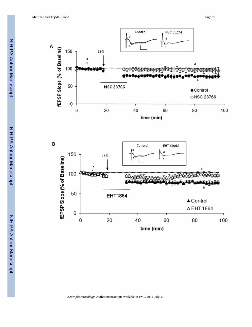
Another major form of long-term synaptic plasticity in the mammalian brain is long-termdepression (LTD). LTD expression involves also formation and elimination of synapticstructures and appears to have also a role in learning and memory. Thus, it is reasonable tohypothesize that LTD-associated structural changes might be mediated by proteins thatmodulate the remodeling of the actin cytoskeleton, such as Rac1. As for LTP, some forms ofLTD are triggered by synaptic activation and dependent on the activation of the NMDAreceptor. Consequently, we next examined whether Rac1 is also involved in modulation ofLTD by inducing depotentiation in hippocampal slices treated with Rac1 inhibitors. NMDAreceptor-dependent LTD was induced by low frequency stimulation (LFS) delivered at 1 Hzfor 15 min. This LTD protocol produced a significant lasting decrease in fEPSP slope incontrol slices perfused with ACSF. However, perfusion of slices with either NSC23766 (30µM; Figure 7A, p<0.05) or EHT1864 (60 µM; Figure 7B, p<0.05) significantly impairedLTD induction.
A second major form of LTD requires mGluR receptors. This form of LTD can bechemically induced by application of the group I mGluR agonist 3,5-dihydroxypehnylglycine (DHPG). LTD was induced in hippocampal slices with 100 µMDHPG for 5 min. All experiments were performed in the presence of the N-methyl-D-aspartate inhibitor APV (100 µM). In this case, perfusion of slices with NSC23766 (30 µM)or EHT1864 (60 µM), even though it induced LTD, did not provoke the long-lasting
Martinez and Tejada-Simon Page 6
Neuropharmacology. Author manuscript; available in PMC 2012 July 1.
NIH
-PA Author Manuscript
NIH
-PA Author Manuscript
NIH
-PA Author Manuscript
depression of synaptic transmission seen in slices perfused with ACSF and used as controls(Figure 7C and 7D). Our results show that upon pharmacological inhibition of Rac1,mGluR-dependent LTD was altered but at a lower level than the effect reported for NMDAreceptor-dependent LTD.
4. DISCUSSIONRac1 signaling pathway has been implicated in learning (Diana et al., 2007; Haditsch et al.,2009; Martinez et al., 2007), as well as some forms of cognitive disorders and X-linkedmental retardation syndromes (Chelly and Mandel, 2001). These cognitive disorders areassociated with abnormalities in dendritic spine structure, which is dependent of the actincytoskeleton regulated by Rac1. Previous finding from studies of animals in which LTP andLTD were impaired as the result of either genetic defects, lesions or pharmacologicaltreatments, suggest that LTP and LTD may be involved in certain types of hippocampal-dependent memory (Nakao et al., 2002; Naie and Manahan-Vaughan 2005; Schmitt et al.,2005). Thus, in this study we focused on elucidating the function of Rac1 in long-termsynaptic plasticity. We considered these experiments to be important in view of differentpsychiatric disorders featuring cognitive impairment, structural deficiencies and aberrantplasticity, where, experimentally, a link between these phenomena still needs to beestablished. For example, there is strong evidence that mGluR-LTD is involved in thepathogenesis of Fragile X syndrome (FXS), as hippocampal plasticity is abnormal in amouse model for this cognitive disorder (Huber et al., 2002). These mice, as well as FXSpatients also present abnormally long, thin dendritic spines (Comery et al., 1997; Irwin etal., 2000), as well as defects in cognition (Comery et al., 1997; Galvez and Greenough,2005; Irwin et al., 2000; Purpura, 1974) that could be caused by the aberrant expression ofLTD reported. Likewise, alterations in neuroplasticity as well as behavioral abnormalitieshave been seen in other psychiatric disorders such as depression and schizophrenia (Elhardtet al., 2010).
Our work herein shows that the small GTPase Rac1 has a critical role in long-term synapticplasticity. We have shown that LTP induction results in rapid Rac1 and PAK1 activation,which can be blocked by the inhibition of NMDA receptor activation. Pharmacologicalinhibition or Rac1 showed a pronounced deficit in long-term potentiation as well as long-term depression deficit at CA1 neurons, without altering receptor activation and calciuminflux (as indicated by unaffected CaMKII), events acting upstream of Rac1 in the inductionof long-term plasticity.
Studies by our laboratory and others have employed a genetic approach, investigatingsynaptic plasticity in Rac1 deficient mice (Haditsch et al., 2009). The results of these studiesare in agreement with our pharmacology experiments, suggesting that Rac1 is associated andnecessary for the induction of long-term plasticity at CA1 area synapses.
For the first time to our knowledge, two agents that specifically inhibit Rac1 have been usedin electrophysiological experiments to study the role of Rac1 in long-term plasticity in themouse brain. Independent studies showed that their effects were dose dependent andreversible. It is believed that these two drugs affect the activation of Rac1 in different butpossibly complementary ways; one by hindering the interaction of Rac1 with its guanineexchange factors (GEFs) and two by unloading GTP from the activated form of Rac1. GEFproteins typically have the highest affinity for unloaded GTPases (Rac1-GDP), thus thesetwo drugs may have competing mechanisms. Theoretically, GEF affinity could be increasedby EHT1864 unloading the nucleotide GTP. Alternatively, NSC23766 could have greateraffinity for Rac1 after the nucleotide has been unloaded. If this is true, the concomitant use
Martinez and Tejada-Simon Page 7
Neuropharmacology. Author manuscript; available in PMC 2012 July 1.
NIH
-PA Author Manuscript
NIH
-PA Author Manuscript
NIH
-PA Author Manuscript
of both inhibitors could produce a higher and prolonged effect regarding Rac1 outcome insynaptic plasticity. Studies are on the way to study possible synergism of these drugs.
AcknowledgmentsThis work was supported by generous grants from the FRAXA Research Foundation, the Jérôme LeJeuneFoundation and the National Institutes of Health Grant NS48037 (M.V.T.S). We thank Dr. Lindsay Schwarz for herthoughtful commentaries and help editing this manuscript.
REFERENCESBradford MM. A rapid and sensitive method for the quantitation of microgram quantities of protein
utilizing the principle of protein-dye binding. Anal.Biochem. 1976; 72:248–254. [PubMed: 942051]Caron E. Rac signalling: a radical view. Nat Cell Biol. 2003; 5:185–187. [PubMed: 12646870]Chelly J, Mandel JL. Monogenic causes of X-linked mental retardation. Nat. Rev. Gen. 2001; 2:669–
680.Collingridge GL, Kehl SJ, McLennan H. Excitatory amino acids in synaptic transmission in the
Schaffer collateral-commissural pathway of the rat hippocampus. J Physiol. 1983; 334:33–46.[PubMed: 6306230]
Comery TA, Harris JB, Willems PJ, Oostra BA, Irwin SA, Weiler IJ, Greenough WT. Abnormaldendritic spines in fragile X knockout mice: Maturation and pruning deficits. Proc. Natl. Acad. Sci.USA. 1997; 94:5401–5404. [PubMed: 9144249]
Diana G, Valentini G, Travaglione S, Falzano L, Pieri M, Zona C, Meschini S, Fabbri A, Fiorentini C.Enhancement of learning and memory after activation of cerebral Rho GTPases. Proc. Natl. Acad.Sci. USA. 2007; 104(2):636–641. [PubMed: 17202256]
Dickson BJ. Rho GTPases in growth cone guidance. Curr Opi Neurobiol. 2001; 11:103–110.Elhardt M, Martinez LA, Tejada-Simon MV. Neurochemical, behavioral and architectural changes
after chronic inactivation of NMDA receptors in mice. Neurosci. Lett. 2010; 468(2):166–171.[PubMed: 19895868]
Galvez R, Greenough WT. Sequence of abnormal dendritic spine development in primarysomatosensory cortex of a mouse model of the fragile X mental retardation syndrome. Am.J. Med.Genet. 2005; 135:155–160. [PubMed: 15880753]
Gao Y, Dickerson JB, Guo F, Zheng J, Zheng Y. Rational design and characterization of a RacGTPase-specific small molecule inhibitor. Proc. Natl. Acad. Sci. USA. 2004; 101(20):7618–7623.[PubMed: 15128949]
Haditsch U, Leone DP, Farinelli M, Chrostek-Grashoff A, Brakebusch C, Mansuy IM, McConnell SK,Palmer TD. A central role for the small GTPase Rac1 in hippocampal plasticity and spatiallearning and memory. Mol. Cell. Neurosci. 2009; 41:409–419. [PubMed: 19394428]
Harris KM, Kater SB. Dendritic spines: cellular specializations imparting both stability and flexibilityto synaptic function. Annu Rev Neurosci. 1994; 17:341–371. [PubMed: 8210179]
Huber KM, Gallagher SM, Warren ST, Bear MF. Altered synaptic plasticity in a mouse model offragile X mental retardation. Proc Natl. Acad. Sci USA. 2002; 99:7746–7750. [PubMed:12032354]
Humeau Y, Popoff MR, Kojima H, Doussau F, Poulain B. Rac GTPase plays an essential role inexocytosis by controlling the fusion competence of release sites. J Neurosci. 2002; 22:7968–7981.[PubMed: 12223550]
Irwin SA, Galvez R, Greenough WT. Dendritic Spine Structural Anomalies in Fragile-X MentalRetardation Syndrome. Cereb. Cortex. 2000; 10:1038–1044. [PubMed: 11007554]
Luo L. Rho GTPases in neuronal morphogenesis. Nature Rev Neurosci. 2000; 1:173–180. [PubMed:11257905]
Martinez LA, Klann E, Tejada-Simon MV. Translocation and activation of Rac in the hippocampusduring associative contextual fear learning. Neurobiol. Learn. Mem. 2007; 88(1):104–113.[PubMed: 17363298]
Martinez and Tejada-Simon Page 8
Neuropharmacology. Author manuscript; available in PMC 2012 July 1.
NIH
-PA Author Manuscript
NIH
-PA Author Manuscript
NIH
-PA Author Manuscript
Naie K, Manahan-Vaughan DP. Pharmacological antagonism of metabotropic glutamate receptor 1regulates lont-term potentiation and spatial reference memory in the dentate gyrus of freelymoving rats via N-methyl-D-aspartate and metabotropic glutamate receptor-dependentmechanisms. Eur. J. Neurosci. 2005; 21:411–421. [PubMed: 15673440]
Nakao K, Ikegaya Y, Yamada MK, Nishiyama N, Natsuki N. Hippocampal long-term depression as anindex of satial working memory. Eur. J. Neurosci. 2002; 16:970–974. [PubMed: 12372034]
Nikolic M, Chernoff J. High midsummer for small GTPases. Trends Cell Biol. 2002; 12:495–497.[PubMed: 12446103]
Purpura DP. Dendritic spine "Dysgenesis" and mental retardation. Science. 1974; 186:1126–1128.[PubMed: 4469701]
Ridley AJ. Rho family proteins: coordinating cell responses. Trends Cell Biol. 2001a; 11:471–477.[PubMed: 11719051]
Ridley AJ. Rho GTPases and cell migration. J Cell Sci. 2001b; 114:2713–2722. [PubMed: 11683406]Schmitt WB, Sprengel R, Mack V, Draft RW, Seeburg PH, Deacon RM, Rawlins JN, Bannerman DM.
Restoration of spatial working memory by genetic rescue of GluR-A-deficient mice. Nat.Neurosci. 2005; 8:270–272. [PubMed: 15723058]
Shutes A, Onesto C, Picard V, Leblond B, Schweighoffer F. Specificity and mechanism of action ofEHT 1864, a novel small molecule inhibitor of Rac family small GTPases. J. Biol. Chem. 2007;282:35666–35678. [PubMed: 17932039]
Sin WC, Haas K, Ruthazer ES, Cline HT. Dendrite growth increased by visual activity requiresNMDA receptor and Rho GTPases. Nature. 2002; 419:475–480. [PubMed: 12368855]
Tejada-Simon MV, Villasana LE, Serrano F, Klann E. NMDA receptor activation inducestranslocation and activation of Rac in mouse hippocampal area CA1. Biochem Bioph Res Comm.2006; 343:504–512.
Van Aelst L, D'Souza-Schorey C. Rho GTPases and signaling networks. Genes & Develop. 1997;11:2295–2322. [PubMed: 9308960]
Waetzig V, Herdegen T. A single c-Jun N-terminal kinase isoform (JNK3-p54) is an effector in bothneuronal differentiation and cell death. J Biol Chem. 2003; 278:567–572. [PubMed: 12401814]
Martinez and Tejada-Simon Page 9
Neuropharmacology. Author manuscript; available in PMC 2012 July 1.
NIH
-PA Author Manuscript
NIH
-PA Author Manuscript
NIH
-PA Author Manuscript
Figure 1. Activation of Rac1 is associated with LTPA. Hippocampal slices were stimulated at CA3 afferents and responses were recorded fromarea CA1 dendrites. Four trains of high frequency stimulation (HFS, 1 sec at 100 Hz spaced5 min apart; indicated by arrows) induces LTP in hippocampal slices. Right: shows fEPSPsample traces recorded during baseline (dashed line, a) and 2hrs after fourth HFS (solid line,b). Scale 1mV/5ms. n=7. B. LTP is associated with NMDA receptor activation. Wildtypehippocampal slices were subjected to four trains of HFS (1 sec at 100 Hz spaced 5 minapart; indicated by arrows) in presence of APV (100 µM), an NMDA receptor antagonistthat blocks LTP (n=6). C. At various time points (2, 6, 10 and 120 min) after the fourthstimulation leading to LTP, hippocampal slices were collected, area CA1 dissected,
Martinez and Tejada-Simon Page 10
Neuropharmacology. Author manuscript; available in PMC 2012 July 1.
NIH
-PA Author Manuscript
NIH
-PA Author Manuscript
NIH
-PA Author Manuscript
homogenized and assayed for Rac1 activation. Representative western blot depictingexpression of Rac1-GTP, total Rac1, phospho-PAK1 and total PAK1 in stimulated (+) andunstimulated (−) slices subjected to HFS while treated or not with APV (100 µM). n=6. D.Data quantification (mean ± SEM, n=6) representing expression of Rac1-GTP and phospho-PAK1 after HFS. * denotes statistically significance respect to unstimulated slices (p<0.05,Student t-test).
Martinez and Tejada-Simon Page 11
Neuropharmacology. Author manuscript; available in PMC 2012 July 1.
NIH
-PA Author Manuscript
NIH
-PA Author Manuscript
NIH
-PA Author Manuscript
Figure 2. Activation of Rac1 is necessary for induction of long-term potentiation (LTP)Effect of (A) NSC23766 and (B) EHT 1864 on baseline synaptic transmission. Baselineresponses were recorded for 30 min before slices were perfused for 50 min with NSC23766(30 µM) or EHT1864 (60 µM) (n=12). Responses were recorded for an additional 20–30min after washout of the drug. C. Four trains of HFS (1 sec at 100 Hz; indicated by arrows)spaced 5 min apart induced LTP in vehicle-treated (ACSF) control hippocampal slices (),but was attenuated (p<0.05) when HFS was delivered in the presence of NSC23766 (30 µM;[]). Inset: Sample traces recorded during baseline (dashed lines, a, c) and 2hrs post-HFS(solid lines, b, d). Scale 1mV/5ms. D. Representative immunoblots depicting expression of
Martinez and Tejada-Simon Page 12
Neuropharmacology. Author manuscript; available in PMC 2012 July 1.
NIH
-PA Author Manuscript
NIH
-PA Author Manuscript
NIH
-PA Author Manuscript
phospho-CaMKII and total CaMKII in vehicle- and NSC23766-treated slices after HFSstimulation. (n=7).
Martinez and Tejada-Simon Page 13
Neuropharmacology. Author manuscript; available in PMC 2012 July 1.
NIH
-PA Author Manuscript
NIH
-PA Author Manuscript
NIH
-PA Author Manuscript
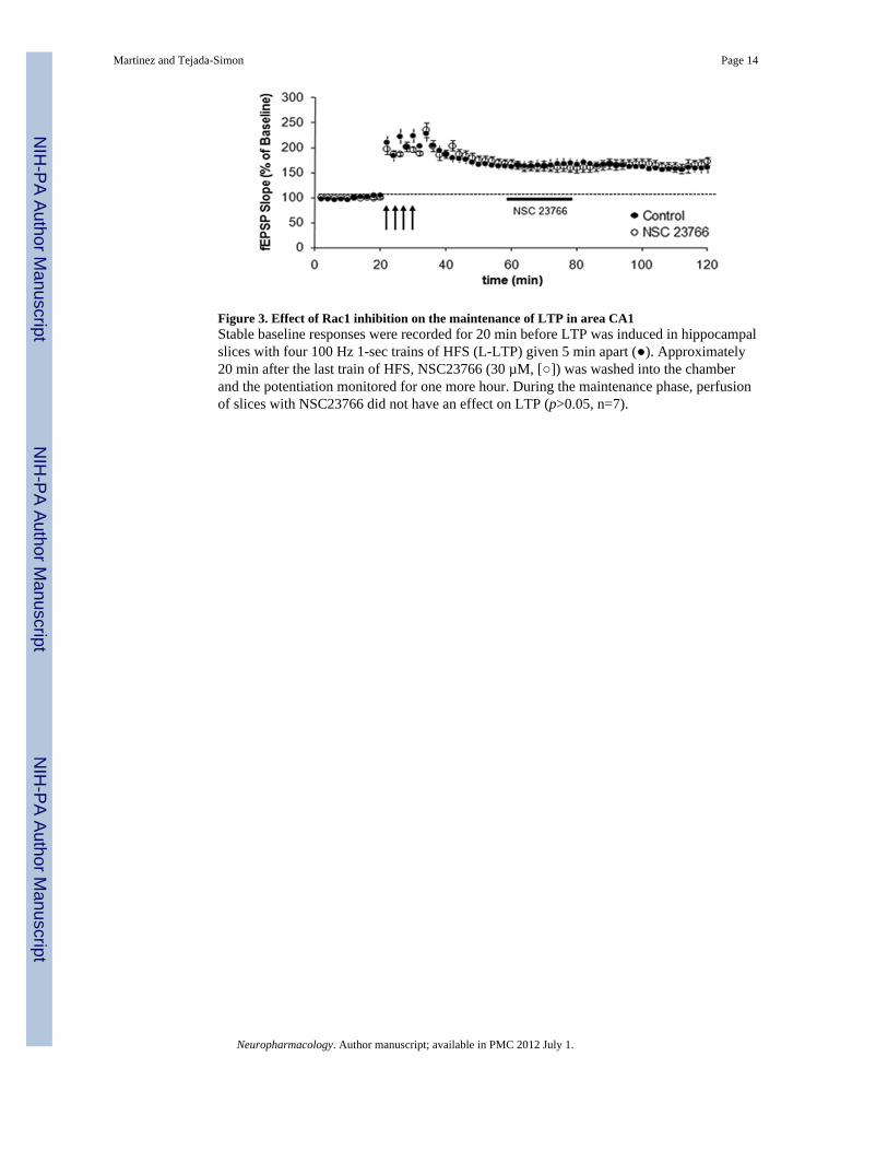
Figure 3. Effect of Rac1 inhibition on the maintenance of LTP in area CA1Stable baseline responses were recorded for 20 min before LTP was induced in hippocampalslices with four 100 Hz 1-sec trains of HFS (L-LTP) given 5 min apart (). Approximately20 min after the last train of HFS, NSC23766 (30 µM, []) was washed into the chamberand the potentiation monitored for one more hour. During the maintenance phase, perfusionof slices with NSC23766 did not have an effect on LTP (p>0.05, n=7).
Martinez and Tejada-Simon Page 14
Neuropharmacology. Author manuscript; available in PMC 2012 July 1.
NIH
-PA Author Manuscript
NIH
-PA Author Manuscript
NIH
-PA Author Manuscript
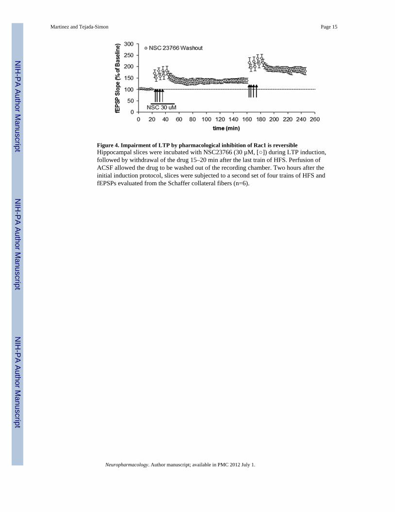
Figure 4. Impairment of LTP by pharmacological inhibition of Rac1 is reversibleHippocampal slices were incubated with NSC23766 (30 µM, []) during LTP induction,followed by withdrawal of the drug 15–20 min after the last train of HFS. Perfusion ofACSF allowed the drug to be washed out of the recording chamber. Two hours after theinitial induction protocol, slices were subjected to a second set of four trains of HFS andfEPSPs evaluated from the Schaffer collateral fibers (n=6).
Martinez and Tejada-Simon Page 15
Neuropharmacology. Author manuscript; available in PMC 2012 July 1.
NIH
-PA Author Manuscript
NIH
-PA Author Manuscript
NIH
-PA Author Manuscript
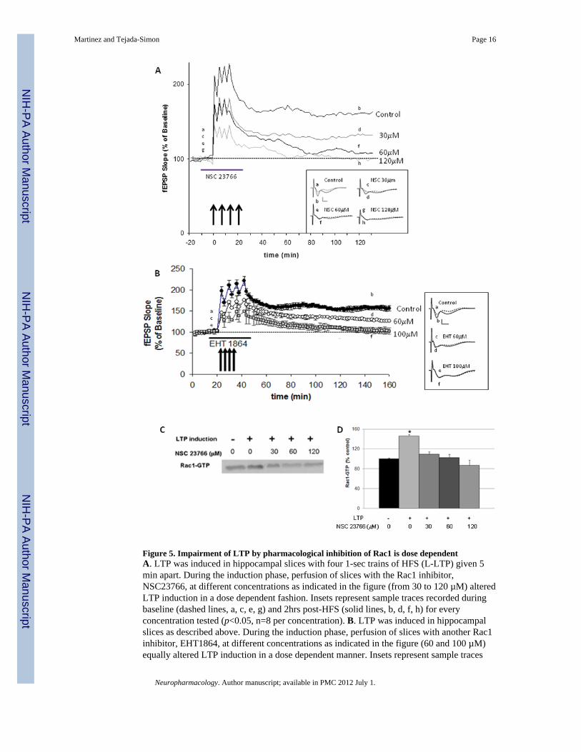
Figure 5. Impairment of LTP by pharmacological inhibition of Rac1 is dose dependentA. LTP was induced in hippocampal slices with four 1-sec trains of HFS (L-LTP) given 5min apart. During the induction phase, perfusion of slices with the Rac1 inhibitor,NSC23766, at different concentrations as indicated in the figure (from 30 to 120 µM) alteredLTP induction in a dose dependent fashion. Insets represent sample traces recorded duringbaseline (dashed lines, a, c, e, g) and 2hrs post-HFS (solid lines, b, d, f, h) for everyconcentration tested (p<0.05, n=8 per concentration). B. LTP was induced in hippocampalslices as described above. During the induction phase, perfusion of slices with another Rac1inhibitor, EHT1864, at different concentrations as indicated in the figure (60 and 100 µM)equally altered LTP induction in a dose dependent manner. Insets represent sample traces
Martinez and Tejada-Simon Page 16
Neuropharmacology. Author manuscript; available in PMC 2012 July 1.
NIH
-PA Author Manuscript
NIH
-PA Author Manuscript
NIH
-PA Author Manuscript
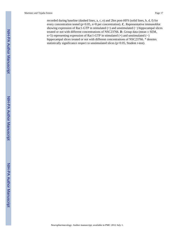
recorded during baseline (dashed lines, a, c, e) and 2hrs post-HFS (solid lines, b, d, f) forevery concentration tested (p<0.05, n=8 per concentration). C. Representative immunoblotshowing expression of Rac1-GTP in stimulated (+) and unstimulated (−) hippocampal slicestreated or not with different concentrations of NSC23766. D. Group data (mean ± SEM,n=5) representing expression of Rac1-GTP in stimulated (+) and unstimulated (−)hippocampal slices treated or not with different concentrations of NSC23766. * denotesstatistically significance respect to unstimulated slices (p<0.05, Student t-test).
Martinez and Tejada-Simon Page 17
Neuropharmacology. Author manuscript; available in PMC 2012 July 1.
NIH
-PA Author Manuscript
NIH
-PA Author Manuscript
NIH
-PA Author Manuscript
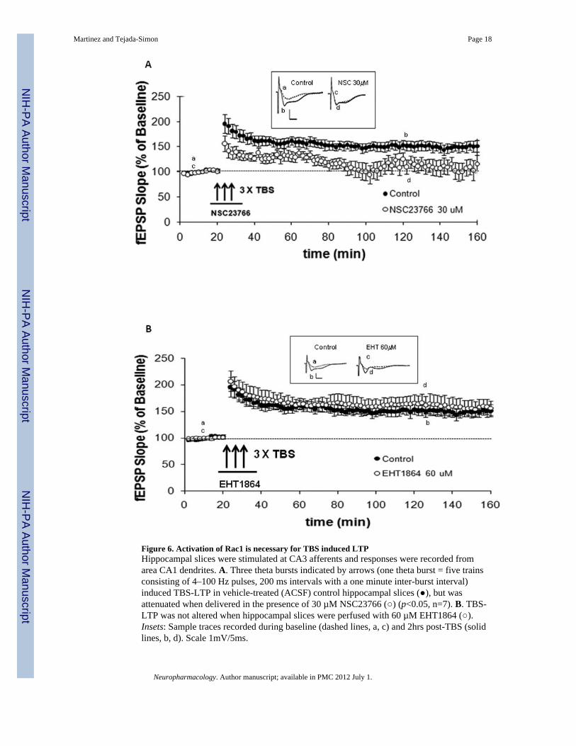
Figure 6. Activation of Rac1 is necessary for TBS induced LTPHippocampal slices were stimulated at CA3 afferents and responses were recorded fromarea CA1 dendrites. A. Three theta bursts indicated by arrows (one theta burst = five trainsconsisting of 4–100 Hz pulses, 200 ms intervals with a one minute inter-burst interval)induced TBS-LTP in vehicle-treated (ACSF) control hippocampal slices (), but wasattenuated when delivered in the presence of 30 µM NSC23766 () (p<0.05, n=7). B. TBS-LTP was not altered when hippocampal slices were perfused with 60 µM EHT1864 ().Insets: Sample traces recorded during baseline (dashed lines, a, c) and 2hrs post-TBS (solidlines, b, d). Scale 1mV/5ms.
Martinez and Tejada-Simon Page 18
Neuropharmacology. Author manuscript; available in PMC 2012 July 1.
NIH
-PA Author Manuscript
NIH
-PA Author Manuscript
NIH
-PA Author Manuscript
Martinez and Tejada-Simon Page 19
Neuropharmacology. Author manuscript; available in PMC 2012 July 1.
NIH
-PA Author Manuscript
NIH
-PA Author Manuscript
NIH
-PA Author Manuscript
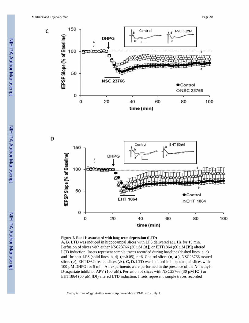
Figure 7. Rac1 is associated with long-term depression (LTD)A, B. LTD was induced in hippocampal slices with LFS delivered at 1 Hz for 15 min.Perfusion of slices with either NSC23766 (30 µM [A]) or EHT1864 (60 µM [B]) alteredLTD induction. Insets represent sample traces recorded during baseline (dashed lines, a, c)and 1hr post-LFS (solid lines, b, d). (p<0.05), n=6. Control slices (, ), NSC23766 treatedslices (), EHT1864 treated slices (). C, D. LTD was induced in hippocampal slices with100 µM DHPG for 5 min. All experiments were performed in the presence of the N-methyl-D-aspartate inhibitor APV (100 µM). Perfusion of slices with NSC23766 (30 µM [C]) orEHT1864 (60 µM [D]) altered LTD induction. Insets represent sample traces recorded
Martinez and Tejada-Simon Page 20
Neuropharmacology. Author manuscript; available in PMC 2012 July 1.
NIH
-PA Author Manuscript
NIH
-PA Author Manuscript
NIH
-PA Author Manuscript
during baseline (dashed lines, a, c) and 1hr post-DHPG (solid lines, b, d). (p<0.05, n=6).Control slices (), NSC23766 treated slices (), EHT1864 treated slices ().
Martinez and Tejada-Simon Page 21
Neuropharmacology. Author manuscript; available in PMC 2012 July 1.
NIH
-PA Author Manuscript
NIH
-PA Author Manuscript
NIH
-PA Author Manuscript


