Embed Size (px)
Citation preview
HAL Id: hal-02333382https://hal-amu.archives-ouvertes.fr/hal-02333382
Submitted on 25 Oct 2019
HAL is a multi-disciplinary open accessarchive for the deposit and dissemination of sci-entific research documents, whether they are pub-lished or not. The documents may come fromteaching and research institutions in France orabroad, or from public or private research centers.
L’archive ouverte pluridisciplinaire HAL, estdestinée au dépôt et à la diffusion de documentsscientifiques de niveau recherche, publiés ou non,émanant des établissements d’enseignement et derecherche français ou étrangers, des laboratoirespublics ou privés.
Distributed under a Creative Commons Attribution| 4.0 International License
Allosteric regulation of a prokaryotic small Ras-likeGTPase contributes to cell polarity oscillations in
bacterial motilityJyoti Baranwal, Sebastien Lhospice, Manil Kanade, Sukanya Chakraborty,
Priyanka Rajendra Gade, Shrikant Harne, Julien Herrou, Tam Mignot,Pananghat Gayathri
To cite this version:Jyoti Baranwal, Sebastien Lhospice, Manil Kanade, Sukanya Chakraborty, Priyanka Rajendra Gade,et al.. Allosteric regulation of a prokaryotic small Ras-like GTPase contributes to cell polarity os-cillations in bacterial motility. PLoS Biology, Public Library of Science, 2019, 17 (9), pp.e3000459.�10.1371/journal.pbio.3000459�. �hal-02333382�
RESEARCH ARTICLE
Allosteric regulation of a prokaryotic small
Ras-like GTPase contributes to cell polarity
oscillations in bacterial motility
Jyoti BaranwalID1, Sebastien Lhospice2, Manil Kanade1, Sukanya Chakraborty1, Priyanka
Rajendra Gade1, Shrikant HarneID1, Julien Herrou2, Tam Mignot2, Pananghat GayathriID
1*
1 Indian Institute of Science Education and Research, Pune, India, 2 CNRS-Aix Marseille University,
Laboratoire de Chimie Bacterienne, Marseille, France
Abstract
Mutual gliding motility A (MglA), a small Ras-like GTPase; Mutual gliding motility B (MglB),
its GTPase activating protein (GAP); and Required for Motility Response Regulator
(RomR), a protein that contains a response regulator receiver domain, are major compo-
nents of a GTPase-dependent biochemical oscillator that drives cell polarity reversals in the
bacterium Myxococcus xanthus. We report the crystal structure of a complex of M. xanthus
MglA and MglB, which reveals that the C-terminal helix (Ct-helix) from one protomer of the
dimeric MglB binds to a pocket distal to the active site of MglA. MglB increases the GTPase
activity of MglA by reorientation of key catalytic residues of MglA (a GAP function) combined
with allosteric regulation of nucleotide exchange by the Ct-helix (a guanine nucleotide
exchange factor [GEF] function). The dual GAP-GEF activities of MglB accelerate the rate
of GTP hydrolysis over multiple enzymatic cycles. Consistent with its GAP and GEF activi-
ties, MglB interacts with MglA bound to either GTP or GDP. The regulation is essential for
cell polarity, because deletion of the Ct-helix causes bipolar localization of MglA, MglB, and
RomR, thereby causing reversal defects in M. xanthus. A bioinformatics analysis reveals
the presence of Ct-helix in homologues of MglB in other bacterial phyla, suggestive of the
prevalence of the allosteric mechanism among other prokaryotic small Ras-like GTPases.
Introduction
Small Ras-like GTPases are ubiquitous in eukaryotes and perform varied functions, including
cell signaling and motility. Recently, such GTPases have been identified in prokaryotes too
[1,2]. The soil bacterium Myxococcus xanthus exhibits 2 types of motility—adventurous gliding
motility and social motility, which are essential for its normal life cycle [3]. The localization of
motility complexes, and thereby the direction of movement, is determined by an oscillatory
system, which includes Mutual gliding motility A (MglA), a small Ras-like GTPase, and
Mutual gliding motility B (MglB), its GTPase activating protein (GAP) as major components
[1,4,5]. Mutual gliding motility (mgl) was identified as a locus essential for both social and
PLOS Biology | https://doi.org/10.1371/journal.pbio.3000459 September 27, 2019 1 / 30
a1111111111
a1111111111
a1111111111
a1111111111
a1111111111
OPEN ACCESS
Citation: Baranwal J, Lhospice S, Kanade M,
Chakraborty S, Gade PR, Harne S, et al. (2019)
Allosteric regulation of a prokaryotic small Ras-like
GTPase contributes to cell polarity oscillations in
bacterial motility. PLoS Biol 17(9): e3000459.
https://doi.org/10.1371/journal.pbio.3000459
Academic Editor: Ann M. Stock, Rutgers
University-Robert Wood Johnson Medical School,
UNITED STATES
Received: May 29, 2019
Accepted: September 4, 2019
Published: September 27, 2019
Copyright: © 2019 Baranwal et al. This is an open
access article distributed under the terms of the
Creative Commons Attribution License, which
permits unrestricted use, distribution, and
reproduction in any medium, provided the original
author and source are credited.
Data Availability Statement: Numerical data for all
the graphs reported in the paper are given in S1
Data. The coordinates of the crystal structures
reported in the manuscript have been deposited in
the PDB and the accession numbers are 5YMX
(MxMglA–GDP) and 6IZW (MxMglAB–GTPγS).
Funding: PG acknowledges by funding from
Department of Science and Technology (DST)
Innovation in Science Pursuit for Inspired Research
(INSPIRE) Faculty Research Grant (IFA12-LSBM-
adventurous motilities in M. xanthus, comprising the 2 genes mglA and mglB [1,3]. Studies on
MglA and MglB showed that these proteins exhibit a mutually exclusive localization pattern
[4,6,7]. MglA localizes to the leading pole presumably in its GTP-bound state [4], whereas
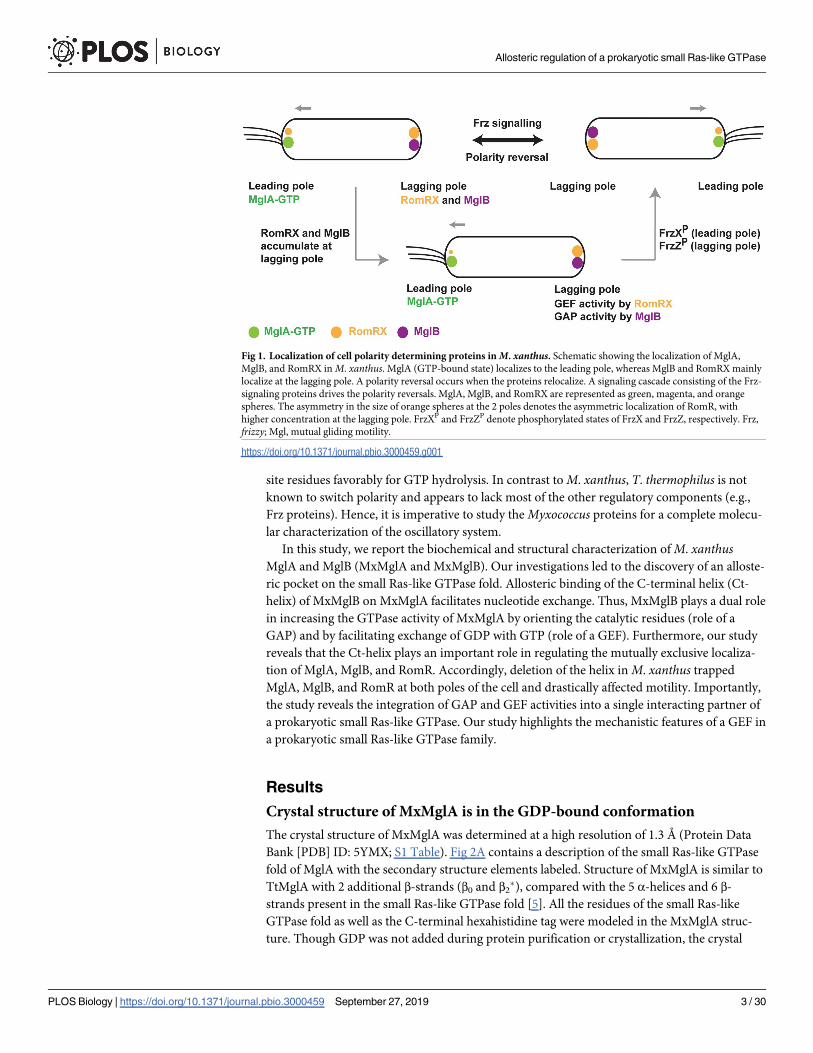
MglB localizes at the lagging pole (Fig 1). These studies [1,3–7] established the role of the pro-
karyotic small Ras-like GTPase in cell polarity determination and motility, which are functions
analogous to its eukaryotic counterparts [8,9].
A remarkable feature of Myxococcus motility is the frequent reversals in its cell polarity in
response to environmental cues [10]. M. xanthus changes its direction of movement by swap-
ping the leading and lagging poles. The reversal frequency is regulated by the chemosensory-
like frizzy (Frz) pathway, which relays environmental signals to the downstream MglA-MglB
polarity control system [11–13] (Fig 1). High-resolution time-lapse experiments revealed that
MglA and MglB relocalize sequentially during a reversal [4,7,11]. Importantly, the Frz system
does not establish polarity, but its activity is required for inversion of the polarity axis. Another
protein involved in the polarity control module is RomR, a protein with a response regulator
receiver-like domain [14,15]. It was recently shown that RomR and its interacting partner
RomX are essential for recruiting MglA-GTP to the poles [16]. Frz-signaling and RomR stimu-
lates the pole-to-pole exchange of MglA and MglB [11,13–15]. All together, these proteins
form a so-called gated relaxation oscillator, which functions to drive the polarity reversals in
response to environmental signals [11].
Regulated reversals are centrally controlled by the MglA GTPase cycle. Though it is estab-
lished that MglB functions as a GAP for MglA [4,7], proteins that function as guanine nucleo-
tide exchange factors (GEFs) [17] for MglA were not identified until very recently [16]. The
complex of RomR and RomX has been recently demonstrated to function as a GEF for MglA
[16]. In vivo, the RomRX complex recruits MglA to the leading pole at the time of reversal.
Between reversals, RomRX dissociates slowly and relocalizes to the lagging cell pole where
RomR interacts with MglB [14,15]. Thus, both GAP and GEF activities accumulate at the lag-
ging cell pole, preparing the next reversal event (Fig 1). However, at this stage, the cell will only
reverse if signals from Frz are sufficiently strong. When this occurs, 2 response regulators
FrzXP (phosphorylated FrzX) and FrzZP (phosphorylated FrzZ) bind to opposite poles, which
trigger the detachment of MglA from the leading cell pole and its relocalization to the lagging
cell pole. MglB is subsequently detached and relocated to the opposite pole, completing the
switch.
Many questions remain unaddressed concerning the exact sequence of events that take
place at the poles. Specifically, the presence of antagonizing GAP and GEF activities at both
cell poles requires that this balance be regulated. The accumulation of RomRX at the lagging
pole is not sufficient to drag MglA to the lagging cell pole, most likely because the MglB GAP
activity predominates. Accumulation of FrzXP at the lagging pole could switch this balance in
favor of the GEF and provoke the switch. In this study, we show that MglB is in fact a bifunc-
tional enzyme and that MglB regulation could be key for the control of this enzymatic balance
at the pole.
Previously, biochemical and structural studies with homologous proteins from Thermusthermophilus demonstrated that MglB is the GAP of MglA [4,5]. MglA and MglB from T. ther-mophilus (TtMglA and TtMglB) share 62% and 29% sequence identities, respectively, with the
M. xanthus counterparts. Unlike the majority of small Ras-like GTPases, both the active site
residues of the GTPase were found within TtMglA itself (Arg53A and Gln82A; residue names
with superscript “A” denote residues of MglA) instead of being contributed by the GAP [5]. A
dimer of TtMglB interacts with a monomer of TtMglA. The structural studies revealed that β2
strand of TtMglA undergoes a screw-type movement during the interaction between TtMglA
and TtMglB in the presence of GTP analogues [5]. The β-screw movement positions the active
Allosteric regulation of a prokaryotic small Ras-like GTPase
PLOS Biology | https://doi.org/10.1371/journal.pbio.3000459 September 27, 2019 2 / 30
52), Science and Engineering Research Board
(SERB) extramural grant (EMR/2014/00563),
SERB Women Excellence Award, Indian National
Science Academy (INSA) Young Scientist
Research Award, and Indo-French Centre for
Promotion of Advanced Research (CEFIPRA)
Collaborative Research Programme Grant. JB and
MK acknowledge Council of Scientific and
Industrial Research (CSIR) and INSPIRE
respectively for fellowships. TM acknowledges
CEFIPRA and Agence Nationale de la Recherche
(ANR) “BACTOCOMPASS”. The funders had no
role in study design, data collection and analysis,
decision to publish, or preparation of the
manuscript.
Competing interests: The authors have declared
that no competing interests exist.
Abbreviations: Ct-helix, C-terminal helix; FP,
fluorescent protein; Frz, frizzy; FrzXP,
phosphorylated FrzX; FrzZP, phosphorylated FrzZ;
GAP, GTPase activating protein; GEF, guanine
nucleotide exchange factor; GDP, guanosine di-
nucleotide; GppNHp, guanosine-5’-[(β,γ)-imido]
triphosphate; GNP, guanosine-5’-[(β,γ)-imido]
triphosphate; GTP, guanosine tri-nucleotide;
GTPγS, guanosine 5’-O-[gamma-thio]triphosphate;
HPLC, high pressure liquid chromatography; IAA,
isoamyl alcohol; LDH, lactate dehydrogenase;
mant, 2’/3’-O-(N-Methyl-anthraniloyl); mgl, mutual
gliding motility; MglA, Mutual gliding motility A;
MglB, Mutual gliding motility B; MxMglA, M.
xanthus MglA; MxMglB, M. xanthus MglB; nG,
neonGreen; PCR, Polymerase Chain Reaction;
PDB, Protein Data Bank; PFS, Perfect Focus
System; Pi, inorganic phosphate; PK, pyruvate
kinase; Rbl/LC7, Roadblock/LC7; RomR, Required
for Motility Response Regulator; SLIC, sequence-
and ligation-independent cloning; SMART, Simple
Modular Architecture Research Tool; TtMglA, T.
thermophilus MglA; TtMglB, T. thermophilus MglB;
WT, wild type; YFP, yellow fluorescent protein.
site residues favorably for GTP hydrolysis. In contrast to M. xanthus, T. thermophilus is not
known to switch polarity and appears to lack most of the other regulatory components (e.g.,
Frz proteins). Hence, it is imperative to study the Myxococcus proteins for a complete molecu-
lar characterization of the oscillatory system.
In this study, we report the biochemical and structural characterization of M. xanthusMglA and MglB (MxMglA and MxMglB). Our investigations led to the discovery of an alloste-
ric pocket on the small Ras-like GTPase fold. Allosteric binding of the C-terminal helix (Ct-
helix) of MxMglB on MxMglA facilitates nucleotide exchange. Thus, MxMglB plays a dual role
in increasing the GTPase activity of MxMglA by orienting the catalytic residues (role of a
GAP) and by facilitating exchange of GDP with GTP (role of a GEF). Furthermore, our study
reveals that the Ct-helix plays an important role in regulating the mutually exclusive localiza-
tion of MglA, MglB, and RomR. Accordingly, deletion of the helix in M. xanthus trapped
MglA, MglB, and RomR at both poles of the cell and drastically affected motility. Importantly,
the study reveals the integration of GAP and GEF activities into a single interacting partner of
a prokaryotic small Ras-like GTPase. Our study highlights the mechanistic features of a GEF in
a prokaryotic small Ras-like GTPase family.
Results
Crystal structure of MxMglA is in the GDP-bound conformation
The crystal structure of MxMglA was determined at a high resolution of 1.3 Å (Protein Data
Bank [PDB] ID: 5YMX; S1 Table). Fig 2A contains a description of the small Ras-like GTPase
fold of MglA with the secondary structure elements labeled. Structure of MxMglA is similar to
TtMglA with 2 additional β-strands (β0 and β2�), compared with the 5 α-helices and 6 β-
strands present in the small Ras-like GTPase fold [5]. All the residues of the small Ras-like
GTPase fold as well as the C-terminal hexahistidine tag were modeled in the MxMglA struc-
ture. Though GDP was not added during protein purification or crystallization, the crystal
Fig 1. Localization of cell polarity determining proteins in M. xanthus. Schematic showing the localization of MglA,
MglB, and RomRX in M. xanthus. MglA (GTP-bound state) localizes to the leading pole, whereas MglB and RomRX mainly
localize at the lagging pole. A polarity reversal occurs when the proteins relocalize. A signaling cascade consisting of the Frz-
signaling proteins drives the polarity reversals. MglA, MglB, and RomRX are represented as green, magenta, and orange
spheres. The asymmetry in the size of orange spheres at the 2 poles denotes the asymmetric localization of RomR, with
higher concentration at the lagging pole. FrzXP and FrzZP denote phosphorylated states of FrzX and FrzZ, respectively. Frz,
frizzy; Mgl, mutual gliding motility.
https://doi.org/10.1371/journal.pbio.3000459.g001
Allosteric regulation of a prokaryotic small Ras-like GTPase
PLOS Biology | https://doi.org/10.1371/journal.pbio.3000459 September 27, 2019 3 / 30
Allosteric regulation of a prokaryotic small Ras-like GTPase
PLOS Biology | https://doi.org/10.1371/journal.pbio.3000459 September 27, 2019 4 / 30
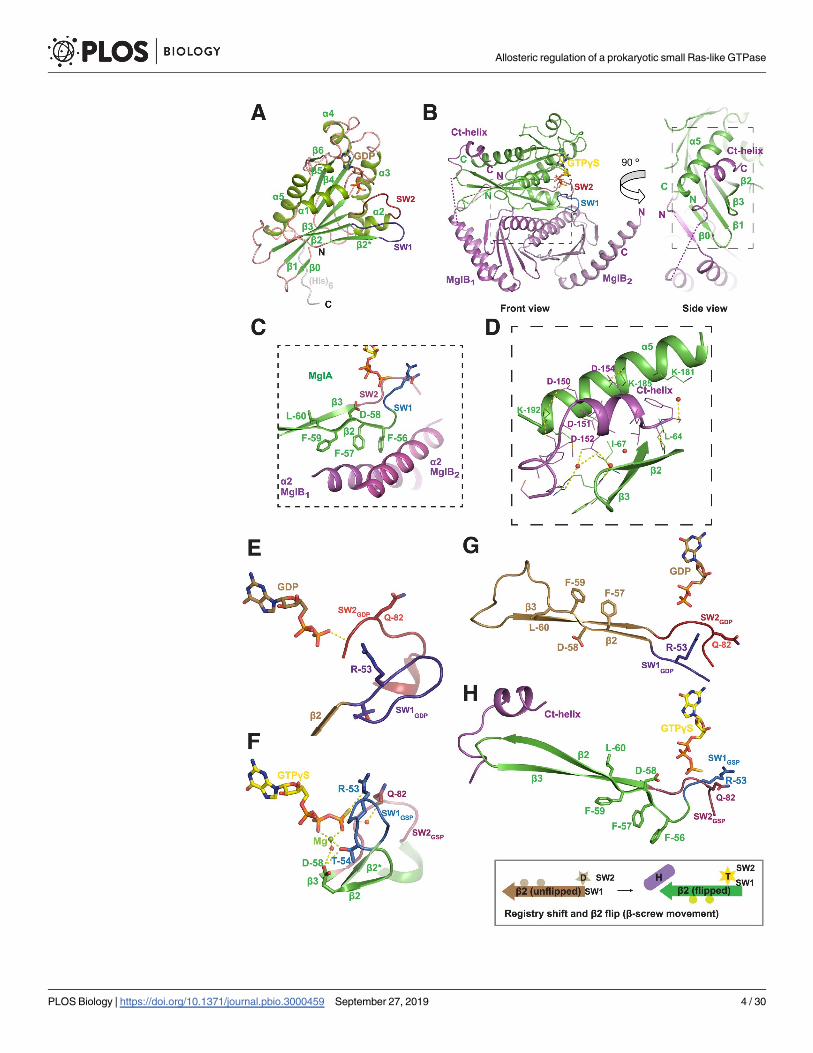
structure of MxMglA contained GDP in the nucleotide-binding pocket (Fig 2A, S1A Fig).
High-performance liquid chromatography (HPLC) analysis of the protein sample reconfirmed
that the bound ligand is GDP (S1A Fig, S1B Fig).
Structure of MxMglAB–GTPγS complex revealed a binding site for
MxMglB Ct-helix
Crystal structure of the complex of MxMglA and MxMglB in the presence of guanosine 5’-O-
[gamma-thio]triphosphate (GTPγS, a substrate analogue) (MxMglAB–GTPγS), was deter-
mined at a resolution of 2.4 Å (PDB ID: 6IZW; S1 Table). In the structure, a monomer of
MxMglA was bound to a dimer of MxMglB (Fig 2B). MxMglA interacts with the MxMglB
dimer through an extensive hydrophobic interface involving β2 strand of MxMglA and α2 heli-
ces of MxMglB monomers (Fig 2C), an interface similar to that of TtMglAB [5].
One of the novel features of the MxMglAB–GTPγS complex is the asymmetry between the
2 protomers of MxMglB (labeled MglB1 and MglB2; Fig 2B). MglB1 and MglB2 were asymmet-
ric at their N- and C-terminal residues in the MxMglAB–GTPγS complex. The core of
MxMglB, ranging from residues 10 to 131, belongs to the Roadblock/LC7 (Rbl/LC7) domain
fold [18]. In MglB1, the N-terminal amino acids (2–7) comprised a β-strand that formed
hydrogen bonds with the β0 strand of MxMglA, thus continuing the central β-sheet of the
MxMglA fold (Fig 2B). α1 helix of the Rbl/LC7 fold started from Glu11B (residue names with
superscript B denote residues of MglB) of MglB1. In contrast, in MglB2, residues 1 to 7 were
disordered, and the α1 helix comprised ordered residues starting from Tyr8B.
Fig 2. Crystal structures of M. xanthus MglA and MglAB–GTPγS complex. (A) Crystal structure of MxMglA bound to
GDP. The secondary structure elements are labeled. GDP is shown in stick representation in light brown, and the catalytic
loops switch 1 (blue) and switch 2 (red) are labeled as SW1 and SW2. The rest of the loops are shown in light pink, whereas
the secondary structure elements are shown in shades of green. The C-terminal hexa-histidine tag (His)6, which is ordered in
the crystal structure, is shown in gray. (B) Crystal structure of the complex of MxMglA (green) bound to GTPγS (yellow) and
MxMglB (magenta). “Front” view and a 90˚ rotated view (side view) are shown. The 2 protomers of MxMglB are labeled as
MglB1 and MglB2. The C-terminal helix of MxMglB (Ct-helix), the respective N- and C-terminal ends, switch 1 (blue; SW1)
and switch 2 (red; SW2), and relevant secondary structures of MxMglA are labeled. GTPγS is shown in stick representation
in yellow. Dotted magenta line connects the ends on either sides of the stretch of disordered residues in MglB1. Boxed
regions in the front and side view panels highlight the major interfaces between MxMglA with the Rbl/LC7 domain of
MxMglB (box with short dashes) and the Ct-helix of MxMglB (box with long dashes), respectively. (C) Residues of β2 strand
(Phe56A, Phe57A, and Phe59A) of MxMglA (green) form a hydrophobic surface that interacts with MxMglB (magenta). The
relevant secondary structure elements are labeled, and switch 1 and 2 (SW1 and SW2) are colored in blue and red,
respectively. The region corresponds to a zoomed view of the boxed region (short dashes) in the front view of panel B. (D)
The interacting interface of the Ct-helix of MxMglB (magenta) and MxMglA (green). The side chains of relevant interface
residues are shown in stick representation and labeled. Water molecules are represented as red spheres, and dotted yellow
lines represent hydrogen bond interactions. The region corresponds to a zoomed view of the boxed region (long dashes) in
the side view of panel B. (E and F) Conformational changes in switch 1 (SW1; 2 shades of blue) and switch 2 (SW2; 2 shades
of red) in the presence of MxMglB illustrates the mechanism of GAP activity (by reorientation of active site residues Arg53A
and Glu82A). MxMglA–GDP conformation and MglA from the MxMglAB–GTPγS complex are shown in panels E and F,
respectively. The 2 structures from a superposed orientation are shown as separate panels. Secondary structures extending
from the switch loops for MxMglA–GDP conformation and MglA from the MxMglAB–GTPγS complex are shown in brown
and green, respectively. Switch 1 (or 2) of the 2 structures are labeled SW1 (or 2) and SW1 (or 2) using GDP and GSP as
subscripts for the MxMglA–GDP and MxMglAB–GTPγS complexes, respectively. (G and H) Ct-helix of MxMglB (magenta)
and GTP (represented by GTPγS shown in stick representation) bind to the C- and N-terminal ends of the β2 strand of
MxMglA, respectively. MxMglA–GDP conformation and MglA from the MxMglAB–GTPγS complex are shown in panels G
and H, respectively. The 2 structures from a superposed orientation are shown as separate panels. The unflipped and flipped
states of the strand are shown (brown, MxMglA–GDP; green, MglA conformation in MxMglAB–GTPγS structure; switch 1
and switch 2 are in shades of blue and red, respectively). A schematic representation of the registry shift and β2 flip is shown
below, with the same color scheme. A 6-pointed yellow star and a 5-pointed light brown star represent GTP (labeled as “T’)
and GDP (labeled as “D”), respectively. The β2 strand is represented by an arrow. The residues that form the interface are
schematically shown by 2 circles on the bottom or top of the β2 strand representation in the flipped and unflipped states of
the strand, respectively. Ct-helix of MxMglB is schematically shown by a cylinder that is labeled “H.” Ct-helix, C-terminal
helix; GAP, GTPase activating protein; GTPγS, guanosine 5’-O-[gamma-thio]triphosphate; Mgl, mutual gliding motility;
MxMglA, M. xanthus MglA; MxMglB, M. xanthus MglB; Rbl, Roadblock; SW1, switch 1; SW2, switch 2.
https://doi.org/10.1371/journal.pbio.3000459.g002
Allosteric regulation of a prokaryotic small Ras-like GTPase
PLOS Biology | https://doi.org/10.1371/journal.pbio.3000459 September 27, 2019 5 / 30
At the C terminus, density was absent for residues 131 to146. However, residues 147 to 157
of the MglB1 protomer could be modeled into the electron density (S1D Fig, S1E Fig), which
adopted a helical conformation. Henceforth, this will be referred to as Ct-helix (C-terminal
helix of MxMglB). In the MglB2 protomer, electron density was absent for residues beyond
130. The Ct-helix of MglB1 bound to a pocket in MxMglA distal from the nucleotide-binding
site (Fig 2B and Fig 2D). The binding pocket in MxMglA was formed by the residues of the α5
helix, β2 strand, and the loop connecting β2 and β3 strands (β2–β3 loop) of MxMglA (inter-
switch region of small Ras-like GTPases [19]; Fig 2D). The corresponding β2 to β3 loop was
disordered in the TtMglAB structure [5] (S1C Fig).
The conformational features described below are consistent with the previously determined
structures of the corresponding complex from TtMglAB [5] (S1C Fig). Some of the prominent
features are the optimal orientation of the active site residues (Fig 2E, Fig 2F) and the β-screw
movement (Fig 2G, Fig 2H), which were also observed in the TtMglAB structures [5]. The con-
formational changes between MxMglA–GDP and the MxMglA–GTPγS bound to MxMglB
resulted in movements of switch 1 and switch 2 such that Arg53A and Gln82A of MxMglA are
now oriented towards the catalytic water (Fig 2E, Fig 2F). Another interesting observation is
the orientation of the residues Thr54A and Asp58A of MxMglA, which coordinate the Mg2+
ion bound to GTPγS (Fig 2F). The β-screw movement exposed hydrophobic residues of
MxMglA toward the MxMglB interface and facilitated MxMglAB interaction (Fig 2C, Fig 2H).
Ct-helix allosterically affected the GTPase activity
To elucidate the functional role of the MglB1 Ct-helix, we compared features of GTPase stimu-
lation and MxMglA interaction with full-length MxMglB (MglB) and a truncated version of
MxMglB lacking the Ct-helix (MglBΔCt; S1F Fig). The GTPase activity was monitored by esti-
mation of released GDP using a coupled enzyme assay [20] or phosphate release using mala-
chite green–based assay [21]. The reactions were carried out with excess GTP (substrate) and
hence measured multiple turnovers of the GTPase. Both approaches for monitoring enzyme
activity showed similar rates of hydrolysis. The GTPase activity of MxMglA on its own was
negligibly low, whereas the activity increased in the presence of MxMglB (Fig 3A, Fig 3B, Fig
3C, S2A Fig). This confirmed the role of MglB as a GAP, consistent with published results
[4,5,7]. Control experiments were performed with only MxMglB (no MxMglA) and excess of
MxMglB (S2A Fig). Similar GTPase activities were observed for MxMglA in the presence of
MxMglB with or without a C-terminal hexahistidine tag (Fig 3C), and hence all further experi-
ments were performed using the construct with C-terminal hexahistidine tag.
We found that the GTP hydrolysis rate was approximately 2-fold lower in the presence of
MxMglBΔCt compared with MxMglB (Fig 3A, Fig 3B, Fig 3C). The GTPase activity remained
low despite using 5 times excess of MxMglBΔCt (Fig 3A, Fig 3B), suggesting that the effect was
not due to a reduced affinity of MxMglBΔCt to MxMglA. We compared the GTPase activities
with that of the active site mutant MxMglAQ82L (referred to as MxMglAQ) in the presence of
MxMglB (S2B Fig, S2C Fig; Fig 3B). The GTPase activity of MxMglABΔCt was, however, higher
than that of MxMglAQB.
Next, we proceeded to confirm that the interactions observed in the crystal structure
between Ct-helix and the binding pocket of MxMglA are indeed relevant for the observed
increase in GTPase activity. Analysis of the crystal structure of MxMglAB–GTPγS complex
showed that Asp150B, Asp151B, and Asp152B on the MxMglB Ct-helix formed water-mediated
as well as direct interactions with MxMglA (Fig 2D). Lys181A and Lys185A of the α5 helix of
MxMglA interacted with Phe157B and Asp154B of the MxMglB Ct-helix, respectively (Fig 2D).
Leu64A and Ile67A of MxMglA were on the β2 to β3 loop (interswitch region) and were within
Allosteric regulation of a prokaryotic small Ras-like GTPase
PLOS Biology | https://doi.org/10.1371/journal.pbio.3000459 September 27, 2019 6 / 30
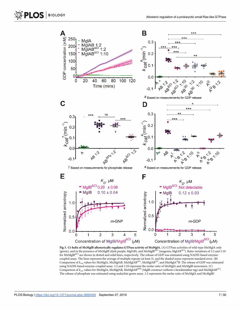
Fig 3. Ct-helix of MxMglB allosterically regulates GTPase activity of MxMglA. (A) GTPase activities of wild-type MxMglA only
(green), and in the presence of MxMglB (dark purple; MglAB), and MxMglBΔCt (magenta; MglABΔCt). Ratio variations of 1:2 and 1:10
for MxMglBΔCt are shown in dotted and solid lines, respectively. The release of GDP was estimated using NADH-based enzyme-
coupled assay. The lines represent the average of multiple repeats (at least 3), and the shaded zones represent standard error. (B)
Comparison of kcat values for MxMglA, MxMglAB, MxMglABΔCt, MxMglAB3D, and MxMglAQB. The release of GDP was estimated
using NADH-based enzyme-coupled assay. 1:2 and 1:10 represent the molar ratio of MxMglA and MxMglB monomers. (C)
Comparison of kcat values for MxMglA, MxMglAB, MxMglABΔH6 (MglB construct without a hexahistidine tag) and MxMglABΔCt.
The release of phosphate was estimated using malachite green assay. 1:2 represents the molar ratio of MxMglA and MxMglB/
Allosteric regulation of a prokaryotic small Ras-like GTPase
PLOS Biology | https://doi.org/10.1371/journal.pbio.3000459 September 27, 2019 7 / 30
interacting distances from the MxMglB Ct-helix (Fig 2D). There was a reduction in GTPase
activity in the presence of MxMglB3D (Fig 3B), a mutant of MxMglB in which Asp150B,
Asp151B, and Asp152B on the Ct-helix were replaced with alanine. Additionally, we studied
the GTPase activities of the MxMglA mutants MxMglAK (a double mutant of K181A and
K185A) and MxMglAL (a double mutant of L64A and I67A; S2B Fig, S2D–S2F Fig).
MxMglB-stimulated GTPase activity of the MxMglA mutants was less than the wild type
(Fig 3D). The GTPase activity remained lower despite 5-fold increase in the concentration of
MxMglB (Fig 3D). The above data demonstrated that mutation of residues in the helix-binding
pocket affected rate of nucleotide hydrolysis, even though they were located away from the
GTPase active site. This implied that MxMglB Ct-helix played an allosteric role in regulating
the GTPase.
Ct-helix facilitated interaction between MxMglB and GDP-bound MxMglA
Binding affinities of MxMglA with MxMglB and MxMglBΔCt in the presence of GDP and
GTP, respectively, were estimated using fluorescence anisotropy measurements of mant-labeled nucleotides (fluorescent nucleotide analogues m-GDP or m-GppNHp, referred to as
m-GNP hereafter) bound to MxMglA. Binding studies showed that m-GNP-bound MxMglA
(MxMglA–m-GNP) bound to both MxMglB and MxMglBΔCt, respectively (Fig 3E, S2A Table).
The binding affinities of MxMglB to MxMglAK and MxMglAL in presence of m-GNP were
also not affected drastically despite the mutations (S2F Fig, S2B Table). Hence, we concluded
that the lower GTPase activities observed for these mutant constructs were not due to
decreased affinity between the GTPase and MxMglB constructs.
Interestingly, MxMglB interacted with MxMglA in presence of either m-GNP or m-GDP
(Fig 3E, Fig 3F). However, MxMglBΔCt bound to MxMglA in the presence of only m-GNP but
not m-GDP (Fig 3E, Fig 3F). The binding affinities (reported as KD values) between MxMglB
and m-GNP–and m-GDP–bound MxMglA (0.10 ± 0.04 μM and 0.12 ± 0.03 μM, respectively)
were comparable to the binding affinity of 0.29 ± 0.08 μM between MxMglBΔCt and m-GNP-
bound MxMglA (Fig 3F, S2A Table). KD between MxMglBΔCt and m-GDP-bound MxMglA
could not be estimated, because the binding was insignificant (Fig 3F, S2A Table).
Thus, the Ct-helix appears to be essential for stabilizing the interaction between MxMglB
and GDP-bound MxMglA. In the previous study with TtMglB, the binding between TtMglB
and TtMglA was observed only in the presence of GNP (or GTP analogues), a characteristic
feature of a GAP. Our studies imply that nucleotide hydrolysis by MxMglA is not sufficient for
disrupting the MxMglAB complex in M. xanthus. Also, interaction with the GDP-bound state
of a GTPase is a characteristic feature of a GEF [17]. Because we observed that MxMglA and
MxMglB interact in the presence of GDP, we proceeded to check whether MxMglB could also
perform a GEF function.
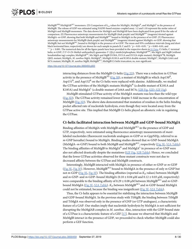
MxMglBΔH6/MxMglABΔCt monomers. (D) Comparison of kcat values for MxMglA, MxMglAK, and MxMglAL in the presence of
MxMglB. The release of GDP was estimated using NADH-based enzyme-coupled assay. 1:2 and 1:10 represent the molar ratios of
MxMglA and MxMglB monomers. The data shown for MxMglA and MxMglAB have been duplicated from panel B for the sake of
comparison. (E) Fluorescence anisotropy measurements for MxMglB (dark purple) and MxMglBΔCt (magenta) titrated against
MxMglA–m-GNP, showing that both MxMglB and MxMglBΔCt bound to MxMglA in the presence of m-GNP. (F) Fluorescence
anisotropy measurements for MxMglB (dark purple) and MxMglBΔCt (magenta) titrated against MxMglA–m-GDP, showing that
MxMglB, but not MxMglBΔCt, bound to MxMglA in the presence of m-GDP. The mean and 95% confidence intervals (long and short
black horizontal lines, respectively) are shown for each sample in panels B, C and D. �p = 0.01–0.05, ��p = 0.001–0.01, and���p< 0.001. The numerical data for all the figure panels have been provided in the respective sheets in S1 Data. Ct-helix, C-terminal
helix; m-GNP, 2’/3’-O-(N-Methyl-anthraniloyl)-guanosine-5’-[(β,γ)-imido]triphosphate; MxMglABΔH6, Mx MglA and MglB (without
hexahistidine tag) complex; MxMglAB3D, Mx MglA and MglB D150A, D151A and D152A triple mutant); MxMglAQB, MxMglA
Q82L mutant; MxMglA, M. xanthus MglA; MxMglAK, MxMglA K181A and K185A double mutant; MxMglAL, MxMglA L64A and
I67A mutant; MxMglB, M. xanthus MglB; MxMglBΔCt, MxMglB Ct-helix truncation; ns, non-significant.
https://doi.org/10.1371/journal.pbio.3000459.g003
Allosteric regulation of a prokaryotic small Ras-like GTPase
PLOS Biology | https://doi.org/10.1371/journal.pbio.3000459 September 27, 2019 8 / 30
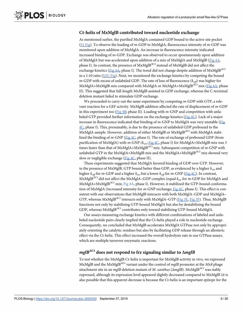
Ct-helix of MxMglB contributed toward nucleotide exchange
As mentioned earlier, the purified MxMglA contained GDP bound to the active site pocket
(S1 Fig). To observe the loading of m-GDP to MxMglA, fluorescence intensity of m-GDP was
monitored upon addition of MxMglA. An increase in fluorescence intensity indicated
increased binding of m-GDP. Exchange was observed to occur spontaneously upon addition
of MxMglA but was accelerated upon addition of a mix of MxMglA and MxMglB (Fig 4A,
phase I). In contrast, the presence of MxMglBΔCt instead of MxMglB did not affect the
exchange kinetics (Fig 4A, phase I). The trend did not change despite addition of MxMglBΔCt
in a 1:10 ratio (S2G Fig). Next, we monitored the exchange kinetics by competing the bound
m-GDP with excess of unlabeled GDP. The rate of loss of fluorescence (koff) was higher for
MxMglA+MxMglB mix compared with MxMglA or MxMglA+MxMglBΔCt mix (Fig 4A, phase
II). This suggested that full-length MxMglB assisted in GDP exchange, whereas the C-terminal
deletion mutant failed to stimulate GDP exchange.
We proceeded to carry out the same experiment by competing m-GDP with GTP, a rele-
vant reaction for a GEF activity. MxMglB addition affected the rate of displacement of m-GDP
in this experiment too (Fig 4B; phase II). Loading with m-GNP and competition with unla-
beled GTP provided further information on the exchange kinetics (Fig 4C). Lack of a major
increase in fluorescence indicated that binding of m-GNP to MxMglA was very unstable (Fig
4C, phase I). This, presumably, is due to the presence of unlabeled GDP prebound to the
MxMglA sample. However, addition of either MxMglB or MxMglBΔCt with MxMglA stabi-
lized the binding of m-GNP (Fig 4C, phase I). The rate of exchange of prebound GDP (from
purification of MxMglA) with m-GNP (kon; Fig 4C, phase I) for MxMglA+MxMglB mix was 3
times faster than that of MxMglA+MxMglBΔCt mix. Subsequent competition of m-GNP with
unlabeled GTP in the MxMglA+MxMglB mix and the MxMglA+MxMglBΔCt mix showed very
slow or negligible exchange (Fig 4C, phase II).
These experiments suggested that MxMglA favored binding of GDP over GTP. However,
in the presence of MxMglB, GTP bound better than GDP, as evidenced by a higher kon and
higher koff for m-GDP and a higher kon but a lower koff for m-GNP (Fig 4C). In contrast,
MxMglBΔCt did not affect the MxMglA–GDP complex (equal kon for m-GDP for MxMglA and
MxMglA+MxMglBΔCt mix; Fig 4A, phase I). However, it stabilized the GTP-bound conforma-
tion of MxMglA (increased intensity for m-GNP exchange; Fig 4C, phase I). This effect is con-
sistent with our observations that MxMglB interacts with both MxMglA–GDP and MxMglA–
GTP, whereas MxMglBΔCt interacts only with MxMglA–GTP (Fig 3E, Fig 3F). Thus, MxMglB
functions not only by stabilizing GTP-bound MxMglA but also by destabilizing the bound
GDP, whereas MxMglBΔCt contributes only toward stabilizing GTP-bound MxMglA.
Our assays measuring exchange kinetics with different combinations of labeled and unla-
beled nucleotide pairs clearly implied that the Ct-helix played a role in nucleotide exchange.
Consequently, we concluded that MxMglB accelerates MxMglA GTPase not only by appropri-
ately orienting the catalytic residues but also by facilitating GDP release through an allosteric
effect via the Ct-helix. This effect increased the overall hydrolysis rate in our GTPase assays,
which are multiple turnover enzymatic reactions.
mglBΔCt does not respond to frz signaling similar to ΔmglBTo test whether the MxMglB Ct-helix is important for MxMglB activity in vivo, we expressed
MxMglB and the MxMglBΔCt variant under the control of mglB promoter at the Mx8 phage
attachment site in an mglB deletion mutant of M. xanthus (ΔmglB). MxMglBΔCt was stably
expressed, although its expression level appeared slightly decreased compared to MxMglB (it is
also possible that this apparent decrease is because the Ct-helix is an important epitope for the
Allosteric regulation of a prokaryotic small Ras-like GTPase
PLOS Biology | https://doi.org/10.1371/journal.pbio.3000459 September 27, 2019 9 / 30
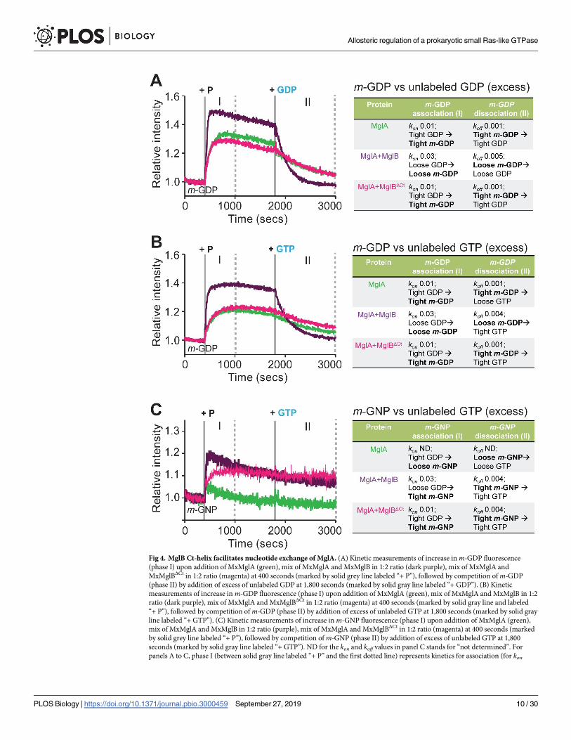
Fig 4. MglB Ct-helix facilitates nucleotide exchange of MglA. (A) Kinetic measurements of increase in m-GDP fluorescence
(phase I) upon addition of MxMglA (green), mix of MxMglA and MxMglB in 1:2 ratio (dark purple), mix of MxMglA and
MxMglBΔCt in 1:2 ratio (magenta) at 400 seconds (marked by solid grey line labeled “+ P”), followed by competition of m-GDP
(phase II) by addition of excess of unlabeled GDP at 1,800 seconds (marked by solid gray line labeled “+ GDP”). (B) Kinetic
measurements of increase in m-GDP fluorescence (phase I) upon addition of MxMglA (green), mix of MxMglA and MxMglB in 1:2
ratio (dark purple), mix of MxMglA and MxMglBΔCt in 1:2 ratio (magenta) at 400 seconds (marked by solid gray line and labeled
“+ P”), followed by competition of m-GDP (phase II) by addition of excess of unlabeled GTP at 1,800 seconds (marked by solid gray
line labeled “+ GTP”). (C) Kinetic measurements of increase in m-GNP fluorescence (phase I) upon addition of MxMglA (green),
mix of MxMglA and MxMglB in 1:2 ratio (purple), mix of MxMglA and MxMglBΔCt in 1:2 ratio (magenta) at 400 seconds (marked
by solid grey line labeled “+ P”), followed by competition of m-GNP (phase II) by addition of excess of unlabeled GTP at 1,800
seconds (marked by solid gray line labeled “+ GTP”). ND for the kon and koff values in panel C stands for “not determined”. For
panels A to C, phase I (between solid gray line labeled “+ P” and the first dotted line) represents kinetics for association (for kon
Allosteric regulation of a prokaryotic small Ras-like GTPase
PLOS Biology | https://doi.org/10.1371/journal.pbio.3000459 September 27, 2019 10 / 30
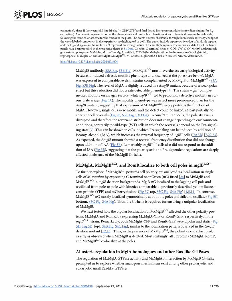
MxMglB antibody; S3A Fig, S3B Fig). MxMglBΔCt must nevertheless carry biological activity
because it induced a drastic motility phenotype and localized at the poles (see below). MglA
was expressed to comparable levels in strains complemented by MxMglB or MxMglBΔCt (S3A
Fig, S3B Fig). The level of MglA is slightly reduced in a ΔmglB mutant because of a weak polar
effect but this reduction did not create detectable phenotype [7]. The strain mglB+ comple-
mented motility on an agar surface, while mglBΔCt+ led to profoundly defective motility in col-
ony plate assays (Fig 5A). The motility phenotype was in fact more pronounced than for the
ΔmglB mutant, suggesting that expression of MxMglBΔCt deeply perturbs the function of
MglA. However, single cells were motile, and the defect could be linked, at least partially, to
aberrant cell reversals (Fig 5B, S3C Fig, S3D Fig). In ΔmglB mutant cells, the polarity axis is
disrupted and therefore the reversal distribution does not change depending on environmental
conditions, contrarily to wild-type (WT) cells in which the reversals depend on the Frz-signal-
ing state [7]. This can be shown in cells in which Frz signaling can be induced by addition of
isoamyl alcohol (IAA), which increases the reversal frequency of mglB+ cells (Fig 5B) [7,22,23].
As expected, the ΔmglB mutant showed a reversal frequency distribution that did not change
upon addition of IAA (Fig 5B). Remarkably, mglBΔCt+ cells also did not respond to the addi-
tion of IAA (Fig 5B), suggesting that the polarity axis and Frz-dependent regulations are deeply
affected in absence of the MxMglB Ct-helix.
MxMglA, MxMglBΔCt, and RomR localize to both cell poles in mglBΔCt+
To further explore if MxMglBΔCt perturbs cell polarity, we analyzed its localization in single
cells of M. xanthus by expressing C-terminal neonGreen (nG) fused [24] to MxMglB and
MxMglBΔCt in mglB deletion backgrounds. MglB-nG localized to the lagging cell pole and
oscillated from pole-to-pole with kinetics comparable to previously described yellow fluores-
cent protein (YFP) and mCherry fusions (Fig 5C top, S3C Fig, S4A Fig) [4,5,11]. In contrast,
MxMglBΔCt-nG mostly localized symmetrically at both the poles and failed to oscillate (Fig 5C
bottom, S3C Fig, S4A Fig). Thus, the Ct-helix is required for ensuring a unipolar localization
of MxMglB.
We next tested how the bipolar localization of MxMglBΔCt affected the other polarity pro-
teins, MxMglA and RomR, by expressing MxMglA-YFP or RomR-GFP, respectively, in the
mglBΔCt+ strain. Remarkably, both MxMglA-YFP and RomR-GFP were bipolar and static (Fig
5D, Fig 5E [top], S4B Fig, S4C Fig), similar to the localization pattern observed in the ΔmglBdeletion mutant [12,13]. Thus, in the presence of MxMglBΔCt, the polarity axis is disrupted,
exactly as observed when MxMglB is deleted. Most strikingly, all 3 proteins MxMglA, RomR,
and MxMglBΔCt co-localize at the poles.
Allosteric regulation in MglA homologues and other Ras-like GTPases
The regulation of MxMglA GTPase activity and MxMglAB interaction by MxMglB Ct-helix
prompted us to explore whether analogous mechanisms exist among other prokaryotic and
eukaryotic small Ras-like GTPases.
estimation), phase II (between solid line labeled “+ GDP/GTP” and final dotted line) represents kinetics for dissociation (for koffestimation). A schematic representation of the observations and probable explanations at each phase is shown on the right side,
following the same color scheme for the font as in the plots. The events directly observable through fluorescence intensity change of
the mant-labeled component in the experiment are highlighted in bold. The panels include representative plots of multiple repeats,
and the kon and koff values (in units of s-1) represent the average values of the multiple repeats. The numerical data for all the figure
panels have been provided in the respective sheets in S1 Data. Ct-helix, C-terminal helix; m-GDP, 2’/3’-O-(N-Methyl-anthraniloyl)-
guanosine-diphosphate; MxMglA, M. xanthus MglA; m-GNP, 2’/3’-O-(N-Methyl-anthraniloyl)-guanosine-5’-[(β,γ)-imido]
triphosphate; MxMglB, M. xanthus MglB; MxMglBΔCt, M. xanthus MglB with Ct-helix truncated; ND, not determined.
https://doi.org/10.1371/journal.pbio.3000459.g004
Allosteric regulation of a prokaryotic small Ras-like GTPase
PLOS Biology | https://doi.org/10.1371/journal.pbio.3000459 September 27, 2019 11 / 30
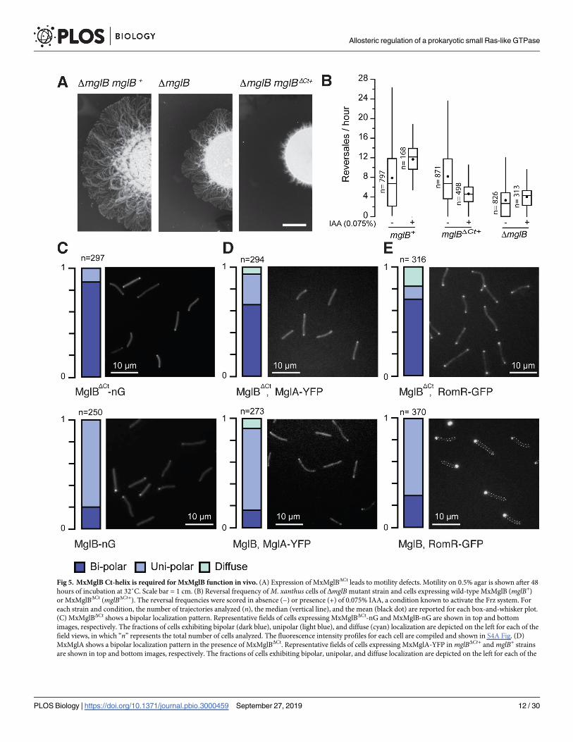
Fig 5. MxMglB Ct-helix is required for MxMglB function in vivo. (A) Expression of MxMglBΔCt leads to motility defects. Motility on 0.5% agar is shown after 48
hours of incubation at 32˚C. Scale bar = 1 cm. (B) Reversal frequency of M. xanthus cells of ΔmglB mutant strain and cells expressing wild-type MxMglB (mglB+)
or MxMglBΔCt (mglBΔCt+). The reversal frequencies were scored in absence (−) or presence (+) of 0.075% IAA, a condition known to activate the Frz system. For
each strain and condition, the number of trajectories analyzed (n), the median (vertical line), and the mean (black dot) are reported for each box-and-whisker plot.
(C) MxMglBΔCt shows a bipolar localization pattern. Representative fields of cells expressing MxMglBΔCt-nG and MxMglB-nG are shown in top and bottom
images, respectively. The fractions of cells exhibiting bipolar (dark blue), unipolar (light blue), and diffuse (cyan) localization are depicted on the left for each of the
field views, in which “n” represents the total number of cells analyzed. The fluorescence intensity profiles for each cell are compiled and shown in S4A Fig. (D)
MxMglA shows a bipolar localization pattern in the presence of MxMglBΔCt. Representative fields of cells expressing MxMglA-YFP in mglBΔCt+ and mglB+ strains
are shown in top and bottom images, respectively. The fractions of cells exhibiting bipolar, unipolar, and diffuse localization are depicted on the left for each of the
Allosteric regulation of a prokaryotic small Ras-like GTPase
PLOS Biology | https://doi.org/10.1371/journal.pbio.3000459 September 27, 2019 12 / 30
Earlier, it has been shown that MglA and MglB, which exist in almost all phyla of eubac-
teria, can be categorized into 5 phylogenetically related groups [25]. Genes encoding MglB-
like proteins are either associated with genes encoding MglA-like protein in the same operon,
called coupled MglBs, or without an associated MglA gene (orphan MglBs) [25] (S3 Table).
We analyzed these sequences and found that 66 coupled and 25 orphan MglBs possessed a C-
terminal extension (more than 15 amino acids beyond the Rbl/LC7 fold; S3 Table). A total of
56 of the 66 coupled MglB sequences showed a predicted helical region containing 2 to 4 aspar-
tates or glutamates (D/E rich), whereas the C-terminal extensions of the remaining MglB
sequences contained proline-rich stretches with no predicted secondary structure (Fig 6A, S3
Table).
Out of these 56 coupled sequences, the α5 helix (which interacts with the Ct-helix) of 48
MglA sequences possessed positively charged residues (either lysines or arginines; Fig 6B).
These positively charged residues were not conserved in other MglA sequences (Fig 6B). This
suggested that the Ct-helix of MglB and its binding pocket on MglA have coevolved. Interest-
ingly, we found MglB having the D/E rich Ct-helix to be present in all the 5 phylogenetic
groups of MglA-like proteins, including a variety of bacteria and also in the archaea Methano-bacterium paludis (S5 Fig). This indicated that the mechanism of allosteric regulation of the
MglA GTPase by the MglB Ct-helix could be widespread in prokaryotic Ras-like GTPases and
not limited to Myxococcus. Because the allosteric regulation by full-length MglB has not been
characterized for any other prokaryotic MglA-like and MglB-like proteins, the mechanism
proposed here would be relevant toward understanding their function.
Discussion
MxMglB Ct-helix facilitates nucleotide exchange allosterically
Our results revealed that the Ct-helix is essential for MxMglB to interact with MxMglA in the
GDP-bound state. Deletion of the helix resulted in the formation of MxMglAB complex in the
presence of only GTP but not GDP. This prompted us to identify the structural features that
enable MxMglB to bind MxMglA–GDP. The β-screw movement (flipping of MxMglA β2
strand) appears to be essential for the interaction of MxMglB with MxMglA. This conforma-
tional change exposes residues that form a major part of MxMglAB interface.
An inspection of the MxMglAB–GTPγS structure revealed that the switch 1 loop and the
binding pocket of the Ct-helix are at the N- and C-terminal ends of the β2 strand of MxMglA,
respectively (Fig 2H). Hence, we propose that the flipped state of the β2 strand that favors
MxMglB binding can be achieved in 2 ways—by interaction of either the γ phosphate of GTP
with the switch 1 loop or the Ct-helix with the β2 to β3 loop (Fig 2H). The flipped state achieved
by the Ct-helix interaction and the consequent stabilization by the Rbl/LC7 fold of MxMglB
results in the formation of a complex with MxMglA–GDP. As a consequence of the flipped
state of the β2 strand, MxMglA in the MxMglAB complex will potentially possess a nucleotide-
binding pocket that favors binding of GTP over GDP, consistent with our observations from
the nucleotide exchange assays (Fig 4). Thus, the proposed model provides an explanation for
field views, in which “n” represents the total number of cells analyzed. The fluorescence intensity profiles for each cell are compiled and shown in S4B Fig. (E)
RomR shows a bipolar localization pattern in the presence of MxMglBΔCt. Representative fields of cells expressing RomR-GFP in mglBΔCt+ and mglB+ strains are
shown in top and bottom images, respectively. The fractions of cells exhibiting bipolar, unipolar, and diffuse localization are depicted on the left for each of the
field views, in which “n” represents the total number of cells analyzed. In the bottom panel (RomR-GFP, mglB+), dashed lines indicate bacterial contour. The
fluorescence intensity profiles for each cell are compiled and shown in S4B Fig. The numerical data for all the figure panels have been provided in the respective
sheets in S1 Data. Ct-helix, C-terminal helix; Frz, frizzy; IAA, isoamyl alcohol; MxMglA, M. xanthus MglA; MxMglB, M. xanthus MglB; MxMglBΔCt, MxMglB with
Ct-helix truncated; nG, neonGreen; YPF, yellow fluorescent protein.
https://doi.org/10.1371/journal.pbio.3000459.g005
Allosteric regulation of a prokaryotic small Ras-like GTPase
PLOS Biology | https://doi.org/10.1371/journal.pbio.3000459 September 27, 2019 13 / 30
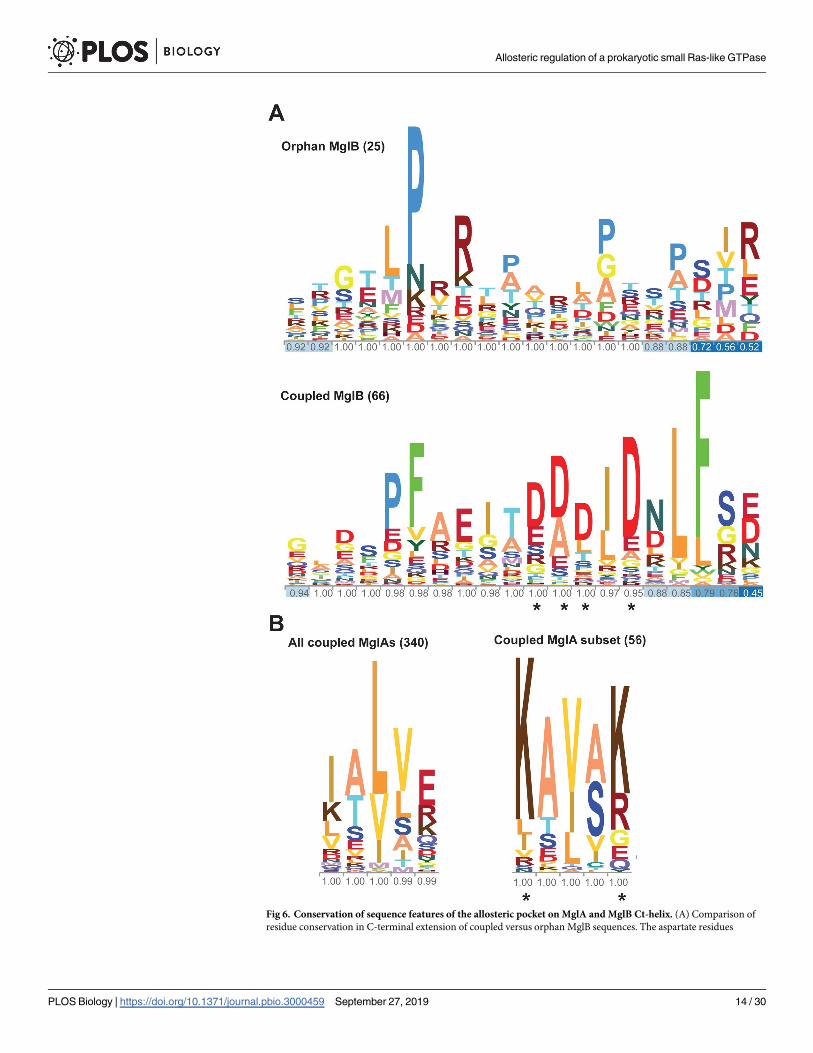
Fig 6. Conservation of sequence features of the allosteric pocket on MglA and MglB Ct-helix. (A) Comparison of
residue conservation in C-terminal extension of coupled versus orphan MglB sequences. The aspartate residues
Allosteric regulation of a prokaryotic small Ras-like GTPase
PLOS Biology | https://doi.org/10.1371/journal.pbio.3000459 September 27, 2019 14 / 30
MxMglAB–GDP complex formation and a mechanistic insight into the allosteric action of Ct-
helix for facilitating nucleotide exchange (Fig 7A).
Interestingly, like MglB, many eukaryotic GEFs such as DENN-1B domain, TRAPP-I, and
Mon1-Ccz1, have the Rbl/LC7 fold [26]. Furthermore, we found that the intersubunit orienta-
tion in MglAB is conserved in the GTPase-GEF complexes of Rab35 with DENN-1B domain
[27], Rab Ypt1p with TRAPP-I [28], and Ypt7 with Mon1-Ccz1 [29] (S6 Fig). However, unlike
in MglAB complex, the mechanism of GEF action in these eukaryotic systems is based on a
glutamate or aspartate residue interacting directly with switch 1 or switch 2 (S6A Fig, S6B Fig).
These residues are present in extensions from the Rbl/LC7 fold or are contributed by other
interacting partners that form the GEF complex [26].
Mechanism of dual GAP and GEF action by MxMglB
We propose that MxMglB increases the rate of GTP hydrolysis by MxMglA through 2 comple-
mentary ways. Firstly, Rbl/LC7 domain of MxMglB stabilizes the flipped β2 strand interface,
which orients the MxMglA catalytic residues favorably. Secondly, its Ct-helix, a flexible ex-
tension from the main Rbl/LC7 fold, retains the flipped state of the β2 strand post GTP hydro-
lysis and, consequently, stabilizes the MxMglAB–GDP complex. This interaction results in a
nucleotide-binding pocket that accelerates release of GDP from the complex and favors GTP-
binding. Thus, by facilitating GDP-to-GTP exchange MxMglB enhances the rate of GTP
hydrolysis by MxMglA, increasing its potency as a GAP. Indeed, our in vitro results show that
MxMglBΔCt exhibits reduced GAP activity, which we attribute to a defect in nucleotide
exchange.
Role of Ct-helix and exchange activity of MxMglB in the polarity oscillation
cycle
In vivo, we propose that the MxMglB Ct-helix is critical for the balance between GAP
and GEF activities at the lagging pole. In absence of the Ct-helix, all 3 proteins—MglA,
MxMglBΔCt, and RomR—localize to both cell poles. Thus, the MxMglB Ct-helix is absolutely
required to enable the cascade of interactions that establish polarity and permit its switch.
Co-localization of both MxMglA and MxMglBΔCt suggest that MxMglBΔCt does not support
its GAP function in vivo, despite exhibiting only a reduction in GAP activity in vitro. In
between reversals in wild-type cells, MxMglB and RomRX stably co-localize at the lagging pole
[11,14,15]. MxMglA is efficiently excluded from the lagging pole, suggesting that the GAP
activity of MxMglB predominates over the GEF activity of RomRX. Thus the MglB Ct-helix
possibly overcomes the action of RomRX.
How could this function at the molecular level? Analysis of RomR revealed a stretch of neg-
atively charged residues (Glu-rich) akin to that of MxMglB Ct-helix (S7A Fig). Thus, similar to
MxMglB, the RomRX complex could facilitate GDP exchange from MxMglA by acting
through the allosteric pocket on MxMglA. In this process, the C-terminal Glu-rich region of
implicated in interaction with the allosteric pocket on MglA are highlighted by “�” in the sequence conservation logo.
The fractional occupancy of amino acids in the sequence alignment is shown for each amino acid position. The
numbers within brackets denote the number of sequences in the alignment. The residues are colored in 20 different
shades. (B) Comparison of residue conservation in the α5 helix of MglA sequences coupled with MglB sequences
possessing a negatively charged C-terminal extension versus that of all coupled MglA sequences. The lysine residues
that potentially interact with the Ct-helix of MglB are highlighted by “�” in the sequence conservation logo. The
fractional occupancy of amino acids in the sequence alignment is shown for each amino acid position. The numbers
within brackets denote the number of sequences in the alignment. Ct-helix, C-terminal helix; MglA, Mutual Gliding
motility A; MglB, Mutual Gliding motility B.
https://doi.org/10.1371/journal.pbio.3000459.g006
Allosteric regulation of a prokaryotic small Ras-like GTPase
PLOS Biology | https://doi.org/10.1371/journal.pbio.3000459 September 27, 2019 15 / 30
RomR might thus assist nucleotide exchange, whereas RomX could assist in stabilizing the
flipped β2 strand of MxMglA. We tested this hypothesis by replacing the Ct-helix of MxMglB
with the Ct-helix from RomR (S7A Fig, S7B Fig) and carrying out GTPase activity measure-
ments in the presence of the chimeric MglB (MglBRhelix). Indeed, we observed that the chime-
ric construct accelerated the GTPase activity of MglA similar to that of wild-type MxMglB
(S7C Fig), supporting the hypothesis that the Ct-helix of RomR is capable of binding at the
allosteric pocket of MxMglA. At the lagging pole, the MxMglB Ct-helix and the RomR Glu-
rich helix might compete for binding at the MxMglA helix-binding pocket, which would dis-
place RomRX from MxMglA. When the MglB Ct-helix is deleted, RomRX could therefore
interact with MxMglA thus blocking MxMglBΔCt from exerting its GAP activity. This would
allow RomRX to exert its GEF activity at the lagging pole too and stabilize the anchoring of
MglA to both the poles.
Additional regulation of the Ct-helix must take place to determine the nucleotide specificity
of MxMglB binding to MxMglA. Additional regulators in vivo will potentially determine the
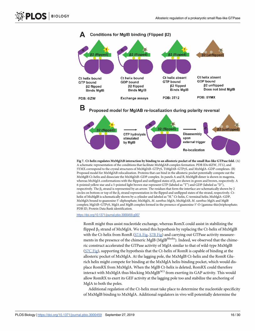
Fig 7. Ct-helix regulates MxMglAB interaction by binding to an allosteric pocket of the small Ras-like GTPase fold. (A)
A schematic representation of the conditions that facilitate MxMglAB complex formation. PDB IDs 6IZW, 3T12, and
5YMX correspond to the crystal structures of MxMglAB–GTPγS, TtMglAB–GTPγS, and MxMglA–GDP complexes. (B)
Proposed model for MxMglAB relocalization. Proteins that can bind in the allosteric pocket potentially compete out the
MxMglB Ct-helix and dissociate the MxMglAB–GDP complex. In panels A and B, MxMglB dimer is shown in magenta,
whereas MxMglA conformations with the flipped and unflipped states of β2 are shown in green and brown, respectively. A
6-pointed yellow star and a 5-pointed light brown star represent GTP (labeled as “T”) and GDP (labeled as “D”),
respectively. The β2 strand is represented by an arrow. The residues that form the interface are schematically shown by 2
circles on bottom or top of the β2 strand representation in the flipped and unflipped states of the strand, respectively. Ct-
helix of MxMglB is schematically shown by a cylinder and labeled as “H.” Ct-helix, C-terminal helix; MxMglA–GDP,
MxMglA bound to guanosine 5’-diphosphate; MxMglA, M. xanthus MglA; MxMglAB, M. xanthus MglA and MglB
complex; MglAB–GTPγS, MglA and MglB complex formed in the presence of guanosine 5’-O-[gamma-thio]triphosphate;
PDB ID, Protein Data Bank identification.
https://doi.org/10.1371/journal.pbio.3000459.g007
Allosteric regulation of a prokaryotic small Ras-like GTPase
PLOS Biology | https://doi.org/10.1371/journal.pbio.3000459 September 27, 2019 16 / 30
accessibility of the Ct-helix and thus the localization of MxMglA and MxMglB. At the pole, the
exact molecular organization of the signaling complexes is not known. Other proteins that par-
ticipate in the polarity switch and that are present at the lagging pole, for example, the MglB-
like protein MglC, can potentially regulate the Ct-helix availability [30]. During the switch,
action of FrzXP could, for example, block the inhibitory effect of MglB on RomRX and thus
initiate the switch. This possibility could be explored in the future. Further characterization of
other polarity determining components and their interactions with MxMglAB are therefore
essential to discriminate the GAP and GEF-active phases of MxMglB during the polarity rever-
sal cycle.
Insights gained from the prokaryotic small Ras-like GTPase MxMglA and its interacting
partner MxMglB highlight the dual role of MxMglB both as a GAP and a GEF. The novel
mechanism of GEF action is based on an allosteric interaction with the small Ras-like GTPase.
Based on sequence analysis, it appears to be a conserved mechanism among prokaryotic small
Ras-like GTPases. The study also opens up a new avenue for design of compounds or factors
that can modulate the enzyme activity by targeting the newly discovered allosteric binding
pocket of the universal Ras-like fold.
Methods and materials
Protein expression and purification
Protein constructs, cloning, and overexpression. Genes corresponding to mglA and
mglB were amplified from M. xanthus genomic DNA (obtained from DSMZ, Germany, cata-
logue number 16526) using suitable primers (S4 Table) and cloned into pHis17 vector
(obtained from Lowe lab, MRC LMB, Cambridge; refer Addgene plasmid #78201 for vector
backbone) between the restriction enzyme sites NdeI (New England Biolabs Inc) and BamHI
(New England Biolabs Inc). Single-point mutants of MxMglA and deletion constructs of
MxMglB were generated using a site-directed mutagenesis strategy utilizing PCR-based meth-
ods followed by DpnI (New England Biolabs Inc) digestion, transformation, and screening of
positive clones. All clones were confirmed by sequencing. The list of clones and the primers
used to generate them are summarized in S5 and S4 Tables. All chemicals were procured from
Sigma Aldrich, unless otherwise mentioned.
Protein expression. Plasmids containing the gene of interest were transformed into suit-
able Escherichia coli strains, and culture was grown at 37˚C and at 30˚C post induction. E. colistrain BL21-DE3 was used for expressing MxMglA wild type and its mutants. The cultures
were grown in LB media containing 50 μg/ml of ampicillin and induced with 0.5 mM IPTG at
OD600 value of 0.8, whereas MxMglB and its mutants were expressed in the strain BL21-AI
and induced with 0.02% L-arabinose (SRL Chemicals, India) at OD600 value of 0.6. Seleno-
methionine-labeled proteins for MxMglBL156M and MxMglBI148M have been expressed by the
feedback inhibition method [31].
Purification of MxMglA(His6). For purification, harvested cells were resuspended in the
lysis buffer L (50 mM Tris, 200 mM NaCl [pH 8.0], and 10% glycerol) and then spun at
39,000g for 45 minutes at 4˚C. Supernatant was loaded on to a 5-ml HisTrap (GE Lifesciences)
column because the presence of hexahistidine tag at the C-terminus facilitated the binding of
overexpressed protein of interest to the column. The column was equilibrated with binding
buffer (Buffer A: 50 mM Tris [pH 8.0], 200 mM NaCl) prior to loading the supernatant and
the bound protein was washed and eluted with a step gradient of 2%, 5%, 10%, 20%, 50%, and
100% of Buffer B (Buffer A containing 500 mM imidazole). The fractions containing the pro-
tein were pooled, concentrated, and loaded onto Superdex75, 10/300 (GE Lifesciences). The
protein was eluted into 50 mM Tris (pH 8.0), 50 mM NaCl. Fractions containing the protein
Allosteric regulation of a prokaryotic small Ras-like GTPase
PLOS Biology | https://doi.org/10.1371/journal.pbio.3000459 September 27, 2019 17 / 30
were pooled, concentrated, flash frozen, and stored at −80˚C. MxMglA mutants, i.e., MglAK,
MglAL, and MglAQ, were also purified using a similar protocol.
Purification of MxMglB. MxMglB(His6), MxMglBΔCt(His6), and MxMglBRhelix(His6)
were purified using the same protocol as described for MxMglA(His6). For MxMglB con-
structs without histidine tag, i.e., MxMglB, MxMglB3D, and selenomethionine-labeled
MxMglBL156M and MxMglBI148M, ion-exchange chromatography was used to purify the pro-
teins. First, the cells were resuspended in the lysis buffer (50 mM Tris [pH 8.0], 200 mM NaCl,
10% glycerol), and then spun at 39,000g for 45 minutes at 4˚C. An ion-exchange column, QHP
(5 ml, GE Lifesciences) was used to purify the protein. Buffers used for binding and elution
were Buffer A (50 mM Tris [pH 8.0], 50 mM NaCl), and a linear gradient of Buffer A with
Buffer B (50 mM Tris [pH 8.0], 1 M NaCl), respectively, ranging from 0% to 50% Buffer B over
20 column volumes. Fractions containing the protein of interest were pooled. This protein was
dialyzed into Buffer A25 (50 mM Tris [pH 8.0], 25 mM NaCl) and again spun at 39,000g at
4˚C, filtered, and loaded on MonoQ 10/100 (GE Lifesciences) to remove minor impurities
present. Binding and elution buffers were the same as earlier. Fractions from the MonoQ run
were checked on SDS-PAGE, and those containing protein of interest were concentrated and
stored at −80˚C.
Crystallization and structure determination
MxMglA crystallization. About 500 conditions of commercially available screens (Molec-
ular dimensions, Hampton Research) were screened using Mosquito crystallization robotic
system, using drop sizes consisting of 100 nl of protein at a concentration of 10 mg/ml and 100
nl of crystallization cocktail, in 96-well sitting drop plates (MRC plate, SWISS-SCI). Initial hits
were obtained in many of the conditions, and, further, it was reproduced and optimized to get
well-diffracting crystals. For crystallization of MxMglA, protein was diluted to a final concen-
tration of 10 mg/ml in A50 buffer (50 mM Tris [pH 8.0], 50 mM NaCl). Diffraction quality
crystals were obtained using the following conditions in a drop ratio of 1:1 volumes for protein
and crystallization condition: (i) 0.1 M sodium cacodylate (pH 6.5), 40% v/v 2,4-methyl pen-
tane diol, and 5% PEG 8000; (ii) 0.1 M imidazole (pH 8.0), 30% w/v 2,4-methyl pentane diol,
and 10% w/v PEG 4000; (iii) 0.1 M sodium citrate (pH 5.6) and 35% w/v tertiary-Butanol; 20%
ethylene glycol was included as cryoprotectant in the parent condition during crystal freezing.
MxMglAB crystallization. MxMglA and MxMglB were mixed in a ratio of 1:1 (consider-
ing monomeric molecular weight of MglB) in A50 buffer containing 5 mM MgCl2 and 2 mM
GTPγS (Sigma). The concentration of the proteins used to crystalize was 4 mg/ml of each.
Screening for crystallization hits yielded a few conditions that were further optimized to get
the desirable crystals. The crystals were obtained in 2 different conditions, namely, (i) PEG
4000 (8%) and ammonium sulphate (200 mM) and (ii) PEG 3350 (12%) and ammonium sul-
phate (200 mM). Diffraction quality crystals were obtained at a drop ratio of 1:1 volumes of
protein to crystallization buffer. Crystallization conditions containing 20% PEG 400 were used
as cryoprotectant during freezing of crystals. Crystals of MxMglA and selenomethionine-
labeled MxMglB complex were obtained in the same crystallization conditions.
Structure solution and refinement. Diffraction data from the crystals were collected at
the home source using Rigaku Micromax 007 X-ray generator, and higher resolution data and
anomalous data were collected at the synchrotron sources at Diamond Light Source, Harwell,
UK, and ESRF, Grenoble. Data reduction was performed using IMOSFLM [32] or XDS [33],
and scaling using AIMLESS [34] in CCP4 package [35]. MxMglA and MxMglB structures
were solved by molecular replacement using the TtMglAB structure (PDB code: 3T12). Molec-
ular replacement was performed using PHASER [36] available in CCP4 package. Refinement
Allosteric regulation of a prokaryotic small Ras-like GTPase
PLOS Biology | https://doi.org/10.1371/journal.pbio.3000459 September 27, 2019 18 / 30
was carried out using PHENIX package [37] and model building using Coot [38]. The refined
structures have been deposited in the PDB with accession numbers 5YMX (MxMglA–GDP)
and 6IZW (MxMglAB–GTPγS).
In order to confirm the registry of the amino acids belonging to the Ct-helix of MxMglB,
selenomethionine-labeled protein for mutant constructs of MxMglB where Ile148B and
Leu156B, respectively, were mutated to methionines, were purified, and selenomethionine-
labeled MxMglB was used for obtain MxMglAB crystals in the same crystallization condition.
The anomalous data from the crystals were collected, and it was confirmed that the correct
amino acids were modeled into the electron density. The anomalous signal from the methio-
nines in these 2 mutants, respectively, confirmed the registry of the amino acids of the
MxMglB Ct-helix (S1D and E Fig).
Fluorescence anisotropy experiments
Binding studies using fluorescent anisotropy measurements. Binding of fluorescently-
labeled nucleotides, mant-GDP (m-GDP) or mant-GppNHp (m-GNP) (Jena Bioscience) with
the proteins were monitored by measuring the change in anisotropy [39]. The excitation and
emission wavelengths used for monitoring the mant-labeled nucleotide fluorescence were 360
nm and 440 nm, respectively. The experiments were performed on Fluoromax-4 (Horiba),
with a sample volume of 200 μl in a cuvette, and excitation and emission slit widths of 5 nm.
Nucleotide binding of MxMglA and its mutants were estimated by measuring the change in
fluorescence anisotropy of the mant-nucleotide upon titration with increasing concentrations
of the protein. Protein samples (MxMglA or its mutants) were titrated against a fixed concen-
tration of mant-labeled nucleotide (100 nM). Each reading corresponds to 10 averaged single
point anisotropy values. The initial value of anisotropy of the mant-labeled nucleotide was sub-
tracted from all the values. GraphPad Prism was used to plot the values against the concentra-
tion of protein (concentration of MxMglA) and to fit the data to the binding equation for
estimation of KD. Because it is known that MxMglA has a single binding site for nucleotides,
equation for one site-specific binding, as given below, was used to fit the data points. The equa-
tion is y = Bmax × x� (KD + x), where y represents the bound fraction (fluorescence anisotropy
based readout), Bmax is the maximum value obtained, x is the concentration of MxMglA, and
KD is the binding constant.
The values were normalized by dividing each of them by the maximum value of anisotropy,
Bmax. The normalized anisotropy values were plotted to obtain a binding curve. Because
MxMglA was purified in the GDP-bound form, the measurements from this experiment are
not absolute affinities of MxMglA (or its mutants) to the nucleotides but a reliable measure of
exchange of the prebound GDP with m-GDP or m-GNP.
Similarly, anisotropy measurements for increasing amounts of MxMglB/MxMglBΔCt with
nucleotide-bound MxMglA provided information about the binding affinity of MxMglB to
nucleotide-bound MxMglA. A mix of 400 nM of mant-labeled nucleotide with 2 μM of
MxMglA was titrated with increasing concentrations of MxMglB/MxMglBΔCt. The same calcu-
lations as described above were performed to obtain the binding affinities (KD). In this case,
the x-axis represents the concentration of MxMglB dimer. Because MxMglA binds to one
dimer of MxMglB, equation for one site-specific binding was used to fit the data points.
Each data point is an average of at least 3 independent measurements, and the standard
error is shown in which each data represent 3 to 5 repeats. Error bars represent standard error.
Nucleotide exchange assays. Intensity of fluorescence emission by mant-labeled nucleo-
tide (GDP or GMPPNP [GNP]; Jena Bioscience) at 440 nm was monitored after the excitation
at 360 nm. All the kinetic measurements for MxMglA, MxMglA with MxMglB, and their
Allosteric regulation of a prokaryotic small Ras-like GTPase
PLOS Biology | https://doi.org/10.1371/journal.pbio.3000459 September 27, 2019 19 / 30
mutants were performed on Fluoromax-4 (Horiba), with a sample volume of 200 μl in a
cuvette and excitation and emission slit widths of 2 nm. m-GDP/GNP (800 nM) was added
with Buffer A50 (50 mM Tris, 50 mM NaCl, 5mM MgCl2 [pH 8.0]) in a quartz cuvette (10 × 2
mm path length), and the fluorescence intensity was monitored initially for 400 seconds. The
protein, i.e., MxMglA or the mix of MxMglAB/MxMglABΔCt (3 μM of MxMglA and 6 μM
MxMglB/MxMglBΔCt monomer), was added in the cuvette at 400 seconds after stabilization of
the signal from only mant-nucleotide. Consequently, the fluorescence was recorded for 1,400
seconds. The binding was estimated in terms of increase in fluorescence intensity. At 1,800
seconds, the mant-labeled nucleotide was competed out with excess of unlabeled nucleotide
(GDP or GTP; 500 μM), resulting in a decrease of fluorescence intensity because of release of
mant-labeled nucleotide from the protein. For plotting the relative intensities from the mea-
surements, each value was divided by the average of first 200 readings (400 seconds). These
accumulation and decay reactions were fitted to exponential binding equations as given below
to estimate the kon and koff values.
P + N1! PN1 + N2! PN2 + N1, where P represents protein, N1 is the labeled nucleotide,
N2 is the unlabeled nucleotide, and PN denotes the protein-nucleotide complex.
For estimation of kon, PNt = PNmax (1 − e−kt).
For estimation of koff, PNt = PNmax − PNmin (e−kt) + PNmin
Here, PNt represents the amount of the complex at time t, PNmax is the maximum amount
of the complex upon association, and PNmin is the minimum amount of the complex after
dissociation.
GTP hydrolysis assay
NADH coupled assay. GTPase activity measurements were made using a coupled enzyme
based assay in which the conversion of NADH to NAD+ is coupled to the utilization of GDP
produced [20]. The GTPase or ATPase hydrolyses the nucleotide while pyruvate kinase (PK)
uses GDP (or ADP) and phosphoenol pyruvate (PEP) to produce GTP (or ATP) and pyruvate.
Lactate dehydrogenase (LDH) utilizes NADH and pyruvate to produce NAD+ and lactate.
Because the utilization of NADH is equivalent to the GDP produced by the GTPase reaction,
the actual read out is decrease in absorbance of NADH, which in turn estimates the GDP pro-
duced by the GTPase. The amount of NADH utilized in the reaction was measured by moni-
toring the NADH absorbance at 340 nm using a multiplate reader, Varioskan Flash (Thermo
Scientific). A master mix was prepared in Buffer A50 containing PEP (1mM; Sigma), GTP
(1mM; Jena Bioscience), NADH (600 μm; Sigma), and PK/LDH mix (approximately 25 U/ml;
Sigma). All the components of NADH reaction were mixed in a total reaction volume of 200 μl
and added in a 96-well plate. The reaction was initiated by addition of MxMglA or MxMglAB
mix and their relevant mutant constructs. MxMglA and its mutants were used at 10 μM and
MxMglB and its mutants at concentrations 20 μM and 200 μM (in a molar ratio of 1:2 and 1:10
considering monomeric molecular weight of MxMglB).
Readings were recorded at the interval of 20 seconds for 2 hours. The absorbance value of
buffer containing GTP was subtracted from all the values. Also, the absorbance at the 0 time
point for each reaction was subtracted from all readings of the reaction. The absorbance read-
ings were represented as concentration of GDP released, by using a conversion factor based on
the standard graph obtained by plotting the absorbance values for known concentrations of
NADH. kcat values were calculated and plotted using GraphPad Prism. Readings from 0 to
7,200 seconds were fitted to a line using linear regression, and the slope of the fitted line was
calculated to obtain the amount of GDP released (in μM) per unit time. kcat was calculated as
the amount of GDP released per unit time per unit enzyme concentration. This value was used
Allosteric regulation of a prokaryotic small Ras-like GTPase
PLOS Biology | https://doi.org/10.1371/journal.pbio.3000459 September 27, 2019 20 / 30
further to compare the enzyme activities among all the mutants. The significance was calcu-
lated using GraphPad Prism, using one-way ANOVA with Tukey test of 95% confidence inter-
val, and significance was marked on the scatter plot.
Phosphate release measurements using malachite green assay. This assay was per-
formed using MxMglA at a concentration of 10 μM, and MxMglB in molar ratio 1:2 (20 μM,
considering monomeric molecular weight of MxMglB) with excess of GTP, i.e., 1 mM GTP in
buffer A50 (50 mM Tris, 50 mM NaCl [pH 8.0]) containing 5 mM MgCl2. All the enzyme
activity assays were performed at 30˚C. Readings were taken at different time points, i.e., 0, 15,
30, 45, 60, 75, 90, 105, and 120 minutes. To stop the reaction, the sample was heated at 65˚C
and then malachite green reagent (prepared as described in [21]) was added. The absorbance
was recorded at 630 nm after 20 minutes of incubation, using a plate reader (Varioskan,
Thermo Scientific). Amount of Pi (inorganic phosphate) released in μM was calculated using a
standard curve, obtained by mixing a set of known concentrations of NaH2PO4 with malachite
green solution. The experiments were repeated multiple times, and the slope was calculated
from every repeat. Readings from 0 to 120 minutes were fitted to a line using linear regression,
and the slope of the fitted line was calculated to obtain the amount of Pi released (in μM) per
unit time. kcat was calculated as Pi released per μM enzyme concentration for each reaction.
kcat values were plotted, and significance was calculated on GraphPad Prism, using one-way
ANOVA with Tukey test of 95% confidence interval, and significance was marked on the scat-
ter plot.
HPLC for bound nucleotide estimation
Purified MxMglA was diluted to 2 to 3 mg/ml concentration in buffer A50 and then heated at
65˚C. The heated protein sample was spun at 21,000g for 10 minutes at 4˚C. Then supernatant
was filtered with 0.22 μm cellulose acetate filter (Corning), and the sample was loaded on the
DNAPac 200 ion-exchange column (Thermo Fisher). Buffer A (2 mM Tris [pH 8.0]) was used
as binding buffer. The runs were performed at a flow rate of 0.8 ml/min, with steps of 100%
Buffer A for 3 minutes, followed by linear gradients of 0% to 30% Buffer B (2 mM Tris [pH
8.0], 1.25 M NaCl) for 10 minutes, and 30% to 100% B for 12 minutes. In S1B Fig, the peak
profile obtained for 18 μM of MglA was compared with that of 50 μM GDP solution, with
35 μl of sample injection volume for both. The absorbance at 260 nm, indicative of the pres-
ence of GDP, was plotted against retention time.
Thermal shift assay [40]
In a 25-μl reaction volume containing 2 μM of protein in buffer A50, SYPRO Orange dye
(Sigma; available as a 5,000× concentration in DMSO) was added to a final concentration of
5×, in 96-well multiwell PCR plates. Biorad CFX96 Real-Time System was used to monitor the
change in fluorescence of SYPRO Orange (Sigma-Aldrich). The mix was heated from 4˚C to
90˚C with an increment of 1.2˚C per minute. Readings were recorded at intervals of 0.4˚C.
The melting temperature (Tm) was calculated based on the differential of the fluorescence
intensity versus temperature plot (dF/dT). This gave an estimate of the stability of the protein.
In vivo experiments
Myxococcus strains, plasmids, growth conditions, and genetic constructs. Primers,
plasmids, and strains used for this study are listed in S4 Table, S5 Table and S6 Table, respec-
tively. In general, M. xanthus strains were grown at 32˚C in CYE rich media as previously
described by Bustamante et al. [23]. Plasmids were introduced in M. xanthus by electropora-
tion. Complementation, expression of the fusion, and mutant proteins were obtained by
Allosteric regulation of a prokaryotic small Ras-like GTPase
PLOS Biology | https://doi.org/10.1371/journal.pbio.3000459 September 27, 2019 21 / 30
ectopic integration of the genes of interest at the Mx8-phage attachment site [7] under the con-
trol of their own promoter in appropriate deletion backgrounds. E. coli cells were grown under
standard laboratory conditions in Luria-Bertani broth supplemented with antibiotics, if
necessary.
Construction of Myxococcus strains. Following an initial step of PCR amplification of
the relevant gene fragments for complementation, the fragments were cloned into the
pSWU19 vector by the one-step sequence- and ligation-independent cloning (SLIC) method
reported previously by Jeong et al. [41]. Specific details of PCR amplification are given below,
whereas S4 Table, S5 Table and S6 Table contain the list of primers, plasmids, and strains used.
pSWU19 mglBΔCt, pSWU19 mglBΔCt-neon green, and pSWU19 mglB-neon green for com-
plementation of the mglB deletion were constructed by amplifying 200 bp upstream the mglBcoding sequence (promoter) with the mglBΔCt gene (417 bp of mglB gene), mglBΔCt gene (417
bp) and the linker with neon green, and the mglB gene and the linker with neon green, respec-
tively. We chose to use neonGreen because this fluorescent protein (FP) is exceptionally bright
and strictly monomeric, preventing possible artifacts due to the formation of weak FP dimers
[24].
Phenotypic assays. Soft agar motility was performed as previously described by Busta-
mante et al. [23]. In general, cells were grown up to an OD600 between 0.4 to 0.8 and concen-
trated at OD600 = 5. Then they were spotted (10 μl) on 0.5% agar in CYE (soft agar). Colonies
were photographed after 48 hours.
Fluorescence imaging and fluorescence intensity measurements. Protocol followed is as
reported previously by Guzzo et al. [11]. For phase-contrast and fluorescence microscopy, cells
from exponentially growing cultures were concentrated to OD600 = 2 by centrifugation of 1 ml
of culture and resuspended in the corresponding volume of TPM buffer (10 mM Tris-HCl
[pH 7.6], 8 mM MgSO4, and 1 mM KH2PO4). Then a drop of 2 μl was deposited on a coverslip
and covered with a 1.5% agar pad with TPM buffer. Microscopic analysis was performed using
an automated and inverted epifluorescence microscope TE2000-E-Perfect Focus System (PFS;
Nikon, France) with a 100×/1.4 DLL objective and a CoolSNAP HQ2 camera (Photometrics).
Images were recorded and processed with Metamorph software (Molecular Devices). All fluo-
rescence images were acquired with a minimal exposure time to minimize bleaching and pho-
totoxicity effects.
Reversal scoring assay. Protocol followed is as reported previously by Guzzo et al. [11].
For this assay, cells were grown at 32˚C in CYE medium. After overnight incubation, 1 ml of
cells was centrifuged for 5 minutes at 7,000 rpm, and pellets were resuspended in TPM
medium (10 mM Tris [pH 7.6], 8 mM MgSO4, 1 mM KH2PO4) to OD600 of 2 units. A total of
2 μl of cells were spotted on coverslip below a TPM 1.5% agar pad with addition of 0.075%
IAA in melted agar before pouring the pad, allowing modulation of Frz-signaling intensity
with IAA. IAA solutions were made in TPM buffer containing 1mM CaCl2. After 10 minutes
of incubation at room temperature, time-lapse experiments were performed using an auto-
mated and inverted epifluorescence microscope TE2000-E- PFS (Nikon, France), with a 40x/
0.75 DLL “Plan-Apochromat” objective and an ORCA-Flash4.0 LT PLUS Digital CMOS cam-
era (#C11440-42U30 Hamamatsu Photonics). The microscope is equipped with a PFS that
automatically maintains focus. Images were recorded with NIS-Elements AR 4.60 software
(Nikon).
Cell tracking. Image analysis was performed with a newly developed MicrobeJ/FIJI-based
tracking procedure. Cells were detected on MicrobeJ by thresholding the phase-contrast
images after stabilization. Cells were tracked using MicrobeJ on a minimum of 20 frames by
calculating all object distances between 2 consecutive frames and selecting the nearest objects.
The computed trajectories were systematically verified manually, and, when errors were
Allosteric regulation of a prokaryotic small Ras-like GTPase
PLOS Biology | https://doi.org/10.1371/journal.pbio.3000459 September 27, 2019 22 / 30
encountered, the trajectories were removed. Reversals were scored when cells switched their
direction of movement with a distance of reversal at least 0.3 μm long and the angle between
the segments was less than 60˚. The reversal frequency was calculated by dividing the number
of reversals by the duration of the track to generate a reversal frequency per hour. Trajectories
with a “distance mean” less than 0.3 μm were removed from the data set. For each strain, 2
movies at least per biological replicate acquired on 2 different days were analyzed and plotted
on box plots with MicrobeJ.
Cluster counting. Image analysis was performed on FIJI with the plugin cell counter;
fluorescence images were acquired with a 1-second exposure time for the strains expressing
MxMglA-YFP and MxMglB-neonGreen (MglB-nG). A 200 ms exposure time was used for the
strain expressing RomR-GFP. The fluorescent intensity profiles of the different strains were
generated using MicrobeJ. Briefly, cells were straightened, and the pole of the cells with the
strongest fluorescence signal was chosen to orient the cells. Cells with abnormal shapes,
unusual fluorescence signals, or dividing were removed from the data set. For each strain, the
fluorescence intensity profiles of hundreds of single cells were thus plotted on a same graph.
Western blot. Samples were grown at 32˚C in CYE medium. When optical density at 600
nm (OD600) reached 0.4 to 1, a volume of culture equivalent to 1 ml was centrifuged for 5 min-
utes at 7,000 rpm. The pellet corresponding to whole cells was resuspended in 1X SDS-PAGE
loading buffer containing β-mercaptoethanol for getting 10 OD600 units and heated for 10
minutes at 99˚C. Proteins samples equivalent to 1 OD600 units were separated by SDS-PAGE.
Electrophoresis was performed at 180 V for 50 minutes at room temperature using 10% SDS-
polyacrylamide gel. For Western blotting, proteins were transferred from gels onto nitrocellu-
lose membranes. The membranes were blocked 1 hour at room temperature in Tris-buffered
saline (pH 7.6), 5% milk, 0.2% Tween 20 (for MglA) or in Tris-buffered saline (pH 7.6), 2%
milk, 0.2% Tween 20 (for MglB) and incubated with primary antibodies directed against
MxMglA (dilution at 1:5,000) or MxMglB (dilution at 1:2,500) in blocking buffer overnight at
4˚C. Details of primary antibodies used are available in the work by Zhang et al. [7]. After 2
washings of 5 minutes with Tris-buffered saline (pH 7.6), 0.2% Tween, 20 membranes were
incubated with Goat Anti-Rabbit IgG (H + L)-HRP Conjugate (#1706515 BioRad) in the
respective blockings buffers. The peroxidase reaction was developed by chemiluminescence
(SuperSignal West Pico Chemiluminescent Substrate #34080 Thermo Scientific) scanned
and analyzed with ImageQuant LAS 4000 and TL analysis software (GE Healthcare life
sciences).
Sequence and structure analyses
The sequences were downloaded using the list given in the work by Wuichet and Søgaard-
Andersen [25] from UniProt. The presence of the Ras-like GTPase and Rbl/LC7 domains,
respectively, for the MglA and MglB sequences were confirmed using SMART (Simple Modu-
lar Architecture Research Tool) [42] analysis. The sequences were analyzed using JalView [43].
The MglB sequences were sorted according to their lengths to identify the sequences with C-
terminal extension (longer than 15 amino acids beyond the last amino acid of the Rbl/LC7
fold). The coupled MglA sequences corresponding to MglB sequences with C-terminal exten-
sion were identified and analyzed for the presence of positively charged residues in α5 helix of
MglA. Conservation-based logos were obtained from the sequence alignments using Skylign
[44]. Phylogentic trees were generated using the default settings in Clustal Omega [45]. The
tree data were analyzed using iTol [46]. The structures of small Ras-like GTPases and interac-
tors containing Rbl/LC7 domain fold were identified using DALI server [47] by giving
MxMglA and MxMglB structures as input.
Allosteric regulation of a prokaryotic small Ras-like GTPase
PLOS Biology | https://doi.org/10.1371/journal.pbio.3000459 September 27, 2019 23 / 30
Supporting information
S1 Fig. Confirmation of bound GDP and amino acid registry of MxMglB Ct-helix. (A) Elec-
tron density for the bound GDP (composite omit map F0-Fc shown at 2.5 σ). GDP is shown in
ball-and stick representation. (B) HPLC profile confirming the presence of GDP (GDP from
the protein extract is shown in black solid line whereas plot for the GDP standard in gray dot-
ted line). The numerical data for the figure panel have been provided in the respective sheets
in S1 Data. (C) Superposition of TtMglAB (gray; PDB 3T12) and MxMglAB structures
(MxMglA in green, MxMglB in magenta). Cα traces of the 2 structures are shown. (D and E)
Anomalous signal for registry confirmation of MxMglB Ct-helix sequence. Two different
mutants of MxMglB were generated, I148M and L156M, and anomalous data collected from
the crystals of MxMglAB–GTPγS complex, produced using selenomethionine-labeled
MxMglB. Panel D represents anomalous map (orange mesh shown at 5σ) and crystal structure
of MxMglAB–GTPγS complex determined with MxMglBI148M mutant, whereas panel E repre-
sents the anomalous map calculated for MxMglBL156M (orange mesh shown at 4σ), shown on
superposed crystal structure of MxMglBI148M mutant. Leu156B and the methionine residues
(MSE for selenomethionine) are labeled. MxMglA (green) and MxMglB (2 shades of magenta
for MglB1 and MglB2) are shown as Cα traces, with the methionine side chains and Leu156B
shown in stick representation. (F) Sequence alignment of full-length MglB from M. xanthus(MxMglB) and T. thermophilus (TtMglB) and the truncated sequence of TtMglB in the crystal
structure (3T1S). The amino acids deleted in the MxMglBΔCt construct are highlighted on the
MxMglB sequence within the black box. Ct-helix, C-terminal helix; GTPγS, guanosine 5’-O-
[gamma-thio]triphosphate; HPLC, high pressure liquid chromatography; Mx, M. xanthus;MxMglA, M. xanthus MglA; MglAB, MglA and MglB complex; MxMglB, M. xanthus MglB;
MxMglBΔCt, MxMglB with Ct-helix truncated; PDB, Protein Data Bank; Tt, Thermus thermo-philus.(TIF)
S2 Fig. Mutational analysis for elucidating the role of residues involved in catalysis and
allostery. (A) GTPase activities of wild-type MxMglA only (green), MxMglB only (dotted blue
line) and MxMglAB (dark purple, MglAB 1:2; and light purple, MglAB 1:10). Ratio variations
of 1:2 and 1:10 for MxMglB are shown in dotted and solid lines, respectively. The release of
GDP was estimated using NADH-based enzyme-coupled assay. The lines represent the aver-
age of multiple repeats (at least 3), and the shaded zones represent standard error. The data
shown for MxMglA and MxMglAB have been duplicated from Fig 3A for the sake of compari-
son. Reaction for MxMglAB 1:10 ratio has been repeated only twice and hence the line repre-
sents an average of 2 repeats without an error estimate. (B) Thermal shift assay for MxMglA
(green), MxMglAK (magenta), MxMglAL (blue), MxMglAQ (orange) demonstrating that the
Tm for unfolding remains similar. (C) Fluorescence anisotropy measurements for MxMglB
titrated against m-GDP–bound (dark orange) and m-GNP–bound (light orange) MxMglAQ,
showing that binding affinities to MxMglB were not affected for the mutant (refer S2B Table
for KD values). (D) Fluorescence anisotropy measurements for MxMglA (green, circle),
MxMglAK (magenta, square), MxMglAL (blue, triangle) titrated against m-GNP, demonstrat-
ing that the binding affinity for GNP remains similar (refer S2C Table for apparent KD values).
(E) Fluorescence anisotropy measurements for MxMglA (green, circle), MxMglAK (magenta,
square), MxMglAL (blue, triangle) titrated against m-GDP, demonstrating that the binding
affinity for GDP remains similar (refer S2C Table for apparent KD values). (F) Fluorescence
anisotropy measurements for MxMglB titrated against m-GNP- (light shade) or m-GDP–
bound (dark shade) mutants of MxMglA, namely, MxMglAK (magenta) and MxMglAL (blue).
(G) Kinetic measurements of increase in m-GDP fluorescence upon addition of MxMglA
Allosteric regulation of a prokaryotic small Ras-like GTPase
PLOS Biology | https://doi.org/10.1371/journal.pbio.3000459 September 27, 2019 24 / 30
(green), mix of MxMglA and MxMglB in 1:2 ratio (dark purple), mix of MxMglA and
MxMglBΔCt in 1:2 (magenta, solid line) and 1:10 (magenta, dotted line) ratios. m-GDP was
added into the cuvette, and the fluorescence intensity monitored for 500 seconds, when either
MxMglA or MxMglA+MxMglB or MxMglA+MxMglBΔCt were added. The time point of addi-
tion at 500 seconds is marked by a solid gray line labeled with “+ P.” The numerical data for all
the figure panels have been provided in the respective sheets in S1 Data. m-GDP, 2’/3’-O-
(N-Methyl-anthraniloyl)-guanosine-5’diphosphate; m-GNP, 2’/3’-O-(N-Methyl-anthrani-
loyl)-guanosine-5’-[(β,γ)-imido]triphosphate; MxMglA, M. xanthus MglA; MxMglAK,
MxMglA K181A and K185A double mutant; MxMglAL, MxMglA L64A and I67A double
mutant; MxMglAQ, MxMglA Q82L mutant; MxMglAB, M. xanthus MglA and MglB;
MxMglB, M. xanthus MglB; MxMglBΔCt, MxMglB with Ct-helix truncation; Tm, melting tem-
perature.
(TIF)
S3 Fig. In vivo experiments in M. xanthus demonstrate that the Ct-helix is essential for
polarity oscillations. (A) Stability of MglBΔCt in vivo. Detection of MglB by Western blots in
extracts from WT (DZ2), mglB deletion mutant and mglB deletion mutant complemented by
mglBΔCt (mglBΔCt+), or mglB+. (B) Stability of MxMglA in WT and mglB mutant complemented
by mglBΔCt or mglB. Detection of MglA by Western blots in extracts from WT (DZ2), mglBdeletion mutant and mglB deletion mutant complemented by mglBΔCt (mglBΔCt+), or mglB+. (C)
MxMglB Ct-helix is required for MxMglB oscillations. Micrographs show representative single
cell examples. Scale bar = 4 μm. The curve represents average intensity profiles of the leading
cell pole for MxMglB-nG (blue) or MxMglBΔCt-nG calculated for n cells and shown before and
after the reversal event (marked by time = 0, dotted line). Note that MxMglBΔCt-nG fluctuations
are centered at 0.5, indicating that the protein remains largely bipolar across the reversal event.
Deviation: Standard error of the mean. (D) MglB Ct-helix is required for MglA-YFP oscilla-
tions. Micrographs show representative single-cell examples. Scale bar = 4 μm. The curve repre-
sents average intensity profiles of the leading cell pole for MxMglA-YFP calculated for n cells
and shown before and after the reversal event (marked by time = 0, dotted line). Note that
MxMglA-YFP fluctuations are centered at 0.5 when MglBΔCt is expressed, indicating that the
protein remains largely bipolar across the reversal event. Deviation: Standard error of the mean.
Ct-helix, C-terminal helix; MglA, Mutual gliding motility A; MglB, Mutual gliding motility B;
MglBΔCt, MglB with Ct-helix truncated; MxMglA, M. xanthus MglA; MxMglB, M. xanthusMglB; nG, neonGreen; WT, wild type; YFP, yellow fluorescent protein.
(TIF)
S4 Fig. Truncation of the Ct-helix of MxMglB results in bipolar localization of MxMglBΔCt,
MxMglA, and RomR. (A) Fluorescence intensity profiles for MxMglBΔCt-nG localization
from individual cells (n represents the number of cells analyzed) of ΔmglB strain comple-
mented with MxMglBΔCt-nG (top) and MxMglB-nG (bottom), respectively. The brightest pole
was chosen as the lagging pole in cells expressing MxMglB-nG. (B) Fluorescence intensity pro-
files for MxMglA-YFP localization from individual cells (n represents the number of cells ana-
lyzed) in the background of ΔmglB strain complemented with MxMglBΔCt (top) and MxMglB
(bottom), respectively. The brightest pole was chosen as the leading pole in cells expressing
MxMglA-YFP. (C) Fluorescence intensity profiles for RomR-GFP localization from individual
cells (n represents the number of cells analyzed) in the background of ΔmglB strain comple-
mented with MxMglBΔCt (top) and MxMglB (bottom), respectively. The brightest pole was
chosen as the lagging pole in cells expressing RomR-GFP. The numerical data for the figure
panel have been provided in the respective sheets in S1 Data. Ct-helix, C-terminal helix;
MxMglA, M. xanthus MglA; MxMglB, M. xanthus MglB; MxMglBΔCt, MglB with Ct-helix
Allosteric regulation of a prokaryotic small Ras-like GTPase
PLOS Biology | https://doi.org/10.1371/journal.pbio.3000459 September 27, 2019 25 / 30
truncated; nG, neonGreen;RomR, Required for Motility Response Regulator;YFP, yellow fluo-
rescent protein.
(TIF)
S5 Fig. Conservation of D/E rich Ct-helix in MglA-like proteins. A phylogenetic tree based
on MglB sequences with a Ct-helix extension (longer than 15 residues beyond the RBl/LC7
domain) and a corresponding positively charged α5 helix. The bacterial phyla demonstrate
that these belong to varied classes and are not restricted to Myxococcus. MxMglB (UniProt ID:
Q1DB03) and TtMglB (UniProt ID: Q5SJ82) are highlighted in shaded boxes, whereas the
sequences for which the positively charged residues are not present in the corresponding cou-
pled MglA sequences are highlighted by a star. Ct-helix, C-terminal helix; D/E, aspartate or
glutamate; MglA, Mutual gliding motility A; MglB, Mutual gliding motility B; MxMglB, M.
xanthus MglB; RBl/LC7, Roadblock/LC7; TtMglB, T. thermophilus MglB.
(TIF)
S6 Fig. Eukaryotic GTPases associated with GEFs of Roadblock/LC7 domain. (A)
MxMglAB complex superposed on Rab GTPase and its GEF Mon1-Ccz1 complex (PDB ID:
5LDD). (B) MxMglAB complex superposed on Yptp1 GTPase and TRAPP1 complex (PDB
ID: 3CUE). The GTPase domains are shown in shades of green (dark green for eukaryotic
GTPases, and pale green for MxMglA), the Rbl/LC7 domains in shades of magenta (dark
shades for eukaryotic proteins and light pink for MxMglB), and insertions to the Rbl/LC7 fold
and other associated proteins in gray. The insertions to the Rbl/LC7 fold or associated protein
loops that contribute to the GEF activity are highlighted in dark orange and boxed, whereas
the Ct-helix of MglB is in light orange. Ct-helix, C-terminal helix; GEF, guanine nucleotide
exchange factor; MglB, Mutual gliding motility B; MxMglB, M. xanthus MglB; M. xanthusMglB; PDB ID, Protein Data Bank identification; Rbl/LC7, Roadblock/LC7.
(TIF)
S7 Fig. Comparison of MxMglB Ct-helix with RomR Glu-rich sequence. (A) Sequence
alignment of RomR Glu-rich C-terminal sequence with MxMglB Ct-helix sequence. Nega-
tively charged residues are shown in blue, hydrophobic residues in red, and positively charged
residues in pink. The conservation of amino acids is marked according to ClustalO format
[45]. “�,” “:,” and “.” denote identity, similarity, and conservation of polar residues. (B) Second-
ary structure prediction of C-terminal end of RomR highlighting that the C-terminal region
has helical features (highlighted by pink cylinder). Only the helical region (residues 375 to
417) following a long unstructured stretch from 125 to 370 residues is shown. The figure was
generated using PSIPRED [48]. (C) GTPase activities of wild-type MxMglA only (green) and
in the presence of MxMglB (dark purple; MglAB), MxMglBΔCt (magenta; MglABΔCt), and
MxMglBRhelix (orange; MglABRhelix). MxMglA and MxMglB variants were used in a ratio of
1:2 considering monomeric molecular weight of MxMglB. The release of GDP was estimated
using NADH-based enzyme-coupled assay. The lines corresponding to MxMglAB and
MxMglBRhelix are shown with shaded zones depicting the error represent the average of at least
3 repeats, whereas MxMglBΔCt and MxMglA are shown without highlighting error because the
data in this figure panel represent only 2 repeats. The numerical data for the figure panel have
been provided in the respective sheets in S1 Data. Ct-helix, C-terminal helix; Glu, glutamate;
MglABΔCt, MglA and MglB with Ct-helix truncated; MglABRhelix, MglA and MglB with RomR
helix instead of Ct-helix; MxMglA, M. xanthus MglA; MxMglB, M. xanthus MglB; MxMglBΔCt,
MglB with Ct-helix truncated; MxMglBRhelix, MglB with RomR helix instead of Ct-helix;
RomR, Required for Motility Response Regulator.
(TIF)
Allosteric regulation of a prokaryotic small Ras-like GTPase
PLOS Biology | https://doi.org/10.1371/journal.pbio.3000459 September 27, 2019 26 / 30
S1 Table. Crystallographic data collection and refinement statistics.
(XLSX)
S2 Table. Summary of binding affinities estimated by fluorescence anisotropy measure-
ments.
(XLSX)
S3 Table. Summary of prokaryotic MglA and MglB sequences analyzed with respect to the
presence of the Ct-helix in MglB and an associated MglA. Ct-helix, C-terminal helix; MglA,
Mutual gliding motility A; MglB, Mutual gliding motility B.
(XLSX)
S4 Table. List of primers for generating the relevant constructs.
(XLSX)
S5 Table. List of constructs.
(XLSX)
S6 Table. M. xanthus strains used for study.
(XLSX)
S1 Data. Numerical data for all the graphs reported in the paper are given in S1_Data. The
coordinates of the crystal structures reported in the manuscript have been deposited in the PDB
and the accession numbers are 5YMX (MxMglA–GDP) and 6IZW (MxMglAB–GTPγS). GTPγS,
guanosine 5’-O-[gamma-thio]triphosphate; MxMglA, M. xanthus MglA; PDB, Protein Data Bank.
(XLSX)
Acknowledgments
Synchrotron facilities at ESRF, Grenoble, and Diamond Light Source, UK, and macromolecular
crystallography facility at IISER Pune are acknowledged. We thank EMBL-DBT and EMBL staff
Dr. Babu A. Manjasetty for data collection at BM14 beamline, ESRF; EMBL-ESRF and DBT India
for facilitating data collection at ID29, ESRF; and beamline scientists at I03 and I04 (MX14198,
MX15369, MX16452, and MX17251) at the Diamond Light Source, UK. We acknowledge
Jazleena P. J. and Sonal Lagad for assistance in cloning. JB and MK acknowledge CSIR and
INSPIRE, respectively, for fellowships. SC and SH acknowledge IISER Pune for fellowships.
Author Contributions
Conceptualization: Jyoti Baranwal, Tam Mignot, Pananghat Gayathri.
Data curation: Tam Mignot, Pananghat Gayathri.
Formal analysis: Jyoti Baranwal, Sebastien Lhospice, Manil Kanade, Sukanya Chakraborty,
Shrikant Harne, Julien Herrou, Tam Mignot, Pananghat Gayathri.
Funding acquisition: Tam Mignot, Pananghat Gayathri.
Investigation: Jyoti Baranwal, Sebastien Lhospice, Manil Kanade, Sukanya Chakraborty,
Priyanka Rajendra Gade, Shrikant Harne, Julien Herrou, Tam Mignot, Pananghat
Gayathri.
Methodology: Jyoti Baranwal, Sebastien Lhospice, Manil Kanade, Sukanya Chakraborty,
Priyanka Rajendra Gade, Shrikant Harne, Julien Herrou, Tam Mignot, Pananghat
Gayathri.
Allosteric regulation of a prokaryotic small Ras-like GTPase
PLOS Biology | https://doi.org/10.1371/journal.pbio.3000459 September 27, 2019 27 / 30
Project administration: Tam Mignot, Pananghat Gayathri.
Resources: Tam Mignot, Pananghat Gayathri.
Software: Tam Mignot, Pananghat Gayathri.
Supervision: Tam Mignot, Pananghat Gayathri.
Validation: Tam Mignot, Pananghat Gayathri.
Visualization: Jyoti Baranwal, Sebastien Lhospice, Manil Kanade, Sukanya Chakraborty,
Julien Herrou, Tam Mignot, Pananghat Gayathri.
Writing – original draft: Tam Mignot, Pananghat Gayathri.
Writing – review & editing: Jyoti Baranwal, Manil Kanade, Sukanya Chakraborty, Tam
Mignot, Pananghat Gayathri.
References1. Hartzell PL. Complementation of sporulation and motility defects in a prokaryote by a eukaryotic
GTPase. Proc. Natl. Acad. Sci. USA. 1997; 94: 9881–9886. https://doi.org/10.1073/pnas.94.18.9881
PMID: 9275220
2. Keilberg D & Søgaard-Andersen L. Regulation of bacterial cell polarity by small GTPases. Biochemistry.
2013; 53: 1899–1907.
3. Hodgkin J & Kaiser D. Genetics of gliding motility in Myxococcus xanthus (Myxobacterales): two gene
systems control movement. Mol. Gen. Genet. 1979; 171: 177–191.
4. Leonardy S, et al. Regulation of dynamic polarity switching in bacteria by a Ras-like G-Protein and its
cognate GAP. EMBO J. 2010; 29: 2276–2289. https://doi.org/10.1038/emboj.2010.114 PMID:
20543819
5. Miertzschke M, et al. Structural analysis of the Ras-like G Protein MglA and its cognate GAP MglB and
implications for bacterial polarity. EMBO J. 2011; 30: 4185–4197. https://doi.org/10.1038/emboj.2011.
291 PMID: 21847100
6. Mauriello E.M., et al. Bacterial motility complexes require the actin-like protein, MreB and the Ras homo-
logue, MglA. EMBO J. 2010; 29: 315–326. https://doi.org/10.1038/emboj.2009.356 PMID: 19959988
7. Zhang Y, Franco M, Ducret A & Mignot T. A bacterial Ras-like small GTP-binding protein and its cog-
nate GAP establish a dynamic spatial polarity axis to control directed motility. PLoS Biol. 2010; 8(7):
e1000430. https://doi.org/10.1371/journal.pbio.1000430 PMID: 20652021
8. Mauriello EMF. Cell polarity/motility in bacteria: closer to eukaryotes than expected? EMBO J. 2010;
29: 2258–2259. https://doi.org/10.1038/emboj.2010.144 PMID: 20648047
9. Etienne-Manneville S. Cdc42 –the centre of polarity. J. Cell Sci. 2004; 117: 1291–1300. https://doi.org/
10.1242/jcs.01115 PMID: 15020669
10. Treuner-Lange A & Søgaard-Andersen L. Regulation of cell polarity in bacteria. J. Cell. Biol. 2014; 206:
7–17. https://doi.org/10.1083/jcb.201403136 PMID: 25002676
11. Guzzo M, et al. A gated relaxation oscillator mediated by FrzX controls morphogenetic movements in
Myxococcus xanthus. Nat. Microbiol. 2018; 3: 948–959. https://doi.org/10.1038/s41564-018-0203-x
PMID: 30013238
12. Igoshin OA, Goldbeter A, Kaiser D, & Oster G. A biochemical oscillator explains several aspects of Myx-
ococcus xanthus behavior during development. Proc. Natl. Acad. Sci. USA. 2004; 101: 15760–15765.
https://doi.org/10.1073/pnas.0407111101 PMID: 15496464
13. Eckhert E, Rangamani P, Davis AE, Oster G & Berleman JE. Dual biochemical oscillators may control
cellular reversals in Myxococcus xanthus. Biophys. J. 2014; 107: 2700–2711. https://doi.org/10.1016/j.
bpj.2014.09.046 PMID: 25468349
14. Keilberg D, Wuichet K, Drescher F & Søgaard-Andersen L. A response regulator interfaces between
the Frz chemosensory system and the MglA/MglB GTPase/GAP module to regulate polarity in Myxo-
coccus xanthus. PLoS Genet. 2012; 8(9): e1002951. https://doi.org/10.1371/journal.pgen.1002951
PMID: 23028358
15. Zhang Y, Mathilde G, Ducret A, Yue-Zhong L & Mignot T. A dynamic response regulator protein modu-
lates G-protein dependent polarity in the bacterium Myxococcus xanthus. PLoS Genet. 2012; 8(8):
e1002872. https://doi.org/10.1371/journal.pgen.1002872 PMID: 22916026
Allosteric regulation of a prokaryotic small Ras-like GTPase
PLOS Biology | https://doi.org/10.1371/journal.pbio.3000459 September 27, 2019 28 / 30
16. Szadkowski D, et al. Spatial control of the GTPase MglA by localized RomR/RomX GEF and MglB GAP
activities enables Myxococcus xanthus motility. Nat. Microbiol. 2019; 4: 1344–1355. https://doi.org/10.
1038/s41564-019-0451-4 PMID: 31110363
17. Cherfils J & Zeghouf M. Regulation of small GTPases by GEFs, GAPs, and GDIs. Physiol Rev. 2013;
93: 269–309. https://doi.org/10.1152/physrev.00003.2012 PMID: 23303910
18. Koonin EV & Aravind L. Dynein light chains of the Roadblock/LC7 group belong to and ancient protein
superfamily implicated in NTPase regulation. Curr Biol. 2000; 10: R774 –R776. https://doi.org/10.1016/
s0960-9822(00)00774-0 PMID: 11084347
19. Wittinghofer A & Vetter I. Structure-function relationships of the G-domain, a canonical switch motif.
Annu. Rev. Biochem. 2011; 80: 943–971. https://doi.org/10.1146/annurev-biochem-062708-134043
PMID: 21675921
20. Ingerman E, Jodi N. A continuous, regenerative coupled GTPase assay for dynamin-related proteins.
Meth. Enzymol. 2005; 404: 611–619. https://doi.org/10.1016/S0076-6879(05)04053-X PMID:
16413304
21. Geladopoulos TP, Sotiroudis TG & Evangelopoulos AE. A malachite green colorimetric assay for pro-
tein phosphatase activity. Anal. Biochem. 1991; 192: 112–116. https://doi.org/10.1016/0003-2697(91)
90194-x PMID: 1646572
22. Guzzo M, et al. Evolution and design governing signal precision and amplification in a bacterial chemo-
sensory pathway. PLoS Genet. 2015; 11(8): e1005460. https://doi.org/10.1371/journal.pgen.1005460
PMID: 26291327
23. Bustamante VH, Martinez-Flores I, Vlamakis HC & Zusman DR. Analysis of the Frz signal transduction
system of Myxococcus xanthus shows the importance of the conserved C-terminal region of the cyto-
plasmic chemoreceptor FrzCD in sensing signals. Mol. Microbiol. 2004; 53: 1501–1513. https://doi.org/
10.1111/j.1365-2958.2004.04221.x PMID: 15387825
24. Shaner NC, et al. A bright monomeric green fluorescent protein derived from Branchiostoma lanceola-
tum. Nat. Methods. 2013; 10: 407–409. https://doi.org/10.1038/nmeth.2413 PMID: 23524392
25. Wuichet K & Søgaard-Andersen L. Evolution and diversity of the Ras superfamily of small GTPases in
prokaryotes. Genome Biol. Evol. 2014; 7: 57–70. https://doi.org/10.1093/gbe/evu264 PMID: 25480683
26. Levine TP, et al. Discovery of Longin domain and Roadblock domains that form platforms in small
GTPases in Ragulator and TRAPP-II. Small GTPases. 2013; 4: 62–69. https://doi.org/10.4161/sgtp.
24262 PMID: 23511850
27. Wu X, et al. Insights regarding guanine nucleotide exchange from the structure of a DENN-domain pro-
tein complexed with its Rab GTPase substrate. Proc. Natl. Acad. Sci. USA. 2011; 108: 18672–18677.
https://doi.org/10.1073/pnas.1110415108 PMID: 22065758
28. Kiontke S, et al. Architecture and mechanism of the late endosomal Rab-like Ypt7 guanine nucleotide
exchange factor complex Mon1-Ccz1. Nat. Commun. 2017; 8: 14034. https://doi.org/10.1038/
ncomms14034 PMID: 28051187
29. Cai Y, et al. The structural basis for activation of the Rab Ypt1p by the TRAPP membrane-tethering
complexes. Cell. 2008; 133
30. McLoon AL, et al. MglC, a paralog of Myxococcus xanthus GTPase activating protein MglB, plays a
divergent role in motility regulation. J. Bacteriol. 2016; 198: 510–520. https://doi.org/10.1128/JB.
00548-15 PMID: 26574508
31. van Duyne GD, Standaert RF, Karplus PA, Schreiber SL, & Clardy, J. Atomic structures of the human
immunophilin FKBP-12 complexes with FK506 and rapamycin. J. Mol. Biol. 1993; 229: 105–124.
https://doi.org/10.1006/jmbi.1993.1012 PMID: 7678431
32. Battye TGG, Kontogiannis L, Johnson O, Powell HR, Leslie AGW. iMOSFLM: a new graphical interface
for diffraction-image processing with MOSFLM. Acta Cryst. D. 2011; 67: 271–281.
33. Kabsch W. Integration, scaling, space-group assignment and post-refinement. Acta Cryst. D. 2010; 66:
133–144.
34. Evans PR, & Murshudov GN. How good are my data and what is the resolution? Acta Cryst. D. 2013;
69: 1204–1214.
35. Winn MD, et al. Overview of the CCP4 suite and current developments. Acta Cryst. D. 2011; 67: 235–
242.
36. McCoy AJ, et al. Phaser crystallographic software. J. Appl. Cryst. 2007; 40: 658–674.
37. Adams PV, et al. PHENIX: a comprehensive Python-based system for macromolecular structure solu-
tion. Acta Cryst. D. 2010; 66: 213–221.
38. Emsley P, Lohkamp B, Scott WG & Cowtan K. Features and development of Coot. Acta Cryst. D. 2010;
66: 486–501.
Allosteric regulation of a prokaryotic small Ras-like GTPase
PLOS Biology | https://doi.org/10.1371/journal.pbio.3000459 September 27, 2019 29 / 30
39. Moerke NJ. Fluorescence polarization (FP) assays for monitoring peptide-protein or nucleic acid-protein
binding. Curr. Protoc. Chem. Biol. 2009; 1: 1–15. https://doi.org/10.1002/9780470559277.ch090102
PMID: 23839960
40. Ericsson UB, Hallberg BM, DeTitta GT, Dekker N, & Nordlund P. Thermofluor-based high-throughput
stability optimization of proteins for structural studies. Anal. Biochem. 2006; 357: 289–298. https://doi.
org/10.1016/j.ab.2006.07.027 PMID: 16962548
41. Jeong J.-Y, et al. One-step sequence- and ligation-independent cloning as a rapid and versatile cloning
method for functional genomics studies. Appl. Environ. Microbiol. 2012; 78: 5440–5443. https://doi.org/
10.1128/AEM.00844-12 PMID: 22610439
42. Letunic I, & Bork P. 20 years of the SMART protein domain annotation server. Nucleic Acids Res. 2017;
46: D493 –D496.
43. Waterhouse AM, Procter JB, Martin DMA, Clamp M & Barton GJ. Jalview Version 2- a multiple
sequence alignment editor and analysis workbench. Bioinformatics. 2009; 25: 1189–1191. https://doi.
org/10.1093/bioinformatics/btp033 PMID: 19151095
44. Wheeler TJ, Clements J, & Finn RD. Skylign: a tool for creating informative, interactive logos represent-
ing sequence alignments and profile hidden Markov models. BMC Bioinformatics. 2014; 15: 7. https://
doi.org/10.1186/1471-2105-15-7 PMID: 24410852
45. Sievers F, & Higgins DG. Clustal Omega for making accurate alignment of many protein sequences.
Protein Sci. 2018; 27: 135–145. https://doi.org/10.1002/pro.3290 PMID: 28884485
46. Letunic I, & Bork P. Interactive Tree of Life (iTOL) v4: recent updates and new developments. Nucleic
Acids Res. 2019; 47: W256 –W259. https://doi.org/10.1093/nar/gkz239 PMID: 30931475
47. Holm L, & Laakso LM. Dali server update. Nucleic Acids Res. 2016; 44: W351 –W355. https://doi.org/
10.1093/nar/gkw357 PMID: 27131377
48. Jones DT. Protein secondary structure prediction based on position- specific scoring matrices. J. Mol.
Biol. 1999; 292: 195–202. https://doi.org/10.1006/jmbi.1999.3091 PMID: 10493868
Allosteric regulation of a prokaryotic small Ras-like GTPase
PLOS Biology | https://doi.org/10.1371/journal.pbio.3000459 September 27, 2019 30 / 30


