Embed Size (px)
Citation preview
RESEARCH PAPERbph_957 1012..1022
Pharmacological modulationby celecoxib of cachexiaassociated withexperimental arthritis andatherosclerosis in rabbitsFI Romero1, MJ Martínez-Calatrava1, O Sánchez-Pernaute1, O Gualillo2,
R Largo1 and G Herrero-Beaumont1
1Joint and Bone Research Unit, Fundación Jiménez Díaz, Universidad Autónoma, Madrid, Spain,
and 2Laboratory of Neuroendocrine Interactions in Rheumatology and Inflammatory Disease,
University Clinical Hospital, Santiago de Compostela, Spain
CorrespondenceFredeswinda I. Romero, Joint andBone Research Unit, IISFundación Jiménez Díaz, Avenidade los Reyes Católicos 2, Madrid28040, Spain. E-mail:fromero@fjd.es----------------------------------------------------------------
Keywordsrheumatoid arthritis; animalmodels; cachexia; PGE2
inhibition; atherosclerosis----------------------------------------------------------------
Received18 March 2010Revised13 May 2010Accepted2 June 2010
BACKGROUND AND PURPOSENon-steroidal anti-inflammatory drugs improve inflammatory cachexia in several conditions. Thus, we have explored inhibitionof cyclooxygenase-2 (COX-2) in an experimental model of rheumatoid cachexia in rabbits.
EXPERIMENTAL APPROACHChronic arthritis was induced in immunized rabbits by repeated intra-articular injections of ovalbumin. To increase the degreeof systemic inflammation and also to induce atherosclerotic lesions, the animals were fed a hyperlipidaemic diet (2%cholesterol and 6% peanut oil) and were given an endothelial injury of the femoral artery. Rabbits were randomized to receivethe COX-2 inhibitor celecoxib (10 mg·kg-1·day-1) or no treatment. After 4 weeks, sera, peripheral mononuclear cells andvessel specimens were collected.
KEY RESULTSInhibition of COX-2 by celecoxib modulated the systemic inflammatory response and increased total cholesterol andtriglyceride levels. Celecoxib also minimized weight loss and prevented serum albumin fall. At a vascular level, celecoxibreduced COX-2 protein in the femoral arterial wall, but did not modify size or the macrophage infiltration of femoral lesionsnor the percentage of rabbits with spontaneous aortic plaques.
CONCLUSIONS AND IMPLICATIONSOur animal model induced a severe inflammatory cachexia, comparable to that of persistently active rheumatoid arthritis. Theinhibition of COX-2 by celecoxib improves this state, suggesting that COX products play an important role in itsdevelopment, without affecting the development or the progression of vascular lesions. Overall, these results suggest thatcelecoxib might be considered as a new therapeutic tool for the treatment of rheumatoid cachexia.
AbbreviationsAIA, antigen-induced arthritis; BCM, body cell mass; BMI, body mass index; COX-2, cyclooxygenase-2; CRP, C-reactiveprotein; EMSA, electrophoretic mobility shift assay; HDL, high-density lipoprotein; IL-6, interleukin-6; mRNA,messenger ribonucleic acid; NF-kB, nuclear factor-kB; NSAID, non-steroidal anti-inflammatory drugs; PBMC, peripheralblood mononuclear cells; PBS, phosphate buffered saline; PCR, polymerase chain reaction; PGE2, prostaglandin E2; RA,rheumatoid arthritis; RC, rheumatoid cachexia; TNF, tumour necrosis factor
Introduction
Rheumatoid arthritis (RA) is a chronic inflammatorydisease, which mainly affects the synovium but also
exhibits systemic manifestations, including ‘rheu-matoid cachexia’ (RC) (Roubenoff et al., 1994), char-acterized by a lower lean mass, with or withoutgreater fat mass (Giles et al., 2008b; Summers et al.,
BJP British Journal ofPharmacology
DOI:10.1111/j.1476-5381.2010.00957.xwww.brjpharmacol.org
1012 British Journal of Pharmacology (2010) 161 1012–1022 © 2010 The AuthorsBritish Journal of Pharmacology © 2010 The British Pharmacological Society
2008). The incidence may vary from 10% up to 2/3of patients depending on the measure considered(Roubenoff et al., 1992; Morley et al., 2006). Theconsequences of this condition are physical inactiv-ity, increasing weakness and a decreased functionalstatus. This complication, which appears earlyduring the course of the disease, may not be evidenton clinical examination as these changes in bodycomposition are not usually followed by modifica-tions in body mass index (Giles et al., 2008b). Theaverage loss of body cell mass (BCM), which com-prises lean tissue mass and, to a lesser extent, vis-ceral and immune cell mass, in these patients rangesbetween 13% and 15% (Roubenoff et al., 1992;1994). The mechanism underlying this complica-tion in RA is not well understood, but a severe cata-bolic state driven by pro-inflammatory cytokines,particularly tumour necrosis factor (TNF)-a andinterleukin (IL)-6, can be blamed for the loss of BCMseen in this condition (Roubenoff et al., 1994;Arshad et al., 2007). However, although BCM deple-tion is strongly correlated with parameters of diseaseactivity, it is only partially restored when the diseaseis clinically well controlled (Walsmith et al., 2004).Together with most of these classical pro-inflammatory cytokines, there is increasing evi-dence about the contribution of a dys-regulatedadipose tissue and its protein products (adipokines)to RC (Giles et al., 2009). Indeed, the endocrinesecretory pattern of these factors by adipose tissue ismarkedly impaired in RA patients and might be asignificant contributor to alterations of vascularfunction (including endothelial dysfunction), pro-thrombotic tendency and low-grade inflammation.Intriguingly, these features are also typical of vis-ceral obesity suggesting that both extreme weightconditions (obesity or severe underweight) predis-pose patients to increased cardiovascular risk.
As in the general population, central obesity is agood predictor of cardiovascular risk in RA (Inabaet al., 2007). By contrast, underweight rheumatoidpatients show an increase in all-cause and cardio-vascular mortality, which has been partly attributedto systemic inflammation (Kremers et al., 2004;Escalante et al., 2005).
So far, a specific treatment for RC has not yetbeen established. Therapies focused on controllinginflammation, such as disease-modifying anti-rheumatic drugs, do not appear to reverse this con-dition. Furthermore, although cytokine-specifictherapies such as TNF-blocking agents showedpromising results in preclinical models of arthritis-induced cachexia (Granado et al., 2006), availabledata in humans are controversial (Marcora et al.,2006; Metsios et al., 2007). On the other hand, thereare quite a few studies showing that non-steroidal
anti-inflammatory drugs (NSAIDs) and particularly,cyclooxygenase (COX)-2 inhibitors, improvecachexia in patients with cancer (Lundholm et al.,2004; Mantovani et al., 2006; Lai et al., 2008; Man-tovani and Madeddu, 2008). It is important to stressthat these agents are widely prescribed in RApatients but data looking at their effects in RC arescarce (Arshad et al., 2007). Preclinical models ofchronic arthritis in rats have been used to studydifferent aspects of RC (Roubenoff et al., 1997;Granado et al., 2006; 2007; Martín et al., 2008) andsome data point towards a beneficial effect of COX-2inhibition in this complication (Largo et al., 2008).However, the potential implications for cardiovas-cular risk when vascular lesions already exist havenot been simultaneously assessed.
Our group has developed an experimental modelof arthritis plus atherosclerosis (Largo et al., 2008),which allows us to study the accelerated atheroscle-rosis and the cachexia associated with RA, in orderto test the effect of pharmacological interventionsfor RA on both complications.
Methods
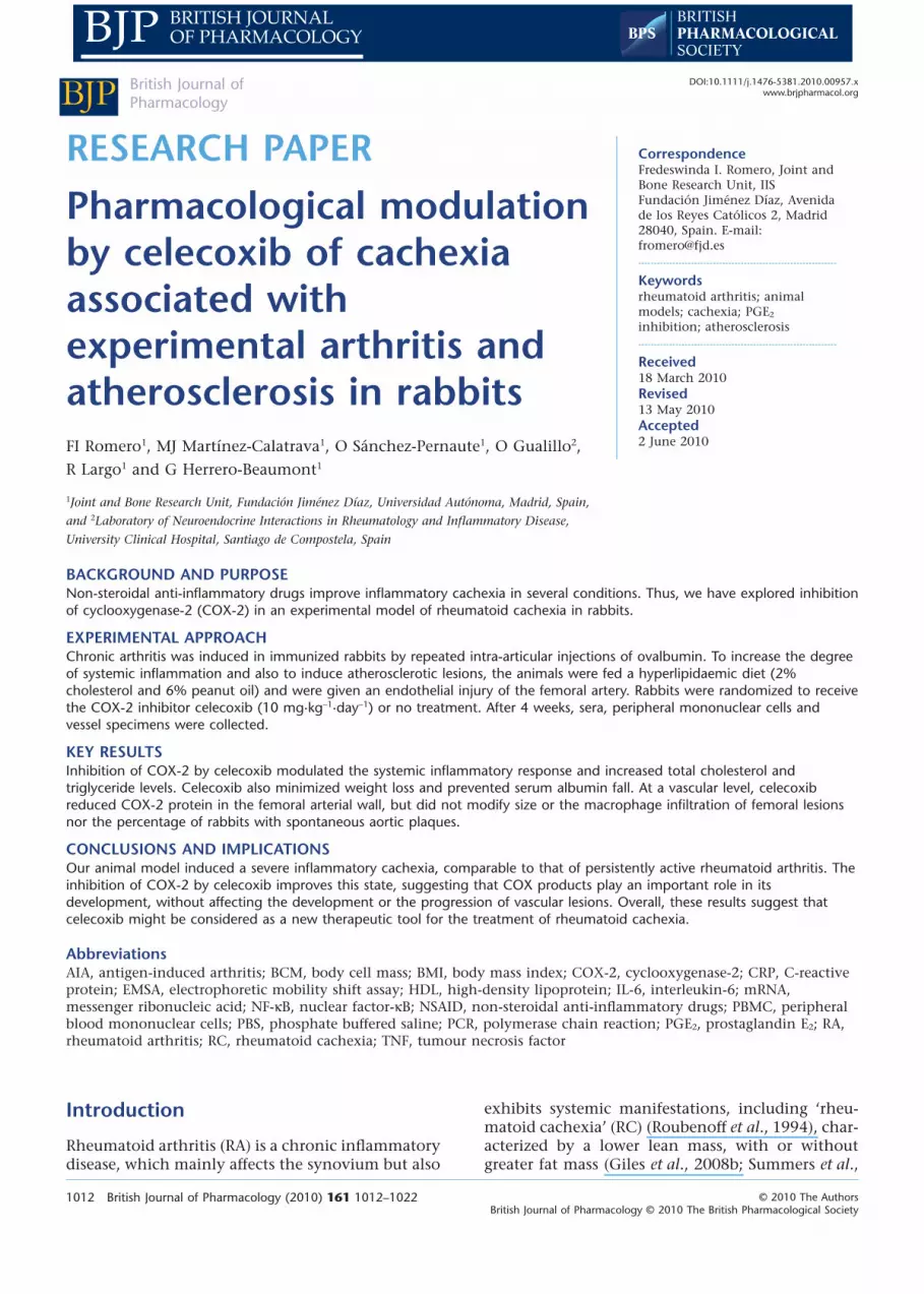
AnimalsAll animal care and experimental protocols for thisstudy complied with the Spanish regulations andthe Guidelines for the Care and Use of LaboratoryAnimals drawn up by the National Institutes ofHealth (USA) and were approved by the Institu-tional Ethics Committee of the Fundación JiménezDíaz Hospital. Forty-four male New Zealand whiterabbits were purchased when they weighed 3.0 �0.3 kg (Granja San Bernardo, Navarra, Spain).Rabbits were randomly assigned to three groups:healthy (n = 15), RC (RC group; n = 15) andRC rabbits treated with oral celecoxib at10 mg·kg-1·day-1 (CXB group; n = 14). The celecoxibwas in capsules (Celebrex; Pfizer) and given by oralgavage. They were fed with 85 g·day-1 of standardrabbit chow or hypercholesterolaemic diet andwater ad libitum according with the experimentalanimal model recently published (Largo et al.,2008). A schematic representation of the experi-mental model of RC is shown in Figure 1.
Body-weight measurements were recorded atbaseline and at the end of the experiment, andbody-weight gain was calculated. All rabbits werekilled by an overdose of pentobarbital. The femoralarteries and the thoracic aortas were removed andfixed in 4% buffered paraformaldehyde, dehydratedand embedded in paraffin. Another piece of femoralartery was snap-frozen and stored at -70°C formolecular biology studies.
BJPPGE2 inhibition in RA-associated cachexia
British Journal of Pharmacology (2010) 161 1012–1022 1013

Biochemical measurementsAt the end of the study, 10 mL blood was obtainedand centrifuged to obtain serum. The total serumcholesterol, triglycerides and high-density lipopro-tein (HDL) cholesterol levels were determined byenzymatic methods (Sigma-Aldrich, Inc). SerumC-reactive protein (CRP) and IL-6 levels were mea-sured using specific commercial enzyme-linkedimmunosorbent assays (Alpha Diagnostic Interna-tional Inc, San Antonio, TX, USA and R&D SystemsInc, Minneapolis, MN, USA respectively). Totalprotein levels were determined from serum totalprotein content by final point biuret method usingDDPP Hitachi Modular automatic equipment. Thesensitivity of the assay was 2 g·L-1. The intra- and
inter-analysis coefficients of variation were 0.7%and 1.22% respectively. Once the total proteinlevel was obtained, albumin was calculated as a per-centage by capillary electrophoresis with Capillarysequipment (Sebia).
Isolation of peripheral bloodmononuclear cellsPeripheral blood mononuclear cells (PBMC) wereisolated from total blood with Lymphoprep(Hernández-Presa et al., 2002). Thereafter, cells wereprocessed to obtain the nuclear extracts (Largo et al.,2003) or dissolved in Trizol Reagent (Roche Diagnos-tics) to extract total RNA from the lysates.
Electrophoretic mobility shift assayNuclear protein extracts pooled from mononuclearcells were prepared as described by Hernández-Presaet al. (2002), and the protein concentration in eachsample was quantified by the BCA method (ThermoScientific, Meridian Road, USA). A consensus oligo-nucleotide for nuclear factor-kB (NF-kB) (PromegaBiotech Iberica) was end-labelled with 32P using 10units of T4 polynucleotide kinase (Promega BiotechIberica), and the nuclear extracts were then equili-brated for 10 min in binding buffer before addingthe labelled probe (Largo et al., 2003). The specific-ity of the assay was tested. Samples were resolved on4% non-denaturing acrylamide gels in Tris-boratebuffer, which were exposed to X-ray film to deter-mine the NF-kB.
RNA extraction and real-time polymerasechain reactionTotal RNA was extracted from femoral arteries orPBMC using the Trizol method (Roche Diagnostic).First strand cDNA was synthesized from 1 mg of totalRNA using the High-Capacity cDNA Reverse Tran-scription Kit according to the manufacturer’sinstructions (Applied Biosystems, Stockholm,Sweden). Polymerase chain reaction primers andprobes were designed by Applied Biosystems (Vidalet al., 2007) and the endogenous control of ourassays was the eukaryotic 18S rRNA. Thermalcycling and florescence detection were performedon an ABI Prism 7500 Sequence Detection Systemwith ABI Prism 7500 SDS software (Applied Biosys-tems, Stockholm, Sweden). Thermal cycling wascarried out for 10 min at 95°C followed by 40 cyclesof 15 s at 95°C and 1 min at 60°C. Gene expressionvalues were calculated using the e-2DDCt method.
Western blot analysisTotal proteins were isolated from femoral arterieswith Trizol (Roche Diagnostics) and they were
A
B
IMMUNIZATION OVA INJECTION
–2 –1 0 1 2 3 4 weeks
De-endothelization
surgeryEnd of study
CXB (10 mg kg–1 day–1)
Atherogenic diet
80
70
60
50
40
30
20
10
0
1.4
1.2
1
0.8
0.6
0.4
0.2
0
Alb
um
in (
g L
–1)
Healthy RC CXB
AlbuminWeight gain
*
*
*##
Weig
ht g
ain
(kg)
Figure 1Effect of the combination of chronic antigen-induced arthritis (AIA)and atherosclerosis in parameters associated with cachexia. (A) Sche-matic representation of the experimental model. (B) Serum albuminlevels and changes in weight-gaining healthy rabbits, rabbits withboth chronic AIA and atherosclerosis, untreated (rheumatoidcachexia, RC) and treated with celecoxib (CXB). *P < 0.05 versushealthy controls; #P < 0.05 versus RC.
BJP FI Romero et al.
1014 British Journal of Pharmacology (2010) 161 1012–1022

resolved on 10% acrylamide-SDS gels. After transferto polyvinylidene difluoride membranes, the lysateswere probed with antibodies against COX-2 (SantaCruz Biotech) and a-tubulin (Sigma – Aldrich,Inc18). Briefly, the membranes were blocked in 5%skimmed milk in phosphate buffered saline-Tween20 (PBS-Tween 20) for 1 h at room temperature, andincubated overnight at 4°C with the primary anti-bodies diluted 1/500 in PBS containing 0.3% Tween20 and 3% bovine serum albumin. Antibodybinding was detected by enhanced chemoluminis-cence using peroxidise-conjugated secondary anti-bodies diluted 1/1000 in PBS-Tween 20, and theresults were expressed in arbitrary densitometricunits normalized to the a-tubulin levels.
Histopathological analysis of vascular lesionsArteries were divided into four equal fragments andembedded in a single paraffin block. These blockswere then cross-sectioned into 4 mm thick serial sec-tions and multiple sections from each block werechosen at regular intervals and stained withhaematoxylin-eosin or orcein. These sections werethen analysed qualitatively to identify the zone withthe most severe stenosis where morphometric andimmunohistochemical studies were performed.Morphometry was performed using the Olympussemiautomatic analytic system with Micro Imagesoftware (version 1.0 for Windows). Slide photomi-crographs were captured with an Olympus micro-scope (BH-2) connected to a video camera(Hernández-Presa et al., 2002; Vidal et al., 2007).Intima and media thickness were measured infemoral arteries and the results were expressed asintima/media thickness ratios.
Immunohistochemistry in femoral lesionsWe identified macrophages in the site where thefemoral lesions showed the maximal stenosis usinga monoclonal anti-rabbit macrophage antibody(RAM11, Dako Corporation, Glostrup, Denmark)according to a protocol described previously(Hernández-Presa et al., 2002; Vidal et al., 2007).Tissues, previously counterstained with haematoxy-lin, were mounted in Pertex (Medite), and thestained area was analysed in digital photomicro-graphs and expressed as a percentage per squaremillimetre of tissue (Hernández-Presa et al., 2002;Vidal et al., 2007). The negative controls involveddetection with an IgG isotype.
Data analysisThe values for lipids and the data from the morpho-metric analysis, immunohistochemistry, electro-phoretic mobility shift assay, Western blots andreal-time polymerase chain reaction are expressed as
the mean � SEM, and they were analysed using theMann–Whitney U-test. Where multiple compari-sons were performed, the Kruskal–Wallis test wasused. The null hypothesis was rejected in each sta-tistical test when the P-value was less than 0.05. Allstatistical analyses were performed using WindowsSPSS version 11.0 software (SPSS, Inc, Chicago, IL,USA).
Results
Weight change and albuminRabbits with RC showed lower body-weight gainthan healthy animals (0.34 � 0.02 kg vs. 1.06 �0.03 kg, P < 0.001 in both cases). Albumin levelswere lower in sera from RC rabbits compared withthe healthy group (P = 0.001; Figure 1). Rabbits withRC treated with celecoxib (CXB group) showed lessweight loss (P = 0.009 vs. RC) and maintained serumconcentrations of albumin within the normal range(P = 0.075 vs. healthy, Figure 1).
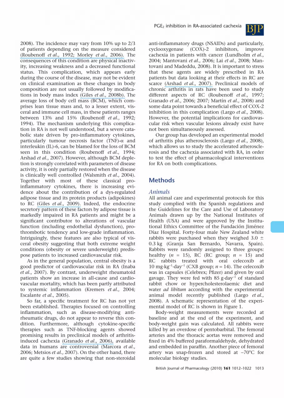
Parameters of systemic inflammationAs described (Largo et al., 2008), the induction ofatherosclerosis plus chronic arthritis leading to RCsignificantly increased levels of both CRP and IL-6in serum (P < 0.001 vs. healthy animals) (Figure 2).The administration of celecoxib to rabbits with RCreduced serum IL-6 and CRP levels (P = 0.037 and P= 0.001, respectively, vs. RC) (Figure 2A).
COX-2 and CCL2 gene expression in PBMCIn previous work, we have found that PBMC fromrabbits with RC show an up-regulation in the geneexpression of COX-2 and the chemokine CCL2,compared with healthy animals (data not shown).Here, we looked for the gene expression of theseproteins in rabbits with or without treatment, andfound that celecoxib did not prevent the increase inboth COX-2 (11.3 � 3.5-fold vs. 7.8 � 1.4-fold, P =0.87) and CCL2 expression (2 � 0.6-fold vs. 3.3 �0.6-fold, P = 0.22) on PBMC.
NF-kB activation in PBMCNF-kB activation was determined by electrophoreticmobility shift assay in nuclear extracts from thePBMC. In these cells, there was an increase in theNF-kB activity in the RC group (P = 0.026 vs. healthyanimals). The administration of celecoxib led to areduction in NF-kB activation in PBMC in compari-son with untreated animals (P = 0.053) (Figure 2B).
Lipid profileRabbits with RC showed lower levels of HDL andhigher levels of total cholesterol and triglycerides
BJPPGE2 inhibition in RA-associated cachexia
British Journal of Pharmacology (2010) 161 1012–1022 1015

than healthy animals. Treatment with celecoxib,increased levels of total cholesterol and triglycerides(P < 0.001 vs. RC), and decreased HDL cholesterollevels (P < 0.001, vs. RC) (Table 1).
Quantification of vascular lesions in thefemoral arteriesAll the animals from the groups with RC – RC andCXB groups– developed a stenotic lesion in thefemoral artery characterized by a hyperplasic trans-formation of the intima and foam cell infiltration.The intima/media thickness ratio was quantified atthe site of maximal stenosis. The administration ofcelecoxib did not modify this parameter in theinjured femoral arteries when compared with RCanimals (Figure 3). No lesions were developed in thevessels of the healthy rabbits.
Inflammatory changes in the femoral arteriesAll the femoral arteries from the groups with RC (RCand CXB groups) displayed macrophage infiltrationat the neointima, assessed with RAM11 staining. Asinflammatory changes at the vessel wall are relatedto the progression of the vascular lesion, we hadexpected that a COX-2 inhibitor could suppress orabolish this component. However, celecoxib treat-ment did not decrease the macrophage density inthe femoral lesion (Figure 3). We had previouslyfound that COX-2 protein expression and CCL2mRNA were up-regulated in femoral extracts ofrabbits with RC, with regard to hyperlipidaemicrabbits (Largo et al., 2008). So we looked for theeffect of celecoxib on these markers of arthritis-associated vascular damage. There was a significantfall in COX-2 protein levels in rabbits treated withcelecoxib compared with RC (fivefold levels, P =0.004), while there was no effect on the expressionof CCL2 mRNA, which persisted 3.6-fold increasedversus healthy specimens (Figure 4).
Atherosclerotic lesions in the aortaWe also studied segments of the thoracic aorta, asone of the additional effects of experimental arthri-tis upon vascular damage was an increase in theincidence of distant lesions. In this regard, aortic
400
IL-6
C-reative protein
NF-kB
A
B
300
200
100
0
16
12
8
4
0
160
120
80
C-re
ativ
e p
rote
in (m
g·m
L–
1)
IL-6
(p
g·m
L–1)
PB
MC
NF
-kB
bin
din
g
40
0Healthy RC CXB
Healthy RC CXB
*#
#
*#
**
*
Figure 2Effect of celecoxib (CXB) administration to rabbits with rheumatoidcachexia (RC) on parameters of systemic inflammation. (A) IL-6 andC-reactive protein concentrations in serum. *P < 0.01 versus healthycontrols; #P < 0.05 versus non-treated (RC) rabbits. (B) Electro-phoretic mobility shift assay and densitometric analysis of radio-labelled NF-kB bound to the nuclear proteins extracted from theperipheral blood mononuclear cells (PBMC). Lane 1 indicates thecompetitive control of the lane 3 (corresponding to a RC rabbit). *P= 0.022 versus healthy controls. #P = 0.053 versus healthy and RC.
Table 1Lipid levels in serum from healthy, RC and CXB groups of rabbits
(mg·L-1) Healthy (n = 15) RC (n = 15) CXB (n = 14)
Total cholesterol 500 � 40 18 600 � 680* 20 400 � 420*#
HDL 370 � 30 190 � 20* 80 � 10*#
Triglycerides 740 � 100 900 � 100* 1220 � 140*#
Data are shown as mean � SEM. The healthy group were untreated, the RC group had rheumatoid cachexia and the CXB group hadrheumatoid cachexia and were treated with oral celecoxib (10 mg·day-1). Blood was taken at the end of the experimental period (seeFigure 1A).*P < 0.05 versus healthy.#P < 0.05 versus RC.
BJP FI Romero et al.
1016 British Journal of Pharmacology (2010) 161 1012–1022

3.5
3
2.5
2
1.5
1
0.5
0
A B C
Fem
ora
l IM
T r
ati
o
Healthy RC
RC CXB
E F
CXB
*
*
60
50
40
30
20
10
0
B
Fem
ora
l R
AM
11
sta
inin
g (
%)
Healthy RC CXB
**
Figure 3Quantification of vascular lesions in injured femoral arteries. Upper panel, neointimal hyperplasia. (A) Intima/media thickness ratio (IMT). *P < 0.05versus healthy controls. Representative haematoxylin-eosin stained femoral sections of rheumatoid cachexia (RC)- and celecoxib (CXB)-treatedrabbits are shown: (B) RC rabbit; (C) CXB-treated rabbit (magnification, 100¥). Lower panel, macrophage detection. (D) Quantification ofmacrophage staining in the neointimal area. *P < 0.05 versus healthy controls. Representative samples of RAM11 immunohistochemistry in RC-and CXB-treated rabbits are shown: (E) RC rabbit; (F) CXB-treated rabbit (magnification, 100¥).
Healthy
140
6
5
4
3
2
1
0
Fem
ora
l C
OX
-2
Pro
tein
Exp
ressio
n
Fem
ora
l C
OX
-2
Gen
e E
xp
ressio
n
120
100
80
60
40
20
0
A
C
B
RC CXB
Healthy RC CXB
P = 0.057
Healthy RC CXB
*#
*
*8
6
4
2
0
Fem
ora
l C
CL
2
Gen
e E
xp
ressio
n
D
Healthy RC CXB
*
*
COX-2
Figure 4COX-2 and CCL2 expression in the femoral artery. Top, COX-2 protein expression: (A) densitometric analysis of Western blot studies; (B) arepresentative Western blot of COX-2 in femoral arteries. Bottom (C and D): analysis of COX-2 (C) and CCL2 (D) mRNA expression measured byreal-time polymerase chain reaction method. *P < 0.05 versus healthy controls; #P < 0.05 versus rheumatoid cachexia (RC) rabbits. CXB, celecoxib.
BJPPGE2 inhibition in RA-associated cachexia
British Journal of Pharmacology (2010) 161 1012–1022 1017

lesions were observed in 60% of animals from RCgroup and in 57% of celecoxib-treated rabbits (P >0.05). No aortic lesions were detected in the rabbitsfed with a standard diet (healthy).
The lesions in the aorta were highly variable insize and appearance, and they consisted of lipid-richdeposits adhering to the aortic wall that were infil-trated with foam cells and other mononuclear cells.The larger ones contained elastic fibres, fibroblast-like cells and collagen deposition. In some cases,fatty streaks were found inside the media layer, asso-ciated with the rupture of elastic fibres, as observedwith orcein (Figure 5).
Discussion
The excessive synthesis of pro-inflammatory cytok-ines, such as IL-6, TNF-a and IL-1, is thought to bethe most important cause of RC (Roubenoff et al.,1992). It has been reported that high levels of thesecytokines lead to an increased hepatic protein syn-thesis (Walsmith and Roubenoff, 2002) and to asevere protein depletion and loss of weight, attrib-
uted in part to NF-kB activation (Guttridge et al.,2000; Acharyya et al., 2007). In agreement, wefound that serum albumin levels fell in parallel tothe increase in systemic inflammation, and NF-kBactivation in PBMC. In addition, pro-inflammatorycytokines induce the production of prostaglandin E2
and some studies also involve COX-2 activation inthe induction of cachectic status by insulin-likegrowth factor-I axis inhibition and activation of theubiquitin-mediated proteolytic system and musclewasting (Ganey et al., 2001; Davis et al., 2004;Granado et al., 2007). In rabbits with cachexia, wehave found COX-2 gene up-regulation at theinjured tissues, that is, the synovial membrane(Largo et al., 2008) and the femoral arteries. Here,selective inhibition of COX-2 with celecoxibreduced systemic inflammation and NF-kB activa-tion, and ameliorated the loss of weight and thereduction in circulating albumin of cachecticrabbits. As previous studies suggested, this effectcould be linked to regulation of TNF-a (Kontureket al., 2006; Granado et al., 2007). Indeed, it hasbeen reported that the inhibitory effect of NSAIDson NF-kB signalling might be responsible for the
A B
70
60
50
40
30
20
10
0
Healthy RC CXB
%R
abbits w
ith a
ort
ic lesio
ns
C
Figure 5Presence of atherosclerotic plaque in the aorta of the rabbits. Low magnification photomicrographs (40¥) show the distribution of plaques inrepresentative aortas from rheumatoid cachexia (RC)- (A) and celecoxib (CXB)-treated (B) rabbits, stained with orcein. The plaques are shown indetail in high magnification (200¥). (C) Graph bar. Percentage of rabbits with aortic plaque. *P < 0.05 versus healthy controls.
BJP FI Romero et al.
1018 British Journal of Pharmacology (2010) 161 1012–1022

suppression of muscle wasting induced by the acti-vation of the ubiquitin-proteasome pathway, whichhas been linked to the increase in TNF-a geneexpression observed in cachexia (Wyke et al., 2004).
Although increased levels of triglycerides are acommon finding in most examples of cachecticstatus (Khovidhunkit et al., 2004), lipolysis is notincreased and adipose lipogenesis is reduced inanimal models of arthritis-induced cachexia (Martínet al., 2008). On the other hand, celecoxib hasshown to decrease the expression of fatty acid syn-thase, a key enzyme in triglyceride synthesis, bydown-regulating c-Jun N-terminal kinase-1 (Lu andArcher, 2007), in animal models fed a high-fat diet.Surprisingly, in our study, triglycerides were furtherincreased in the celecoxib-treated group suggestingthat this drug may reverse the effect of inflamma-tion on lipid metabolism. In addition and, by con-trast with other species, inflammation induceshypercholesterolaemia in rodents and rabbits(Cabana et al., 1983; 1996; Khovidhunkit et al.,2004). Available data about the effect of COX-2 inhi-bition on cholesterol metabolism are controversial.While genetic COX-2 deficiency results in hyperc-holesterolaemia, which is further increased onatherogenic diets (Narasimha et al., 2007), celecoxibtreatment of apoE-deficient mice fed a high-fat dietdid not modify plasma cholesterol levels, although aslight activation of hydroxy-methyl-glutaryl-CoAreductase, the rate-limiting enzyme in cholesterolbiosynthesis, has been reported (Metzner et al.,2007). We also found a reduction in HDL levelsin rabbits with RC. During inflammation, serumHDL not only decreases but can also becomepro-inflammatory (Van Lenten et al., 1995). Cele-coxib administration, despite decreasing pro-inflammatory cytokines, paradoxically lowered HDLand this effect was accompanied by an increase intriglycerides. Moreover, the relationship betweenhypertriglyceridaemia and low HDL has been previ-ously described in acute and in non-acute phaseconditions (Gotto, 1990; Cabana et al., 1996). Inaddition, rabbits are deficient in hepatic lipase, soHDL triglyceride hydrolysis might be furthercompromised (Clay et al., 1989). As we did notdetermine HDL composition, we cannot say if thedecrease in the HDL levels was due to its enrichmentin triglycerides and/or a decrease in pro-inflammatory HDL synthesis.
On the other hand, treatment with celecoxib didnot worsen vascular lesions induced in rabbits withcachexia. The apparently adverse cardiovascularlipid profile developed by the celecoxib-treatedgroup, higher triglyceride and total cholesterollevels and lower HDL cholesterol than their non-treated counterparts, did not either result in a
higher incidence of atherosclerotic lesions on intactaortas. These data are consistent with the results ofMetzner et al. (2007), who did not find differencesin lesion sizes in apoE-/- mice on a high-fat dietwhen treated with coxibs while apoE-/- mice on achow diet developed larger lesions when treated,suggesting that COX-2 inhibition may not affect thelate stages of atherogenesis, once plaque generationhas already been initiated. Indeed, the vascularlesions in femoral arteries of celecoxib-treatedanimals did show a lesser degree of inflammation asmeasured by COX-2 abundance. We should stressthat more than lipid levels, lipoprotein content isimportant in the development of inflammatory vas-cular lesions (Van Lenten et al., 2007).
Mechanisms other than inhibition of COX-2 andprostaglandin E2 biosynthesis, such as a decrease insmooth muscle cell proliferation and neointimalhyperplasia through inhibition of protein kinase Bsignalling (Yang et al., 2004), have been describedwith celecoxib in vivo. In this context, we did notfind the inhibition in CCL2 expression, which hasbeen previously reported (Wang et al., 2005; Tegederand Geisslinger, 2006), although there was a ten-dency towards a lower expression in CXB animals.
In addition, the fall in CRP levels, a parameterthat has been linked to a higher cardiovascular risk,seen with celecoxib might contribute to the reducedseverity of inflammatory vascular lesions. In RA,cachexia is a common aberration of body composi-tion and it is characterized by the concurrentdecrease in fat-free mass and increase in fat mass,including central obesity. This may contributeclearly to the increased morbidity as well as themortality associated with RA. Cytokine-drivenhypermetabolism and protein degradation linked tothe disease causes reduction of fat-free mass, whichis, at the same time, combined with a significantincrease of fat mass. This status can be readilydefined as ‘cachectic obesity’, a condition resem-bling morbid obesity and which may contribute toincreased risk of cardiovascular diseases in RApatients. ‘Cachectic obesity’ could be considered atruly ‘silent killer’ of these patients (Kumar andArmstrong, 2008). In addition, the increased fatmass, which in most cases can be considered asfrank obesity, is responsible of the pro-inflammatorystatus that perpetuates disease mechanisms associ-ated with RA, including cardiovascular disease,immune dys-regulation and inflammatory path-ways, which are, of course, capital potential targetsfor therapy. Interestingly, fat mass is an importantcontributor to CRP levels in RA (Giles et al., 2008a)and a recent study in hyperlipidaemic rats showedthat celecoxib decreased intra-abdominal adiposetissue mass (Lu and Archer, 2007). Although it is
BJPPGE2 inhibition in RA-associated cachexia
British Journal of Pharmacology (2010) 161 1012–1022 1019

tempting to do so, we cannot speculate on this as wehave not performed studies of body composition.
Summarizing, the major two contributions ofour results are, on one hand, the development of anexperimental model, which allows us to explorefurther the molecular mechanisms underlying RCand to simultaneously test the effect of drugs on thecardiovascular risk associated with RA. On the otherhand, our results show that the use of an inhibitorof COX-2 improves RC without worsening alreadyestablished vascular lesions or facilitating new ones.
Although the published literature and availableexpert’s guidelines do not recommend the general-ized use of NSAIDs, and particularly of COX-2inhibitors, in patients at increased cardiovascularrisk, in the light of our study, this advice should notbe extended to diseases with an important inflam-matory component, such as active arthritis. In thiscontext, our results are consistent with a very recentphase II non-randomized study, which confirmedthat celecoxib, at a moderate dosage and for dura-tion of less than 6 months, did not increase cardio-vascular risk (Mantovani et al., 2010). Thus, theimprovement of RA-associated cachexia with thesedrugs may decrease the related poor clinical out-comes without having a negative effect on the asso-ciated cardiovascular risk, which is a key factor ofmorbidity and mortality in these patients. However,we have to keep in mind that our data are limited toan experimental model. Thus, further clinicalstudies in this field are needed in order to considercachexia as a new indication for NSAIDs in thisgroup of patients.
Acknowledgements
We thank Dr Concha de la Piedra and Petra Rubiofor their collaborations in biochemical and histo-pathological studies. This work was supported bythe Spanish Ministry of Health through the Fondode Investigación Sanitaria, Instituto de Salud CarlosIII [CP03/0011, PI06/0032]; the Spanish Ministry ofScience and Innovation [SAF 2006/2704]; and theMutua Madrileña Automovilística Foundation.
Conflicts of interest
The authors have no conflicts of interest to declare.
References
Acharyya S, Villalta SA, Bakkar N, Bupha-Intr T,Janssen PM, Carathers M et al. (2007). Interplay ofIKK/NF-kappaB signaling in macrophages and myofibers
promotes muscle degeneration in Duchenne musculardystrophy. J Clin Invest 117: 889–901.
Arshad A, Rashid R, Benjamin K (2007). The effect ofdisease activity on fat free mass and resting energyexpenditure in patients with rheumatoid arthritis versusnoninflammatory arthropathies/soft tissue rheumatism.Mod Rheumatol 17: 470–475.
Cabana VG, Gewurz H, Siegel JN (1983).Inflammation-induced changes in rabbit CRP andplasma lipoproteins. J Immunol 130: 1736–1742.
Cabana VG, Lukens JR, Rice KS, Hawkins TJ, Getz GS(1996). HDL content and composition in acute phaseresponse in three species: triglyceride enrichment ofHDL a factor in its decrease. J Lipid Res 37: 2662–2674.
Clay MA, Hopkins GJ, Ehnholm CP, Barter PJ (1989).The rabbit as an animal model of hepatic lipasedeficiency. Biochim Biophys Acta 1002: 173–181.
Davis TW, Zweifel BS, O’Neil JM, Heuvelman DM,Abegg AL, Hendrich TO et al. (2004). Inhibition ofcyclooxigenase-2 by celecoxib reverses tumor-inducedwasting. J Pharmacol Exp Ther 308: 929–934.
Escalante A, Haas R, Del Rincón I (2005). Paradoxicaleffect of body mass index on survival in rheumatoidarthritis. Arch Intern Med 165: 1624–1629.
Ganey PE, Barton YW, Kinser S, Sneed RA, Barton CC,Roth RA (2001). Involvement of cyclooxigenase-2 in thepotentiation of allyl alcohol-induced liver injury bybacterial lipopolysaccharide. Toxicol Appl Pharmacol174: 113–121.
Giles JT, Bartlett SJ, Andersen R, Thompson R,Fontaine KR, Bathon JM (2008a). Association of body fatwith C-reactive protein in rheumatoid arthritis. ArthritisRheum 58: 2632–2641.
Giles JT, Ling SM, Ferruci L, Fontaine KR, Bathon JM(2008b). Abnormal body composition phenotypes inolder rheumatoid arthritis patients: association withdisease characteristics and pharmacotherapies. ArthritisRheum 59: 807–815.
Giles JT, Allison M, Bingham CO Jr 3rd, Scott WM Jr,Bathon JM (2009). Adiponectin is a mediator of theinverse association of adiposity with radiographicdamage in rheumatoid arthritis. Arthritis Rheum 61:1248–1256.
Gotto AM Jr (1990). Interrelationship of triglycerideswith lipoproteins and high density lipoproteins. Am JCardiol 66: 20A–23A.
Granado M, Priego T, Martín I, Vara E,López-Calderón A, Villanúa MA (2006). Anti-tumornecrosis factor agent PEG-sTNFRI improves the growthhormone/insulin-like growth factor-I system inadjuvant-induced arthritic rats. Eur J Pharmacol 536:204–210.
Granado M, Martín AI, Villanúa MA, López-Calderón A(2007). Experimental arthritis inhibits the insulin-likegrowth factor-I axis and induces muscle wasting
BJP FI Romero et al.
1020 British Journal of Pharmacology (2010) 161 1012–1022

through cyclooxygenase-2 activation. Am J PhysiolEndocrinol Metab 292: E1656–E1665.
Guttridge DC, MayoMW MLV, Wang CY, Baldwin AS Jr(2000). NF-kappaB-induced loss of MyoD messengerRNA: possible role in muscle decay and cachexia.Science 289: 2363–2366.
Hernández-Presa MA, Martin-Ventura JL, Ortego M,Gómez-Hernández A, Tuñón J, Hernández-Vargas P et al.(2002). Atorvastatin reduces the expression ofcyclooxygenase-2 in a rabbit model of atherosclerosisand in cultured vascular smooth muscle cells.Atherosclerosis 160: 49–58.
Inaba M, Tanaka K, Goto H, Usami T, Azumi K,Kubota H et al. (2007). Independent association ofincreased trunk fat with increased arterial stiffening inpostmenopausal patients with rheumatoid arthritis. JRheumatol 34: 290–295.
Khovidhunkit W, Min-Sun K, Memon RA, Shigenaga JK,Moser AH, Feingold KR et al. (2004). Effects of infectionand inflammation on lipid and lipoprotein metabolism:mechanisms and consequences to the host. J Lipid Res45: 1169–1196.
Konturek PC, Rembiasz K, Burnat G, Konturek SJ,Tusinela M, Bielanski W et al. (2006). Effects ofcyclooxigenase-2 inhibition on serum and tumorgastrins and expression of apoptosis-related proteins incolorectal cancer. Dig Dis Sci 51: 779–787.
Kremers HM, Nicola PJ, Crowson CS, Ballman KV,Gabriel SE (2004). Prognostic importance of low bodymass index in relation to cardiovascular mortality inrheumatoid arthritis. Arthritis Rheum 50: 3450–3457.
Kumar N, Armstrong DJ (2008). Cardiovascular disease –the silent killer in rheumatoid arthritis. Clin Med 8:384–387.
Lai V, George J, Richey L, Kim HJ, Cannon T, Shores Cet al. (2008). Results of a pilot study of the effects ofcelecoxib on cancer cachexia in patients with cancer ofthe head, neck and gastrointestinal tract. Head Neck 30:67–74.
Largo R, Alvarez-Soria MA, Diez-Ortego I, Calvo E,Sánchez-Pernaute O, Egido J et al. (2003). Glucosamineinhibits IL-1b-induced NFkB activation in humanosteoarthritic chondrocytes. Osteoarthritis Cartilage 11:290–298.
Largo R, Sánchez-Pernaute O, Marcos ME,Moreno-Rubio J, Aparicio C, Granado R et al. (2008).Chronic arthritis aggravates vascular lesions in rabbitswith atherosclerosis: a novel model of atherosclerosisassociated with chronic inflammation. Arthritis Rheum58: 2723–2734.
Lu S, Archer MC (2007). Celecoxib decreases fatty acidsynthase expression via down-regulation of c-JunN-Terminal Kinase-1. Exp Biol Med 232: 643–653.
Lundholm K, Daneryd P, Corner U, Hyltander A,Bosaeus I (2004). Evidence that long-termCOX-treatment improves energy homeostasis and body
composition in cancer patients with progressivecachexia. Int J Oncol 24: 505–512.
Mantovani G, Madeddu C (2008). Cyclooxygenase-2inhibitors and antioxidants in the treatment ofcachexia. Curr Opin Support Palliat Care 2: 275–281.
Mantovani G, Macciò A, Madeddu C, Gramignano G,Lusso MR, Serpe R et al. (2006). A phase II study withantioxidants, both in the diet and supplemented,pharmaconutritional support, progestagen, andanti-cyclooxigenase-2 showing efficacy and safety inpatients with cancer-related anorexia/cachexia andoxidative stress. Cancer Epidemiol Biomarkers Prev 15:1030–1034.
Mantovani G, Macció A, Madeddu C, Serpe R, Antoni G,Massa E et al. (2010). Phase II nonrandomized study ofthe efficacy and safety of COX-2 inhibitor celecoxib onpatients with cancer cachexia. J Mol Med 88: 85–92.
Marcora SM, Chester KR, Mittal G, Lemmey AB,Maddison PJ (2006). Randomized phase 2 trial ofanti-tumor necrosis factor therapy for cachexia inpatients with early rheumatoid arthritis. Am J Clin Nutr84: 1463–1472.
Martín AI, Castillero E, Granado M, López-Menduiña M,Villanúa MA, López-Calderón A (2008). Adipose tissueloss in adjuvant arthritis is associated with a decrease inlipogenesis, but not with an increase in lipolysis. JEndocrinol 197: 111–119.
Metsios GS, Stavropoulos-Kalinoglou A, Douglas KMJ,Koutedakis Y, Nevill AM, Panoulas VF et al. (2007).Blockade of tumour necrosis factor-a in rheumatoidarthritis: effects on components of rheumatoid cachexia.Rheumatology 46: 1824–1827.
Metzner J, Popp L, Marian C, Schmidt R,Manderscheid C, Renne C et al. (2007). The effects ofCOX-2 selective and non-selective NSAIDs on theiniciation and progression of athrosclerosis in ApoE-/-
mice. J Mol Med 85: 623–633.
Morley JE, Thomas DR, Wilson MMG (2006). Cachexia:Pathophysiology and clinical relevance. Am J Clin Nutr83: 735–743.
Narasimha A, Watanabe J, Lin JA, Hama S,Langenbach R, Navab M et al. (2007). A novelanti-atherogenic role for COX-2: potencial mechanismfor the cardiovascular side effects of COX-2 inhibitors.Prostaglandins Other Lipid Mediat 84: 24–33.
Roubenoff R, Roubenoff RA, Ward LM, Holland SM,Hellmann DB (1992). Rheumatoid cachexia: depletionof lean body mass in rheumatoid artritis. Possibleassociation with tumor necrosis factor. J Rheumatol 19:1505–1510.
Roubenoff R, Roubenoff RA, Cannon JG, Kehayias JJ,Zhuang H, Dawson-Hughes B et al. (1994). Rheumatoidcachexia: cytokine-driven hypermetabolismaccompanying reduced body cell mass in chronicinflammation. J Clin Invest 93: 2379–2386.
Roubenoff R, Freeman LM, Smith DE, Abad LW,Dinarello CA, Kehayias JJ (1997). Adjuvant arthritis as amodel of inflammatory cachexia. Arthritis Rheum 40:534–539.
BJPPGE2 inhibition in RA-associated cachexia
British Journal of Pharmacology (2010) 161 1012–1022 1021

Summers GD, Deighton CM, Rennie MJ, Booth AH(2008). Rheumatoid cachexia: a clinical perspective.Rheumatology (Oxford) 47: 1124–1131.
Tegeder I, Geisslinger G (2006). Cardiovascular risk withcyclooxygenase inhibitors: general problem withsubstance specific differences. Naunyn SchmiedebergsArch Pharmacol 373: 1–17.
Van Lenten BJ, Hama SY, de Beer FC, Stafforini DM,McIntyre TM, Prescott SM et al. (1995).Anti-inflammatory HDL becomes pro-inflammatoryduring the acute phase response. J Clin Invest 96:2758–2767.
Van Lenten BJ, Wagner AC, Navab M,Anantharamaiah GM, Hama S, Reddy ST et al. (2007).Lipoprotein inflammatory properties and serum amyloidA levels but not cholesterol levels predict lesion area incholesterol-fed rabbits. J Lipid Res 48: 2344–2353.
Vidal C, Gómez-Hernández A, Sánchez-Galán E,González A, Ortega L, Gómez-Gerique JA et al. (2007).Licofenole, a balanced inhibitor of cyclooxigenase andlypoxygenase reduces inflammation in a rabbit modelof atherosclerosis. J Pharmacol Exp Ther 320:108–116.
Walsmith J, Roubenoff R (2002). Cachexia inrheumatoid arthritis. Int J Cardiol 85: 89–99.
Walsmith J, Abad L, Kehayias J, Roubenoff R (2004).Tumor necrosis factor-a production is associated withless body cell mass in women with rheumatoid arthritis.J Rheumatol 31: 23–29.
Wang K, Tarakji K, Zhou Z, Zhang M, Forudi F, Zhou Xet al. (2005). Celecoxib, a selective cyclooxygenase-2inhibitor, decreases monocyte chemoattractant protein-1expression and neointimal hyperplasia in the rabbitatherosclerotic balloon injury model. J CardiovascPharmacol 45: 61–67.
Wyke SM, Russell ST, Tisdale MJ (2004). Induction ofproteasome expression in skeletal muscle is attenuatedby inhibitors of NF-kappa B activation. Br J Cancer 91:1742–1750.
Yang HM, Kim HS, Park KW, You HJ, Jeon SI, Youn SWet al. (2004). Celecoxib, a cyclooxygenase-2 inhibitor,reduces neointimal hyperplasia through inhibition ofAkt signalling. Circulation 110: 301–308.
BJP FI Romero et al.
1022 British Journal of Pharmacology (2010) 161 1012–1022


