Embed Size (px)
Citation preview

Photocycle and Vectorial Proton Transfer in a Rhodopsin from theEukaryote Oxyrrhis marinaChristian Janke,† Frank Scholz,‡ Johanna Becker-Baldus,§ Clemens Glaubitz,§ Phillip G. Wood,†
Ernst Bamberg,† Josef Wachtveitl,‡ and Christian Bamann*,†
†Max-Planck-Institut fur Biophysik, Max-von-Laue Strasse 3, 60438 Frankfurt am Main, Germany‡Institute of Physical and Theoretical Chemistry and §Institute for Biophysical Chemistry & Centre for Biomolecular MagneticResonance, Johann Wolfgang Goethe-Universitat, Max-von-Laue Strasse 7, 60438 Frankfurt am Main, Germany
*S Supporting Information
ABSTRACT: Retinylidene photoreceptors are ubiquitouslypresent in marine protists as first documented by theidentification of green proteorhodopsin (GPR). We presenta detailed investigation of a rhodopsin from the protistOxyrrhis marina (OR1) with respect to its spectroscopicproperties and to its vectorial proton transport. Despite itshomology to GPR, OR1’s features differ markedly in its pHdependence. Protonation of the proton acceptor starts at pHbelow 4 and is sensitive to the ionic conditions. The mutationof a conserved histidine H62 did not influence the pKa value ina similar manner as in other proteorhodopsins where the charged histidine interacts with the proton acceptor forming the so-called His-Asp cluster. Mutational and pH-induced effects were further reflected in the temporal behavior upon light excitationranging from femtoseconds to seconds. The primary photodynamics exhibits a high sensitivity to the environment of the protonacceptor D100 that are correlated to the different initial states. The mutation of the H62 does not affect photoisomerization atneutral pH. This is in agreement with NMR data indicating the absence of the His-Asp cluster. The subsequent steps in thephotocycle revealed protonation reactions at the Schiff base coupled to proton pumping even at low pH. The main electrogenicsteps are associated with the reprotonation of the Schiff base and internal proton donor. Hence, OR1 shows a different theme ofthe His-Asp organization where the low pKa of the proton acceptor is not dominated by this interaction, but by otherelectrostatic factors.
The heterotrophic dinoflagellate Oxyrrhis marina representsan important model organism for marine ecology.1 The
morphospecies Oxyrrhis marina derived from an early branchpoint in the evolution of dinoflagellates. Therefore it possessescytological features which are distinct from the phylumdinophyceae such as nuclear division.2 Its phylogenetic positionrenders Oxyrrhis marina a model organism for the evolution ofgenes and organelles.3 Two recent publications revealed theexistence of a strongly expressed rhodopsin in Oxyrrhis marina(named OR1). It belongs to the proteorhodopsin (pR)family4,5 and has been presumably acquired from lateral genetransfer similar to other pR-like molecules.6 Differentconclusions were drawn about its biological function fromphototactic and cell localization experiments. A photosensoricfunction of a rhodopsin was found in phototactic experimentscorroborated by the response sensitivity toward hydroxylaminethat bleaches retinal containing photoreceptors.5 On thecontrary, Slamovits et al. found an endomembrane localizationof a strongly expressed rhodopsin that they attributed to aphototrophic function.4
In eukaryotes, several type I rhodopsins have been identifiedand characterized before that are not members of the pR family,in particular from algae7,8 or from fungi.9 In several cases, their
involvement in phototaxis is well documented,10 while in otherstheir native function is elusive. In host systems, they can workas a proton pump leading to speculation about theircontribution to light energy conversion (e.g., ref 11). In fact,the awareness of retinal-based photosynthesis started with thediscovery of bacteriorhodopsin (bR).12 After the identificationof a eubacterial green proteorhodopsin (GPR),13 retinaldependent light energy fixation turned into a globalphenomenon by the ubiquitous presence of pR-like genes inall kingdoms.14 All pRs described so far share the generalfeatures of type I rhodopsins with the seven transmembranehelices and a retinal chromophore linked to a lysine side chainvia a protonated Schiff base (pSB).15−17 As in the proton pumpbR, absorption of a photon initiates a photocycle in GPRconsisting of a series of spectrally distinct intermediates (K, L,M, N, O).17,18 The primary process in this photocycle is anisomerization of the retinal chromophore from its all-trans stateto 13-cis leading to the formation of the first ground statephotoproduct (termed K-intermediate in bR).19 Subsequently,
Received: October 17, 2012Revised: March 26, 2013Published: March 27, 2013
Article
pubs.acs.org/biochemistry
© 2013 American Chemical Society 2750 dx.doi.org/10.1021/bi301412n | Biochemistry 2013, 52, 2750−2763

the Schiff base deprotonates and the released proton movestoward the extracellular side with D97 (D85 in bR) as theprimary proton acceptor. Reprotonation of the Schiff base fromE108 (D96 in bR) located at the cytoplasmic side establishesthe vectorial proton transport.The primary proton acceptor D97 in GPR has an unusual
high pKa of 7.7 that is provoked by the conserved H75 residueforming a pH-dependent hydrogen-bonded proton dyad withD97 as it has been shown by solid-state NMR measurements.20
The close proximity of these two residues has also beendescribed in the solution NMR structure of GPR21 and the X-ray structure of xanthorhodopsin (XR).16 The protonation stateof D97 in GPR defines the vectoriality of the protontransport.22 At high pH with a deprotonated D97, an outwardtransport occurs as in bR although the sequence of theindividual proton transfer steps differs. This is mainly due to adifferent proton release complex toward the extracellular side
that leads to a retarded proton release in the photocycle.17 Theoutward proton transport declines as the proton acceptor D97is protonated at low pH,23 where an inward transport has beendescribed in a two photon photocycle.22 However, GPR is ableto create a proton gradient that is high enough to be utilized forATP synthesis in vivo.24,25 As pRs appear ubiquitous in marinemicroorganisms, their ecological role has been estimated tohave a high impact, either for primary energy fixation or as apower supply to drive different cellular processes in the waythat has been discussed in the case of OR1.14
The general role of the conserved histidine in the pR familyhas been challenged by the recently described rhodopsin fromExiguobacterium sibiricum (ESR).26 Here, the histidine stabilizesthe deprotonated proton acceptor D85 in the initial state andduring the photocycle in such pH-dependent manner thatproton pumping is retained even at low pH. The titration ofD85 shows complex features indicating coupling to at least one
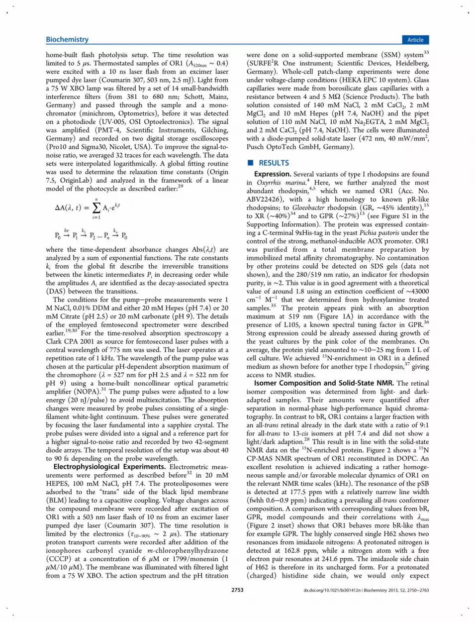
Figure 1. pH-titration of OR1. (A) Absorbance spectra of OR1 wild type under salt-free conditions and the D100N mutant (gray lines) at pH 7.2(1) and pH 2.3 (2). Wild type spectra are normalized to A280nm, and the D100N is scaled to the maximum at 519 of the wild type at pH 7.4. (B)Difference spectra of OR1 at acidic pH values under salt-free conditions (dotted lines) and with 1 M NaCl (line). The wild type spectrum at pH 7.2was taken as reference (Sref). (C) Absorbance spectra of OR1 wild type and the D100N mutant at 1 M NaCl (gray lines) at pH 7.4 (1) and pH 2.3(2). All spectra were as well normalized to A280 nm. D100N is scaled to the maximum at 519 of the wild type at pH 7.0 (D) Transition from Sref toS600nm followed by the absorbance changes at 600 nm. The dotted lines indicate the half-maximal amplitude change and are color coded according tothe ionic conditions in the legend. The absorbance changes are normalized to the protein concentration at 280 nm. (E) Absorbance spectra of OR1H62N at different pH values under salt-free conditions (dotted lines) and with 1 M NaCl (lines). (F) The pH dependence of the λmax values for thewild type (squares) and the H62N (triangles) mutant under different ionic conditions.
Biochemistry Article
dx.doi.org/10.1021/bi301412n | Biochemistry 2013, 52, 2750−27632751

other group. Only at high pH, when the histidine is presumablyin its uncharged state, or in histidine deficient mutants, thedeprotonated M-state accumulates in ESR and its amountcorrelates with the pH-titration curve of the proton acceptor.Our studies were motivated by the question of the proton
pumping ability of the eukaryotic pR OR1 and its relation tothe photocycle and by exploring a putative role of theconserved histidine H62. We address these topics by acombination of spectroscopic and electrophysiological techni-ques. First we describe the expression and initial stateproperties of OR1. Solid-state NMR data specifically allowsthe determination of the uncharged state of H62 at neutral pH.The pH-titration data that give information about the chargestate of the counterion at the pSB revealed a high complexityand sensitivity toward the ionic conditions: Two transitions areobserved upon lowering the pH below 4 that we interpret asthe protonation of the proton acceptor D100 coupled to atleast one further group. In the first transition, only a fraction ofthe molecules are protonated resembling in their spectralproperties the D100N mutant at neutral pH. The secondprotonation event requires high ionic strength to be observablein the wild type and the D100N mutant. This requirement isabolished by the replacement of H62 while keeping thetransition’s low pKa. However, the first transition is notobservable any more in the H62N mutant. In a second step, weprobe the photoisomerization kinetics in different initial statesin ultrafast pump−probe experiments that are very sensitive tothe electronic and hydrogen-bonding environment of the pSB.The results are discussed in comparison to the D100N as areference of a protonated counterion and to the H62N mutantthat shows no difference to the wild type at neutral pH in linewith the absence of the His-Asp cluster. In the third part, weturn to later steps of the photocycle and its relation to theproton pumping. In accordance with the titration data and theresults from the pump−probe measurements, the deprotonatedstate of the Schiff base (M-state) can be followed at low pH andcurrent measurements of reconstituted OR1 can be correlatedto proton pumping even at pH 2.4. The H62N mutation hadno strong impact on the general appearance of thedeprotonated state, but modulated its accumulation duringthe photocycle. A part of this effect seems to be independent ofthe charge of the histidine and could be rather related to itsinfluence on other groups than D100. Therefore, we findanother variation of the His-Asp theme in OR1.
■ EXPERIMENTAL PROCEDURESCloning and Expression. The nucleotide sequence of OR1
(Acc. No. ABV22426) was optimized to human codon usageand synthesized (Sloning Biotechnology GmbH, Puchheim,Germany). The or1 gene was cloned into the BamHI/HindIIIsite of the vector pCDNA 3.1 (−) containing a C-terminal eyfpgene for expression in NG108−15 cells. The cells weretransfected with cDNA using either Effectene (Qiagen) orLipofectamine 2000 (Invitrogen). For protein purification thegene was cloned into the EcoRI/NotI site of the vector pPiK9K(Invitrogen). Point mutations were generated by PCR site-directed mutagenesis. The Pichia pastoris strain SMD1163 (his4,pep4, prB1) (Invitrogen) was transformed with 3 μg oflinearized vector (pPik9K::opsin_or1). Selection on geniticinand cell culture was followed by the manufacturer’sinstructions. The expression of OR1 was induced with 1%methanol and 1 μM all-trans retinal during the late logarithmicphase. After cell lysis with glass beads (0.25 mm in diameter),
the total membrane fraction was prepared by differentialcentrifugation (1 h, 100000g) and the membranes weresolubilized in 1% dodecylmaltoside (DDM, Glycon Biochem-icals) in 20 mM Hepes, 1 M NaCl, pH 7.4. The protein waspurified on a Ni-NTA column. We used ClustalX 2.0 forsequence alignments.27 Retinal extraction was carried out asdescribed earlier.28 15N-labeling was performed in bufferedminimal methanol medium (BMM, Invitrogen) containing 5 g/L 15N-ammonium chloride. For black lipid membrane experi-ments OR1 was reconstituted into proteoliposomes with aprotein:lipid ratio of 1:5 (w/w) consisting of dioleoyl-phosphatidylcholine. For NMR experiments the protein:lipidratio was increased to 1:1 (w/w).
Solid-State NMR. Magic angle spinning (MAS)-NMRexperiments were carried out on a Bruker Avance III 850MHz solid-state NMR spectrometer using a triple resonance 4mm DVT probehead. 15N cross-polarization experiments wereperformed using standard settings. A sample spin rate of 12kHz was used with the sample temperature set to 270 Kresulting in a real temperature inside of the MAS rotor ofapproximately 280 K. A recycle delay of 3 s, a contact time of 2ms and proton decoupling of 85 kHz during a 20 msacquisition period were applied. The spectrum shown in Figure2 was recorded using 8k scans. 15N chemical shift referencing
was done via the gyromagnetic ratios through the 13C chemicalshift of admantane with respect to DSS (4,4-dimethyl-4-silapentane-1-sulfonic acid).
Spectroscopy and Data Analysis. Static spectra weretaken with a U3000 double-beam spectrophotometer (Hitachi,Japan). Titration experiments were done in a buffer containing10 mM citrate, 10 mM Mes, 10 mM Hepes and 10 mM Triswith varying concentration of NaCl. Small volumes of acid andbase were used to adjust the pH between recordings of thespectra. Light-induced absorption changes were recorded with a
Figure 2. 15N-CP-MAS NMR spectrum of U-15N-OR1 at pH 7.4reconstituted in DOPC. The signal of the protonated Schiff base(pSB) is observed at 177.5 ppm. The 15N imidazole ring resonances ofhighly conserved H62 are detected at 162.8 ppm and 241.6 ppm. Theinset shows the correlation between λmax and
15N chemical shifts of thepSB obtained from retinal derivatives with all-trans polyene chainswith different halide counterions.65 OR1 as well as all-trans bR(BR568) deviate not as much from model behavior as GPR.66.
Biochemistry Article
dx.doi.org/10.1021/bi301412n | Biochemistry 2013, 52, 2750−27632752
home-built flash photolysis setup. The time resolution waslimited to 5 μs. Thermostated samples of OR1 (A520nm ∼ 0.4)were excited with a 10 ns laser flash from an excimer laserpumped dye laser (Coumarin 307, 503 nm, 2.5 mJ). Light froma 75 W XBO lamp was filtered by a set of 14 small-bandwidthinterference filters (from 381 to 680 nm; Schott, Mainz,Germany) and passed through the sample and a mono-chromator (minichrom, Optometrics), before it was detectedon a photodiode (UV-005, OSI Optoelectronics). The signalwas amplified (PMT-4, Scientific Instruments, Gilching,Germany) and recorded on two digital storage oscilloscopes(Pro10 and Sigma30, Nicolet, USA). To improve the signal-to-noise ratio, we averaged 32 traces for each wavelength. The datasets were interpolated logarithmically. A global fitting routinewas used to determine the relaxation time constants (Origin7.5, OriginLab) and analyzed in the framework of a linearmodel of the photocycle as described earlier:29
∑λΔ = ·=
tA( , ) A ei
n
ik t
1
i
→ → →P P P ... P Phv k
nk
0 1 2 0n1
where the time-dependent absorbance changes Abs(λ,t) areanalyzed by a sum of exponential functions. The rate constantski from the global fit describe the irreversible transitionsbetween the kinetic intermediates Pi in decreasing order whilethe amplitudes Ai are identified as the decay-associated spectra(DAS) between the transitions.The conditions for the pump−probe measurements were 1
M NaCl, 0.01% DDM and either 20 mM Hepes (pH 7.4) or 20mM Citrate (pH 2.5) or 20 mM carbonate (pH 9). The detailsof the employed femtosecond spectrometer were describedearlier.19,30 For the time-resolved absorption spectroscopy aClark CPA 2001 as source for femtosecond laser pulses with acentral wavelength of 775 nm was used. The laser operates at arepetition rate of 1 kHz. The wavelength of the pump pulse waschosen at the particular pH-dependent absorption maximum ofthe chromophore (λ = 527 nm for pH 2.5 and λ = 522 nm forpH 9) using a home-built noncollinear optical parametricamplifier (NOPA).31 The pump pulses were adjusted to a lowenergy (20 nJ/pulse) to avoid multiexcitation. The absorptionchanges were measured by probe pulses consisting of a single-filament white-light continuum. These pulses were generatedby focusing the laser fundamental into a sapphire crystal. Theprobe pulses were divided into a signal and a reference part fora higher signal-to-noise ratio and recorded by two 42-segmentdiode arrays. The temporal resolution of the setup was about 40to 90 fs depending on the probe wavelength.Electrophysiological Experiments. Electrometric meas-
urements were performed as described before32 in 20 mMHEPES, 100 mM NaCl, pH 7.4. The proteoliposomes wereadsorbed to the “trans” side of the black lipid membrane(BLM) leading to a capacitive coupling. Voltage changes acrossthe compound membrane were recorded after excitation ofOR1 with a 503 nm laser flash of 10 ns from an excimer laserpumped dye laser (Coumarin 307). The time resolution islimited by the electronics (τ10−90% ∼ 2 μs). The stationaryproton transport currents were recorded after addition of theionophores carbonyl cyanide m-chlorophenylhydrazone(CCCP) at a concentration of 6 μM or 1799/monensin (1μM/10 μM). The membrane was illuminated with filtered lightfrom a 75 W XBO. The action spectrum and the pH titration
were done on a solid-supported membrane (SSM) system33
(SURFE2R One instrument; Scientific Devices, Heidelberg,Germany). Whole-cell patch-clamp experiments were doneunder voltage-clamp conditions (HEKA EPC 10 system). Glasscapillaries were made from borosilicate glass capillaries with aresistance between 4 and 5 MΩ (Science Products). The bathsolution consisted of 140 mM NaCl, 2 mM CaCl2, 2 mMMgCl2 and 10 mM Hepes (pH 7.4, NaOH) and the pipetsolution of 110 mM NaCl, 10 mM Na2EGTA, 2 mM MgCl2and 2 mM CaCl2 (pH 7.4, NaOH). The cells were illuminatedwith a diode-pumped solid-state laser (472 nm, 40 mW/mm2,Pusch OptoTech GmbH, Germany).
■ RESULTSExpression. Several variants of type I rhodopsins are found
in Oxyrrhis marina.4 Here, we further analyzed the mostabundant rhodopsin,4,5 which we named OR1 (Acc. No.ABV22426), with a high homology to known pR-likerhodopsins; to Gloeobacter rhodopsin (GR, ∼45% identity),15
to XR (∼40%)34 and to GPR (∼27%)13 (see Figure S1 in theSupporting Information). The protein was expressed contain-ing a C-terminal 9xHis-tag in the yeast Pichia pastoris under thecontrol of the strong, methanol-inducible AOX promoter. OR1was purified from a total membrane preparation byimmobilized metal affinity chromatography. No contaminationby other proteins could be detected on SDS gels (data notshown), and the 280/519 nm ratio, an indicator for rhodopsinpurity, is ∼2. This value is in good agreement with a theoreticalvalue of around 1.8 using an extinction coefficient of ∼43000cm−1 M−1 that we determined from hydroxylamine treatedsamples.35 The protein appears pink with an absorptionmaximum at 519 nm (Figure 1A) in accordance with thepresence of L105, a known spectral tuning factor in GPR.36
Strong expression could be already assessed during growth ofthe yeast cultures by the pink color of the membranes. Onaverage, the protein yield amounted to ∼10−25 mg from 1 L ofcell culture. We achieved 15N-enrichment in OR1 in a definedmedium as shown before for another type I rhodopsin,37 givingaccess to NMR studies.
Isomer Composition and Solid-State NMR. The retinalisomer composition was determined from light- and dark-adapted samples. Their amounts were quantified afterseparation in normal-phase high-performance liquid chroma-tography. In contrast to bR, OR1 contains a larger fraction withan all-trans retinal already in the dark state with a ratio of 9:1for all-trans to 13-cis isomers at pH 7.4 and did not show alight/dark adaption.28 This result is in line with the solid-stateNMR data on the 15N-enriched protein. Figure 2 shows a 15NCP-MAS NMR spectrum of OR1 reconstituted in DOPC. Anexcellent resolution is achieved indicating a rather homoge-neous sample and/or favorable molecular dynamics of OR1 onthe relevant NMR time scales (kHz). The resonance of the pSBis detected at 177.5 ppm with a relatively narrow line width(fwhh 0.6−0.9 ppm) indicating a prevailing all-trans conformercomposition. A comparison with corresponding values from bR,GPR, model compounds and their correlations with λmax(Figure 2 inset) shows that OR1 behaves more bR-like thanfor example GPR. The highly conserved single H62 shows tworesonances from imidazole nitrogens: A protonated nitrogen isdetected at 162.8 ppm, while a nitrogen atom with a freeelectron pair resonates at 241.6 ppm. The imidazole side chainof H62 is therefore in its uncharged form. For a protonated(charged) histidine side chain, we would only expect
Biochemistry Article
dx.doi.org/10.1021/bi301412n | Biochemistry 2013, 52, 2750−27632753
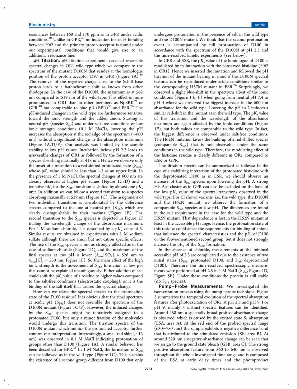
resonances between 160 and 170 ppm as in GPR under acidicconditions.20 Unlike in GPR,20 no indication for an H-bondingbetween H62 and the primary proton acceptor is found underour experimental conditions that would give rise to anadditional resonance line.pH Titration. pH titration experiments revealed reversible
spectral changes in OR1 wild type which we compare to thespectrum of the mutant D100N that resides at the homologueposition of the proton acceptor D97 in GPR (Figure 1A).38
The removal of the negative charge close to the Schiff baseproton leads to a bathochromic shift as known from otherrhodopsins. In the case of the D100N, the maximum is at 562nm compared to 519 nm of the wild type. This effect is morepronounced in OR1 than in other members as NpSRII39 orGPR,40 but comparable to blue pR (BPR)36 and ESR.26 ThepH-induced changes in the wild type are furthermore sensitivetoward the ionic strength and the added anion. Starting atneutral pH (species Sref) and under salt-free conditions or lowionic strength conditions (0.1 M NaCl), lowering the pHincreases the absorption at the red edge of the spectrum (∼600nm) without a significant change in the absorption maximum(Figure 1A/D/F). Our analysis was limited by the samplestability at low pH values. Incubation below pH 2.3 leads toirreversible changes of OR1 as followed by the formation of aspecies absorbing maximally at 418 nm. Hence we observe onlythe onset of a transition to a red-shifted protonated state (S600)whose pKa value should be less than ∼3 as an upper limit. Inthe presence of 1 M NaCl, the spectral changes at 600 nm arealready observed at higher pH values (Figure 1C/D) and atentative pKa for the S600 transition is shifted by almost one pKaunit. In addition we can follow a second transition to a speciesabsorbing maximally at 529 nm (Figure 1C). The assignment oftwo individual transitions is corroborated by the differencespectra compared to the one at neutral pH (Sref), which areclearly distinguishable by their maxima (Figure 1B). Thesecond transition to the S566 species is depicted in Figure 1Ftrailing the wavelength change of the absorbance maximum.For 1 M sodium chloride, it is described by a pKa value of 3.Similar results are obtained in experiments with 1 M sodiumsulfate although there are anion but not cation specific effects:The rise of the S600 species is not as strongly affected as in thecase of sodium chloride (Figure 1D), and the maximum of thefinal species at low pH is lower (λmax[SO4] = 526 nm vsλmax[Cl] = 530 nm, Figure 1F). So the main effect of the highionic strength is the assessment of S566 formation at low pHthat cannot be explained unambiguously: Either addition of saltcould shift the pKa value of a residue to higher values comparedto the salt-free conditions (electrostatic coupling), or it is thebinding of the salt itself that causes the spectral change.How can we relate the spectral species to the protonation
state of the D100 residue? It is obvious that the final spectrumat acidic pH (S566) does not resemble the spectrum of theD100N mutant (Figure 1C/F). However, the induced changesby the S600 species might be tentatively assigned to aprotonated D100, but only a minor fraction of the moleculeswould undergo this transition. The titration spectra of theD100N mutant which mimics the protonated acceptor furtherconfirm our interpretation. Interestingly, a small red-shift (∼13nm) was observed in 0.1 M NaCl indicating protonation ofgroups other than D100 (Figure 1A). A similar behavior hasbeen described for BPR.36 In 1 M NaCl, the formation of S566can be followed as in the wild type (Figure 1C). This sustainsthe existence of a second group different from D100 that only
undergoes protonation in the presence of salt in the wild typeand the D100N mutant. We think that the second protonationevent is accompanied by full protonation of D100 inaccordance with the spectrum of the D100N at pH 2.5 andthe time-resolved kinetic experiments (see below).In GPR and ESR, the pKa value of the homologue of D100 is
modulated by its interaction with the conserved histidine (H62in OR1). Hence we inserted the mutation and followed the pHtitration of the mutant bearing in mind if the D100N spectralfeatures can be reproduced under acidic conditions similar tothe corresponding H57M mutant in ESR.26 Surprisingly, weobserved a slight blue-shift in the spectrum albeit of the ionicconditions (Figure 1 E, F) when going from neutral pH 7.4 topH 4 where we observed the biggest increase in the 600 nmabsorbance for the wild type. Lowering the pH to 2 induces asimilar red-shift in the mutant as in the wild type. The pKa valueof this transition and the wavelength of the absorbancemaximum are again affected by the ionic conditions (Figure1F), but both values are comparable to the wild type. In fact,the biggest difference is observed under salt-free conditions.The H62N mutation favors the build-up of a red-shifted species(comparable S566) that is not observable under the sameconditions in the wild type. Therefore, the modulating effect ofthe histidine residue is clearly different in OR1 compared toESR or GPR.The titration spectra can be summarized as follows: In the
case of a stabilizing interaction of the protonated histidine withthe deprotonated D100 as in ESR, we should observe anincrease of the S600 species upon lowering the pH. A strongHis-Asp cluster as in GPR can also be excluded on the basis ofthe low pKa value of the spectral transitions observed in thewild type. For all shown variants, i.e., the wild type, the D100Nand the H62N mutant, we observe the formation of acomparable S566 species at low pH. The differences are foundin the salt requirement in the case for the wild type and theH62N mutant. That dependence is lost in the H62N mutant atleast in the accessible pH range. Hence, the protonation state ofthis residue could affect the requirements for binding of anionsthat influence the spectral characteristics and the pKa of D100or the above-mentioned second group, but it does not stronglyincrease the pKa of the S566 formation.In the absence of chloride, measurements at the minimal
accessible pH of 2.3 are complicated due to the existence of twoinitial states (S600, protonated D100, and Sref, deprotonatedD100). Therefore the time-resolved spectroscopic measure-ments were performed at pH 2.5 in 1 M NaCl (S566, Figure 1D,Figure 5E). Under these conditions the protein is still stable(no S418 species).
Pump−Probe Measurements. We investigated theisomerization process using the pump−probe technique. Figure3 summarizes the temporal evolution of the spectral absorptionfeatures after photoexcitation of OR1 at pH 2.5 and pH 9. ForpH 9, mainly 5 distinct spectral features can be identified.Around 450 nm a spectrally broad positive absorbance changeis observed, which is caused by the excited state S1 absorption(ESA; area A). At the red end of the probed spectral range(630−750 nm) the sample exhibits a negative difference bandthat is attributed to the stimulated emission (SE; area B). Ataround 520 nm a negative absorbance change can be seen thatwe assign to the ground state bleach (GSB; area C). The strongpositive absorption feature from 560 to 640 nm is observedthroughout the whole investigated time range and is composedof the ESA at early delay times and the photoproduct
Biochemistry Article
dx.doi.org/10.1021/bi301412n | Biochemistry 2013, 52, 2750−27632754

absorption (PA) at the end of the investigated time range (D).This species is the product state of the primary reaction and issupposed to be analogous to the K-intermediate in thephotocycle of bR containing the isomerized retinal in itsground state. Moreover at pH 9, a positive absorption featurecan be observed between 550 and 600 nm that lasts for severalhundred femtoseconds (E). This signal can be attributed to awave packet evolution on the S1 potential energy surface. Thelatter is absent in the spectra of the wild type at pH 2.5. Allother spectral features are conserved at this pH, although thedynamics are dramatically retarded.For a better visualization of the pH effect on the primary
reaction, single transients at characteristic wavelengths for bothpH values are presented in Figure 4A. The overall decay of thespectral features is accelerated under alkaline conditions asreflected in the values for the time constants at pH 2.5 and 9(Table 1). The corresponding DAS are depicted in Figure 4B.The first time constant τ1 is similar for pH 2.5 and pH 9 andcontains amplitudes in the region of the ESA and the SE region
and is therefore attributed to a wave packet motion on the S1potential energy surface.The biggest spectral differences appear for the time constants
τ2 and τ3. At pH 2.5 the spectra of both τ2 and τ3 showcontributions of the ESA decay in the spectral range around450 nm, the GSB recovery (550 nm), photoproduct formationand the SE decay. The high similarity of the spectra indicatesthat the underlying processes are identical and represent abiexponential decay of the excited state to the ground state.The spectral signature of τ2 at pH 9 is similar to τ2 at pH 2.5,whereas for τ3 the contribution of the SE is lost, indicating thatthe excited state decays faster at alkaline pH. The spectra of theinfinite time constant at both pH values resemble each other
Figure 3. Time-resolved absorption changes after photoexcitation ofOR1 at pH 2.5 (top) and pH 9 (bottom) with a 527 nm pulse (pH2.5) and a 522 nm pulse (pH 9). The spectra are color-coded: redindicates positive, green zero and blue negative absorption. The timescale is linear until 1 ps and logarithmic from 1 ps to 1 ns. The ground-state spectra are shown on the right side. See text for details.
Figure 4. (A) Transient absorption change at pH 2.5 and pH 9. Thesolid line represents a sum of several exponential decays obtained by aglobal fit analysis. For comparison, the transient absorption change ofGPR at pH 9 is shown. The data were taken from ref 55. (B) Decayassociated spectra of the time constants obtained by the global fittinganalysis for acidic and alkaline pH.
Figure 5. Light-induced absorption changes of OR1 under differentpH conditions at 283 K. (A, C, E) Time course of spectral changes atcharacteristic wavelengths for the M-state (398 nm), the ground statebleaching (541 nm) and red-shifted states (578 nm) after flashexcitation at 503 nm. The traces are normalized to the maximalamplitude of the 460 nm signal at pH 9.5 (A), pH 7.4 (C) and pH 2.5(E), respectively. (B, D, F) Decay associated amplitude spectra derivedfrom a global fit analysis for all 14 measured wavelengths at theselected pH values. The trace at 398 nm was smoothed byinterpolation for clarity.
Table 1. Time Constants Obtained from a Global FitAnalysis of the Data from the Pump−Probe Measurementson OR1 in Comparison to Literature Data for GPRa
τ0/fs τ1/ps τ2/ps τ3/ps τ4
OR1 pH 2.5 <70 0.30 ± 0.03 2.1 ± 0.2 21 ± 2 ∞OR1 pH 7 <70 0.27 1.0 9.8 ∞OR1 pH 9 <70 0.27 ± 0.2 1.0 ± 0.1 9.5 ± 1 ∞D100N pH 7 <70 0.3 ± 0.03 2.2 ± 0.2 11 ± 1 ∞GPR pH 655 0.15 1 ± 0.1 16 ± 1 ∞GPR pH 955 0.14 0.3 ± 0.03 9.5 ± 1 ∞GPR D97N40 <0.2 1.4 20
aThe zeroth time constant contains coherent effects from the pumpand the probe pulse, and only a higher limit is indicated.
Biochemistry Article
dx.doi.org/10.1021/bi301412n | Biochemistry 2013, 52, 2750−27632755

and represent the difference absorption spectrum between theK-intermediate and the ground state.Lowering the pH leads to the protonation of acid residues of
the protein. For other proteins it is well-known that a negativelycharged primary proton acceptor functions as an effectivecatalyst for photoisomerization.41 Therefore we investigated theprimary photodynamics of the OR1 D100N mutant (FiguresS2 to S4 in the Supporting Information). The reactiondynamics are accelerated compared to wild type at pH 2.5but are slower than wild type at pH 7 and 9. Remarkably, theprimary photodynamics are not affected upon H62N mutation(Figures S5 and S6 in the Supporting Information), supportingthe ssNMR data that there is no interaction between H62 andD100.20
Light-Induced Absorption Changes. The subsequentevents of the photocycle were investigated by analyzing light-induced absorption changes with a time resolution of 5 μs at283 K in detergent. As shown in Figure 5 and Table 2 weselected pH values with the proton acceptor D100 either in aprotonated (pH 2.5, S566) or in a deprotonated state (pH 9.5).Additionally, we included the data at pH 7.4 (Sref) forcomparison with the electrical measurements. The time courseof the changes is depicted at wavelengths characteristic for theobserved photointermediates, the deprotonated Schiff base(398 nm), the initial state (460 nm, 541 nm) and red-shiftedintermediates (578 nm), respectively. The spectral properties ofthe photointermediates can be inferred from the DAS (Figure5B/D/F) obtained from a global fit analysis. The correspond-ing time constants for the processes represent the transitionsbetween the kinetic intermediates starting from P1 to Pn.At all measured pH values an early red-shifted intermediate,
identical with P1, is observed that is in accordance with thephotoproduct of the isomerization from the pump−probemeasurements (Figure 5A, 578 nm). At pH 9.5, our data showthe decay of the K-like intermediate and the build-up of M-likestate (τ1) with a deprotonated Schiff base with a maximalabsorption around 400 nm. The M-like intermediate and red-shifted intermediates persist over the whole time range. Inanalogy to bR, we term the latter ones the N-like and O-likeintermediates. Reprotonation of the Schiff base and therecovery of the initial state occurs in several processes (τ2 toτ5). Hence, the kinetic intermediates P2 to P5 showcontributions from several spectral species, the M-like andthe N/O-like, respectively.At pH 2.5 (initial state S566), we could only detect red-shifted
species. A deprotonated M-like intermediate was not observed
comparable to the data from GPR17 at pH 5 with a protonatedprimary acceptor. The red-shifted intermediate with amaximum at around 594 nm in the difference spectrum tothe wild type rises in the microsecond time-range, whereas thedecay to the ground state at pH 2.5 is a three-step process(τ2−τ4). Inspection of the time constants at different pH valuesreveals that the photocycle is slightly accelerated under alkalineand acidic conditions compared to neutral pH (Table 2).Six time constants are needed to describe our data at pH 7.4.
As at pH 9.5, only four spectral intermediates are present anddescribe the spectral characteristics of the kinetic intermediatesP1 to P6. Starting again from the K-like intermediate (P1), theM-like intermediate is formed in the microsecond to milli-second time range described by the process τ1 to τ3 (Figure5D). Like at pH 9.5, the M-like state seems to equilibrate withother red-shifted intermediates that lead to a complex mixtureof different spectral species for P2 and P3. The concentration ofthe M-state is strongly reduced compared to the data at pH 9.5.This behavior is similar to ESR.26 In contrast to pH 9.5, the riseand the accumulation of a late red-shifted intermediate (N/O-like) can clearly be followed at 578 nm (Figure 5C) that isformed within a process described by a time constant of 4.2 ms(τ4). The return to the ground state shows a biphasic behaviorto the ground-state (τ5 and τ6). Additionally, the temperaturedependence of the photocycle was measured between 283 and308 K at pH 7.4 to further constrain the determination of thekinetic intermediates P1 to P6. The first time constant is notresolved at higher temperatures. Thus, we ignore τ1. All otherapparent rate constants follow an Arrhenius-like behavior, andtheir derived Eyring parameters are summarized in Table 2.The values for the enthalpy and entropy of activation are in asimilar range as observed for other rhodopsins like bR.29
Environmental and Mutational Effects on the Photo-cycle. In reference to the titration data we use the presence ofthe M-state as an indicator for a deprotonated counterion,while its actual accumulation during the photocycle depends onthe formation and decay rate. The assignment of D100 as theproton acceptor upon M-formation is supported by our mutantdata. The rate of M-formation is speeded up in the D100Emutant similar to the homologue mutation in bR42 (Figure S7in the Supporting Information). Similar to the absence of theproton acceptor in the D100N mutant, we see no M-state atpH 2.5 and 1 M NaCl when starting the photocycle from S566(Figure 5E). This is changed by any other condition where wealways infer a mixture of initial states (Sref and S600) from thetitration data and hence a fraction with a deprotonated proton
Table 2. Time Constants from the Global Fit Analysis of the Light-Induced Absorption Changes (Vis) at Three Different pHValues, the Thermodynamic Activation Parameters Determined from the Eyring Equation at pH 7.4 and the Comparison of theSpectroscopic Data with the Electrogenic Steps from the Analysis of the Voltage Signala
τ1 [μs] τ2 [μs] τ3 [ms] τ4 [ms] τ5 [ms] τ6 [ms]
vis 283 K pH 2.5 27 ± 2 560 ± 22 4.8 ± 0.2 72 ± 4pH 7.4 15 ± 1 240 ± 32 1.0 ± 0.1 4.2 ± 0.2 34 ± 1 210 ± 8pH 9.5 8.6 ± 1 140 ± 5 2.9 ± 0.2 32 ± 4 94 ± 10
vis pH 7.4 ΔH‡ [kJ mol−1] 86 ± 24 85 ± 6.4 63 ± 3.2 78 ± 1.8 56 ± 2ΔS‡ [J K−1 mol−1] 136 ± 83 111 ± 22 24 ± 11 58 ± 6.4 −35 ± 6.9
vis pH 7.4, 293 K (proteoliposomes) τi 67 0.9 9.1 110BLM, pH 7.4, 293 K τi 1.0 ± 0.1 16± 2 10 ± 22
rel amplitudes 0.31 ± 0.01 0.32 ± 0.02 0.37 ± 0.02aThe given standard errors derived from the global fit analysis.
Biochemistry Article
dx.doi.org/10.1021/bi301412n | Biochemistry 2013, 52, 2750−27632756

acceptor, i.e., pH 4 with and without salt or pH 2.5 without salt(Figure 6A), although the effect seems to be different on the
rise of the M-state. Aside from the effect on the initial state, theionic strength and the ion composition did not change thephotocycle kinetics significantly (Figure 6A), leading to acomparable time course and to the presence of the sameintermediates in the photocycle at pH 7.4 (Figure 6A) althoughthe M-state is less populated in the absence of salt. So the light-induced absorbance changes agree with our titration data andthe expected effect on the M-formation requiring adeprotonated proton acceptor in the initial state.In ESR, the pH dependence of the M-state is strongly
influenced by the conserved histidine. Therefore, we comparedthe light-induced changes between OR1 wild type and theH62N mutant (Figure 6 B−D). At pH 9.5, there is nosignificant difference between the two variants and we observethe same spectral intermediates in the H62N mutant as well. AtpH 7.4, the photocycle time is accelerated compared to the wildtype (66 ms vs 200 ms, Table S2 in the SupportingInformation). A similar phenomenon has been described forGPR.20,43 Furthermore, we detect a higher accumulation of theM-state in the mutant due to a faster rise during τ2 (Table S2and Figure S8 in the Supporting Information). At pH 4 thekinetics of M-rise and M-decay are faster than in the wild type.We detect only low levels of the M-state where we have for theinitial states a fraction of the wild type molecules in S600 butonly a slight blue shift for the H62N mutant. Hence, the H62Ndoes not reveal the coupling between M-formation and theD100 spectral change as in the wild type. Furthermore, there isno drastic change in the pH dependence of the M-formationbetween wild type and H62N, i.e., no indication for a stronginteraction between these two groups (Figure 6B−D).OR1 Is a Proton Pump and Electrogenic Properties.
The homology to bR and GPR and the presence of E111 at theproton donor position suggest OR1 to pump protons uponlight activation. We followed up this question by patch-clamp
recordings that reveal light-induced outward currents up to 300pA (Figure 7A), i.e., proton transfer from the cytoplasm to the
extracellular side. At neutral pH, we observe a linear current−voltage relationship (Figure 7B) as known from otherrhodopsins like bR44 or GPR22 with an apparent reversalpotential of the pump current at −170 mV. This experimentproves the proton pumping activity of OR1, and it is furthersustained by electrometric measurements with an artificialmembrane bilayer system that selectively probes the nature ofthe transported ion by protonophores and the proton reactionsat the pSB by the blue light effect (Figure S9 in the SupportingInformation).In a next step, we tested a change in the pump direction by
pH variation as it has been assessed for GPR with a protonatedproton acceptor at low pH.18 Here, a solid supportedmembrane system (SSM)33 allowed a solution exchangebetween the measurements. The amplitude of the peak currentcorrelates with the proton transport activity. We used GPR as areference and measured light-induced currents at pH 7.4 andpH 4 (Figure 7C) proving the change in the pump directionupon protonation of the proton acceptor D97.22 ReconstitutedOR1 adsorbed to a SSM-sensor generates currents up to 2 nAat pH 7.4. To control the stability of the system over time, thepeak amplitude at pH 7.4 was used as a reference. A titrationfrom pH 7.4 to pH 1.4 with 0.1 M NaCl as backgrounddecreased the amplitude of the signal to the level of the lightartifact (Figure 7D). The effect was reversible, but we could notdetect any change in the sign of the current at low pH evenwhen the sample was incubated for more than ninety minutes.At pH 2.4 with Sref and S600 as the initial states, only half of thecharge was transported compared to pH 7.4 as calculated fromthe integral in Figure 7D. Hence, we still correlate this value toproton pumping activity of OR1 and not as the result of atransient deprotonation without a complete pumping cycle. In
Figure 6. (A) The figure shows light-induced absorption changes at400 nm of OR1 wild type at different pH values either in 1 M NaCl orin the absence of chloride ions. All spectra were normalized to ground-state bleaching at 460 nm and absorption. The traces at 398 nm havebeen smoothed for clarity using a Savitzky−Golay filter. (B−D) Light-induced absorption changes of the H62N mutant in presence of 1 MNaCl at pH 4 (B), 7.4 (C) and 9 (D) were compared to wild type ofOR1. The spectra were normalized to 460 nm wavelength. Themeasurements at 398 nm were smoothed by interpolation.
Figure 7. (A) Whole-cell patch-clamp experiments at pH 7.4 (n = 4)of NG108-15 cells expressing OR1-EYFP. The curves depictphotocurrents upon illumination (472 nm, 40 mW/mm2, gray bar)at holding potentials between −100 mV and +80 mV in Δ20 mVsteps. The current density varied between 0.4 and 4.1 A/F indicatingmost probably varying OR1 concentrations in the plasma membrane.(B) Current−voltage relationship from patch-clamp recordings. (C)Control measurements with GPR at pH 7.4 and after changing to pH4 upon illumination with a light filtered by 495 nm cutoff filter (graybar). (D) Transient currents of OR1 after incubation at different pHvalues and 0.1 M NaCl in SSM experiments. The light-induced artifacton the gold surface is depicted as comparison.
Biochemistry Article
dx.doi.org/10.1021/bi301412n | Biochemistry 2013, 52, 2750−27632757

that case we would expect a highly diminished number oftransported charges compared to pH 7.4.To deduce the single proton transfer steps, we looked into
the charge displacement that is associated with the individualtransition of the intermediates. For the correlation with thespectral data, we performed time-resolved BLM measurementsas described earlier32 and measured the light-induced changesof OR1 reconstituted in proteoliposomes. Compared to thedetergent conditions, the M-state formation is similar inproteoliposomes (Figure 8), although this process is described
with a single time constant (τ1 to τ3 for detergent, Table 2)mainly because of the lower signal-to-noise ratio. On themillisecond time scale the M-state accumulates to higherconcentration in proteoliposomes due to a slower reprotona-tion. A similar phenomenon has been observed for ESR26 andGPR45 and might be related to the surface pH of the liposomes.As shown in Figure 7 the electrogenic steps take place duringthe decay of the M-like intermediate (τ4 = 1 ms) and decay ofthe N/O-like intermediate (τ5 = 16 ms; τ6 = 160 ms). Inparticular P4−P6 from the time-resolved spectroscopic data atpH 7.4 are mainly correlated with our obtained signal. Thecorresponding amplitudes reveal that all processes contributeequally to the electrogenicity of the proton transfer (Table 2).In contrast, the electrogenic steps in bR are associated to therise and decay of the M-intermediate at neutral pH.46 For OR1,the deprotonation of the Schiff base hardly contributes to itselectrogenicity. Here, it is rather the reprotonation presumablyfrom E111 and the reestablishment of the proton acceptor(D100, deprotonated) and the proton donor (E111, proto-nated). Furthermore, the results demonstrate that the photo-cycle and the associated proton transfer of OR1 are slowcompared to bR.
■ DISCUSSIONExpression. We successfully expressed OR1 in Pichia
pastoris and described its spectroscopic and functional behaviorin detail by various spectroscopic and electrophysiologicalmethods. Interestingly, the expression yields are exceptionallyhigh compared to other membrane proteins expressed in Pichiapastoris.47 In comparison, the protein yield for other type I
rhodopsins ranges from 0.5 mg/L for wild type channelrho-dopsin-248 up to 5 mg/L for some channelrhodopsin mutantsor a rhodopsin from Leptosphaeria maculans.37 For the latterprotein, an isotope labeling protocol was already successfullydemonstrated in Pichia pastoris. Here, we further providesupport for using this host system to gain access to structuraland functional studies of seven transmembrane helical receptorsby NMR spectroscopy.21,37,49
Spectral Features. OR1 is a proton pump retainingstructural and mechanistic properties of GPR and XR. Asignificant difference is found in the low pKa value of thecounterion below 3 in OR1. This reports changes in theelectrostatic environment of the counterion D100. All thedescribed experiments, the static and time-resolved spectro-scopic measurements and the data from the charge transportexperiments as well, are all in accordance with a pKa-shift of thecounterion to acidic pH (pKa <3) compared to GPR (pKa7.7).18 Therefore, the high pKa value of the counterion is not ageneral feature of the pR family, as it can be seen from thecomparison with the pKa values from other members of thisfamily and their decreasing order:
∼ > ∼ > ∼ > ≤
∼
pR XR GR OR
ESR
( 7) ( 6) ( 5) 1( 3)18 50 51
26
We have identified three spectral species in our titrationmeasurements that set the basis for the kinetic experiments.Our reference state was defined at pH 7.4 with Sref whoseabsorbance maximum is independent of the ionic conditions orof the presence of H62. At pH 7.4, the solid-state NMRexperiments revealed H62 to be in a neutral state and theabsence of hydrogen bonding interaction to this residue. Bygoing to pH 9, we observe almost no spectral change and thephotoisomerization rate followed in the pump−probe measure-ments is not altered reflecting a similar charge and geometricalarrangement of the chemical groups around the chromophore(see below). However, the amount of M-state formationstrongly increases at alkaline pH that could be related to thecoupling of the proton acceptor to another protonatable group.H62 increases M-formation at neutral pH, but showsqualitatively the same tendency as the wild type with a muchhigher accumulation of M at high pH (Figure 6B,C). Hence,the concentration of the M-state is not correlated to the chargeof H62 in the initial state.The titration of the D100 does not follow a simple titration
curve and is furthermore complicated by its ionic strengthsensitivity. Such a property is not unusual and has beendescribed before for bR as the result of protonation of otherresidues.52 The coupling of the counterion to a secondionizable group gains functional relevance in the protontransport mechanism as in bR, where counterion protonationlowers the pKa of the proton release site,53 or in ESR betweenthe proton acceptor and the conserved histidine H57.26 Thesephenomena are documented again in complex titration curves.Hereby, we infer also for OR1 that the spectral shift followsmainly the protonation state of the counterion itself. For OR1,we cannot describe the wild type and the mutant titration datain a minimal scheme with H62 and D100 as the protonationsites as going from Sref to S600 (single protonated) and S566(double protonated). The scheme is justified, because theabsorbance at 600 nm decreases again (Figure 1D). If theD100N at neutral pH represents the single protonated species,we would expect a high accumulation of the same species in the
Figure 8. Comparison of light-induced absorption changes at 293 Kand 0.1 M NaCl with voltage build-up from the BLM measurements(U) overlaid by a black line derived from a fit (see Table 2). The light-induced absorption changes were obtained from OR1 either indetergent [(1) 400 nm, smoothed with a Savitzky−Golay filter] orreconstituted in DOPC-liposomes [(2) 579 nm; (3) 400 nm; (4) 460nm].
Biochemistry Article
dx.doi.org/10.1021/bi301412n | Biochemistry 2013, 52, 2750−27632758

titration of the H62N mutant. As this is not the case, otherprotonatable groups must be involved in the formation of theS566 species to explain the pH-dependency of the wild type. Stillone has to be cautious about the conclusions from the mutantdata because we do not know so far if the nature and theprotonation state of the S566 species is identical in the mutantsand in the wild type. Further experiments that follow the chargestate of the H62 in the wild type and the D100N could help foran unambiguous assignment. The small spectral changes at pH2.4 (0 M NaCl) are in contrast to the clear shift of theabsorbance maximum of the D100N mutant (Δe ∼ 0.2 eV; λmax= 562 nm). This could be simply the result from a much lowerpKa value of D100 in the wild type leading to the lowaccumulation of S600 under the accessible conditions. With Srefand the D100N as principal components, we would estimate afraction of less than 30% of the molecules. Such a notion wouldbe in line with the presence of the M-state in the light-inducedabsorption changes at pH 2.5 in salt-free conditions (Figure6A) and the charge transport measurements (Figure 7).The presence of chloride increases the pKa value of D100
(S600 formation). As we observe the same spectral speciesdevoid of the chloride ion, we do not assume a tight binding ofchloride close to the retinal, e.g., to D100 itself. Instead,chloride ions (and to a lesser extent sulfate ions) couldmodulate the interaction between D100 and further groups inOR1 either by binding to the group alone or as HCl. As aresult, we could not find conditions with the S600 as dominantinitial state. However, the spectral shift and the decrease in M-formation argue for a fractional protonation of the protonacceptor in this species. The location of the D100 and thephenotype of the D100N and D100E mutant providearguments for its assignment as the proton acceptor.The H62 could be involved in the formation of the S600 as
mutating the residue abolishes its accumulation. Therefore, onecould speculate about a protonation state of OR1 where oneproton is shared between H62 and D100 as it has beensuggested for XR50 and demonstrated for GPR.20 Instead ofpopulating S600 by further decreasing the pH, a secondprotonation reaction arises as assessed by the formation ofS566. The mutant D100N undergoes the same salt and pH-dependent transition to S566 (Figure 1A,C), so that otherprotonatable groups in addition to D100 must be involved inthe wild type and in the D100N mutant in the formation of thisspecies. Prime candidates would be other residues involved inthe complex counterion formation like the homologue residueD212 in bRwhose chloride-dependent protonation reactioncauses the formation of the so-called acid purple species54butnot the conserved histidine H62 in OR1 that still forms S566. InS566, the proton acceptor D100 is fully protonated as we see noM-formation and a retarded photoisomerization rate.In ESR, the presence of the homologue histidine (H57)
stabilizes the deprotonated state of the counterion whoseprotonation can only be followed at pH values below 3.26 Theremoval of the histidine shifts the pKa value to 6.3 andreestablishes the same spectral features as in the protonacceptor mutant D85N. Our results are in clear contrast andreveal another variation of the histidine theme in the pR family.The behavior of the H62N mutant does not support a stronginteraction between the histidine and the proton acceptor inthe initial state, because the spectral transition to the S566occurs at low pKa values under any ionic condition (Figure 1F),i.e., the pKa of the proton acceptor remains low independent ofthe conserved histidine. Also the salt sensitivity of the pKa for
the S566 transition is comparable to that of the wild type, so thatwe do not assume H62 to be part of the salt binding site.
Ultrafast Spectroscopy. Pump−probe spectroscopy re-cords the primary reaction step in the photocycle of OR1, e.g.,the photoisomerization and the formation of the K-likeintermediate. In the global fit analysis, five time constantswere necessary to approximate the transient data set (Table 1).The time constant τ0 is lower than the time resolution of thelaser system and is therefore not further considered. The timeconstants τ1−τ4 can be compared to the ones found for GPRreported earlier55 and are attributed to the following processes:After photon absorption the retinal in its all-trans conformationis excited to the first excited electronic state (S1). The first timeconstant τ1 can be assigned to the motion of the initiallyprepared wave packet out of the Franck−Condon region. Thepropagation on the multidimensional energy surface includesseveral retinal stretching followed by torsional modes (τ2)leading to the S1/S0 conical intersection with the electronicground state (τ2, τ3), where one double bond is twisted by90°.56,57 For GPR it was postulated that a part of the excitedstate population will not reach the CI directly, but end up in astate on the S1 potential energy surface separated from the CIby an energetic barrier (τ3). They can access the CI on apicosecond time scale.19 For OR1 this mechanism is plausiblefor pH 2.5 but is not valid for pH 9, since the DAS of τ3 lacks ofcontribution from SE. Therefore this time constant must beattributed to processes occurring on the S0 surface. Afterreaching the CI the reaction path splits into two channels,where the molecules can adopt either the all-trans or the 13-cisconformation, which is called the K-intermediate.As in GPR,19 the primary photodynamics of OR1 are
strongly accelerated by increasing the pH. This behavior is mostlikely caused by changed electrostatics associated with analtered hydrogen-bonding network. A key factor is theprotonation state of the primary proton acceptor: It is assumedthat the first excited electronic state of retinal proteins exhibits apolar character. In comparison to the electronic chargedistribution in the ground state, the photoexcitation movespartial positive charge toward the hydrocarbon tail of thechromophore.58 An immediate change in dipole moment by 12D upon photoexcitation causes a polarization of the retinal inbR.59,60 The partial positive charge can be stabilized bynegatively charged amino acids near the C13 position of theretinal. As a result the double bond character is reduced at thisposition and therefore the energy barrier for isomerization isdecreased.61 Furthermore a negatively charged primary protonacceptor is known to affect the nature of the S1 potential energysurface, leading to a deeper slope toward the CI.62 Thenecessity of a negatively charged primary proton acceptor forfast isomerization is illustrated by the observation that theprimary dynamics is also slowed down upon D100N mutation.The primary photodynamics of OR1 D100N is more similar toOR1 at pH 2.5 than to OR1 at the same pH, e.g., a retardedphotoisomerization rate and a biexponential decay of the S1state (Figure S4 in the Supporting Information) that areproperties also found in other rhodopsins like bR or GPR(Table 1, Table S1 in the Supporting Information). However,the reaction mechanism for OR1 D100N is acceleratedcompared to OR1 at pH 2.5. Taking into account that theabsorption maximum is more red-shifted for OR1 D100N (λMax= 562 nm) than for OR1 at pH 2.5 (λMax = 529 nm) suggeststhat either there are conformational changes within the retinalbinding pocket for OR1 D100N or other groups are
Biochemistry Article
dx.doi.org/10.1021/bi301412n | Biochemistry 2013, 52, 2750−27632759

additionally protonated in the wild type at pH 2.5 as speculatedin the titration data for S566. This would also explain the altereddynamics found for OR1 D100N and OR1 at pH 2.5, althoughboth species exhibit a neutral primary proton acceptor. TheD100N mutant was not stable at low pH to assess differencesbetween the mutant and the wild type at low pH when bothvariants have undergone the transition to S566.Although the overall transient absorption spectra in Figure 3
look similar to GPR reported earlier, there are significantdifferences: The time constants given in Table 1 may suggest afaster decay for the primary reaction of GPR compared to OR1,and the fast component (τ2) is more pronounced in OR1,leading to a faster overall reaction dynamics as indicated inFigure 4, where individual transients for OR1 and GPR arecompared. Moreover, the DAS of τ3 exhibit no contribution tothe stimulated emission suggesting a single-exponential decayof the S1 state for OR1. In contrast to this a biexponential decaywas observed for pH 2.5. A similar behavior was also reportedfor bR (see Table S1 in the Supporting Information), althoughthe time constant of the single-exponential decay of the S1 stateis 2-fold slower for OR1 at alkaline pH compared to bR. ForGPR a contribution to SE is observed for τ2 and τ3 at pH 6 andpH 9, suggesting a biexponential decay for both pH values. Thereaction rate and the underlying mechanism strongly dependon the nature of the S1 potential energy surface. Quantummechanical calculation reveals that the shape of the S1 surface ismainly determined by the relative orientation of acidic sidechains of the complex counterion toward the Schiff base.62
Therefore our data suggest that the stabilization of theprotonated Schiff base through coordination with the complexcounterion is unique for OR1.With respect to the role of the conserved histidine, a
comparison of pump−probe measurements of GPR wild typeand H75N reveals the strong dependence of the photo-isomerization rate on the protonation state of D97.20 Its pKa islowered upon H75N mutation, and the rate of photo-isomerization is affected as well. For OR1 it was found thatthe pKa is unaffected upon H62N mutation. Therefore theprimary photodynamics are similar for OR1 wild type andH62N (Figures S5 and S6 in the Supporting Information),indicating the absence of the His-Asp cluster OR1 atphysiological pH in accordance with the solid-state NMR data.Light-Induced Absorption Changes and Electrometric
Measurements. Besides the K-like state, the deprotonatedSchiff base (M-like) and late red-shifted intermediates (N- andO-like) are detected. So far, we cannot clearly discriminatebetween the N-state (relaxed 13-cis retinal and deprotonatedE111) and the O-state (all-trans retinal and protonated E111).The presence of the N-like state is mainly a consequence of thedifferent accumulation of the O-like state at pH 7.4 and pH 9.5(Figure 5A/C). Furthermore the absorbance changes exhibit astrong pH-dependence which can be best pursued bycomparing the course of deprotonated species at differentpH’s (Table 2, Figure 5 and Figure 6A). Especially, the highaccumulation of the M-state at high pH is noteworthy.Although the deprotonation of the Schiff base occurs at pH7.4 and pH 9.5 with nearly the same kinetics (τ1 ∼10 μs), theamplitude of the deprotonated species is significantly higher atpH 9.5. Therefore the interaction between the Schiff base andthe counterion seems to be influenced by a protonatable groupat alkaline pH that leads to the stabilization of the neutralcomplex as suggested for ESR.26 Here, it was suggested that theconserved histidine is the responsible factor. However, we
could show that H62 is already neutral at pH 7.4 (Figure 2)although we cannot fully rule out effects of the lipidenvironment in the solid-state NMR data compared to thedetergent micelles. Indications might come from the transientabsorbance measurements (Figure 8) that depict a slowerreprotonation of the SB. In ESR, it shifted the pKa of thehistidine by two pK units. Titration experiments on OR1reconstituted in liposomes were qualitatively similar for the S600and S566 transitions (data not shown), but so far we cannotassess the effect directly on H62.Moreover, the amplitude spectra reveal that at pH 7.4 the
second build-up of deprotonated species (τ3) and thereprotonation of the Schiff Base (τ4) display the main pH-dependent changes to pH 9.5 (τ3). As for the titration, wecould speculate about the influence of another pH sensitivegroup coupled to the protonation state of the proton acceptorleading to either a splitting of the photocycle or the presence oftwo differently protonated initial states with two photocycles.Interestingly, the H62N exerts its strongest effect at pH 7.4 onthe multiphasic M-rise in the wild type, thereby accelerating thesecond build-up of the deprotonated species. As for thetitration data, the histidine has a modulating effect on theprimary proton acceptor also during the photocycle that seemsto be not coupled to the protonation of H62. The effect wouldbe the stabilization of the neutral SB and counterion complex(M-state) either by the altered electrostatics, by a differenthydrogen-bonding network or by affecting the reprotonationrate from an internal proton donor. This property continues tobe present during the reprotonation of the SB affecting theamplitude of the N-/O-like intermediates getting moreaccumulated at pH 7.4. Apparently, the photocycleespeciallyat pH 7.4reveals even more complexity than as described in alinear photocycle. Thus, additional experiments (e.g., time-resolved vibrational spectroscopy) have to be performed to getdeeper insights to the complex behavior of OR1.In the electrometric measurements, we show the linkage
between the photocycle and vectorial proton transport with itsmain electrogenic steps for OR1 (Figure 8) mainly coupled tothe reprotonation. We observe a very fast positive offset in ourpotential signal that is often observed during the K-state inrhodopsins. Interestingly, we could not detect a big electro-genicity upon deprotonation of the SB that is different to GPRwhen measured in cell suspensions,63 but similar to in vitrocurrent measurements.38 The reprotonation of the Schiff baseoccurs with τ4 (∼1 ms) while τ5 and τ6 are coupled to the decayof the N/O-like intermediate of OR1 and show thereprotonation of E111 and proton release to the extracellularsurface. Therefore the sequence of the protonation reactions issimilar to other members of the pR family. In GR and GPR,experiments with the pH-sensitive dye pyranine revealed apreceding uptake of a proton before the release of a proton tothe extracellular side. Those reactions were also coupled to theN/O-like intermediates.15,17 The altered behavior compared tobR was explained with a different proton release group (E194and E204 in bR) that is absent in the pR family and the causefor fast proton release in bR.
Role of H62 and Biological Function. The conservedhistidine is an indicative sequence marker for the pR family. Itsfunction seems to be diverse and context sensitive. In GPR, theHis-Asp cluster forms a pH-dependent hydrogen bond shiftingthe pKa of the proton acceptor to high values.20 In contrast forESR, the interaction stabilizes the deprotonated state of theproton acceptor in the initial state and during the photocycle.26
Biochemistry Article
dx.doi.org/10.1021/bi301412n | Biochemistry 2013, 52, 2750−27632760

Here for OR1, we see only a minor effect on the protonacceptor. OR1 has a more similar phenotype in its titrationbehavior than, e.g., bR while it shows a stronger pHdependence of its M-formation and M-decay. But thisobservation seems not to be solely correlated to the chargestate of the histidine. This is in accordance to the previousreports stating that the histidine is not necessary for protonpumping or the sole factor for the high pKa.
20 Therefore, therole of the histidine in OR1 might rather be related to thekinetic modulation of proton transfer steps and here especiallywith a coupling to the so far unknown proton release group inthe pR family.Oxyrrhis marina is a heterotrophic organism. Still, strong
expression of OR1 is observed in Oxyrrhis cells, which raises thequestion about the rhodopsin’s role under physiologicalconditions. As described in the introduction, part a photo-trophic4 as well as a phototactic function have been proposedby two independent studies.5 Our results with low pKa value ofthe counterion, pump activity and charge translocation over awide pH range would clearly suggest a general involvement inenergy fixation. Initial patch-clamp data (Figure 7 A,B) clearlydemonstrated the light-induced pumping of OR1. Interestingly,the titration data from the electrometric measurements (Figure6 D) indicates that vectorial proton transport is not reversedeven under very acidic conditions. This is in clear contrast toGPR,18,22 but again similar to ESR,26 reflecting the differentenvironment at the pSB in different pRs. Therefore the low pKavalue of the counterion would guarantee OR1 to pump protonsinto an acidified compartment as proposed as possible functionby Slamovits et al.. A McpT-like sensor, which detects thechange in membrane potential generated by OR1 as knownfrom Halobacterium salinarium for bR,64 could be one possibleexplanation for OR1’s phototactic activity. Furthermore thestrong expression of OR1 in vivo might result in the presence ofthe rhodopsin in internal compartment and in a small fractionalso in the plasma membrane enabling phototaxis. A secondminor group of rhodopsins found in Oxyrrhis marina are bR-like proteins, suggesting as well a role in energy fixation. It isnot known whether both rhodopsin genes are encoded in thesame strain or not. But the fact that both seem to be involved inthe energy metabolism of the cell makes it unlikely that acombination of both proteins in the dinoflagellate could explainthe contradictory results.In conclusion, we presented an initial characterization from a
eukaryotic member of the pR family. OR1 acts as a protonpump within a broad pH range whose pumping cycle isconnected to a bR-like photocycle. The conserved histidineresidue shows enigmatic features that have to be addressedmore specifically with spectroscopic techniques.
■ ASSOCIATED CONTENT
*S Supporting InformationFurther experimental results (Tables S1 and S2 and FiguresS1−S9). This material is available free of charge via the Internetat http://pubs.acs.org.
■ AUTHOR INFORMATION
Corresponding Author*Max-Planck-Institut fur Biophysik, Abteilung fur Biophysikali-sche Chemie, Max-von-Laue Strasse 3, 60438 Frankfurt amMain, Germany. Phone: ++49-69-63032007. Fax: ++49-69-63032222. E-mail: [email protected].
FundingThis work was supported by the Max Planck Society, the DFG(Sonderforschungsbereich 807 to J.W., C.G. and E.B.) and theCenter of Excellence Frankfurt Macromolecular Complexes.NotesThe authors declare no competing financial interest.
■ ACKNOWLEDGMENTSWe thank Dr. Katrin Feldbauer (MPI) and Dr. Mirka-KristinVerhoefen (JWG) for critical reading of the manuscript. Wewould like to thank Dr. Ionela Radu (Freie Universitat Berlin)for discussions and access to unpublished data. Furthermorethe authors thank Dr. Christine Keipert for support with theSURFE2R-System. Additionally we would like to thank HelgaHusmann for her help in the figure preparation, Stefan Geys forsupport with NG108-15 cells and Heike Biehl for excellenttechnical support.
■ ABBREVIATIONS USEDOR1, Oxyrrhis marina rhodopsin; bR, bacteriorhodopsin; GR,gloeobacter rhodopsin; GPR, green proteorhodopsin; pR,proteorhodopsin; sR, sensory rhodopsin; BLM, black lipidmembrane; RT, room temperature; SSM, solid supportedmembrane; XR, xanthorhodopsin; DAS, decay-associatedspectra; MAS, magic angle spinning; pSB, protonated Schiffbase; ESR, rhodopsin from Exiguobacterium sibiricum
■ REFERENCES(1) Montagnes, D. J. S., Lowe, C. D., Roberts, E. C., Breckels, M. N.,Boakes, D. E., Davidson, K., Keeling, P. J., Slamovits, C. H., Steinke,M., Yang, Z., and Watts, P. C. (2011) An introduction to the specialissue: Oxyrrhis marina, a model organism? J. Plankton Res. 33, 549−554.(2) Gao, X. P., and Li, J. Y. (1986) Nuclear division in the marinedinoflagellate Oxyrrhis marina. J. Cell Sci. 85, 161−175.(3) Slamovits, C. H., and Keeling, P. J. (2011) Contributions ofOxyrrhis marina to molecular biology, genomics and organelleevolution of dinoflagellates. J. Plankton Res. 33, 591−602.(4) Slamovits, C. H., Okamoto, N., Burri, L., James, E. R., andKeeling, P. J. (2011) A bacterial proteorhodopsin proton pump inmarine eukaryotes. Nat. Commun. 2, 183.(5) Hartz, A. J., Sherr, B. F., and Sherr, E. B. (2011) Photoresponse inthe Heterotrophic Marine Dinoflagellate Oxyrrhis marina. J. EukaryoticMicrobiol. 58 (2), 171−177.(6) Frigaard, N. U., Martinez, A., Mincer, T. J., and DeLong, E. F.(2006) Proteorhodopsin lateral gene transfer between marineplanktonic Bacteria and Archaea. Nature 439, 847−850.(7) Nagel, G., Ollig, D., Fuhrmann, M., Kateriya, S., Musti, A. M.,Bamberg, E., and Hegemann, P. (2002) Channelrhodopsin-1: a light-gated proton channel in green algae. Science 296, 2395−2398.(8) Tsunoda, S. P., Ewers, D., Gazzarrini, S., Moroni, A., Gradmann,D., and Hegemann, P. (2006) H+-pumping rhodopsin from themarine alga Acetabularia. Biophys. J. 91, 1471−1479.(9) Waschuk, S. A., Bezerra, A. G., Shi, L., and Brown, L. S. (2005)Leptosphaeria rhodopsin: Bacteriorhodopsin-like proton pump from aeukaryote. Proc. Natl. Acad. Sci. U.S.A. 102, 6879−6883.(10) Sineshchekov, O. A., Jung, K. H., and Spudich, J. L. (2002) Tworhodopsins mediate phototaxis to low- and high-intensity light inChlamydomonas reinhardtii. Proc. Natl. Acad. Sci. U.S.A. 99, 8689−8694.(11) Jung, K.-H. (2007) The Distinct Signaling Mechanisms ofMicrobial Sensory Rhodopsins in Archaea, Eubacteria and Eukarya.Photochem. Photobiol. 83, 63−69.(12) Oesterhelt, D., and Stoeckenius, W. (1971) Rhodopsin-likeprotein from the purple membrane of Halobacterium halobium. Nat.New Biol. 233, 149−152.
Biochemistry Article
dx.doi.org/10.1021/bi301412n | Biochemistry 2013, 52, 2750−27632761

(13) Beja, O., Aravind, L., Koonin, E. V., Suzuki, M. T., Hadd, A.,Nguyen, L. P., Jovanovich, S. B., Gates, C. M., Feldman, R. A., Spudich,J. L., Spudich, E. N., and DeLong, E. F. (2000) Bacterial Rhodopsin:Evidence for a New Type of Phototrophy in the Sea. Science 289,1902−1906.(14) Fuhrman, J. A., Schwalbach, M. S., and Stingl, U. (2008)Proteorhodopsins: an array of physiological roles? Nat. Rev. Microbiol.6, 488−494.(15) Miranda, M. R. M., Choi, A. R., Shi, L. C., Bezerra, A. G., Jung,K. H., and Brown, L. S. (2009) The Photocycle and ProtonTranslocation Pathway in a Cyanobacterial Ion-Pumping Rhodopsin.Biophys. J. 96, 1471−1481.(16) Luecke, H., Schobert, B., Stagno, J., Imasheva, E. S., Wang, J. M.,Balashov, S. P., and Lanyi, J. K. (2008) Crystallographic structure ofxanthorhodopsin, the light-driven proton pump with a dualchromophore. Proc. Natl. Acad. Sci. U.S.A. 105, 16561−16565.(17) Dioumaev, A. K., Brown, L. S., Shih, J., Spudich, E. N., Spudich,J. L., and Lanyi, J. K. (2002) Proton transfers in the photochemicalreaction cycle of proteorhodopsin. Biochemistry 41, 5348−5358.(18) Friedrich, T., Geibel, S., Kalmbach, R., Chizhov, I., Ataka, K.,Heberle, J., Engelhard, M., and Bamberg, E. (2002) Proteorhodopsin isa light-driven proton pump with variable vectoriality. J. Mol. Biol. 321,821−838.(19) Huber, R., Kohler, T., Lenz, M. O., Bamberg, E., Kalmbach, R.,Engelhard, M., and Wachtveitl, J. (2005) pH-dependent photo-isomerization of retinal in proteorhodopsin. Biochemistry 44, 1800−1806.(20) Hempelmann, F., Holper, S., Verhoefen, M. K., Woerner, A. C.,Kohler, T., Fiedler, S. A., Pfleger, N., Wachtveitl, J., and Glaubitz, C.(2011) His75-Asp97 Cluster in Green Proteorhodopsin. J. Am. Chem.Soc. 133 (12), 4645−4654.(21) Reckel, S., Gottstein, D., Stehle, J., Lohr, F., Verhoefen, M. K.,Takeda, M., Silvers, R., Kainosho, M., Glaubitz, C., Wachtveitl, J.,Bernhard, F., Schwalbe, H., Guntert, P., and Dotsch, V. (2011)Solution NMR structure of proteorhodopsin. Angew. Chem., Int. Ed. 50,11942−11946.(22) Lorinczi, E., Verhoefen, M.-K., Wachtveitl, J., Woerner, A. C.,Glaubitz, C., Engelhard, M., Bamberg, E., and Friedrich, T. (2009)Voltage- and pH-Dependent Changes in Vectoriality of PhotocurrentsMediated by Wild-type and Mutant Proteorhodopsins uponExpression in Xenopus Oocytes. J. Mol. Biol. 393, 320−341.(23) Dioumaev, A. K., Wang, J. M., Balint, Z., Varo, G., and Lanyi, J.K. (2003) Proton transport by proteorhodopsin requires that theretinal Schiff base counterion Asp-97 be anionic. Biochemistry 42,6582−6587.(24) Yoshizawa, S., Kawanabe, A., Ito, H., Kandori, H., and Kogure,K. (2012) Diversity and functional analysis of proteorhodopsin inmarine Flavobacteria. Environ. Microbiol. 14 (5), 1240−1248.(25) Martinez, A., Bradley, A. S., Waldbauer, J. R., Summons, R. E.,and DeLong, E. F. (2007) Proteorhodopsin photosystem geneexpression enables photophosphorylation in a heterologous host.Proc. Natl. Acad. Sci. U.S.A. 104, 5590−5595.(26) Balashov, S. P., Petrovskaya, L. E., Lukashev, E. P., Imasheva, E.S., Dioumaev, A. K., Wang, J. M., Sychev, S. V., Dolgikh, D. A., Rubin,A. B., Kirpichnikov, M. P., and Lanyi, J. K. (2012) Aspartate-histidineinteraction in the retinal schiff base counterion of the light-drivenproton pump of Exiguobacterium sibiricum. Biochemistry 51, 5748−5762.(27) Larkin, M. A., Blackshields, G., Brown, N. P., Chenna, R.,McGettigan, P. A., McWilliam, H., Valentin, F., Wallace, I. M., Wilm,A., Lopez, R., Thompson, J. D., Gibson, T. J., and Higgins, D. G.(2007) Clustal W and Clustal X version 2.0. Bioinformatics 23, 2947−2948.(28) Scherrer, P., Mathew, M. K., Sperling, W., and Stoeckenius, W.(1989) Retinal isomer ratio in dark-adapted purple membrane andbacteriorhodopsin monomers. Biochemistry 28, 829−834.(29) Chizhov, I., Chernavskii, D. S., Engelhard, M., Mueller, K. H.,Zubov, B. V., and Hess, B. (1996) Spectrally silent transitions in thebacteriorhodopsin photocycle. Biophys. J. 71, 2329−2345.
(30) Helbing, J., Bregy, H., Bredenbeck, J., Pfister, R., Hamm, P.,Huber, R., Wachtveitl, J., De Vico, L., and Olivucci, M. (2004) A fastphotoswitch for minimally perturbed peptides: investigation of thetrans → cis photoisomerization of N-methylthioacetamide. J. Am.Chem. Soc. 126, 8823−8834.(31) Wilhelm, T., Piel, J., and Riedle, E. (1997) Sub-20-fs pulsestunable across the visible from a blue-pumped single-pass noncollinearparametric converter. Opt. Lett. 22, 1494−1496.(32) Ruitenberg, M., Kannt, A., Bamberg, E., Ludwig, B., Michel, H.,and Fendler, K. (2000) Single-electron reduction of the oxidized stateis coupled to proton uptake via the K pathway in Paracoccusdenitrificans cytochrome c oxidase. Proc. Natl. Acad. Sci. U.S.A. 97,4632−4636.(33) Pintschovius, J., and Fendler, K. (1999) Charge translocation bythe Na+/K+-ATPase investigated on solid supported membranes:rapid solution exchange with a new technique. Biophys. J. 76, 814−826.(34) Balashov, S. P., Imasheva, E. S., Boichenko, V. A., Anton, J.,Wang, J. M., and Lanyi, J. K. (2005) Xanthorhodopsin: a proton pumpwith a light-harvesting carotenoid antenna. Science 309, 2061−2064.(35) Scharf, B., Hess, B., and Engelhard, M. (1992) Chromophore ofsensory rhodopsin II from Halobacterium halobium. Biochemistry 31,12486−12492.(36) Wang, W. W., Sineshchekov, O. A., Spudich, E. N., and Spudich,J. L. (2003) Spectroscopic and photochemical characterization of adeep ocean proteorhodopsin. J. Biol. Chem. 278, 33985−33991.(37) Fan, Y., Shi, L., Ladizhansky, V., and Brown, L. (2011) Uniformisotope labeling of a eukaryotic seven-transmembrane helical proteinin yeast enables high-resolution solid-state NMR studies in the lipidenvironment. J. Biomol. NMR 49, 151−161.(38) Varo, G., Brown, L. S., Lakatos, M., and Lanyi, J. K. (2003)Characterization of the photochemical reaction cycle of proteorho-dopsin. Biophys. J. 84, 1202−1207.(39) Verhoefen, M. K., Lenz, M. O., Amarie, S., Klare, J. P., Tittor, J.,Oesterhelt, D., Engelhard, M., and Wachtveitl, J. (2009) Primaryreaction of sensory rhodopsin II mutant D75N and the influence ofazide. Biochemistry 48, 9677−9683.(40) Verhoefen, M. K., Neumann, K., Weber, I., Glaubitz, C., andWachtveitl, J. (2009) Primary reaction dynamics of proteorhodopsinmutant D97N observed by femtosecond infrared and visiblespectroscopy. Photochem. Photobiol. 85, 540−546.(41) Scholz, F., Bamberg, E., Bamann, C., and Wachtveitl, J. (2012)Tuning the Primary Reaction of Channelrhodopsin-2 by Imidazole,pH, and Site-Specific Mutations. Biophys. J. 102, 2649−2657.(42) Butt, H. J., Fendler, K., Bamberg, E., Tittor, J., and Oesterhelt,D. (1989) Aspartic acids 96 and 85 play a central role in the functionof bacteriorhodopsin as a proton pump. EMBO J. 8, 1657−1663.(43) Bergo, V. B., Sineshchekov, O. A., Kralj, J. M., Partha, R.,Spudich, E. N., Rothschild, K. J., and Spudich, J. L. (2009) His-75 inProteorhodopsin, a Novel Component in Light-driven ProtonTranslocation by Primary Pumps. J. Biol. Chem. 284, 2836−2843.(44) Nagel, G., Kelety, B., Mockel, B., Buldt, G., and Bamberg, E.(1998) Voltage dependence of proton pumping by bacteriorhodopsinis regulated by the voltage-sensitive ratio of M1 to M2. Biophys. J. 74,403−412.(45) Krebs, R. A., Alexiev, U., Partha, R., DeVita, A. M., and Braiman,M. S. (2002) Detection of fast light-activated H+ release and Mintermediate formation from proteorhodopsin. BMC Physiol. 2, 5.(46) Heberle, J., and Dencher, N. A. (1992) Surface-bound opticalprobes monitor protein translocation and surface potential changesduring the bacteriorhodopsin photocycle. Proc. Natl. Acad. Sci. U.S.A.89, 5996−6000.(47) Macauley-Patrick, S., Fazenda, M. L., McNeil, B., and Harvey, L.M. (2005) Heterologous protein production using the Pichia pastorisexpression system. Yeast 22, 249−270.(48) Bamann, C., Kirsch, T., Nagel, G., and Bamberg, E. (2008)Spectral Characteristics of the Photocycle of Channelrhodopsin-2 andIts Implication for Channel Function. J. Mol. Biol., 686−694.(49) Takamatsu, S., Hoshino, K., Matsumoto, K., Miyasaka, T., andShimoyama, I. (2011) The photo charge of a bacterioRhodopsin
Biochemistry Article
dx.doi.org/10.1021/bi301412n | Biochemistry 2013, 52, 2750−27632762

electrochemical cells measured by a charge amplifier. IEICE Electron.Express 8, 505−511.(50) Imasheva, E. S., Balashov, S. P., Wang, J. M., and Lanyi, J. K.(2006) pH-Dependent transitions in xanthorhodopsin. Photochem.Photobiol. 82, 1406−1413.(51) Hashimoto, K., Choi, A. R., Furutani, Y., Jung, K.-H., andKandori, H. (2010) Low-Temperature FTIR Study of GloeobacterRhodopsin: Presence of Strongly Hydrogen-Bonded Water and Long-Range Structural Protein Perturbation upon Retinal Photoisomeriza-tion. Biochemistry 49, 3343−3350.(52) Varo , G., and Lanyi, J. K. (1989) Photoreactions ofbacteriorhodopsin at acid pH. Biophys. J. 56, 1143−1151.(53) Balashov, S. P., Imasheva, E. S., Govindjee, R., and Ebrey, T. G.(1996) Titration of aspartate-85 in bacteriorhodopsin: what it saysabout chromophore isomerization and proton release. Biophys. J. 70,473−481.(54) Kelemen, L., Galajda, P., Szaraz, S., and Ormos, P. (1999)Chloride ion binding to bacteriorhodopsin at low pH: An infraredspectroscopic study. Biophys. J. 76, 1951−1958.(55) Lenz, M. O., Huber, R., Schmidt, B., Gilch, P., Kalmbach, R.,Engelhard, M., and Wachtveitl, J. (2006) First steps of retinalphotoisomerization in proteorhodopsin. Biophys. J. 91, 255−262.(56) Garavelli, M., Vreven, T., Celani, P., Bernardi, F., Robb, M. A.,and Olivucci, M. (1998) Photoisomerization path for a realistic retinalchromophore model: The nonatetraeniminium cation. J. Am. Chem.Soc. 120, 1285−1288.(57) Schapiro, I., Ryazantsev, M. N., Frutos, L. M., Ferre, N., Lindh,R., and Olivucci, M. (2011) The Ultrafast Photoisomerizations ofRhodopsin and Bathorhodopsin Are Modulated by Bond LengthAlternation and HOOP Driven Electronic Effects. J. Am. Chem. Soc.133, 3354−3364.(58) Gonzalez-Luque, R., Garavelli, M., Bernardi, F., Merchan, M.,Robb, M. A., and Olivucci, M. (2000) Computational evidence in favorof a two-state, two-mode model of the retinal chromophorephotoisomerization. Proc. Natl. Acad. Sci. U.S.A. 97, 9379−9384.(59) Schenkl, S., van Mourik, F., van der Zwan, G., Haacke, S., andChergui, M. (2005) Probing the Ultrafast Charge Translocation ofPhotoexcited Retinal in Bacteriorhodopsin. Science 309, 917−920.(60) Mathies, R., and Stryer, L. (1976) Retinal has a highly dipolarvertically excited singlet-state - implications for vision. Proc. Natl. Acad.Sci. U.S.A. 73, 2169−2173.(61) Song, L., Elsayed, M. A., and Lanyi, J. K. (1993) ProteinCatalysis of the Retinal Subpicosecond Photoisomerization in thePrimary Process of Bacteriorhodopsin Photosynthesis. Science 261,891−894.(62) Cembran, A., Bernardi, F., Olivucci, M., and Garavelli, M.(2004) Counterion controlled photoisomerization of retinal chromo-phore models: a computational investigation. J. Am. Chem. Soc. 126,16018−16037.(63) Sineshchekov, O. A., and Spudich, J. L. (2004) Light-inducedintramolecular charge movements in microbial rhodopsins in intact E.coli cells. Photochem. Photobiol. Sci. 3, 548−554.(64) Koch, M. K., and Oesterhelt, D. (2005) MpcT is the transducerfor membrane potential changes in Halobacterium salinarum. Mol.Microbiol. 55, 1681−1694.(65) Hu, J., Griffin, R. G., and Herzfeld, J. (1994) Synergy in thespectral tuning of retinal pigments: complete accounting of the opsinshift in bacteriorhodopsin. Proc. Natl. Acad. Sci. U.S.A. 91, 8880−8884.(66) Pfleger, N., Worner, A. C., Yang, J., Shastri, S., Hellmich, U. A.,Aslimovska, L., Maier, M. S. M., and Glaubitz, C. (2009) Solid-stateNMR and functional studies on proteorhodopsin. Biochim. Biophys.Acta, Bioenerg. 1787, 697−705.
Biochemistry Article
dx.doi.org/10.1021/bi301412n | Biochemistry 2013, 52, 2750−27632763


