Embed Size (px)
Citation preview

Phylogenetic analyses and detectionof viridans streptococci based onsequences and denaturing gradientgel electrophoresis of the rodshape-determining protein geneIkuri Konishi1, Tomonori Hoshino1*, Yoshio Kondo1,2,Kan Saito1, Miyuki Nishiguchi1, Kyoko Sato1 andTaku Fujiwara1
1Department of Pediatric Dentistry, Nagasaki University Graduate School of Biomedical Sciences,Nagasaki, Japan; 2Division of Microbiology and Oral Infection, Nagasaki University Graduate Schoolof Biomedical Sciences, Nagasaki, Japan
Background: Population analysis of viridans streptococci is important because these species are associated
with dental caries, bacteremia, and subacute endocarditis, in addition to being important members of the
human oral commensal microbiota.
Design: In this study, we phylogenetically analyzed the rod shape-determining protein gene (rodA), which is
associated with cellular morphology, cell division, and sensitivity for antibiotics, and demonstrated that the
diversity of the rodA gene is sufficient to identify viridans streptococci at the species level. Moreover, we
developed a more convenient denaturing gradient gel electrophoresis (DGGE) method based on the diversity
of the rodA gene (rodA-DGGE) for detecting nine dominant streptococcal species in human saliva, namely,
Streptococcus sanguinis, Streptococcus oralis, Streptococcus mitis, Streptococcus parasanguinis, Streptococcus
gordonii, Streptococcus vestibularis, Streptococcus salivarius, Streptococcus mutans, and Streptococcus
sobrinus.
Results: This rodA-DGGE method proved useful in detecting viridans streptococci without cultivation,
isolation, and phenotypic characterization.
Conclusion: Analysis of the oral microbiota by rodA-DGGE offers a higher resolution than the conventional
DGGE using 16S rDNA and may be an alternative in the microbial diagnosis of streptococcal infection.
Keywords: population analysis; oral microbiota; streptococcus; subacute bacterial endocarditis; dental caries; saliva
Received: 9 June 2009; Revised: 29 July 2009; Accepted: 31 July 2009; Published: 28 August 2009
Denaturing gradient gel electrophoresis (DGGE)
analysis of the 16S rRNA gene (16S rDNA) is
used for investigating entire bacterial commu-
nities without cultivation (1). The advantage of this
method is that it uses 16S rDNA, which is present in
all bacteria and can be amplified with a set of universal
bacterial primers. Bacterial species are differentiated on
the basis of differential migration on a denaturing
gradient gel due to their melting behavior, which is based
on the difference in the G�C content. A bar code-like
profile is obtained, with each band presumably represent-
ing a different microorganism within the microbial
communities (1�3). This molecular technique has become
an important tool for studying complex bacterial com-
munities and has been applied for the analyses of various
microbiotas, such as those found in environmental
biofilms, food fermentation processes, feces, intestine,
gastrointestinal tract infections, corneal ulcer, and vagi-
nosis (4�10).
DGGE analysis using 16S rDNA (16S rDNA-DGGE)
has also been applied for analyzing the microflora found
in the periodontal pocket, dental plaque, and saliva in
order to identify the pathogens causing periodontitis,
dental caries, and halitosis (11�14). These reports indicate
(page number not for citation purpose)
�ORIGINAL ARTICLE
Journal of Oral Microbiology 2009. # 2009 Ikuri Konishi et al. This is an Open Access article distributed under the terms of the Creative CommonsAttribution-Noncommercial 3.0 Unported License (http://creativecommons.org/licenses/by-nc/3.0/), permitting all non-commercial use, distribution, andreproduction in any medium, provided the original work is properly cited. Citation: Journal of Oral Microbiology 2009.
1
DOI: 10.3402/jom.v1i0.2015

that the pathogenic bacteria of oral diseases, such as
Porphyromonas gingivalis, Aggregatibacter actinomycetem-
comitans, Prevotella intermedia, Fusobacterium nuclea-
tum, and Streptococcus mutans, are clearly detected by
16S rDNA-DGGE. However, viridans streptococci,
which are closely associated with bacteremia and sub-
acute bacterial endocarditis (SBE), have not been clearly
detected. One of the reasons is that the 16S rDNA genes
of several viridans streptococci are highly homologous
and that there is evidence of homologous recombination
between species (15). The 16S rDNA genes of Strepto-
coccus mitis, Streptococcus oralis, and Streptococcus
pneumoniae, in particular, exhibit more than 99% se-
quence homology with each other (16). Therefore, it is
difficult to clearly identify these species by 16S rDNA-
DGGE as this method is based on differences in the G�C content. Hence, our hypothesis was that some other
gene, the evolutionary rate of which is higher than that of
16S rDNA, would be a useful target for DGGE analysis
aiming at detecting the abovementioned streptococci.
Previously, during the sequencing of the glucosyltrans-
ferase gene (gtfR) of S. oralis (17), we found three open
reading frames upstream of gtfR. By using the basic local
alignment search tool, one of them was identified as the
rod shape-determining protein (RodA) gene (rodA),
which is widely present in gram-positive and gram-
negative bacteria. This gene determines cellular morphol-
ogy and is associated with peptidoglycan degradation
during elongation and septation (18). In addition, since it
has been suggested that rodA of Streptococcus thermo-
philus is associated with oxidative stress defense and
streptonigrin tolerance (19, 20), it becomes one of the
interesting genes in the study of bacterial response against
environmental stress. Our preliminary alignment analysis
revealed 78% similarity of rodA of S. oralis to that of
S. mitis; further, this similarity was lower than that
between 16S rDNA of S. oralis and that of S. mitis. Thus,
we thought that the streptococcal rodA gene would
exhibit sufficient phylogenetic diversity to aid in identify-
ing viridans streptococci by DGGE.
In this study, we investigated the prevalence of the rodA
genes in viridans streptococci, and phylogenetically
analyzed them as the identification tool of those species.
In addition, we developed a DGGE method based on the
phylogenetic diversity of the rodA gene for detecting
dominant streptococci in human oral cavity.
Methods
Sampling of streptococcal strains in salivaThe study group comprised eight healthy adult volun-
teers. The study protocol was approved by the Ethics
Board of the Institute of Dentistry, Nagasaki University,
and informed consent was obtained from all the subjects.
Non-stimulated saliva was used as the clinical sample in
this study, since it roughly represents a summary of the
oral microbiota of the teeth, tongue, and mucosa of the
upper respiratory tract. These samples were obtained by
collecting whole saliva in a sterile tube before tooth
brushing. The samples were serially diluted and inocu-
lated on mitis-salivarius (MS) agar (Difco Laboratories,
Detroit, MI). From the inoculated MS agar plates, 20
colonies were randomly selected and streptococcal strains
in saliva were isolated. These clinical isolates were
identified by a combination of phenotypic characteriza-
tion performed using STREPTOGRAM (Wako Pure
Chemicals, Osaka, Japan) (21), polymerase chain reac-
tion (PCR) based on the species-specific variety of gtf
genes (22), and sequencing of the 16S rRNA gene (16)
and were examined for the rodA sequence according to
the following method.
Bacterial strains and cultureThe reference strains used in this study were taken from
our own culture collection (Table 1) (21, 23). These were
selected as the streptococcal species that could be
detected in the oral cavity. The strains designated
ATCC, NCTC, CCUG, and GTC were obtained from
the American Type Culture Collection, National Collec-
tion of Type Cultures (Colindale, London, England),
Culture Collection of the University of Goteborg (Go-
teborg, Sweden), and Gifu Type Culture Collection
(Gifu, Japan), respectively. These organisms were routi-
nely cultured in brain heart infusion broth (BHI; Difco
Laboratories) and on 5% defibrinated sheep blood agar
(Nissui Pharmaceutical Co. Ltd., Tokyo, Japan).
Preparation of DNA for PCRDNA of the cultured bacteria was obtained as previously
described (17). In brief, the organisms were grown in BHI
broth at 378C for 18 h, collected, and then washed by
centrifugation. The cells were suspended in a solution of
50 mM NaCl and 10 mM Tris-HCl (pH 7.4) and then
digested with mutanolysin (final concentration, 33.3 U
mL�1; Sigma-Aldrich Co., St. Louis, MO) at 508C for 1
h. Thereafter, the cells were lysed by adding N-lauroyl
sarcosine (final concentration, 1.5%) and Ethylenediamin
etetraacetic acid (EDTA) (final concentration, 10 mM).
The lysate was treated with RNase (0.3 mg mL�1; Wako
Pure Chemicals) and proteinase K (0.3 mg mL�1; Sigma-
Aldrich Co.). DNA was purified from the cell lysate by
phenol, phenol-chloroform, and chloroform extractions
and collected by ethanol precipitation.
Further, the bacterial DNA from the saliva samples
was extracted as previously described (22). In brief, the
organisms in the saliva were harvested from 500 mL of the
samples by centrifugation at 16,000�g for 10 min. The
bacterial cells were heated in a microwave oven at 500 W
for 5 min to destroy the cell walls and then digested in 100
mL of 200 U mL�1 mutanolysin (Sigma-Aldrich Co.) at
Ikuri Konishi et al.
2(page number not for citation purpose)

508C for 1 h. The lysate was treated with 80 mL of nuclei
lysis solution (Promega, Madison, WI) at 808C for 5 min,
and the proteins were removed by centrifugation after
adding 60 mL of protein precipitation solution (Promega).
The DNA was then purified by phenol-chloroform
extraction and collected by ethanol precipitation.
Alignment analysis and construction of thephylogenetic treeClustalX software (24), downloaded from http://www.
ebi.ac.uk, was used to align the sequences. Phylogenetic
tree was constructed by using the neighbor-joining
algorithm with MEGA 4 (25) on the basis of nucleotide
sequences by using the maximum composite likelihood
model. The corresponding parameter of the neighbor-
joining algorithm was set as ‘complete deletion.’
Phylogenetic analysis of the known rodA sequencesTo evaluate the appropriateness of the phylogenetic
reconstruction based on rodA genes and to design the
primers used in this study, relevant, available gene
sequences of rodA and 16S rDNA genes were obtained
from the GenBank database and analyzed. The phyloge-
netic distances were calculated with the abovementioned
algorithm and parameter using MEGA 4.
Design of PCR primersThe primers used in this study to amplify the fragments
of the rodA gene and to determine their sequence were
Rd_uni-F (5?-CCDTCAGARTTTATGAAGATWTCC-
3?) and Rd_uni-R (5?-AATCATATCHSWYTCDCG-
DACWGG-3?). Approximately 520-bp-long fragment of
the rodA gene was amplified with these primers. These
oligonucleotide primers were designed on the basis of
conserved sequences, which were identified by aligning
the streptococcal rodA genes in the GenBank nucleotide
database. The DGGE sample was amplified using
Rd_uni-F and a primer constructed by the addition of
the GC clamp (CGCCCGGGGCGCGCCCCGGGC
GGCCCGGGGGCACCGGGGG-) to Rd_uni-R.
PCR conditionsThe rodA gene was amplified by performing PCR in 50 mL
of a reaction mixture containing 0.5-U Takara Ex TaqTM
Hot Start (HS) Version (Takara, Kyoto, Japan), 0.5 mM of
the oligonucleotide primers, template DNA
(B20 ng mL�1), and 1.5 mM of MgCl2, according to
the manufacturer’s instructions. Amplification was per-
formed using GeneAmp† System 9700 (Applied Biosys-
tems, Foster City, CA), under the following conditions: 35
cycles of denaturation at 988C for 10 s, primer annealing
at 488C for 30 s, and extension at 728C for 30 s. The PCR
products were analyzed by 1.5% agarose gel electrophor-
esis, after staining with ethidium bromide. The amplicons
were then purified with a Qiagen PCR purification kit
(QIAGEN GmbH, Hilden, Germany) and used as the
template for subsequent sequencing and amplification of
the DGGE sample. The DGGE samples were amplified
by PCR by changing the annealing temperature to 538Cand using the purified amplicons as the template.
Sequencing and analysis of the streptococcalrodA genesThe cycle sequencing reaction of the purified amplicons
were performed by using BigDye Terminator v3.1 cycle
sequencing kit (Applied Biosystems) and the products
Table 1. Streptococcus strains used in this study
Streptococcus group Accession number
Species Strain rodA 16S rDNA
Mitis group
S. mitis NCTC 12261T SMT1128a D38482
S. pseudop-
neumoniae
CCUG 49455T AB441144b AY485599
S. oralis ATCC 10557 AB439009b AB355617
S. gordonii ATCC 10558T AB441145b AY485606
S. sanguinis ATCC 10556T AB441146b DQ303192
S. parasanguniis ATCC 15912T AB441147b DQ303191
S. infantis ATCC 27375T AB441148b AB008315
S. australis ATCC 700641T AB441149b AY485604
S. cristatus NCTC 12479T AB441150b AB008313
S. peroris GTC848T AB441151b AB008314
Salivarius group
S. salivarius NCTC 8618T AB441152b AB355616
S. vestibularis ATCC 49125T AB441153b AY188353
S. thermophilus ATCC 19258T AB441154b X68418
Anginosus group
S. anginosus ATCC 33397T AB441155b AB355609
S. constellatus
subsp. constellatus
ATCC 27823T AB441156b AB355606
S. constellatus
subsp. pharyngis
CCUG 46377T AB441157b AY309095
S. intermedius ATCC 27355T AB441158b AF104671
Mutans group
S. mutans NCTC 10449T AB441159b AB294730
S. sobrinus ATCC 27351 AB441160b AF439398
Bovis group
S. equinus ATCC 33317c AB441161b AB002482
S. gallolyticus
subsp.
macedonicus
CCUG 39970T AB441162b AF459431
TT means type strain.aTIGR locus name in the genome database of TIGR.bAccession number of a sequence determined in this study.cThis strain is the type strain of once ‘Streptococcus bovis.’
DGGE analysis of streptococcal rodA genes
3(page number not for citation purpose)

Fig. 1. (Continued)
Ikuri Konishi et al.
4(page number not for citation purpose)

were examined with an automatic DNA sequencer (ABI
Prism 3,100; Applied Biosystems), according to the
manufacturer’s instructions. The resultant sequences
were aligned using ClustalX and then analyzed with
MEGA 4, as described above.
DGGE analysisIn this study, the DGGE analyses were performed with
the DCodeTM universal mutation detection system (Bio-
Rad Laboratories, Hercules, CA), according to the
manufacturer’s instructions. The DGGE analysis was
initially performed with a perpendicular denaturing
gradient gel in order to determine the conditions for
that with a parallel gradient gel. To determine the
streptococcal rodA genes having the highest and lowest
melting temperature (Tm) among nine dominant strepto-
coccal species in the human oral cavity, S. mitis,
Streptococcus sanguinis, S. oralis, Streptococcus gordonii,
Streptococcus parasanguinis, Streptococcus salivarius,
Streptococcus vestibularis, S. mutans, and Streptococcus
sobrinus (26, 27), the Tm values of the rodA amplicons
were calculated using the following formula: Tm�81.58C�16.6 (log10[Na�])�0.41(%[G�C])�(500/n) (28).
The samples with genes having the highest and lowest
Tm values were applied on a perpendicular denatur-
ing gradient gel containing 6% of acrylamide and
0�70% linear gradient of denaturant (100% denaturant
was equivalent to 7 mol �L�1 urea and 40% deinonized
formamide) and separated at 80 V for 2 h at 568C. On
the basis of the result obtained, the appropriate constant
denaturant concentration in the DGGE analysis to detect
the selected nine streptococcal species was manually
determined by changing it into every 2% from the
dissociable denaturant concentration of the lowest-
Tm rodA fragment to that of the highest-Tm fragment.
The electrophoresis patterns of nine streptococcal refer-
ence strains were adopted as the reference markers
to identify these species. In the constant denaturing
gel electrophoresis, a gel containing 8% (w/v) of acryl-
amide and 28% of denaturant was used, and electro-
phoresis was performed at 260 V for 6 h at 568C.
The electrophoresed gels were visualized by staining
with SYBR† Gold (Invitrogen Corp., Carlsbad, CA).
Registration of sequencesThe newly determined rodA sequences were deposited in
DDBJ. Their accession numbers are shown in Table 1.
Results
Phylogenetic analysis of reference rodA genes fromselected gram-positive bacteria and sequenceanalysis of streptococcal rodA genesAt first, in order to investigate the prevalence of rodA gene
in Streptococcus spp., we performed phylogenetic analysis
of the sequences obtained from members of the genus
Streptococcus and related genera. We obtained 26 rodA
genes, derived from the members of the genera Strepto-
coccus, Lactococcus, Lactobacillus, Leuconostoc, and
Listeria, from the GenBank database. The phylogenetic
tree of these genes was constructed, with its root
representing the rodA gene of Listeria monocytogenes
(Fig. 1A). This tree clearly divides the tested strains at the
genus and species levels. Therefore, the streptococcal rodA
genes were considered to exhibit sufficient phylogenetic
diversity to classify the genus at the species level. More-
over, when the sequence similarities of rodA genes among
S. pneumoniae R6, S. mitis NCTC 12261, and S. oralis
ATCC 10557 were calculated, the similarity between S.
pneumoniae and S. mitis, S. pneumoniae and S. oralis, and
S. oralis and S. mitis was 93%, 77%, and 78%, respectively.
These similarity values were smaller than those of the 16S
rRNA genes among the three abovementioned strains.
Next, we performed the alignment analysis of the
streptococcal rodA gene sequences retrieved in order to
design the universal PCR primers that could amplify all
the streptococcal rodA genes. As a result, we constructed
the primers that could amplify approximately 520-bp-
long fragment from the N-terminus region of 1,200-bp-
total length of the rodA gene and the amplicons isolated
from 21 nonhemolytic streptococci were sequenced (Table
1). In the phylogenetic analysis, approximately 370-bp
sequence determined with fidelity was used.
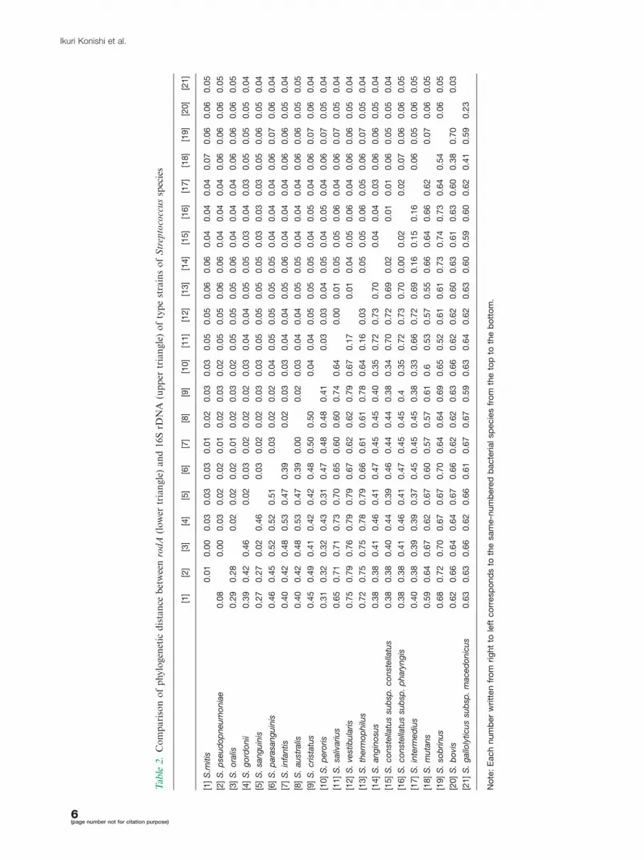
Phylogenetic analysis of the streptococcalrodA genesThe phylogenetic distances of the rodA (approximately
370 bp) and 16S rDNA (approximately 1,200 bp) genes
among streptococcal strains in Table 1 were computed
with MEGA 4 by using the following: bootstrap analyses
(500 replicates), gap/missing data (complete deletion),
model (maximum composite likelihood), and substitution
(d; transition�transversion) (Table 2). The mean phylo-
genetic distance of the rodA and 16S rDNA genes
was calculated and found to be 0.527 (SE, 0.474) and
0.043 (SE, 0.005), respectively. For this reason, the base
Fig. 1. Phylogenetic analyses of the sequences of the rodA gene. A, Phylogenetic tree derived from gram-positive bacteria’s rodA
gene, which was obtained from the GenBank database. The numbers in parentheses indicate the GenBank accession numbers.
Comparison of the phylogenetic trees of partial sequences of rodA and 16S rDNA constructed using the reference strains of
nonhemolytic streptococci in Table 1. B, Phylogenetic tree derived from partial sequences of rodA (370 bp) and C, Phylogenetic
tree derived from partial sequences of 16S rDNA (1,277 bp). The scales of both phylogenetic trees were adjusted so that they
were equivalent.
DGGE analysis of streptococcal rodA genes
5(page number not for citation purpose)
Tab
le2.
Co
mp
ari
son
of
ph
ylo
gen
etic
dis
tan
ceb
etw
een
rod
A(l
ow
ertr
ian
gle
)a
nd
16
SrD
NA
(up
per
tria
ng
le)
of
typ
est
rain
so
fS
trep
toco
ccu
ssp
ecie
s
[1]
[2]
[3]
[4]
[5]
[6]
[7]
[8]
[9]
[10]
[11]
[12]
[13]
[14]
[15]
[16]
[17]
[18]
[19]
[20]
[21]
[1]
S.m
itis
0.0
10.0
00.0
30.0
30.0
30.0
10.0
20.0
30.0
30.0
50.0
50.0
60.0
60.0
40.0
40.0
40.0
70.0
60.0
60.0
5
[2]
S.
pse
ud
op
neu
mo
nia
e0.0
80.0
00.0
30.0
20.0
20.0
10.0
20.0
30.0
20.0
50.0
50.0
60.0
60.0
40.0
40.0
40.0
60.0
60.0
60.0
5
[3]
S.
ora
lis0.2
90.2
80.0
20.0
20.0
20.0
10.0
20.0
30.0
20.0
50.0
50.0
50.0
60.0
40.0
40.0
40.0
60.0
60.0
60.0
5
[4]
S.
go
rdo
nii
0.3
90.4
20.4
60.0
20.0
30.0
20.0
20.0
20.0
30.0
40.0
40.0
50.0
50.0
30.0
40.0
30.0
50.0
50.0
50.0
4
[5]
S.
san
gu
inis
0.2
70.2
70.0
20.4
60.0
30.0
20.0
20.0
30.0
30.0
50.0
50.0
50.0
50.0
30.0
30.0
30.0
50.0
60.0
50.0
4
[6]
S.
para
san
gu
inis
0.4
60.4
50.5
20.5
20.5
10.0
30.0
20.0
20.0
40.0
50.0
50.0
50.0
50.0
40.0
40.0
40.0
60.0
70.0
60.0
4
[7]
S.
infa
ntis
0.4
00.4
20.4
80.5
30.4
70.3
90.0
20.0
30.0
30.0
40.0
40.0
50.0
60.0
40.0
40.0
40.0
60.0
60.0
50.0
4
[8]
S.
au
stra
lis0.4
00.4
20.4
80.5
30.4
70.3
90.0
00.0
20.0
30.0
40.0
40.0
50.0
50.0
40.0
40.0
40.0
60.0
60.0
50.0
5
[9]
S.
crist
atu
s0.4
50.4
90.4
10.4
20.4
20.4
80.5
00.5
00.0
40.0
40.0
50.0
50.0
50.0
40.0
50.0
40.0
60.0
70.0
60.0
4
[10]
S.
pero
ris
0.3
10.3
20.3
20.4
30.3
10.4
70.4
80.4
80.4
10.0
30.0
30.0
40.0
50.0
40.0
50.0
40.0
60.0
70.0
50.0
4
[11]
S.
saliv
ariu
s0.6
50.7
10.7
10.7
30.7
00.6
50.6
00.6
00.7
40.6
40.0
00.0
10.0
50.0
50.0
60.0
40.0
60.0
70.0
50.0
4
[12]
S.
vest
ibu
laris
0.7
50.7
90.7
60.7
90.7
90.6
70.6
20.6
20.7
90.6
70.1
70.0
10.0
40.0
50.0
60.0
40.0
60.0
60.0
50.0
4
[13]
S.
therm
op
hilu
s0.7
20.7
50.7
50.7
80.7
90.6
60.6
10.6
10.7
80.6
40.1
60.0
30.0
50.0
50.0
60.0
50.0
60.0
70.0
50.0
4
[14]
S.
an
gin
osu
s0.3
80.3
80.4
10.4
60.4
10.4
70.4
50.4
50.4
00.3
50.7
20.7
30.7
00.0
40.0
40.0
30.0
60.0
60.0
50.0
4
[15]
S.
co
nst
ella
tus
sub
sp.
co
nst
ella
tus
0.3
80.3
80.4
00.4
40.3
90.4
60.4
40.4
40.3
80.3
40.7
00.7
20.6
90.0
20.0
10.0
10.0
60.0
50.0
50.0
4
[16]
S.
co
nst
ella
tus
sub
sp.
ph
ary
ng
is0.3
80.3
80.4
10.4
60.4
10.4
70.4
50.4
50.4
0.3
50.7
20.7
30.7
00.0
00.0
20.0
20.0
70.0
60.0
60.0
5
[17]
S.
inte
rmed
ius
0.4
00.3
80.3
90.3
90.3
70.4
50.4
50.4
50.3
80.3
30.6
60.7
20.6
90.1
60.1
50.1
60.0
60.0
50.0
60.0
5
[18]
S.
mu
tan
s0.5
90.6
40.6
70.6
20.6
70.6
00.5
70.5
70.6
10.6
0.5
30.5
70.5
50.6
60.6
40.6
60.6
20.0
70.0
60.0
5
[19]
S.
sob
rin
us
0.6
80.7
20.7
00.6
70.6
70.7
00.6
40.6
40.6
90.6
50.5
20.6
10.6
10.7
30.7
40.7
30.6
40.5
40.0
60.0
5
[20]
S.
bo
vis
0.6
20.6
60.6
40.6
40.6
70.6
60.6
20.6
20.6
30.6
60.6
20.6
20.6
00.6
30.6
10.6
30.6
00.3
80.7
00.0
3
[21]
S.
gallo
lyticu
ssu
bsp
.m
aced
on
icu
s0.6
30.6
30.6
60.6
20.6
60.6
10.6
70.6
70.5
90.6
30.6
40.6
20.6
30.6
00.5
90.6
00.6
20.4
10.5
90.2
3
No
te:
Each
num
ber
writt
en
fro
mrig
ht
tole
ftco
rresp
ond
sto
the
sam
e-n
um
bere
db
acte
rialsp
ecie
sfr
om
the
top
toth
eb
ott
om
.
Ikuri Konishi et al.
6(page number not for citation purpose)

substitution rates in the rodA genes were observed to be
considerably higher than those in the 16S rDNA
sequences, though the phylogenetic distance between
the rodA genes of Streptococcus australis and Strepto-
coccus infantis was 0.00. The phylogenetic tree also
indicated that the evolutionary rates of the rodA genes,
except those of S. australis and S. infantis, were higher
than those of the 16S rDNA sequences (Fig. 1B and C).
Thus, it was revealed that the phylogenetic analysis based
on the rodA sequences of Streptococcus spp. except S.
australis and S. infantis, would be able to differentiate the
species that are closely related by 16S rDNA analysis.
Determination of the conditions for DGGE withconstant denaturing gradient gelAs for the nine dominant streptococcal species in the
human oral cavity, the G�C content (%) and Tm value
(8C) were estimated from these sequence data in order to
determine the conditions for the subsequent DGGE
analysis. The rodA genes of S. sobrinus and S. mutans
showed the highest (49.78C) and lowest (37.68C) Tm
values, respectively. The fragments of the genes obtained
from these two strains, which were amplified with the GC-
clamped primer set, were applied to perpendicular dena-
turing gradient gel to determine the optimal concentration
Fig. 2. Negative image of the rodA-DGGE analyses. The appropriate denaturant concentration in the following experiment was
determined by the ethidium bromide-stained perpendicular DGGE gel. The rodA gene fragments of S. sobrinus (with highest
Tm) and S. mutans (with lowest Tm) were amplified with the GC-clamped primer set. The amplicons were applied on the same
perpendicular denaturing gradient gel containing 6% of acrylamide and 0�70% linear gradient of denaturant and
electrophoresed at 80 V for 2 h at 568C. The electrophoretic bands of these two amplicons were separated at concentrations
of the denaturant ranging from 22 to 56% (A). The rodA-DGGE analysis with parallel constant denaturant gel was applied to
detection of viridans streptococci in saliva. In this experiment, a parallel constant denaturant gel containing 8% (w/v) of
acrylamide and 28% of constant denaturant was used, and the GC-clamped rodA gene fragments were separated at 260 V for 6 h
at 568C in 0.5� Tris-acetate-EDTA buffer. As the reference markers to identify the streptococcal species, the GC-clamped rodA
fragments of S. sobrinus (sob), S. sanguinis (san), S. oralis (ora), S. mitis (mit), S. vestibularis (ves), S. salivarius (sal), S.
parasanguinis (par), S. gordonii (gor), and S. mutans (mut) were used. The Arabic numerals identify the individual subjects (B).
DGGE analysis of streptococcal rodA genes
7(page number not for citation purpose)

of the denaturant (Fig. 2A). It was observed that these two
amplicons separated between 22% and 56% of the
denaturant and that the double-strand rodA fragment of
S. mutans and S. sobrinus started to denature at 22% and
34% of the denaturant, respectively. Thus, the appropriate
constant denaturant concentration in the DGGE analysis
to detect the nine streptococcal species was fixed at 28%. In
this condition, the rodA fragments from the nine species
showed clearly different mobility (Fig. 2B) in order of the
estimated Tm value except S. sanguinis and S. mitis (data
not shown).
Detection of nine Streptococcus species by DGGEanalysis based on the diversity of the rodA geneAs shown in Fig. 2B, the electrophoresis patterns of the
rodA gene fragments of the clinical samples were
compared with those of nine reference streptococcal
strains, and the streptococci present in the saliva samples
were expected (Table 3). Then, we extracted the DNA
fragments from the band on the DGGE gel in Fig. 2B
and carried out direct sequence. The sequence of the
streptococci asterisked in Table 3 were consistent with the
rodA sequence of the corresponding bacterial species
isolated from MS agar, although the fragments extracted
from thin bands in lanes 2, 3, and 4 could not be
sequenced. On the other hand, all species listed in Table 3
were contained in the sample of streptococci isolated
from MS agar plate cultures. Thus, it was revealed that
the rodA-DGGE analysis could detect the streptococcal
species as well as the cultivation method.
DiscussionRodA is the molecule that participates with penicillin-
binding protein 2 in peptidoglycan synthesis and cell
division (29). Peptidoglycan synthesis by these two
molecules has been investigated in association with
susceptibility to antibiotics such as penicillin (30), and
many studies on this subject have been reported not only
on gram-negative bacilli such as Escherichia coli or
Salmonella (31�33) but also on gram-positive cocci such
as S. pneumoniae and viridans streptococci (34�41).
Therefore, it is important that peptidoglycan synthesis
and cell division by RodA and penicillin-binding protein
2 is studied in these streptococci in the future. Further, it
is thought that phylogenetic analysis of the streptococcal
rodA gene, which is one of the genes associated with the
abovementioned biological activities, is more important
as the initial step in the investigation of the mechanism
underlying drug resistance in streptococcal infection.
However, until now, there is no report on the phyloge-
netic analysis of the streptococcal rodA gene. In the
present study, we investigated the prevalence of the rodA
gene in viridans streptococci and determined the phylo-
genetic relationship of the streptococcal rodA gene in
certain representative gram-positive bacteria by con-
structing a phylogenetic tree. It was revealed that the
genus Streptococcus, together with the genus Lactococcus,
formed one cluster of cocci in the dendrogram rooted by
genus Listeria, while Leuconostoc mesenteroides was
classified in the bacilli cluster.
In the field of the water examination, since the
microbial population associated with denitrification can-
not be precisely identified by 16S rDNA-DGGE alone,
DGGE methods based on nirS and nirK have been
developed and applied (42�44). Similarly, the application
of the DGGE to analysis of other housekeeping and/or
prevalent genes as an alternative or supplement to 16S
rDNA-DGGE is thought to offer a higher resolution to
analyses of complex microbial populations such as that of
the human oral cavity. In this study, for developing the
DGGE method for the analysis of the oral streptococcal
population, we used the rodA gene. We first performed a
phylogenetic analysis of the rodA gene derived from the 21
streptococcal species that may be isolated from the oral
cavity. The analysis results revealed that the phylogenetic
tree of rodA classified these streptococci, except
S. australis and S. infantis, with a phylogenetic resolution
that was 10 times that of the phylogenetic tree based on
16S rDNA. These results suggested that the rodA gene
possesses sufficient genetic diversity to identify viridans
streptococci and that phylogenetic analysis of this gene
may be an efficient tool for their classification and
identification. However, this phylogenetic analysis has a
potential limitation, since any analysis of oral streptococci
based on single gene loci may be flawed by inter-species
homologous recombination, which is not uncommon in
these bacteria (15, 45). In the present study, one possible
example of this may be the lack of discrimination between
S. australis and S. infantis. For this reason, we should
make an allowance for homologous recombination in the
identification based on single gene loci.
Next, we succeeded in developing a DGGE method
based on the diversity of rodA (rodA-DGGE) in order to
Table 3. The rodA-DGGE analysis of clinical samples
No. Detected streptococci
1 S. gordonii*, S. salivarius*, S. oralis*
2 S. gordonii*, S. salivarius*, S. mitis, S. oralis
3 S. mutans*, S. gordonii*, S. parasanguinis, S. salivarius*, S.
mitis*, S. sanguinis*
4 S. gordonii*, S. salivarius, S. mitis*
5 S. gordonii*, S. parasanguinis*, S. salivarius*, S. oralis*
6 S. gordonii*, S. salivarius*, S. vestibularis*, S. mitis*
7 S. mutans*, S. gordonii*, S. salivarius*, S. vestiburalis*
8 S. salivarius*, S. vestibularis*, S. mits*, S. oralis
*The asterisked streptococci were consistent with the rodA
sequence of the corresponding bacterial species.
Ikuri Konishi et al.
8(page number not for citation purpose)

detect nine selected streptococcal species, which fre-
quently are isolated from the oral cavity and associated
with oral disease such as dental caries, and systemic
disease such as bacteremia and SBE (21). As we could
detect and differentiate the nine species in saliva by rodA-
DGGE without cultivation, it was suggested that our
developed method is an efficient initial screening test for
the detection of the pathogenic and commensal strepto-
cocci derived from the human oral cavity. Especially, in
the situation where the SBE-causing streptococci had
been isolated, our rodA-DGGE would provide the
opportunity to simultaneously evaluate the existence of
the infecting organism in the oral cavity, blood, the
infected organ, and saliva of the patient. Even though a
causative organism cannot be identified by this DGGE
method, sequencing analysis followed by Basic Local
Alignment Search Tool (BLAST) search of the tested
rodA fragment will be able to identify the species among
the other 12 Streptococcus species that were not adopted
as reference markers. Although these 12 Streptococcus
species are minor in human saliva, e.g. Streptococcus
anginosus is one of the bacteria associated with bacter-
emia (21). For this reason, a database of rodA gene
sequences as well as other housekeeping genes is im-
portant (45). Moreover, we believe that identification
based on a combination of phylogenetic analysis of rodA
gene and 16S rDNA offers a higher resolution and
overcomes weaknesses in each method. For example,
although our method could not distinguish S. infantis
and S. australis, the phylogenetic analysis of 16S rDNA
could classify these species (46, 47). On the other hand,
although the phylogenetic analysis of 16S rDNA could
not clearly identify S. mitis, S. oralis, and S. pneumoniae
(16), our method could divide them.
In conclusion, we showed the prevalence and phyloge-
netic analysis of rodA gene in viridans streptococci and
demonstrated that identification based on the diversity of
rodA genes, containing rodA-DGGE, was convenient and
effective as an initial screening of viridans streptococci. In
our future clinical study, we will apply the phylogenetic
analysis of rodA gene containing rodA-DGGE to the
detection of SBE-causative streptococci by using the
bacterial DNA samples extracted from the isolated strep-
tococci, the blood, infected organ, and saliva of their host.
Acknowledgements
This work was supported by grants-in-aid from the Japan Society for
the Promotion of Science (18592243).
Conflict of interest and fundingThis study was supported by Nagasaki University
Graduate School of Biomedical Sciences, Nagasaki,
Japan. There is no conflict of interest in the present
study for any of the authors.
References
1. Muyzer G, de Waal EC, Uitterlinden AG. Profiling of complex
microbial populations by denaturing gradient gel electrophoresis
analysis of polymerase chain reaction-amplified genes coding for
16S rRNA. Appl Environ Microbiol 1993; 59: 695�700.
2. Bonin P, Michotey V, Mouzdahir A, Rontani J-F. Anaerobic
biodegradation of squalene: using DGGE to monitor the
isolation of denitrifying bacteria taken from enrichment cul-
tures. FEMS Microbial Ecol 2002; 42: 37�49.
3. Muyzer G. DGGE/TGGE a method for identifying genes from
natural ecosystems. Curr Opin Microbiol 1999; 2: 317�22.
4. Anukam KC, Reid G. Organisms associated with bacterial
vaginosis in Nigerian women as determined by PCR-DGGE
and 16S rRNA gene sequence. Afr Health Sci 2007; 7: 68�72.
5. Heilig HG, Zoetendal EG, Vaughan EE, Marteau P, Akkermans
AD, de Vos WM. Molecular diversity of Lactobacillus spp. and
other lactic acid bacteria in the human intestine as determined
by specific amplification of 16S ribosomal DNA. Appl Environ
Microbiol 2002; 68: 114�23.
6. Peixoto RS, da Costa Coutinho HL, Rumjanek NG, Macrae A,
Rosado AS. Use of rpoB and 16S rRNA genes to analyse
bacterial diversity of a tropical soil using PCR and DGGE. Lett
Appl Microbiol 2002; 35: 316�20.
7. Randazzo CL, Pitino I, De Luca S, Scifo GO, Caggia C. Effect
of wild strains used as starter cultures and adjunct cultures on
the volatile compounds of the Pecorino Siciliano cheese. Int J
Food Microbiol 2008; 122: 269�78.
8. Schabereiter-Gurtner C, Maca S, Kaminsky S, Rolleke S, Lubitz
W, Barisani-Asenbauer T. Investigation of an anaerobic micro-
bial community associated with a corneal ulcer by denaturing
gradient gel electrophoresis and 16S rDNA sequence analysis.
Diagn Microbiol Infect Dis 2002; 43: 193�9.
9. Songjinda P, Nakayama J, Kuroki Y, Tanaka S, Fukuda S,
Kiyohara C. Molecular monitoring of the developmental
bacterial community in the gastrointestinal tract of Japanese
infants. Biosci Biotechnol Biochem 2005; 69: 638�41.
10. Zoetendal EG, Akkermans AD, De Vos WM. Temperature
gradient gel electrophoresis analysis of 16S rRNA from human
fecal samples reveals stable and host-specific communities of
active bacteria. Appl Environ Microbiol 1998; 64: 3854�9.
11. Burton JP, Chilcott CN, Moore CJ, Speiser G, Tagg JR. A
preliminary study of the effect of probiotic Streptococcus
salivarius K12 on oral malodour parameters. J Appl Microbiol
2006; 100: 754�64.
12. Li Y, Ku CY, Xu J, Saxena D, Caufield PW. Survey of oral
microbial diversity using PCR-based denaturing gradient gel
electrophoresis. J Dent Res 2005; 84: 559�64.
13. Zijnge V, Harmsen HJ, Kleinfelder JW, van der Rest ME,
Degener JE, Welling GW. Denaturing gradient gel electrophoresis
analysis to study bacterial community structure in pockets of
periodontitis patients. Oral Microbiol Immunol 2003; 18: 59�65.
14. Zijnge V, Welling GW, Degener JE, van Winkelhoff AJ, Abbas
F, Harmsen HJ. Denaturing gradient gel electrophoresis as a
diagnostic tool in periodontal microbiology. J Clin Microbiol
2006; 44: 3628�33.
15. Kilian M, Pouslen K, Blomqvist T, Havarstein LS, Bek-
Thomsen M, Tettelin H, et al. Evolution of Streptococcus
pneumoniae and its close commensal relatives. PLos ONE
2008; 16: e2683.
16. Kawamura Y, Hou XG, Sultana F, Miura H, Ezaki T.
Determination of 16S rRNA sequences of Streptococcus mitis
and Streptococcus gordonii and phylogenetic relationships
among members of the genus Streptococcus. Int J Syst Bacteriol
1995; 45: 406�8.
DGGE analysis of streptococcal rodA genes
9(page number not for citation purpose)

17. Fujiwara T, Hoshino T, Ooshima T, Sobue S, Hamada S.
Purification, characterization, and molecular analysis of the
gene encoding glucosyltransferase from Streptococcus oralis.
Infect Immun 2000; 68: 2475�83.
18. Uehara T, Park JT. Growth of Escherichia coli: significance of
peptidoglycan degradation during elongation and septation.
J Bacteriol 2008; 190: 3914�22.
19. Thibessard A, Fernandez A, Gintz B, Leblond-Bourget N,
Decaris B. Effects of rodA and pbp2b disruption on cell
morphology and oxidative stress response of Streptococcus
thermophilus CNRZ368. J Bacteriol 2002; 184: 2821�6.
20. Thibessard A, Borges F, Fernandez A, Gintz B, Decaris B,
Leblond-Bourget N. Identification of Streptococcus thermophi-
lus CNRZ368 genes involved in defense against superoxide
stress. Appl Environ Microbiol 2004; 70: 2220�9.
21. Hoshino T, Fujiwara T, Kilian M. Use of phylogenetic and
phenotypic analyses to identify nonhemolytic streptococci iso-
lated from bacteremic patients. J Clin Microbiol 2005; 43: 6073�5.
22. Hoshino T, Kawaguchi M, Shimizu N, Hoshino N, Ooshima T,
Fujiwara T. PCR detection and identification of oral strepto-
cocci in saliva samples using gtf genes. Diagn Microbiol Infect
Dis 2004; 48: 195�9.
23. Kilian M, Mikkelsen L, Henrichsen J. Taxonomic studies of
viridans streptococci: description of Streptococcus gordonii sp.
nov. and emended descriptions of Streptococcus sanguis (White
and Niven 1946), Streptococcus oralis (Bridge and Sneath 1982),
and Streptococcus mitis (Andrewes and Horder 1906). Int J Syst
Bacteriol 1989; 48: 921�7.
24. Jeanmougin F, Thompson JD, Gouy M, Higgins DG, Gibson
TJ. Multiple sequence alignment with Clustal X. Trends
Biochem Sci 1998; 23: 403�5.
25. Tamura K, Dudley J, Nei M, Kumar S. MEGA4: Molecular
Evolutionary Genetics Analysis (MEGA) software version 4.0.
Mol Biol Evol 2007; 24: 1596�9.
26. Bek-Thomsen M, Tettelin H, Hance I, Nelson KE, Kilian M.
Population diversity and dynamics of Streptococcus mitis,
Streptococcus oralis, and Streptococcus infantis in the upper
respiratory tracts of adults, determined by a nonculture strategy.
Infect Immun 2008; 76: 1889�96.
27. Kononen E, Jousimies-Somer H, Bryk A, Kilp T, Kilian M.
Establishment of streptococci in the upper respiratory tract:
longitudinal changes in the mouth and nasopharynx up to 2
years of age. J Med Microbiol 2002; 51: 723�30.
28. Sambrook J, Russell D. Molecular cloning, a laboratory manual,
third ed. New York: Cold Spring Harbor Laboratory Press; 2001.
29. Ishino F, Park W, Tomioka S, Tamaki S, Takase I, Kunugita K.
Peptidoglycan synthetic activities in membranes of Escherichia
coli caused by overproduction of penicillin-binding protein 2
and rodA protein. J Biol Chem 1986; 261: 7024�31.
30. Bylund JE, Haines MA, Walsh K, Bouloc P, D’Ari R, Higgins ML.
Buoyant density studies of several mecillinam-resistant and division
mutants of Escherichia coli. J Bacteriol 1991; 173: 5396�402.
31. Costa CS, Anton DN. Round-cell mutants of Salmonella
typhimurium produced by transposition mutagenesis: lethality
of rodA and mre mutations. Mol Gen Genet 1993; 236: 387�94.
32. de Pedro MA, Donachie WD, Holtje JV, Schwarz H. Consti-
tutive septal murein synthesis in Escherichia coli with impaired
activity of the morphogenetic proteins RodA and penicillin-
binding protein 2. J Bacteriol 2001; 183: 4115�26.
33. Iwai N, Nagai K, Wachi M. Novel S-benzylisothiourea compound
that induces spherical cells in Escherichia coli probably by acting
on a rod-shape-determining protein(s) other than penicillin-
binding protein 2. Biosci Biotechnol Biochem 2002; 66: 2658�62.
34. Bilavsky E, Eliahou R, Keller N, Yarden-Bilavsky H, Harel L,
Amir J. Effect of benzathine penicillin treatment on antibiotic
susceptibility of viridans streptococci in oral flora of patients
receiving secondary prophylaxis after rheumatic fever. J Infect
2008; 56: 244�8.
35. Doern GV, Richter SS, Miller A, Miller N, Rice C, Hellmann K.
Antimicrobial resistance among Streptococcus pneumoniae in the
United States: have we begun to turn the corner on resistance to
certain antimicrobial classes? Clin Infect Dis 2005; 41: 139�48.
36. Dowson CG, Hutchison A, Woodford N, Johnson AP, George
RC, Spratt BG. Penicillin-resistant viridans streptococci have
obtained altered penicillin-binding protein genes from penicillin-
resistant strains of Streptococcus pneumoniae. Proc Natl Acad
Sci U S A 1990; 87: 5858�62.
37. Fujitani S, Rowlinson MC, George WL. Penicillin G-resistant
viridans group streptococcal endocarditis and interpretation of
the American Heart Association’s Guidelines for the Treatment
of Infective Endocarditis. Clin Infect Dis 2008; 46: 1064�6.
38. Richter SS, Heilmann KP, Coffman SL, Huynh HK, Bruegge-
mann AB, Pfaller MA. The molecular epidemiology of peni-
cillin-resistant Streptococcus pneumoniae in the United States,
1994�2000. Clin Infect Dis 2002; 34: 330�9.
39. Sevillano D, Aguilar L, Alou L, Gimenez MJ, Gonzalez N,
Torrico M. Beta-lactam activity against penicillin-resistant Strep-
tococcus pneumoniae strains exhibiting higher amoxicillin versus
penicillin minimum inhibitory concentration values: an in vitro
pharmacodynamic simulation. Chemotherapy 2008; 54: 84�90.
40. Westling K, Julander I, Ljungman P, Heimdahl A, Thalme A,
Nord CE. Reduced susceptibility to penicillin of viridans group
streptococci in the oral cavity of patients with haematological
disease. Clin Microbiol Infect 2004; 10: 899�903.
41. Westling K, Julander I, Ljungman P, Jalal S, Nord CE, Wretllnd
B. Viridans group streptococci in blood culture isolates in a
Swedish university hospital: antibiotic susceptibility and identi-
fication of erythromycin resistance genes. Int J Antimicrob
Agents 2006; 28: 292�6.
42. Desnues C, Michotey VD, Wieland A, Zhizang C, Fourcans A,
Duran R. Seasonal and diel distributions of denitrifying and
bacterial communities in a hypersaline microbial mat (Camar-
gue, France). Water Res 2007; 41: 3407�19.
43. Goregues CM, Michotey VD, Bonin PC. Molecular, biochem-
ical, and physiological approaches for understanding the
ecology of denitrification. Microb Ecol 2005; 49: 198�208.
44. Shoji T, Nittami T, Onuki M, Satoh H, Mino T. Microbial
community of biological phosphorus removal process fed with
municipal wastewater under different electron acceptor condi-
tions. Water Sci Technol 2006; 54: 81�9.
45. Bishop CJ, Aanenses DM, Jordan GE, Kilian M, Hanage WP,
Spratt BG. Electronic taxonomy: assigning strains to bacterial
species via the internet. BMC Biology 2009; 7: 3. doi:10.1186/
1741-7007-7-3
46. Kawamura Y, Hou XG, Todome Y, Sultana F, Hirose K, Shu
SE, Ezaki T, Ohkuni H. Streptococcus peroris sp. nov. and
Streptococcus infantis sp. nov., new members of the Streptococ-
cus mitis group, isolated from human clinical specimens. Int J
Syst Bacteriol 1998; 48: 921�7.
47. Willcox MD, Zhu H, Knox KW. Streptococcus australis sp. nov.,
a novel oral streptococcus. Int J Syst Evol Microbiol 2001; 51:
1277�81.
*T. HoshinoDepartment of Pediatric DentistryNagasaki University Graduate School of Biomedical Sciences1-7-1 SakamotoNagasaki 852-8588, JapanTel: �81-95-819-7674Fax: �81-95-819-7675Email: [email protected]
Ikuri Konishi et al.
10(page number not for citation purpose)


