Embed Size (px)
Citation preview

Accepted Manuscript
PLA-ZnO nanocomposite films: water vapor barrier properties and specific end-
use characteristics
Roberto Pantani, Giuliana Gorrasi, Giovanni Vigliotta, Marius Murariu,
Philippe Dubois
PII: S0014-3057(13)00414-X
DOI: http://dx.doi.org/10.1016/j.eurpolymj.2013.08.005
Reference: EPJ 6196
To appear in: European Polymer Journal
Received Date: 30 December 2012
Revised Date: 30 July 2013
Accepted Date: 4 August 2013
Please cite this article as: Pantani, R., Gorrasi, G., Vigliotta, G., Murariu, M., Dubois, P., PLA-ZnO nanocomposite
films: water vapor barrier properties and specific end-use characteristics, European Polymer Journal (2013), doi:
http://dx.doi.org/10.1016/j.eurpolymj.2013.08.005
This is a PDF file of an unedited manuscript that has been accepted for publication. As a service to our customers
we are providing this early version of the manuscript. The manuscript will undergo copyediting, typesetting, and
review of the resulting proof before it is published in its final form. Please note that during the production process
errors may be discovered which could affect the content, and all legal disclaimers that apply to the journal pertain.

1
PLA-ZnO nanocomposite films: water vapor barrier properties and specific
end-use characteristics
Roberto Pantani[a]*
, Giuliana Gorrasi[a]
, Giovanni Vigliotta[b]
, Marius Murariu[c]
and Philippe
Dubois[c]
[a] Department of Industrial Engineering, University of Salerno, via Giovanni Paolo II, 84084 Fisciano (SA)-Italy
[b] Department of Chemistry and Biology University of Salerno, via Giovanni Paolo II, 84084 Fisciano (SA)-Italy
[c] Center of Innovation and Research in Materials & Polymers (CIRMAP), Laboratory of Polymeric and Composite Materials (LPCM), University of Mons & Materia Nova Research Centre, Place du Parc 20, 7000 Mons, Belgium
* e-mail: [email protected]
PLA nanocomposite films with multifunctional characteristics such as mechanical, anti-UV,
antibacterial, electrical, gas barrier properties are potentially of high interest as packaging
biomaterials. Occasionally, desired and beneficial effects obtained by addition nanofillers come
along with some drawbacks, leading to the sharp drop in the molecular weights of the polyester
chains, and consequently an important loss of mechanical and thermal properties. Novel PLA-
ZnO nanocomposite films were produced by melt-compounding PLA with 0.5-3% ZnO rod-like
nanoparticles. The surface treatment of nanofiller by silanization (with triethoxy caprylylsilane)
was necessary to obtain a better dispersion and to limit the decrease of molecular mass of PLA.
The morphology, molecular, thermo-mechanical and transport properties to water vapor of PLA-
ZnO films were analyzed with respect to the neat PLA. According to DSC and to XRD, the

2
produced films were essentially amorphous. The changes in PLA permeation properties were
strongly dependent on temperature and nanofiller loading. The well dispersed ZnO nanoparticles
within the polyester matrix were effective in increasing the tortuosity of the diffusive path of the
penetrant molecules. The activation energy remained similar for PLA and PLA-1% ZnO, but was
found greater at higher loading of ZnO (3%), confirming the increased difficulty of travelling
molecules to diffuse through PLA. In comparison to the neat PLA (presenting no antimicrobial
efficacy), the nanocomposites were active against both Gram positive and Gram negative
bacteria, stronger antibacterial activity being evidenced after 7 days elapsed time. By considering
the multifunctional properties of PLA-ZnO nanocomposites, the films produced by extrusion can
be considered a promising alternative as environmental-friendly packaging materials.
Keywords: Poly(lactic acid); nanocomposite films; zinc oxide; packaging; films; water vapor
permeability.

3
1. Introduction
The need to extend the shelf life of packaged food has brought the research to innovative
solutions along with the changing needs of consumers, making the "packaging" a constantly
evolving field [1-3]. The current trend is directing the research towards the development of
innovative solutions both for functional packaging (active packaging and nanocomposite
materials) and low environmental impact (biodegradable materials, recyclable packaging with
reduced size). Most films, used to preserve food stuff, have been produced from synthetic
polymers. Nevertheless, for environmental reasons, attention has lately been focused on
biodegradable polymers for the preparation of food packaging films [4–13]. These films are
usually loaded with antimicrobial agents that come into contact with food stuff, act on food-born
microorganisms and inhibit their growth [14-21]. Therefore, the current research has been
focused on the search for new bactericides that can effectively reduce the harmful effects of
microorganisms. With the emergence of nanotechnology, the search for effective biocidal agents
has focused on the development of nanostructure of coinage metals like silver, copper, zinc and
gold [22]. However, the high cost of silver and gold metals has limited their use as antibacterial
agents on industrial basis. Therefore, currently metal oxide nanoparticles, such as ZnO, have
emerged out as a new class of important materials that are increasingly being developed for use
in research and health-related applications, because of their low cost, easy availability, and
unique chemical and physical properties. Recently, several reports have described the
antimicrobial activity of ZnO nanoparticles [23-26].
Polylactide (PLA) is an environmental friendly, economical and commercially available
polymer that offers great potential as disposable packaging material [27-31].

4
ZnO nanoparticles are well-known environmentally friendly and multifunctional inorganic
additives that could be considered as nanofillers for various polymers providing properties like
antibacterial effect or intensive ultraviolet absorption [32, 33]. In this context, it is certainly of
interest to endow PLA with antibacterial properties (and intensive ultraviolet absorption) and
addition of ZnO nanoparticles to PLA could represent a relevant approach [34-36].
Unfortunately, the addition of untreated ZnO nanoparticles into PLA at melt-processing
temperature leads to severe degradation of the polyester matrix, ascribed to the transesterification
reactions and ‘unzipping’ depolymerization of PLA. By contrary, as reported elsewhere [34],
ZnO adequately surface-treated by selected silanes can lead to PLA-based nanocomposites
characterized by quite good preservation of PLA intrinsic molecular parameters and related
physicochemical characteristics.
In this paper we report the preparation of PLA-ZnO nanocomposites in a composition range from
0.5 to 3% nanofiller, followed by the extrusion of films. This is one of the first studies
concerning the main characteristics of PLA-ZnO nanocomposite films, with a special focus on
the water vapor barrier properties resulted from nanofiller incorporation. The morphology of the
so-produced films and their thermal, mechanical and transport properties (sorption (S), diffusion
(D), permeability (P)) to water vapor were analyzed. The investigation of transport properties
was conducted at three different temperatures to evaluate also the activation energy of the
diffusion phenomenon. In order to test the used ZnO as antimicrobial agent in the prepared film
nanocomposites, two different types of bacteria (Gram-positive and Gram-negative) were used to
evaluate the degree of inhibition of bacterial growth depending on the incubation time and filler
amount.

5
2. Experimental
2.1 Materials
Poly(L,L-lactide) – hereafter called PLA, supplier NatureWorks LLC, was a grade designed
for realization of films (4032D) with Mn= 133 000, dispersity, Mw/Mn = 1.9, whereas according
to producer information the other characteristics are as follows: D isomer = 1.4%; relative
viscosity = 3.94; residual monomer = 0.14%. Commercially available ZnO nanofillers (rod-like
particles) were kindly supplied by Umicore Zinc Chemicals (Belgium) as Zano 20 Plus (surface
coated with a silane especially suitable for the treatment of metal oxides, i.e., triethoxy
caprylylsilane; ZnO content: 96.2 ± 0.5%, bulk density: 360 g/L). A thermal stabilizer, Ultranox
626A, (bis (2,4-di-t-butylphenyl) pentaerythritol diphosphite) was used at 0.3% in PLA.
Throughout this contribution, all percentages are given as wt %.
2.2 Preparation of nanocomposites and films
PLA-ZnO nanocomposites were produced by melt compounding PLA with up to 3% ZnO
nanofiller in a Leistritz twin-screw extruder (type ZSE 18 HP-40D, diameter of screws (D) = 18
mm, L/D = 40). The previously dried granules of PLA and additives were first mixed in a
Rondol turbo-mixer (2000 rpm, 2 min) with 0.5, 1, 2 and 3% ZnO, followed by dosing and melt
compounding in twin-screw extruder (throughput of 1.5 kg/h, speed of the screws = 100 rpm,
temperature of the molten polymer ~185 °C). For the sake of comparison, unfilled PLA
containing only thermal stabilizer was processed in similar conditions of melt-compounding. The
granules of unfilled PLA and PLA-ZnO nanocomposites were dried (at 80 °C overnight, under
vacuum) and used for the production of films by extrusion. Films with a thickness of about 150
µm were obtained using a DSM twin-screw microcompounder (batch-volume: 15 mL, speed of

6
screws: 70 rpm, temperature of molten polymer: 185-190 °C) equipped with a flat die (width: 35
mm, die opening: 0.4 mm) and a DSM Xplore microfilm device.
2.3 Methods of characterization
Molecular weight parameters (number average molar mass, Mn, and dispersity index, Mw/Mn)
of unfilled PLA and PLA-ZnO films were determined by size exclusion chromatography (SEC).
Recovery of PLA from selected compositions for molecular weight parameters determination
was carried out by firstly dissolving the samples in chloroform. The metallic residues were
removed by liquid–liquid extraction with a 0.1N HCl aqueous solution, step followed by
intensively washing with demineralized water. Finally, PLA was recovered by precipitation in an
excess of heptane. After filtration and drying, PLA solutions were prepared in chloroform (10
mg polymer / 5 ml solvent). Molecular weight parameters of pristine PLA and those of PLA
extracted from the studied nanocomposites were determined by SEC after a previously filtration
of PLA solutions using filters of 0.45 µm.
X-ray diffraction measurements (XRD) were performed with a Bruker diffractometer
(equipped with a continuous scan attachment and a proportional counter) with Ni-filtered Cu Kα
radiation (λ = 1.54050 Å).
Differential scanning calorimetry (DSC) analysis was carried out on samples with a mass
ranging between 8 and 12 mg. The tests were carried out by means of a DTA Mettler Toledo
(DSC 30) under nitrogen atmosphere. The samples were heated from -60 °C to 250 °C at 10
°C/min. To ensure reliability of the data, heat flow and temperature were calibrated with standard
materials, i.e., indium and zinc. The events of interest, i.e., the glass transition temperature (Tg)
and the associated enthalpy of relaxation (∆Hrel), cold crystallization temperature (Tc), enthalpy
of cold crystallization (∆Hc), melting temperature (Tm) and melting enthalpy (∆Hm), were

7
evaluated. The degree of crystallinity (χc) was determined by subtracting ∆Hc from ∆Hm, and by
considering a melting enthalpy of 93 J/g for 100% crystalline PLA [34].
Barrier properties (sorption, diffusion and permeability) were evaluated using a microbalance
SMS DVS Advantage-2 system. This system has a sensitivity of ±1.0µg, and allows the
measurements of mass changes due to sorption or desorption of vapor molecules. In this work
the chosen method consisted in submitting the sample to pressure steps at constant temperature.
The tests were conducted using water vapor in a nitrogen atmosphere at 30 °C. The starting
samples were dry, square films having a thickness of 150µm and a side of 15mm. The
experimental protocol considered steps of relative humidity from 0 to 80%. The chosen
temperatures were: 15; 30 and 45 °C.
Tensile testing measurements were performed using a Lloyd LR 10K tensile bench on strips
cut from films (63.5 x 10 x ~0.15 mm3) at a speed rate of 0.5 mm/min, with the distance of 40
mm between grips. All tests were carried out on specimens previously conditioned for at least 48
hours at 20 (±2) °C under a relative humidity of 50 (±3) % and the values were averaged out
over minimum five measurements from each sample.
Opacity measurements were performed using a Konica Minolta CM-2500d X-Rite SP60 Series
spectrophotometer. Following the ASTM E284 (“Terminology of Appearance”), the opacity was
defined as ability of a thin film to hide a surface behind and in contact with it, expressed as the
ratio of the reflectance factor (Rb) when the material is backed by a black surface to the
reflectance factor (Rw) when it is backed by a white surface (usually having a reflectance factor
of 0.89). The opacity (O) was calculated using the relationship:
O (%) = (Rb/Rw) x 100 1

8
In order to evaluate the antibacterial effect at different ZnO loading, Escherichia coli and
Staphylococcus aureus were pre-inoculated in Luria-Bertani (LB) medium (10 gL-1 trypton, 5
gL-1 yeast extract, 10 gL-1 NaCl) at 37 °C and grown for 12 h in aerobiosis at 250 rpm.
Subsequently, bacteria were collected by centrifugation for 10 min at 3500 g, re-suspended at
concentration of 0.005 OD600 in presence of 1 cm2 of polymeric films, each cut into four equal
parts, and incubated in aerobiosis to 37 °C at 250 rpm. E. coli more resistant to oligotrophic
conditions was re-suspended in distilled, sterile water, while S. aureus more sensitive to lack of
nutrients was re-suspended in diluted (50% vol/vol) peptone water (1 gL-1 peptone, 8.5 gL-1
NaCl). For cell survival determination at indicated times 50 µl of different dilutions of each
suspension were spread on LB agar dishes (15 gL-1 agar), incubated for 24 hours and colony
forming units (CFU) calculated. The obtained values were used to calculate the antibacterial
activity (A) as previously reported [34]. Briefly, was applied the formula:
A = F – G 2
F represents the growth values in presence of unfilled PLA samples (control/without ZnO),
while G corresponds to the growth values of filled samples (PLA-ZnO films). They are
calculated according to the formula F = Log (C24h/7d - Log C0) and G = Log (T24h/7d - Log T0). C
and T are CFU detected respectively for control and filled nanocomposite material at different
times, i.e., 0, 24 hours and 7 days.
Antibacterial efficacy was tested on films with 1, 2 and 3% ZnO. The PLA-ZnO films were
considered “antimicrobial” when achieving an A superior to 2 (reduction in bacteria number > 99
%).
Transmission electron microscopy (TEM): TEM images of nanofiller and selected PLA-ZnO
nanocomposites were obtained with a Philips CM200 apparatus using an accelerator voltage of

9
up to 120 kV. The nanocomposite samples (70–80 nm thick) were prepared with a Leica Ultracut
UCT ultracryomicrotome by cutting at -100 °C.
3. Results and discussion
Preliminary considerations
In order to make PLA matrix less susceptible to the catalytic action of ZnO nanoparticles
during the melt blending process and subsequent film processing, the nanofiller surface treatment
with silane agents was considered as a prerequisite, leading to significant enhancements of both
thermo-mechanical and molecular properties with respect to the use of untreated ZnO [34]. In
relation to the transport properties of PLA films, it is noteworthy to mention that according to H.
Tsuji et al. [37], the changes in Mn of PLLA films in the range of 9 104 - 5 105 g mol-1and D-
lactide unit content up to 50%, lead to insignificant effects on the water vapor transmission rate
(WVTR). In contrast, the WVTR of PLA films were found to decrease monotonically with
increasing the degree of crystallinity from 0 to 20%, while levelling off for values exceeding
30%.
Table 1 reports the modification of PLA molecular characteristics upon films production. PLA
is very sensitive to hydrolysis, shearing and to the contact with Zn-based products. The
molecular characterization of unfilled PLA and PLA-ZnO nanocomposite films confirms the
expected decreasing of Mn and changes of dispersity index after the melt-compounding and
following the second processing to produce films by extrusion.
The Mn of PLA was about 101 000Da, whereas the films containing 1 and 3% ZnO were
characterized by a Mn of 78 500Da and 68 200Da, respectively. Accordingly, a noticeable

10
decrease of Mn (about 32 000Da) and significant change of dispersity index are seen even for the
neat PLA film. However, as previously reported [34], once more it is confirmed that the addition
of silane treated ZnO does not lead to a dramatic drop of PLA molecular weights, which was not
the case of untreated nanofiller. Based on previous data reported in the literature [37], for an
easier interpretation of the results we will assume that the barrier proprieties of the films are
marginally influenced by the molecular parameters, whereas the information concerning the level
of crystallinity is presented in the next section.
Table 1. Determination of molecular weights and dispersity index of neat PLA and PLA-ZnO
films
Entry Sample (%, by weight) Dispersity index Mn [Da]
1 PLA (granules) 1.9 133 000
2 PLA (0% ZnO) 2.6 101 200
3 PLA- 1% ZnO 2.6 78 500
4 PLA- 3% ZnO 2.9 68 200
Morphology and crystallinity
In the case of the studied nanocomposite films, the system contains a continuous phase
represented by the PLA matrix and a dispersed phase, the ZnO nanofiller. Following the TEM
investigations (Fig. 1a) the nanoparticles are characterized by a rod-like morphology, typically
with diameters of ~15-30 nm and a length up to around 100 nm. Mineral surfaces covered with
hydroxyl groups such as ZnO are generally very receptive to the bonding with alkoxysilanes and
more than one (mono) layer of silane is usually applied onto the surface of the filler, which could

11
play the role of compatibilizer and dispersion agent. In the nanocomposites with 1% naanofiller
the ZnO nanoparticles are quite well distributed and dispersed through the PLA matrix (Fig. 1b
and c), since the presence of aggregates is difficult to be observed in the TEM pictures.
Furthermore, by increasing the loading of ZnO at 3% the distribution of the nanofiller is
remaining reasonably good (Fig. 1d), whereas the TEM images realized at higher magnification
(Fig. 1e) are proving that are only few zones where some ZnO nanorods are associated as small
clusters.
On the other hand, even though the loading (up to 3%) and consequently the volume fraction
of ZnO nanofiller is not so high in nanocomposite films, it is believed that the nanoparticles can
provoke a tortuosity to water vapor, pure or mixed gas, such in the case of other
(nano)composites, finally leading to changes of PLA inherent permeation properties. [38-40]

12
Figure 1 (a – e). TEM images at different magnifications to illustrate the morphology of ZnO
nanoparticles (a) and those of PLA nanocomposites filled with 1% ZnO (b and c) and 3% ZnO (d
and e)

13
Furthermore, in semi-crystalline polymers, the crystalline regions are considered to be
impermeable to the vapor molecules, and in this context it is of interest to have information
about the morphology and structures of initial samples as they are evaluated by the different
techniques. However, by considering the specific experimental procedure, it is assumed that the
PLA crystallinity of prepared films could be mainly influenced by the molecular characteristics,
the content of D-isomer, the parameters of processing by extrusion (residence time, shear,
drawing and cooling to produce the films), etc. The quantification of PLA crystallinity on films
was carried out mainly by DSC whereas XRD was used as additional tool of investigation.
Table 2. Calorimetric data from DSC thermograms of PLA and PLA-ZnO films
Sample Tg
(°C)
∆Hrel
(J/g)
Tc
(°C)
∆Hc
(J/g)
Tm
(°C)
∆Hm
(J/g)
χc
(%)
PLA 64 5.8 108 26.4 169 32.5 6.6
PLA- 0.5% ZnO 65 5.6 110 32.2 170 35.0 3.0
PLA- 1% ZnO 64 6.1 111 34.2 171 36.8 2.8
PLA- 2% ZnO 64 4.7 110 32.3 171 35.1 3.0
PLA- 3% ZnO 63 5.5 111 34.1 172 36.7 2.8
Figure 2 shows the DSC thermograms of all nanocomposites and of neat PLA, whereas the
calorimetric parameters are summarized in Table 2. All samples are characterized by a marked
exothermic crystallization as confirmation of the amorphous or low crystalline starting
structures. The glass transition temperature (Tg) and the endothermic enthalpy of relaxation
(∆Hrel) do not show significant modifications with nanofiller addition during the first DSC scan.
This phenomenon of enthalpic relaxation is typical for a polymeric material in the glassy state
that undergoes physical ageing [28, 41]. The samples display a similar cold crystallization

14
temperature (Tc), whereas the nanocomposites show higher enthalpy of cold crystallization (∆Hc)
with respect to neat PLA. However, it is of interest to note that all nanocomposite films were
characterized by a very low degree of crystallinity (χc), i.e., about 3%, while neat PLA showed a
slightly higher value, i.e., 6.5%. Accordingly, the analyzed films are largely characterized by an
amorphous structure, results that are not surprising by considering the poor crystallization ability
of PLA [42, 43]. Furthermore, it is assumed that the eventual orientations and rearrangements of
macromolecular chains into a crystalline structure under the specific extrusion conditions are
rather negligible. Following the DSC analysis, it is evident that the addition of up to 3% ZnO
(thus surface-treated with silane agents), does not lead in this typical experiment to an increase of
PLA crystallinity, a conclusion that is also supported by the results of XRD investigations.
Figure 2. DSC thermograms of neat PLA and all PLA-ZnO nanocomposites. The curves are
arbitrarily shifted along the vertical axis
Figure 3 shows the XRD patterns of neat PLA and all studied nanocomposite films. On one
hand, all samples show a broad intensity with a maximum appearing at approximately 2θ = 17°

15
indicating mainly an amorphous structure [44]. Furthermore, according to the XRD
measurements and consistent with the DSC results, the samples filled with 1 - 3% ZnO are
completely amorphous or only slightly crystalline. On the other hand, due to some traces of
crystallinity, only the neat PLA and the nanocomposites film containing the lower amount of
ZnO (0.5%) exhibit an evident crystallization peak (close to 2θ of ~ 16.5°) ascribed to the
diffraction of (200) and/or (110) planes of the typical orthorhombic crystal of PLA. [45].
Furthermore, the specific peaks evidenced at 2θ= 31.6° and 2θ= 36.2° can be ascribed
respectively to the diffraction planes of (100) and (101) of the crystalline form of ZnO, while
their intensity is clearly increasing with nanofiller loading [46]. Finally, because the investigated
PLA samples were mainly amorphous, in the discussion of results it was considered that the
crystallinity is not an additional parameter that can influence the transport properties.
Figure 3. X-ray diffraction patterns for PLA and all PLA-ZnO nanocomposites
Barrier properties
It is well known that biodegradable polyesters, like PLA, are moisture sensitive [47-48], so the
analysis of transport properties (sorption, diffusion and permeability) to water vapor is of

16
fundamental importance for developing new packaging materials. Water molecules, at different
activity and temperatures, could have effect on the microbial growth and influence the shelf life
of the packed products. On the other hand, the presence of filler could influence the transport
phenomena of small molecules through a polymeric matrix either from a thermodynamic or a
kinetic viewpoint. For this purpose, we performed the analysis of the thermodynamic parameter,
sorption (S), the kinetic parameter, diffusion (D), and their product, the permeability (P = S x D)
at three different temperatures (i.e., 15, 30 and 45 °C) on unfilled PLA and PLA filled with 1%
and 3% ZnO.
Table 3. Sorption parameters, S (wt%/atm), evaluated from isotherms of Fig. 4 using Eq. 3
Sample →
Temperature , °C↓
PLA PLA-
1% ZnO
PLA-
3% ZnO
15 44.90 46.15 46.05
30 20.58 20.50 20.22
45 9.94 10.00 9.98
Sorption
Some modifications of polymers by swelling, hydrolysis or plasticization can result from the
sorption of water, which can be accelerated by the presence of polar and hydrophilic groups. [47]
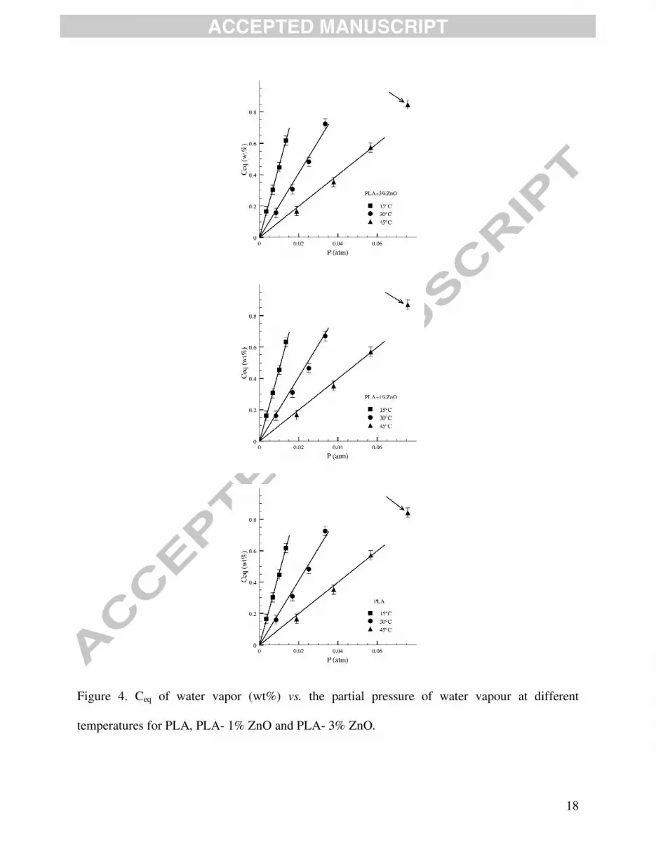
Sorption isotherms were obtained for PLA and nanocomposites loaded with 1% and 3% ZnO.
Figure 4 reports the equilibrium concentration of water vapor, Ceq (wt%), as function of water
partial pressure, P (atm) at the investigated temperatures for unfilled PLA and PLA-ZnO
nanocomposite films. All samples show an ideal behaviour at low pressure, that allowed
evaluating the sorption coefficient, S, from Henry’s law:

17
Ceq= S x P 3
For isotherms at 15 and 30 °C we considered the first four points, very well interpolated from
Henry’s model. In the case of isotherms evaluated at 45 °C, for the evaluation of sorption, we
have taken into account the first three points, which ensure an ideal interaction matrix-penetrant.
It is evident that, in all cases, at high pressure the isotherms deviate from linearity following a
Flory-Huggins mode of sorption [48] (see the arrows in the plots). According to this behaviour
there is preference for the formation of penetrant-penetrant pairs, so that the solubility coefficient
continuously increases with penetrant pressure. The first molecules sorbed tend to locally loose
the polymer structure and make it easier for the following molecules to enter. These isotherms
are observed when the penetrant effectively plasticizes the polymer, being a strong solvent or
swelling agent for the polymer, like is water vapour for PLA matrix. Table 3 reports the S
(wt%/atm) parameters, evaluated according to Eq. 3, for the analyzed samples at the three
studied temperatures. At constant temperature, there is not evident variation of S with increasing
ZnO loading. Comparing the data evaluated at different temperatures, it is possible to observe a
decreasing of sorption with increasing the temperature, in agreement with the thermodynamics of
the system.
18
Figure 4. Ceq of water vapor (wt%) vs. the partial pressure of water vapour at different
temperatures for PLA, PLA- 1% ZnO and PLA- 3% ZnO.

19
Table 4: Thermodynamic diffusion coefficients, Do (cm2/s), at different temperatures (15; 30, 45 °C) and diffusion activation energy, ED (kJ/mol) for PLA and nanocomposite films loaded with 1% and 3% ZnO. Do and ED values should be considered as having an accuracy of about ±10%. Sample
Temperature,
°C
Diffusion Coefficient, Do (cm2/s)
Diffusion Activation Energy (ED), kJ/mol
PLA
15
30
40
3.27 10-8
7.75 10-8
10.4 10-8
29.60
PLA- 1% ZnO
15
30
40
3.75 10-8
5.71 10-8
11.7 10-8
28.73
PLA- 3% ZnO
15
30
40
2.51 10-8
4.85 10-8
11.1 10-8
37.63
Diffusion
Following the increasing of sample weight as function of time, it was possible to evaluate the
diffusion coefficient, D, at different water pressures [49]. The equation used to evaluate the
diffusion parameter at different water vapor partial pressures is as follows:
4
in which M(t) is the mass of the sample at each time, M0 is the value at the beginning of the
test, M∞ is the value at equilibrium, h is the thickness of the sample. Eq. 4 is valid for times for
which the ratio [(M-M0)/(M∞-M0)] is larger than 0.4. Plotting ln(d Mt/dt) vs. time, the value of D

20
(cm2/s) is calculated at each partial pressure from the slope of the curve. Figure 5 shows an
example of the procedure adopted to determine the diffusivity. On the left axis it is possible to
read measurements of the normalized mass of PLA at 30 °C (step of humidity from 0 to 20%).
The squares show the calculations performed according to Eq. 4. The range of points to fit was
always determined by a mass change between 40% and 90%. Accordingly, for a time long
enough the considered model fits very well the experimental data. By the knowledge of sample
thickness, the diffusion coefficient is evaluated from the slope of the line.
Figure 5. Example to illustrate the procedure adopted to determine the diffusion coefficients.
The diffusion parameters obtained using Eq. 4 were plotted as function of water percentage
sorbed at the different pressures. For polymer-solvent systems, the diffusion parameter is usually
not constant, but depends on the vapor concentration, according to the empirical Eq. 5:
D = Do exp (γ Ceq) 5
where Do (cm2/s) is the zero concentration diffusion coefficient (related to the fractional free
volume and to the microstructure of the polymer); γ is a coefficient which depends on the
fractional free volume and on the effectiveness of the penetrant to plasticize the matrix.

21
Figure 6 reports the diffusion coefficient, D (cm2/s), as function of the equilibrium
concentration of water vapor, Ceq (wt%), for unfilled PLA and nanocomposite films (1 and 3%
ZnO) at 15, 30 and 45 °C. It is evident that the diffusion is independent of water vapor
concentration at any investigated temperature, so that the Do values extrapolated at Ceq= 0, with
very good degree of approximation can be considered equal to D at any vapor pressure. Do
values are reported in Table 4. As expected, on increasing the temperature, Do increases.
Therefore, the diffusion process is faster by increasing polymer free volume and at higher
mobility of polymeric chains. At constant temperature a slight decrease of D with ZnO loading
can be observed, more evident at T= 30 °C. A great number of data in literature suggest that the
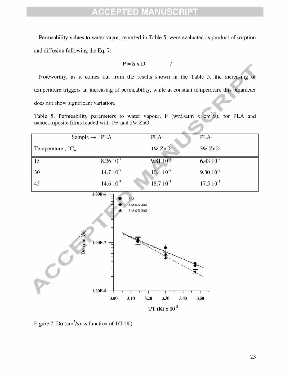
diffusion coefficient of vapors in polymers depends on temperature, via an Arrhenius’ law on a
narrow range of temperatures following the Eq. 6:
6
where Do represents the limit value of the transport coefficient for T (temperature) →∞ and ED
(kJ/mol) is the diffusion activation energy, i.e., the energy level that a molecule must reach to
diffuse inside the polymeric matrix. In our case, D being constant with Ceq, it was possible to
plot Do vs. 1/T (T in K) to evaluate the activation energies (Figure 7) that are reported in Table 4.
It is evident that such values remain quite constant for PLA and PLA- 1% ZnO, but are higher
for the sample containing 3% ZnO, confirming the increased difficulty of the travelling
molecules to diffuse into the polyester matrix.

22
0.00 0.20 0.40 0.60 0.80 1.00
Ceq (wt%)
1.00E-8
1.00E-7
1.00E-6
D (
cm
/s)
2
PLA+3% ZnO
15°C
30°C
45°C
0.00 0.20 0.40 0.60 0.80 1.00
Ceq (wt%)
1.00E-8
1.00E-7
1.00E-6
D (
cm /s
)2
PLA+1%ZnO
15°C
30°C
45°C
0.00 0.20 0.40 0.60 0.80 1.00
Ceq (wt%)
1.00E-8
1.00E-7
1.00E-6
D (
cm
/s)
2
PLA
15°C
30°C
45°C
Figure 6. Diffusion coefficient, D (cm2/s), as function of Ceq of water vapour (wt%) at different
temperatures for neat PLA and nanocomposites loaded with 1% and 3% ZnO.
23
Permeability values to water vapor, reported in Table 5, were evaluated as product of sorption
and diffusion following the Eq. 7:
P = S x D 7
Noteworthy, as it comes out from the results shown in the Table 5, the increasing of
temperature triggers an increasing of permeability, while at constant temperature this parameter
does not show significant variation.
Table 5. Permeability parameters to water vapour, P (wt%/atm x cm2/s), for PLA and nanocomposite films loaded with 1% and 3% ZnO
3.00 3.10 3.20 3.30 3.40 3.50
1/T (K) x 10
1.00E-8
1.00E-7
1.00E-6
Do
(cm
/s
)2
PLA
PLA+1% ZnO
PLA+3% ZnO
3
Figure 7. Do (cm2/s) as function of 1/T (K).
Sample →
Temperature , °C↓
PLA PLA-
1% ZnO
PLA-
3% ZnO
15 8.26 10-7 9.81 10-7 6.43 10-7
30 14.7 10-7 10.4 10-7 9.30 10-7
45 14.6 10-7 18.7 10-7 17.5 10-7

24
Antibacterial properties
To assess the antimicrobial effects of ZnO, the nanocomposite films were evaluated against E.
coli and S. aureus, respectively Gram-negative and Gram-positive bacteria. Samples of unfilled
PLA and PLA-ZnO nanocomposites with a surface area of about 1 cm2 and thickness of ~ 150
µm were incubated with each bacterial suspension at 37 °C and constant agitation, harvested at
different times and plated on a rich medium and incubated for 24 h. Subsequently, the colony
forming units (CFU) were determined and the antibacterial activity (A) was measured as
differences between logarithm values of growth of untreated (neat PLA) and treated (PLA-ZnO)
samples.
First of all, it is noteworthy mentioning that the mechanisms responsible for the antibacterial
activity of ZnO nanoparticles are still not fully clear. However, the antibacterial effect is mainly
ascribed to the photocatalytic generation of a large number of reactive oxygen species
(superoxide anions and hydroxyl radicals, generation of hydrogen peroxide), to the formation of
Zn2+ ions or the capture of ZnO onto the cell membrane, etc. [26, 50].
The values of antibacterial activity are summarized in Table 6. On one hand, for E. coli at 24 h
it was found that in presence of ZnO the number of CFU decreased with respect to the control,
but the antimicrobial effect was low (> 70 % of killed bacteria). On the other hand, after 7 days
(see also Fig. 8a) the antibacterial effect was high for all concentrations and even very good for
nanocomposite films containing low amount of nanofiller, i.e., 1% ZnO. In fact following the
values of A that are higher than 2 after 7 days (Table 6) the antimicrobial performance is
considered good or very good, which means a reduction of bacterial population of 99% or more.
For sake of comparison, it is of interest to note that in the absence of ZnO, for the neat PLA the
number of CFU after a similar incubation time is mostly unchanged (see Fig. 8a).

25
In S. aureus antibacterial properties occurred only with films containing 3% ZnO. Similarly to
E. coli, significant reduction of population (>99%) was obtained after 7 days of treatment (A=
2.85) (Table 6 and Fig. 8b).
Table 6. Antibacterial activity (A) of selected PLA-ZnO films against E. coli and S. aureus after different incubation times
Following these results, it is assumed that under the specific experimental conditions the
antibacterial activities to both bacteria were time-dependent. Moreover, similarly to the behavior
of other metal-polymer formulations [51], it is believed that the release of antimicrobial
products, such as reactive oxygen species and/or of Zn2+ ions, can require longer time than 24h
to obtain the minimum inhibitory concentration and optimal effectiveness. The different
sensitivity of E. coli and S. aureus to the concentration of ZnO may be due to structural
differences between the cell wall of Gram positive and Gram-negative bacteria. However this
could also be due to the characteristic of S. aureus to form aggregates which would protect more
internal cells from lower doses of antimicrobial products. Finally as remark, growth (G) values
Sample Average (log CFU) Growth24h Growth 7 d (day) Antibacterial
Activity (24 h) (A24h)
Antibacterial
Activity (7 d) (A7d) 0 h 24h 7d
Escherichia coli
PLA 6.76 6.59 6.62 F= -0.17 F = -0.14 (A24h=F24h–G%_24h) (A7d = F7d – G%_7d)
PLA- 1% ZnO 6.66 5.89 3.20 G1.0% = -0.77 G1.0% = -3.46 0.60 3.32
PLA- 2% ZnO 6.76 5.84 4.38 G2.0% = -0.92 G2.0% = -2.38 0.75 2.24
PLA- 3% ZnO 6.70 6.00 4.30 G3.0% = -0.70 G3.0% = -2.40 0.53 2.26
Stapylococcus aureus
PLA 7.16 7.08 4.98 F = -0.08 F=-2.18 (A24h = F24h – G%_24h) (A7d = F7d – G%_7d)
PLA- 3% ZnO 7.11 6.81 2.08 G3.0% = -0.30 G3.0 = -5.03 0.22 2.85

26
resulted significantly negative for all nanocomposites, as attended by a toxic effect of ZnO,
however, slightly negative values were also found for control populations (F), indicating an
absence of growth and a reduction of the microbial populations in experimental conditions. This
occurred because to reduce interference of nanocomposite materials with the substances
dissolved in growth media, microorganisms were suspended in distilled water (E. coli) or in
diluted peptone water (S. aureus), both non suitable to support the bacterial growth. Nonetheless,
under these conditions the bacteria tested may remain viable several weeks. Thus, based on
antimicrobial activity, ZnO containing nanocomposites showed a high antibacterial effect toward
analyzed Gram positive and negative microorganisms, but the bacterial sensitivity to them was
different, with E. coli killed efficiently at concentrations apparently innocuous for Gram positive.
Mechanical properties and additional end-use characteristics
Mechanical parameters were evaluated from stress-strain curves following the tensile tests
performed at ambient temperature. As shown in Table 7, the addition of surface-treated ZnO into
PLA leads to the slight increase of the rigidity (Young’s modulus, E) whereas both tensile stress
at yield (σy) and at break (σb) reveal interesting values for packaging applications (e.g., σy is in
the range 39 - 44 MPa) as compared to the neat PLA matrix (σy of 45 MPa). However, especially
at high ZnO loading (3%), a slight reduction of the tensile stress was observed, that is reasonably
associated to some diminution of molecular weights and to formation of low molecular products
as aforementioned. Furthermore, like the neat PLA, the nanocomposite films show low
elongation at yield (εy) and break (εb), without an apparent correlation with the nanofiller
amount. To improve this parameter of interest in the perspective of further applications, the
addition of a plasticizer into PLA-ZnO nanocomposite films can be supplementary considered.

27
a
b
Figure 8 (a, b). Evolution of log10 CFU (colony forming units) at various time intervals of E.
coli (a) and S. aureus (b) on the surface of PLA and PLA-ZnO nanocomposite films

28
Table 7: Comparative mechanical properties of PLA and PLA–ZnO nanocomposite films (standard deviations are given in brackets)
*Distance between grips of 40 mm.
The opacity is a parameter that must be considered for packaging applications, since it affects
the appearance of the products. Figure 9 reports the experimental data obtained on films of about
150 micron thickness. The opacity is only slightly increased with a higher ZnO amount. The
opacities were ~10% higher for the films containing 3% ZnO. Such effect, due to the interference
of nanoparticles with the light, must be taken into account since this is relevant for packaging
applications.
Sample
(%- by weight)
E
(MPa)
σy
(MPa)
σb
(MPa)
εy *
(%)
εb*
(%)
PLA 2700 (±200) 45 (±3) 42 (±3) 2.5 (±0.3) 9.3 (±2.4)
PLA- 1% ZnO 2900 (±300) 44 (±5) 41 (±5) 3.3 (±0.7) 13.0 (±4.1)
PLA- 2% ZnO 3000 (±150) 42 (±4) 39 (±3) 1.9 (±0.1) 7.1 (±1.5)
PLA- 3% ZnO 2800 (±100) 39 (±2) 35 (±3) 2.0 (±0.3) 12.9 (±3.0)

29
Figure 9. Opacity of 150 micron-thick films vs. the ZnO content
Finally it is of interest to note that the additional investigations (thermogravimetric analyses
(TGA) and UV–Visible absorption spectra, results not shown here) confirm the previously
reported results [34], respectively, addition of ZnO is leading in some decrease of PLA thermal
stability, whereas once more, at above 1% ZnO the films were characterized by a total anti-UV
protection and good transmittance of the visible light (400–800 nm). By considering the
multifunctional properties that are specific for PLA-ZnO films (mechanical performances, anti-
UV, antibacterial, barrier characteristics) with respect to PLA or other PLA nanocomposites and
additional potential features, such as the possibility of utilization as self-cleaning materials, these
new nanocomposites can present a high interest in traditional and special applications as
biosourced packaging materials.

30
4. Conclusions
The work was focused on the production and characterization of PLA-ZnO films to highlight
their key-characteristics. PLA-ZnO nanocomposites were produced by melt-compounding PLA
and 0.5 - 3% surface-treated ZnO rod-like nanoparticles, step followed by the production of
films. It was demonstrated that it is possible to obtain competitive thin films characterized by
adequate ZnO dispersion and quite good preservation of molecular and mechanical properties
particularly when the polyester/nanofiller interface is adequately tuned via a triethoxy
caprylylsilane surface-treatment. According to DSC and consistent with XRD the produced films
were mostly amorphous. They were analyzed with special attention paid to the water vapor
barrier properties (sorption, diffusion, permeability) which resulted from the nanofiller
incorporation. It was assumed that the well dispersed nanoparticles through the polyester matrix
can provoke certain tortuosity to water vapor molecules. The changes in PLA inherent
permeation properties were mainly dependent on the temperature and nanofiller loading. The
values of energy activation were higher for the films containing 3% ZnO with respect to the neat
PLA, confirming the increased difficulty for molecules to diffuse through the PLA matrix. The
nanocomposite films were characterized by a good antibacterial activity against Gram-positive
and Gram-negative bacteria and the antibacterial performance was found to be time-dependent.
By considering the multifunctional properties of PLA-ZnO films (anti-UV, antibacterial,
mechanical, etc.), such nanocomposites produced by extrusion could be considered as a very
promising approach in the production of environmental-friendly packaging materials.

31
Acknowledgements
The authors from University of Salerno thank Ms Laura Bassi for carrying out part of the
experiments of transport properties during her work of thesis for bachelor in Chemical
Engineering.
The authors from UMONS & Materia Nova thank the Wallonia Region, Nord-Pas de Calais
Region and European Community for the financial support in the frame of the INTERREG –
NANOLAC project. They thank to Yoann Paint and Anne-Laure Dechief for assistance in the
preparation and characterization of samples, NANOLAC partners and all mentioned companies
for supplying raw materials.
CIRMAP acknowledges supports by the Région Wallonne in the frame of OPTI²MAT
program of excellence, by the Interuniversity Attraction Pole program of the Belgian Federal
Science Policy Office (PAI 6/27) and by FNRS-FRFC.

32
5. References
[1] S. Burt, Essential oils: their antibacterial properties and potential applications in foods - a
review, Int J Food Microbiol, 94 (2004) 223-253.
[2] H. Moller, S.P. Grelier, P. Pardon, W. Coma, Antimicrobial and physicochemical
properties of chitosan-HPMC-based films, J Agr Food Chem, 52 (2004) 6585-6591.
[3] G. Findenig, S. Leimgruber, R. Kargl, S. Spirk, K. Stana-Kleinschek, V. Ribitsch, Creating
Water Vapor Barrier Coatings from Hydrophilic Components, ACS Appl. Mater. Interfaces, 4
(2012) 3199-3206.
[4] S. Khalloufi, J. Giasson, C. Ratti, Water activity of freeze dried mushrooms and berries,
Can Agr Eng, 42 (2000) 51-56.
[5] A. Cagri, Z. Ustunol, E.T. Ryser, Antimicrobial edible films and coatings, J Food Protect,
67 (2004) 833-848.
[6] M. Vargas, A. Albors, A. Chiralt, C. Gonzalez-Martinez, Characterization of chitosan-oleic
acid composite films, Food Hydrocolloids, 23 (2009) 536-547.
[7] G. Findenig; S. Leimgruber; R. Kargl; S. Spirk; K. Stana-Kleinschek; V. Ribitsch, Creating
Water Vapor Barrier Coatings from Hydrophilic Components. ACS Appl. Mater. Interfaces
2012, 4, 3199-3206.
[8] T. Bourtoom, M.S. Chinnan, Preparation and properties of rice starch-chitosan blend
biodegradable film, Lwt-Food Sci Technol, 41 (2008) 1633-1641.
33
[9] M.A. Bertuzzi, E.F. Castro Vidaurre, M. Armada, J.C. Gottifredi, Water vapor permeability
of edible starch based films, Journal Of Food Engineering, 80 (2007) 972-978.
[10] M. Perez-Mateos, P. Montero, M.C. Gomez-Guillen, Formulation and stability of
biodegradable films made from cod gelatin and sunflower oil blends, Food Hydrocolloids, 23
(2009) 53-61.
[11] T. Sivarooban, N.S. Hettiarachchy, M.G. Johnson, Physical and antimicrobial properties
of grape seed extract, nisin, and EDTA incorporated soy protein edible films, Food Research
International, 41 (2008) 781-785.
[12] M.A. Del Nobile, A. Conte, A.L. Incoronato, O. Panza, Antimicrobial efficacy and release
kinetics of thymol from zein films, Journal of Food Engineering, 89 (2008) 57-63.
[13] P. Suppakul, J. Miltz, K. Sonneveld, S.W. Bigger, Active packaging technologies with an
emphasis on antimicrobial packaging and its applications, Journal of Food Science, 68 (2003)
408-420.
[14] S. Eswaranandam, N.S. Hettiarachchy, M.G. Johnson, Antimicrobial activity of citric,
lactic, malic, or tartaric acids and nisin-incorporated soy protein film against Listeria
monocytogenes, Escherichia coli O157:H7, and Salmonella gaminara, Journal of Food Science,
69 (2004) FMS79-FMS84.
[15] A. Chaibi, L.H. Ababouch, K. Belasri, S. Boucetta, F.F. Busta, Inhibition of germination
and vegetative growth of Bacillus cereus T and Clostridium botulinum 62A spores by essential
oils, Food Microbiology, 14 (1997) 161-174.

34
[16] K.W. Kim, I.W. Roh, K.M. Kim, I.S. Jang, S.D. Ha, K.B. Song, S.K. Park, W.Y. Lee, K.
Youn, D.H. Bae, Antimicrobial edible film developed from defatted corn germ meal fermented
by Bacillus subtilis, J Microbiol Biotechn, 16 (2006) 597-604.
[17] C.M. Gucbilmez, A. Yemenicioglu, A. Arslanoglu, Antimicrobial and antioxidant activity
of edible zein films incorporated with lysozyme, albumin proteins and disodium EDTA, Food
Research International, 40 (2007) 80-91.
[18] A. Conte, B. Speranza, M. Sinigaglia, M.A. Del Nobile, Effect of lemon extract on
foodborne microorganisms, J Food Protect, 70 (2007) 1896-1900.
[19] U. Costantino, V. Bugatti, G. Gorrasi, F. Montanari, M. Nocchetti, L. Tammaro, V.
Vittoria, New Polymeric Composites Based on Poly(epsilon-caprolactone) and Layered Double
Hydroxides Containing Antimicrobial Species, Acs Applied Materials & Interfaces, 1 (2009)
668-677.
[20] V. Bugatti, U. Costantino, G. Gorrasi, M. Nocchetti, L. Tammaro, V. Vittoria, Nano-
hybrids incorporation into poly(epsilon-caprolactone) for multifunctional applications:
Mechanical and barrier properties, European Polymer Journal, 46 (2010) 418-427.
[21] G. Gorrasi, V. Bugatti, V. Vittoria, Pectins filled with LDH-antimicrobial molecules:
Preparation, characterization and physical properties, Carbohydrate Polymers, 89 (2012) 132-
137.
[22] I. Sondi, B. Salopek-Sondi, Silver nanoparticles as antimicrobial agent: a case study on E-
coli as a model for Gram-negative bacteria, Journal of Colloid and Interface Science, 275 (2004)
177-182.
35
[23] A. Yadav, V. Prasad, A.A. Kathe, S. Raj, D. Yadav, C. Sundaramoorthy, N.
Vigneshwaran, Functional finishing in cotton fabrics using zinc oxide nanoparticles, B Mater
Sci, 29 (2006) 641-645.
[24] S. Chandramouleeswaran, S. T. Mhaske, A. A. Kathe, P. V. Varadarajan, V. Prasad, N.
Vigneshwaran, Functional behaviour of polypropylene/ZnO–soluble starch nanocomposites,
Nanotechnology 18 (2007) 385702-9
[25] K.H. Tam, A.B. Djuri, C.M.N. Chan, Y.Y. Xi, C.W. Tse, Y.H. Leung, W.K. Chan, F.C.C.
Leung, D.W.T. Au, Antibacterial activity of ZnO nanorods prepared by a hydrothermal method,
Thin Solid Films, 516 (2008) 6167-6174.
[26] N. Padmavathy, R. Vijayaraghavan, Enhanced bioactivity of ZnO nanoparticles-an
antimicrobial study, Sci Technol Adv Mat, 9 (2008).
[27] D. Garlotta, A literature review of poly(lactic acid), Journal of Polymers and the
Environment, 9 (2001) 63-84.
[28] L.T. Lim, R. Auras, M. Rubino, Processing technologies for poly(lactic acid), Progress in
Polymer Science, 33 (2008) 820-852.
[29] K.M. Nampoothiri, N.R. Nair, R.P. John, An overview of the recent developments in
polylactide (PLA) research, Bioresource Technology, 101 (2010) 8493-8501.
[30] K. Rezwan, Q.Z. Chen, J.J. Blaker, A.R. Boccaccini, Biodegradable and bioactive porous
polymer/inorganic composite scaffolds for bone tissue engineering, Biomaterials, 27 (2006)
3413-3431.

36
[31] A.P. Gupta, V. Kumar, New emerging trends in synthetic biodegradable polymers -
Polylactide: A critique, European Polymer Journal, 43 (2007) 4053-4074.
[32] Y.Q. Li, S.Y. Fu, Y.W. Mai, Preparation and characterization of transparent ZnO/epoxy
nanocomposites with high-UV shielding efficiency, Polymer, 47 (2006) 2127-2132.
[33] S.C. Li, Y.N. Li, Mechanical and Antibacterial Properties of Modified Nano-ZnO/High-
Density Polyethylene Composite Films with a Low Doped Content of Nano-ZnO, Journal of
Applied Polymer Science, 116 (2010) 2965-2969.
[34] M. Murariu, A. Doumbia, L. Bonnaud, A.L. Dechief, Y. Paint, M. Ferreira, C. Campagne,
E. Devaux, P. Dubois, High-Performance Polylactide/ZnO Nanocomposites Designed for Films
and Fibers with Special End-Use Properties, Biomacromolecules, 12 (2011) 1762-1771.
[35] Ph. Dubois, M. Murariu, bioplastics Magazine, 7(1) (2012) 46-49.
[36] P.O. Bussiere, S. Therias, J-L. Gardette, M. Murariu, Ph. Dubois, M. Baba, Effect of ZnO
nanofillers treated with triethoxy caprylylsilane on the isothermal and non-isothermal
crystallization of poly(lactic acid), Physical Chemistry Chemical Physics,14 (2012) 12301-
12308.
[37] H. Tsuji, R. Okino, H. Daimon, K. Fujie, Water vapor permeability of poly(lactide)s:
Effects of molecular characteristics and crystallinity, Journal of Applied Polymer Science, 99
(2006) 2245-2252.
[38] A. Sorrentino, G. Gorrasi, V. Vittoria, Potential perspectives of bio-nanocomposites for
food packaging applications, Trends in Food Science and Technology, 18 (2007) 84-95.

37
[39] H.M.C. de Azeredo, Nanocomposites for food packaging applications, Food Research
International, 42 (2009) 1240-1253.
[40] G. Gorrasi, V. Vittoria, M. Murariu, A.D.S. Ferreira, M. Alexandre, P. Dubois, Effect of
filler content and size on transport properties of water vapor in PLA/calcium sulfate composites,
Biomacromolecules, 9 (2008) 984-990.
[41] L.C.E. Struik, Physical Aging In Plastics And Other Glassy Materials, Polymer
Engineering And Science, 17 (1977) 165-173.
[42] R. Pantani, F. De Santis, A. Sorrentino, F. De Maio, G. Titomanlio, Crystallization
kinetics of virgin and processed poly(lactic acid), Polymer Degradation And Stability, 95 (2010)
1148-1159.
[43] F. De Santis, R. Pantani, G. Titomanlio, Nucleation and crystallization kinetics of
poly(lactic acid), Thermochimica Acta, 522 (2011) 128-134.
[44] M. Pluta, Morphology and properties of polylactide modified by thermal treatment, filling
with layered silicates and plasticization, Polymer, 45 (2004) 8239-8251.
[45] T.M. Wu, C.Y. Wu, Biodegradable poly(lactic acid)/chitosan-modified montmorillonite
nanocomposites: Preparation and characterization, Polymer Degradation and Stability, 91 (2006)
2198-2204.
[46] Z. Yang, X.L. Zong, Z.Z. Ye, B.H. Zhao, Q.L. Wang, P. Wang, The application of
complex multiple forklike ZnO nanostructures to rapid and ultrahigh sensitive hydrogen
peroxide biosensors, Biomaterials, 31 (2010) 7534-7541.

38
[47] J. Ahmed, S.K. Varshney, Polylactides-Chemistry, Properties and Green Packaging
Technology: A Review, Int J Food Prop, 14 (2011) 37-58.
[48] H.B. Hopfenberg, V. Stannett, American Chemical Society. Division of Organic Coatings
and Plastics Chemistry., Permeability of plastic films and coatings to gases, vapors, and liquids :
[Proceeding of the Borden Award Symposium of the Division of Organic Coatings and Plastics
Chemistry of the American Chemical Society, honoring Professor Vivian T. Stannett], Plenum
Press, New York, 1974.
[49] M. Partini, G. Gorrasi, R. Pantani, Distribution of Morphology and Transport Properties
of Water Vapor in Injection-Molded Biodegradable Aliphatic Polyester, J Appl Polym Sci, 117
(2010) 2831-2838.
[50] S. Therias, J.-F. Larche, P.-O. Bussiere, J.-L. Gardette, M. Murariu, P. Dubois,
Photochemical Behavior of Polylactide/ZnO Nanocomposite Films. Biomacromolecules, 13
(2012) 3283-3291.
[51] R. Kumar, H. Munstedt, Silver ion release from antimicrobial polyamide/silver
composites, Biomaterials, 26 (2005) 2081-2088.


39
Nanocomposites of PLA and surface-treated ZnO rod-like nanoparticles.
Changes in PLA inherent permeation properties dependent on temperature and loading
Good antibacterial activity against Gram-positive and Gram-negative bacteria
The antibacterial performance was found to be time-dependent.


