Embed Size (px)
Citation preview

Preconditioning-mimetics Bradykinin and DADLE Activate PI3-kinase Through Divergent Pathways
Michael V. Cohen1,2, Sebastian Philipp1,*, Thomas Krieg1,**, Lin Cui1, Atsushi Kuno1,Viktoriya Solodushko1, and James M. Downey11 Department of Physiology, University of South Alabama, College of Medicine, Mobile, AL
2 Department of Medicine, University of South Alabama, College of Medicine, Mobile, AL
AbstractWe previously reported that pharmacological preconditioning of rabbit hearts with acetylcholineinvolves activation of phosphatidylinositol 3-kinase (PI3-K) through transactivation of the epidermalgrowth factor receptor (EGFR). Transactivation is thought to be initiated by cleavage of membrane-bound proheparin-binding EGF-like growth factor (HB-EGF) by a membrane metalloproteinase thusreleasing HB-EGF which binds to the EGFR. This pathway leads to redox signaling with thegeneration of reactive oxygen species (ROS) by mitochondria. We tested whether preconditioning’sphysiological triggers, bradykinin and opioid, also signal through the EGFR. Both bradykinin andthe synthetic δ-opioid agonist DADLE increased ROS production in isolated cardiomyocytes byapproximately 50%. DADLE’s effect was abrogated by either metalloproteinase inhibitor III (MPI)or the diphtheria toxin mutant CRM-197 which blocks heparin-binding EGF shedding indicating thatDADLE signals through EGFR transactivation. MPI also blocked DADLE’s infarct-sparing effectin whole hearts. Additionally, blocking Src kinase (a component of the EGFR’s signaling complex)with PP2 or PI3-K with wortmannin blocked DADLE’s effect on cardiomyocyte ROS productionand PP2 blocked DADLE’s salvage of ischemic myocardium. Finally, DADLE increasedphosphorylation of Akt and extracellular signal-regulated protein kinases (ERK) 1/2 in leftventricular myocardium, and this increase was blocked by the EGFR antagonist AG1478. On theother hand, neither MPI nor CRM-197 prevented bradykinin from increasing ROS production, andMPI did not affect bradykinin’s infarct-sparing effect in intact hearts. Conversely, both PP2 andwortmannin blocked bradykinin’s effect on ROS generation and also aborted bradykinin’scardioprotective effect in intact hearts. While bradykinin also increased phosphorylation of Akt andERK in myocardium, that increase was not affected by AG1478. Hence bradykinin, unlikeacetylcholine or opioid, does not transactivate EGFR, although all 3 agonists do signal through Srcand PI3-K.
Keywordsbradykinin; epidermal growth factor; opioids; preconditioning; transactivation
Address for correspondence: Michael V. Cohen, M.D., Department of Physiology, MSB 3050, University of South Alabama, Collegeof Medicine Mobile, AL 36688, Tel 251-460-6812, FAX 251-460-6464, [email protected]*Current address: Department of Cardiology, West German Heart Center Essen, University of Duisberg-Essen, Essen, Germany**Current address: Department of Cardiology, Ernst-Moritz-Arndt University Greifswald, Greifswald, GermanyPublisher's Disclaimer: This is a PDF file of an unedited manuscript that has been accepted for publication. As a service to our customerswe are providing this early version of the manuscript. The manuscript will undergo copyediting, typesetting, and review of the resultingproof before it is published in its final citable form. Please note that during the production process errors may be discovered which couldaffect the content, and all legal disclaimers that apply to the journal pertain.
NIH Public AccessAuthor ManuscriptJ Mol Cell Cardiol. Author manuscript; available in PMC 2008 April 1.
Published in final edited form as:J Mol Cell Cardiol. 2007 April ; 42(4): 842–851.
NIH
-PA Author Manuscript
NIH
-PA Author Manuscript
NIH
-PA Author Manuscript
Ischemic preconditioning is universally recognized as a potent cardioprotective intervention.Many investigators have studied this phenomenon to learn how protection is accomplished inthe hope that insights could be extrapolated to the clinical arena. Of the many autacoids releasedby ischemic myocardium adenosine, bradykinin, and opioids appear to be the principalendogenous triggers of preconditioning. Although all 3 of these agonists bind to surfacemembrane Gi protein-coupled receptors (GiPCR), the intracellular signaling initiated by eachis not identical. For example, the protection from both bradykinin and opioids is dependent onopening of mitochondrial ATP-dependent potassium channels (mitoKATP) and production ofreactive oxygen species (ROS) which then act as second messengers to activate protein kinaseC (PKC) [1]. However, adenosine bypasses mitoKATP and ROS production, and appears toactivate PKC directly through the dissociated G proteins [1]. It is unclear how theses 3 GiPCRscan lead to such diversity of signaling, but these redundant pathways do insure continuing cellprotection even after one of the pathways is blocked.
Several years ago we sought to map preconditioning’s trigger pathway. We initially used themuscarinic receptor agonist acetylcholine (ACh) since the M2 receptor is also a GiPCR andprotects by similarly opening mitoKATP and releasing ROS [1]. Although ACh is not releasedby ischemic cells and therefore is not a physiological mediator of preconditioning, we foundit to be a convenient agonist with which to work since its receptors are so well characterized.Krieg et al. [2,3] found evidence that ACh signaled through transactivation of the epidermalgrowth factor receptor (EGFR). The current theory is that receptor binding activates amembrane metalloproteinase (MMP) that in turn cleaves heparin-binding epidermal growthfactor-like growth factor (HB-EGF) from a membrane-associated proHB-EGF. The liberatedHB-EGF then binds to the ectodomain of the EGF receptor (EGFR) monomer causing it todimerize with another monomer which transactivates their intrinsic tyrosine kinase activityleading to autophosphorylation of their tyrosine residues. This recruits Src kinase andphosphatidylinositol 3-kinase (PI3-K) forming an active signaling complex. Activation of thelatter results in phosphorylation of membrane phospholipids, activation of phosphoinositide-dependent kinases, phosphorylation of Akt and in turn nitric oxide synthase, and productionof NO with downstream activation of PKG [4]. The latter then opens mitoKATP with subsequentrelease of ROS [5,6].
We recently observed that the cardioprotection initiated by the δ-opioid [D-Ala2, D-Leu5]-enkephalin acetate (DADLE) was abrogated by blockade of MMP with the broad-spectrumMP inhibitor III (MPI) [3]. Similarly Cao et al. [7] blocked the ability of Met5-enkephalin,another synthetic δ-opioid agonist, to prolong survival of isolated rabbit cardiomyocytes duringsimulated ischemia by treating them with AG1478, an EGFR kinase inhibitor. Bothobservations suggest that δ-opioids also trigger preconditioning through the EGFR. We couldfind no studies testing whether bradykinin also signals through EGFR transactivation inmyocardium. Because of the documented diversity of agonist-induced signaling, we thoughtit important to establish whether proHB-EGF cleavage and EGFR transactivation occurredwith both δ-opioid and bradykinin-stimulated cardioprotection as well. Consequently we testeda number of known blockers of this pathway on both the ability of bradykinin and DADLE tostimulate ROS production in isolated rabbit cardiomyocytes and to limit infarct size in isolatedrabbit hearts exposed to ischemia/reperfusion to determine whether or not these receptors signalthrough the EGFR.
METHODSThis study was performed in accordance with The Guide for the Care and Use of LaboratoryAnimals (National Academy Press, Washington, DC, 1996).
Cohen et al. Page 2
J Mol Cell Cardiol. Author manuscript; available in PMC 2008 April 1.
NIH
-PA Author Manuscript
NIH
-PA Author Manuscript
NIH
-PA Author Manuscript

Adult rabbit myocytesRabbit ventricular myocytes were isolated as described previously in detail [8]. Briefly heartsof New Zealand White rabbits were excised and retrogradely perfused with calcium-free Krebs-Henseleit-HEPES buffer containing collagenase (type 2, Worthington, Inc., Lakewood, NJ)(200 U/ml) at 37°C. Viable myocytes were separated by repetitive slow-speed centrifugationand made calcium tolerant by stepwise restoration of calcium in the medium to 1.25 mM.Usually 30–35 million viable, calcium-tolerant cells were extracted per heart. Greater than65% were rod-shaped.
Immediately after the isolation and separation procedure, cells were plated on laminin coated24-well plates (Becton Dickinson, Bedford, MA) using creatine (5 mM), L-carnitine (2 mM),and taurine (5 mM) supplemented medium 199 (CCT-medium 199) as described by Piper etal. [9] and Mitcheson et al. [10]. Penicillin (100 U/ml) and streptomycin (100μg/ml) wereadded as antibiotics. Cells were stored in incubators at 37°C in air enriched with 5% CO2. Afirst medium change was performed after 3–4 h; afterwards cells were undisturbed and allowedto equilibrate for at least 18 h.
Experimental designEach experiment started with a change of medium for 10 min. The medium was then removedand replaced with one containing the drug or drug + blocker (if required) and reducedMitoTracker Red (1μM) as a dye. This reduced form of the probe is non-fluorescent. Whenthe probe is oxidized by ROS, it then becomes fluorescent. The oxidized product is bound tothiol groups and proteins within mitochondria. After incubation with MitoTracker for 15 mincells were washed twice with fresh MitoTracker-free CCT medium. The wash serves to removethe unbound and thus voltage-dependent pool of dye held in the cells. After washing,fluorescence becomes stable for at least 30 min.
In experiments in which the effect of the blockers MPI (3μM), wortmannin (100 nM), and 4-amino-5-(4-chlorophenyl)-7-(t-butyl)pyrazolo[3,4-d] pyrimidine (PP2, 1μM) on ROSproduction by bradykinin (500 nM) or DADLE (20 nM) was to be examined, the blocker waspresent in the medium during the 10-min period prior to addition of MitoTracker and theagonist. When CRM-197 (10μg/ml), a non-toxic mutant of diphtheria toxin, was used, thepretreatment period was 90 min. Previous investigations have documented that the describedprotocol of a timed incubation followed by washing permits reliable measurement of ROSgeneration and minimizes any possible influence of changing mitochondrial transmembranepotential [11,12].
Measurement of ROS productionExperiments were designed such that four different conditions were always simultaneouslyevaluated. Mitochondrial ROS generation was analyzed by measuring the fluorescence of atleast 40 individual, rod-shaped cells that were randomly selected within each well. The averagefluorescence for the selected cells in each well was computed and compared to the averagesingle cell fluorescence in the respective control well in the same chamber. Thus the treatedcells were compared only to untreated cells of the same age and isolated and stained with thesame MitoTracker lot. Single cell fluorescence was quantified as described previously [12].Each set of experiments was repeated five to eight times on different days with cells of differentages. Approximately 200–400 typical rod-shaped cells contributed data for each experiment.
Isolated heart modelAs previously described [1], a 2–0 silk suture was passed around a branch of the left coronaryartery of New Zealand White rabbits to form a snare by passing the ends of the thread through
Cohen et al. Page 3
J Mol Cell Cardiol. Author manuscript; available in PMC 2008 April 1.
NIH
-PA Author Manuscript
NIH
-PA Author Manuscript
NIH
-PA Author Manuscript

a small vinyl tube. The heart was rapidly excised, mounted on a Langendorff apparatus by theaortic root, and perfused with oxygenated, warmed Krebs buffer. Perfusion pressure was setat 75 mmHg by adjusting the height of the reservoir. A fluid-filled latex balloon was insertedinto the left ventricle and inflated to set an end-diastolic pressure of 5 mmHg. All hearts wereallowed to equilibrate for 30 min before the protocols were started.
For the infarct investigations eight groups of hearts were studied. All hearts were subjected to30 min of regional ischemia and then reperfused for 2 h. Control hearts received no treatment.The second group of hearts was treated with DADLE (20 nM) for 5 min followed by 10 minof washout before the long ischemia. A third group was also treated with DADLE. In additionMPI (3μM) was added to the perfusate for 15 min starting 5 min before and ending 5 min afterDADLE treatment. This protocol allowed 5 min of washout before coronary occlusion. Afourth group of hearts was treated with DADLE and the Src kinase inhibitor PP2 (1μM). Thenext group of hearts was treated for 5 min with bradykinin (0.4μM) followed by 10 min ofwashout before the 30 min ischemia. The next 3 groups were co-treated with bradykinin as ingroup 5 and either MPI, PP2, or the PI3-K antagonist wortmannin (100 nM) infused with thesame schedule as MPI in group 3.
Infarct size measurementAs previously detailed [1] risk zone was delineated with 1–9μM green fluorescentmicrospheres (Duke Scientific Corp., Palo Alto, CA) and infarction with triphenyltetrazoliumchloride (TTC) staining. The areas of infarct and risk zone were determined by planimetry ofeach slice and volumes calculated by multiplying each area by slice thickness and summingthem for each heart. Infarct size is expressed as a percentage of the risk zone.
Biochemical measurementsIsolated, buffer-perfused rabbit hearts were treated with either DADLE (20 nM) or bradykinin(0.4μM) for 5 min. Transmural left ventricular biopsies weighing approximately 25 mg wereobtained with a motorized biopsy tool before and at the end of the infusion of the agonist andejected into liquid nitrogen within 1 s of excision. In 2 additional groups of hearts AG1478(300 nM) was added to the perfusate for 10 min starting 5 min before either DADLE orbradykinin.
Tissues were prepared as previously described [2]. Electrophoresis of supernatant on 10%SDS-polyacrylamide gel was followed by transfer to nitrocellulose membrane. To identifyphosphorylated kinases membranes were probed with monoclonal antibodies for phospho-Akt(Ser 473) and phospho-ERK 1/2 (Thr 202/Thr 204) followed by incubation with a secondaryantibody conjugated to horseradish peroxidase. Immunoreactive proteins were detected byenhanced chemiluminescence with LumiGLO. Band densities were determined with Sigmagelsoftware. To test for differences in protein loading and errors introduced during the transferprocess each membrane was stained with Ponceau S dye. When a difference in Ponceau stainingwas apparent the density of the phosphorylated protein was corrected for the difference inprotein loading. Samples from each heart were run on the same gel. Because each blot differsin its exposure time, strength of the antibody, and strength of the ECL chemical, it is impossibleto compare densities between blots. However we have found that preischemic phosphorylationof Akt and ERK are very reproducible and therefore all band densities are expressed as apercentage of the corresponding baseline sample. Accordingly exposure time for each blot wasadjusted to give a density that would allow accurate scanning of all lanes.
ChemicalsAll drugs required for cell isolation and culture were purchased from Sigma Chemical (St.Louis, MO). Reduced MitoTracker Red was obtained from Molecular Probes (Eugene, OR),
Cohen et al. Page 4
J Mol Cell Cardiol. Author manuscript; available in PMC 2008 April 1.
NIH
-PA Author Manuscript
NIH
-PA Author Manuscript
NIH
-PA Author Manuscript
CRM-197, DADLE, wortmannin and Ponceau S from Sigma, MPI and PP2 from Calbiochem(La Jolla, CA), and AG1478 from Alexis Biochemicals (San Diego, CA). Either distilled wateror DMSO was used to dissolve the drugs and to prepare stock solutions. The final DMSOconcentration was kept below 1%.
Phospho-Akt antibody to Ser 473 (#4251), HRP-linked anti-rabbit IgG antibody, cell lysisbuffer, and LumiGLO were purchased from Cell Signaling Technology (Beverly, MA), whileanti-ERK 1/2 (#05–481) was obtained from Upstate (Lake Placid, NY).
Data analysisFluorescence measurements provide data in arbitrary units (a.u.). To remove the variabilitycaused by different MitoTracker lots, cell age, and environmental conditions, average cellfluorescence was calculated and compared to that of simultaneously studied control cells asdescribed above. Therefore, fluorescence data are provided as a percentage of the respectivecontrol (mean ± SEM). To further minimize the possible influence of these variables on thedata, ANOVA for repeated measures with Tukey’s post hoc testing was used to evaluatedifferences in mean fluorescence of the groups within the same experiment. Baselinehemodynamic variables and risk zone and infarct size data amongst groups were compared byone-way ANOVA with Student-Newman-Keuls post hoc testing. Temporal changes inhemodynamics in a given group were evaluated by one-way ANOVA for repeated measureswith Tukey’s post hoc testing. Changes in phospho-Akt and phospho-ERK levels wereevaluated by Student’s paired t-test. A value of p< 0.05 was considered significant.
RESULTSIsolated cardiomyocytes
DADLE—Exposing cardiomyocytes to DADLE led to a 55.0 ± 8.8% increase in fluorescenceand thus ROS production (Fig. 1). Co-incubation with MPI prevented the DADLE effect onROS generation, while MPI in the absence of DADLE had no effect on ROS production.
ProHB-EGF is the primary membrane attachment site for diphtheria toxin. The diphtheria toxinmutant, CRM-197, specifically inhibits the mitogenic activity of HB-EGF [13]. To test whetherDADLE acts through HB-EGF-dependent activation of EGFR to generate ROS incardiomyocytes, we measured ROS in cells pretreated with CRM-197 prior to stimulation withDADLE. As expected, the increased ROS production triggered by DADLE was totallyabolished by CRM-197 (Fig. 1). CRM-197 on its own had no effect on cell fluorescence.
We next probed the downstream involvement of Src kinase and PI3-K in DADLE’s effect onROS generation. Again DADLE increased ROS production by more than 50% (Fig. 2). Butboth PP2 and wortmannin blocked DADLE’s effect on ROS. Neither PP2 nor wortmanninalone had virtually any effect on ROS production.
Bradykinin—In parallel studies bradykinin was substituted for DADLE in the aboveexperiments. Bradykinin significantly increased ROS production by 30.5 % (Fig. 3), andsurprisingly this increase was not affected by co-treatment with MPI. Furthermore the robustbradykin-induced increase in ROS (53.6 ± 4.5%) was not diminished by CRM-197 either (Fig.4).
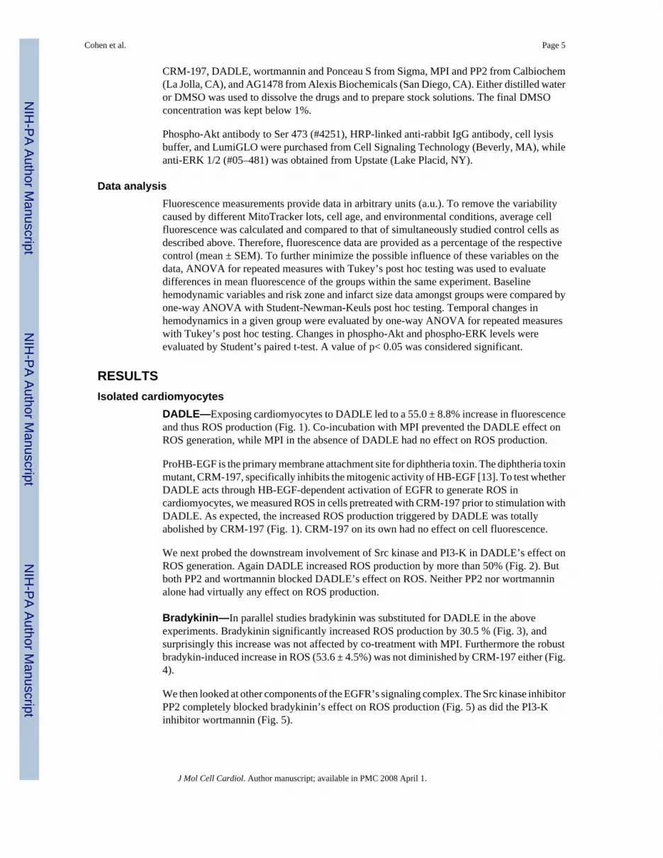
We then looked at other components of the EGFR’s signaling complex. The Src kinase inhibitorPP2 completely blocked bradykinin’s effect on ROS production (Fig. 5) as did the PI3-Kinhibitor wortmannin (Fig. 5).
Cohen et al. Page 5
J Mol Cell Cardiol. Author manuscript; available in PMC 2008 April 1.
NIH
-PA Author Manuscript
NIH
-PA Author Manuscript
NIH
-PA Author Manuscript

Isolated rabbit heart-infarct sizeHemodynamics are presented in Table 1. There were no differences in baseline heart rate, leftventricular developed pressure, or coronary flow among the groups. Wortmannin modestlydecreased left ventricular developed pressure and coronary flow. Heart weights werecomparable in all groups (Table 2). Risk zone volume was inexplicably smaller in the heartsco-treated with bradykinin and MPI.
DADLE—Pre-treatment of hearts with DADLE before ischemia/reperfusion was veryprotective and decreased infarction from 29.8 ± 2.4% of the area at risk in control hearts to10.9 ± 2.2% (p<0.01) (Table 2 and Fig. 6). Both MPI and PP2 blocked DADLE’s salutaryeffect. Previous studies have demonstrated lack of independent effect of MPI [14,3] and PP2[14,3] on infarct size.
Bradykinin—Bradykinin also had a marked cardioprotective effect (13.4 ± 1.4% infarction,p<0.01 vs Control) (Table 2 and Fig. 7). Whereas MPI had no effect on this protection (12.6± 6.9% infarction), both PP2 and wortmannin aborted the protective effect. A previous studydemonstrated that wortmannin has no effect on infarct size in untreated hearts [15]. Becauseof the smaller risk zone volume in one of the bradykinin groups, infarct size was plotted againstrisk zone volume in all groups and regression lines for each relationship compared (data notshown). Regression lines for all protected groups (DADLE, bradykinin, bradykinin + MPI)were significantly different from those for unprotected groups (Control, DADLE + MPI,DADLE + PP2, bradykinin + PP2, bradykinin + wortmannin).
Isolated rabbit heart-phospho-Akt and phospho-ERKBoth DADLE (n=4) and bradykinin (n=4) elicited significant increases in Akt phosphorylation(Fig. 8), although the response following DADLE was more robust. The EGFR antagonistAG1478 effectively blocked any response to DADLE. Although there was a trend for AG1478to also lower the level of Akt phosphorylation with bradykinin, it was not significant. Apparentdifferences in baseline phosphorylation in the blots reflect differing exposure times betweenblots rather than any differences in baseline phosphorylation.
Additionally both DADLE (n=5) and bradykinin (n=4) also produced significant increases inphosphorylation of ERK 1/2 (p44/p42) (Fig. 9). As seen with Akt the response to DADLE wasnearly double that to bradykinin. AG1478 completely blocked DADLE’s effect on ERKphosphorylation, whereas bradykinin’s effect was minimally diminished. Because of thegreater variability, the level of phosphorylated ERK 2 after AG1478 was neither significantlygreater than that at baseline nor was it smaller than that with bradykinin alone.
DISCUSSIONSignal transduction in preconditioning is thought to have two phases: the trigger phaseoccurring before ischemia and the mediator phase occurring during and after ischemia. Theformer involves in sequential order: binding of bradykinin and opioid to GiPCRs, activationof PI3-K, phosphorylation of Akt, generation of nitric oxide, activation of PKG, opening ofmitoKATP, and generation of ROS. Redox signaling by the ROS is then thought to activatePKC which we believe to be the defining event that puts the cell into the preconditioned state.Adenosine A1 receptors were found to activate PKC through a pathway that bypasses the redoxsignaling. In the present study we provide further evidence that opioid activates PI3-K throughthe EGFR similar to what was seen with muscarinic receptors. On the other hand we could findno evidence that bradykinin uses the EGFR to activate PI3-K.
Cohen et al. Page 6
J Mol Cell Cardiol. Author manuscript; available in PMC 2008 April 1.
NIH
-PA Author Manuscript
NIH
-PA Author Manuscript
NIH
-PA Author Manuscript

We originally identified involvement of the EGFR in preconditioning’s trigger pathway usingpharmacological preconditioning with ACh. While all of our evidence was indirect it wassubstantial. We first observed that ACh triggered Akt phosphorylation in rabbit hearts [2]. Asexpected that increase was dependent on PI3-K, but unexpectedly it could also be blocked bygenistein, a tyrosine kinase blocker, and by the Src kinase blocker PP2. Src kinase is knownto be a component in EGFR’s signaling complex. That suggested EGFR transactivation, whichis known to be coupled to muscarinic receptors in other tissues [16,17,18]. Accordingly theselective EGFR antagonist AG1478 blocked the pathway. We also saw phosphorylation oftyrosine groups on the EGFR. Unfortunately AG1478 cannot be used in infarction studies inrabbit hearts because it is a direct preconditioning agent [3]. It also generates mitochondrialROS in cardiomyocytes [3]. That forced us to look for more indirect evidence of EGFRinvolvement. Prenzel and colleagues [17] proposed that ACh activates the EGFR by triggeringmetalloproteinase-induced shedding of HB-EGF from a membrane bound precursor. Wetherefore tested whether a metalloproteinase inhibitor could block ACh’s protection and foundthat it could [3]. We also found that CRM-197 blocked ROS production in cardiomyocytes.Unfortunately CRM-197 is too expensive to use in whole hearts. The next obvious questionwas whether either of the physiological triggers of preconditioning, opioid or bradykinin, alsouses the EGFR for signaling. The pathways used by the two receptors to trigger preconditioningbeyond PI3-K appear to be identical to that used by ACh: all 3 use NOS, PKG, mitoKATP andredox signaling to activate PKC [1,19,4]. Furthermore Cao et al. [7] found that AG1478 couldblock a δ-opioid agonist’s protection in a cardiomyocyte model of ischemic injury suggestingthat the opioid receptors do indeed couple through the EGFR. Our present results confirm theirconclusion. Surprisingly we could find no evidence of EGFR involvement in bradykinin’striggering. Nonetheless downstream signaling steps involving Src kinase and PI3-K are intact.Therefore, we conclude that the bradykinin signal rejoins the pathway used by opioid and AChat a point distal to the EGFR.
How solid is the evidence that bradykinin does not signal through the EGFR? MPI, ahydroxamate-based inhibitor of metalloproteinases, is known to block MMP-1,-2, and-13 withIC50 <10nM, MMP-7 with IC50 = 10–100nM, and MMP-3 with IC50 = 135 nM. It is not knownwhich metalloproteinase is responsible for HB-EGF cleavage [17]. Suzuki et al. [20] originallyproposed that MMP-3 was the responsible metalloproteinase, but Hao et al. [21] identifiedMMP-7 as a major HB-EGF sheddase. Increasing information suggests that a unique group ofmetalloproteases called a disintegrin and metalloprotease (ADAM) which includes severalmembers, specifically ADAM 9, 10, 12, and 17, supports EGFR transactivation by initiatingshedding of HB-EGF [22,23,24,25,26,27,28]. Although HB-EGF shedding appears to becritical for transactivation to initiate several biologic processes in cardiac tissue, specificinhibitors which can distinguish between the various MMPs or even designate an ADAM asthe responsible protease are not available. Thus bradykinin could still be activating ametalloproteinase or metalloprotease that was not blocked by MPI. On the other hand, thefailure of CRM-197 to block bradykinin’s effect in isolated cardiomyocytes strongly arguesagainst EGFR dependence in the pathway. Furthermore, the failure of AG1478, an EGFRantagonist, to block the ability of bradykinin to increase Akt and ERK phosphorylation isadditional evidence that this GiPCR does not signal through EGFR. Because bradykinin-induced phosphorylation of ERK 2 and Akt appeared to be attenuated following exposure ofthe heart to AG1478, the data could suggest that some of bradykinin’s signaling was throughthe EGFR in addition to an alternate coupling. Unfortunately the statistics are not strongenough to confirm that a true attenuation had occurred. Our data also do not excludeinvolvement of a different membrane growth factor receptor such as the platelet-derived growthfactor receptor that can transactivate and recruit Src kinase to form a signaling module that canthen activate PI3-K.
Cohen et al. Page 7
J Mol Cell Cardiol. Author manuscript; available in PMC 2008 April 1.
NIH
-PA Author Manuscript
NIH
-PA Author Manuscript
NIH
-PA Author Manuscript

Prior examinations of the effect of AG1478 on bradykinin’s ability to activate andphosphorylate downstream MAPK and specifically ERK1/2 have yielded mixed results. Inagreement with our observations Graness et al. [29] observed that AG1478 did not affectbradykinin’s activaton of MAPK in A431 cells. On the other hand, Adomeit [30] in COS-7cells, Yang [31] in rat vascular smooth muscle cells, and Mukhin [32] in mIMCD-3 cellsreported that AG1473 inhibited bradykinin-induced activation of MAPK. Thus bradykinin’seffect on EGFR and transactivation may be tissue-and cell-specific. Our experiments are thefirst in myocardium and demonstrate that bradykinin causes phosphorylation of Akt andERK1/2 without the involvement of EGFR.
PP2 blocked ROS production in isolated cardiomyocytes and cardioprotection in intact heartsby both bradykinin and DADLE, suggesting involvement of Src kinase. The latter ispresumably downstream of EGFR in the case of DADLE, whereas its activation by bradykininis effected in the absence of EGFR occupancy. It is noteworthy that Fryer et al. [33] wereunable to block DADLE’s infarct-sparing effect in in vivo rats with PP2. However, their chosendose of PP2 was based on the agent’s known IC50 in an in vitro system, and, therefore, mightnot have accounted for factors encountered in intact animals such as protein binding. Sinceonly one dose of PP2 was used and no other parameter of adequacy of PP2 blockade wasmonitored, it is possible that the amount injected was insufficient to block Src kinase in theheart. Of interest Audet et al. [34] have noted the stimulatory effect on ERK phosphorylationof the δ-opioid receptor ligand TICP in HEK293s cells was blocked by PP2, whereas PP2paradoxically increased ERK phosphorylation triggered by a second δ-opioid receptor ligandDPDPE. Therefore, the PP2 response may also be tissue-and cell-specific.
It is unknown how two GiPCR agonists, bradykinin and DADLE, cleaving a common Giprotein can produce such diversity of signaling. But from a teleological viewpoint this diversitywould be expected to increase the survival advantage of the organism. We have nowdemonstrated at least 3 variations of signaling during the trigger phase (adenosine, ACh-opioidand bradykinin), and the differences ensure a high probability of cardioprotection even afterblockade of a single signaling moiety.
Acknowledgements
This work was supported in part by grants HL-20648 and HL-50688 from the Heart, Lung and Blood Institute of theNational Institutes of Health.
References1. Cohen MV, Yang X-M, Liu GS, Heusch G, Downey JM. Acetylcholine, bradykinin, opioids, and
phenylephrine, but not adenosine, trigger preconditioning by generating free radicals and openingmitochondrial KATP channels. Circ Res 2001;89:273–278. [PubMed: 11485978]
2. Krieg T, Qin Q, McIntosh EC, Cohen MV, Downey JM. ACh and adenosine activate PI3-kinase inrabbit hearts through transactivation of receptor tyrosine kinases. Am J Physiol 2002;283:H2322–H2330.
3. Krieg T, Cui L, Qin Q, Cohen MV, Downey JM. Mitochondrial ROS generation followingacetylcholine-induced EGF receptor transactivation requires metalloproteinase cleavage of proHB-EGF. J Mol Cell Cardiol 2004;36:435–443. [PubMed: 15010282]
4. Oldenburg O, Qin Q, Krieg T, Yang X-M, Philipp S, Critz SD, et al. Bradykinin induces mitochondrialROS generation via NO, cGMP, PKG, and mitoKATP channel opening and leads to cardioprotection.Am J Physiol 2004;286:H468–H476.
5. Pain T, Yang X-M, Critz SD, Yue Y, Nakano A, Liu GS, et al. Opening of mitochondrial KATP channelstriggers the preconditioned state by generating free radicals. Circ Res 2000;87:460–466. [PubMed:10988237]
Cohen et al. Page 8
J Mol Cell Cardiol. Author manuscript; available in PMC 2008 April 1.
NIH
-PA Author Manuscript
NIH
-PA Author Manuscript
NIH
-PA Author Manuscript

6. Costa ADT, Garlid KD, West IC, Lincoln TM, Downey JM, Cohen MV, et al. Protein kinase Gtransmits the cardioprotective signal from cytosol to mitochondria. Circ Res 2005;97:329–336.[PubMed: 16037573]
7. Cao Z, Liu L, Van Winkle DM. Met5-enkephalin-induced cardioprotection occurs via transactivationof EGFR and activation of PI3K. Am J Physiol 2005;288:H1955–H1964.
8. Armstrong S, Downey JM, Ganote CE. Preconditioning of isolated rabbit cardiomyocytes: inductionby metabolic stress and blockade by the adenosine antagonist SPT and calphostin C, a protein kinaseC inhibitor. Cardiovasc Res 1994;28:72–77. [PubMed: 7509260]
9. Piper HM, Probst I, Schwartz P, Hütter FJ, Spieckermann PG. Culturing of calcium stable adult cardiacmyocytes. J Mol Cell Cardiol 1982;14:397–412. [PubMed: 7175947]
10. Mitcheson JS, Hancox JC, Levi AJ. Cultured adult cardiac myocytes: future applications, culturemethods, morphological and electrophysiological properties. Cardiovasc Res 1998;39:280–300.[PubMed: 9798514]
11. Oldenburg O, Qin Q, Sharma AR, Cohen MV, Downey JM, Benoit JN. Acetylcholine leads to freeradical production dependent on KATP channels, Gi proteins, phosphatidylinositol 3-kinase andtyrosine kinase. Cardiovasc Res 2002;55:544–552. [PubMed: 12160951]
12. Krenz M, Oldenburg O, Wimpee H, Cohen MV, Garlid KD, Critz SD, et al. Opening of ATP-sensitivepotassium channels causes generation of free radicals in vascular smooth muscle cells. Basic ResCardiol 2002;97:365–373. [PubMed: 12200636]
13. Mitamura T, Higashiyama S, Taniguchi N, Klagsbrun M, Mekada E. Diphtheria toxin binds to theepidermal growth factor (EGF)-like domain of human heparin-binding EGF-like growth factor/diphtheria toxin receptor and inhibits specifically its mitogenic activity. J Biol Chem 1995;270:1015–1019. [PubMed: 7836353]
14. Qin Q, Downey JM, Cohen MV. Acetylcholine but not adenosine triggers preconditioning throughPI3-kinase and a tyrosine kinase. Am J Physiol 2003;284:H727–H734.
15. Baines CP, Wang L, Cohen MV, Downey JM. Myocardial protection by insulin is dependent onphosphatidylinositol 3-kinase but not protein kinase C or KATP channels in the isolated rabbit heart.Basic Res Cardiol 1999;94:188–198. [PubMed: 10424237]
16. Keely SJ, Uribe JM, Barrett KE. Carbachol stimulates transactivation of epidermal growth factorreceptor and mitogen-activated protein kinase in T84 cells. Implications for carbachol-stimulatedchloride secretion. J Biol Chem 1998;273:27111–27117. [PubMed: 9765228]
17. Prenzel N, Zwick E, Daub H, Leserer M, Abraham R, Wallasch C, et al. EGF receptor transactivationby G-protein-coupled receptors requires metalloproteinase cleavage of proHB-EGF. Nature1999;402:884–888. [PubMed: 10622253]
18. Cheng K, Zimniak P, Raufman J-P. Transactivation of the epidermal growth factor receptor mediatescholinergic agonist-induced proliferation of H508 human colon cancer cells. Cancer Res2003;63:6744–6750. [PubMed: 14583469]
19. Oldenburg O, Cohen MV, Yellon DM, Downey JM. Mitochondrial KATP channels: role incardioprotection. Cardiovasc Res 2002;55:429–437. [PubMed: 12160940]
20. Suzuki M, Raab G, Moses MA, Fernandez CA, Klagsbrun M. Matrix metalloproteinase-3 releasesactive heparin-binding EGF-like growth factor by cleavage at a specific juxtamembrane site. J BiolChem 1997;272:31730–31737. [PubMed: 9395517]
21. Hao L, Du M, Lopez-Campistrous A, Fernandez-Patron C. Agonist-induced activation of matrixmetalloproteinase-7 promotes vasoconstriction through the epidermal growth factor-receptorpathway. Circ Res 2004;94:68–76. [PubMed: 14656925]
22. Izumi Y, Hirata M, Hasuwa H, Iwamoto R, Umata T, Miyado K, et al. A metalloprotease-disintegrin,MDC9/meltrin-γ/ADAM9 and PKCδ are involved in TPA-induced ectodomain shedding ofmembrane-anchored heparin-binding EGF-like growth factor. EMBO J 1998;17:7260–7272.[PubMed: 9857183]
23. Weskamp G, Cai H, Brodie TA, Higashyama S, Manova K, Ludwig T, et al. Mice lacking themetalloprotease-disintegrin MDC9 (ADAM9) have no evident major abnormalities duringdevelopment or adult life. Mol Cell Biol 2002;22:1537–1544. [PubMed: 11839819]
Cohen et al. Page 9
J Mol Cell Cardiol. Author manuscript; available in PMC 2008 April 1.
NIH
-PA Author Manuscript
NIH
-PA Author Manuscript
NIH
-PA Author Manuscript

24. Yan Y, Shirakabe K, Werb Z. The metalloprotease Kuzbanian (ADAM10) mediates thetransactivation of EGF receptor by G protein-coupled receptors. J Cell Biol 2002;158:221–226.[PubMed: 12119356]
25. Asakura M, Kitakaze M, Takashima S, Liao Y, Ishikura F, Yoshinaka T, et al. Cardiac hypertrophyis inhibited by antagonism of ADAM12 processing of HB-EGF: metalloproteinase inhibitors as anew therapy. Nat Med 2002;8:35–40. [PubMed: 11786904]
26. Sahin U, Weskamp G, Kelly K, Zhou H-M, Higashiyama S, Peschon J, et al. Distinct roles forADAM10 and ADAM17 in ectodomain shedding of six EGFR ligands. J Cell Biol 2004;164:769–779. [PubMed: 14993236]
27. Mifune M, Ohtsu H, Suzuki H, Nakashima H, Brailoiu E, Dun NJ, et al. G protein coupling andsecond messenger generation are indispensable for metalloprotease-dependent, heparin-bindingepidermal growth factor shedding through angiotensin II type-1 receptor. J Biol Chem2005;280:26592–26599. [PubMed: 15905175]
28. Higashiyama S, Nanba D. ADAM-mediated ectodomain shedding of HB-EGF in receptor cross-talk.Biochim Biophys Acta 2005;1751:110–117. [PubMed: 16054021]
29. Graness A, Hanke S, Boehmer FD, Presek P, Liebmann C. Protein-tyrosine-phosphatase-mediatedepidermal growth factor (EGF) receptor transinactivation and EGF receptor-independent stimulationof mitogen-activated protein kinase by bradykinin in A431 cells. Biochem J 2000;347:441–447.[PubMed: 10749673]
30. Adomeit A, Graness A, Gross S, Seedorf K, Wetzker R, Liebmann C. Bradykinin B2 receptor-mediated mitogen-activated protein kinase activation in COS-7 cells requires dual signaling via bothprotein kinase C pathway and epidermal growth factor receptor transactivation. Mol Cell Biol1999;19:5289–5297. [PubMed: 10409720]
31. Yang C-M, Lin M-I, Hsieh H-L, Sun C-C, Ma Y-H, Hsiao L-D. Bradykinin-induced p42/p44 MAPKphosphorylation and cell proliferation via Src, EGF receptors, and PI3-K/Akt in vascular smoothmuscle cells. J Cell Physiol 2005;203:538–546. [PubMed: 15573401]
32. Mukhin YV, Garnovsky EA, Ullian ME, Garnovskaya MN. Bradykinin B2 receptor activatesextracellular signal-regulated protein kinase in mIMCD-3 cells via epidermal growth factor receptortransactivation. J Pharmacol Exp Ther 2003;304:968–977. [PubMed: 12604671]
33. Fryer RM, Wang Y, Hsu AK, Nagase H, Gross GJ. Dependence of δ1-opioid receptor-inducedcardioprotection on a tyrosine kinase-dependent but not a Src-dependent pathway. J Pharmacol ExpTher 2001;299:477–482. [PubMed: 11602657]
34. Audet N, Paquin-Gobeil M, Landry-Paquet O, Schiller PW, Piñeyro G. Internalization and Src activityregulate the time course of ERK activation by delta opioid receptor ligands. J Biol Chem2005;280:7808–7816. [PubMed: 15632168]
Cohen et al. Page 10
J Mol Cell Cardiol. Author manuscript; available in PMC 2008 April 1.
NIH
-PA Author Manuscript
NIH
-PA Author Manuscript
NIH
-PA Author Manuscript

Figure 1.DADLE leads to a significant increase in generation of reactive oxygen species in adult rabbitventricular myocytes. Co-treatment with either metalloproteinase inhibitor III (MPI) orCRM-197, a mutant diphtheria toxin, completely abolished this action, whereas neither MPInor CRM-197 alone had any effect. Data are presented as a change in cell fluorescence (mean± SEM) expressed as a percentage of that of simultaneously studied untreated controlcardiomyocytes.
Cohen et al. Page 11
J Mol Cell Cardiol. Author manuscript; available in PMC 2008 April 1.
NIH
-PA Author Manuscript
NIH
-PA Author Manuscript
NIH
-PA Author Manuscript

Figure 2.DADLE significantly increased production of reactive oxygen species by adult rabbitventricular myocytes. This increased production was blocked when cells were additionallyexposed to either PP2, an antagonist of Src kinase, or wortmannin, an inhibitor ofphosphatidylinositol 3-kinase. Neither PP2 nor wortmannin alone had any influence.
Cohen et al. Page 12
J Mol Cell Cardiol. Author manuscript; available in PMC 2008 April 1.
NIH
-PA Author Manuscript
NIH
-PA Author Manuscript
NIH
-PA Author Manuscript

Figure 3.When rabbit cardiomyocytes were exposed to bradykinin, they produced significantly morereactive oxygen species (ROS) than simultaneously studied untreated cells. This increasedROS production was not aborted by metalloproteinase inhibitor III (MPI), casting doubt ontransactivation of the epidermal growth factor receptor being part of bradykinin’s triggerpathway. MPI had no independent effect on ROS generation.
Cohen et al. Page 13
J Mol Cell Cardiol. Author manuscript; available in PMC 2008 April 1.
NIH
-PA Author Manuscript
NIH
-PA Author Manuscript
NIH
-PA Author Manuscript

Figure 4.Bradykinin significantly increased production of reactive oxygen species (ROS) by rabbitcardiomyocytes and this effect was not affected by the mutant diphtheria toxin CRM-197.Because the latter prevents cleavage from its inactive precursor of heparin binding epidermalgrowth factor-like growth factor, an essential component in the transactivation of the epidermalgrowth factor receptor (EGFR), its failure to limit ROS generation effectively excludesparticipation of EGFR in signaling triggered by bradykinin. CRM-197 had no independenteffect on ROS production.
Cohen et al. Page 14
J Mol Cell Cardiol. Author manuscript; available in PMC 2008 April 1.
NIH
-PA Author Manuscript
NIH
-PA Author Manuscript
NIH
-PA Author Manuscript
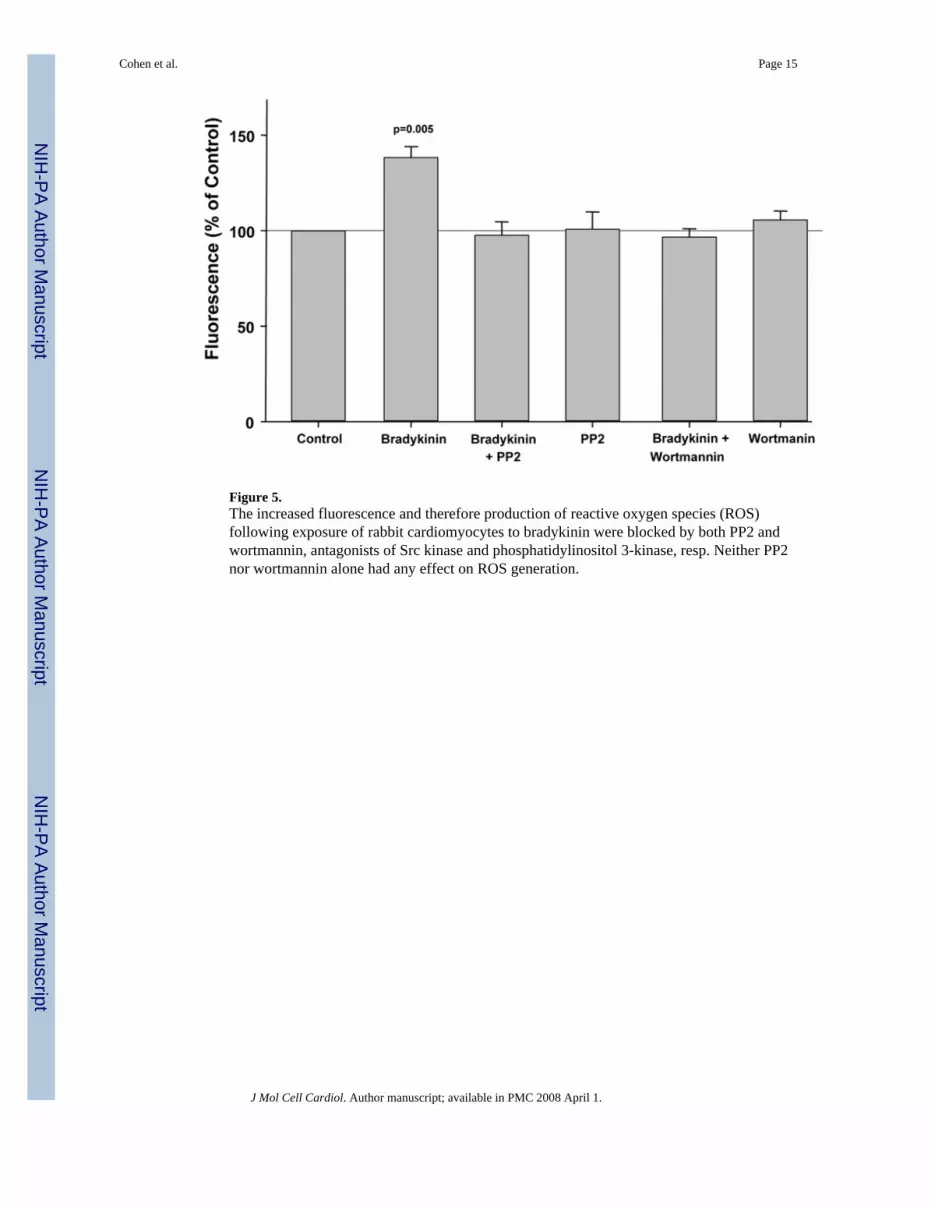
Figure 5.The increased fluorescence and therefore production of reactive oxygen species (ROS)following exposure of rabbit cardiomyocytes to bradykinin were blocked by both PP2 andwortmannin, antagonists of Src kinase and phosphatidylinositol 3-kinase, resp. Neither PP2nor wortmannin alone had any effect on ROS generation.
Cohen et al. Page 15
J Mol Cell Cardiol. Author manuscript; available in PMC 2008 April 1.
NIH
-PA Author Manuscript
NIH
-PA Author Manuscript
NIH
-PA Author Manuscript

Figure 6.Effect of DADLE, metalloproteinase inhibitor III (MPI), and PP2 on infarct size expressed asa percentage of the risk zone in isolated rabbit hearts. Open circles represent individual datapoints, while closed circles represent group averages with SEM. DADLE significantlydecreased infarction following a 30-min coronary occlusion/2-h reperfusion, and this salutaryeffect was aborted by both MPI and the Src kinase antagonist PP2. These results supportinvolvement of transactivation of the epidermal growth factor receptor in the intracellularsignaling triggered by DADLE.
Cohen et al. Page 16
J Mol Cell Cardiol. Author manuscript; available in PMC 2008 April 1.
NIH
-PA Author Manuscript
NIH
-PA Author Manuscript
NIH
-PA Author Manuscript
Figure 7.Infarct size as a percentage of the risk zone following a 30-min coronary occlusion/2-hreperfusion in isolated rabbit hearts. Open circles represent individual data points, while closedcircles represent group averages with SEM. Bradykinin salvaged ischemic myocardium.Metalloproteinase inhibitor III (MPI) had no effect on bradykinin’s cardioprotective effectsuggesting cleavage of heparin binding epidermal growth factor-like growth factor andtherefore subsequent transactivation of the epidermal growth factor receptor were not essentialfor bradykinin’s protective effect. These results distinguish bradykinin from DADLE.However, both PP2 and wortmannin, Src kinase and phosphatidylinositol 3-kinase antagonists,resp., did block bradykinin’s salutary effect implicating these kinases in the downstreamsignaling cascade. The latter observations suggest bradykinin and DADLE do have signalingsteps in common.
Cohen et al. Page 17
J Mol Cell Cardiol. Author manuscript; available in PMC 2008 April 1.
NIH
-PA Author Manuscript
NIH
-PA Author Manuscript
NIH
-PA Author Manuscript

Figure 8.
Cohen et al. Page 18
J Mol Cell Cardiol. Author manuscript; available in PMC 2008 April 1.
NIH
-PA Author Manuscript
NIH
-PA Author Manuscript
NIH
-PA Author Manuscript

A. Representative western blot of phosphorylated Akt in myocardial samples before and afterexposure to either bradykinin (Brady) or DADLE. The additional effect of the EGFR antagonistAG1478 (AG) on the change in phosphorylation induced by the agonist is demonstrated. Allsamples from each heart were run on the same blot. Differences in the baseline densities derivefrom differing exposure times of the individual blots rather than any differences in baselinephosphorylation. Because it is impossible to compare western blot data between blots all dataare normalized to the baseline (BL) sample for each heart. B.Effect of bradykinin (Brady) andDADLE on phosphorylation of Akt determined by a phospho-specific antibody in serialbiopsies from isolated rabbit hearts with and without simultaneous exposure to AG1478 (AG).*p<0.05 vs baseline
Cohen et al. Page 19
J Mol Cell Cardiol. Author manuscript; available in PMC 2008 April 1.
NIH
-PA Author Manuscript
NIH
-PA Author Manuscript
NIH
-PA Author Manuscript

Figure 9.
Cohen et al. Page 20
J Mol Cell Cardiol. Author manuscript; available in PMC 2008 April 1.
NIH
-PA Author Manuscript
NIH
-PA Author Manuscript
NIH
-PA Author Manuscript

A. Representative western blot of phosphorylated ERK 1/2 (p44/p42) in myocardial samplesbefore and after exposure to either bradykinin (Brady) or DADLE. The additional effect of theEGFR antagonist AG1478 (AG) on the change in phosphorylation induced by the agonist isdemonstrated. All samples from each heart were run on the same blot. Differences in thebaseline densities derive from differing exposure times of the individual blots rather than anydifferences in baseline phosphorylation. Because it is impossible to compare western blot databetween blots all data are normalized to the baseline (BL) sample for each heart. B. Effect ofbradykinin (Brady) and DADLE on phosphorylation of ERK 1/2 determined by a phospho-specific antibody in serial biopsies from isolated rabbit hearts with and without simultaneousexposure to AG1478 (AG). All data are normalized to the baseline (BL) sample for each heart.*p<0.025 **p<0.001 vs baseline
Cohen et al. Page 21
J Mol Cell Cardiol. Author manuscript; available in PMC 2008 April 1.
NIH
-PA Author Manuscript
NIH
-PA Author Manuscript
NIH
-PA Author Manuscript

NIH
-PA Author Manuscript
NIH
-PA Author Manuscript
NIH
-PA Author Manuscript
Cohen et al. Page 22Ta
ble
1H
emod
ynam
ics
Bas
elin
eD
rug
Tre
atm
ent
30 m
in O
cclu
sion
30 m
in R
eper
fusi
onH
R (b
pm)
LV
DP
(mm
Hg)
CF
(ml/
min
/g)
HR
(bpm
)L
VD
P (m
mH
g)C
F (m
l/m
in/g
)H
R (b
pm)
LV
DP
(mm
Hg)
CF
(ml/
min
/g)
HR
(bpm
)L
VD
P (m
mH
g)C
F (m
l/m
in/g
)C
ontro
l19
6±3
112±
48.
8±0.
418
9±5
42±2
*3.
8±0.
4*19
2±7
74±4
*6.
4±0.
5†D
AD
LE20
5±8
119±
29.
2±0.
520
2±6
117±
38.
6±0.
619
7±2
56±6
*4.
8±0.
4*18
9±4
86±7
*6.
8±0.
5*D
AD
LE +
MPI
186±
612
0±2
9.5±
0.5
192±
911
4±3
8.6±
0.3‡
196±
438
±6*
4.1±
0.3*
204±
278
±8*
6.4±
0.4*
DA
DLE
+ P
P221
2±6
118±
39.
4±0.
420
2±6
111±
57.
9±0.
520
2±7
61±8
*4.
5±0.
7*21
0±5
82±6
*5.
4±0.
7*br
adyk
inin
204±
411
4±2
11.1
±0.6
201±
510
8±1
10.6
±0.7
190±
846
±9*
5.2±
1.1*
194±
774
±2†
7.3±
0.9*
brad
ykin
in +
MPI
204±
510
8±2
8.9±
0.5
206±
796
±38.
2±0.
419
2±12
35±5
*3.
4±0.
2*21
2±5
66±4
*5.
8±0.
3*br
adyk
inin
+ P
P222
2±9
116±
39.
0±0.
720
8±7‡
106±
78.
3±0.
620
6±9†
52±8
*4.
4±0.
6*20
8±7‡
73±9
*6.
3±0.
6*br
adyk
inin
+ W
ort
200±
510
9±2
7.4±
0.2
204±
479
±10†
5.6±
0.4†
200±
033
±5*
3.1±
0.3*
197±
352
±7*
3.7±
0.4*
Mea
n ±
SEM
Abb
revi
atio
ns: b
rady
kini
n =
brad
ykin
in C
F =
coro
nary
flow
DA
DLE
= [D
-Ala
2 , D
-Leu
5 ]-e
nkep
halin
ace
tate
HR
= h
eart
rate
LV
DP
= le
ft ve
ntric
ular
dev
elop
ed p
ress
ure
MPI
= m
etal
lopr
otei
nase
inhi
bito
r PP2
= 4
-am
ino-
5-(4
-chl
orop
heny
l)-7(
t-but
yl)p
yraz
olo[
3,4-
d]py
rimid
ine
Wor
t = w
ortm
anni
n
Stat
istic
al si
gnifi
canc
e of
diff
eren
ce b
etw
een
base
line
and
expe
rimen
tal p
oint
:
* p<0.
001
† p<0.
01
‡ p<0.
05
J Mol Cell Cardiol. Author manuscript; available in PMC 2008 April 1.

NIH
-PA Author Manuscript
NIH
-PA Author Manuscript
NIH
-PA Author Manuscript
Cohen et al. Page 23Ta
ble
2In
farc
t siz
eN
BW
(kg)
HW
(g)
Ris
k Z
one
(cm
3 )In
farc
t (cm
3 )I/
R (%
)C
ontro
l7
2.5±
0.2
8.0±
0.6
1.43
±0.0
90.
42±0
.03
29.8
±2.4
DA
DLE
62.
2±0.
17.
3±0.
41.
00±0
.11
0.12
±0.0
3*10
.9±2
.2*
DA
DLE
+ M
PI5
2.2±
0.1
6.3±
0.3
1.11
±0.0
60.
32±0
.03
28.8
±1.8
DA
DLE
+ P
P26
2.3±
0.1
7.3±
0.3
1.22
±0.0
30.
36±0
.02
29.6
±0.8
brad
ykin
in7
2.3±
0.1
7.0±
1.0
1.27
±0.1
50.
18±0
.04†
13.4
±1.4
‡br
adyk
inin
+ M
PI5
2.3±
0.0
6.9±
0.3
0.89
±0.1
0‡0.
12±0
.07*
12.6
±6.9
†br
adyk
inin
+ P
P26
2.2±
0.0
6.8±
0.3
1.29
±0.1
30.
38±0
.05
29.0
±2.1
brad
ykin
in +
Wor
t6
2.3±
0.0
7.0±
0.4
1.23
±0.1
40.
46±0
.06
33.4
±5.3
Mea
n ±
SEM
Abb
revi
atio
ns: s
ee T
able
1 B
W =
bod
y w
eigh
t HW
= h
eart
wei
ght I
/R =
ratio
of i
nfar
ct si
ze to
risk
zon
e vo
lum
e N
= n
umbe
r of a
nim
als
Stat
istic
al si
gnifi
canc
e of
diff
eren
ce b
etw
een
Con
trol a
nd e
xper
imen
tal g
roup
:
* p<0.
001
† p<0.
01
‡ p<0.
05
J Mol Cell Cardiol. Author manuscript; available in PMC 2008 April 1.
![Novel pyrazolo[1,5-a]pyridines as p110α-selective PI3 kinase inhibitors: Exploring the benzenesulfonohydrazide SAR](https://img.pdfslide.net/doc/110x75/634c7245d2302b71f40cd024/novel-pyrazolo15-apyridines-as-p110-selective-pi3-kinase-inhibitors-exploring.jpg)


