Embed Size (px)
Citation preview
J. Exp. Med.
The Rockefeller University Press • 0022-1007/2000/11/1289/11 $5.00Volume 192, Number 9, November 6, 2000 1289–1299http://www.jem.org/cgi/content/full/192/9/1289
1289
Host Cell Invasion by
Trypanosoma cruzi
Is Potentiated
by Activation of Bradykinin B
2
Receptors
By Julio Scharfstein,
*
Veronica Schmitz,
*
Veronica Morandi,
‡
Marcia M. A. Capella,
*
Ana Paula C. A. Lima,
*
Alexandre Morrot,
*
Luiz Juliano,
§
and Werner Müller-Esterl
i
From the
*
Instituto de Biofísica Carlos Chagas Filho, Universidade Federal do Rio de Janeiro, CEP
21990-400 Rio de Janeiro, Brazil; the
‡
Department of Cell Biology and Genetics, Universidade do Estado do Rio de Janeiro, Rio de Janeiro 20550-013, Brazil; the
§
Department of Biophysics, Escola Paulista de Medicina-Universidade Federal de São Paolo, São Paulo 04044-000, Brazil; and the
i
Institute for Biochemistry II, University of Frankfurt Medical School, D-60590
Frankfurt, Germany
Abstract
The parasitic protozoan
Trypanosoma cruzi
employs multiple molecular strategies to invade abroad range of nonphagocytic cells. Here we demonstrate that the invasion of human primaryumbilical vein endothelial cells (HUVECs) or Chinese hamster ovary (CHO) cells overexpress-
ing the B
2
type of bradykinin receptor
(CHO-B
2
R) by tissue culture trypomastigotes is subtlymodulated by the combined activities of kininogens, kininogenases, and kinin-degrading pepti-dases. The presence of captopril, an inhibitor of bradykinin degradation by kininase II, drasti-cally potentiated parasitic invasion of HUVECs and CHO-B
2
R, but not of mock-transfectedCHO cells, whereas the B
2
R antagonist HOE 140 or monoclonal antibody MBK3 to bradyki-nin blocked these effects. Invasion competence correlated with the parasites’ ability to liberatethe short-lived kinins from cell-bound kininogen and to elicit vigorous intracellular free cal-
cium ([Ca
2
1
]
i
) transients through B
2
R. Invasion was impaired by membrane-permeable cys-teine proteinase inhibitors such as Z-(SBz)Cys-Phe-CHN
2
but not by the hydrophilic inhibitor
1-trans-epoxysuccinyl-
l
-leucyl-amido-(4-guanidino) butane or cystatin C, suggesting that ki-nin release is confined to secluded spaces formed by juxtaposition of host cell and parasiteplasma membranes. Analysis of trypomastigote transfectants expressing various cysteine pro-teinase isoforms showed that invasion competence is linked to the kinin releasing activity ofcruzipain, herein proposed as a factor of virulence in Chagas’ disease.
Key words:
Trypanosoma cruzi
• bradykinin • cruzipain • cysteine proteinases • kinin receptors
Introduction
Chagas’ disease, the chronic infection by the parasitic pro-tozoan
Trypanosoma cruzi
,
is a major cause of cardiomyopa-thy in rural Latin America. Transmitted by blood-suckingtriatomine insects, the infective forms of
T. cruzi
(trypo-mastigotes) rapidly enter the bloodstream, from where theydisseminate the infection to multiple tissues. After invadingmacrophages, muscle, and other nucleated cells, the trypo-mastigotes escape from endocytic vacuoles and migrate intothe cytoplasm where they transform into round-shapedamastigotes, the replicating forms. Within 5–6 d, the hostcells rupture, releasing large numbers of trypomastigotes
and amastigotes into interstitial spaces. Acute pathology andparasite tissue load subside with the onset of immunity, butthe pathogen is not eradicated. After years of asymptomaticinfection, 10–24% of the patients develop a severe chroniccardiomyopathy characterized by myocarditis, fibrosis, mi-crocirculatory lesions, cardiomegaly, and conduction sys-tem abnormalities (1–3).
At the cellular level,
T. cruzi
trypomastigotes invadenonphagocytic cells by a unique mechanism distinct fromphagocytosis (4, 5). Penetration by tissue culture trypomas-tigotes (TCTs)
1
is preceded by energy-dependent adhesive
Address correspondence to Julio Scharfstein, Instituto de Biofísica Car-los Chagas Filho, Centro de Ciencias de Saude, Universidade Federal doRio de Janeiro, Cidade Universitária, CEP 21990-400 Rio de Janeiro,Brazil; Phone: 55-21-280-2718; Fax: 55-21-280-8193; E-mail:[email protected]
1
Abbreviations used in this paper:
ACE, angiotensin I–converting enzyme;BK, bradykinin; [CA
2
1
]
i
, intracellular free calcium; CHO, Chinese ham-ster ovary; DTT, dithiothreitol; H-kininogen, high molecular weightkininogen; HUVEC, human primary umbilical vein endothelial cell;TCT, tissue culture trypomastigote.
1290
Kinin Pathway Involved in
T. cruzi
Infection
interactions (6) involving the parasites’ surface glycopro-teins (7, 8) and negatively charged host surface molecules(9). Depending on the host cell–parasite combination stud-ied, invasion requires activation of the TGF-
b
signalingpathway (10) or stimulation of host cell receptors coupledto heterotrimeric G proteins (11, 12). Efforts to character-ize the hitherto unknown Ca
2
1
-signaling agonist pointedto a crucial role of a cytosolic parasitic serine protease of 80kD, oligopeptidase B (13). Although null mutants gener-ated by targeted deletion of the oligopeptidase B gene werepoorly infective (14), purified or recombinant oligopepti-dase B alone failed to induce intracellular free calcium([Ca
2
1
]
i
)
transients in the mammalian cells (13). Becauseaddition of recombinant oligopeptidase B to null parasiteextracts reconstituted [Ca
2
1
]
i
signaling, it was suggestedthat the agonistic activity was generated by oligopeptidaseB–mediated processing of a cytoplasmic
T. cruzi
precursormolecule (14).
Other clues to understand the role of
T. cruzi
proteasesin host cell invasion emerged from in vitro assays per-formed with synthetic inhibitors of cruzipain (15), the par-asite’s major cysteine proteinase (16–18). Encoded by mul-tiple polymorphic genes (19, 20), this cathepsin L–likeproteinase is the most extensively characterized isoform ex-pressed by replicating forms of the parasite (16–18, 21).Given the broad pH range of the activity profile and thehigh stability of cruzipain (17), the finding of antigen de-posits of this molecule in foci of myocardial inflammation(22) suggested that this proteinase may contribute to pa-thology. Our findings that the substrate specificity of cruzi-pain resembles that of tissue kallikrein and that cruzipainreleases the bradykinin (BK)-like vasoactive peptide lysyl-bradykinin (“kallidin”) from its large precursor forms, high(H-) and low (L-) molecular weight kininogens (23), sug-gested that
T. cruzi
may directly trigger the kinin systemthrough the activity of this cysteine proteinase.
Here we demonstrate that the short-lived kinin peptidesand their cognate G protein–coupled cellular receptors (24)are engaged in the signaling mechanisms leading to
T. cruzi
invasion. We also show that invasion of cells that overex-press the constitutive B
2
subtype of BK receptor is criticallymodulated by the kinin-degrading activity of host kininaseII, also known as the angiotensin I–converting enzyme(ACE). The finding that activation of the proinflammatorykinin cascade by trypomastigotes potentiates invasion mayshed light on the molecular basis of Chagas’ disease patho-physiology.
Materials and Methods
Cells and Parasites.
Chinese hamster ovary (CHO) cells trans-fected with the cDNA encoding the rat B
2
type of BK receptor
(
B
2
R; CHO-B
2
R) or mock-transfected CHO cells (CHO-mock)were used (25). Subclone rB2CHO12/4 showed a maximum
3
H-BK binding activity of 1.3 pmol/mg of protein at passage 2.CHO cells were cultured in HAM’s F12, each supplementedwith 10% (vol/vol) of FCS at 37
8
C in a humidified atmospherecontaining 5% CO
2
. Vero cells were cultivated in DMEM with
10% FCS. Human primary umbilical vein endothelial cells (HU-VECs) were obtained by treatment of umbilical veins with a 0.1%(wt/vol) collagenase IV solution (Sigma-Aldrich). Primary HU-VECs were seeded in 25-cm
2
flasks (Corning) coated with 2%porcine skin gelatin, and grown in M199 medium supplementedwith 2 mM glutamine, 2.5
m
g/ml amphotericin B, 100
m
g/mlpenicillin, 100
m
g/ml gentamycin, 0.13% sodium bicarbonate,and 20% FCS. Cells were maintained at 37
8
C in a humidified 5%CO
2
atmosphere until they reached confluency. After treatmentwith 0.02% trypsin/0.02% EDTA, HUVECs were seeded into24-well plates with gelatin-coated glass coverslips and cultivatedat 37
8
C for several days before being used in invasion assays.
T. cruzi
epimastigotes (Dm28c clone) were cultivated at 28
8
Cin LIT medium containing 10% FCS. TCTs were harvested fromthe supernatants of infected Vero cultures maintained in DMEMsupplemented with 2% FCS (TCT-FCS). TCT transfectantsoverexpressing Dm28c genes encoding the major cruzipain (18)isoform (for simplicity, hereafter designated cruzipain-1) or cruz-ipain-2 (20, 23) were obtained by cloning full-length copies ofeach of these into the SmaI-HindIII sites of pTEX plasmid (26).Log phase Dm28c epimastigotes were transfected by electropora-tion with a single pulse of 450 kV, 500
m
F in an electroporator(Bio-Rad Laboratories). The parasites were selected for growthin LIT medium containing 10% FCS and 200
m
g/ml of geneticin(Sigma-Aldrich) for six consecutive weeks and reselected at 800
m
g/ml of geneticin for four additional weeks. Metacyclogenesiswas done by incubating stationary phase–transfected epimasti-gotes in Grace’s medium, pH 5.5, including 800
m
g/ml of gene-ticin for 7 d at 27
8
C. TCT transfectants were collected from Verocell supernatants 3–4 d after infection with the metacyclics. Plas-mid contents were stable for at least 7 wk of culture in the ab-sence of the selecting drug; TCT transfectants were tested in in-vasion assays after a 3-wk passage. The cysteine proteinaseactivity contained in cell lysates from transfected or wild-typeparasites was measured as the rate of hydrolysis of
e
-
l
-NH
2
-Cap-
l
-(SBz)C-MCA (20
m
M) in Na
2
HPO
4
, 50 mM Na
2
HPO
4
, 200mM NaCl, and 5 mM EDTA, pH 7.0, supplemented with 2.5mM dithiothreitol (DTT), at 37
8
C. To prepare the parasite celllysates, freshly released TCTs were washed twice in HBSS andresuspended in 300
m
l of PBS, pH 7.2, containing 2 mM EDTA.Then, parasites were subjected to freeze and thaw cycles (twotimes), followed by the addition of Triton X-100 to 1%. Sampleswere kept on ice for 10 min and soluble material was recoveredby centrifugation at 13,000
g
. Protein concentration was deter-mined by the Dc-protein kit (Bio-Rad Laboratories). Peptidaseactivity was measured in lysates normalized to 2
m
g/ml protein(final concentration). Enzyme stability tests were performed bymixing 2
m
l of lysates (1 mg/ml) to 100
m
l of 0.1 M glycine, pH12, for 5 s. Assay buffer was added to 1 ml and the peptidase ac-tivity was measured as described above.
Cell Invasion Assays.
CHO-B
2
R, CHO-mock, or nativeHUVECs were plated on 13-mm round coverslips at a density of2.5
3
10
4
cells/cm
2
in appropriate medium supplemented with10% FCS and cultivated in 24-well plates for 48 h at 37
8
C in a5% CO
2
atmosphere. Before addition of TCTs, coverslips withattached cells were washed three times with HBSS and kept inserum-free medium containing 1 mg/ml BSA. The parasitesadded to the wells were freshly released from cultures of infectedVero cells cultivated in DMEM-FCS (2%). After removing cellu-lar debris by low speed centrifugation (169
g
), the parasite suspen-sion was diluted three times in HBSS, spun down at 2,000
g
, andgently resuspended in DMEM-BSA or M199-BSA (1 mg/mleach). Invasion assays with CHO cells or HUVECs were done in
1291
Scharfstein et al.
a total volume of 0.6 ml of DMEM-BSA or M199-BSA, respec-tively, at a parasite/host cell ratio of 2:1 for 3 h at 37
8
C, unlessotherwise specified. When required, the medium was supple-mented with 25
m
M of captopril (CAP medium). The effects ofreceptor antagonists or antibodies were assayed by adding to CAPmedium 0.1
m
M HOE 140 (Aventis), 5–100 nM BK (Calbio-chem), or 200 nM of mAbs. In some experiments, CAP mediumwas supplemented with purified H-kininogen (9 nM) 5 min be-fore adding TCTs to HUVECs. Likewise, the effects of proteaseinhibitors were tested by adding 10 or 75
m
M 1-trans-epoxysuc-cinyl-
l
-leucylamido-(4-guanidino) butane (E-64), 10
m
M leu-peptin (Sigma-Aldrich), 10
m
M Z-(SBz)Cys-Phe-CHN
2
(15), or1
m
M recombinant human cystatin C (from Dr. Magnus Abra-hamson, Lund University, Lund, Sweden) to the CAP medium.The effect of cruzipain-1 on invasion was evaluated by adding10-fold dilutions of the activated protease to wells (0–10 nM finalconcentration) immediately after the addition of the parasites.The interaction was stopped by removing TCTs and washingcells three times with HBSS. Monolayers were fixed with Bouinand stained with Giemsa. Invasion was quantified by counting thenumber of intracellular parasites in a total of 100 cells per cover-slip. Values represent means
6
SD of at least three independentexperiments, each done in triplicate under “blinded” conditions.Statistical analysis was done by one-way analysis of variance at a
P
5
0.05 significance level.
Digital Imaging Fluorescence Microscopy.
The changes in [Ca
2
1
]
i
were determined using the fluorescent dye Fura 2-AM (Molecu-lar Probes). Customized chambers used in these experimentswere designed as follows: 30-mm plastic Petri dishes were drilledleaving 1-cm diameter holes in the center covered at the bottomby thin glass coverslips (0.7 mm) mounted with silicone glue.The plates were extensively washed and sterilized under a UVlamp for 20 min before use; coverslips for HUVECs contained2% porcine gelatin. CHO cells and HUVECs were plated at 2
3
105 cells per dish in the appropriate medium supplemented with10 and 20% of FCS, respectively, and cultivated for 24 h at 378Cin 5% CO2. The monolayers were washed three times withHBSS and incubated for 20 min at 378C in a 5% CO2 atmosphereusing serum-free HAM’s F12 medium, pH 7.4, supplementedwith 12.5 mM Hepes, 1 mg/ml BSA, 2.5 mM probenecid(Sigma-Aldrich), 25 mM captopril, and 6 mM Fura 2-AM. Afterrinsing three times to remove extracellular dye, the cells weremaintained at 378C for 15 min in the same medium without Fura2-AM. Fura-loaded cells were analyzed in an Axiovert 100 mi-croscope under an oil immersion 403 objective (ZEISS). Fluo-rescence images were collected by a digital CCD camera using a510-nm filter. [Ca21]i was monitored at 36–378C by alternatingthe excitation wavelengths between 334 and 380 nm using theAttofluor Ratio System (ZEISS). Raw fluorescence images weredigitalized to a pixel assay, point density readings were taken foreach image, and a visual display of the 340/380-nm ratio wasproduced. Before adding parasites or purified proteases, the varia-tion of [Ca21]i of all cells in the field was monitored for 2 min;20–30 cells which did not present spontaneous [Ca21]i transientswere chosen as regions of interest. After initial monitoring, thecellular responsiveness to kinin receptor agonists was assessed byadding BK (5–50 nM) to CHO-B2R or HUVECs preloadedwith Fura 2-AM, using CAP medium. Vigorous [Ca21]i tran-sients were observed for nearly 100% of the monitored cells.Specificity of BK-induced transients was checked by adding 100nM of HOE 140 to the monolayers before 50-nM agonist expo-sure. CHO-mock failed to induce significant [Ca21]i elevationsup to 50 nM BK. When indicated, 100 nM of HOE 140 was ap-
plied to the cells before the application of enzyme or parasites.Assays with TCTs were carried out at a parasite/host cell ratio of10:1. Responding cells were observed under light phase to con-firm interaction with the parasites. The Attograph software wasused to generate tracings representing [Ca21]i transients of indi-vidual cell responses, as well as average responses of 20–30 cells(n 5 30). Purified cruzipain-1 was tested at 5 nM, after a 15-minactivation of a stock solution (10-fold) with 2.5 mM DTT inPBS, pH 7.2. Controls included the addition of DTT-containingbuffer alone or activated cruzipain-1 pretreated with 75 mM E-64for 30 min. The specificity of HOE 140 was tested by pretreatingHUVECs with 100 nM of the B2R antagonist before stimulatingthe cells with 14 nM a-thrombin (Dr. Russolina Zingali, FederalUniversity of Rio de Janeiro, Rio de Janeiro, Brazil) or with 50nM BK. In the last series of experiments, the [Ca21]i concentra-tions were determined by the two-point calibration in vivomethod (27) after sequentially adding 20 mM ionomycin (Sigma-Aldrich) and 10 mM EGTA to the cultures.
Purified Proteins and Abs. Cruzipain (GP57/51) was isolatedfrom crude aqueous extracts of Dm28c epimastigotes as described(28). Recombinant cruzain (cruzipain-1) lacking the COOH-terminal extension (18) was expressed in Escherichia coli (Dr. J.H.McKerrow, University of California at San Francisco, San Fran-cisco, CA). Recombinant cruzipain-2 devoid of the COOH ter-minus (20) was expressed in Saccharomyces cerevisiae (23) andpartially purified by affinity chromatography on thiolpropyl–Sepharose 6B (Amersham Pharmacia Biotech); SDS-PAGE re-vealed a major band of 29 kD. Purified H-kininogen was fromCalbiochem. mAbs MBK3 (IgG1) recognizing the BK epitope indomain D4 of human and bovine H-/L-kininogens, HKL16 di-rected to domain D6H of human H-kininogens (29), mAb212 di-rected to cruzipain-1 (17), and IgG1 myeloma protein (MOPC31c; Sigma-Aldrich) were used. Antiserum to cruzipain-2 (rab222) was raised in rabbit chimeric protein, where a fragment (Tyr147 to Trp 187) from the central domain of cruzipain-2 (20) hadbeen fused to glutathione transferase.
Cruzipain Isoforms Released by Trypomastigotes. TCTs wereharvested from supernatants of infected Vero cells, washed withHBSS, and resuspended at 2 3 107 cells/ml in DMEM withoutFCS. After incubation at 378C for 2 h, the cell suspension wascentrifuged at 3,000 g, and the supernatant was filtered through0.2-mm Millipore filters. 200 ml of the supernatant was tested forproteolytic activity (see above). For the control, 30 mM E-64 (fi-nal concentration) was added to the supernatants. Immunoab-sorption of cruzipain isozymes was carried out by treating 600 mlof the TCT supernatant for 2 h at room temperature with 10% ofprotein A–agarose beads (Sigma-Aldrich) previously coated with10 ml of mAb212 ascites or with 50 ml of rab 222 antiserum. Forcontrols, irrelevant mAb or preimmune serum was used. Afterwashing with PBS, the hydrolytic activity associated to the resinwas measured with 50 mM fluorogenic substrate in the presenceor absence of E-64.
Kinin Release Assays. The kinin concentration in reactionmixtures of human H-kininogen and cruzipain was measured bycompetitive ELISA (Markit-M; Dainippon). After activatingstock solutions of Dm28c cruzipain-1 (50 nM) with 2.5 mMDTT, dilutions of the protease were mixed (2.5–10 nM) withH-kininogen (10 nM) in 200 ml of a reaction buffer containing50 mM Na2HPO4, pH 6.5, 200 mM NaCl, 5 mM EDTA, and0.25 mM DTT. After incubation for 30 min at 378C, the reactionwas stopped by adding 75 mM E-64, 1 mg/ml BSA, and 25 mMcaptopril; samples were deproteinized with ice-cold TCA. Afterdiluting the soluble fractions with the supplier’s buffer, the sam-
1292 Kinin Pathway Involved in T. cruzi Infection
ples were applied to the ELISA plate. A standard curve was con-structed with synthetic BK. Assays with recombinant cruzipainisozymes (20 nM) were performed as described above. Controlsperformed with enzyme buffer alone did not induce kinin re-lease. The kinin-releasing activity of trypomastigotes was mea-sured after incubating 10 nM of H-kininogen with TCTs (3 3106 cells) in 250 ml of Ham’s F12, 12.5 mM Hepes, 25 mM cap-topril, and 1 mg/ml BSA, pH 6.5, for 30 min at 378C. For thecontrol, TCTs were preincubated with 75 mM E-64 for 30 minat 378C before the addition of H-kininogen. The reaction wasstopped by adding E-64 (final concentration of 10 mM) to thecell suspension. Parasites were centrifuged at 3,000 rpm for 10min, the supernatants were filtered through 0.2-mm Millipore fil-ters, and kinin release was quantified as above. Experiments weredone in triplicates.
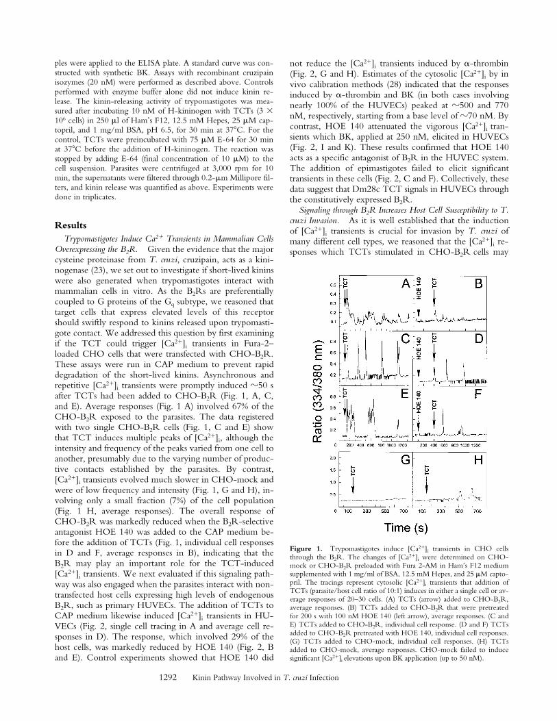
ResultsTrypomastigotes Induce Ca21 Transients in Mammalian Cells
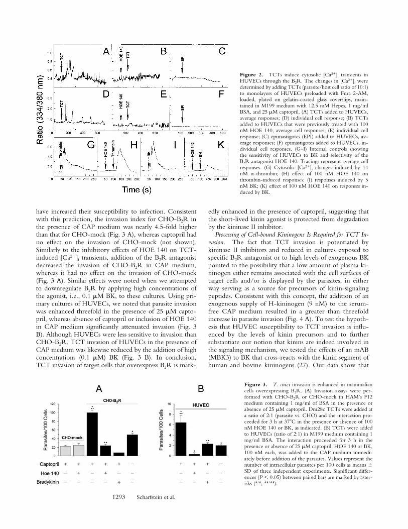
Overexpressing the B2R. Given the evidence that the majorcysteine proteinase from T. cruzi, cruzipain, acts as a kini-nogenase (23), we set out to investigate if short-lived kininswere also generated when trypomastigotes interact withmammalian cells in vitro. As the B2Rs are preferentiallycoupled to G proteins of the Gq subtype, we reasoned thattarget cells that express elevated levels of this receptorshould swiftly respond to kinins released upon trypomasti-gote contact. We addressed this question by first examiningif the TCT could trigger [Ca21]i transients in Fura-2–loaded CHO cells that were transfected with CHO-B2R.These assays were run in CAP medium to prevent rapiddegradation of the short-lived kinins. Asynchronous andrepetitive [Ca21]i transients were promptly induced z50 safter TCTs had been added to CHO-B2R (Fig. 1, A, C,and E). Average responses (Fig. 1 A) involved 67% of theCHO-B2R exposed to the parasites. The data registeredwith two single CHO-B2R cells (Fig. 1, C and E) showthat TCT induces multiple peaks of [Ca21]i, although theintensity and frequency of the peaks varied from one cell toanother, presumably due to the varying number of produc-tive contacts established by the parasites. By contrast,[Ca21]i transients evolved much slower in CHO-mock andwere of low frequency and intensity (Fig. 1, G and H), in-volving only a small fraction (7%) of the cell population(Fig. 1 H, average responses). The overall response ofCHO-B2R was markedly reduced when the B2R-selectiveantagonist HOE 140 was added to the CAP medium be-fore the addition of TCTs (Fig. 1, individual cell responsesin D and F, average responses in B), indicating that theB2R may play an important role for the TCT-induced[Ca21]i transients. We next evaluated if this signaling path-way was also engaged when the parasites interact with non-transfected host cells expressing high levels of endogenousB2R, such as primary HUVECs. The addition of TCTs toCAP medium likewise induced [Ca21]i transients in HU-VECs (Fig. 2, single cell tracing in A and average cell re-sponses in D). The response, which involved 29% of thehost cells, was markedly reduced by HOE 140 (Fig. 2, Band E). Control experiments showed that HOE 140 did
not reduce the [Ca21]i transients induced by a-thrombin(Fig. 2, G and H). Estimates of the cytosolic [Ca21]i by invivo calibration methods (28) indicated that the responsesinduced by a-thrombin and BK (in both cases involvingnearly 100% of the HUVECs) peaked at z500 and 770nM, respectively, starting from a base level of z70 nM. Bycontrast, HOE 140 attenuated the vigorous [Ca21]i tran-sients which BK, applied at 250 nM, elicited in HUVECs(Fig. 2, I and K). These results confirmed that HOE 140acts as a specific antagonist of B2R in the HUVEC system.The addition of epimastigotes failed to elicit significanttransients in these cells (Fig. 2, C and F). Collectively, thesedata suggest that Dm28c TCT signals in HUVECs throughthe constitutively expressed B2R.
Signaling through B2R Increases Host Cell Susceptibility to T.cruzi Invasion. As it is well established that the inductionof [Ca21]i transients is crucial for invasion by T. cruzi ofmany different cell types, we reasoned that the [Ca21]i re-sponses which TCTs stimulated in CHO-B2R cells may
Figure 1. Trypomastigotes induce [Ca21]i transients in CHO cellsthrough the B2R. The changes of [Ca21]i were determined on CHO-mock or CHO-B2R preloaded with Fura 2-AM in Ham’s F12 mediumsupplemented with 1 mg/ml of BSA, 12.5 mM Hepes, and 25 mM capto-pril. The tracings represent cytosolic [Ca21]i transients that addition ofTCTs (parasite/host cell ratio of 10:1) induces in either a single cell or av-erage responses of 20–30 cells. (A) TCTs (arrow) added to CHO-B2R,average responses. (B) TCTs added to CHO-B2R that were pretreatedfor 200 s with 100 nM HOE 140 (left arrow), average responses. (C andE) TCTs added to CHO-B2R, individual cell response. (D and F) TCTsadded to CHO-B2R pretreated with HOE 140, individual cell responses.(G) TCTs added to CHO-mock, individual cell responses. (H) TCTsadded to CHO-mock, average responses. CHO-mock failed to inducesignificant [Ca21]i elevations upon BK application (up to 50 nM).
1293 Scharfstein et al.
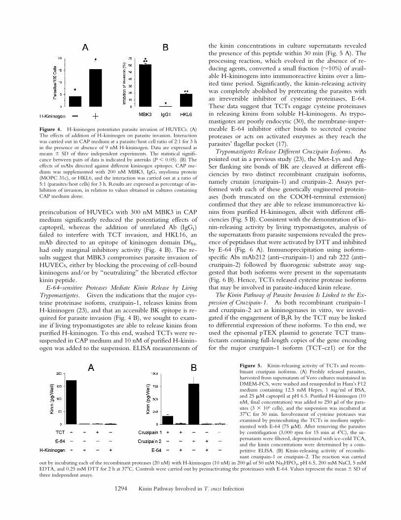
have increased their susceptibility to infection. Consistentwith this prediction, the invasion index for CHO-B2R inthe presence of CAP medium was nearly 4.5-fold higherthan that for CHO-mock (Fig. 3 A), whereas captopril hadno effect on the invasion of CHO-mock (not shown).Similarly to the inhibitory effects of HOE 140 on TCT-induced [Ca21]i transients, addition of the B2R antagonistdecreased the invasion of CHO-B2R in CAP medium,whereas it had no effect on the invasion of CHO-mock(Fig. 3 A). Similar effects were noted when we attemptedto downregulate B2R by applying high concentrations ofthe agonist, i.e., 0.1 mM BK, to these cultures. Using pri-mary cultures of HUVECs, we noted that parasite invasionwas enhanced threefold in the presence of 25 mM capto-pril, whereas absence of captopril or inclusion of HOE 140in CAP medium significantly attenuated invasion (Fig. 3B). Although HUVECs were less sensitive to invasion thanCHO-B2R, TCT invasion of HUVECs in the presence ofCAP medium was likewise reduced by the addition of highconcentrations (0.1 mM) BK (Fig. 3 B). In conclusion,TCT invasion of target cells that overexpress B2R is mark-
edly enhanced in the presence of captopril, suggesting thatthe short-lived kinin agonist is protected from degradationby the kininase II inhibitor.
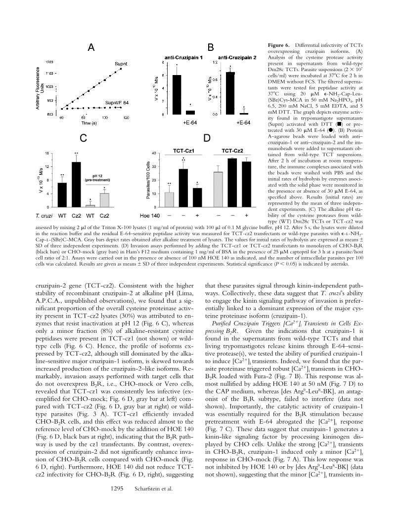
Processing of Cell-bound Kininogens Is Required for TCT In-vasion. The fact that TCT invasion is potentiated bykininase II inhibitors and reduced in cultures exposed tospecific B2R antagonist or to high levels of exogenous BKpointed to the possibility that a low amount of plasma ki-ninogen either remains associated with the cell surfaces oftarget cells and/or is displayed by the parasites, in eitherway serving as a source for precursors of kinin-signalingpeptides. Consistent with this concept, the addition of anexogenous supply of H-kininogen (9 nM) to the serum-free CAP medium resulted in a greater than threefoldincrease in parasite invasion (Fig. 4 A). To test the hypoth-esis that HUVEC susceptibility to TCT invasion is influ-enced by the levels of kinin precursors and to furthersubstantiate our notion that kinins are indeed involved inthe signaling mechanism, we tested the effects of an mAB(MBK3) to BK that cross-reacts with the kinin segment ofhuman and bovine kininogens (27). Our data show that
Figure 2. TCTs induce cytosolic [Ca21]i transients inHUVECs through the B2R. The changes in [Ca21]i weredetermined by adding TCTs (parasite/host cell ratio of 10:1)to monolayers of HUVECs preloaded with Fura 2-AM,loaded, plated on gelatin-coated glass coverslips, main-tained in M199 medium with 12.5 mM Hepes, 1 mg/mlBSA, and 25 mM captopril. (A) TCTs added to HUVECs,average responses; (D) individual cell response; (B) TCTsadded to HUVECs that were previously treated with 100nM HOE 140, average cell responses; (E) individual cellresponse; (C) epimastigotes (EPI) added to HUVECs, av-erage responses; (F) epimastigotes added to HUVECs, in-dividual cell responses. (G–I) Internal controls showingthe sensitivity of HUVECs to BK and selectivity of theB2R antagonist HOE 140. Tracings represent average cellresponses. (G) Cytosolic [Ca21]i changes induced by 14nM a-thrombin; (H) effect of 100 nM HOE 140 onthrombin-induced responses; (I) responses induced by 5nM BK; (K) effect of 100 nM HOE 140 on responses in-duced by BK.
Figure 3. T. cruzi invasion is enhanced in mammaliancells overexpressing B2R. (A) Invasion assays were per-formed with CHO-B2R or CHO-mock in HAM’s F12medium containing 1 mg/ml of BSA in the presence orabsence of 25 mM captopril. Dm28c TCTs were added ata ratio of 2:1 (parasite vs. CHO) and the interaction pro-ceeded for 3 h at 378C in the presence or absence of 100nM HOE 140 or BK, as indicated. (B) TCTs were addedto HUVECs (ratio of 2:1) in M199 medium containing 1mg/ml BSA. The interaction proceeded for 3 h in thepresence or absence of 25 mM captopril. HOE 140 or BK,100 nM each, was added to the CAP medium immedi-ately before addition of the parasites. Values represent thenumber of intracellular parasites per 100 cells as means 6SD of three independent experiments. Significant differ-ences (P , 0.05) between paired bars are marked by aster-isks (*/*; **/**).
1294 Kinin Pathway Involved in T. cruzi Infection
preincubation of HUVECs with 300 nM MBK3 in CAPmedium significantly reduced the potentiating effects ofcaptopril, whereas the addition of unrelated Ab (IgG1)failed to interfere with TCT invasion, and HKL16, anmAb directed to an epitope of kininogen domain D6H,had only marginal inhibitory activity (Fig. 4 B). The re-sults suggest that MBK3 compromises parasite invasion ofHUVECs, either by blocking the processing of cell-boundkininogens and/or by “neutralizing” the liberated effectorkinin peptide.
E-64–sensitive Proteases Mediate Kinin Release by LivingTrypomastigotes. Given the indications that the major cys-teine proteinase isoform, cruzipain-1, releases kinins fromH-kininogen (23), and that an accessible BK epitope is re-quired for parasite invasion (Fig. 4 B), we sought to exam-ine if living trypomastigotes are able to release kinins frompurified H-kininogen. To this end, washed TCTs were re-suspended in CAP medium and 10 nM of purified H-kinin-ogen was added to the suspension. ELISA measurements of
the kinin concentrations in culture supernatants revealedthe presence of this peptide within 30 min (Fig. 5 A). Theprocessing reaction, which evolved in the absence of re-ducing agents, converted a small fraction (z10%) of avail-able H-kininogens into immunoreactive kinins over a lim-ited time period. Significantly, the kinin-releasing activitywas completely abolished by pretreating the parasites withan irreversible inhibitor of cysteine proteinases, E-64.These data suggest that TCTs engage cysteine proteinasesin releasing kinins from soluble H-kininogens. As trypo-mastigotes are poorly endocytic (30), the membrane-imper-meable E-64 inhibitor either binds to secreted cysteineproteases or acts on activated enzymes as they reach theparasites’ flagellar pocket (17).
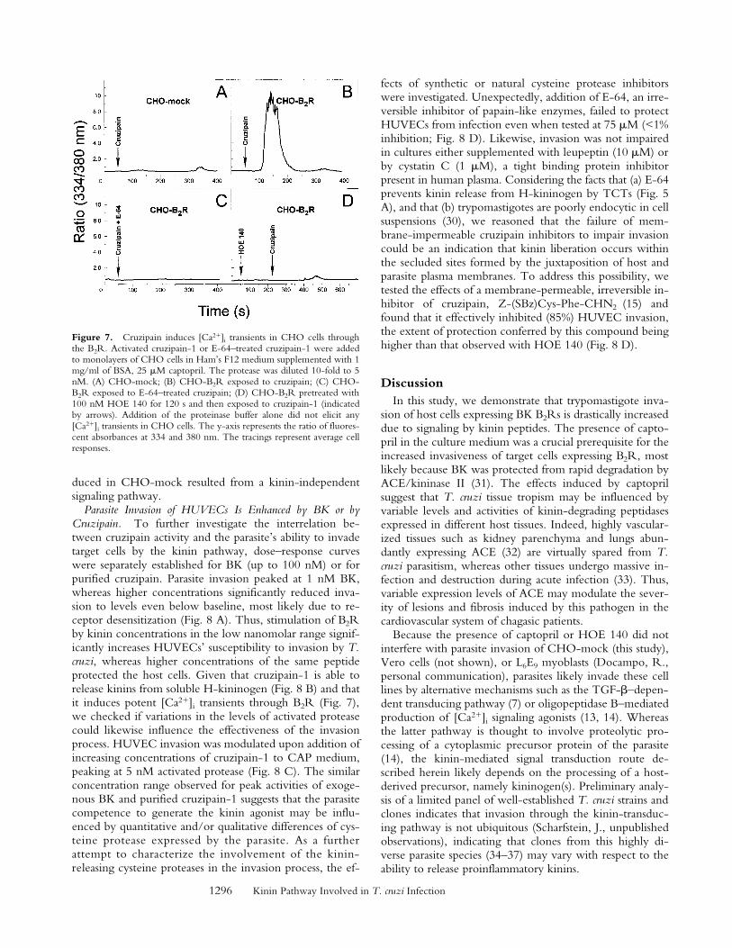
Trypomastigotes Release Different Cruzipain Isoforms. Aspointed out in a previous study (23), the Met-Lys and Arg-Ser flanking site bonds of BK are cleaved at different effi-ciencies by two distinct recombinant cruzipain isoforms,namely cruzain (cruzipain-1) and cruzipain-2. Assays per-formed with each of these genetically engineered protein-ases (both truncated on the COOH-terminal extension)confirmed that they are able to release immunoreactive ki-nins from purified H-kininogen, albeit with different effi-ciencies (Fig. 5 B). Consistent with the demonstration of ki-nin-releasing activity by living trypomastigotes, analysis ofthe supernatants from parasite suspensions revealed the pres-ence of peptidases that were activated by DTT and inhibitedby E-64 (Fig. 6 A). Immunoprecipitation using isoform-specific Abs mAb212 (anti–cruzipain-1) and rab 222 (anti–cruzipain-2) followed by fluorogenic substrate assay sug-gested that both isoforms were present in the supernatants(Fig. 6 B). Hence, TCTs released cysteine protease isoformsthat may be involved in parasite-induced kinin release.
The Kinin Pathway of Parasite Invasion Is Linked to the Ex-pression of Cruzipain-1. As both recombinant cruzipain-1and cruzipain-2 act as kininogenases in vitro, we investi-gated if the engagement of B2R by the TCT may be linkedto differential expression of these isoforms. To this end, weused the episomal pTEX plasmid to generate TCT trans-fectants containing full-length copies of the gene encodingfor the major cruzipain-1 isoform (TCT-cz1) or for the
Figure 4. H-kininogen potentiates parasite invasion of HUVECs. (A)The effects of addition of H-kininogen on parasite invasion. Interactionwas carried out in CAP medium at a parasite/host cell ratio of 2:1 for 3 hin the presence or absence of 9 nM H-kininogen. Data are expressed asmeans 6 SD of three independent experiments. The statistical signifi-cance between pairs of data is indicated by asterisks (P , 0.05). (B) Theeffects of mAbs directed against different kininogen epitopes. CAP me-dium was supplemented with 200 nM MBK3, IgG1 myeloma protein(MOPC 31c), or HKL6, and the interaction was carried out at a ratio of5:1 (parasites/host cells) for 3 h. Results are expressed as percentage of in-hibition of invasion, in relation to values obtained in cultures containingCAP medium alone.
Figure 5. Kinin-releasing activity of TCTs and recom-binant cruzipain isoforms. (A) Freshly released parasites,harvested from supernatants of Vero cultures maintained inDMEM-FCS, were washed and resuspended in Ham’s F12medium containing 12.5 mM Hepes, 1 mg/ml of BSA,and 25 mM captopril at pH 6.5. Purified H-kininogen (10nM, final concentration) was added to 250 ml of the para-sites (3 3 106 cells), and the suspension was incubated at378C for 30 min. Involvement of cysteine proteases wasexamined by preincubating the TCTs in medium supple-mented with E-64 (75 mM). After removing the parasitesby centrifugation (3,000 rpm for 15 min at 48C), the su-pernatants were filtered, deproteinized with ice-cold TCA,and the kinin concentrations were determined by a com-petitive ELISA. (B) Kinin-releasing activity of recombi-nant cruzipain-1 or cruzipain-2. The reaction was carried
out by incubating each of the recombinant proteases (20 nM) with H-kininogen (10 nM) in 200 ml of 50 mM Na2HPO4, pH 6.5, 200 mM NaCl, 5 mMEDTA, and 0.25 mM DTT for 2 h at 378C. Controls were carried out by preinactivating the proteinases with E-64. Values represent the mean 6 SD ofthree independent assays.
1295 Scharfstein et al.
cruzipain-2 gene (TCT-cz2). Consistent with the higherstability of recombinant cruzipain-2 at alkaline pH (Lima,A.P.C.A., unpublished observations), we found that a sig-nificant proportion of the overall cysteine proteinase activ-ity present in TCT-cz2 lysates (30%) was attributed to en-zymes that resist inactivation at pH 12 (Fig. 6 C), whereasonly a minor fraction (8%) of alkaline-resistant cysteinepeptidases were present in TCT-cz1 (not shown) or wild-type cells (Fig. 6 C). Hence, the profile of isoforms ex-pressed by TCT-cz2, although still dominated by the alka-line-sensitive major cruzipain-1 isoform, is skewed towardsincreased production of the cruzipain-2–like isoforms. Re-markably, invasion assays performed with target cells thatdo not overexpress B2R, i.e., CHO-mock or Vero cells,revealed that TCT-cz1 was consistently less infective (ex-emplified for CHO-mock; Fig. 6 D, gray bar at left) com-pared with TCT-cz2 (Fig. 6 D, gray bar at right) or wild-type parasites (Fig. 3 A). TCT-cz1 efficiently invadedCHO-B2R cells, and this effect was reduced almost to thereference level of CHO-mock by the addition of HOE 140(Fig. 6 D, black bars at right), indicating that the B2R path-way is used by the cz1 transfectants. By contrast, overex-pression of cruzipain-2 did not significantly enhance inva-sion of CHO-B2R cells compared with CHO-mock (Fig.6 D, right). Furthermore, HOE 140 did not reduce TCT-cz2 infectivity for CHO-B2R (Fig. 6 D, right), suggesting
that these parasites signal through kinin-independent path-ways. Collectively, these data suggest that T. cruzi’s abilityto engage the kinin signaling pathway of invasion is prefer-entially linked to a dominant expression of the major cys-teine proteinase isoform (cruzipain-1).
Purified Cruzipain Triggers [Ca21]i Transients in Cells Ex-pressing B2R. Given the indications that cruzipain-1 isfound in the supernatants from wild-type TCTs and thatliving trypomastigotes release kinins through E-64–sensi-tive protease(s), we tested the ability of purified cruzipain-1to induce [Ca21]i transients. Indeed, we found that the par-asite proteinase triggered robust [Ca21]i transients in CHO-B2R loaded with Fura-2 (Fig. 7 B). This response was al-most nullified by adding HOE 140 at 50 nM (Fig. 7 D) tothe CAP medium, whereas [des Arg9-Leu8-BK], an antag-onist of the B1R subtype, failed to interfere (data notshown). Importantly, the catalytic activity of cruzipain-1was essentially required for the B2R stimulation becausepretreatment with E-64 abrogated the [Ca21]i response(Fig. 7 C). These data suggest that cruzipain-1 generates akinin-like signaling factor by processing kininogen dis-played by CHO cells. Unlike the strong [Ca21]i transientsin CHO-B2R, cruzipain-1 induced only a minor [Ca21]i
response in CHO-mock (Fig. 7 A). This low response wasnot inhibited by HOE 140 or by [des Arg9-Leu8-BK] (datanot shown), suggesting that the minor [Ca21]i transients in-
Figure 6. Differential infectivity of TCTsoverexpressing cruzipain isoforms. (A)Analysis of the cysteine protease activitypresent in supernatants from wild-typeDm28c TCTs. Parasite suspensions (2 3 107
cells/ml) were incubated at 378C for 2 h inDMEM without FCS. The filtered superna-tants were tested for peptidase activity at378C using 20 mM e-NH2-Cap-Leu-(SBz)Cys-MCA in 50 mM Na2HPO4, pH6.5, 200 mM NaCl, 5 mM EDTA, and 5mM DTT. The graph depicts enzyme activ-ity found in trypomastigote supernatants(Supnt) activated with DTT (j) or pre-treated with 30 mM E-64 (d). (B) ProteinA–agarose beads were loaded with anti–cruzipain-1 or anti–cruzipain-2 and the im-munobeads were added to supernatants ob-tained from wild-type TCT suspensions.After 2 h of incubation at room tempera-ture, the immune complexes associated withthe beads were washed with PBS and theinitial rates of hydrolysis by enzymes associ-ated with the solid phase were monitored inthe presence or absence of 30 mM E-64, asspecified above. Results (initial rates) arerepresented by the mean of three indepen-dent experiments. (C) The alkaline pH sta-bility of the cysteine proteases from wild-type (WT) Dm28c TCTs or TCT-cz2 was
assessed by mixing 2 ml of the Triton X-100 lysates (1 mg/ml of protein) with 100 ml of 0.1 M glycine buffer, pH 12. After 5 s, the lysates were dilutedin the reaction buffer and the residual E-64–sensitive peptidase activity was measured for TCT-cz2 transfectants or wild-type parasites with e-l-NH2-Cap-l-(SBz)C-MCA. Gray bars depict rates obtained after alkaline treatment of lysates. The values for initial rates of hydrolysis are expressed as means ±SD of three independent experiments. (D) Invasion assays performed by adding the TCT-cz1 or TCT-cz2 transfectants to monolayers of CHO-B2R(black bars) or CHO-mock (gray bars) in Ham’s F12 medium containing 1 mg/ml of BSA in the presence of 25 mM captopril for 3 h at a parasite/hostcell ratio of 2:1. Assays were carried out in the presence or absence of 100 nM HOE 140 as indicated, and the number of intracellular parasites per 100cells was calculated. Results are given as means ± SD of three independent experiments. Statistical significance (P , 0.05) is indicated by asterisks.
1296 Kinin Pathway Involved in T. cruzi Infection
duced in CHO-mock resulted from a kinin-independentsignaling pathway.
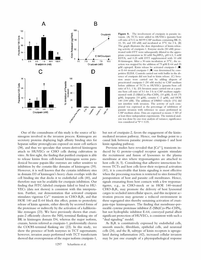
Parasite Invasion of HUVECs Is Enhanced by BK or byCruzipain. To further investigate the interrelation be-tween cruzipain activity and the parasite’s ability to invadetarget cells by the kinin pathway, dose–response curveswere separately established for BK (up to 100 nM) or forpurified cruzipain. Parasite invasion peaked at 1 nM BK,whereas higher concentrations significantly reduced inva-sion to levels even below baseline, most likely due to re-ceptor desensitization (Fig. 8 A). Thus, stimulation of B2Rby kinin concentrations in the low nanomolar range signif-icantly increases HUVECs’ susceptibility to invasion by T.cruzi, whereas higher concentrations of the same peptideprotected the host cells. Given that cruzipain-1 is able torelease kinins from soluble H-kininogen (Fig. 8 B) and thatit induces potent [Ca21]i transients through B2R (Fig. 7),we checked if variations in the levels of activated proteasecould likewise influence the effectiveness of the invasionprocess. HUVEC invasion was modulated upon addition ofincreasing concentrations of cruzipain-1 to CAP medium,peaking at 5 nM activated protease (Fig. 8 C). The similarconcentration range observed for peak activities of exoge-nous BK and purified cruzipain-1 suggests that the parasitecompetence to generate the kinin agonist may be influ-enced by quantitative and/or qualitative differences of cys-teine protease expressed by the parasite. As a furtherattempt to characterize the involvement of the kinin-releasing cysteine proteases in the invasion process, the ef-
fects of synthetic or natural cysteine protease inhibitorswere investigated. Unexpectedly, addition of E-64, an irre-versible inhibitor of papain-like enzymes, failed to protectHUVECs from infection even when tested at 75 mM (<1%inhibition; Fig. 8 D). Likewise, invasion was not impairedin cultures either supplemented with leupeptin (10 mM) orby cystatin C (1 mM), a tight binding protein inhibitorpresent in human plasma. Considering the facts that (a) E-64prevents kinin release from H-kininogen by TCTs (Fig. 5A), and that (b) trypomastigotes are poorly endocytic in cellsuspensions (30), we reasoned that the failure of mem-brane-impermeable cruzipain inhibitors to impair invasioncould be an indication that kinin liberation occurs withinthe secluded sites formed by the juxtaposition of host andparasite plasma membranes. To address this possibility, wetested the effects of a membrane-permeable, irreversible in-hibitor of cruzipain, Z-(SBz)Cys-Phe-CHN2 (15) andfound that it effectively inhibited (85%) HUVEC invasion,the extent of protection conferred by this compound beinghigher than that observed with HOE 140 (Fig. 8 D).
DiscussionIn this study, we demonstrate that trypomastigote inva-
sion of host cells expressing BK B2Rs is drastically increaseddue to signaling by kinin peptides. The presence of capto-pril in the culture medium was a crucial prerequisite for theincreased invasiveness of target cells expressing B2R, mostlikely because BK was protected from rapid degradation byACE/kininase II (31). The effects induced by captoprilsuggest that T. cruzi tissue tropism may be influenced byvariable levels and activities of kinin-degrading peptidasesexpressed in different host tissues. Indeed, highly vascular-ized tissues such as kidney parenchyma and lungs abun-dantly expressing ACE (32) are virtually spared from T.cruzi parasitism, whereas other tissues undergo massive in-fection and destruction during acute infection (33). Thus,variable expression levels of ACE may modulate the sever-ity of lesions and fibrosis induced by this pathogen in thecardiovascular system of chagasic patients.
Because the presence of captopril or HOE 140 did notinterfere with parasite invasion of CHO-mock (this study),Vero cells (not shown), or L6E9 myoblasts (Docampo, R.,personal communication), parasites likely invade these celllines by alternative mechanisms such as the TGF-b–depen-dent transducing pathway (7) or oligopeptidase B–mediatedproduction of [Ca21]i signaling agonists (13, 14). Whereasthe latter pathway is thought to involve proteolytic pro-cessing of a cytoplasmic precursor protein of the parasite(14), the kinin-mediated signal transduction route de-scribed herein likely depends on the processing of a host-derived precursor, namely kininogen(s). Preliminary analy-sis of a limited panel of well-established T. cruzi strains andclones indicates that invasion through the kinin-transduc-ing pathway is not ubiquitous (Scharfstein, J., unpublishedobservations), indicating that clones from this highly di-verse parasite species (34–37) may vary with respect to theability to release proinflammatory kinins.
Figure 7. Cruzipain induces [Ca21]i transients in CHO cells throughthe B2R. Activated cruzipain-1 or E-64–treated cruzipain-1 were addedto monolayers of CHO cells in Ham’s F12 medium supplemented with 1mg/ml of BSA, 25 mM captopril. The protease was diluted 10-fold to 5nM. (A) CHO-mock; (B) CHO-B2R exposed to cruzipain; (C) CHO-B2R exposed to E-64–treated cruzipain; (D) CHO-B2R pretreated with100 nM HOE 140 for 120 s and then exposed to cruzipain-1 (indicatedby arrows). Addition of the proteinase buffer alone did not elicit any[Ca21]i transients in CHO cells. The y-axis represents the ratio of fluores-cent absorbances at 334 and 380 nm. The tracings represent average cellresponses.
1297 Scharfstein et al.
One of the conundrums of this study is the source of ki-ninogens involved in the invasion process. Kininogens aresecretory proteins displaying high affinity binding sites forheparan sulfate proteoglycans exposed on most cell surfaces(38), and thus we speculate that serum-derived kininogensattach to HUVECs or CHO cells during cultivation invitro. At first sight, the finding that purified cruzipain is ableto release kinins from cell-bound kininogens seems para-doxical because papain-like enzymes are rather sensitive toinhibition by the cystatin-like domains of kininogens (39).However, it is well known that the cystatin inhibitory sitesin domain D3 of kininogen’s heavy chain overlaps with thecell binding site that docks it to endothelial cells (40), andtherefore may not be available for cruzipain inhibition. Ourfinding that FITC-labeled cruzipain failed to bind to HU-VECs (data not shown) is consistent with this interpreta-tion. Further, our demonstration that activated cruzipainstimulates vigorous Ca21 transients in CHO-B2R, and thatHOE 140 and E-64 block this effect, points to proteolyticrelease of kinin agonists, either directly by secreted forms ofthe proteinase or indirectly by the activation of a kallikrein-like zymogen (23). We have previously shown that cruzi-pain-2 efficiently cleaves the NH2-terminal flanking site ofBK in kininogen domain D4, whereas the major isoform,cruzain, herein referred as cruzipain-1, preferentially cleavesthe COOH-terminal flanking site (23). In this study, weshow the presence of both isozymes in TCT supernatants;however, invasion assays performed with TCT transfectantsshowed that overexpression of the major isoform cruzipain-1,
but not of cruzipain-2, favors the engagement of the kinin-mediated invasion pathway. Hence, our findings point to acausal link between parasitic protease cruzipain-1 and hostkinin signaling pathway.
Previous studies have revealed that [Ca21]i transients in-duced by G protein–coupled receptor agonists stimulatethe recruitment and fusion of lysosomes to the plasmamembrane at sites where trypomastigotes are attached tohost cells (4, 5). Considering that adhesive interactions be-tween TCTs and host cells favor their reciprocal activation(41), it is conceivable that kinin signaling is most effectivewhen the processing reaction is restricted to sites formed byjuxtaposition of host and parasite cell membranes. Hence,signals emanating from host contacts with a few trypomas-tigotes, e.g., in CHO-mock or in HOE 140–treatedCHO-B2R, may promote the delivery of host lysosomalcargos to secluded intercellular spaces, and this incipient ac-tivation process may generate a reduced environment inthese segregated sites thereby sustaining activation of cruzi-pain-type kininogenases. The finding that membrane-per-meable cysteine proteinase inhibitor Z-(SBz)Cys-Phe-CHN2,but not hydrophilic inhibitor E-64, conveyed partial albeitsignificant protection of HUVECs, is consistent with such a“dual signaling” model.
As B2R is constitutively expressed by endothelial cells,smooth muscle, fibroblasts, epithelial cells, and neuronalcells (24), and the B1 subtype of kinin receptors is upregu-lated during inflammation (42), increased cellular invasionmay be just one example of a physiopathological response
Figure 8. The involvement of cruzipain in parasite in-vasion. (A) TCTs were added to HUVECs (parasite/hostcell ratio of 5:1) in M199 CAP medium containing BK (0,10, 50, and 100 nM) and incubated at 378C for 3 h. (B)The graph illustrates the dose dependence of kinin-releas-ing activity of cruzipain-1. Enzyme stocks (50 nM) preac-tivated with DTT were subsequently diluted to the appro-priate concentration in 50 mM Na2HPO4, pH 6.5, 5 mMEDTA, and 0.25 mM DTT supplemented with 10 nMH-kininogen. After a 30-min incubation at 378C, the re-action was stopped by the addition of 75 mM E-64 and 25mM captopril. Kinin release by activated cruzipain-1 (d)or E-64–treated cruzipain-1 (r) was determined by com-petitive ELISA. Controls carried out with buffer in the ab-sence of cruzipain did not lead to kinin release. (C) Inva-sion assays were carried out by adding aliquots ofpreactivated cruzipain-1 (50 nM stocks) to CAP mediumbefore addition of TCTs to HUVECs (parasite/host cellratio of 5:1, 1 h). (D) Invasion assays carried out at a para-site/host cell ratio of 5:1 for 3 h in CAP medium supple-mented with Z-(SBz)Cys-Phe-CHN2 (10 mM), E-64 (75mM), leupeptin (10 mM), cystatin C (1 mM), and HOE140 (100 nM). The addition of DMSO vehicle (1%) didnot interfere with invasion. The activity of each com-pound was expressed as the percentage of inhibition ofparasite invasion with reference to assays performed inCAP medium alone. Data are expressed as means 6 SD ofat least three independent experiments. The statistical anal-ysis was done by one-way analysis of variance; significancewas considered at *P , 0.05.
1298 Kinin Pathway Involved in T. cruzi Infection
that kinins and their metabolites may induce in tissuesexposed to T. cruzi. For example, triggering of kinin re-ceptors from vascular endothelial cells may facilitate thetransmigration of the trypomastigotes across capillaries,modulate the expression of vascular adhesion molecules,and/or promote plasma leakage into interstitial spaces, andthus contribute to the microvascular changes observed inpatients with chronic cardiomyopathy (43). Unraveling thedelicate interplay between the parasite cysteine proteinasesand the multiple components of the kinin cascade systemmay provide fresh insights into the molecular pathogenesisof Chagas’ disease.
We thank Leila F.C. Duarte and Alda M. Alves for technical assis-tance, and Dr. Anibal Gil Lopes for free access to digital imagingfluorescence microscopy.
This work was supported by grants from Pronex II, ConselhoNacional de Pesquisas, Fundacão de Amparo à Pesquisa do Rio deJaneiro, Fundacão Universitária José Bonifácio, and the DeutscheForschungsgemeinschaft. The authors dedicate this work to thememory of the late Prof. Carlos Chagas Filho.
Submitted: 7 February 2000Revised: 27 September 2000Accepted: 28 September 2000
References1. Andrade, Z.A. 1999. Immunopathology of Chagas’ disease.
Mem. Inst. Oswaldo Cruz. 94(Suppl. I):71–80.2. Rossi, M. 1990. Microvascular changes as a cause of chronic
cardiomyopathy in Chagas’ disease. Am. Heart J. 120:233–236.
3. Morris, S.A., H. Tanowitz, M. Wittner, and J.P. Bilezikjan.1990. Pathophysiological insights into the cardiomyopathy ofChagas’ disease. Circulation. 82:1900–1909.
4. Tardieux, I., P. Webster, J. Ravesloot, W. Boron, J.A. Lunn,and N.W. Andrews. 1992. Lysosome recruitment and fusionare early events required for Trypanosoma invasion of mam-malian cells. Cell. 71:1117–1130.
5. Burleigh, B.A, and N.W. Andrews. 1995. The mechanismsof Trypanosoma cruzi invasion of mammalian cells. Annu. Rev.Microbiol. 49:175–200.
6. Schenckman, S., E.S. Robbins, and V. Nussenzweig. 1991.Attachment of Trypanosoma cruzi to mammalian cells requiresparasite energy and invasion can be independent of the targetcell cytoskeleton. Infect. Immun. 59:645–654.
7. Schenckman, S., M.S. Jiang, G.W. Hart, and V. Nussenz-weig. 1991. A novel cell surface trans-sialidase of Trypanosomacruzi generates a stage-specific epitope required for invasionof mammalian cells. Cell. 65:1117–1125.
8. Giordano, R., R. Chammas, S.S. Veiga, W. Colli, and M.J.Alves. 1994. An acidic component of the heterogeneous Tc-85 protein family from the surface of Trypanosoma cruzi is alaminin binding glycoprotein. Mol. Biochem. Parasitol. 65:85–94.
9. Herrera, E.M., M. Ming, E. Ortega-Barria, and M.E. Pereira.1994. Mediation of Trypanosoma cruzi invasion by heparansulfate receptors on host cells and penetrin counter-receptorson the trypanossomes. Mol. Biochem. Parasitol. 65:73–83.
10. Ming, M., M.E. Ewen, and M.E. Pereira. 1995. Trypanosomainvasion of mammalian cells requires activation of the TGF-b
signaling pathway. Cell. 82:287–296.11. Tardieux, I., M.H. Nathanson, and N.W. Andrews. 1994.
Role in host cell invasion of Trypanosoma cruzi–induced cyto-solic free Ca21 transients. J. Exp. Med. 179:1017–1022.
12. Docampo, R., and S.N.J. Moreno. 1996. The role of Ca21 inthe process of cell invasion by intracellular parasites. Parasitol.Today. 12:61–65.
13. Burleigh, B.A., E.V. Caler, P. Webster, and N.W. Andrews.1997. A cytosolic serine endopeptidase from Trypanosomacruzi is required for the generation of Ca21 signaling in mam-malian cells. J. Cell Biol. 136:609–620.
14. Caler, E.V., S.V. de Avalos, P.A Haynes, N.W. Andrews,and B.A. Burleigh. 1998. Oligopeptidase B-dependent sig-naling mediates host cell invasion by Trypanosoma cruzi.EMBO (Eur. Mol. Biol. Organ.) J. 17:4975–4986.
15. Meirelles, M.N., L. Juliano, E. Carmona, S.G. Silva, E.M.Costa, A.C. Murta, and J. Scharfstein. 1992. Inhibitors of themajor cysteinyl proteinase (GP57/51) impair host cell inva-sion and arrest the intracellular development of Trypanosomacruzi in vitro. Mol. Biochem. Parasitol. 52:175–184.
16. Cazzulo, J.J, R. Cousi, A. Raimondi, C. Wernstedt, and U.Hellman. 1989. Further characterization and partial aminoacid sequence of a cysteine protease (cruzipain) from Try-panosoma cruzi. Mol. Biochem. Parasitol. 33:33–42.
17. Murta, A.C., P.M. Persechini, T. Souto-Padron, W. deSouza, J.A. Guimarães, and J. Scharfstein. 1990. Structuraland functional identification of GP57/51 antigen of Trypano-soma cruzi as a cysteine proteinase. Mol. Biochem. Parasitol. 43:27–38.
18. Eakin, A.E., A.A Mills, G. Harth, J.H. McKerrow, and C.S.Craik. 1992. The sequence, organization and expression ofthe major cysteine protease (cruzain) from Trypanosoma cruzi.J. Biol. Chem. 267:7411–7420.
19. Campetella, O., J. Henrickson, L. Aslund, A.C.C. Frasch, U.Petterson, and J.J. Cazzulo. 1992. The major cysteine pro-tease (cruzipain) of Trypanosoma cruzi is encoded by polymor-phic tandemly repeated organized genes located in differentchromosomes. Mol. Biochem. Parasitol. 50:225–234.
20. Lima, A.P.C.A., D.C. Tessier, D.Y. Thomas, J. Scharfstein,A.C. Storer, and T. Vernet. 1994. Identification of new cys-teine protease gene isoforms in Trypanosoma cruzi. Mol. Bio-chem. Parasitol. 6:333–338.
21. McGrath, M.E., A.E. Eakin, J.C. Engel, J.H. McKerrow,C.S. Craik, and R.J. Fletterick. 1995. The crystal structure ofcruzain: a therapeutic target for Chagas’ disease. J. Mol. Biol.247:251–259.
22. Morrot, A., D.S. Strickland, M.L. Higuchi, M. Reis, R.Pedrosa, and J. Scharfstein. 1997. Human T cell responseagainst the major cysteine proteinase (cruzipain) of Trypano-soma cruzi: role of the multifunctional a2-macroglobulin re-ceptor in antigen presentation by monocytes. Int. Immunol.9:825–834.
23. Del Nery, E., M.A. Juliano, A.P.C.A. Lima, J. Scharfstein,and L. Juliano. 1997. Kininogenase activity by major cys-teinyl proteinase (cruzipain) from Trypanosoma cruzi. J. Biol.Chem. 272:25713–25718.
24. Bhoola, K.D, C.D. Figueroa, and K. Worthy. 1992. Bioreg-ulation of kinins: kallikreins, kininogens, and kininases. Phar-macol. Rev. 44:1–80.
25. Quitterer, U., C. Schroeder, W. Müller-Esterl, and H.Rehm. 1995. Effects of bradykinin and endothelin-1 on thecalcium homeostasis of mammalian cells. J. Biol. Chem. 270:1992–1999.
1299 Scharfstein et al.
26. Kelly, J.M., P. Das, and A.M. Thomas. 1994. An approach tofunctional complementation by introduction of large frag-ments into Trypanosoma cruzi and Leishmania donovani using acosmid shuttle vector. Mol. Biochem. Parasitol. 65:51–62.
27. Kaufmann, J., M. Haasemann, S. Modrow, and W. Müller-Esterl. 1993. Structural dissection of the multi-domain kinin-ogens. Fine mapping of the target epitopes of antibodies in-terfering with their functional properties. J. Biol. Chem. 268:9079–9091.
28. Grynkiewicz, G., M. Poenie, and R.Y. Tsien. 1985. A newgeneration of Ca21 indicators with greatly improved fluores-cence properties. J. Biol. Chem. 260:3440–3450.
29. Lima, A.P.C.A., J. Scharfstein, A.C. Storer, and R. Menard.1992. Temperature-dependent substrate inhibition of thecysteine proteinase (GP57/51) from Trypanosoma cruzi. Mol.Biochem. Parasitol. 56:335–338.
30. de Souza, W. 1995. Structural organization of the cell surfaceof pathogenic protozoa. Micron. 26:405–430.
31. Ferreira, S.H., L.J. Greene, V.A. Alabaster, Y.S. Bakhle, andJ.R. Vane. 1970. Activity of various fractions of bradykininpotentiating factor against angiotensin I converting enzyme.Nature. 225:379–380.
32. Danilov, S.M., A.I. Faerman, O. Yu Printseva, A.V Mar-tynov, I. Yu Sakharov, and I.N. Trakht, 1987. Immunohis-tochemical study of angiotensin-converting enzyme in hu-man tissues using monoclonal antibodies. Histochemistry. 87:487–490.
33. Lenzi, H.L., D.N. Oliveira, M.T. Lima, and C.R. Gattass.1996. Trypanosoma cruzi: paninfectivity of CL strain duringmurine acute infection. Exp. Parasitol. 84:16–27.
34. Miles, M.A., A. Souza, M. Povoa, J.J. Shaw, R. Lainson, andP.J. Toye. 1978. Isozymic heterogeneity of Trypanosoma cruziin the first autochtonous patients with Chagas’ disease inAmazonia Brazil. Nature. 272:819–821.
35. Tibayrenc, M., P. Ward, A. Moya, and F.J. Ayala. 1986.
Natural populations of Trypanosoma cruzi, the agent of Cha-gas’ disease, have a complex multiclonal structure. Proc. Natl.Acad. Sci. USA. 83:115–119.
36. Otavio-Souto, R.P., O. Fernandes, A.M. Macedo, D.A.Campbell, and B. Zingales. 1996. DNA markers define twomajor phylogenetic lineages of Trypanosoma cruzi. Mol. Bio-chem. Parasitol. 83:141–152.
37. de Diego, J.A., M.T. Palau, C. Gamallo, and P. Penin. 1998.Relationship between histopathological findings and phylo-genetic divergence in Trypanosoma cruzi. Trop. Med. Int.Health. 3:222–233.
38. Renné, T., J. Dedio, G. David, and W. Müller-Esterl. 2000.H-kininogen utilizes heparan sulfate proteoglycans for accu-mulation on endothelial cells. J. Biol. Chem. 275:33688–33696.
39. Stoka, V., M. Nycander, B. Lenarcic, C. Labriola, J.J. Caz-zulo, I. Björk, and V. Turk. 1995. Inhibition of cruzipain,the major cysteine proteinase of the protozoan parasite, Try-panosoma cruzi, by proteinase inhibitors of the cystatin super-family. FEBS Lett. 370:101–104.
40. Herwald, H., A.A.K. Hasan, J. Godovac-Zimmermann, A.H.Schmaier, and W. Müller-Esterl. 1995. Identification of anendothelial cell binding site on kininogens’ domain D3. J.Biol. Chem. 270:14634–14642.
41. Moreno, S.N.J., J. Silva, A.E. Vercesi, and R. Docampo.1994. Cytosolic-free calcium elevation in Trypanosoma cruzi.J. Exp. Med. 180:1535–1540.
42. Marceau, F. 1995. Kinin B1 receptors: a review. Immunophar-macology. 30:1–26.
43. Higuchi, M.L., S. Fukasawa, T. de Brito, L.C. Parzianello, G.Belloti, and J.A.F. Ramires. 1999. Different microcirculatoryand interstitial matrix patterns in idiopathic dilated cardiomy-opathy and Chagas’ disease: a three dimensional confocal mi-croscopy study. Heart. 8:1–6.


