Embed Size (px)
Citation preview
Prefrontal Contributions to Delayed Spatial and Object Alternation:A Positron Emission Tomography Study
David H. ZaldVanderbilt University
Clayton CurtisUniversity of California, Berkeley
Bradley S. FolleyVanderbilt University
JoseV. PardoVeterans Affairs Medical Center, Minneapolis, Minnesota,
and University of Minnesota
Delayed alternation tasks are frequently used as probes of frontal lobe functioning. To clarifythe neural substrates of delayed alternation performance in humans, the authors measuredregional cerebral blood flow with H2
15O positron emission tomography in healthy subjects asthey performed delayed spatial and object alternation. Consistent with the monkey lesionliterature, increased dorsolateral prefrontal activity emerged during delayed spatial alternationbut not delayed object alternation, whereas orbitofrontal activations emerged in both alter-nation tasks. The possible cognitive processes contributing to the orbitofrontal and dorsolat-eral prefrontal involvement in delayed alternation are discussed. Additional activationslocalized to several nonfrontal regions suggest caution in interpreting alternation deficits inpatients as strictly reflecting frontal lobe impairment.
Delayed alternation tasks have been widely used asprobes of frontal lobe functions in both humans and ani-mals. These tasks require subjects to select one of twoobjects on each trial, with the correct response correspond-ing to the object or location that the subject did not chooseon the previous trial. Such tasks are typically conceptualizedas working memory tasks because they require the subjectto hold on line and update information on a trial-by-trialbasis.
Several lesion studies in monkeys implicate the dorsolat-eral prefrontal cortex (DLPFC) as a critical substrate forperforming delayed spatial alternations (DSA; Butters &Pandya, 1969; Goldman, Rosvold, Vest, & Galkin, 1971;Miller & Orbach, 1972; Mishkin, 1957; Mishkin, Vest,Waxler, & Rosvold, 1969; Stamm & Weber-Levine, 1971).
Autoradiographic data in monkeys further confirm the en-gagement of the DLPFC in DSA (Friedman & Goldman-Rakic, 1994). The deficit in DSA that arises followingDLPFC lesions is typically thought to reflect the specificinvolvement of the DLPFC in spatial working memory(Goldman et al., 1971; Goldman-Rakic, 1987). Selectivelesions of the DLPFC do not typically impair performanceof delayed object alternation (DOA) tasks (Mishkin & Man-ning, 1978; Mishkin et al., 1969). In contrast, deficits inboth DSA and DOA often emerge following lesions toventrolateral–lateral orbital regions along the inferior con-vexity (Butters, Butter, Rosen, & Stein, 1973; Miller &Orbach, 1972; Mishkin & Manning, 1978; Mishkin et al.,1969).
Despite their widespread use with animals, and increas-ing use as neuropsychological probes in humans (Freedman,1990, 1994; Freedman, Black, Ebert, & Binns, 1998; Freed-man & Oscar-Berman, 1986; Gross-Isseroff et al., 1996;Oscar-Berman, Zola-Morgan, Oberg, & Bonner, 1982; Zo-har, Hermesh, Weizman, Voet, & Gross-Isseroff, 1999),only two published neuroimaging studies have examinedthe neural correlates of alternation tasks. Gold, Berman,Randolph, Goldberg, and Weinberger (1996) reported wide-spread orbitofrontal (OFC) and dorsolateral frontal activa-tions in a hybrid task that combined elements of alternationand delayed response paradigms. However, the hybrid na-ture of the task makes it difficult to directly interpret theseresults with reference to standard DSA and DOA tasks.More recently, Curtis, Zald, Lee, and Pardo (2000) reporteda positron emissions tomography (PET) study of object andspatial alternation performed with a minimal 1-s intertrialdelay. Activations emerged in the medial orbital gyrus,inferior parietal lobule, and right anterior hippocampus dur-ing both the object and the spatial alternation conditionsrelative to a sensorimotor control task. This confirmed the
David H. Zald and Bradley S. Folley, Department of Psychol-ogy, Vanderbilt University; Clayton Curtis, Department of Psy-chology, Wills Neuroscience Institute, University of California,Berkeley; Jose´ V. Pardo, Cognitive Neuroimaging Unit and Divi-sion of Neuroscience Research, Veterans Affairs Medical Center,Minneapolis, Minnesota, and Department of Psychiatry, Univer-sity of Minnesota.
This work was supported in part by Veterans Affairs MedicalCenter, Minneapolis, Minnesota, Vanderbilt University, and Uni-versity of Minnesota. Technical support was provided through thePositron Emission Tomography Imaging Service and the Cogni-tive Neuroimaging Unit of the Veterans Affairs Medical Center.We thank the volunteers in this study for their patience andgenerosity.
Correspondence concerning this article should be addressed toJoseV. Pardo, Cognitive Neuroimaging Unit (11P), PsychiatryService, Veterans Affairs Medical Center, One Veterans Drive,Minneapolis, Minnesota 55417. E-mail: [email protected]
Neuropsychology In the public domain2002, Vol. 16, No. 2, 182–189 DOI: 10.1037//0894-4105.16.2.182
182
involvement of the human OFC in alternation tasks. How-ever, no significant activations emerged in the DLPFC re-gion even during the spatial alternation condition. Onepossible cause for this lack of activation relates to the brief1-s delay. Numerous studies in both animals and humansindicate the importance of delay length to the level ofprefrontal involvement in various cognitive tasks (Fuster,1989; Goldman-Rakic, 1987). Almost all studies of DSA inmonkeys use 5-s delays. Interesting to note, Miller andOrbach (1972) observed that monkeys with DLPFC lesionscan perform spatial alternations when there is no delaypresent, perform poorly when their view of the objects istransiently blocked (for less than a second), and performeven worse when their view of the objects is obstructed for5 s between each trial. Given these data, which suggest adelay-dependent effect of DLPFC lesions on DSA perfor-mance, it seems reasonable to ask whether a DSA task witha 5-s delay engages the DLPFC in humans. To determinethe neural correlates of DSA, we asked subjects to performDSA while rCBF was estimated with H2
15O PET. Forpurpose of comparison, a small sample of subjects per-formed a DOA task while undergoing PET scanning.
Method
Subjects
Ten healthy volunteers (7 right-handed men and 3 right-handedwomen; mean age � 24 years, range � 20–29) participated in theDSA study. A separate group of 6 healthy subjects (4 right-handedmen, 1 left-handed man, and 1 right-handed woman; mean age� 31 years, range � 21–45) participated in the DOA study. Allsubjects underwent a computerized psychiatric screening and wereexcluded if they demonstrated any current or past Axis I psychi-atric disorders. Subjects also underwent a brief medical screeninginterview to rule out a history of neurological disorders. Allsubjects completed written informed consent approved by theMinneapolis Veterans Affairs Medical Center’s Radioactive DrugResearch Committee and Human Studies Committee.
Alternation Tasks
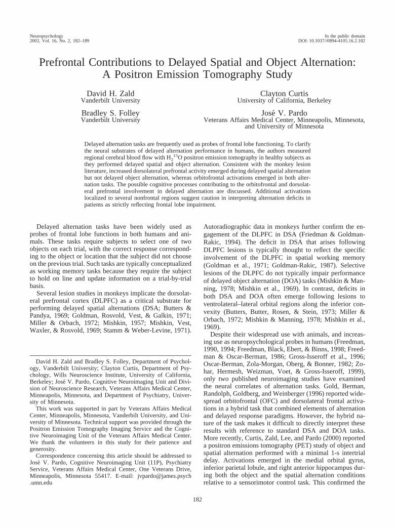
All tasks were presented using an IBM compatible personalcomputer, with a 37-cm diagonal monitor positioned approxi-mately 50 cm in front of the subject’s eyes. Figure 1 provides aschematic diagram of the DSA and DOA tasks. In both conditions,two-dimensional line drawings of 3-D objects appeared on acomputer monitor. The specific objects were selected because theypossessed visual features that were easy to distinguish from eachother but did not lend themselves to verbal labeling. To select anobject, subjects controlled a cursor with the use of a stylus andtouch pad, which functioned like a mouse. The cursor positionreturned to a central point after each response to prevent the use ofthe cursor’s position from the preceding trial as a cue on thecurrent trial. Automated software recorded all subject responses todetermine accuracy and adherence to task demands. Once subjectsindicated their response, the objects were left on the screen for anadditional 1 s accompanied by the word correct (colored green) orincorrect (colored red). On the first trial, the subject’s choice wascounted correct regardless of the subject’s selection. The positionof the two objects changed across trials according to a Gellermannrandomization schedule (Gellermann, 1933). In both the DSA and
the DOA tasks, a 5-s delay was interposed between trials duringwhich time the screen remained dark, except for a fixation pointthat subjects were instructed to fixate on at all times. Prior toscanning, all subjects were informed of the rule that governscorrect performance, and they practiced performing the tasks to acriterion of five consecutively correct trials. The control conditionfor both the DSA and the DOA consisted of stimuli presented inthe exact same format as in the DSA and DOA conditions, exceptthat on each trial a 10-point–font asterisk was embedded randomlyon one of the two figures. Subjects were instructed to choosewhichever object contained the asterisk. Scan order was approxi-mately counterbalanced across the alternation and sensorimotorcontrol conditions. Because one of our motivations was to examineareas becoming active (or showing more activity) with increasingdelay demands, we also collected data while subjects performedspatial alternation with a 1-s delay (SA-1s). Nine subjects receivedboth DSA and SA-1s conditions (each of these subjects wasincluded as part of a previous report on SA-1s; Curtis et al., 2000).These subjects additionally received a separate sensorimotor con-trol condition with 1-s intertrial delay (as opposed to the 5-sintertrial delay used for the DSA and DOA tasks).
PET Imaging and Analysis
Regional cerebral blood flow (rCBF) was estimated from tissueradioactivity using a Siemens ECAT 953B camera (Siemens,Knoxville, TN) with septa retracted, a slow-bolus injection of
Figure 1. Schematic illustration of the DSA (top) and DOA(bottom) tasks. For each trial during the DSA condition, subjectsselected the spatial location that was not chosen on the precedingtrial, regardless of the object at that location. During the DOAcondition, subjects selected the object that was not chosen on thepreceding trial, regardless of its location.
183DELAYED ALTERNATION
H215O (0.25 mCi/kg infused at a constant rate over 30 s; Silbers-
weig et al., 1993), a 90-s scan acquisition, and a 10-min interscaninterval. Scanning began with radiotracer arrival into the brain,which was timed to occur during the second to third trial of thetask condition. Images were reconstructed using a 3-D reconstruc-tion algorithm with a Hanning filter (0.5 cycles/pixel; Kinahan &Rogers, 1989) and were corrected for attenuation with a measuredtwo-dimensional transmission scan. Measured coincidences werecorrected for random detections and electronic dead time, but nocorrections were made for decay or scatter. Normalization forglobal activity (1,000 counts), coregistration within each studysession, placement of the intercommissural line from image fidu-cials, nonlinear warping of each subject’s scans to a referencestereotactic atlas (Talairach & Tournoux, 1988), and statisticalanalyses were accomplished with software developed and pro-vided by Minoshima and coworkers (Minoshima, Berger, Lee, &Mintun, 1992; Minoshima, Koeppe, Frey, & Kuhl, 1994;Minoshima et al., 1993). Images were blurred with a 6.0-mm 3-Dgaussian filter, producing a final image resolution of 10-mm fullwidth at half maximum. We adopted a significance threshold ofp � .0001 on the basis of previous studies of the rate of falsepositive foci arising in bootstrapping analysis (Zald, Lee, Fluegel,& Pardo, 1998). Statistics reflect the maximum magnitude of rCBFincrease in a given region relative to the global variance of allpixels within the brain (Worsley, Evans, Marrett, & Neelin, 1993).Identification of cerebellar foci used the atlas of Schmahmann,Doyon, Toga, Petrides, and Evans (2000), whereas identificationof orbital foci followed the nomenclature and probability maps ofChiavaras, LeGoualher, Evans, and Petrides (2001) and the cyto-architectural nomenclature of Ongur and Price (2000).
Results
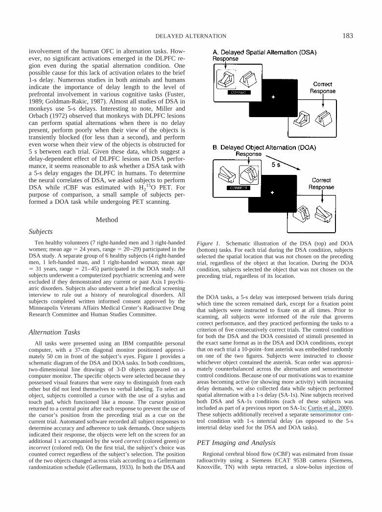
All subjects performed the DSA tasks at 100% accuracy.Table 1 displays the results of the contrast between the DSAcondition and the sensorimotor control task. Consistent withthe lesion literature, performance of the DSA task wasassociated with significantly increased rCBF in a portion ofthe dorsolateral prefrontal cortex (middle frontal gyrus,BA 9, see Figure 2A). As can be seen from Figure 2, thisactivation represents a discrete focus that appears strictlylateralized to the left hemisphere. Another discrete focusemerged in the left posterior orbital gyrus (see Figure 2B).Unexpectedly, the largest volume focus localized to theprecuneus region (Figure 2C). In 2 subjects, this focus laynear the edge of the PET camera’s field of view. Because
this could result in interpolation errors during 3-D imagereconstruction, we reanalyzed the data without these sub-jects. However, this did not substantially alter the locationor magnitude of the focus. Finally, an additional cluster offoci localized to the superior frontal gyrus.
The present data suggest performing DSA engages adiscrete portion of the DLPFC, whereas previous data didnot indicate activation of the DLPFC at a 1-s delay. To morecarefully examine this difference, we performed a region-of-interest analysis comparing activations in the DSA con-dition (relative to the sensorimotor control condition) withactivations emerging during the SA-1s condition (relative toa sensorimotor condition using a 1-s delay). We limited thisanalysis to 9 subjects who completed both the DSA condi-tion and the briefer SA-1s condition to ensure that differ-ences did not arise as a consequence of using differentsamples. A 2-pixel radius spherical region of interest wascentered on the peak coordinate of the DLPFC activation inthe DSA-sensorimotor control contrast and on the corre-sponding position in the 1-s spatial alternation–sensorimo-tor (1-s intertrial delay) contrast. In the DSA condition,rCBF increased by a mean of 8.1% (SD � 5.5%) across theregion of interest, whereas rCBF showed no substantialchange (M � �1.2%, SD � 5.9%) in the SA-1s contrast.This greater activity occurred despite the fact that therewere almost four times more trials in the SA-1s conditionthan in the DSA condition, t(8) � 4.39, p � .005.
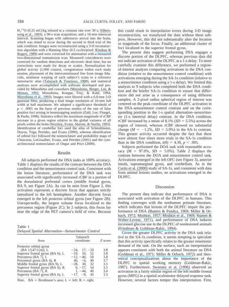
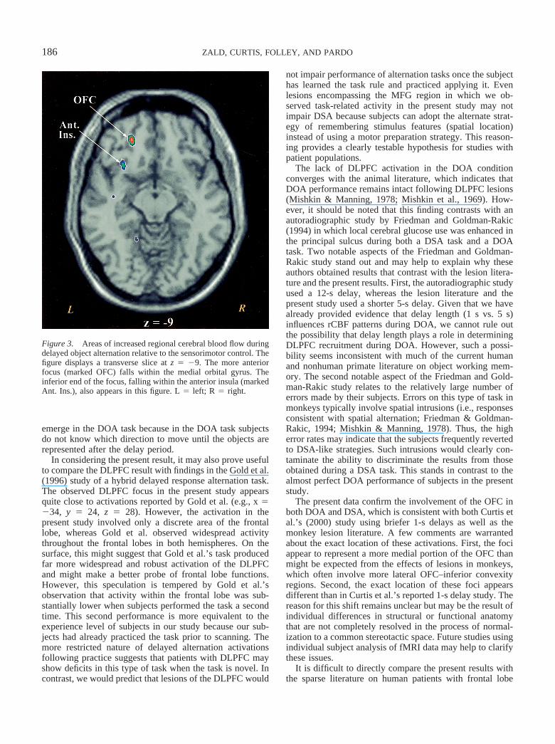
Subjects performed the DOA task with reasonable accu-racy (M � 97.0%, SD � 5.0%). Table 2 displays thecontrast between the DOA and sensorimotor control task.Activations emerged in the left OFC (see Figure 3), anteriorinsula, supramarginal gyrus, and cerebellum. As in theCurtis et al. (2000) study of SA-1s, and consistent with datafrom animal lesions studies, no activations emerged in theDLPFC.
Discussion
The present data indicate that performance of DSA isassociated with activation of the DLPFC in humans. Thisfinding converges with the nonhuman primate literature,which indicates that lesions of the DLPFC impair the per-formance of DSA (Butters & Pandya, 1969; Miller & Or-bach, 1972; Mishkin, 1957; Mishkin et al., 1969; Stamm &Weber-Levine, 1971), and performance of DSA inducesincreased glucose use in the DLPFC of nonlesioned animals(Friedman & Goldman-Rakic, 1994).
Given the greater DLPFC activity in the DSA task rela-tive to the SA-1s condition, it seems tempting to speculatethat this activity specifically relates to the greater mnemonicdemand of the task. On the surface, such an interpretationappears consistent with both the animal literature on DSA(Goldman et al., 1971; Miller & Orbach, 1972) and theo-retical conceptualizations about the importance of theDLPFC to spatial working memory (Goldman-Rakic,1987). Furthermore, Sweeney et al. (1996) observed anactivation in a fairly similar region of the left middle frontalgyrus (MFG) in a spatial oculomotor delayed response task.However, several factors temper this interpretation. First,
Table 1Delayed Spatial Alternation—Sensorimotor Control
AreaTalairachcoordinate Z score
Posterior orbital gyrus(BA 13,47/12m), L �24, 17, �20 3.8
Superior frontal gyrus (BA 6), L �19, 10, 52 3.8Precuneus (BA 7), L �12, �46, 50 3.8Precentral gyrus (BA 4), R 46, �6, 40 3.7Middle frontal gyrus (BA 9), L �28, 30, 32 3.5Superior frontal gyrus (BA 6), R 6, �17, 54 3.5Precuneus (BA 7), R 1, �44, 40 3.4Superior frontal gyrus (BA 6), L �17, �8, 45 3.3
Note. BA � Brodmann’s area; L � left; R � right.
184 ZALD, CURTIS, FOLLEY, AND PARDO
the left MFG has not emerged as a significant focus ofactivation in numerous other spatial working memory stud-ies (see D’Esposito et al., 1998, for review). This portion ofthe left MFG or regions proximal to it have sometimesemerged in tasks involving the monitoring of auditory,verbal, or nonspatial visual information in working memory(Braver et al., 1997; Petrides, Alivisatos, Meyer, & Evans,1993; Schumacher et al., 1996; Smith, Jonides, & Koeppe,1996; Zatorre, Evans, & Meyer, 1994). Thus, to the extentthat the focus is attributable to working memory, it does notappear to specifically reflect spatial working memory per se.Indeed, it should not be assumed that the activity reflects themaintenance of sensory information (either in terms of a
spatial code or a left–right verbalization) during the delayperiod. Instead, the activation may reflect motor preparationbecause subjects know the location of the next correctresponse throughout the entire delay. Thus, to perform theDSA task, one can maintain either a retrospective represen-tation of the sensory information (i.e., the object) or aprospective representation of the upcoming motor com-mand. In a study of DLPFC activity during the delay periodof delayed response tasks, Quintana and Fuster (1992) ob-served cells in the DLPFC whose activity was linked to thedirection of a postdelay motor response (see also Rainer,Rao, & Miller, 1999). In other words, these cells showactivity related to motor preparation as opposed to holdingsensory information on line. The importance of the DLPFCfor motor preparation in delayed responses tasks has beenconfirmed in studies of patients with frontal lesions (Fer-reira et al., 1998) and in functional magnetic resonanceimaging (fMRI) investigations with healthy subjects(D’Esposito, Ballard, Zarahn, & Aguirre, 2000; Pochon etal., 2001). The left lateralization of the observed DLPFCactivity in the present study might thus relate to a right-handmotor preparation in the present study (all subjects re-sponded with their right hand). The motor preparation issuemight also explain why a similar DLPFC activation did not
Figure 2. Areas of increased regional cerebral blood flow during delayed spatial alternationrelative to the sensorimotor control. A: Transverse slice displaying the middle frontal gyrus focusat z � 32. B: Sagittal slice displaying the posterior orbital focus at x � �24. C: Sagittal slicedisplaying the precuneus focus at x � �12. All figures were produced by resampling the positronemission tomography Z-score image to a 1-mm3 pixel resolution and templating the data to displayonly Z scores greater than 2.5 and by overlaying the resulting data on a high-resolution Talairachwarped anatomical magnetic resonance image. Z scores are color coded according to the color barat the bottom left side of the figure. L � left; R � right.
Table 2Delayed Object Alternation—Sensorimotor Control
AreaTalairachcoordinate Z score
Cerebellum (Lobule V), R 12, �58, �18 3.7Medical orbital sulcus (BA 11), L �21, 39, �9 3.7Anterior insula, L �30, 14, �2 3.4Supramarginal gyrus (BA 40), L �53, �49, 36 3.3
Note. R � right; BA � Brodmann’s area; L � left.
185DELAYED ALTERNATION
emerge in the DOA task because in the DOA task subjectsdo not know which direction to move until the objects arerepresented after the delay period.
In considering the present result, it may also prove usefulto compare the DLPFC result with findings in the Gold et al.(1996) study of a hybrid delayed response alternation task.The observed DLPFC focus in the present study appearsquite close to activations reported by Gold et al. (e.g., x ��34, y � 24, z � 28). However, the activation in thepresent study involved only a discrete area of the frontallobe, whereas Gold et al. observed widespread activitythroughout the frontal lobes in both hemispheres. On thesurface, this might suggest that Gold et al.’s task producedfar more widespread and robust activation of the DLPFCand might make a better probe of frontal lobe functions.However, this speculation is tempered by Gold et al.’sobservation that activity within the frontal lobe was sub-stantially lower when subjects performed the task a secondtime. This second performance is more equivalent to theexperience level of subjects in our study because our sub-jects had already practiced the task prior to scanning. Themore restricted nature of delayed alternation activationsfollowing practice suggests that patients with DLPFC mayshow deficits in this type of task when the task is novel. Incontrast, we would predict that lesions of the DLPFC would
not impair performance of alternation tasks once the subjecthas learned the task rule and practiced applying it. Evenlesions encompassing the MFG region in which we ob-served task-related activity in the present study may notimpair DSA because subjects can adopt the alternate strat-egy of remembering stimulus features (spatial location)instead of using a motor preparation strategy. This reason-ing provides a clearly testable hypothesis for studies withpatient populations.
The lack of DLPFC activation in the DOA conditionconverges with the animal literature, which indicates thatDOA performance remains intact following DLPFC lesions(Mishkin & Manning, 1978; Mishkin et al., 1969). How-ever, it should be noted that this finding contrasts with anautoradiographic study by Friedman and Goldman-Rakic(1994) in which local cerebral glucose use was enhanced inthe principal sulcus during both a DSA task and a DOAtask. Two notable aspects of the Friedman and Goldman-Rakic study stand out and may help to explain why theseauthors obtained results that contrast with the lesion litera-ture and the present results. First, the autoradiographic studyused a 12-s delay, whereas the lesion literature and thepresent study used a shorter 5-s delay. Given that we havealready provided evidence that delay length (1 s vs. 5 s)influences rCBF patterns during DOA, we cannot rule outthe possibility that delay length plays a role in determiningDLPFC recruitment during DOA. However, such a possi-bility seems inconsistent with much of the current humanand nonhuman primate literature on object working mem-ory. The second notable aspect of the Friedman and Gold-man-Rakic study relates to the relatively large number oferrors made by their subjects. Errors on this type of task inmonkeys typically involve spatial intrusions (i.e., responsesconsistent with spatial alternation; Friedman & Goldman-Rakic, 1994; Mishkin & Manning, 1978). Thus, the higherror rates may indicate that the subjects frequently revertedto DSA-like strategies. Such intrusions would clearly con-taminate the ability to discriminate the results from thoseobtained during a DSA task. This stands in contrast to thealmost perfect DOA performance of subjects in the presentstudy.
The present data confirm the involvement of the OFC inboth DOA and DSA, which is consistent with both Curtis etal.’s (2000) study using briefer 1-s delays as well as themonkey lesion literature. A few comments are warrantedabout the exact location of these activations. First, the fociappear to represent a more medial portion of the OFC thanmight be expected from the effects of lesions in monkeys,which often involve more lateral OFC–inferior convexityregions. Second, the exact location of these foci appearsdifferent than in Curtis et al.’s reported 1-s delay study. Thereason for this shift remains unclear but may be the result ofindividual differences in structural or functional anatomythat are not completely resolved in the process of normal-ization to a common stereotactic space. Future studies usingindividual subject analysis of fMRI data may help to clarifythese issues.
It is difficult to directly compare the present results withthe sparse literature on human patients with frontal lobe
Figure 3. Areas of increased regional cerebral blood flow duringdelayed object alternation relative to the sensorimotor control. Thefigure displays a transverse slice at z � �9. The more anteriorfocus (marked OFC) falls within the medial orbital gyrus. Theinferior end of the focus, falling within the anterior insula (markedAnt. Ins.), also appears in this figure. L � left; R � right.
186 ZALD, CURTIS, FOLLEY, AND PARDO
lesions (Freedman et al., 1998; Freedman & Oscar-Berman,1986). These studies have examined the error rates of pa-tients while they acquire the task. Because the subjects arenot explicitly told the alternation rule in these studies, thedeficits may reflect a reasoning deficit rather than a specificdeficit in the ability to perform the task. This contrasts withthe present PET study in which we examined subjects afterthey acquired and practiced the task rule. Nevertheless, it isnotable that Freedman and Oscar-Berman (1986) observedthe greatest deficits in both DSA and DOA acquisition whenlesions included the OFC. Freedman et al. (1998) notedDOA acquisition impairments following both ventrolateral–orbitofrontal and more medial frontal (Brodmann areas 10,24, and 32) lesions. Given the lack of medial frontal acti-vations in the present study, it seems plausible that the moremedial frontal regions observed by Freedman et al. (1998)reflect aspects of task acquisition rather than performance ofalready learned alternations.
The present study does not attempt to decompose thecognitive functions leading to OFC involvement in alterna-tion tasks. Mishkin (1964) speculated that the OFC’s con-tribution to alternation tasks relates to the need to inhibit aprepotent response (which would be to respond to the stim-ulus that was rewarded on the previous trial). However,once the subject has learned the task (as is the case in thepresent study), it is not clear that such an inhibition remainsnecessary. Specifically, the prepotency of the win–staystrategy is reduced after the subject has practiced the alter-nation strategy. Subjects do not describe having to specifi-cally inhibit an urge to respond to the previously correctresponse instead of alternating between stimuli. An alterna-tive explanation focuses on the hypothesis that the OFCparticipates in updating and holding on-line informationabout the relationship between stimuli and rewards (Zald &Kim, 2001). In many memory tasks, subjects need only holda representation of whether or not they have seen a stimulusbefore or which of two stimuli has ever been associated witha reward. However, in the alternation paradigm, both stimuliare seen an equal number of times and rewarded an equal oralmost equal number of times. Because of this, the ability toupdate and hold information on line regarding the laststimulus–reward pairing may take on particular importancein the performance of alternation tasks. Of course, for thishypothesis to possess explanatory power in the presentstudy requires the assumption that the knowledge of beingcorrect acts as a reward. Such an assumption requires test-ing, although it is consistent with the recent data implicatingthe OFC in the coding of abstract reward (O’Doherty,Kringelbach, Rolls, Hornak, & Andrews, 2001). A finalhypothesis revolves around the possible role of the OFC inreducing interference. Data from humans with frontal leu-cotomies indicate that the OFC lesions preferentially affecttests of memory under conditions of high interference (Stusset al., 1982). Alternation tasks involve high levels of pro-active interference in that irrelevant memories of prior trialsother than the immediately preceding trial can compete withthe relevant memory of the past trial and interfere withcorrect performance. Indeed, subjects frequently report thatthe hardest part of the task is distinguishing their last re-
sponse from earlier responses. Thus, it may be speculatedthat the OFC contributes to alternation tasks by reducing theeffects of proactive interference. However, until studiesexamine the role of the OFC in specific task subcomponentssuch as interference suppression and working memory forreward, it will remain difficult to determine the precisenature of the OFC contribution to alternation tasks.
The present findings suggest that marked differences inthe neural network subserving performance of alternationtasks occur depending on the modality and delay period ofthe task. However, some caution is necessary in drawingthis conclusion because of the use of different samples ofsubjects in the DSA and DOA conditions. This precludeduse of a factorial design that would allow examination of themain effects of delay and modality. However, application ofsuch a factorial design is problematic even if all subjectshad completed all of the conditions. Specifically, there is aconfound in examining the effect of delay because of thedramatically different number of trials in a scan period.Thus, a failure to observe a focus that emerged in the shortdelay condition may reflect a decreased signal because ofthe sparseness of the responses. However, the emergence ofnew areas such as the DLPFC, which were not seen in theshorter delay trials, likely relate to the increased delay timebecause they emerge despite the far fewer responses pro-duced during the DSA condition compared with the SA-1scondition.
The extent to which nonfrontal regions show activationsduring alternation tasks represents a striking feature of theneuroimaging literature on alternation. Nonfrontal activa-tions range from the hippocampus and parietal cortex atshort delays to the precuneus, cerebellum, and supramar-ginal gyrus at longer delays. Although the activation ofthese areas appears to vary depending on the specific mo-dality (object vs. spatial) or delay length (1 s vs. 5 s), theirpresence suggests a danger in conceptualizing performanceon alternation tasks purely as a measure of frontal lobefunctioning. Other working memory and frontal lobe tasks,although sensitive to frontal lobe lesions, rarely show ex-clusive specificity for frontal lobe damage (Anderson,Damasio, Jones, & Tranel, 1991; Bondi, Kazniak, Bayles, &Vance, 1993; Dudkin, Chueva, Makarov, & Orlov, 1999;Grafman, Jonas, & Salazar, 1990; Reitan & Wolfson, 1994,1995). Instead, such tasks typically engage and depend onnormal functioning within a distributed network of frontal,posterior, and subcortical brain regions (Casey et al., 1998;Chafee & Goldman-Rakic, 1998, 2000; Cohen et al., 1997;Friedman & Goldman-Rakic, 1994; Oliveri et al., 2001;Postle, Stern, Rosen, & Corkin, 2000; Rowe & Passingham,2001). The present results suggest that the neural basis ofdelayed alternation involves similarly widely distributednetworks.
References
Anderson, S. W., Damasio, H., Jones, R. D., & Tranel, D. (1991).Wisconsin Card Sorting Test performance as a measure offrontal lobe damage. Journal of Clinical & Experimental Neu-ropsychology, 13, 909–922.
187DELAYED ALTERNATION
Bondi, M., Kazniak, A. W., Bayles, K. A., & Vance, K. A. (1993).Contributions of frontal system dysfunction to memory andperceptual abilities in Parkinson’s disease. Neuropsychology, 7,89–102.
Braver, T. S., Cohen, J. D., Nystrom, L. E., Jonides, J., Smith,E. E., & Noll, D. C. (1997). A parametric study of prefrontalcortex involvement in human working memory. Neuroimage, 5,49–62.
Butters, N., Butter, C., Rosen, J., & Stein, D. (1973). Behavioraleffects of sequential and one-stage ablations of orbital prefrontalcortex in the monkey. Experimental Neurology, 39, 204–214.
Butters, N., & Pandya, D. N. (1969, September 19). Retention ofdelayed-alternation: Effect of selective lesions of the sulcuspricipalis. Science, 165, 1271–1273.
Casey, B. J., Cohen, J. D., O’Craven, K., Davidson, R. J., Irwin,W., Nelson, C. A., et al. (1998). Reproducibility of fMRI resultsacross four institutions using a spatial working memory task.Neuroimage, 8, 249–261.
Chafee, M. V., & Goldman-Rakic, P. S. (1998). Matching patternsof activity in primate prefrontal area 8a and parietal area 7ipneurons during a spatial working memory task. Journal ofNeurophysiology, 79, 2919–2940.
Chafee, M. V., & Goldman-Rakic, P. S. (2000). Inactivation ofparietal and prefrontal cortex reveals interdependence of neuralactivity during memory-guided saccades. Journal of Neurophys-iology, 83, 1550–1566.
Chiavaras, M. M., LeGoualher, G., Evans, A., & Petrides, M.(2001). Three-dimensional probabilistic atlas of the human or-bitofrontal sulci in standardized stereotaxic space. Neuroim-age, 13, 479–496.
Cohen, J. D., Perlstein, W. M., Braver, T. S., Nystrom, L. E., Noll,D. C., Jonides, J., & Smith, E. E. (1997, April 10). Temporaldynamics of brain activation during a working memory task.Nature, 386, 604–608.
Curtis, C. E., Zald, D. H., Lee, J. T., & Pardo, J. V. (2000). Objectand spatial alternation tasks with minimal delays activate theright anterior hippocampus proper in humans. Neuroreport, 11,2203–2207.
D’Esposito, M., Aguirre, G. K., Zarahn, E., Ballard, D., Shin,R. K., & Lease, J. (1998). Functional MRI studies of spatial andnonspatial working memory. Cognitive Brain Research, 7,1–13.
D’Esposito, M., Ballard, D., Zarahn, E., & Aguirre, G. K. (2000).The role of prefrontal cortex in sensory memory and motorpreparation: An event-related fMRI study. Neuroimage, 11,400–408.
Dudkin, K. N., Chueva, I. V., Makarov, F. N., & Orlov, I. V.(1999). Short-term memory processes in delayed visual differ-entiation in rhesus macaques after bilateral removal of field 7 ofthe parietal cortex. Neuroscience and Behavioral Physiol-ogy, 29, 483–491.
Ferreira, C. T., Verin, M., Pillon, B., Levy, R., Dubois, B., & Agid,Y. (1998). Spatio-temporal working memory and frontal lesionsin man. Cortex, 34, 83–98.
Freedman, M. (1990). Object alternation and orbitofrontal systemdysfunction in Alzheimer’s and Parkinson’s disease. Brain &Cognition, 14, 134–143.
Freedman, M. (1994). Frontal and parietal lobe dysfunction indepression: Delayed alternation and tactile learning deficits.Neuropsychologia, 32, 1015–1025.
Freedman, M., Black, S., Ebert, P., & Binns, M. (1998). Orbito-frontal function, object alternation and perseveration. CerebralCortex, 8, 18–27.
Freedman, M., & Oscar-Berman, M. (1986). Bilateral frontal lobedisease and selective delayed response deficits in humans. Be-havioral Neuroscience, 100, 337–342.
Friedman, H. R., & Goldman-Rakic, P. S. (1994). Coactivation ofprefrontal cortex and inferior parietal cortex in working memorytasks revealed by 2DG functional mapping in the rhesus mon-key. Journal of Neuroscience, 14, 2775–2788.
Fuster, J. M. (1989). The prefrontal cortex. New York: RavenPress.
Gellermann, L. W. (1933). Chance order of alternating stimuli invisual discrimination experiments. Journal of Genetic Psychol-ogy, 42, 207–208.
Gold, J. M., Berman, K. F., Randolph, C., Goldberg, T. E., &Weinberger, D. R. (1996). PET validation of a novel prefrontaltask: Delayed response alternation. Neuropsychology, 10, 3–10.
Goldman, P. S., Rosvold, H. E., Vest, B., & Galkin, T. W. (1971).Analysis of the delayed-alternation deficit produced by dorso-lateral prefrontal lesions in the rhesus monkey. Journal of Com-parative and Physiological Psychology, 77, 212–220.
Goldman-Rakic, P. S. (1987). Circuitry of primate prefrontal cor-tex and regulation of behavior by representational memory. In F.Plum & V. Mountcastle (Eds.), Handbook of physiology (Vol. 5,pp. 373–417). New Haven, CT: Yale University School ofMedicine.
Grafman, J., Jonas, B., & Salazar, A. (1990). Wisconsin CardSorting Test performance based on location and size of neuro-anatomical lesion in Vietnam veterans with penetrating headinjury. Perceptual and Motor Skills, 71, 1120–1122.
Gross-Isseroff, R., Sasson, Y., Voet, H., Hendler, T., Luca-Haimovici, K., Kandel-Sussman, H., & Zohar, J. (1996). Alter-nation learning in obsessive-compulsive disorder. BiologicalPsychiatry, 39, 733–738.
Kinahan, P. E., & Rogers, J. G. (1989). Analytic 3-D imagereconstruction using all detected events. IEEE Transactions onNuclear Science, 36, 964–986.
Miller, M. H., & Orbach, J. (1972). Retention of spatial alternationfollowing frontal lobe resections in stump-tailed macaques.Neuropsychologia, 10, 291–298.
Minoshima, S., Berger, K. L., Lee, K. S., & Mintun, M. A. (1992).An automated method for rotational correction and centering ofthree-dimensional functional brain images. Journal of NuclearMedicine, 33, 1579–1585.
Minoshima, S., Koeppe, R. A., Frey, K. A., & Kuhl, D. E. (1994).Anatomic standardization: Linear scaling and nonlinear warpingof functional brain images. Journal of Nuclear Medicine, 35,1528–1537.
Minoshima, S., Koeppe, R. A., Mintun, M. A., Berger, K. L.,Taylor, S. F., Frey, K. A., & Kuhl, D. E. (1993). Automateddetection of the intercommissural line for stereotactic localiza-tion of functional brain images. Journal of Nuclear Medi-cine, 34, 322–329.
Mishkin, M. (1957). Effects of small frontal lesions on delayedalternation in monkeys. Journal of Neurophysiology, 20, 615–622.
Mishkin, M. M. (1964). Preservation of central sets after frontallesions in monkeys. In J. M. Warren & K. Akert (Eds.), Thefrontal granular cortex and behavior (pp. 219–241). New York:McGraw-Hill.
Mishkin, M., & Manning, F. J. (1978). Non-spatial memory afterselective prefrontal lesions in monkeys. Brain Research, 143,313–323.
Mishkin, M., Vest, B., Waxler, M., & Rosvold, H. E. (1969). Are-examination of the effects of frontal lesions on object alter-nation. Neuropsychologia, 7, 357–363.
188 ZALD, CURTIS, FOLLEY, AND PARDO
O’Doherty, J., Kringelbach, M. L., Rolls, E. T., Hornak, J., &Andrews, C. (2001). Abstract reward and punishment represen-tations in the human orbitofrontal cortex. Nature Neuro-science, 4, 95–102.
Oliveri, M., Turriziani, P., Carlesimo, G. A., Koch, G., Tomaiuolo,F., Panella, M., & Caltagirone, G. M. (2001). Parieto-frontalinteractions in visual-object and visual-spatial working memory:Evidence from transcranial magnetic stimulation. Cerebral Cor-tex, 11, 606–618.
Ongur, D., & Price, J. L. (2000). The organization of networkswithin the orbital and medial prefrontal cortex of rats, monkeysand humans. Cerebral Cortex, 10, 206–219.
Oscar-Berman, M., Zola-Morgan, S. M., Oberg, R. G., & Bonner,R. T. (1982). Comparative neuropsychology and Korsakoff’ssyndrome: III. Delayed response, delayed alternation and DRLperformance. Neuropsychologia, 20, 187–202.
Petrides, M., Alivisatos, B., Meyer, E., & Evans, A. C. (1993).Functional activation of the human frontal cortex during theperformance of verbal working memory tasks. Proceedings ofthe National Academy of Sciences, USA, 90, 878–882.
Pochon, J. B., Levy, R., Poline, J. B., Crozier, S., Lehericy, S.,Pillon, B., et al. (2001). The role of dorsolateral prefrontalcortex in the preparation of forthcoming actions: An fMRIstudy. Cerebral Cortex, 11, 260–266.
Postle, B. R., Stern, C. E., Rosen, B. R., & Corkin, S. (2000). AnfMRI investigation of cortical contributions to spatial and non-spatial visual working memory. Neuroimage, 11, 409–423.
Quintana, J., & Fuster, J. M. (1992). Mnemonic and predictivefunctions of cortical neurons in a memory task. Neuroreport, 3,721–724.
Rainer, G., Rao, S. C., & Miller, E. K. (1999). Prospective codingfor objects in primate prefrontal cortex. Journal of Neuro-science, 19, 5493–5505.
Reitan, R. M., & Wolfson, D. (1994). A selective and criticalreview of neuropsychological deficits and the frontal lobes.Neuropsychology Review, 4, 161–198.
Reitan, R. M., & Wolfson, D. (1995). Category test and trailmaking test as measures of frontal lobe functions. ClinicalNeuropsychologist, 9, 50–56.
Rowe, J. B., & Passingham, R. E. (2001). Working memory forlocation and time: Activity in prefrontal area 46 relates toselection rather than maintenance in memory. Neuroimage, 14,77–86.
Schmahmann, J. D., Doyon, J., Toga, A. W., Petrides, M., &Evans, A. C. (2000). MRI atlas of the human cerebellum. SanDiego, CA: Academic Press.
Schumacher, E. H., Lauber, E., Awh, E., Jonides, J., Smith, E. E.,& Koeppe, R. A. (1996). PET evidence for an amodal verbalworking memory system. Neuroimage, 3, 79–88.
Silbersweig, D. A., Stern, E., Frith, C. D., Cahill, C., Schnorr, L.,Grootoonk, S., et al. (1993). Detection of thirty-second cogni-tive activations in single subjects with positron emission tomog-raphy: A new low-dose H2
15O regional cerebral blood flowthree-dimensional imaging technique. Journal of CerebralBlood Flow and Metabolism, 13, 617–629.
Smith, E. E., Jonides, J., & Koeppe, R. A. (1996). Dissociatingverbal and spatial working memory using PET. Cerebral Cor-tex, 6, 11–20.
Stamm, J. S., & Weber-Levine, M. L. (1971). Delayed alternationimpairments following selective prefrontal cortical ablations inmonkeys. Experimental Neurology, 33, 263–278.
Stuss, D. T., Kaplan, E. F., Benson, D. F., Weir, W. S., Chiulli, S.,& Sarazin, F. F. (1982). Evidence for the involvement of or-bitofrontal cortex in memory functions: An interference effect.Journal of Comparative and Physiological Psychology, 96,913–925.
Sweeney, J. A., Mintun, M. A., Kwee, S., Wiseman, M. B., Brown,D. L., Rosenberg, D. R., & Carl, J. R. (1996). Positron emissiontomography study of voluntary saccadic eye movements andspatial working memory. Journal of Neurophysiology, 75, 454–468.
Talairach, J., & Tournoux, P. (1988). Co-planar stereotaxic atlasof the human brain. New York: Thieme.
Worsley, K. J., Evans, A. C., Marrett, S., & Neelin, P. (1993). Athree-dimensional statistical analysis for CBF activation studiesin human brain. Journal of Cerebral Blood Flow and Metabo-lism, 12, 900–918.
Zald, D. H., & Kim, S. W. (2001). The orbitofrontal cortex. In S.Salloway, D. Duffy, & P. F. Malloy (Eds.), The frontal lobesand neuropsychiatric illness (pp. 33–70). Washington, DC:American Psychiatric Press.
Zald, D. H., Lee, J. T., Fluegel, K. W., & Pardo, J. V. (1998).Aversive gustatory stimulation activates limbic circuits in hu-mans. Brain, 121, 1143–1154.
Zatorre, R. J., Evans, A. C., & Meyer, E. (1994). Neural mecha-nisms underlying melodic perception and memory for pitch.Journal of Neuroscience, 14, 1908–1919.
Zohar, J., Hermesh, H., Weizman, A., Voet, H., & Gross-Isseroff,R. (1999). Orbitofrontal cortex dysfunction in obsessive-com-pulsive disorder? I. Alternation learning in obsessive-compul-sive disorder: Male–female comparisons. European Neuropsy-chopharmacology, 9, 407–413.
Received June 18, 2001Revision received October 17, 2001
Accepted October 22, 2001 �
189DELAYED ALTERNATION


![Added value of [(18)F]fluorodeoxyglucose positron emission](https://img.pdfslide.net/doc/110x75/633cc1029ae9920b87022f3e/added-value-of-18ffluorodeoxyglucose-positron-emission-.jpg)
