Embed Size (px)
Citation preview

Ultrasound Obstet Gynecol 2005; 25: 50–59Published online in Wiley InterScience (www.interscience.wiley.com). DOI: 10.1002/uog.1823
Preoperative sonographic features of borderline ovariantumors
C. EXACOUSTOS, M. E. ROMANINI, D. RINALDO, C. AMOROSO, B. SZABOLCS, E. ZUPIand D. ARDUINIDepartment of Obstetrics and Gynecology, Universita degli Studi di Roma ‘Tor Vergata’, Rome, Italy
KEYWORDS: borderline ovarian tumors; low malignant potential ovarian tumors; power Doppler; transvaginal sonography
ABSTRACT
Objective To determine the sonographic findings thatdistinguish borderline ovarian tumors (BOT) from bothbenign and invasive malignant tumors, thus allowingconservative treatment and laparoscopic management ofthese tumors.
Methods We reviewed retrospectively transvaginal sono-grams of 33 women who, when evaluated further bysurgery and histology, were found to have BOT. Twenty-three were premenopausal and 10 were postmenopausal(mean age ± SD, 45.8 ± 15.7 years). For each mass, sizeand morphological features and power Doppler char-acteristics were evaluated. We compared these findingswith those of 337 patients with benign ovarian tumorsand those of 82 patients with invasive malignant ovariantumors. Patients with dermoid cysts were not included inthe study.
Results Of the 33 BOT, 15 were mucinous and 18 wereserous cystadenomas. The presence of papillae, defined asa small number of solid tissue projections, 1–15 mm inheight and 1–10 mm in width (base) and length (base),into the cyst cavity from the cyst wall, was significantlymore frequent in BOT (48%) than it was in benign(4%) and invasive (4%) malignant tumors. Intracysticsolid tissue (> 15 mm in height or > 10 mm in width orlength) was observed in 48% of invasive malignant massesbut in only 18% of BOT and in 7% of benign tumors(P < 0.001). No sonographically unilocular, hypoechoic,smooth-walled adnexal cysts were invasively malignantbut three unilocular cysts with a diameter of > 6 cm wereserous BOT. Although close attention was paid to thecyst wall at ultrasound examination we did not observe inthese three cysts the very small papillae which were foundat histological analysis.
Conclusions The most frequent diagnostic feature onimaging BOT is the presence of papillae within the cyst.However, neither papillae nor other sonographic featuresconstituted highly sensitive sonographic markers of BOT.Copyright 2004 ISUOG. Published by John Wiley &Sons, Ltd.
INTRODUCTION
Borderline ovarian tumors (BOT) or tumors of lowmalignant potential are epithelial tumors with a slowgrowth rate and low invasive potential. These tumorstend to occur in younger women and are oftendiagnosed at an earlier stage of disease than are invasivecarcinomas1. Patients with early-stage lesions have anexcellent prognosis, while Stage III lesions with peritonealimplants, which are uncommon, are associated with apoor prognosis. Because of the generally good prognosisassociated with BOT and the fact that they tend to occur inyounger women, fertility-sparing conservative treatmentshave been proposed. Some authors1–4 have proposed anendoscopic approach, after accurate surgical staging, forconservative treatment of BOT.
The identification of patients with ovarian lesionssuspected of being BOT may be helpful in theirmanagement. Whereas transvaginal sonography has beenestablished as a reliable method of differentiating benignfrom malignant adnexal masses, little is known aboutsonographic features in the diagnosis of BOT5–9. The aimof this study was to determine the sonographic findingsthat distinguish BOT from both benign and invasivemalignant tumors, thus making it possible to treat themin a more conservative way.
Correspondence to: Dr C. Exacoustos, Department of Obstetrics and Gynecology, Universita degli Studi di Roma ‘Tor Vergata’ OspedaleGenerale S. Giovanni Calibita ‘Fatebenefratelli’, Isola Tiberina, Rome, Italy (e-mail: [email protected])
Accepted: 19 November 2004
Copyright 2004 ISUOG. Published by John Wiley & Sons, Ltd. ORIGINAL PAPER

Sonography of borderline ovarian tumors 51
METHODS
We reviewed retrospectively transvaginal sonograms ofwomen with adnexal tumors who were evaluated furtherby surgery and histology. Between January 1997 andApril 2003, 33 patients with BOT underwent transvaginalsonography and were treated at our institution. Thisstudy group was compared with 403 patients with benignovarian tumors and 82 patients with invasive malignanttumors managed at our institution in the same period.All patients underwent gray-scale sonographic and powerDoppler examination of the pelvis within 1 month priorto surgery.
In all patients serum levels of CA 125 were preopera-tively determined and in the case of suspected malignancypresurgical and surgical staging was performed accordingto FIGO criteria10. The CA 125 assays were performedwith an immunoassay method using two monoclonal anti-bodies (CA 125 II Elecsys Sistemas 1010/2010, Centocor,Malvern, PA, USA) with a limit of detection of 0.6 U/mL.Histological classification was performed in accordancewith the WHO classification11,12.
A transvaginal sonographic examination, accompaniedif necessary by a transabdominal examination, wasperformed on all patients using an Esaote Technos(Esaote, Genova, Italy) ultrasound machine with color andpower Doppler capability, equipped with a 2.5–5.5-MHzconvex transabdominal transducer and a 5.5–8.5-MHztransvaginal probe. Size and echostructure of the uterus,endometrial thickness and any irregular findings andintraperitoneal free fluid (pouch of Douglas or ascites)were recorded. Ovarian shape and size were examinedtransvaginally.
For each adnexal mass, longitudinal (LD), transverse(TD) and anteroposterior (APD) diameters were measuredand adnexal volumes were calculated by means ofa prolate ellipsoid formula: volume = 0.5233 × LD ×TD × APD in cm.
The morphology of each adnexal mass was describedaccording to the following different types of sonographicmorphological tumor characteristic:
1. Unilocular cyst: smooth-walled unilocular cyst withclear fluid or dense (echogenic) fluid content.
2. Cyst with septa: smooth-walled cyst with clear fluid ordense (echogenic) fluid content and only septa insidethe cyst, not papillary projections or solid tissue. Septawere further classified as:a) thin septa (≤ 3 mm);b) thick septa (> 3 mm);c) no more than three septa (cyst with 1–3 septa);d) more than three septa (multilocular cyst).
3. Cyst with papillae: cyst with clear fluid or dense(echogenic) fluid content and papillae, defined as asmall number of solid tissue projections, 1–15 mm inheight and 1–10 mm in width (base) and length (base),into the cyst cavity from the cyst wall. Papillae weredescribed further and classified as:a) small papilla (≤ 3 mm in height, length and width);b) large papilla (> 3 mm in height, length and width);
c) only one papilla;d) more than three papillae (cyst with > 3 papillae).
4. Tumors with fluid/solid content: cyst with clear fluidor dense (echogenic) fluid and solid content defined asa solid part > 15 mm in height and > 10 mm in widthand length.
5. Pure solid tumors: tumors composed of only solidtissue.
Each adnexal tumor was classified on the basis of theworst sonographic pattern, according to various publishedscoring systems13–15; in the presence of septa and papillaethe cyst was classified as having papillae, and in thepresence of solid tissue and septa or papillae the solidtissue was considered predominant and the cyst wasclassified as tumor with fluid/solid content. In the caseof multiple papillae the largest was considered, and in thecase of multiple septa the thickest was considered.
The entire adnexal mass was surveyed by powerDoppler imaging. This was performed with gain adjustedto avoid color noise, thus allowing detection of thevessels inside and just outside the tumor. The power gainsetting was set to maximize visualization of the vesselswhile minimizing artifacts. A subjective semiquantitativeassessment of the amount of blood flow (area and colorscale) at the level of the tumor capsule and inside thetumor was made16–20. Vascularization was described asbeing present or absent and, if present, as being central(intratumoral, i.e. in solid tissue, septa or papillae) orperipheral (capsule, pericystic).
When color signals were observed inside the tumor,blood flow velocity waveforms were recorded by placingthe sample volume over the colored area and initiating thepulsed Doppler mode. At least three consecutive correctlyimaged blood flow velocity waveforms were analyzed andthe pulsatility index (PI = maximum systolic velocity −least diastolic velocity/mean velocity) and the resistanceindex (RI = maximum systolic velocity − least diastolicvelocity/maximum systolic velocity) were calculated.If more color signals were detectable peripherally orcentrally in the tumor, at least three different arterieswere analyzed, and the lowest PI and RI values wereused for further analysis. If only peripheral flow wasobserved and no color signal was detectable centrally,the peripheral waveforms were considered for analysis.Otherwise, the PI and RI values of the waveform obtainedfrom the structures inside the tumors (septa, papillae orsolid tissue) were considered for data analysis.
We excluded patients with histologically confirmeddermoid cysts from the analysis. These cysts have char-acteristics similar to those of malignant complex tumorsbut their sonographic appearance is pathognomonic andeasily identified21,22. Subtracting the 66 mature teratomasfrom the 403 benign ovarian tumors observed, we con-sidered 337 benign masses in the final analysis (Table 1).If the adnexal masses were bilateral, the more morpho-logically complex tumor was considered, and if bothmasses were morphologically similar, the largest one wasconsidered in the final statistical analysis. Histological
Copyright 2004 ISUOG. Published by John Wiley & Sons, Ltd. Ultrasound Obstet Gynecol 2005; 25: 50–59.
52 Exacoustos et al.
Table 1 Histological types of benign, borderline and malignant ovarian tumors observed in pre- and postmenopausal women
Histology of ovarian tumor Premenopausal (n (%)) Postmenopausal (n (%)) Total (n (%))
Benign n = 306 n = 97 n = 403Simple cyst 25 (8.2) 28 (28.9) 53 (13.2)Serous cystadenoma 11 (3.6) 12 (12.4) 23 (5.8)Mucinous cystadenoma 15 (4.9) 12 (12.4) 27 (6.7)Endometriosis 116 (37.9) 8 (8.3) 124 (30.8)Mature teratoma 63 (20.6) 3 (3.1) 66 (16.4)Functional cyst 20 (6.5) 3 (3.1) 23 (5.7)Cystadenofibroma 15 (4.9) 4 (4.1) 19 (4.7)Salpingo-ovarian pathology 7 (2.3) 2 (2) 9 (2.2)Paraovarian cyst 21 (6.9) 9 (9.3) 30 (7.4)Ovarian fibroma 2 (0.6) 9 (9.3) 11 (2.7)Ovarian thecoma 4 (1.3) 2 (2) 6 (1.5)Other 7 (2.3) 5 (5.1) 12 (2.9)
Borderline n = 23 n = 10 n = 33Serous cystadenoma 12 (64.3)* 6 (60) 18 (55)Mucinous cystadenoma 11 (35.7)* 4 (40)* 15 (45)
Malignant n = 22 n = 60 n = 82Serous adenocarcinoma 5 (23) 26 (43) 31 (38)Mucinous adenocarcinoma 2 (9) 5 (8) 7 (9)Endometrioid adenocarcinoma 8 (36) 12 (20) 20 (24)Undifferentiated carcinoma 1 (5) 5 (8) 6 (7)Immature teratoma 1 (5) 0 1 (1)Granulosa cells 2 (9) 2 (3) 4 (5)Germ cells 1 (5) 0 1 (1)Brenner’s tumor 1 (5) 2 (3) 3 (4)Metastatic tumor 1 (5) 8 (13) 9 (11)
*P < 0.001 borderline vs. malignant ovarian tumors.
findings were compared with sonographic appearance ofthe adnexal masses.
To evaluate the accuracy of ultrasound in identi-fying BOT, sensitivity, specificity, positive and nega-tive predictive values and likelihood ratios were calcu-lated.
The 95% CIs for prevalence of the presence ofpower Doppler signals in the papillae in the differenttypes of ovarian tumor were calculated according tothe recommended method of Wilson23,24. This methodhas better statistical properties than does the traditionalmethod, having the advantage that it can be used for anydata24.
Statistical analysis was performed using the chi-squaretest and the unpaired Student’s t-test. A P-value of < 0.05was considered significant.
RESULTS
The type and stage of BOT and invasive malignant tumorare reported in Tables 1 and 2. A significantly higherpercentage of Stage Ia was observed in patients withBOT (64%) compared with those with invasive malignantmasses (24%).
The mean ± SD age of patients was 45.8 ± 15.7 yearsfor those with BOT, which was significantly lower thanthe age of those with invasive malignant tumors andsimilar to the age of those with benign tumors (Table 3).The percentage of BOT patients aged less than 35 yearswas also similar to that for benign tumors.
The maximum diameter of BOT was similar to that ofinvasive malignant tumors but significantly greater thanthat of benign tumors. However, the percentage of cysts≤ 5 cm was similar to that of benign and significantlyhigher than that of invasive malignant tumors (Table 3).
The analysis of the different morphological echopat-terns of the adnexal masses is reported in Table 4. Thedistribution of small and large papillae was not signifi-cantly different in BOT and benign and malignant tumors;therefore, we considered only the presence and the numberof papillae. No difference was observed in the distributionof thick or thin septa in BOT and benign and invasivemalignant tumors, but the number of septa did vary inthe different types of mass and multilocular cysts wereconsidered separately.
We observed numerous unilocular smooth-walled cystsin patients with benign tumors and no unilocular cystsin patients with invasive malignant tumors. Moreover,we found three (9%) unilocular smooth-walled cystsin patients with BOT. These three patients were over35 years old and the cyst diameters were > 6 cm (6.5 cm,10 cm and 12 cm). The three cysts were serous BOT, andvery small papillae on the cyst wall were observed onhistological analysis.
Sixteen (48%) BOT showed papillae, and eight (24%)showed septa, of which six (18%) were multilocular.The percentage of pure solid and fluid/solid tumors wassignificantly higher in patients with invasive malignanttumors compared with those with BOT (Table 4).
Copyright 2004 ISUOG. Published by John Wiley & Sons, Ltd. Ultrasound Obstet Gynecol 2005; 25: 50–59.

Sonography of borderline ovarian tumors 53
Table 2 Stages of the borderline and malignant ovarian tumors
Ovarian tumorTotal Cases
(n)Stage Ia(n (%))
Stage Ib(n (%))
Stage Ic(n (%))
Stage II(n (%))
Stage III(n (%))
Stage IV(n (%))
Borderline 33 21 (64)* 4 (12) 5 (15) 1 (3) 2 (6)* 0Malignant† 82 20 (24) 1 (1) 11 (13) 7 (9) 30 (37) 4 (5)
*P < 0.001 borderline vs. malignant ovarian tumors; †Nine cases were metastatic.
Table 3 Comparison between patient age, menopausal status and maximum diameter of benign, malignant and borderline ovarian tumors
Type of ovarian tumor (No. of cases)
Benign (n = 337) Borderline (n = 33) Malignant (n = 82)
AgeRange (years) 12–82 24–74 28–82Mean age ± SD (years) 40.5 ± 14.9§ 45.8 ± 15.7† 57.9 ± 12.9Patients < 35 years (n (%)) 146 (43)§ 12 (36)† 4 (5)
Menopausal status: premenopausal (n (%)) 243 (72)§ 23 (70)† 22 (27)
Tumor maximum diameterMean ± SD (cm) 6.3 ± 3.7§ 9.3 ± 7.2‡ 11.0 ± 6.3≤ 5 cm (n (%)) 154 (46)§ 13 (39)* 13 (16)≤ 8 cm (n (%)) 267 (79)§ 19 (58) 34 (42)
*P < 0.05 borderline vs. malignant ovarian tumors. †P < 0.01 borderline vs. malignant ovarian tumors. ‡P < 0.01 borderline vs. benignovarian tumors. §P < 0.01 benign vs. malignant ovarian tumors.
Table 4 Main morphological characteristics of adnexal tumorscorrelated with histological type of benign, malignant andborderline ovarian tumors
Morphological characteristic
Benign(n = 337)(n (%))
Borderline(n = 33)(n (%))
Malignant(n = 82)(n (%))
Unilocular cyst 224 (67) 3 (9)* 0 (0)§Cyst with septa 54 (16) 8 (24)† 4 (5)§
Cyst with ≤ 3 septa 45 (13) 2 (6) 1 (1)§Multilocular cyst 9 (3) 6 (18)*‡ 3 (4)
Cyst with papillae 14 (4) 16 (48)*† 3 (4)Cyst with 1 papilla 9 (3) 3 (9) 0 (0)Cyst with > 3 papillae 0 (0) 9 (27)*† 3 (4)
Fluid/solid tumor 25 (7) 6 (18)† 39 (48)§Pure solid tumor 20 (6) 0 (0)† 36 (44)§
*P < 0.01 borderline vs. benign ovarian tumors. †P < 0.01borderline vs. malignant ovarian tumors. ‡P < 0.05 borderline vs.malignant ovarian tumors. §P < 0.01 malignant vs. benignovarian tumors.
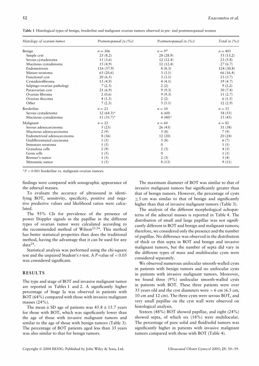
The diagnostic image that appeared to be bestcorrelated with BOT was that of a cyst with papillae (P <
0.01 vs. invasive malignant and benign tumors) (Figure 1).In addition, the multilocular echopattern seemed to befrequent in BOT (P < 0.05 vs. malignant, P < 0.01 vs.benign) (Table 4). Considering the invasive malignanttumors with only papillae, there were two serousadenocarcinomas (Stages Ic and III) and one endometrioidcystadenocarcinoma (Stage III), all well differentiated (oneof the serous tumors was considered BOT at frozen sectionbut the final pathological diagnosis was cancer Grade
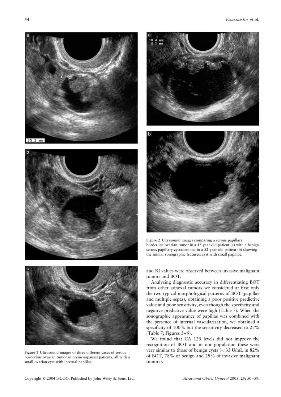
1). Analyzing benign tumors with papillae we observedeight serous tumors (three cystadenofibromas and fiveserous cystadenomas), one mucinous cystadenoma andfive endometriomas (Figure 2).
Table 5 shows our morphological analysis of serousand mucinous tumors only, in order to clarify whetherthere was a difference between BOT, benign and invasivemalignant tumors. Again, the presence of internal papillaeand of multiple septa was the most significant sonographicpattern associated with BOT. Moreover, the sonographicmorphology of mucinous and serous BOT was similar,but seemed to differ between mucinous and serous benigntumors.
Table 6 shows the percentages of tumors with positivefindings on power Doppler imaging and the location ofdetectable blood flow (peripheral or intratumoral). Thepercentage of intratumoral vascularization is reportedwhen present, otherwise the peripheral vascularization isgiven. Qualitative analysis revealed statistically significantdifferences: peripheral vascularization between invasivemalignant tumors and the other adnexal masses differed(P < 0.01 vs. BOT and vs. benign tumors), while therewas no difference between BOT and benign tumors.Intratumoral presence of power Doppler signals, inparticular intrapapillae power Doppler signals, was morefrequent in borderline (56.3%; 95% CI, 33.2–76.9%)and in invasive malignant tumors (66.7%; 95% CI,20.0–93.6%) than it was in benign tumors (0%; 95% CI,0–21.5%). Moreover, statistically significant differenceswere observed in the lowest PI and RI values of benigncompared with borderline and invasive malignant ovariantumors. No statistically significant differences in lowest PI
Copyright 2004 ISUOG. Published by John Wiley & Sons, Ltd. Ultrasound Obstet Gynecol 2005; 25: 50–59.
54 Exacoustos et al.
Figure 1 Ultrasound images of three different cases of serousborderline ovarian tumor in premenopausal patients, all with asmall ovarian cyst with internal papillae.
Figure 2 Ultrasound images comparing a serous papillaryborderline ovarian tumor in a 48-year-old patient (a) with a benignserous papillary cystadenoma in a 52-year-old patient (b) showingthe similar sonographic features: cyst with small papillae.
and RI values were observed between invasive malignanttumors and BOT.
Analyzing diagnostic accuracy in differentiating BOTfrom other adnexal tumors we considered at first onlythe two typical morphological patterns of BOT (papillaeand multiple septa), obtaining a poor positive predictivevalue and poor sensitivity, even though the specificity andnegative predictive value were high (Table 7). When thesonographic appearance of papillae was combined withthe presence of internal vascularization, we obtained aspecificity of 100% but the sensitivity decreased to 27%(Table 7; Figures 3–5).
We found that CA 125 levels did not improve therecognition of BOT and in our population these werevery similar to those of benign cysts (< 35 U/mL in 82%of BOT, 78% of benign and 29% of invasive malignanttumors).
Copyright 2004 ISUOG. Published by John Wiley & Sons, Ltd. Ultrasound Obstet Gynecol 2005; 25: 50–59.

Sonography of borderline ovarian tumors 55
Table 5 Morphological characteristics of benign, borderline and malignant ovarian tumors of serous and mucinous histological type
Benign (n = 123) Borderline (n = 33) Malignant (n = 38)
Morphological characteristic
Mucinous(n = 27)(n (%))
Serous(n = 96)(n (%))
Mucinous(n = 15)(n (%))
Serous(n = 18)(n (%))
Mucinous(n = 7)(n (%))
Serous(n = 31)(n (%))
Unilocular 17 (63.0) 68 (70.8) 0 3 (16.7) 0 0Cyst with septa 8 (29.6) 13 (13.5) 4 (26.7) 4 (22.2) 1 (14.3) 1 (3.2)
Cyst with ≤ 3 septa 3 (11.1) 12 (12.5) 1 (6.7) 1 (5.6) 0 1 (3.2%)Multilocular cyst 5 (18.5) 1 (1.0)‡ 3 (20.0) 3 (16.7)* 1 (14.3) 0
Cyst with papillae 1 (3.7) 8 (8.3) 8 (53.3)*† 8 (44.4)*† 0 2 (6.5)Cyst with 1 papilla 1 (3.7) 6 (6.3) 0 3 (16.7) 0 0Cyst with > 3 papillae 0 0 5 (33.3)*† 4 (22.2)* 0 2 (6.5%)
Fluid/solid content 1 (3.7) 7 (7.3) 3 (20.0) 3 (16.7) 5 (71.4) 14 (45.2)Pure solid content 0 0 0 0 1 (14.3) 14 (45.2)
*P < 0.05 borderline vs. benign ovarian tumors. †P < 0.05 borderline vs. malignant ovarian tumors. ‡P < 0.01 mucinous vs. serous.
Table 6 Power Doppler imaging (PDI) characteristics of ovarian tumors correlated with histological type
PDI characteristic Benign (n = 337) Borderline (n = 33) Malignant (n = 82)
Flow present (% (No. ofcases))
82 (276/337) 97 (32/33) 100 (82/82)
Peripheral (tumors withpositive findings on PDI, %(No. of cases))
71 (195/276)‡ 34 (11/32)† 4 (3/82)
Intratumoral (tumors withpositive findings on PDI, %(No. of cases))
29 (81/276)‡ 66 (21/32)* 96 (79/82)
Intrapapillae (‘cyst withpapillae’, % (No. of cases))
0 (0/14)‡ 56 (9/16)* 67 (2/3)
Lowest PI (mean ± SD) 2.29 ± 0.86‡ 0.91 ± 0.50* 0.75 ± 0.25Lowest RI (mean ± SD) 0.83 ± 0.14‡ 0.45 ± 0.08* 0.50 ± 0.11
*P < 0.01 borderline vs. benign ovarian tumors. †P < 0.01 borderline vs. malignant ovarian tumors. ‡P < 0.01 benign vs. malignant tumors.
Table 7 Comparison of different sonographic criteria and theirdiagnostic accuracy in differentiating borderline from benign andmalignant ovarian tumors
Ovarian cyst with
PapillaePapillae or
multiple septa
Papillae andintrapapillae
flow*
True positive (n) 16 22 9False positive (n) 17 29 2True negative (n) 402 390 417False negative (n) 17 11 24Sensitivity (%) 48 68 27Specificity (%) 96 93 100PPV (%) 48 43 82NPV (%) 96 97 95Positive likelihood ratio 12 10 27Negative likelihood ratio 0.54 0.34 0.73
Prevalence of borderline tumors = 7%. *Papilla with internalvessels seen with power Doppler imaging. PPV, positive predictivevalue; NPV, negative predictive value.
DISCUSSION
Approximately 15% of epithelial ovarian malignanciesare BOT1. It has been clearly established in several
series that these tumors occur in patients who areyounger compared with those who develop invasivecarcinomas and they have been reported to exhibita relatively benign clinical course. Therefore, thepreoperative evaluation of BOT is becoming increasinglyimportant in premenopausal women when the clinician isfaced with the dilemma of avoiding unnecessary surgerywhile identifying possible ovarian cancer in good time.Because of the desirability of preserving ovarian function,particularly if retention of childbearing capacity is anissue, several authors have recommended less radicalsurgery (unilateral oophorectomy or cystectomy) for thegroup of young patients with unilateral BOT1–4,25–27.
It is well known that ultrasound is effective indifferentiating benign from malignant ovarian tumorsand is actually one of the most important presurgicaldiagnostic tests in the management of adnexal lesions.However, in most studies reporting sonographic featuresof ovarian tumors, BOT have been considered togetherwith invasive malignant tumors. Only a few studies1,5–9,most of which involved a small number of cases, havefocused on the sonographic appearance of BOT. Daraiet al.1 reported that the majority (67.7%) of BOT atsonographic examination are multilocular, have thicksepta and are sonolucent or of mixed echogenicity.
Copyright 2004 ISUOG. Published by John Wiley & Sons, Ltd. Ultrasound Obstet Gynecol 2005; 25: 50–59.

56 Exacoustos et al.
Figure 3 Power Doppler ultrasound image showing cyst withvascularized papilla: serous borderline ovarian tumor.
Figure 4 Power Doppler ultrasound image showing cyst withvascularized papilla: well-differentiated serous cystadencarcinomaStage 1c.
Mucinous tumors tend to be larger at presentationbut some authors observed no significant difference indiameter between invasive malignant tumors and BOT6.In our study the maximum diameter of BOT was similarto that of invasive malignant tumors; however, thepercentage of cysts ≤ 5 cm was similar to that of benignones (Table 3).
Furthermore, other studies1,7,8,28,29 have reported thatBOT can exhibit sonographic features such as unilocu-lar smooth anechoic cysts without endophytic papillary
a
b
Figure 5 Ultrasound images showing benign serous cystadenoma:(a) gray-scale image showing small cyst with internal papilla;(b) power Doppler imaging did not show internal vascularizationof the papilla.
growth. Gotlieb et al.8 reported 13% and Emoto et al.7
reported 17% of BOT with a sonographic appearanceof unilocular cysts, while in a prospective study of 1072premenopausal ovarian tumors, Osmers et al.28 demon-strated that malignancy occurred in 0.8% of unilocularsmooth-walled cysts, and that 0.5% of unilocular smooth-walled cysts were BOT. Nevertheless, they found noovarian cancer in women < 20 years old and none inthose with simple ovarian cysts of a mean size < 40 mm,which allowed them to conclude that the risk of finding amalignant growth in a simple ovarian cyst decreases alongwith decreasing tumor size and decreasing age28.
We found no invasive malignancies in simple ovariancysts in our population but we observed unilocularsmooth-walled cysts in three cases (9%) of BOT. All threecysts were detected in premenopausal women and had adiameter > 6 cm. Although our ultrasound examinationof the cyst wall was very careful, we did not observein these three cysts the very small papillae which weredescribed among the histological features. This couldbe due to the fact that the transvaginal sonographicexamination of large cysts may have been less accuratebecause the distal cyst wall was too far away to be clearlyvisible on the screen. On the other hand the sonographic
Copyright 2004 ISUOG. Published by John Wiley & Sons, Ltd. Ultrasound Obstet Gynecol 2005; 25: 50–59.

Sonography of borderline ovarian tumors 57
transabdominal evaluation of large cysts is unable toidentify small papillae because of technical features (suchas the need to use a lower frequency probe because of athick abdominal wall). New machines and transvaginalsonographic probes with different characteristics (largervision angle, possibility of changing frequency, harmonicimaging, three-dimensional imaging) could probablyeliminate these problems; however, large unilocular cystsshould be managed with more caution than are smallerones (≤ 5 cm).
In 85% of BOT we detected sonographic features withsome degree of complexity. The diagnostic image thatseemed to best correlate with BOT was that of a cystwith papillae (48%) or one with multiple septa (18%).These findings were similar to those of other recentstudies8,9, which reported the presence of papillae in63–65% of BOT.
Analyzing benign tumors with papillae we observedeight serous tumors, one mucinous cystadenoma and fiveendometriomas. Benign tumors such as endometriomasoften present a typical echostructure that permits theidentification of the type of tumor (i.e. ‘ground glass’appearance). An experienced sonographer is often able toidentify the type of tumor (endometrioma, sactosalpinx,paraovarian cyst, ovarian fibroid)30–32 and this subjectivepattern recognition has been reported by many authorsto be very accurate, especially for endometriosis30,31,33,34.We therefore focused our morphological analysis only onserous and mucinous tumors (Table 5), and the presenceof internal papillae and of multiple septa was againthe most significant sonographic pattern associated withBOT. No difference in sonographic features was observedbetween mucinous and serous BOT. This can be explainedby the fact that both types of tumor can present a differentappearance on analysis of gross histological section35,36.The mucinous tumors were mostly multicystic andfilled with mucinous material (Figure 6), but couldalso contain solid tissue and endophytic papillae37,38.Papillary projections seemed to be more typical in seroustumors; however, unilocular and septate cysts were alsoobserved35,36,39.
Our power Doppler examination of BOT revealed PIand RI values similar to those of invasive malignanttumors and a vascular distribution (central vs. peripheralvascularization) between those of benign and invasivemalignant tumors. According also to other studies6,8,9, nospecific Doppler velocimetry flow evaluation is currentlyavailable to diagnose BOT. Despite this, Zanetta et al.6
proposed a model for differentiating BOT from invasivemalignant and benign tumors that involved Dopplerflow parameters. This model was based on the presenceof intracystic complexity (either septa or papillae), aPI < 1.0, absence of intratumoral confluence of vesselsand CA 125 levels < 150 U/L in women under the age of60 years, and it yielded an accuracy of 91%. However,the same authors recognized the limited value of thismodel clinically, considering that the fit of such a modelis always greater when derived retrospectively than whenapplied prospectively.
a
b
c
Figure 6 Ultrasound images showing multiseptate cysts: (a) largebenign mucinous cystadenomas; (b) transabdominal and(c) transvaginal views of a mucinous borderline ovarian tumor.
Copyright 2004 ISUOG. Published by John Wiley & Sons, Ltd. Ultrasound Obstet Gynecol 2005; 25: 50–59.

58 Exacoustos et al.
The problem that arises from all the sonographic studieson BOT and from our results is not only the inabilityto differentiate benign tumors from BOT but especiallythe inaccuracy in distinguishing BOT from malignancy.It seems that BOT also have a borderline sonographicappearance and do not have a pathognomonic ortypical sonographic pattern. In fact, the presence ofpapillae can also be found in benign serous tumors40
(Figures 2 and 5) and if internal vascularization of thepapillae is considered, it is very difficult to distinguishBOT from invasive malignant tumors (Figures 3 and 4).Histopathological analysis can help, and gross section andangiogenesis in particular could be helpful in interpretingthese findings. In histopathological specimens it has beenshown recently that microvessel count is more intense ininvasive ovarian carcinomas than it is in BOT41. It hasalso been noted42 that there is little difference betweenBOT and well-differentiated invasive malignant tumors.Tumors that are of higher grading or are undifferentiatedare associated with a greater amount of solid tissuewith marked and irregular vascularization42,43, whereaslow-grade malignant tumors preserve the structuralcharacteristics of the original histological type. Thereforedifferentiation between BOT and invasive malignanttumors seems not to be stage-dependent, but is probablygrade-dependent. In other words, it seems to be verydifficult to distinguish a well-differentiated serous ovariancarcinoma from a serous BOT, since both can appear atultrasound as a cyst with vascularized papillae (Figures 3and 4). A Grade 3 serous carcinoma, however, generallypresents a sonographically more complex structure witha large amount of solid tissue. On the other hand,papillae can also be observed in benign cystadenomasor cystadenofibromas and in these cases perhaps thevascularization inside the papilla could suggest BOT ormalignancy, even though avascular papilla is not alwaysassociated with benign tumors and vice versa (95% CI,0–22%).
Finally, our results show that accurate sonographicexamination can suggest BOT on the basis of the presenceof papillae or multiple septa. However, neither papillaenor septa constitute highly sensitive sonographic markers.The main concern for BOT remains that they must notbe managed with overly aggressive therapy, and thereforea very accurate presurgical diagnosis is desirable. Thismeans that conservative surgery for a suspected BOT, theactual sonographic diagnosis not being able to excludetotally invasive malignancy, has to be associated withfrozen section, and greater care than normal should betaken not to rupture the cyst capsule. However, theprecision of frozen-section diagnosis of BOT has beenquestioned44,45 and the risk of spread of BOT and invasivemalignant tumors treated conservatively has yet to beestablished.
It is to be hoped that further advances in theapplication of three-dimensional and power Dopplerimaging and sonographic contrast media for the detectionof microvessels may be able to identify more accuratelyBOT and prevent the overtreatment of these lesions.
REFERENCES
1. Darai E, Teboul J, Walker F, Benifla JL, Meneux E,Guglielmina JN, Pennehouat G, Renolleau C, Sebban E, Made-lenat P. Epithelial ovarian carcinoma of low malignant poten-tial. Eur J Obstet Gynecol Reprod Biol 1996; 66: 141–145.
2. Candiani M, Vasile C, Sgerzi MR, Nozza A, Maggi F, Maggi R.Borderline ovarian tumors: laparoscopic treatment. Clin ExpObstet Gynecol 1999; 26: 39–43.
3. Seracchioli R, Venturoli S, Colombo FM, Govoni F, Missiroli S,Bagnoli A. Fertility and tumor recurrence rate after conservativelaparoscopic management of young women with early-stageborderline ovarian tumors. Fertil Steril 2001; 76: 999–1004.
4. Camatte S, Morice P, Pautire P, Atallah D, Duvillard P, Cas-taigne D. Fertility results after conservative treatment ofadvanced stage serous borderline tumor of the ovary. BJOG2002; 109: 376–380.
5. Hata K, Hata T, Manabe A, Kitao M. Ovarian tumors of lowmalignant potential: transvaginal Doppler ultrasound features.Gynecol Oncol 1992; 45: 259–264.
6. Zanetta G, Lissoni A, Cha S, Bertalero C, Scalambrino S,Bratina G. Pre-operative morphological and color Dopplerfeatures of borderline ovarian tumours. Br J Obstet Gynaecol1995; 102: 990–996.
7. Emoto M, Udo T, Obama H, Eguchi F, Hachisuga T,Kawarabayashi T. The blood flow characteristics in borderlineovarian tumors based on both color Doppler ultrasound andhistopathological analyses. Gynecol Oncol 1998; 70: 351–356.
8. Gotlieb WH, Soriano D, Achiron R, Zalel Y, Davidson B,Kopolovic J, Novikov I, Ben-Baruch G. CA 125 measurementand ultrasonography in borderline tumors of the ovary. Am JObstet Gynecol 2000; 183: 541–546.
9. Pascual MA, Tresserra F, Grases PJ, Labastida R, Dexeus S.Borderline cystic tumors of the ovary: grey-scale and colorDoppler sonographic findings. J Clin Ultrasound 2002; 30:76–82.
10. International Federation of Gynecology and Obstetrics. Changesin definitions of clinical staging for carcinoma of the cervix andovary. Am J Obstet Gynecol 1987; 156: 263–264.
11. Serov SF, Scully RE, Sobin LH. International histological clas-sification of ovarian tumors (No 9). In Histological Typing ofOvarian Tumors. World Health Organization: Geneva, 1973.
12. Scully RE. World Health Organization. International histologi-cal classification of tumors. In Histological Typing of OvarianTumors. Springer Verlag: Berlin, 1999.
13. Sassone AM, Timor-Tritsch IE, Artner A, Westhoff C, War-ren WB. Transvaginal sonographic characterization of ovariandisease: evaluation of a new scoring system to predict ovarianmalignancy. Obstet Gynecol 1991; 78: 70–76.
14. Timmerman D, Bourne TH, Tailor A, Collins WP, Verrelst H,Vandenberghe K, Vergote I. A comparison of methods forpreoperative discrimination between malignant and benignadnexal masses: the development of a new logistic regressionmodel. Am J Obstet Gynecol 1999; 181: 57–65.
15. Valentin L. Comparison of Lerner score, Doppler ultrasoundexamination, and their combination for discrimination betweenbenign and malignant adnexal masses. Ultrasound ObstetGynecol 2000; 15: 143–147.
16. Timmerman D, Valentin L, Bourne TH, Collins WP, Ver-relst H, Vergote I; International Ovarian Tumor Analysis(IOTA) Group. Terms, definitions and measurements to describethe sonographic features of adnexal tumors: a consensus opin-ion from the International Ovarian Tumor Analysis (IOTA)Group. Ultrasound Obstet Gynecol 2000; 16: 500–505.
17. Cohen LS, Escobar PF, Scharm C, Glimco B, Fishman DA.Three-dimensional power Doppler ultrasound improves thediagnostic accuracy for ovarian cancer prediction. GynecolOncol 2001; 82: 40–48.
18. Marret H, Ecochard R, Giraudeau B, Golfiers F, Raudrant D,Lansac J. Color Doppler energy prediction of malignancy in
Copyright 2004 ISUOG. Published by John Wiley & Sons, Ltd. Ultrasound Obstet Gynecol 2005; 25: 50–59.

Sonography of borderline ovarian tumors 59
adnexal masses using logistic regression models. UltrasoundObstet Gynecol 2002; 20: 597–604.
19. Guerriero S, Alcazar JL, Coccia ME, Ajossa S, Scarselli G,Boi M, Gerada M, Melis GB. Complex pelvic mass as atarget of evaluation of vessel distribution by color Dopplersonography for the diagnosis of adnexal malignancies: resultsof a multicenter European study. J Ultrasound Med 2002; 21:1105–1111.
20. Alcazar JL, Merce LT, Laparte C, Jurado M, Lopez-Garcia G.A new scoring system to differentiate benign from malignantadnexal masses. Am J Obstet Gynecol 2003; 188: 685–692.
21. Caspi B, Appelman Z, Robinerson D, Elchalal U, Zalel Y,Katz Z. Pathognomonic echo patterns of benign cystic teratomasof the ovary: classification, incidence, and accuracy rate ofsonographic diagnosis. Ultrasound Obstet Gynecol 1996; 7:275–280.
22. Zupi E, Exacoustos C, Szabolcs B, Marconi D, Carusotti C,Sbracia M, Arduini D, Lanzi G. Laparoscopic approach to der-moid cysts: combined surgical technique and ultrasonographicevaluation of residual functioning ovarian tissue. J Am AssocGynecol Laparosc 2003; 10: 154–158.
23. Wilson EB. Probable interference, the law of succession andstatistical interference. J Am Stat Assoc 1927; 22: 209–212.
24. Newcombe RG. Two-sided confidence intervals for singleproportions: Comparison of seven methods. Stat Med 1998;17: 857–872.
25. Nation JG, Krepart GV. Ovarian carcinoma of low malignantpotential: Staging and treatment. Am J Obstet Gynecol 1986;154: 290–293.
26. Trope CG, Kærn J, Vergote IB, Kristensen G, Abeler VM. Areborderline tumors of the ovary overtreated both surgically andsystemically? A review of four prospective randomized trialsincluding 253 patients with borderline tumors. Gynecol Oncol1993; 51: 236–243.
27. Tazelaar HD, Bostwick DG, Ballon SC, Hendrickson MR,Kempson RL. Conservative treatment of borderline ovariantumors. Obstet Gynecol 1985; 66: 417–422.
28. Osmers RGW, Osmers M, von Maydell B, Wagner B, Kuhn W.Preoperative evaluation of ovarian tumors in premenopauseby transvaginosonography. Am J Obstet Gynecol 1996; 175:428–434.
29. Osmers RGW, Osmers M, von Maydell B, Wagner B, Kuhn W.Evaluation of ovarian tumors in postmenopausal women bytransvaginal sonography. Eur J Obstet Gynecol Reprod Biol1998; 77: 81–88.
30. Valentin L. Pattern recognition of pelvic masses by gray-scaleultrasound imaging: the contribution of Doppler ultrasound.Ultrasound Obstet Gynecol 1999; 14: 338–347.
31. Valentin L, Hagen B, Tingulstad S, Eik-Nes S. Comparisonof ‘pattern recognition’ and logistic regression models fordiscrimination between benign and malignant pelvic masses: aprospective cross validation. Ultrasound Obstet Gynecol 2001;18: 357–365.
32. Guerriero S, Ajossa S, Lai MP, Mais V, Paoletti AM, Melis GB.Transvaginal ultrasonography associated with colour Dopplerenergy in the diagnosis of hydrosalpinx. Hum Reprod 2000; 15:1568–1572.
33. Guerriero S, Ajossa S, Melis G. Performance of US in thediagnosis of endometrioma. Radiology 2000; 215: 305–307.
34. Exacoustos C, Zupi E, Carusotti C, Rinaldo D, Marconi D,Lanzi G, Arduini D. Staging of pelvic endometriosis: role ofsonographic appearance in determining extension of disease andmodulating surgical approach. J Am Assoc Gynecol Laparosc2003; 10: 378–382.
35. Seidman JD, Ronnett BM, Kurman RJ. Pathology of borderline(low malignant potential) ovarian tumours. Best Pract Res ClinObstet Gynaecol 2002; 16: 499–512.
36. Prat J, de Nictolis M. Serous borderline tumors of the ovary.A long-term follow-up study of 137 cases, including 18 with amicropapillary pattern and 20 with microinvasion. Am J SurgPathol 2002; 26: 1111–1128.
37. Rodriguez IM, Prat J. Mucinous tumors of the ovary. Aclinicopathologic analysis of 75 borderline tumors (of intestinaltype) and carcinomas. Am J Surg Pathol 2002; 26: 139–152.
38. Shappell HW, Riopel MA, Smith Sehdev AE, Ronnett BM,Kurman RJ. Diagnostic criteria of behaviour of ovarianseromucinous (endocervical-type mucinous and mixed cell-type) tumors. Atypical proliferative (borderline) tumors,intraepithelial, microinvasive, and invasive carcinomas. Am JSurg Pathol 2002; 26: 1529–1541.
39. Kurman RJ, Trimble CL. The behaviour of serous tumors oflow malignant potential: are they ever malignant? Int J GynecolPathol 1993; 12: 120–127.
40. Alcazar JL, Errasti T, Minguez JA, Galan MJ, Garcia-Manero M, Ceamanos C. Sonographic features of ovariancystadenofibromas: spectrum of findings. J Ultrasound Med2001; 20: 915–919.
41. Abulafia O, Ruiz JE, Holcomb K, Dimaio TM, Lee Y, Sherer D.Angiogenesis in early-invasive and low-malignant-potentialepithelial ovarian carcinoma. Obstet Gynecol 2000; 95:548–552.
42. Russel P. Surface epithelial-stromal tumors of the ovary. InBlaunstein’s Pathology of the Female Genital Tract (4th edn),Kurman RJ (ed). Springer Verlag: New York, Berlin, Heidelberg,1994; 705–782.
43. Jung SE, Lee JM, Rha SE, Byun JY, Jung JI, Hahn ST. CT andMR imaging of ovarian tumors with emphasis on differentialdiagnosis. Radiographics 2002; 22: 1305–1325.
44. Houck K, Nikrui N, Duska L, Chang Y, Fuller AF, Bell D,Goodman A. Borderline tumors of the ovary: correlationof frozen and permanent histopathologic diagnosis. ObstetGynecol 2000; 95: 839–843.
45. Kayikcioglu F, Pata O, Cengiz S, Tulunay G, Boran N, Yal-van S, Kose MF. Accuracy of frozen section diagnosis in bor-derline ovarian malignancy. Gynecol Obstet Invest 2000; 49:187–189.
Copyright 2004 ISUOG. Published by John Wiley & Sons, Ltd. Ultrasound Obstet Gynecol 2005; 25: 50–59.


