Embed Size (px)
Citation preview

1
2
3
4
5Q1
6
7
89
10
11
12
13
14
15
16
17
18
19
20
21
22
23
24
25
26
27
28
29
30
31
32
3334
35
36
37
38
39
40
41
42
43
44
Biochimica et Biophysica Acta xxx (2008) xxx–xxx
BBAMCR-15958; No. of pages: 10; 4C: 3, 4, 5, 7
Contents lists available at ScienceDirect
Biochimica et Biophysica Acta
j ourna l homepage: www.e lsev ie r.com/ locate /bbamcr
ARTICLE IN PRESS
Review
Principles of lysosomal membrane degradationCellular topology and biochemistry of lysosomal lipid degradation
Heike Schulze, Thomas Kolter, Konrad Sandhoff ⁎LIMES Membrane Biology and Lipid Biochemistry, Kekulé-Institut für Organische Chemie und Biochemie der Universität Bonn, Germany
UN
F
Abbreviations: BMP, bis(monoacylglycero)phosphatdrugs; GlcCer, glucosylceramide; GM3, NeuAcα2,3GalβNAcβ1,4(NeuAcα2,3)Galβ1,4Glcβ1ceramide; GM2-AP,glycosphingolipids; LLBP, lysosomal lipid binding prbodies; NPC, Niemann–Pick disease Type C; SAPs, sphinA–D and GM2-AP); Sap, saposin A–D⁎ Corresponding author. Tel.: +49 228 735834; fax: +4
E-mail address: [email protected] (K. Sandhoff)
0167-4889/$ – see front matter © 2008 Elsevier B.V. Aldoi:10.1016/j.bbamcr.2008.09.020
Please cite this article as: H. Schulze, et albbamcr.2008.09.020
a b s t r a c t
a r t i c l e i n f oArticle history:
Cellular membranes enter t Received 4 July 2008Received in revised form 24 September 2008Accepted 30 September 2008Available online xxxxKeywords:LysosomeGlycosphingolipidSphingolipid activator protein
O
TEDPR
Ohe lysosomal compartment by endocytosis, phagocytosis, or autophagy. Withinthe lysosomal compartment, membrane components of complex structure are degraded into their buildingblocks. These are able to leave the lysosome and can then be utilized for the resynthesis of complexmolecules or can be further degraded. Constitutive degradation of membranes occurs on the surface of intra-endosomal and intra-lysosomal membrane structures. Many integral membrane proteins are sorted to theinner membranes of endosomes and lysosome after ubiquitinylation. In the lysosome, proteins are degradedby proteolytic enzymes, the cathepsins. Phospholipids originating from lipoproteins or cellular membranesare degraded by phospholipases. Water-soluble glycosidases sequentially cleave off the terminalcarbohydrate residues of glycoproteins, glycosaminoglycans, and glycosphingolipids. For glycosphingolipidswith short oligosaccharide chains, the additional presence of membrane-active lysosomal lipid-bindingproteins is required. The presence of lipid-binding proteins overcomes the phase problem of water solubleenzymes and lipid substrates by transferring the substrate to the degrading enzyme or by solubilizing theinternal membranes. The lipid composition of intra-lysosomal vesicles differs from that of the plasmamembrane. To allow at least glycosphingolipid degradation by hydrolases and activator proteins, thecholesterol content of these intraorganellar membranes decreases during endocytosis and the concentrationof bis(monoacylglycero)phosphate, a stimulator of sphingolipid degradation, increases. A considerable part ofour current knowledge about mechanism and biochemistry of lysosomal lipid degradation is derived from aclass of human diseases, the sphingolipidoses, which are caused by inherited defects within sphingolipid andglycosphingolipid catabolism.
© 2008 Elsevier B.V. All rights reserved.
C45
E1. Lysosomal membrane digestion
46
47
48
49
50
51
52
53
54
CORRLysosomes are major degradative compartments of eukaryoticcells. In contrast to the proteasome, lysosomes degrade a wide varietyof structurally diverse substances, such as proteins, glycosaminogly-cans, nucleic acids, oligosaccharides, and complex lipids, into theirbuilding blocks [1]. These can leave the lysosomes either via diffusion,or with the aid of specialized transporters [2]. In the cytosol, thebuilding blocks can be further degraded to fuel energy metabolism orcan re-enter biosynthetic pathways. To provide building blocks ofcomplex macromolecules for salvage and recycling pathways seems tobe an important function of lysosomes. It has been shown, that in not
55
56
57
58
59
60
61
62
63
e; CADs, cationic amphiphilic1,4Glcβ1ceramide; GM2, Gal-GM2 activator protein; GSL,oteins; MVBs, multivesiculargolipid activator proteins (Sap
9 228 737778..
l rights reserved.
., Principles of lysosomal me
very rapidly dividing cells, glycosphingolipids (GSL) are synthesizedpredominantly from sphingoid bases, carbohydrates and sialic acidsreleased by lysosomes. In human foreskin fibroblasts for example, 90%of the glucosylceramide derives from recycling of sphingoid base, only10% is synthesized de novo [3]. Under this aspect, the concept oflysosomes as waste dumps within cells would be a misleadingassociation and should be replaced by the idea of lysosomes asstomachs of the cell, that provide macromolecule constituents andensure lipid homeostasis.
1.1. Endocytosis and autophagy
Eukaryotic cells maintain highly regulated transport systems thatconvey cargo into the cell or exchange membranes and cargo betweencellular organelles. Cellular and foreign cargo, but alsomembranes canreach the endosomal–lysosomal systemvia endocytosis, phagocytosis,autophagy, or direct transport. The various cellular functionsassociated with this process require degradation steps within thelysosomes, where proteins, complex cargo constituents, or complexmembrane lipids have to be cleaved. During endocytosis, cargo entersthe cell via clathrin-dependent or -independent mechanisms in a
mbrane degradation, Biochim. Biophys. Acta (2008), doi:10.1016/j.

C
64
65
66
67
68
69
70
71
72
73
74
75
76
77
78
79
80
81
82
83
84
85
86
87
88
89
90
91
92
93
94
95
96
97
98
99
100
101
102
103
104
105
106
107
108
109
110
111
112
113
114
115
116
117
118
119
120
121
122
123
124
125
126
127
128
129
130
131
132
133
134
135
136
137
138
139
140
141
142
143
144
145
146
147
148
149
150
151
152
153
2 H. Schulze et al. / Biochimica et Biophysica Acta xxx (2008) xxx–xxx
ARTICLE IN PRESS
RE
constitutive or ligand-induced manner [4]. Parts of the plasmamembranewith and without receptor proteins are internalized, trafficthrough the endosomal compartment, and undergo different steps ofsorting, before they are either recycled to the plasma membrane, ordelivered to the lysosome for degradation. They reach the lysosomeeither as intra-lysosomal membrane structures or as part of theperimeter membrane [5,6]. During endosomal maturation, theluminal pH value decreases from values of about 7.2 to below 5 [7].
The endosomal membrane consists of different domain arrange-ments, in which Rab proteins are localized in morphologicallydistinct domains, like in a mosaic. Endosomes comprised of differentdomain arrangements display biochemical and possibly functionaldiversity [8].
Cellular macromolecules can be degraded by different pathways ineukaryotic cells. Ubiquitinylated proteins are degraded by theproteasomal system in the cytosol, bulk cytoplasma and organellesare delivered to the lysosome by (macro)autophagy [9] and cellularmembranes are degraded in the lysosome after endocytosis. Autop-hagy requires a membrane degradation step, before cargo can bedegraded by the lysosomal degradation system. Autophagy representsa unique form of membrane trafficking, in which membranecompartments (autophagosomes) engulf organelles or cytosoliccargo and deliver them to the lysosome for degradation [10]. Undernormal growth conditions, autophagy occurs at a basal level.Starvation dramatically induces autophagy to maintain a pool ofbasic nutrients. Autophagy is evolutionary conserved in eukaryotes.Insights into the molecular pathways of autophagy were mainlygained by genetic approaches in yeast mutants defective in autophagy.Degradation of autophagic bodies occurs in yeast in the vacuole.
Autophagy starts with the formation of autophagosomes, double-membrane-layered vesicles, which enclose cytosol or organelles [11].In yeast, after fusion with the vacuole, the autophagosome is releasedinto the lumen as a single-membrane vesicle and termed autophagicbody. The breakdown of this subvacuolar vesicle depends on the acidicpH of the vacuole [12], and on vacuolar proteinase A and proteinase B(Prb1) [13]. However, the function of Prb1 might be to activatevacuolar zymogens that play a direct role in the breakdown process[14]. Two other proteins have also been implicated in membranedegradation, the putative lipases Aut5 [11] and Aut4 [15].
Another role of autophagy in membrane degradation is that it is asource of bis(monoacylglycero)phosphate (BMP, erroneously alsocalled lysobisphosphatidic acid, Fig. 1). This negatively charged lipidis highly enriched in the internal membranes of the lysosome andrequired for degradation of small GSL [16]. Biosynthetically, BMP isformed during the degradation of phosphatidylglycerol and cardioli-pin, presumably on the surface of intra-lysosomal vesicles [17,18].
UNCO
R 154
155
156
157
158
159
160
161
162
163
164
165
166
167
168
169
170
171Fig. 1. Structure of bis(monoacylglycero)phosphate (BMP). Structure of bis(mono-acylglycero)phosphate (BMP).
Please cite this article as: H. Schulze, et al., Principles of lysosomal mbbamcr.2008.09.020
TEDPR
OOF
Cardiolipin in turn, reaches the lysosome as a component ofmitochondria by macroautophagy. Its degradation leads to formationof BMP on internal membranes.
In eukaryotes, transmembrane proteins destined for lysosomaldegradation are in eukaryotes often monoubiquitinylated and sortedin endosomal multivesicular bodies (MVBs) [19]. MVB formationrequires the sequential action of three endosomal sorting complexesneeded for transport (ESCRT-I,-II,-III) [20]. MVBs follow the pathwayfrom early to late endosomes, and are eventually delivered tolysosomes, where they are degraded together with their proteincargo [21].
1.2. Topology of degradation
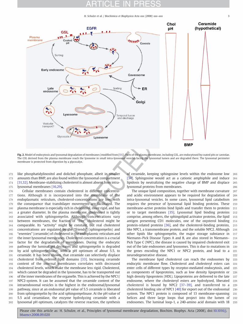
In the endosomal–lysosomal system, a variety of hydrolyticenzymes with acidic pH-optima cleave macromolecules such asproteins, polysaccharides, nucleic acids, glycoconjugates, and phos-pholipids. To protect the interior of the cell from these degradativeenzymes, the integrity of the limiting membrane has to bepreserved during the process of lysosomal degradation. This isachieved by a thick glycocalyx [22] composed of the carbohydratepart of lysosomal integral membrane proteins (LIMPS) and lysoso-mal associated membrane proteins (LAMPS) [23]. The enzymesrequired for lipid degradation cannot be expected to reach theirsubstrates through this glycocalyx, which is composed of glycopro-teins highly N-glycosylated with polylactosamine units. Since theperimeter membrane is protected from degradation, a seconddistinct pool of membranes has to be present in the endosomal/lysosomal compartment. This has been proposed in 1992 [5] and hasbeen confirmed by independent groups. According to our currentview, former parts of the plasma membrane destined for degrada-tion reach the lysosome as part of small intra-lysosomal vesicles, inwhich the former extracytoplasmic membrane leaflet faces thelumen of the lysosome (Fig. 2). These intra-lysosomal vesiclesprovide the platform of membrane degradation. As indicated bystudies with human patients with defects in GSL degradation, thesurface of the inner lysosomal membranes represents the main siteof membrane degradation in eukaryotic cells. Intra-lysosomalmembranes have been initially observed, when different membranedegradation steps are defective. This was the case in cells frompatients with sphingolipid storage diseases such as GM1 gang-liosidosis [24] or combined sphingolipid activator protein (Sap)deficiency [25], where they accumulate as multivesicular storagebodies. Later on, they have been identified as MVBs [21,26].Complementing the media of Sap-precursor-deficient fibroblastswith nanomolar concentrations of purified Sap-precursor reversedthe aberrant accumulation of multivesicular structures, and restoredthe cells ability to degrade glycosphingolipids [27].
1.3. Lipid composition of lysosomal membranes
The lipid composition of limiting membrane and the intra-lysosomal membrane structures differs considerably from eachother and from that of plasma membrane or the limiting membranesof other cellular organelles. In addition, the luminal pH value steadilydecreases to achieve optimal conditions for the action of lysosomalenzymes. Besides the lipid composition, also the protein compositionof the internal membranes is adjusted during endosomal sorting [28].The intra-lysosomal membranes are formed by a lipid-sorting processalong the endocytic pathway, during which its cholesterol contentdecreases and that of the negatively charged lipid BMP increases[16,29]. Due to its unusual sn1,sn1′-configuration, BMP has asufficiently long lifetime in spite of the presence of the lysosomalphospholipases [30]. While intra-lysosomal membranes are enrichedin BMP, this lipid is almost absent in the perimeter membrane,distinguishing the two membrane pools [16,29]. Other anionic lipids
embrane degradation, Biochim. Biophys. Acta (2008), doi:10.1016/j.
DPR
OOF
172
173
174
175
176
177
178
179
180
181
182
183
184
185
186
187
188
189
190
191
192
193
194
195
196
197
198
199
200
201
202
203
204
205
206
207
208
209
210
211
212
213
214
215
216
217
218
219
220
221
222
223
224
225
226
227
228
229
230
231
232
233
Fig. 2.Model of endocytosis and lysosomal degradation of membranes (modified from [51]). Parts of the plasmamembrane, including GSL, are endocytosed by coated pits or caveolae.The GSL derived from the plasma membrane reach the lysosome in small intra-lysosomal vesicles facing the lysosomal lumen and are degraded there. The lysosomal perimetermembrane is protected from digestion by a glycocalyx.
3H. Schulze et al. / Biochimica et Biophysica Acta xxx (2008) xxx–xxx
ARTICLE IN PRESS
UNCO
RREC
like phosphatidylinositol and dolichol phosphate, albeit in smalleramounts than BMP, are also found within the lysosomal compartment[31,32]. Membrane-stabilizing cholesterol is almost absent from intra-lysosomal membranes [16,29].
Cellular membranes contain cholesterol in different concentra-tions. Although it is incorporated into the membrane of theendoplasmatic reticulum, cholesterol-concentrations are low, withthe consequence that transbilayer movements are facilitated. Theplasmamembrane is especially rich in cholesterol, more rigid, and hasa greater diameter. In the plasma membrane, cholesterol is tightlyassociated with sphingomyelin. Although concentrations varybetween membranes, the fraction of “free” cholesterol might becomparable. Its “activity” might be constant, but real cholesterolconcentrations are regulated by the “friends” (sphingomyelin) and“enemies” (ceramide) of cholesterol in the endoplasmic reticulum andthe inner lysosomal membranes. Cholesterol concentration is a crucialfactor for the degradation of membranes. During the endocyticpathway the luminal pH decreases and sphingomyelin is degradedby acid sphingomyelinase, with a pH optimum of about 5.5, toceramide. It has been shown, that ceramide can selectively displacecholesterol from ordered lipid domains [33]. Increasing ceramidecontent of the internal membranes correlates with decreasedcholesterol levels, which make the membrane less rigid. Cholesterol,which cannot be degraded in the lysosome, has to be transported outof the innermembranes of the organelle. This is achieved by the NPC1/NPC2-system. It can be assumed that the ceramide content of theintraendosomal vesicles is the highest in the endosomal/lysosomalpathway, since at an endosomal pH value of 5.5 ceramide is liberatedfrom sphingomyelin by the acid sphingomyelinase. At the pH value of5.5 acid ceramidase, the enzyme hydrolyzing ceramide with alysosomal pH optimum, catalyzes the reverse reaction, the synthesis
Please cite this article as: H. Schulze, et al., Principles of lysosomal mebbamcr.2008.09.020
TEof ceramide, keeping sphingosine levels within the endosome low[34]. Sphingosine would act as a cationic amphiphile and inducelipidosis by neutralizing the negative charge of BMP and displacelysosomal proteins from membranes.
The unique lipid composition, together with membrane curvatureand acidic environment appears to be required for degradation ofintra-lysosomal vesicles. In some cases, lysosomal lipid catabolismrequires the presence of lysosomal lipid binding proteins. Thesemembrane-active proteins bind lipids and transfer them to proteinsor to target membranes [35]. Lysosomal lipid binding proteinscomprise, among others, the sphingolipid activator proteins, the lipidantigen presenting CD1 molecules, one of the oxysterol bindingprotein-related proteins [36], and the cholesterol-binding proteins,like NPC1, a transmembrane protein, and the soluble NPC2. Althoughother lipids like sphingomyelin, the major storage substance inNiemann–Pick Disease Types A and B, are also stored in Niemann–Pick Type C (NPC), the disease is caused by impaired cholesterol exitout of the late endosomes and lysosomes. This is due to mutations inthe genes encoding the NPC1 or NPC2 protein, and lead to aneurodegenerative disease.
The membrane lipid cholesterol can reach the endosomes byendocytic membrane flow. Cholesterol and cholesteryl esters canenter cells of different types by receptor-mediated endocytosis, andas components of lipoproteins, such as low density lipoprotein orhigh density lipoprotein (HDL). Lipoproteins are delivered to the lateendosome, where the cholesteryl esters are hydrolyzed, liberatedcholesterol is bound by NPC2 [37–39], and transferred to acholesterol binding site of NPC1 [40] for export out of the endosomalsystem. The NPC1 protein is composed of 13 membrane-spanninghelices and three large loops that project into the lumen ofendosomes. The luminal loop-1, a 240-amino acid domain with 18
mbrane degradation, Biochim. Biophys. Acta (2008), doi:10.1016/j.

C
234
235
236
237
238
239
240
241
242
243
244
245
246
247
248
249
250
251
252
253
254
255
256
257
258
259
260
261
262
263
264
265
266
267
268
269
270
271
272
273
274
275
276
277
278
279
280
281
282
283
284
285
286
287
288
289
290
291
292
293
294
295
296
297
298
299
300
301
302
303
304
305
306
307
308
309
310
311
312
313
314
315
4 H. Schulze et al. / Biochimica et Biophysica Acta xxx (2008) xxx–xxx
ARTICLE IN PRESS
E
cysteines has been identified as cholesterol and oxysterol bindingsite [40]. The NPC1 protein is suggested to mediate transport oflipophilic molecules through the glycocalyx of lysosomal perimetermembranes and presumably also out of the endosomal–lysosomalsystem [41,42] (Fig. 3).
In fibroblasts from NPC patients, exit of liberated cholesterol fromthe late endosomes and lysosomes is attenuated and it accumulates inthe organelles. Secondarily, other membrane components alsoaccumulate. Storage of neutral glycolipids such as glucosylceramideand lactosylceramide, acidic glycolipids, especially of gangliosidesGM3 and GM2, sphingomyelin (less than in Nieman–Pick disease,Types A and B), BMP, and phospholipids occurs in liver, spleen, brain,and other organs [43]. This can be easily explained by a kind of trafficjam, that occurs when lipids accumulate. Lipids of other structure andhydrophobic proteins can dissolve in the accumulating membranes,precipitate, and prevent further degradation.
1.4. Lysosomal degradation of proteins
Lysosomal proteolytic enzymes, the cathepsins, catalyze thehydrolysis of proteins [44]. Few of the proteinases work as amino-or carboxypeptidases, while most are endopeptidases. Most cathe-psins belong to the aspartic, cysteine, or serine proteinase families ofhydrolytic enzymes. They are expressed in a tissue- or cell type-specific manner and are usually detected within all vesicles of theendocytic pathway. In specific cell types, they can also be secreted andmight fulfill tasks in the direct pericellular surrounding. Functions oflysosomal proteases comprise bulk protein degradation withinlysosomes, antigen processing within early endosomes, proproteinprocessing, prohormone processing, and degradation of matrixconstituents in the extracellular space. In addition, lysosomalproteases have been proposed also to contribute to the initiation ofapoptotic processes within the cytosol [44]. In addition to theirenzymatic function, complexes of lysosomal proteins including thecathepsins lead to enhanced lifetimes of other proteins in thelysosomal environment, as in the case of cathepsin A, neuraminidase,and β-galactosidase [45,46]. Also lipid-modified proteins aredegraded within the lysosomal compartment.
The lysosomal degradation of prenylated and palmitoylatedproteins requires two lysosomal enzymes, palmitoyl-protein thioes-terase (PPT1) and prenylcysteine lyase (PCL) [47]. PCL is amembrane-associated flavin-containing lysosomal monooxygenasethat converts prenylcysteine to a prenyl aldehyde. PPT1 cleaves fattyacids from cysteine residues in proteins during lysosomal protein
UNCO
RR
Fig. 3. In the late endosome ceramide is liberated from sphingomyelin (SM) by the acid sphglucosidase and displaces cholesterol (indicated in red) from the internal membranes. CholesN-terminus of the NPC1 is indicated by a blue dot.
Please cite this article as: H. Schulze, et al., Principles of lysosomal mbbamcr.2008.09.020
TEDPR
OOF
degradation. Deficiency in the enzyme causes a neurodegenerativelysosomal storage disorder, a variant form of infantile neuronalceroid lipofuscinosis.
1.5. Degradation of phospholipids
Glycerophospholipids can be cleaved at various positions bydifferent phospholipases. Lysosomal phospholipases play a criticalrole in the degradation of cellular membranes. Several phospholi-pases, among them isoenzymes of acid phospholipase A, are presentin the lysosome. Lysosomal phospholipase A2 was characterized andshown to have substrate specificity for phosphatidylcholine andphosphatidylethanolamine. It is ubiquitously expressed, but is mosthighly expressed in alveolar macrophages [48,49]. An enzyme withphospholipase A2 and transacylase activity is the 1-O-acylceramidesynthase, which catalyzes in the presence of ceramide the formationof 1-O-acylceramide by transacylation of fatty acids from the sn-2position of phosphatidylcholine or phosphatidylethanolamine. In theabsence of ceramide or other alcohols as acceptors, the enzyme acts asa traditional phospholipase A2 [50].
1.6. Disturbance of endosomal trafficking and lysosomal digestion
In addition to the presence of hydrolyzing enzymes, degradation ofintra-lysosomal membranes requires a low pH value, high BMPcontent, and low cholesterol levels. In some cases e.g. the degradationof most sphingolipids, the water soluble enzymes do not workproperly on membrane-bound lipids and need the support of lipid-binding proteins [51]. If any of these components is defective, lipidsaccumulate, causing secondary accumulation of other lipids andhydrophobic proteins, and finally a breakdown of lysosomal digestion.Then the lysosome can no longer provide the cell with nutrients andbuilding blocks of macromolecules, and the cell starves.
Lysosomal digestion can also be impaired by certain drugs [52].Cationic amphiphilic drugs (CADs) can induce the formation oflamellar bodies, an accumulation of lipids in the lysosome [53].CADs are neutral at a pH value of 7 and can penetrate membranes.Once in the lysosome, CADs are protonated in the acidic environmentand trapped in the lysosome. In their protonated form, they interactwith the negatively charged intra-lysosomal vesicles, displacingenzymes and eventually the lipid binding proteins. Many lysosomalproteins are glycoproteins and polycations at acid pH, so that theyattach to negatively charged BMP-containing membranes. With thevesicle on one side and the glycan part on the other side they are
ingomyelinase (ASM) and from glucosylceramide (GlcCer) by the glucosylceramide-β-terol is transported out of the lysosome by NPC1 and NPC2. The sterol binding site at the
embrane degradation, Biochim. Biophys. Acta (2008), doi:10.1016/j.

316
317
318
319
320
321
322
323
324
325
326
327
328
329
330
331
332
333
334
335
336
337
338
339
340
341
342
343
344
345
346
347
348
349
350
351
352
353
354
355
356
357
358
359
360
361
362
363
364
365
366
367
368
369
370
371
Fig. 5. Structure of ganglioside GM2. Structure of ganglioside GM2.
5H. Schulze et al. / Biochimica et Biophysica Acta xxx (2008) xxx–xxx
ARTICLE IN PRESS
protected from the degradation by cathepsins. This has been at leastdemonstrated for acid sphingomyelinase [54,55] and acid ceramidase[56]. CADs displace the enzymes, and lead to their prematuredegradation, and this induces a lipidosis (Fig. 4). The tricyclicantidepressant desipramine induces rapid intracellular degradationof acid sphingomyelinase and other enzymes, when added to culturedfibroblasts [55]. Surface plasmon resonance studies indicate thatdesipramine interferes with the binding of acid sphingomyelinase tothe lipid bilayers and thereby displaces the enzyme from itsmembrane-bound substrate. The displacement of the enzyme fromthe inner membranes of late endosomes and lysosomes by desipra-minemight render its susceptible to proteolytic cleavage by lysosomalproteases [55,57]. Cationic amphiphilic drugs also induce phospholi-pid accumulation in lysosomes, by inhibition of lysosomal phospho-lipase A2 activity [58], or other mechanisms [52].
1.7. Lysosomal storage diseases
The loss of functional enzymes or activator proteins acting in thedegradation of complex biomolecules leads to the accumulation ofnondegradable enzyme substrates. The corresponding lysosomalstorage diseases are classified according to the accumulating sub-stances as sphingolipidoses, mucopolysaccharidoses, mucolipidoses,glycoprotein and glycogen storage diseases [1]. Insights into themechanism of membrane digestion came from the investigation ofGSL catabolism and the metabolic diseases associated with inheriteddefects in this pathway. Key features of lysosomal sphingolipiddegradation and principles governing membrane digestion arediscussed in this article.
2. Glycosphingolipid degradation
2.1. Structure and function of glycosphingolipids
Together with glycerophospholipids and cholesterol, sphingolipidsand GSL are membrane components of eukaryotic cell surfaces. Theyare characterized by the presence of a sphingoid base within the
UNCO
RREC
372
373
374
375
376
377
378
379
380
381
382
383
384
385
386
387
388
389
390
391
392
393
394
395
396
397
398
399
400
Fig. 4. Drug induced lipidoses. The molecular view (modified from [125]). At acidic pHvalues (4–5) cationic lysosomal lipid binding proteins (LLBPs) (Saps and GM2-AP) andexohydrolases bind to the negatively charged surface of BMP containing innermembranes. To overcome the lipid phase problem, LLBPs bind, extract, and presentmembrane-bound lipids to water-soluble exohydrolases for degradation. CADs enterthe lysosome, concentrate in the organelle and interact with inner membranes.
Please cite this article as: H. Schulze, et al., Principles of lysosomal mebbamcr.2008.09.020
TEDPR
OOF
hydrophobic part of the molecule. In sphingomyelin and the glyco-sphingolipids, a phosphorylcholine or a carbohydrate moiety is boundto the terminal hydroxyl group of ceramide (N-acylsphingosine)(Fig. 5) [59]. Variations in the type, number and linkage of sugarresidues in the oligosaccharide chain give rise to a wide range ofnaturally occurring GSL [60]. Also the lipid moiety can vary in chainlength of the fatty acid and sphingoid base and in the degree ofsaturation and hydroxylation. GSL form cell-type specific pattern,which changes with cell growth, differentiation, viral transformation,ontogenesis, and oncogenesis [61].
Based on the differential miscibility in vitro and on the differentialsolubilities of membrane components in detergent containing solu-tions at low temperature [62], it is believed that lipids of the plasmamembrane segregate into membrane microdomains. According to thisview, GSL, cholesterol, the phosphosphingolipid sphingomyelin, andglycosylphosphatidylinositol-anchored proteins form so-called rafts[63]. So far, the small size and lifetime of these putative objectsprevented their characterization. Single-particle tracking experimentsat high temporal and moderate spatial resolution indicated that singleGPI-anchored proteins are associated with very small (b10 nm)domains with short lifetimes (b0.1 ms) [64]. Observations of labelledlipids in the plasma membrane of living cells by the nanoscopesupport this view [65]. Antibodies and toxins are able to stain distinctmembrane areas, but single molecule experiments indicate that thelipid rafts detected by this method are not preexisting, but induced bythis treatment. Also treatment with detergents has been demon-strated to induce shifts in lipid ratio [66]. Rafts still remain ahypothesis [67,68].
Glycosphingolipids, sphingomyelin, their lysosomal degradationproducts sphingosine and sphingosine-1-phosphate are essential fordevelopment and survival of multicellular organisms [59]. This hasbeen demonstrated in mice lacking acid ceramidase, which do notsurvive the two-cell stage [69]. GSL play a role in cell adhesionphenomena and in the regulation of membrane proteins [70]. Mice,that are not able to form ganglioside GM3 show enhanced insulinreceptor phosphorylation and increased insulin sensitivity [71].Sphingolipid processing is also vital for the barrier function in thestratum corneum of the human skin, where ceramides of complexstructures occurring at high concentrations, either in free form orcovalently bound to proteins, contribute to the water permeabilitybarrier [72,73]. GSL of the developing sperm with highly unsaturatedvery long chain fatty acids in their ceramide moieties are essential formale fertility [74].
In addition to disorders caused by inherited alterations ofsphingolipid metabolism, sphingolipids are involved in a variety ofdiseases. In infectious diseases, sphingolipids are for example used asreceptors by pathogens and can determine pathogen infection andhost defense [75]. In the immune system, glycosphingolipids play arole as antigens (AB0-system, Forssman), but can also stimulate thegeneration of autoantibodies in postinfectious autoimmune diseaseslike Guillain–Barré or Miller–Fisher syndromes [76,77]. In addition,certain gangliosides can induce a CD1d-restricted natural killer T(NKT)-cell response [78].
mbrane degradation, Biochim. Biophys. Acta (2008), doi:10.1016/j.

C
401
402
403
404
405
406
407
408
409
410
411
412
413
414
415
416
417
418
419
420
421
422
423
424
425
426
427
428
429
430
431
432
433
434
435
436
437
438
439
440
441
442
443
444
445
446
447
448
449
450
451
452
453
454
455
456
457
458
459
460
461
462
463
464
465
466
467
468
469
470
471
472
473
474
475
476
477
478
479
480
481
482
483
484
485
486
487
488
489
490
491
492
493
494
495
496
497
498
499
500
501
502
503
504
505
506
507
508
509
510
511
512
513
514
515
516
517
518
519
520
521
522
523
524
525
526
527
528
529
6 H. Schulze et al. / Biochimica et Biophysica Acta xxx (2008) xxx–xxx
ARTICLE IN PRESS
UNCO
RRE
2.2. Biosynthesis of glycosphingolipids
De novo biosynthesis of GSL starts with the formation of ceramideat the cytoplasmic face of the endoplasmic reticulum (ER) [79]. Fromthere, ceramide is transferred to the Golgi apparatus by vesiculartransport, or with the aid of the transfer protein CERT [80]. On thecytoplasmic face of the Golgi apparatus, a glucose residue is added inβ-glycosidic linkage to the 1-position of the ceramide [81,82]. There isevidence that the lipid-binding protein FAPP2 can mediate the non-vesicular transport of newly synthesized glucosylceramide (GlcCer),labelled with a fluorescent dye, to cytosolic surfaces of differentintercellular membranes [83]. Addition of carbohydrate moieties toGlcCer starts at the luminal face of early compartments of the Golgiapparatus [84].
The translocation of GlcCer to reach the luminal face of the Golgimembranes for the biosynthesis of complex glycosphingolipids is acontroversial issue, since some results have been obtained withfluorescent labelled GlcCer, which behaves not always like theendogenous one. Fluorescent-labelled GlcCer can eventually betranslocated by a multidrug transporter, to reach the luminal face ofthe organelle [85]. In skin cells, the lipid transporter ABCA12 has beenimplicated to be involved in GlcCer translocation [86–88]. Halter et al.found, however, that tritium-labelled GlcCer – free of any fluorescenttag – destined for glycolipid synthesis follows a different pathway andtransports back to the ER via FAPP2 [89], where it can translocate tothe luminal surface of the membranes. From there the newlysynthesized GlcCer can reach the luminal face of Golgi membranes,where it becomes substrate of lactosylceramide synthase [89]. This fitsto the view of the Golgi apparatus as a continuous two-dimensionalgradient of proteins and lipids in a structure of interconnectedcisternae that may be progressing [90].
On the luminal side of the Golgi apparatus, membrane-residentglycosyltransferases elongate the carbohydrate chain by the stepwiseaddition of single carbohydrate residues. Due to the topology of thebiosynthesis, the oligosaccharide moieties of most GSL face theextracytoplasmic side of the plasma membrane, or the lumen ofcellular organelles respectively. The limited specificity of some of theglycosyltransferases gives rise to the complex patterns of gangliosideson the cell surface within a combinatorial biosynthetic pathway[60,91]. After their biosynthesis, GSL reach the plasma membranethrough vesicular exocytotic membrane flow.
2.3. Lysosomal glycosphingolipid degradation and sphingolipid activatorproteins
Areas of the plasma membrane bud into clathrin-coated pits, non-clathrincoated pits, caveolae or others, are internalized, if necessaryuncoated, and fuse with early endosomes [92]. Here, parts of theendosomal membrane enriched in components derived from theplasma membrane invaginate and bud into the endosomal lumen.Accordingly, GSL derived from the plasma membrane reach thelysosome on the lysosolic side of small intra-lysosomal vesicles andother membrane structures, these are the platforms for sphingolipidand membrane degradation [6,51]. Degradation of sphingolipidsderived from the cell surface that become part of the limitingmembrane of endosomes and lysosomes, however is prevented bythe thick glycocalyx, which protects the membrane from the attack bydegrading enzymes. The lysosomal degradation of GSL is a sequentialpathway that starts with the stepwise release of monosaccharide unitsfrom the nonreducing end of the oligosaccharide chain, catalyzed byexohydrolases with acidic pH-optima (Fig. 6). The substrates of thewater soluble hydrolases are embedded in intraendosomal and intra-lysosomal membranes [93–95], whereas the soluble enzymes aredissolved in the lumen of the lysosome. In vivo, GSLwith less than foursugar residues [96] are only degraded in the presence of sphingolipidactivator proteins (SAPs). In vitro, these SAPs can often be replaced by
Please cite this article as: H. Schulze, et al., Principles of lysosomal mbbamcr.2008.09.020
TEDPR
OOF
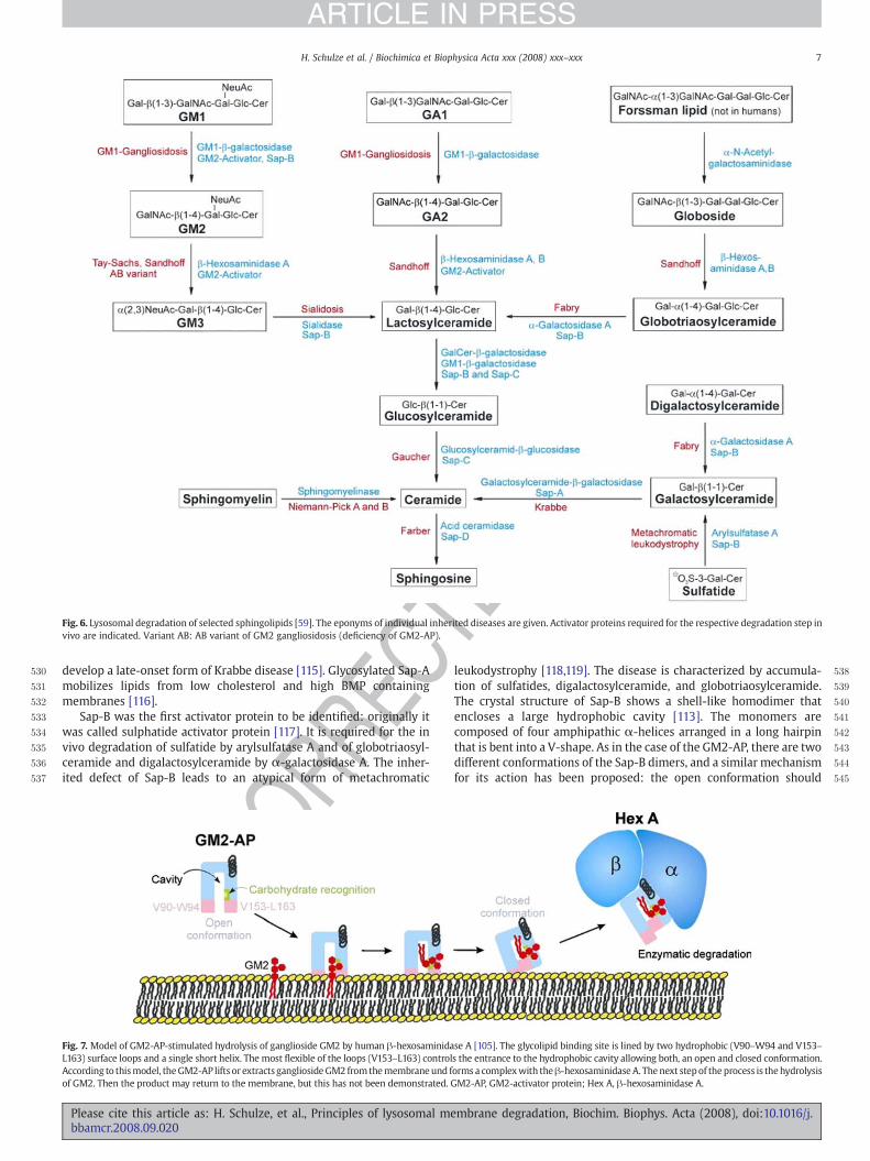
detergents. SAPs mediate the interaction between the membranebound lipid substrate and the water-soluble enzyme, or activate theenzyme directly. The SAPs comprise five small non-enzymaticglycoproteins, encoded by two genes. One gene codes for the precursorof the GM2 activator protein (GM2-AP) [97]. The second gene codes forprosaposin, which is proteolytically processed to four highly homo-logous proteins, the Saps or saposins A–D [98]. These five activatorproteins (GM2-AP and the Saps A–D) act on the surface of the intra-endosomal/intra-lysosomal membrane vesicles, making the lipidsubstrate accessible to the degrading enzyme. The GM2-AP acts as anessential cofactor in the in vivo degradation of ganglioside GM2 by β-hexosaminidase A [99]. Inherited deficiencyof theGM2-AP leads to theAB variant of GM2 gangliosidosis. The GM2-AP is a glycoprotein with17.6 kDa in its deglycosylated form, it bears a N-glycosidically boundoligosaccharide chain and contains four disulfide bridges [100,101].The X-ray crystallographic structure of non-glycosylated GM2-AP,expressed in Escherichia coli allowed an understanding of the role ofthis protein on membrane degradation [102–104]. The GM2-APcontains a hydrophobic cavity that harbors the ceramide moiety ofganglioside GM2. Extraction of the GSL out of the intra-lysosomalmembrane is followed by a conformational change of the lipid loadedactivator, which increases the water solubility of the complex [105](Fig. 7). The major determinant for the interaction with the enzymethat degrades GM2 has been identified as a single short α-helix ofGM2-AP [106]. The GM2-AP can be regarded as a weak detergent withhigh selectivity that inserts into the bilayer of intra-lysosomal lipidvesicles and lifts gangliosides like GM2 andGM1 and other lipids out ofthemembrane, so that they become accessible for the active site of thedegrading enzyme [96]. As a “liftase” [5] the GM2-AP formsstoichiometric, water-soluble glycolipid-protein complexes that arephysiological Michaelis–Menten substrates of β-Hexosaminidase A[99,106]. Physico-chemicalmeasurementswith lipidmonolayers showthat the GM2-AP can insert only if the lateral pressure is below 15–25mNm−1 [107]. It can be assumed that theGM2-AP cannot insert intothe lysosomal perimetermembrane due to its higher `lateral pressure´.The `lateral pressure´ of biological membranes correlates to a rangebetween30 and35mNm−1 [108]. Althoughnodata on intra-lysosomalvesicles are available, it can be assumed that their `lateral pressure´. isquite low due to their low cholesterol and their high BMP content,enabling the GM2-AP to insert into the intra-lysosomal vesicles, butnot into the perimeter membrane.
The Saps A–D are four acidic, not enzymatically active, heat-stableand protease-resistant glycoproteins of about 8–11 kDa [51]. They aremembers of the saposin-like protein family (SAPLIPs), which com-prises proteins and protein domains of several species and differentfunction, all sharing a lipid-binding and membrane perturbingproperties [109]. The first SAPLIP for which structure determinationhas been successful, was the elucidation of NK-lysin by nuclearmagnetic resonance spectroscopy [110]. Up to now, several otherSAPLIP structures have been determined, among them the solutionstructure of Sap-C [111] and the X-ray crystallographic structure ofunglycosylated human recombinant Sap-A [112], Sap-C [113], Sap-B[113], and Sap-D [114]. The proteins show high structural similarity.They are characterized by the presence of three intradomain disulfidelinkages and several hydrophobic residues that form a commonstructural framework. Despite their high degree of homology, the Sapsdiffer in specificity. The absence of different Saps leads to differentsphingolipidoses. Although all Saps are able to mobilize lipids frommembranes, this indicates that additional Sap-specific properties arerequired. The physiological degradation of sphingomyelin does notrequire the presence of an activator protein, since acid sphingomye-linase contains a protein domain with homology to the Saps [57].
The in vivo-degradation of galactosylceramide by galactosylcer-amide-β-galactosidase requires in vivo the presence of Sap-A. Micecarrying a mutation in the Sap-A domain of the Sap-precursor andtherefore lack mature Sap-A accumulate galactosylceramide and
embrane degradation, Biochim. Biophys. Acta (2008), doi:10.1016/j.
EDPR
OOF
530
531
532
533
534
535
536
537
538
539
540
541
542
543
544
545
Fig. 6. Lysosomal degradation of selected sphingolipids [59]. The eponyms of individual inherited diseases are given. Activator proteins required for the respective degradation step invivo are indicated. Variant AB: AB variant of GM2 gangliosidosis (deficiency of GM2-AP).
7H. Schulze et al. / Biochimica et Biophysica Acta xxx (2008) xxx–xxx
ARTICLE IN PRESS
RECdevelop a late-onset form of Krabbe disease [115]. Glycosylated Sap-A
mobilizes lipids from low cholesterol and high BMP containingmembranes [116].
Sap-B was the first activator protein to be identified: originally itwas called sulphatide activator protein [117]. It is required for the invivo degradation of sulfatide by arylsulfatase A and of globotriaosyl-ceramide and digalactosylceramide by α-galactosidase A. The inher-ited defect of Sap-B leads to an atypical form of metachromatic
UNCO
R
Fig. 7. Model of GM2-AP-stimulated hydrolysis of ganglioside GM2 by human β-hexosaminidaL163) surface loops and a single short helix. The most flexible of the loops (V153–L163) controlAccording to thismodel, theGM2-AP lifts or extracts gangliosideGM2 fromthemembraneund fof GM2. Then the product may return to the membrane, but this has not been demonstrated. G
Please cite this article as: H. Schulze, et al., Principles of lysosomal mebbamcr.2008.09.020
T
leukodystrophy [118,119]. The disease is characterized by accumula-tion of sulfatides, digalactosylceramide, and globotriaosylceramide.The crystal structure of Sap-B shows a shell-like homodimer thatencloses a large hydrophobic cavity [113]. The monomers arecomposed of four amphipathic α-helices arranged in a long hairpinthat is bent into a V-shape. As in the case of the GM2-AP, there are twodifferent conformations of the Sap-B dimers, and a similar mechanismfor its action has been proposed: the open conformation should
se A [105]. The glycolipid binding site is lined by two hydrophobic (V90–W94 and V153–s the entrance to the hydrophobic cavity allowing both, an open and closed conformation.orms a complexwith theβ-hexosaminidase A. Thenext stepof theprocess is thehydrolysisM2-AP, GM2-activator protein; Hex A, β-hexosaminidase A.
mbrane degradation, Biochim. Biophys. Acta (2008), doi:10.1016/j.

C
546
547
548
549
550
551
552
553
554
555
556
557
558
559
560
561
562
563
564
565
566
567
568
569
570
571
572
573
574
575
576
577
578
579
580
581
582
583
584
585
586
587
588
589
590
591
592
593
594
595
596
597
598
599
600
601
602
603
604
605
606
607
608
609610611612613614615616617618619620621622623624625626627628629630631632633634635636637638639640641642643644645646647648649650651652653654655656657658659660661662663664665666667668669670671672673674675676677678679680681682683684685686687688689690691692
8 H. Schulze et al. / Biochimica et Biophysica Acta xxx (2008) xxx–xxx
ARTICLE IN PRESS
UNCO
RRE
interact directly with the membrane, promote a reorganization of thelipid alkyl chains, and extract the lipid substrate accompanied by achange to the closed conformation, so that the substrate can beexposed to the enzyme in a water-soluble activator lipid complex[120]. This is consistent with the observation that, after initial binding,glycosylated Sap-B disintegrates the membrane structure and solubi-lizes lipids from it [121].
Sap-C is required for the lysosomal degradation of glucosylcer-amide by glucosylceramide-β-glucosidase. The 20 kDa homodimericprotein was first isolated from the spleens of patients with Gaucherdisease [122]. In contrast to Sap-B, Sap-C binds not only to lipids andmembranes, but also interacts with glucosylceramide-β-glucosidaseand stimulates the enzyme directly [123,124]. The β-glucosidase is awater soluble lysosomal enzyme that can associate to membranes.Sap-C also supports the interaction of the enzyme with the substrateembedded in vesicles containing anionic phospholipids. In addition,Sap-C is able to destabilize these vesicles [125]. Binding of Sap-C tophospholipid vesicles is a pH-controlled, reversible process [126]. Thedeficiency of Sap-C leads to an abnormal juvenile form of Gaucherdisease and an accumulation of glucosylceramide [127,128]. Feeding ofpurified Sap-C to patients fibroblasts reduces the level of glucosylcer-amide storage, whereas Sap-A, -B and -D were not effective [129].Additionally, Sap-C renders glucosylceramide-β-glucosidase moreprotease-resistant inside the cell [130]. The solution structure ofSap-C [111] consists of five tightly packed α-helices that form half of asphere. All charged amino acids are solvent-exposed, whereas thehydrophobic residues are contained within the protein core. Sap-Clocally alters regions of lipid bilayer for subsequent attack by acid β-glucosidase [131].
Sap-D stimulates lysosomal ceramide degradation by acid cerami-dase in cultured cells [129] and in vitro [132]. The detailedphysiological function and mode of action of Sap-D is unclear. It isable to bind to vesicles containing negatively charged lipids and tosolubilize them at an appropriate pH [133]. Sap-D-deficient miceaccumulate ceramides with hydroxylated fatty acids mainly in thebrain and in the kidney [134]. In addition to their function as enzymecofactors, SAPs play an important role in the presentation of lipid andglycolipid antigens. CD1 immunoreceptors present lipid antigens to Tcells. Therefore, the lipid antigens have to be removed from themembranes in which they are embedded to allow loading on CD1molecules.
Antigen presentation by human CD1b [135], and human [136], andmouse CD1d [137] have been studied. Human CD1b especiallyrequires Sap-C to present different types of glycolipid antigens. Invitro, all saposins can exchange phosphatidylserine bound to murineCD1d against glycosphingolipids, but with different activity.
3. Perspective
Cellular membranes are highly dynamic structures. Eukaryoticcells maintain their morphology, endomembrane homeostasis, andcomposition of the intracellular organelles despite continuous inwardand outward membrane flow. The role of endosomes and lysosomesfor lipid homeostasis can be illustrated by one example: Impairmentof cholesterol-efflux out of the lysosomes in NPC1-diseases causes notonly lysosomal accumulation of cholesterol in neuronal cell bodies,but cholesterol-depletion in the axons of neuronal cells, inhibitingaxonal growth [138]. The molecular mechanisms underlying theseprocesses are not completely understood [139]. Control of membranehomeostasis remains an open field.
Acknowledgments
Work in the laboratory of the authors was supported by theDeutsche Forschungsgemeinschaft and the European Union(LipidomicNet).
Please cite this article as: H. Schulze, et al., Principles of lysosomal mbbamcr.2008.09.020
TEDPR
OOF
References
[1] A. Vellodi, Lysosomal storage disorders, Br. J. Haematol. 128 (2005) 413–431.[2] E.M. Strehle, Sialic acid storage disease and related disorders, Genet. Test 7
(2003) 113–121.[3] B.K. Gillard, R.G. Clement, D.M. Marcus, Variations among cell lines in the
synthesis of sphingolipids in de novo and recycling pathways, Glycobiology 8(1998) 885–890.
[4] S. Mayor, R.E. Pagano, Pathways of clathrin-independent endocytosis, Nat. Rev.Mol. Cell. Biol. 8 (2007) 603–612.
[5] W. Fürst, K. Sandhoff, Activator proteins and topology of lysosomal sphingolipidcatabolism, Biochim. Biophys. Acta 1126 (1992) 1–16.
[6] W. Möbius, V. Herzog, K. Sandhoff, G. Schwarzmann, Gangliosides aretransported from the plasma membrane to intralysosomal membranes asrevealed by immuno-electron microscopy, Biosci. Rep. 19 (1999) 307–316.
[7] M. Grabe, G. Oster, Regulation of organelle acidity, J. Gen. Physiol. 117 (2001)329–344.
[8] B. Sonnichsen, S. De Renzis, E. Nielsen, J. Rietdorf, M. Zerial, Distinct membranedomains on endosomes in the recycling pathway visualized by multicolorimaging of Rab4, Rab5, and Rab11, J. Cell Biol. 149 (2000) 901–914.
[9] M. Kundu, C.B. Thompson, Autophagy: basic principles and relevance to disease,Annu. Rev. Pathol. 3 (2008) 427–455.
[10] T. Yoshimori, Autophagy: paying Charon's toll, Cell 128 (2007) 833–836.[11] U.D. Epple, I. Suriapranata, E.L. Eskelinen, M. Thumm, Aut5/Cvt17p, a putative
lipase essential for disintegration of autophagic bodies inside the vacuole,J. Bacteriol. 183 (2001) 5942–5955.
[12] N. Nakamura, A. Matsuura, Y. Wada, Y. Ohsumi, Acidification of vacuoles isrequired for autophagic degradation in the yeast, Saccharomyces cerevisiae,J. Biochem. 121 (1997) 338–344.
[13] K. Takeshige, M. Baba, S. Tsuboi, T. Noda, Y. Ohsumi, Autophagy in yeastdemonstrated with proteinase-deficient mutants and conditions for its induc-tion, J. Cell Biol. 119 (1992) 301–311.
[14] D.J. Klionsky, The molecular machinery of autophagy: unanswered questions,J. Cell. Sci. 118 (2005) 7–18.
[15] I. Suriapranata, U.D. Epple, D. Bernreuther, M. Bredschneider, K. Sovarasteanu, M.Thumm, The breakdown of autophagic vesicles inside the vacuole depends onAut4p, J. Cell. Sci. 113 (Pt) (2000) 4025–4033.
[16] W.Möbius, E. van Donselaar, Y. Ohno-Iwashita, Y. Shimada, H.F. Heijnen, J.W. Slot,H.J. Geuze, Recycling compartments and the internal vesicles of multivesicularbodies harbor most of the cholesterol found in the endocytic pathway, Traffic 4(2003) 222–231.
[17] B. Amidon, A. Brown, M. Waite, Transacylase and phospholipases in thesynthesis of bis(monoacylglycero)phosphate, Biochemistry 35 (1996)13995–14002.
[18] J. Brotherus, O. Renkonen, Subcellular distributions of lipids in cultured BHKcells: evidence for the enrichment of lysobisphosphatidic acid and neutral lipidsin lysosomes, J. Lipid Res. 18 (1977) 191–202.
[19] R.C. Piper, J.P. Luzio, Ubiquitin-dependent sorting of integral membrane proteinsfor degradation in lysosomes, Curr. Opin. Cell Biol. 19 (2007) 459–465.
[20] S. Saksena, J. Sun, T. Chu, S.D. Emr, ESCRTing proteins in the endocytic pathway,Trends Biochem. Sci. 32 (2007) 561–573.
[21] F.G. van der Goot, J. Gruenberg, Intra-endosomal membrane traffic, Trends CellBiol. 16 (2006) 514–521.
[22] C. Peters, K. von Figura, Biogenesis of lysosomal membranes, FEBS Lett. 346(1994) 108–114.
[23] S.R. Carlsson, J. Roth, F. Piller, M. Fukuda, Isolation and characterization ofhuman lysosomal membrane glycoproteins, h-lamp-1 and h-lamp-2. Majorsialoglycoproteins carrying polylactosaminoglycan, J. Biol. Chem. 263 (1988)18911–18919.
[24] K. Suzuki, G.C. Chen, GM1-gangliosidosis (generalized gangliosidosis). Morphol-ogy and chemical pathology, Pathol. Eur. 3 (1968) 389–408.
[25] K. Harzer, B.C. Paton, A. Poulos, B. Kustermann-Kuhn, W. Roggendorf, T. Grisar, M.Popp, Sphingolipid activator protein deficiency in a 16-week-old atypicalGaucher disease patient and his fetal sibling: biochemical signs of combinedsphingolipidoses, Eur. J. Pediatr. 149 (1989) 31–39.
[26] D.J. Gill, H. Teo, J. Sun, O. Perisic, D.B. Veprintsev, Y. Vallis, S.D. Emr, R.L. Williams,Structural studies of phosphoinositide 3-kinase-dependent traffic to multi-vesicular bodies, Biochem. Soc. Symp. (2007) 47–57.
[27] J.K. Burkhardt, S. Huttler, A. Klein, W. Mobius, A. Habermann, G. Griffiths, K.Sandhoff, Accumulation of sphingolipids in SAP-precursor (prosaposin)-deficientfibroblasts occurs as intralysosomal membrane structures and can be completelyreversed by treatment with human SAP-precursor, Eur. J. Cell Biol. 73 (1997)10–18.
[28] K. Umebayashi, The roles of ubiquitin and lipids in protein sorting along theendocytic pathway, Cell Struct. Funct. 28 (2003) 443–453.
[29] von Coburg, A., Untersuchung der Lipidnachbarschaft von derivatisiertem GM1in Modellmembranen und kultivierten Zellen, PhD Thesis, University of Bonn,2004
[30] Y. Matsuzawa, K.Y. Hostetler, Degradation of bis(monoacylglycero)phosphate byan acid phosphodiesterase in rat liver lysosomes, J. Biol. Chem. 254 (1979)5997–6001.
[31] T. Kobayashi, E. Stang, K.S. Fang, P. de Moerloose, R.G. Parton, J. Gruenberg, A lipidassociated with the antiphospholipid syndrome regulates endosome structureand function, Nature 392 (1998) 193–197.
[32] T. Chojnacki, G. Dallner, The biological role of dolichol, Biochem. J. 251 (1988)1–9.
embrane degradation, Biochim. Biophys. Acta (2008), doi:10.1016/j.

693694695696697698699700701702703704705706707708709710711712713714715716717718719720721722723724725726727728729730731732733734735736737738739740741742743744745746747748749750751752753754755756757758759760761762763764765766767768769770771772773774775776777778
779780781782783784785786787788789790791792793794795796797798799800801802803804805806807808809810811812813814815816817818819820821822823824825826827828829830831832833834835836837838839840841842843844845846847848849850851852853854855856857858859860861862863864
9H. Schulze et al. / Biochimica et Biophysica Acta xxx (2008) xxx–xxx
ARTICLE IN PRESS
UNCO
RREC
[33] Megha, E. London, Ceramide selectively displaces cholesterol from ordered lipiddomains (rafts): implications for lipid raft structure and function, J. Biol. Chem.279 (2004) 9997–10004.
[34] N. Okino, X. He, S. Gatt, K. Sandhoff, M. Ito, E.H. Schuchman, The reverse activityof human acid ceramidase, J. Biol. Chem. 278 (2003) 29948–29953.
[35] T. Kolter, F. Winau, U.E. Schaible, M. Leippe, K. Sandhoff, Lipid-binding proteins inmembrane digestion, antigen presentation, and antimicrobial defense, J. Biol.Chem. 280 (2005) 41125–41128.
[36] D. Yan, V.M. Olkkonen, Characteristics of oxysterol binding proteins, Int. Rev.Cytol. 265 (2008) 253–285.
[37] J.O. Babalola, M. Wendeler, B. Breiden, C. Arenz, G. Schwarzmann, S. Locatelli-Hoops, K. Sandhoff, Development of an assay for the intermembrane transfer ofcholesterol by Niemann–Pick C2 protein, Biol. Chem. 388 (2007) 617–626.
[38] S. Xu, B. Benoff, H.L. Liou, P. Lobel, A.M. Stock, Structural basis of sterol binding byNPC2, a lysosomal protein deficient in Niemann–Pick type C2 disease, J. Biol.Chem. 282 (2007) 23525–23531.
[39] S.R. Cheruku, Z. Xu, R. Dutia, P. Lobel, J. Storch, Mechanism of cholesterol transferfrom the Niemann–Pick type C2 protein to model membranes supports a role inlysosomal cholesterol transport, J. Biol. Chem. 281 (2006) 31594–31604.
[40] R.E. Infante, A. Radhakrishnan, L. Abi-Mosleh, L.N. Kinch, M.L. Wang, N.V. Grishin,J.L. Goldstein, M.S. Brown, Purified NPC1 protein: II. Localization of sterol bindingto a 240-amino acid soluble luminal loop, J. Biol. Chem. 283 (2008) 1064–1075.
[41] R.E. Infante, L. Abi-Mosleh, A. Radhakrishnan, J.D. Dale, M.S. Brown, J.L. Goldstein,Purified NPC1 protein. I. Binding of cholesterol and oxysterols to a 1278-aminoacid membrane protein, J. Biol. Chem. 283 (2008) 1052–1063.
[42] R.E. Infante, M.L. Wang, A. Radhakrishnan, H.J. Kwon, M.S. Brown, J.L. Goldstein,NPC2 facilitates bidirectional transfer of cholesterol between NPC1 and lipidbilayers, a step in cholesterol egress from lysosomes, Proc. Natl. Acad. Sci. U. S. A.(2008).
[43] X. Sun, D.L. Marks, W.D. Park, C.L. Wheatley, V. Puri, J.F. O, D.L. Kraft, P.A.Lundquist, M.C. Patterson, R.E. Pagano, K. Snow, Niemann–Pick C variantdetection by altered sphingolipid trafficking and correlation with mutationswithin a specific domain of NPC1, Am. J. Hum. Genet. 68 (2001) 1361–1372.
[44] K. Brix, Lysosomal proteases: revival of the Sleeping Beauty, In: P. Saftig (Ed.),Lysosomes, Eurekah.com, Landes Bioscience, 2005.
[45] H. Galjaard, R. Willemsen, A.T. Hoogeveen, G.M. Mancini, S. Palmeri, F.W.Verheijen, A. D, Molecular heterogeneity in human beta-galactosidase andneuraminidase deficiency, Enzyme 38 (1987) 132–143.
[46] A. d 'Azzo, A. Tessitore, R. Sano, Gangliosides as apoptotic signals in ER stressresponse, Cell Death Differ. 13 (2006) 404–414.
[47] J.Y. Lu, S.L. Hofmann, Thematic review series: lipid posttranslational modifica-tions. Lysosomal metabolism of lipid-modified proteins, J. Lipid Res. 47 (2006)1352–1357.
[48] M. Hiraoka, A. Abe, Y. Lu, K. Yang, X. Han, R.W. Gross, J.A. Shayman, Lysosomalphospholipase A2 and phospholipidosis, Mol. Cell. Biol. 26 (2006) 6139–6148.
[49] A. Abe, M. Hiraoka, S. Wild, S.E. Wilcoxen, R. Paine 3rd, J.A. Shayman, Lysosomalphospholipase A2 is selectively expressed in alveolar macrophages, J. Biol. Chem.279 (2004) 42605–42611.
[50] A. Abe, M. Hiraoka, J.A. Shayman, The acylation of lipophilic alcohols bylysosomal phospholipase A2, J. Lipid Res. 48 (2007) 2255–2263.
[51] T. Kolter, K. Sandhoff, Principles of lysosomal membrane digestion: stimulation ofsphingolipid degradation by sphingolipid activator proteins and anioniclysosomal lipids, Annu. Rev. Cell Dev. Biol. 21 (2005) 81–103.
[52] N. Anderson, J. Borlak, Drug-induced phospholipidosis, FEBS Lett. 580 (2006)5533–5540.
[53] H. Lullmann, R. Lullmann-Rauch, O. Wassermann, Lipidosis induced byamphiphilic cationic drugs, Biochem. Pharmacol. 27 (1978) 1103–1108.
[54] M. Kölzer, N. Werth, K. Sandhoff, Interactions of acid sphingomyelinase and lipidbilayers in the presence of the tricyclic antidepressant desipramine, FEBS Lett.559 (2004) 96–98.
[55] R. Hurwitz, K. Ferlinz, K. Sandhoff, The tricyclic antidepressant desipraminecauses proteolytic degradation of lysosomal sphingomyelinase in humanfibroblasts, Biol. Chem. Hoppe-Seyler 375 (1994) 447–450.
[56] S. Elojeimy, D.H. Holman, X. Liu, A. El-Zawahry, M. Villani, J.C. Cheng, A. Mahdy, Y.Zeidan, A. Bielawska, Y.A. Hannun, J.S. Norris, New insights on the use ofdesipramine as an inhibitor for acid ceramidase, FEBS Lett. 580 (2006) 4751–4756.
[57] M. Kölzer, K. Ferlinz, O. Bartelsen, S.L. Hoops, F. Lang, K. Sandhoff, Functionalcharacterization of the postulated intramolecular sphingolipid activator proteindomain of human acid sphingomyelinase, Biol. Chem. 385 (2004) 1193–1195.
[58] J.A. Handler, A. Badger, C.A. Genell, A.M. Klinkner, S. Kassis, C.R. Waites, P.J.Bugelski, Selective inhibition of phospholipases by atiprimod, a macrophagetargeting antiarthritic compound, Toxicol. Appl. Pharmacol. 159 (1999) 9–17.
[59] T. Kolter, K. Sandhoff, Sphingolipid metabolism diseases, Biochim. Biophys. Acta1758 (2006) 2057–2079.
[60] T. Kolter, R.L. Proia, K. Sandhoff, Combinatorial ganglioside biosynthesis, J. Biol.Chem. 277 (2002) 25859–25862.
[61] S. Hakomori, Glycosphingolipids in cellular interaction, differentiation, andoncogenesis, Annu. Rev. Biochem. 50 (1981) 733–764.
[62] D.A. Brown, E. London, Structure and function of sphingolipid- and cholesterol-rich membrane rafts, J. Biol. Chem. 275 (2000) 17221–17224.
[63] K. Simons, E. Ikonen, Functional rafts in cell membranes, Nature 387 (1997)569–572.
[64] J.F. Hancock, Lipid rafts: contentious only from simplistic standpoints, Nat. Rev.Mol. Cell. Biol. 7 (2006) 456–462.
[65] C. Eggeling, C. Ringemann, R. Medda, G. Schwarzmann, K. Sandhoff, C. vonMiddendorf, A. Schönle and S. W. Hell, Nanoscaledynamics of single lipid
Please cite this article as: H. Schulze, et al., Principles of lysosomal mebbamcr.2008.09.020
TEDPR
OOF
molecules in living cells reveal cholesterol assisted sphingolipid microdomainsin membranes, Nature (submitted for publication)
[66] H. Heerklotz, H. Szadkowska, T. Anderson, J. Seelig, The sensitivity of lipiddomains to small perturbations demonstrated by the effect of Triton, J. Mol. Biol.329 (2003) 793–799.
[67] S. Munro, Lipid rafts: elusive or illusive? Cell 115 (2003) 377–388.[68] D. Lichtenberg, F.M. Goni, H. Heerklotz, Detergent-resistant membranes should
not be identified with membrane rafts, Trends Biochem. Sci. 30 (2005)430–436.
[69] E. Eliyahu, J.H. Park, N. Shtraizent, X. He, E.H. Schuchman, Acid ceramidase is anovel factor required for early embryo survival, FASEB J. 21 (2007) 1403–1409.
[70] S. Hakomori, Structure and function of sphingoglycolipids in transmembranesignalling and cell–cell interactions, Biochem. Soc. Trans. 21 (Pt. 3) (1993)583–595.
[71] T. Yamashita, A. Hashiramoto, M. Haluzik, H. Mizukami, S. Beck, A. Norton, M.Kono, S. Tsuji, J.L. Daniotti, N. Werth, R. Sandhoff, K. Sandhoff, R.L. Proia,Enhanced insulin sensitivity in mice lacking ganglioside GM3, Proc. Natl. Acad.Sci. U. S. A. 100 (2003) 3445–3449.
[72] T. Döring, H. Brade, K. Sandhoff, Sphingolipid metabolism during epidermalbarrier development in mice, J. Lipid Res. 43 (2002) 1727–1733.
[73] Y. Uchida, W.M. Holleran, Omega-O-acylceramide, a lipid essential for mamma-lian survival, J. Dermatol. Sci. 51 (2008) 77–87.
[74] M. Rabionet, A.C. van der Spoel, C.C. Chuang, B. von Tumpling-Radosta, M. Litjens,D. Bouwmeester, C.C. Hellbusch, C. Korner, H. Wiegandt, K. Gorgas, F.M. Platt, H.J.Grone, R. Sandhoff, Male germ cells require polyenoic sphingolipids withcomplex glycosylation for completion of meiosis: a link to ceramide synthase-3,J. Biol. Chem. 283 (2008) 13357–13369.
[75] K.Hanada, Sphingolipids in infectiousdiseases, Jpn. J. Infect. Dis. 58 (2005) 131–148.[76] B. Schwerer, Antibodies against gangliosides: a link between preceding infection
and immunopathogenesis of Guillain–Barre syndrome, Microbes. Infect. 4 (2002)373–384.
[77] N. Yuki, Infectious origins of, and molecular mimicry in, Guillain–Barre andFisher syndromes, Lancet. Infect. Dis. 1 (2001) 29–37.
[78] J.E. Park, D.Y. Wu, M. Prendes, S.X. Lu, G. Ragupathi, N. Schrantz, P.B. Chapman,Fine specificity of natural killer T cells against GD3 ganglioside and identificationof GM3 as an inhibitory natural killer T-cell ligand, Immunology 123 (2008)145–155.
[79] E.C. Mandon, I. Ehses, J. Rother, G. van Echten, K. Sandhoff, Subcellularlocalization and membrane topology of serine palmitoyltransferase, 3-dehydro-sphinganine reductase, and sphinganine N-acyltransferase in mouse liver, J. Biol.Chem. 267 (1992) 11144–11148.
[80] K. Hanada, K. Kumagai, S. Yasuda, Y. Miura, M. Kawano, M. Fukasawa, M.Nishijima, Molecular machinery for non-vesicular trafficking of ceramide, Nature426 (2003) 803–809.
[81] D. Jeckel, A. Karrenbauer, K.N. Burger, G. van Meer, F. Wieland, Glucosylceramideis synthesized at the cytosolic surface of various Golgi subfractions, J. Cell Biol.117 (1992) 259–267.
[82] A.H. Futerman, R.E. Pagano, Determination of the intracellular sites andtopology of glucosylceramide synthesis in rat liver, Biochem. J. 280 (Pt. 2)(1991) 295–302.
[83] G. D 'Angelo, E. Polishchuk, G. Di Tullio, M. Santoro, A. Di Campli, A. Godi, G. West,J. Bielawski, C.C. Chuang, A.C. van der Spoel, F.M. Platt, Y.A. Hannun, R.Polishchuk, P. Mattjus, M.A. De Matteis, Glycosphingolipid synthesis requiresFAPP2 transfer of glucosylceramide, Nature 449 (2007) 62–67.
[84] G. van Echten, H. Iber, H. Stotz, A. Takatsuki, K. Sandhoff, Uncoupling ofganglioside biosynthesis by Brefeldin A, Eur. J. Cell Biol. 51 (1990) 135–139.
[85] P.D. Eckford, F.J. Sharom, The reconstituted P-glycoprotein multidrug transporteris a flippase for glucosylceramide and other simple glycosphingolipids, Biochem.J. 389 (2005) 517–526.
[86] M. Akiyama, Y. Sugiyama-Nakagiri, K. Sakai, J.R. McMillan, M. Goto, K. Arita, Y.Tsuji-Abe, N. Tabata, K. Matsuoka, R. Sasaki, D. Sawamura, H. Shimizu, Mutationsin lipid transporter ABCA12 in harlequin ichthyosis and functional recovery bycorrective gene transfer, J. Clin. Invest. 115 (2005) 1777–1784.
[87] C. Lefevre, S. Audebert, F. Jobard, B. Bouadjar, H. Lakhdar, O. Boughdene-Stambouli, C. Blanchet-Bardon, R. Heilig, M. Foglio, J. Weissenbach, M. Lathrop, J.F. Prud, J. Fischer, Mutations in the transporter ABCA12 are associated withlamellar ichthyosis type 2, Hum. Mol. Genet. 12 (2003) 2369–2378.
[88] D.P. Kelsell, E.E. Norgett, H. Unsworth, M.T. Teh, T. Cullup, C.A. Mein, P.J. Dopping-Hepenstal, B.A. Dale, G. Tadini, P. Fleckman, K.G. Stephens, V.P. Sybert, S.B.Mallory, B.V. North, D.R. Witt, E. Sprecher, A.E. Taylor, A. Ilchyshyn, C.T. Kennedy,H. Goodyear, C. Moss, D. Paige, J.I. Harper, B.D. Young, I.M. Leigh, R.A. Eady, E.A. O,Mutations in ABCA12 underlie the severe congenital skin disease harlequinichthyosis, Am. J. Hum. Genet. 76 (2005) 794–803.
[89] D. Halter, S. Neumann, S.M. van Dijk, J. Wolthoorn, A.M. de Maziere, O.V. Vieira, P.Mattjus, J. Klumperman, G. van Meer, H. Sprong, Pre- and post-Golgi transloca-tion of glucosylceramide in glycosphingolipid synthesis, J. Cell Biol. 179 (2007)101–115.
[90] G.H. Patterson, K. Hirschberg, R.S. Polishchuk, D. Gerlich, R.D. Phair, J. Lippincott-Schwartz, Transport through the Golgi apparatus by rapid partitioning within atwo-phase membrane system, Cell 133 (2008) 1055–1067.
[91] G. Pohlentz, D. Klein, G. Schwarzmann, D. Schmitz, K. Sandhoff, Both GA2,GM2, and GD2 synthases and GM1b, GD1a, and GT1b synthases are singleenzymes in Golgi vesicles from rat liver, Proc. Natl. Acad. Sci. U. S. A. 85 (1988)7044–7048.
[92] F.R. Maxfield, T.E. McGraw, Endocytic recycling, Nat. Rev. Mol. Cell Biol. 5 (2004)121–132.
mbrane degradation, Biochim. Biophys. Acta (2008), doi:10.1016/j.

C
865866867868869870871872873874875876877878879880881882883884885886887888889890891892893894895896897898899900901902903904905906907908909910911912913914915916917918919920921922923924925926927928929930931932933934935936937
93893994094194294394494594694794894995095195295395495595695795895996096196296396496596696796896997097197297397497597697797897998098198298398498598698798898999099199299399499599699799899910001001100210031004100510061007100810091010
1012
10 H. Schulze et al. / Biochimica et Biophysica Acta xxx (2008) xxx–xxx
ARTICLE IN PRESS
NCOR
RE
[93] K. Sandhoff, T. Kolter, Topology of glycosphingolipid degradation, Trends CellBiol. 6 (1996) 98–103.
[94] W.Mobius, V. Herzog, K. Sandhoff, G. Schwarzmann, Intracellular distribution of abiotin-labeled ganglioside, GM1, by immunoelectron microscopy after endocy-tosis in fibroblasts, J. Histochem. Cytochem. 47 (1999) 1005–1014.
[95] W.Mobius, E. van Donselaar, Y. Ohno-Iwashita, Y. Shimada, H.F. Heijnen, J.W. Slot,H.J. Geuze, Recycling compartments and the internal vesicles of multivesicularbodies harbor most of the cholesterol found in the endocytic pathway, Traffic 4(2003) 222–231.
[96] G. Wilkening, T. Linke, G. Uhlhorn-Dierks, K. Sandhoff, Degradation ofmembrane-bound ganglioside GM1. Stimulation by bis(monoacylglycero)phos-phate and the activator proteins SAP-B and GM2-AP, J. Biol. Chem. 275 (2000)35814–35819.
[97] H. Klima, A. Klein, G. van Echten, G. Schwarzmann, K. Suzuki, K. Sandhoff, Over-expression of a functionally active human GM2-activator protein in Escherichiacoli, Biochem. J. 292 (Pt 2) (1993) 571–576.
[98] G. Vielhaber, R. Hurwitz, K. Sandhoff, Biosynthesis, processing, and targeting ofsphingolipid activator protein (SAP)precursor in cultured human fibroblasts.Mannose 6-phosphate receptor-independent endocytosis of SAP precursor,J. Biol. Chem. 271 (1996) 32438–32446.
[99] E. Conzelmann, K. Sandhoff, Purification and characterization of an activatorprotein for the degradation of glycolipids GM2 and GA2 by hexosaminidase A,Hoppe-Seyler Z. Physiol. Chem. 360 (1979) 1837–1849.
[100] W. Fürst, J. Schubert, W. Machleidt, H.E. Meyer, K. Sandhoff, The complete amino-acid sequences of human ganglioside GM2 activator protein and cerebrosidesulfate activator protein, Eur. J. Biochem. 192 (1990) 709–714.
[101] C.G. Schutte, T. Lemm, G.J. Glombitza, K. Sandhoff, Complete localization ofdisulfide bonds in GM2 activator protein, Protein Sci. 7 (1998) 1039–1045.
[102] C.S. Wright, S.C. Li, F. Rastinejad, Crystal structure of human GM2-activatorprotein with a novel beta-cup topology, J. Mol. Biol. 304 (2000) 411–422.
[103] C.S. Wright, Q. Zhao, F. Rastinejad, Structural analysis of lipid complexes of GM2-activator protein, J. Mol. Biol. 331 (2003) 951–964.
[104] C.S. Wright, L.Z. Mi, S. Lee, F. Rastinejad, Crystal structure analysis ofphosphatidylcholine-GM2-activator product complexes: evidence for hydrolaseactivity, Biochemistry 44 (2005) 13510–13521.
[105] M. Wendeler, J. Hoernschemeyer, D. Hoffmann, T. Kolter, G. Schwarzmann, K.Sandhoff, Photoaffinity labelling of the human GM2-activator protein. Mechan-istic insight into ganglioside GM2 degradation, Eur. J. Biochem. 271 (2004)614–627.
[106] M. Wendeler, N. Werth, T. Maier, G. Schwarzmann, T. Kolter, M. Schoeniger, D.Hoffmann, T. Lemm, W. Saenger, K. Sandhoff, The enzyme-binding region ofhuman GM2-activator protein, FEBS J. 273 (2006) 982–991.
[107] A. Giehl, T. Lemm, O. Bartelsen, K. Sandhoff, A. Blume, Interaction of the GM2-activator protein with phospholipid–ganglioside bilayer membranes and withmonolayers at the air–water interface, Eur. J. Biochem. 261 (1999) 650–658.
[108] D. Marsh, Lateral pressure in membranes, Biochim. Biophys. Acta 1286 (1996)183–223.
[109] R.S. Munford, P.O. Sheppard, P.J. O, Saposin-like proteins (SAPLIP) carry outdiverse functions on a common backbone structure, J. Lipid Res. 36 (1995)1653–1663.
[110] E. Liepinsh, M. Andersson, J.M. Ruysschaert, G. Otting, Saposin fold revealed bythe NMR structure of NK-lysin, Nat. Struct. Biol. 4 (1997) 793–795.
[111] E. de Alba, S. Weiler, N. Tjandra, Solution structure of human saposin C: pH-dependent interaction with phospholipid vesicles, Biochemistry 42 (2003)14729–14740.
[112] V.E. Ahn, P. Leyko, J.R. Alattia, L. Chen, G.G. Prive, Crystal structures of saposins Aand C, Protein Sci. 15 (2006) 1849–1857.
[113] V.E. Ahn, K.F. Faull, J.P. Whitelegge, A.L. Fluharty, G.G. Prive, Crystal structure ofsaposin B reveals a dimeric shell for lipid binding, Proc. Natl. Acad. Sci. U. S. A.100(2003) 38–43.
[114] K. Popovic, G.G. Prive, Structures of the human ceramide activator proteinsaposin D, Acta Crystallogr. D. Biol. Crystallogr. 64 (2008) 589–594.
[115] J. Matsuda, M.T. Vanier, Y. Saito, J. Tohyama, K. Suzuki, A mutation in the saposinA domain of the sphingolipid activator protein (prosaposin) gene results in a late-onset, chronic form of globoid cell leukodystrophy in the mouse, Hum. Mol.Genet. 10 (2001) 1191–1199.
[116] S. Locatelli-Hoops, N. Remmel, R. Klingenstein, B. Breiden, M. Rossocha, M.Schoeniger, C. Koenigs, W. Saenger, K. Sandhoff, Saposin A mobilizes lipids fromlow cholesterol and high bis(monoacylglycerol)phosphate-containing mem-branes: patient variant Saposin A lacks lipid extraction capacity, J. Biol. Chem.281 (2006) 32451–32460.
[117] E. Mehl, H. Jatzkewitz, A cerebrosidesulfatase from swine kidney, Hoppe-SeylersZ. Physiol. Chem. 339 (1964) 260–276.
U 1011Please cite this article as: H. Schulze, et al., Principles of lysosomal mbbamcr.2008.09.020
TEDPR
OOF
[118] K.A. Kretz, G.S. Carson, S. Morimoto, Y. Kishimoto, A.L. Fluharty, J.S. O 'Brien,Characterization of a mutation in a family with saposin B deficiency: aglycosylation site defect, Proc. Natl. Acad. Sci. U. S. A. 87 (1990) 2541–2544.
[119] D. Wrobe, M. Henseler, S. Huettler, S.I. Pascual Pascual, A. Chabas, K. Sandhoff, Anon-glycosylated and functionally deficient mutant (N215H) of the sphingolipidactivator protein B (SAP-B) in a novel case of metachromatic leukodystrophy(MLD), J. Inherit. Metab. Dis. 23 (2000) 63–76.
[120] G. Fischer, H. Jatzkewitz, The activator of cerebroside sulphatase. Binding studieswith enzyme and substrate demonstrating the detergent function of theactivator protein, Biochim. Biophys. Acta 481 (1977) 561–572.
[121] N. Remmel, S. Locatelli-Hoops, B. Breiden, G. Schwarzmann, K. Sandhoff, SaposinB mobilizes lipids from cholesterol-poor and bis(monoacylglycero)phosphate-rich membranes at acidic pH. Unglycosylated patient variant saposin B lackslipid-extraction capacity, FEBS J. 274 (2007) 3405–3420.
[122] M.W. Ho, J.S. O, Gaucher's disease: deficiency of ‘acid’-glucosidase andreconstitution of enzyme activity in vitro, Proc. Natl. Acad. Sci. U. S. A. 68(1971) 2810–2813.
[123] S.L. Berent, N.S. Radin, Mechanism of activation of glucocerebrosidase by co-beta-glucosidase (glucosidase activator protein), Biochim. Biophys. Acta 664(1981) 572–582.
[124] D. Fabbro, G.A. Grabowski, Human acid beta-glucosidase. Use of inhibitory andactivating monoclonal antibodies to investigate the enzyme's catalyticmechanism and saposin A and C binding sites, J. Biol. Chem. 266 (1991)15021–15027.
[125] G. Wilkening, T. Linke, K. Sandhoff, Lysosomal degradation on vesicularmembrane surfaces. Enhanced glucosylceramide degradation by lysosomalanionic lipids and activators, J. Biol. Chem. 273 (1998) 30271–30278.
[126] A.M. Vaccaro, F. Ciaffoni, M. Tatti, R. Salvioli, A. Barca, D. Tognozzi, C. Scerch, pH-dependent conformational properties of saposins and their interactions withphospholipid membranes, J. Biol. Chem. 270 (1995) 30576–30580.
[127] H. Christomanou, A. Aignesberger, R.P. Linke, Immunochemical characterizationof two activator proteins stimulating enzymic sphingomyelin degradation invitro. Absence of one of them in a human Gaucher disease variant, Biol. Chem.Hoppe-Seyler 367 (1986) 879–890.
[128] D. Schnabel, M. Schroder, K. Sandhoff, Mutation in the sphingolipid activatorprotein 2 in a patient with a variant of Gaucher disease, FEBS Lett. 284 (1991)57–59.
[129] A. Klein, M. Henseler, C. Klein, K. Suzuki, K. Harzer, K. Sandhoff, Sphingolipidactivator protein D (sap-D) stimulates the lysosomal degradation of ceramide invivo, Biochem. Biophys. Res. Commun. 200 (1994) 1440–1448.
[130] Y. Sun, X. Qi, G.A. Grabowski, Saposin C is required for normal resistance of acidbeta-glucosidase to proteolytic degradation, J. Biol. Chem. 278 (2003)31918–31923.
[131] J.R. Alattia, J.E. Shaw, C.M. Yip, G.G. Prive, Molecular imaging of membraneinterfaces reveals mode of beta-glucosidase activation by saposin C, Proc. Natl.Acad. Sci. U. S. A. 104 (2007) 17394–17399.
[132] T. Linke, G. Wilkening, F. Sadeghlar, H. Mozcall, K. Bernardo, E. Schuchman, K.Sandhoff, Interfacial regulation of acid ceramidase activity. Stimulation ofceramide degradation by lysosomal lipids and sphingolipid activator proteins,J. Biol. Chem. 276 (2001) 5760–5768.
[133] F. Ciaffoni, R. Salvioli, M. Tatti, G. Arancia, P. Crateri, A.M. Vaccaro, Saposin Dsolubilizes anionic phospholipid-containing membranes, J. Biol. Chem. 276(2001) 31583–31589.
[134] J. Matsuda, M. Kido, K. Tadano-Aritomi, I. Ishizuka, K. Tominaga, K. Toida, E.Takeda, K. Suzuki, Y. Kuroda, Mutation in saposin D domain of sphingolipidactivator protein gene causes urinary system defects and cerebellar Purkinje celldegeneration with accumulation of hydroxy fatty acid-containing ceramide inmouse, Hum. Mol. Genet. 13 (2004) 2709–2723.
[135] F. Winau, V. Schwierzeck, R. Hurwitz, N. Remmel, P.A. Sieling, R.L. Modlin, S.A.Porcelli, V. Brinkmann, M. Sugita, K. Sandhoff, S.H. Kaufmann, U.E. Schaible,Saposin C is required for lipid presentation by human CD1b, Nat. Immunol. 5(2004) 169–174.
[136] S.J. Kang, P. Cresswell, Saposins facilitate CD1d-restricted presentation of anexogenous lipid antigen to T cells, Nat. Immunol. 5 (2004) 175–181.
[137] D. Zhou, C. Cantu 3rd, Y. Sagiv, N. Schrantz, A.B. Kulkarni, X. Qi, D.J. Mahuran, C.R.Morales, G.A. Grabowski, K. Benlagha, P. Savage, A. Bendelac, L. Teyton, Editing ofCD1d-bound lipid antigens by endosomal lipid transfer proteins, Science 303(2004) 523–527.
[138] B. Karten, D.E. Vance, R.B. Campenot, J.E. Vance, Trafficking of cholesterol fromcell bodies to distal axons in Niemann Pick C1-deficient neurons, J. Biol. Chem.278 (2003) 4168–4175.
[139] I. Mellman, G.Warren, The road taken: past and future foundations of membranetraffic, Cell 100 (2000) 99–112.
embrane degradation, Biochim. Biophys. Acta (2008), doi:10.1016/j.


