Embed Size (px)
Citation preview

Processing Words with Emotional Connotation: An fMRIStudy of Time Course and Laterality in Rostral Frontal and
Retrosplenial Cortices
M. Allison Cato1,2, Bruce Crosson1,2, Didem Gokcay1, David Soltysik1,Christina Wierenga1, Kaundinya Gopinath1, Nathan Himes1,
Heather Belanger1, Russell M. Bauer1, Ira S. Fischler1,Leslie Gonzalez-Rothi1,2, and Richard W. Briggs1,2
Abstract
& Responses of rostral frontal and retrosplenial cortices to theemotional significance of words were measured using functionalmagnetic resonance imaging (fMRI). Twenty-six strongly right-handed participants engaged in a language task that alternatedbetween silent word generation to categories with positive,negative, or neutral emotional connotation and a baseline taskof silent repetition of emotionally neutral words. Activationuniquely associated with word generation to categories withpositive or negative versus neutral emotional connotationoccurred bilaterally in rostral frontal and retrosplenial cortices.Furthermore, the time courses of activity in these areas differed,indicating that they subserve different functions in processingthe emotional connotation of words. Namely, the retrosplenial
cortex appears to be involved in evaluating the emotionalsalience of information from external sources, whereas therostral frontal cortex also plays a role in internal generation ofwords with emotional connotation. In both areas, activityassociated with positive or negative emotional connotationwas more extensive in the left hemisphere than the right,regardless of valence, presumably due to the language demandsof word generation. The present findings localize specific areasin the brain that are involved in processing emotional meaningof words within the brain’s distributed semantic system. Inaddition, time course analysis reveals diverging mechanisms inanterior and posterior cortical areas during processing of wordswith emotional significance. &
INTRODUCTION
In aggregate, recent functional neuroimaging studiesindicate that processing the meaning of words andobjects involves a complex matrix with three primaryparameters that influence the pattern of brain activationfor any given word or object. These parameters are asfollows: the processing modality of the object, usuallyeither visual or verbal (e.g., Thompson-Schill, Aguirre,D’Esposito, & Farah, 1999; Vandenberghe, Price, Wise,Josephs, & Frackowiak, 1996); the semantic category ofthe word or object (e.g., Chao, Haxby, & Martin, 1999;Ishai, Ungerleider, Martin, Schouten, & Haxby, 1999;Damasio, Grabowski, Tranel, Hichwa, & Damasio,1996; Martin, Wiggs, Ungerleider, & Haxby, 1996); andthe defining attributes of the word or object (e.g.,Pulvermuller, Harle, & Hummel, 2000; Crosson et al.,1999, 2002). Emotional connotation is conceptualized asa semantic attribute conveying, often implicitly, thepleasant or unpleasant nature of an object or experi-
ence. The current study addressed processing emotionalconnotation within the verbal modality.
Recent literature implicates two cortical areas in verbalprocessing of the emotional connotation of words. First,Maddock and Buonocore (1997) reported unique activityin the left posterior cingulate gyrus for processing threat-related words. In a later review, Maddock (1999) sug-gested that the retrosplenial cortex, in particular Brod-mann’s area (BA) 30 and neighboring posterior cingulatecortex (BA 23, 31), may be involved in processing theemotional significance of words and objects. This corticalregion was the most commonly activated area in experi-ments when an emotional condition (words or pictures)was compared to a closely matched neutral condition.Second, Crosson et al. (1999, 2002), with two separateparadigms, found left rostral frontal cortex (BA 9, 10)activation associated with processing the emotional con-notation of words. In one paradigm, subjects engaged insemantic monitoring of words with emotional connota-tion, and in the other task, subjects generated wordseither to categories with emotional connotation or toemotionally neutral categories. Activation of this rostralfrontal area (BA 9, 10) has also been reported in anumber of studies examining processing of visual stimuli
1University of Florida, 2Veterans Affairs Brain RehabilitationResearch Center
D 2004 Massachusetts Institute of Technology Journal of Cognitive Neuroscience 16:2, pp. 167–177
(i.e., slides, film clips) with emotional connotation, pos-itive or negative (e.g., Paradiso et al., 1999; Lane, Reiman,Ahern, Schwartz, & Davidson, 1997; Lane, Reiman, Brad-ley, et al., 1997; for a review, see Teasdale et al., 1999). Nostudies that examined processing the emotional conno-tation of words have reported both rostral frontal andretrosplenial activations. Further, it is not known wheth-er retrosplenial and rostral frontal cortices are involved insimilar or dissimilar aspects of processing emotionalconnotation.
Given what is known about the connectivity and func-tion of the retrosplenial/posterior cingulate cortex andabout the connectivity and function of the rostral frontalcortex, we might hypothesize that these two regions playdifferent roles in evaluation of the emotional connotationof words. While both regions are connected to othercortical regions that have strong connectivity with thelimbic system (Pandya & Yeterian, 1986), the posteriorcingulate cortex has connections with polymodal andsupramodal cortices involved in sensory processing.Thus, it has been suggested that the posterior cingulatecortex is involved in evaluating the motivational signifi-cance of sensory stimuli (Heilman, Watson, & Valenstein,1993). On the other hand, prefrontal cortex can be seenas ‘‘motor cortex of the highest order in that it supportsthe cognitive functions that coordinate the execution ofthe most novel and elaborate actions of the organism’’(Fuster, 1999, p.187). Limbic inputs to the frontal lobe arethought to have motivational significance (Fuster, 1999).Rostral frontal cortex is a rich interface between the limbicsystem that has access to emotional experience and otherareas of the frontal lobes that organize and plan behavior.Thus, one function of the rostral frontal cortex may be toevaluate the emotional and motivational significance ofbehavior as it is planned. To summarize, the retrosplenial/posterior cingulate cortex is likely to be involved inevaluating the emotional significance of incoming infor-mation, but the rostral frontal cortex may evaluate theemotional significance of actions. If this conceptualiza-tion of retrosplenial/posterior cingulate and rostral front-al functions is correct, then these regions should bedifferentially engaged when an activity involves the com-prehension of incoming information with emotional sig-nificance versus when the activity involves actions withemotional significance, respectively.
Another question is laterality of findings when pro-cessing words with emotional connotation. While left-hemisphere dominance for processing language mayalways bias processing in the verbal modality towardthe left hemisphere, the two previous studies by Cross-on et al. (1999, 2002) examining the emotional conno-tation of words did not image right-hemispherefunction, and thus, the role of the nondominant hemi-sphere in processing the emotional connotation ofwords was not evaluated. In addition, these two previ-ous studies did not examine effects of valence (i.e.,positive vs. negative). Indeed, the relative contribution
of the two hemispheres may vary by valence whenprocessing words with emotional connotation. Althoughsome data suggest the right hemisphere is dominant forprocessing emotions (e.g., Gainotti, 1997; Ross, 1997;Heilman, Bowers, Speedie, & Coslett, 1984; Tucker,1981), the valence hypothesis posits that emotionalvalence determines which hemisphere, particularlyfrontal structures, processes emotional stimuli. Specifi-cally, greater left- than right-hemisphere regions arethought to process emotionally positive stimuli, andgreater right- than left-hemisphere regions are thoughtto process emotionally negative stimuli (e.g., Davidson,1995; Sackeim et al., 1982). Canli, Desmond, Zhao,Glover, and Gabrieli (1998) found support for thevalence hypothesis using picture stimuli and functionalmagnetic resonance imaging (fMRI) when rated arousalof the stimuli were equated across positively and nega-tively valenced pictures. In a tachistoscopic study ofwritten words, Ali and Cimino (1997) also providedsupport for the valence hypothesis, despite a large biasfor left-hemisphere processing for both words of posi-tive and negative emotional valence.
In the current study, male and female subjects gener-ated words from categories with positive, negative, andneutral emotional connotation. We found significantactivity bilaterally in both rostral frontal and retrosple-nial/posterior cingulate cortices associated with process-ing the positive or negative emotional connotation ofwords. Given the above analysis of retrosplenial/poste-rior cingulate versus rostral frontal functions, timecourses of activity in these two regions were comparedas an indicator of their different functions. Regardingthese differences, we hypothesized that the rostralfrontal cortex would be active during the generation ofwords with emotional connotation while the retrosple-nial/posterior cingulate area would be involved morewith processing externally presented words with emo-tional connotation (i.e., the category cues). Regardingleft- versus right-hemisphere activity in these two areas,we entertained three competing hypotheses: First, theverbal nature of the task would bias processing towardthe left hemisphere. Second, the emotional nature ofstimuli would bias processing toward the right hemi-sphere for words with positive or negative emotionalconnotation versus emotionally neutral words. Third, forstructures that process emotional connotation, emotion-ally positive words would be processed more in the lefthemisphere, whereas emotionally negative words wouldbe processed more in the right hemisphere.
To evaluate the function and laterality of the rostralfrontal cortex and retrosplenial/posterior cingulate cor-tex in processing words with emotional connotation, aword generation paradigm and whole-brain fMRI wereused with 26 healthy adult participants (13 male, 13female). Blocks of silent generation of words to catego-ries with positive, negative, or neutral emotional conno-tation alternated with blocks of silent repetition of
168 Journal of Cognitive Neuroscience Volume 16, Number 2

words with neutral emotional connotation (Figure 1). Aswas done in the Crosson et al. (1999) study, a baselinetask of neutral word repetition, rather than rest, wasselected to control for linguistic aspects of auditoryword recognition and silent word production, and tocontrol for automatic semantic processing that is asso-ciated with the ‘‘resting state’’ (Binder et al. 1999).Deconvolution of the time series was used to isolatehemodynamic response functions (HRFs) for each of thethree valence types. Region-of-interest (ROI) analyseswere used to examine the HRFs in both rostral frontaland retrosplenial/posterior cingulate areas.
RESULTS
Behavioral Results
Because participants performed the language task silent-ly while in the scanner, behavioral testing using overtword production immediately followed scanning ses-sions. Participants performed one additional run withovert word generation outside the scanner to determinethe average generation rate for categories belonging toeach valence. Participants also rated valence and arousalfor categories with positive, negative, and neutral emo-tional valence. As in the Canli et al. (1998) study, weaimed to equate arousal ratings for positive and negativecategories. The behavioral results, including the meannumber of words generated and mean valence andarousal ratings of the categories within each valence,are listed in Table 1. From the acquired behavioral data, itdid not appear that the sex of the fMRI participantssignificantly influenced the number of words generated,F(1,24) = .33, p = .57, the valence ratings, F(1,24) = 3.02,p = .1, or the arousal ratings, F(1,24) = 2.88, p = .1.
Collapsed across sex of participants, number of wordsgenerated in a 16.5-sec period did not significantly varyby valence type, F(2,48) = 1.05, p = .36. As expected,
valence ratings did significantly differ among positive,negative, and neutral categories, F(2,48) = 193.13,p < .001, in the assumed directions, with ratings for posi-tive categories the highest and ratings for negative categ-ories the lowest (Table 1). Also as expected, arousalratings differed significantly among categories, F(2,48) =85.22, p < .001. Post hoc comparisons revealed signifi-cantly higher arousal ratings for positive and negativecategories than for neutral categories and no significantdifference between arousal ratings of positive and neg-ative categories. In short, categories functioned as theywere designed; that is, they evoked the expected levelsof rated valence and emotional arousal, and rated emo-tional arousal was equated between positive and nega-tive categories. Further, since there were no differencesin number of words generated for categories of differentvalence, generation rate is unlikely to have influencedfMRI comparisons for emotional valence.
Functional Imaging Results
Brain regions associated with emotional connotationwere identified with direct comparisons of the threeword generation tasks, positive, negative, and neutral.These comparisons, collapsed across sex of participants,were performed with a voxelwise one-way repeatedmeasures ANOVA for valence with area under the curveof the HRF as the dependent variable. In addition,because sex effects have been reported in two previousfMRI paradigms involving viewing pictures (Lang et al.,1998) or faces (Schneider, Habel, Kessler, Salloum, &Posse, 2000) with positive or negative emotional conno-tation the effect of sex of participants and its interactionwith valence was investigated using a two-way voxelwiseANOVA. These analyses were conducted with a statisticalthreshold of p < .005 and a cluster threshold of volume>200 Al. For the one-way repeated measures ANOVA forvalence, collapsed across sex of participants, significantactivity was found in both rostral frontal and retrosple-nial/posterior cingulate cortices (Figure 2). The clusterin the rostral frontal cortex extended bilaterally, with themaximum intensity (MI) in the left hemisphere, withTalairach coordinates (Talairach & Tournoux, 1988) of�2, 60, and 12. The cluster was located primarily inmedial BA 10 but also extended to the anterior mostportions of BA 9. The retrosplenial/posterior cingulate
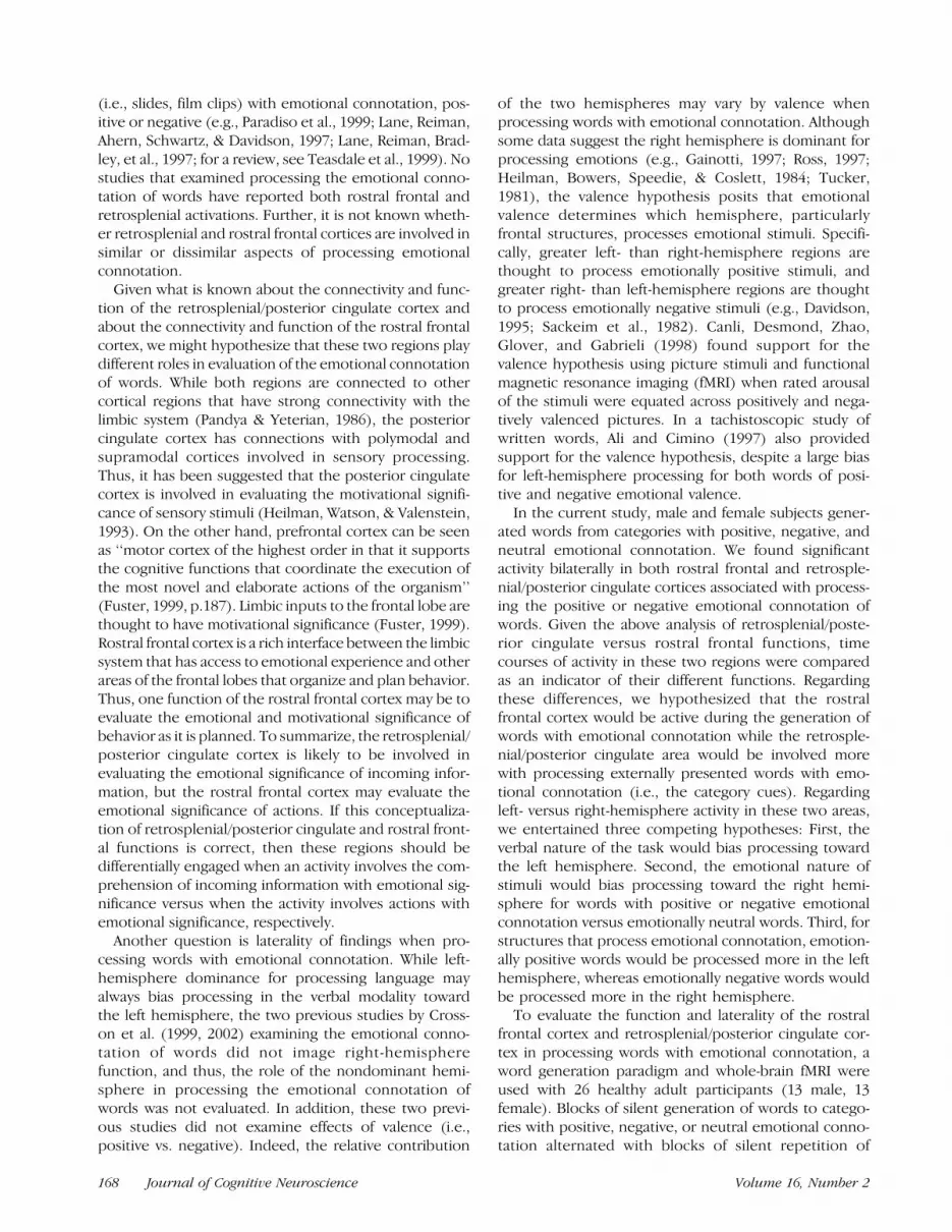
Figure 1. Silent word generation task (white intervals) alternated with
silent word repetition as a baseline task (black intervals) during
scanning. Baseline intervals varied in length (9.9, 16.5, and 23.1 sec)and number of items repeated (4, 7, and 10 words, respectively), and
were pseudorandomly interspersed in the 9.5 cycles of the imaging
runs. Silent word generation alternated between generating words with
positive emotional connotation (e.g., vacations), generating words withnegative emotional connotation (e.g., weapons), and generating
emotionally neutral words (e.g., buildings).
Table 1. Behavioral Results of the fMRI Participants (N = 26)Collapsed Across Sex
Positive Negative Neutral
M SD M SD M SD
Words generated 6.44 1.42 6.29 1.92 6.72 1.45
Valence rating 8.00 .99 2.28 1.09 5.33 .41
Arousal rating 6.11 1.94 4.99 1.83 1.90 1.61
Cato et al. 169

cluster also extended bilaterally, with the MI at midline,0, �48, and 30. This cluster, which centered upon thesame region as that reported by Maddock and Buono-core (1997), was located primarily in BA 30 as well as theportion of BA 23 superior and posterior to the spleniumof the corpus callosum.
To further examine areas of activity found to besignificant on the one-way ANOVA, a mask of thesignificant clusters was generated and subjected tofollow-up voxelwise t tests for positive versus neutralemotional connotation, for negative versus neutral emo-tional connotation, for neutral versus emotional conno-tation, and for positive versus negative emotionalconnotation with a statistical threshold of p < .005and a cluster threshold of volume >200 Al. Table 2shows all clusters of significant activity for positive versusneutral and negative versus neutral t tests that wereconsistent with significant areas of activity on theANOVA. For positive versus neutral emotional connota-tion, significant clusters of bilateral activity were notedfor both rostral frontal and retrosplenial/posterior cin-gulate cortices. Likewise, for negative versus neutralemotional connotation, significant clusters of bilateralactivity were noted for both rostral frontal and retro-splenial/posterior cingulate cortices. Other than theseareas of a priori interest, additional regions of activationwere circumscribed to the left middle frontal gyrus andareas along the left temporal lobe and left temporopar-ietal junction (Table 2).
Brain regions more involved in the generation ofwords to neutral than emotional connotation wereexamined with a follow-up voxelwise t test in whichactivity associated with words to positive and negativeemotional connotations was collapsed. Table 3 revealsall clusters of significant activity for the neutral versusemotional t test that were consistent with significantareas of activity on the ANOVA. Brain regions moreactive during generation of words with neutral emotion-al connotation included a posterior cingulate regionlateral and inferior to the retrosplenial region associatedwith positive and negative emotional connotations. Thisregion of activity extended back to the peristriate cortex(BA 19). The two other clusters of activation identifiedwith the neutral versus emotional t test were circum-scribed to the occipital cortex suggesting relatively morevisualization during neutral word generation comparedto emotional word generation. Two other brain regionswere relatively more active during neutral word gener-ation, one centering in the left precentral gyrus and theother centering in the left middle temporal gyrus. Thus,generation of neutral words engaged a different regionof the left retrosplenial/posterior cingulate cortex thatwas lateral and inferior to the area involved in process-ing words with positive or negative emotional connota-tion. The rostral frontal area that was engaged duringgeneration to words with positive and negative emo-tional connotation was not active during generation ofneutral words. While the lateral frontopolar cortex has
Figure 2. Activity in rostral
frontal and in retrosplenial/
posterior cingulate cortexassociated with the main effect
of valence, across participants,
red = p < .005; yellow = p <
.001, along with correspondingaverage hemodynamic
response functions (HRFs).
Eleven images were modeled tocapture the entire
hemodynamic response across
the blocked word generation
task for positive, negative, andneutral categories for these
areas.
170 Journal of Cognitive Neuroscience Volume 16, Number 2

been activated during generation of neutral informationin other paradigms (for a review, see Christoff & Gabri-eli, 2000), we did not find a similar pattern of activityincrease during neutral word generation in this region.As neutral word generation was directly compared toother word generation tasks (also involving complexprocessing of internally generated information), thisarea of activity may have been cancelled by comparableactivity associated with the comparison tasks.
Time Course Analysis
Activation in rostral frontal and retrosplenial/posteriorcingulate cortices was found when comparing genera-tion of words with emotional connotation (positive ornegative) versus generation of emotionally neutralwords (Figure 2). To further determine the time courseassociated with these differences, a mask of each regionwas generated, and averaged HRFs for each valencewere entered into a repeated measures two-way Valence� Time (image number) ANOVA to examine differencesin HRF signal amplitudes in the time domain between
the three valence types. To model the entire HRFassociated with the 5 images of blocked word genera-tion, we specified 11 images (beginning at the onset ofthe first image of word generation) to be modeled inorder to capture the entire HRF from onset to return tobaseline.
For the rostral frontal cortex, the Valence � Timeinteraction was significant, F(20,500) = 10.37, p < .0001.In the case of this cortex, contrasts revealed that fromImages 3 through 8, the HRF amplitudes for positive andnegative emotional connotation were significantly great-er than the HRF amplitude for neutral emotional con-notation. For the retrosplenial/posterior cingulatecortex, the Valence � Time interaction was also signif-icant, F(20,500) = 13.78, p < .0001. Contrasts revealedthat during Images 3 through 7, the HRF amplitudes forpositive and negative emotional connotation were sig-nificantly greater than the HRF amplitude for neutralemotional connotation. For both ROIs, the courses forpositive and negative emotional connotation did notsignificantly differ at any time point other than duringImage 7 in the retrosplenial/posterior cingulate cortex.
Table 2. Volumes of Tissue (>200 Al) Showing Significant Activity Changes ( p < .005) for Positive and Negative versus NeutralWord Generation
LocationPositive > Neutral Anatomic Area(max t loc) max t, volume in A l
Negative > Neutral Anatomic Area(max t loc) max t, volume in A l
Rostral frontal cortex L and R BAs 9, 10 (�3, 60, 12),t = 6.12, 3035 Al
L and R BAs 9, 10(�4, 60, 29), t = 5.17, 1807 Al
Retrosplenial/posteriorcingulate cortex
L and R, BAs 23, 30, 31(0, �48, 30), t = 5.53, 3422 Al
L and R BAs 23, 30, 31(0, �47, 29), t = 4.50, 1042 Al
Middle frontal gyrus L BA 9 (�22, 42, 31),t = 3.83, 244 Al
Temporoparietal junction L BAs 22, 40 (�57, �51, 24),t = 4.44, 772 Al;L BAs 22, 39 (�48, �63, 16),t = 4.32, 595 Al
L BA 22, 40 (�54, �44, 23),t = 3.96, 300 Al; L BAs 22, 39(�40, �55, 26), t = 4.84, 872 Al
Superior and middletemporal gyrus
L BAs 21, 22 (�50, �24, 0),t = 4.25, 518 Al
L BA 21 (�50, �30, 1), t = 4.16, 413 Al
BA = Brodmann’s area (according to Talairach & Tournoux, 1988); max t = maximum t within given cluster of activity. Other abbreviations are asfollows: L = left, R = right.
Table 3. Volumes of Tissue (>200 Al) Showing Significant Activity Changes ( p < .005) for Neutral versus Emotional (Positive andNegative Categories Collapsed) Word Generation
Location Anatomic Area (max t loc) max t, volume in Al
Posterior cingulate/occipital cortex L BA 30, extending to BA 19(�12, �55, 12), t = �4.83, 1021 Al
Precentral and middle frontal gyrus L BA 6 (�43, 4, 36), t = �4.23, 586 Al
Middle and superior temporal gyrus L BA 37 (�59, �49, �9), t = �5.13, 1667 Al
Lingual gyrus/other visual areas L BAs 19, 17 (�29, �68, 32), t = �5.63, 970 Al
BAs 18, 19 (�15, �74, 7), t = �4.15, 439 Al
BA = Brodmann’s area (according to Talairach & Tournoux, 1988); max t = maximum t value within given cluster of activity. Other abbreviations areas follows: L = left, R = right.
Cato et al. 171

Thus, in both rostral frontal and retrosplenial/poste-rior cingulate cortex, HRF amplitudes for words withpositive and negative emotional connotation were con-sistently greater than the HRF amplitude for emotionallyneutral words. Furthermore, visual comparison of theHRF curves between rostral frontal and retrosplenial/posterior cingulate cortex revealed different timecourses within valence across these areas. To confirmthese observable differences, for each valence, the aver-aged HRF for each of the two brain regions was enteredinto a repeated measures two-way Brain Region � Time(image number) ANOVA to determine if the signalamplitudes within valence differed as a function of brainregion down the time domain.
For positive valence, the Area � Time interaction termwas significant, F(10,250) = 12.02, p < .0001. Contrastsrevealed that from Images 4 through 7, the amplitude ofthe HRF to positive valence in the rostral frontal cortexwas significantly greater than that of the HRF in theretrosplenial/posterior cingulate cortex. Thus, after Im-age 3, the HRF in the retrosplenial/posterior cingulatecortex begins to decline whereas that in the rostralfrontal cortex continues to rise to peak. This differencein courses carries out to Image 7, after which both HRFsin both cortices fall to near-baseline levels. Therefore, inresponse to positively valenced categories, the HRF inthe retrosplenial/posterior cingulate cortex peaks muchearlier (Image 3) and follows a much shorter time coursethan the HRF in the rostral frontal cortex, which appearsto peak at Image 4 and return to baseline at Image 8.Indeed, the time course in the retrosplenial/posteriorcingulate cortex is approximately what would be ex-pected from a response to a single event, the presenta-tion of the positively valenced category at the beginningof word generation trials. In contrast, the rostral frontalHRF seems representative of the entire generationepoch, returning to baseline levels a few seconds aftertermination of the generation trial.
For negative valence, the interaction term was againsignificant, F(10,250) = 9.44, p < .0001. Contrastsrevealed that from Image 1 to 2, the HRF for theretrosplenial/posterior cingulate cortex has a significant-ly steeper rise than that of the rostral frontal cortex, orthat the rostral frontal cortex exhibits a lag or delay,whereas the retrosplenial/posterior cingulate cortexdoes not. From Image 3 to 4, the courses again signi-
ficantly diverge: the HRF in the rostral frontal cortexrises to peak while that of the retrosplenial/posteriorcingulate cortex descends towards baseline. Similar tothe differences found for positive valence, the HRF inthe retrosplenial/posterior cingulate cortex follows amuch shorter time course and the HRF amplitude issignificantly greater for the rostral frontal cortex forImages 4 through 6.
Finally, for neutral valence, the interaction term wassignificant, F(10,250) = 9.57, p < .0001. Contrastsrevealed that from Image 3 to 4, trajectories of the HRFsin the two cortical areas diverge due to a sharp drop ofthe retrosplenial/posterior cingulate HRF to below base-line levels. From Images 4 to 6 where the retrosplenial/posterior cingulate HRF is at below baseline levels, theamplitude of the rostral frontal HRF was significantlygreater than that of the retrosplenial/posterior cingulateHRF. The sharp rise of the HRF curve in the retrosple-nial/posterior cingulate cortex back to baseline levelsfrom Image 6 to 8 also resulted in significant differencesin trajectories of the neutral HRF in the retrosplenial/posterior cingulate area versus that of the neutral HRF inthe rostral frontal area.
Laterality Analysis
For the rostral frontal and retrosplenial/posterior cingu-late regions, where activity extended across the midline,it was important to examine lateralization in the contextof valence. As noted previously, there were three com-peting hypotheses regarding lateralization and valence:(a) that the left hemisphere would dominate processingbecause the stimuli were verbal, (b) that the righthemisphere would dominate processing because ofemotional content, and (c) that the left hemispherewould be more involved in processing positive emo-tions, and the right hemisphere would be more involvedin processing negative emotions. The clusters weredivided into right and left hemisphere components(Table 4). In comparisons to neutral emotional conno-tation, both positive emotional connotation and nega-tive emotional connotation demonstrate a clear majorityof the cluster in the left hemisphere. These data areincompatible with the right-hemisphere hypothesis ofemotional processing. Because negative emotional con-notation showed an even greater left-hemisphere bias
Table 4. Relative Contribution of the Left and Right Hemispheres for Positive and Negative versus Neutral Word Generation( p < .005)
Location Positive > Neutral Negative > Neutral
L rostral frontal (BAs 9, 10) 2366 Al; 78% total cluster 1726 Al; 96% total cluster
R rostral frontal (BAs 9, 10) 669 Al; 22% total cluster 81 Al; 4% total cluster
L retrosplenial/posterior cingulate cortex (BAs 23, 30, 31) 2211 Al; 65% total cluster 858 Al; 82% total cluster
R retrosplenial/posterior cingulate cortex (BAs 23, 30, 31) 1211 Al; 35% total cluster 184 Al; 18% total cluster
172 Journal of Cognitive Neuroscience Volume 16, Number 2

than positive emotional connotation for both rostralfrontal and retrosplenial/posterior cingulate cortices,the data are also incompatible with the hypothesis thatnegative emotional connotation would show a right-hemisphere bias. Thus, at least for these two corticalregions, data are most consistent with a left-hemispherebias due to the verbal nature of the stimuli.
As mentioned earlier, other regions of activity thatwere significant on the one-way valence ANOVA andsubsequent positive versus neutral and negative versusneutral t tests were circumscribed to the left hemisphere(Table 2) for both positive emotional connotation andnegative emotional connotation. Again, these data areconsistent with a left-hemisphere bias because of theverbal nature of the stimuli and are inconsistent withother hypotheses.
Also with respect to laterality, the voxelwise t test forgenerating words with negative versus positive emotion-al connotation revealed only two significant clusters ofactivity. In both cases, activity either favored the lefthemisphere or was located entirely within the lefthemisphere. Activity for the retrosplenial/posterior cin-gulate region was greater for positive than negativeemotional connotation. This region centered in the lefthemisphere (MI = �2, �59, 14), with some activitycrossing over to the right hemisphere. The second areashowed greater activity for negative than positive emo-tional connotation and was located in the left inferiorfrontal sulcus (MI = �46, 18, 28). Thus, activity differ-ences in generating words with positive versus negativeemotional connotation are located primarily in the lefthemisphere regardless of valence.
Finally, a Sex � Valence ANOVA with repeated meas-ures on valence was also performed to assess sex differ-ences and the interaction of sex with valence. Twoclusters for the sex main effect survived the statisticalthreshold of p < .005 with a cluster threshold of volume>200 Al. The first was in the right precentral gyrus(MI = 50, �4, 27), and the second was in the leftcerebellum (MI = �28, �42, �22). In both cases, theclusters were more active for men than for women. Thus,regardless of valence, men showed more activity inthe right precentral gyrus and the left cerebellum thanwomen. The interaction of sex of participants andvalence of words generated showed no significant areasof activity.
DISCUSSION
The major finding of the current study is that generatingwords with positive or negative emotional connotationled to unique activity bilaterally in rostral frontal andretrosplenial/posterior cingulate cortices when directlycompared to generating words with neutral emotionalconnotation. In addition, examination of time courses inthese two areas revealed differences that may corre-spond to unique roles during processing of emotional
connotation. With respect to laterality, activation in theseareas, while bilateral, showed consistently greater later-alization to the left hemisphere. The relative proportionof these sizable clusters in the right hemisphere did notindicate any support for either the valence hypothesis orthe right-hemisphere hypothesis. Rather, it appears thatthe verbal nature of the task led to a strong left-hemi-sphere bias regardless of valence. In addition, the sex ofparticipants did not further influence differences in brainactivation associated with word generation to positive,negative, and neutral categories.
An examination of the hemodynamic response curvesin the rostral frontal cortex and in retrosplenial/posteriorcingulate cortex (Figure 2) provides clues to their re-spective functions in mediating processing of emotionalconnotation. First, it is apparent that generation topositive and negative emotionally valenced categories,but not neutral categories, led to significant activation inthese areas. Despite this common pattern, the timecourses between these areas are quite different. Thefunctional activity in the retrosplenial/posterior cingu-late cortex begins to increase immediately after thestimulus presentation, peaks relatively early after stimu-lus presentation, and then returns to baseline before theend of word generation, much like an event-relatedresponse. We suggest that the role of this cortex is inthe evaluation of an external stimulus with emotionalsalience, which in this case was the category cue. Thishypothesis is consistent with the assessment of Heilmanet al. (1993) that the posterior cingulate cortex evaluatesthe motivational significance of external stimuli.
In the case of the rostral frontal cortex, the responseis slightly delayed, remains throughout the active gen-eration period, and then lingers for an additional 4 to5 sec after word generation has ended before falling tobaseline. Therefore, the rostral frontal cortex is impli-cated in the process of generation of words withemotional connotation. This finding raises two inter-esting questions. First, is the rostral frontal cortexinvolved in evaluating emotional connotation only rel-ative to some kind of behavioral output (e.g., wordgeneration) or does it have a more general function ofprocessing emotional connotation in any circumstance?In addressing this first question, it should be noted thatthis is the third demonstration of rostral frontal activityin processing words with emotional connotation. In thefirst instance (Crosson et al., 1999), generation ofwords with emotional connotation was compared togeneration of emotionally neutral words, similar to thecurrent study. However, Crosson et al. (1999) did notdistinguish between positive and negative connotation.By contrast, the current study establishes that therostral frontal cortex is active during word generationfor both words with positive emotional connotationand words with negative emotional connotation. Inthe second instance (Crosson et al., 2002), semanticmonitoring of words with emotional connotation was
Cato et al. 173

compared to semantic monitoring of sensory stimuli(i.e., auditory presentation of words with emotionalconnotation vs. emotionally neutral words). Subjects inthis experiment were not just listening to words butwere actively involved in making semantic decisionsabout the words. Thus, it can be argued that all studiesthat have shown rostral frontal activity in processingwords with emotional connotation have involved activeoutput of some kind. A future direction would be toevaluate whether this area of activity occurs duringprocessing of emotional connotation when no behav-ioral output is required.
The second question regarding the sustained activityin the rostral frontal cortex is whether activity in thisarea only occurs in the presence of some level ofsustained mood change (positive or negative). Indeed,at least two previous imaging studies using visualstimuli and mood induction procedures also reportedbrain activity in this rostral frontal area (Teasdale et al.,1999; Lane, Reiman, Ahern, et al., 1997). Unfortunately,as this particular experiment involving word generationwas not intended to serve as a mood induction proce-dure (MIP), we did not assess whether mood changesoccurred during participation in the experiment. Gen-erally, MIPs in imaging experiments have involvedsustained exposure (typically longer than 16 sec) ofemotionally evocative material (almost always with avisual component) of one valence. The Teasdale et al.(1999) study and the Lane, Reiman, Ahern, et al (1997)study, both of which were attempting mood induction,involved longer stimulus exposures (30 and 120 sec,respectively) within valence than the current studyinvolving only 16.5-sec periods of within-valence expo-sure. Also during MIPs, in most previous imagingexperiments (e.g., Schneider et al., 2000; Lane, Reiman,Ahern, et al., 1997) and otherwise (e.g., the Velten MIP;for a review, see Gerrads-Hesse, Spies, & Hesse, 1994),the participant is instructed to attempt to experiencethe desired mood state. In contrast, we did not provideany suggestions or directions regarding mood induc-tion. Thus, our procedures differed from typical MIPs. Itis possible that the rostral frontal cortex (BA 9, 10) isinvolved both in experiencing mood and in processingemotional connotation, in much the same way that thepremotor cortex is involved in both action planning andin processing action semantics (i.e., action verb andtool knowledge; see Kellenbach, Brett, & Patterson,2003; Pulvermuller, 2001; Chao & Martin, 2000; Pulver-muller et al., 2000). Nonetheless, we must concede thatwe did not attempt to measure if changes in moodoccurred during our experiment. Therefore, futurestudies of emotional processing should assess self-rat-ings of mood in addition to valence and arousal ratingsalong with psychophysiological measures to furtherexplore this question.
Differences in type and intensity of emotional stimuliacross studies of emotional processing may contribute
to inconsistencies in this literature in terms of thelaterality of findings and the presence or absence ofsex differences. In terms of laterality, while the currentstudy did not support either the valence or right-hemi-sphere hypothesis, a likely explanation is that the lan-guage demands of this task outweighed any hemisphericdifferences that might occur in response to the valenceof a stimulus. In other words, engaging language cortexin the act of word generation may have dampened right-hemisphere activity secondary to transcallosal inhibitionof cortices not devoted to language generation (Gazza-niga, 1998). One functional imaging study that has foundsupport for the valence hypothesis used pictures ratherthan words (Canli et al., 1998). Thus, the use of wordswith this study, rather than pictures, may be the sourceof the discrepancies between the current findings andthose of Canli et al. (1998) that support the valencehypothesis. In a similar vein, the use of words ratherthan pictures or film clips in this study may have alsoprecluded findings of sex differences in processingpositive versus negative emotional connotation thathave been reported in two previous functional imagingstudies (Schneider, et al, 2000; Lang et al., 1998). Futurestudies that attempt to disentangle these variations inmethodologies would be helpful in determining whatvariables (e.g., processing modality, emotional valence,type of emotional stimuli, duration of exposure) influ-ence direction of findings of hemispheric specializationand sex differences.
In closing, the current study is unique not only inshowing activity of both rostral frontal and retrosple-nial/posterior cingulate cortices in processing wordswith emotional connotation, but it also speaks ofdifferences in the roles of these cortices in processingemotional connotation. Based on the different timecourses within these areas as well as what is knownabout their respective functions, we are suggesting thatthe retrosplenial/posterior cingulate cortex is involvedin processing the emotional significance of sensorystimuli, while the rostral frontal cortex may be involvedin processing emotional connotation when generationof some kind of active output is required. Regardingthe status of emotional connotation as a semanticattribute, this is a provocative hypothesis. We knowof no other instance in which it has been suggestedthat the cortex that processes a specific semanticattribute depends on whether an external stimulus isbeing evaluated or whether some significant output isbeing generated. With respect to this hypothesis, futureresearch should address two questions: First, can thehypothesis be verified in other research designs? Sec-ond, if the hypothesis can be further verified foremotional connotation, is this anterior–posterior divi-sion for response generation versus sensory processingunique to emotional connotation, or can it be appliedto other semantic attributes such as visual form orobject function?
174 Journal of Cognitive Neuroscience Volume 16, Number 2

METHODS
Participants
Twenty-six (13 men, 13 women) strongly right-handed[Edinburgh Handedness Inventory (Oldfield, 1971),M = 81.9, SD = 14.3] volunteers participated (age 18–49 years, M = 31.5 years, SD = 9.9; education 13–20 years, M = 16.4 years, SD = 1.7 years). Male andfemale participants did not significantly differ in terms ofyears of education, t(24) = �.56, p = .58, or age, t(24) =�.95, p = .35. Potential participants were excluded ifthey reported a history of neurological disease, majorpsychiatric disturbance, learning disability, attention def-icit disorder, or substance abuse, or if they reportedcurrent use of psychoactive medications. Potential riskswere explained, and informed consent was obtainedfrom participants according to institutional guidelinesestablished by the Health Center Institutional ReviewBoard at the University of Florida.
Materials and Methods
Experimental Stimuli
Emotional and neutral categories were extrapolatedfrom nouns listed in the Affective Norms for EmotionalWords (ANEW, Bradley, Cuthbert, & Lang, 1988) corpus.This corpus provides words with ratings on a valencedimension from negative to positive (range, 1.00 to 9.00)and an arousal dimension from low to high arousal(range, 1.00 to 9.00). Categories were chosen to berepresentative of items from this corpus with positive,negative, and neutral emotional connotation. ANEW wasalso used to select emotionally neutral words for therepetition baseline condition. A behavioral pilot studywith 12 different subjects (6 males, 6 females; age 25–34 years, M = 28.5, SD = 3.4; education 18–21 years,M = 19.0, SD = .95) was conducted to equate numberof words generated in a 16.5-sec time period across thevalence of categories and to equate arousal ratings acrosspositive and negative categories. We wanted to equatearousal ratings between positive and negative categories,as laterality differences have only been found in the pastwhen arousal has been equated (Canli et al., 1998). As aresult of the pilot study, 45 of 80 piloted categories wereselected for the fMRI study.
Experimental Tasks
During each of four functional imaging runs, blocks ofsilently generating words to categories with positive,negative, or neutral emotional connotation alternatedwith a baseline task of silent repetition of emotionallyneutral words (Figure 1). A blocked presentation formatwas used because pilot studies indicated a blocked par-adigm produced hemodynamic responses of greateramplitude and consistency than an event-related para-digm. The use of a baseline of neutral word repetition,rather than rest, has also been found to increase sensitiv-
ity of comparisons between word generation tasks(Crosson et al., 1999). During category generation, par-ticipants heard a category and silently generated as manyexemplars as possible during a 16.5-sec word generationperiod. Categories with positive (e.g., desserts), negative(e.g., disasters), or neutral (e.g., containers) emotionalconnotation were presented in a pseudorandom order,such that each functional run consisted of three catego-ries from each valence (positive, negative, and neutral).During alternating baseline periods of variable length(either 9.9, 16.5, or 23.1 sec, distributed within runs in apseudorandomized order), participants silently repeatedemotionally neutral words, with 4 words to repeat duringthe 9.9-sec baseline; 7 words to repeat during the 16.5-secbaseline; and 10 words to repeat during the 23.1-secbaseline. The number of words to repeat was matchedwith the mean number of words generated in a 16.5-secperiod, determined by the behavioral pilot.
The onset of generation blocks was marked by thecue, ‘‘generate,’’ while baseline repetition blocks com-menced with the cue, ‘‘repeat.’’ All words, cues, andcategories were delivered binaurally via acousticallyinsulated air conduction tubes connected to hollowfoam earplugs using a Kenwood KR-A4070 amplifier,Realistic 31-2005 Tenband stereo frequency equalizer,and JBL 16-� speaker. Prior to the experiments, auditorythreshold in the scanner was determined for eachsubject using recognition of target words during scanneroperation. Volume was adjusted by adding a constant35 dB to each subject’s auditory threshold.
Four of five possible category lists were administeredto each experimental subject during the four imagingruns, with the order of these runs pseudorandomizedacross participants. Following scanning, a fifth categorylist was administered outside of the scanner to provideinformation about the generation rate for categories ofemotionally positive, negative, and neutral valence. Thelist administered outside the scanner was varied be-tween subjects in a random fashion with the constraintthat each list be used approximately an equal number oftimes. Following completion of the fifth list, fMRI par-ticipants rated all 45 categories for valence and arousal.
Image Acquisition
Each experimental run consisted of nine 16.5-sec periodsof word generation during which 5 images were acquired.The baseline state was repetition of emotionally neutralwords for 9.9, 16.5, or 23.1 sec (corresponding to 3, 5, and7 images respectively). Length of baseline periods wasvaried pseudorandomly to mitigate low-frequency peri-odic and quasiperiodic physiologic artifacts. Each lengthof baseline was used an equal number of times, exceptthat an extra 16.5-sec baseline period was added to theend of each run. Thus, for each functional imaging run,there was an average of 10 images per word generation–baseline cycle, with 95 images total per functional run. By
Cato et al. 175

staggering the baseline condition, the onset of the exper-imental task (word generation) was also aperiodic. Thelength of word generation blocks did not vary so that asingle HDR could be modeled for each block using thedeconvolution technique.
Whole-brain imaging was performed on a 1.5-T GESigna scanner using a dome-shaped quadrature radiofrequency head coil. The head was aligned such that theinterhemispheric fissure was within 18 of vertical. ForfMRI sequences, 22 slices (5.8–7.0 mm thick) wereacquired. The plane between the eleventh and twelfthfunctional image slices was centered at the interhemi-spheric fissure for equal coverage of both hemispheres.Images were obtained using a two-spiral gradient-echosequence with the following parameters: TE = 40 msec,TR = 1650 msec, FA = 658, FOV = 180 mm. Afterfunctional image acquisition, structural images wereacquired for 124 1.3-mm-thick sagittal slices, using a3-D spoiled GRASS volume acquisition (TE = 7 msec,TR = 27 msec, FA = 458, NEX = 1, FOV = 24 cm, matrixsize = 256 � 192).
Image Analysis
Functional images were analyzed and overlaid ontoanatomic images with Analysis of Functional Neuro-images (AFNI) software (Cox, 1996). To minimize ef-fects of head motion, time series of images werespatially registered in 3-D space. For each subject, meanslice signal intensities were normalized to the grandmean of slice intensity across functional runs. Voxelswhere the standard deviation of the signal changeexceeded 5% of the mean signal were set to zero todecrease large vessel effects and residual motion arti-fact. Images were visually inspected for gross artifactsand were viewed in cineloop to detect residual motion.If any time series of a subject was judged to contain asignificant number of images with gross artifacts orresidual motion, the subject’s data were removed fromfurther analyses. Data from 3 of 29 subjects whoparticipated were eliminated because of motion artifact,leaving the 26 subjects who were described above.
Four runs of 95 images each were concatenated into asingle time series of 380 images for each of the 22functional image slices. Then, HRFs were deconvolvedfrom the 380-image time series on a voxel-by-voxel basis.Each HRF was modeled using the 11 TR periods thatdirectly followed each category cue. Eleven TR periodswere modeled in order to capture the entire hemody-namic response from its onset to its return to baseline.For each voxel, a single HRF was deconvolved separatelyfor positive, negative, and neutral emotional connotation.Magnitude of response for each condition was operation-ally defined as area under the curve of the HRF.
Anatomic and functional images were interpolated tovolumes with 1-mm3 voxels, coregistered, and convertedto the stereotaxic coordinate space of Talairach and
Tournoux (1988) using AFNI. HRFs within the 1-mm3
voxels were spatially smoothed using a 3-mm full-widthhalf-maximum (FWHM) Gaussian filter to compensatefor variability in structural and functional anatomy acrossparticipants. A voxelwise one-way repeated measuresANOVA was performed to determine the effect of va-lence on the pattern of brain activation during wordgeneration across participants. The effect of sex ofparticipants and its interaction with valence was alsoinvestigated using a two-way voxelwise ANOVA. In bothcases, area under the HRF curve was the dependentvariable. Cluster analyses on resulting statistical para-metric maps were conducted. Post hoc t tests wereperformed on significant clusters identified by eachANOVA to evaluate the direction of differences in HRFs(area under the curve) between the conditions. For boththe ANOVA and the subsequent t tests, a volume thresh-old of 200 Al and a statistical probability threshold ofp < .005 were used.
ROI analyses were performed within two clusters thatexhibited visibly different temporal evolutions of theHRF according to valence and to spatial region. Forthe HRFs of each of these two brain areas, a two-wayrepeated measures ANOVA was performed for eachbrain region across valence type (3 levels) and numberof TR periods or images modeled in deconvolution(11 levels). In addition, a two-way repeated measuresANOVA was performed for each valence type acrossbrain regions (2 levels) and number of TR periods orimages modeled in deconvolution (11 levels).
Acknowledgments
Supported by the University of Florida Brain Institute and grantR01-DC03455 from NIDCD. Results of this study weresubmitted by the first author to the graduate school at theUniversity of Florida in partial fulfillment of the requirementsfor the degree of Doctor of Philosophy. Thanks to Douglas C.Noll for spiral scan sequences, Jeffrey R. Fitzsimmons for headcoil, and Wayne King for constructing the air conduction soundtransducer. Additional thanks to the Veterans Affairs Rehabil-itation Research and Development Service for support duringthe composition of this manuscript.
Reprint requests should be sent to M. Allison Cato, Postdoc-toral Fellow, VISN 22 MIRECC Program, Psychology Service116B, Department of Veterans Affairs, 3350 La Jolla VillageDrive, San Diego, CA 92161, or via e-mail: [email protected].
The data reported in this experiment have been deposited inthe fMRI Data Center (http://www.fmridc.org). The accessionnumber is 2-2003-113YQ.
REFERENCES
Ali, N., & Cimino, C. R. (1997). Hemispheric lateralizations ofperception and memory for emotional verbal stimuli innormal individuals. Neuropsychology, 11, 114–125.
Binder, J. R., Frost, J. A., Hammeke, T. A., Bellgowan, P. S. F.,Rao, S. M., & Cox, R. W. (1999). Conceptual processingduring the conscious resting state: A functional MRI study.Journal of Cognitive Neuroscience, 11, 80–93.
176 Journal of Cognitive Neuroscience Volume 16, Number 2

Bradley, M. M., Cuthbert, B. N., & Lang, P. J. (1988). Affectivenorms for English words (ANEW): Technical manual andaffective ratings. Gainesville: University of Florida.
Canli, T., Desmond, J. E., Zhao, Z., Glover, G., & Gabrieli, J. D.E. (1998). Hemispheric asymmetry for emotional stimulidetected with fMRI. NeuroReport, 9, 3223–3239.
Chao, L. L., Haxby, J. V., & Martin, A. (1999). Attribute-basedneural substrates in temporal cortex for perceiving andknowing about objects. Nature Neuroscience, 2, 913–919.
Chao, L. L., & Martin, A. (2000). Representation of manipulableman-made objects in the dorsal stream. NeuroImage, 12,478–484.
Christoff, K., & Gabrieli, J. D. E. (2000). The frontopolar cortexand human cognition: Evidence for a rostrocaudalhierarchical organization within the human prefrontalcortex. Psychobiology, 28, 168–186.
Cox, R. W. (1996). AFNI: Software for analysis and visualizationof functional magnetic resonance neuroimages. Computersand Biomedical Research, 29, 162–173.
Crosson, B., Cato, M. A., Sadek, J., Radonovich, K., Gokcay, D.,Bauer, R., Fischler, I., Maron, L., Auerbach, E., Browd, S.,Freeman, A., & Briggs, R. (2002). Semantic monitoring ofwords with emotional connotation during fMRI:Contribution of left-hemisphere limbic association cortex.Journal of the International Neuropsychological Society,8, 607–622.
Crosson, B., Radonovich, K., Sadek, J. R., Gokcay, D., Bauer, R.M., Fischler, I. S., Cato, M. A., Maron, L., Auerbach, E. J.,Browd, S. R., & Briggs, R. W. (1999). Left-hemisphereprocessing of emotional connotation during wordgeneration. NeuroReport, 10, 2449–2455.
Damasio, H., Grabowski, T. J., Tranel, D., Hichwa, R. D., &Damasio, A. R. (1996). A neural basis for lexical retrieval.Nature, 380, 499–505.
Davidson, R. J. (1995). Cerebral asymmetry, emotion, andaffective style. In R. J. Davidson & K. Hugdahl (Eds.), Brainasymmetry (pp. 361–387). Cambridge: MIT Press.
Fuster, J. M. (1999). Cognitive functions of the frontal lobes. InB. L. Miller & J. L. Cummings (Eds.), The human frontallobes (pp. 187–195). New York: The Guilford Press.
Gainotti, G. (1997). Emotional, psychological and psychosocialproblems of aphasic patients: An introduction. Aphasiology,11, 635–650.
Gazzaniga, M. S. (1998). Cerebral lateralization and specializa-tion. In M. S. Gazzaniga, R. B. Ivry, & G. M. Mangun (Eds.),Cognitive neuroscience: The biology of the mind(pp. 340–341). New York: W. W. Norton.
Gerrads-Hesse, A., Spies, K., & Hesse, F. W. (1994). Experimentalinductions of emotional states and their effectiveness: Areview. British Journal of Psychology, 85, 55–78.
Heilman, K. M., Bowers, D., Speedie, L., & Coslett, H. B. (1984).Comprehension of affective and nonaffective prosody.Neurology, 34, 917–921.
Heilman, K. M., Watson, R. T., & Valenstein, E. (1993). Neglectand related disorders. In K. M. Heilman & E. Valenstein(Eds.), Clinical neuropsychology (pp. 279–336). New York:Oxford University Press.
Ishai, A., Ungerleider, L. G., Martin, A., Schouten, J. L., & Haxby,J. V. (1999). Distributed representation of objects in thehuman ventral visual pathway. Proceedings of the NationalAcademy of Sciences, U.S.A., 96, 9379–9384.
Kellenbach, M., Brett, M., & Patterson, K. (2003). Actions speaklouder than functions: The importance of manipulability andaction in tool representation. Journal of CognitiveNeuroscience, 15, 30–46.
Lane, R. D., Reiman, E. M., Ahern, G. L., Schwartz, G. E., &Davidson, R. J. (1997). Neuroanatomical correlates of
happiness, sadness, and disgust. American Journal ofPsychiatry, 154, 926–933.
Lane, R. D., Reiman, E. M., Bradley, M. M., Lang, P. J., Ahern,G. L., Davidson, R. J., & Schwartz, G. E. (1997).Neuroanatomical correlates of pleasant and unpleasantemotion. Neuropsychologia, 35, 1437–1444.
Lang, P. J., Bradley, M. M., Fitzsimmons, J. R., Cuthbert, B. N.,Scott, J. D., Moulder, B., & Nangia, V. (1998). Emotionalarousal and activation of the visual cortex: An fMRI analysis.Psychophysiology, 35, 199–210.
Maddock, R. J. (1999). The retrosplenial cortex and emotion:New insights from functional neuroimaging of the humanbrain. Trends in Neuroscience, 22, 310–316.
Maddock, R. J., & Buonocore, M. H. (1997). Activation of leftposterior cingulate gyrus by the auditory presentation ofthreat-related words: An fMRI study. Psychiatry Research:Neuroimaging Section, 75, 1–14.
Martin, A., Wiggs, C. L., Ungerleider, L. G., & Haxby, J. V.(1996). Neural correlates of category-specific knowledge.Nature, 379, 649–652.
Oldfield, R. C. (1971). The assessment and analysis ofhandedness: The Edinburgh Inventory. Neuropsychologia,9, 97–113.
Pandya, D. N., & Yeterian, E. H. (1986). Architecture andconnections of cortical association areas. In A. Peters & E. G.Jones (Eds.), Cerebral cortex (pp. 3–61). New York: PlenumPress.
Paradiso, S., Johnson, D. L., Andreasen, N. C., O’Leary, D. S.,Watkins, G. L., Ponto, L. L. B., & Hichwa, R. D. (1999).Cerebral blood flow changes associated with attribution ofemotional valence to pleasant, unpleasant, and neutral visualstimuli in a PET study of normal subjects. American Journalof Psychiatry, 156, 1618–1629.
Pulvermuller, F. (2001). Brain reflections of words and theirmeanings. Trends in Cognitive Sciences, 5, 517–524.
Pulvermuller, F., Harle, M., & Hummel, F. (2000).Neurophysiological distinction of verb categories.NeuroReport: For Rapid Communication of NeuroscienceResearch, 11, 2789–2793.
Ross, E. D. (1997). Cortical representation of the emotions. InM. R. Trimble & J. L. Cummings (Eds.), Contemporarybehavioral neurology (pp. 107–216). Woburn:Butterworth-Heinemann.
Sackeim, H. A., Greenberg, M. S., Weiman, A. L., Gur, R. C.,Hungerbuhler, J. P., & Geschwind, N. (1982). Hemisphericasymmetry in the expression of positive and negativeemotions—neurologic evidence. Archives of Neurology, 39,210–218.
Schneider, F., Habel, U., Kessler, C., Salloum, J. B., & Posse, S.(2000). Gender differences in regional cerebral activityduring sadness. Human Brain Mapping, 9, 226–238.
Talairach, J., & Tournoux, P. (1988). Co-planar stereotaxicatlas of the human brain. New York: Thieme Medical.
Teasdale, J. D., Howard, R. J., Cox, S. G., Ha, Y., Brammer, M. J.,Williams, S. C. R., & Checkley, S. A. (1999). Functional MRIstudy of the cognitive generation of affect. AmericanJournal of Psychiatry, 156, 209–215.
Thompson-Schill, S. L., Aguirre, G. K., D’Esposito, M., & Farah,M. J. (1999). A neural basis for category and modalityspecificity of semantic knowledge. Neuropsychologia, 37,671–676.
Tucker, D. M. (1981). Lateral brain function, emotion, andconceptualization. Psychological Bulletin, 89, 19–46.
Vandenberghe, R., Price, C., Wise, R., Josephs, O., &Frackowiak, R. (1996). Functional anatomy of a commonsemantic system for words and pictures. Nature, 383,254–256.
Cato et al. 177


