Embed Size (px)
Citation preview
Saudi Journal of Biological Sciences (2014) xxx, xxx–xxx
King Saud University
Saudi Journal of Biological Sciences
www.ksu.edu.sawww.sciencedirect.com
ORIGINAL ARTICLE
Cytotoxic and apoptotic activities
of Amorphophallus campanulatus (Roxb.) Bl. tuberextracts against human colon carcinoma cell line
HCT-15
* Corresponding author. Tel.: +91 481 2731035; fax: +91 481
2731009.
E-mail address: [email protected] (M.S. Latha).
Peer review under responsibility of King Saud University.
Production and hosting by Elsevier
1319-562X ª 2014 Production and hosting by Elsevier B.V. on behalf of King Saud University.
http://dx.doi.org/10.1016/j.sjbs.2014.01.004
Please cite this article in press as: Ansil, P.N. et al., Cytotoxic and apoptotic activities of Amorphophallus campanulatus (Roxb.) Bl. tuberagainst human colon carcinoma cell line HCT-15. Saudi Journal of Biological Sciences (2014), http://dx.doi.org/10.1016/j.sjbs.2014.01.0
P.N. Ansil a, P.J. Wills b, R. Varun b, M.S. Latha a,*
a Biochemistry and Pharmacognosy Research Laboratory, School of Biosciences, Mahatma Gandhi University, P.D. Hills P.O.,
Kottayam, Kerala 686560, Indiab MIMS Research Foundation, Mankavu P.O., Calicut, Kerala 673007, India
Received 1 November 2013; revised 5 January 2014; accepted 14 January 2014
KEYWORDS
Amorphophallus
campanulatus;
Colon cancer;
MTT;
DAPI;
JC-1;
Annexin-V
Abstract Colorectal cancer is one of the leading causes of cancer death worldwide and is the third
most common form of malignancy in both men and women. Several possible colon cancer chemo-
preventive agents are found in edible plants. Amorphophallus campanulatus (Roxb.) Blume (family:
Araceae) is a tuber crop, largely cultivated throughout the plains of India for using its corm as food.
This tuber has also been traditionally used for the treatment of abdominal tumors, liver diseases,
piles etc. The aim of this study was to evaluate the dose-dependent cytotoxic and apoptosis inducing
effects of the sub fractions of A. campanulatus tuber methanolic extract (ACME) viz. petroleum
ether fraction (PEF), chloroform fraction (CHF), ethyl acetate fraction (EAF) and methanolic frac-
tion (MEF) on the colon cancer cell line, HCT-15. Antiproliferative effects of the sub fractions of
ACME were studied by MTT assay. Apoptotic activity was assessed by DAPI, Annexin V-FITC
and JC-1 fluorescent staining. The chemotherapeutic drug, 5-flurouracil (5-FU) was used as positive
drug control. The sub fractions of ACME significantly inhibited the proliferation of HCT-15 cells in
a dose-dependent manner. In addition, the extracts were found to induce apoptosis and were con-
firmed by DAPI, Annexin V-FITC and JC-1 fluorescent staining. A pronounced results of cytotoxic
and apoptotic activities were observed in the cells treated with 5-FU and CHF, whereas, EAF and
MEF treated cells exhibited a moderate result and the least effect was observed in PEF treated cells.
Our results suggested that, among the sub fractions of ACME, CHF had potent cytotoxic and
extracts04
2 P.N. Ansil et al.
Please cite this article in press as: Ansil,against human colon carcinoma cell lin
apoptotic activity and thus it could be explored as a novel target for anticancer drug development.
Furthermore, these findings confirm that the sub fractions of ACME dose-dependently suppress the
proliferation of HCT-15 cells by inducing apoptosis.
ª 2014 Production and hosting by Elsevier B.V. on behalf of King Saud University.
1. Introduction
Colorectal cancer is the third most common malignant neo-plasm worldwide and has become one of the major causes ofcancer mortality (Jemal et al., 2004; American Cancer Society,
2011). Epidemiological studies have revealed a number of riskfactors for colorectal cancer including age, family history ofcolon cancer or inflammatory bowel disease, smoking, alcohol
consumption, obesity and diet (Kirkegaard et al., 2010). Hu-man colon cancer development is often characterized at anearly stage by hyperproliferation of the colonic epithelium
leading to the formation of adenomas. This is mainly a conse-quence of dysregulated cell cycle control and/or suppressedapoptosis as usually observed in colon cancers (Gryfe et al.,1997; Potten, 1997). In addition, several studies have reported
that the loss of control of apoptosis results in cancer initiationand progression and thus many new treatment strategiestargeting apoptosis are feasible and may be used in the treat-
ment of various types of cancers (Tu et al., 1996; Vitale-Crosset al., 2004; Tian et al., 2007; Wong, 2011).
Nowadays, large numbers of natural compounds have been
identified that are pharmacologically highly effective againstcancerous cells. It is reported that the consumption of a dietrich in phytochemicals can reduce the risk of cancer. Fruits
and vegetables, which contain a diverse range of phytochemi-cals, are suggested to have properties important in the preven-tion of cancer. It includes antioxidant, anti-inflammatory andantiproliferative activities as well as modulatory effects on sub-
cellular signaling pathways, induction of cell cycle arrest, andapoptosis (Block et al., 1992; Aviram et al., 2000; Kaplanet al., 2001; Afaq et al., 2005 Mahassni and Al-Reemi, 2013).
Among the various possible experimental models of cancer,human cancer cell lines have been the most widely used. Theyhave retained the hallmarks of cancer cells namely self suffi-
ciency in growth control, insensitivity to antigrowth signals, es-cape from checkpoints (apoptosis), genetic instability andinvasiveness (van Staveren et al., 2009). HCT-15 cells, usedin the present study, are one of the human adenocarcinoma
cell lines extensively used in colon cancer studies. These cellswere established from colorectal cancer after surgical resectionbefore the chemotherapeutic treatment of a tumor (Tompkins
et al., 1974).In the present study, our aim was to investigate the cyto-
toxic and apoptosis inducing effects of the sub fractions of
Amorphophallus campanulatus tuber methanolic extract(ACME) namely, petroleum ether fraction (PEF), chloroformfraction (CHF), ethyl acetate fraction (EAF) and methanolic
fraction (MEF) on the colon cancer cell line, HCT-15.A. campanulatus (Roxb.) Blume belonging to the family ofAraceae is a perennial herb commonly known as elephant footyam. It is mostly a tuber crop of south East Asian origin and is
largely cultivated throughout the plains of India for using itscorm (bulb) as food (Das et al., 2009). Further, the plant is
P.N. et al., Cytotoxic and apoptote HCT-15. Saudi Journal of Biolo
valuable as medicine particularly the corm has been used
traditionally for the treatment of abdominal tumors, abdomi-nal pain, liver diseases, piles etc. Besides, the corm has been re-ported to possess antioxidant, antibacterial, antifungal,
hepatoprotective and cytotoxic activities (Khan et al., 2007;Ansil et al., 2012). The cytotoxic and apoptotic potential ofthe sub fractions of ACME, investigated in the present study,was compared with 5-fluorouracil (5-FU) – a pyrimidine ana-
log used in the treatment of colorectal cancer (Mohapatraet al., 2011). Furthermore, the present study was a step to-wards the isolation and characterization of the pharmacologi-
cally active phytochemical constituents from the crude extractof A. campanulatus tuber.
2. Materials and methods
2.1. Chemicals
Fetal bovine serum (FBS), N-2-Hydroxyethylpiperazine-N-2-ethane-sulfonic acid (HEPES) and 40,6-diamidino-2-phenylin-
dole (DAPI) were procured from Sigma Chemical Co., St.Louis, MO, USA. 5-flourouracil (5-FU) was purchased fromBiochem Pharmaceutical Industries, Mumbai, India. RPMI
Medium and antibiotic–antimycotic were purchased fromGibco, Grand Island, N.Y, USA. Cell Proliferation Assaykit (3-(4,5-dimethylthiazol-2-yl)-2,5diphenyltetrazoliumbro-mide, [MTT]) was purchased from HiMedia, India. 5,50,6,60
Tetrachloro-1,10,3,30-tetraethylbenzimidazolylcarbocyanineiodide (JC-1) was purchased from Invitrogen, Carlsbad, CA,USA. Annexin V-fluorescein isothiocyanate (FITC) kit was
supplied by Calbiochem, La Jolla, CA, USA. Dimethyl sulfox-ide (DMSO) was obtained from Merck, Mumbai, India. Allthe other chemicals used were also of high purity grade.
2.2. Cell culture
HCT-15 cell line was procured from National Centre for CellScience (NCCS), Pune, India and grown as a monolayer in
RPMI (Rosewell Park Memorial Institute) medium containingHEPES and sodium bicarbonate supplemented with 10% FBSand 1% antibiotic–antimycotics. Cells were maintained in a
tissue culture flask kept in a humidified incubator (5% CO2
in air at 37 �C) with a medium change every 2–3 days. Whenthe cells reached 70–80% confluence, they were harvested with
trypsin – EDTA (ethylene diamine tetra acetate) and seededinto a new tissue culture flask.
2.3. Collection and extraction of the plant material
A. campanulatus tubers were harvested from the Kottayamdistrict, Kerala, India and authenticated. A voucher specimen(SBSBRL.02) is maintained in the institute. The shade-dried tu-
ic activities of Amorphophallus campanulatus (Roxb.) Bl. tuber extractsgical Sciences (2014), http://dx.doi.org/10.1016/j.sjbs.2014.01.004
Cytotoxic and apoptotic activities of Amorphophallus campanulatus (Roxb.) Bl. tuber 3
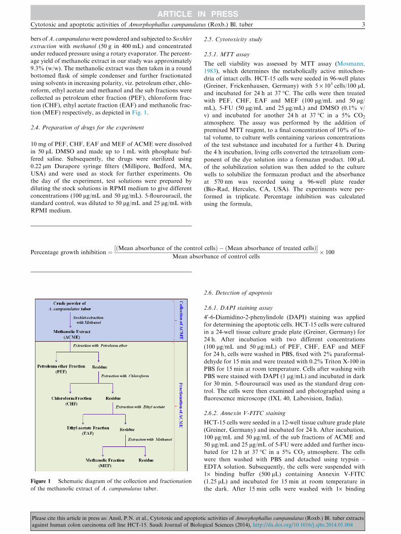
bers ofA. campanulatuswere powdered and subjected toSoxhletextraction with methanol (50 g in 400 mL) and concentratedunder reduced pressure using a rotary evaporator. The percent-
age yield of methanolic extract in our study was approximately9.3% (w/w). The methanolic extract was then taken in a roundbottomed flask of simple condenser and further fractionated
using solvents in increasing polarity, viz. petroleum ether, chlo-roform, ethyl acetate and methanol and the sub fractions werecollected as petroleum ether fraction (PEF), chloroform frac-
tion (CHF), ethyl acetate fraction (EAF) and methanolic frac-tion (MEF) respectively, as depicted in Fig. 1.
2.4. Preparation of drugs for the experiment
10 mg of PEF, CHF, EAF and MEF of ACME were dissolvedin 50 lL DMSO and made up to 1 mL with phosphate buf-fered saline. Subsequently, the drugs were sterilized using
0.22 lm Durapore syringe filters (Millipore, Bedford, MA,USA) and were used as stock for further experiments. Onthe day of the experiment, test solutions were prepared by
diluting the stock solutions in RPMI medium to give differentconcentrations (100 lg/mL and 50 lg/mL). 5-flourouracil, thestandard control, was diluted to 50 lg/mL and 25 lg/mL with
RPMI medium.
Percentage growth inhibition ¼ ðMean absorbance of the control½Mean absor
Figure 1 Schematic diagram of the collection and fractionation
of the methanolic extract of A. campanulatus tuber.
Please cite this article in press as: Ansil, P.N. et al., Cytotoxic and apoptotagainst human colon carcinoma cell line HCT-15. Saudi Journal of Biolo
2.5. Cytotoxicity study
2.5.1. MTT assay
The cell viability was assessed by MTT assay (Mosmann,
1983), which determines the metabolically active mitochon-dria of intact cells. HCT-15 cells were seeded in 96-well plates(Greiner, Frickenhausen, Germany) with 5 · 103 cells/100 lLand incubated for 24 h at 37 �C. The cells were then treated
with PEF, CHF, EAF and MEF (100 lg/mL and 50 lg/mL), 5-FU (50 lg/mL and 25 lg/mL) and DMSO (0.1% v/v) and incubated for another 24 h at 37 �C in a 5% CO2
atmosphere. The assay was performed by the addition ofpremixed MTT reagent, to a final concentration of 10% of to-tal volume, to culture wells containing various concentrations
of the test substance and incubated for a further 4 h. Duringthe 4 h incubation, living cells converted the tetrazolium com-ponent of the dye solution into a formazan product. 100 lLof the solubilization solution was then added to the culture
wells to solubilize the formazan product and the absorbanceat 570 nm was recorded using a 96-well plate reader(Bio-Rad, Hercules, CA, USA). The experiments were per-
formed in triplicate. Percentage inhibition was calculatedusing the formula,
cellsÞ � ðMean absorbance of treated cellsÞ�bance of control cells
� 100
2.6. Detection of apoptosis
2.6.1. DAPI staining assay
40-6-Diamidino-2-phenylindole (DAPI) staining was appliedfor determining the apoptotic cells. HCT-15 cells were culturedin a 24-well tissue culture grade plate (Greiner, Germany) for
24 h. After incubation with two different concentrations(100 lg/mL and 50 lg/mL) of PEF, CHF, EAF and MEFfor 24 h, cells were washed in PBS, fixed with 2% paraformal-dehyde for 15 min and were treated with 0.2% Triton X-100 in
PBS for 15 min at room temperature. Cells after washing withPBS were stained with DAPI (1 lg/mL) and incubated in darkfor 30 min. 5-flourouracil was used as the standard drug con-
trol. The cells were then examined and photographed using afluorescence microscope (IXL 40, Labovision, India).
2.6.2. Annexin V-FITC staining
HCT-15 cells were seeded in a 12-well tissue culture grade plate(Greiner, Germany) and incubated for 24 h. After incubation,100 lg/mL and 50 lg/mL of the sub fractions of ACME and
50 lg/mL and 25 lg/mL of 5-FU were added and further incu-bated for 12 h at 37 �C in a 5% CO2 atmosphere. The cellswere then washed with PBS and detached using trypsin –
EDTA solution. Subsequently, the cells were suspended with1· binding buffer (500 lL) containing Annexin V-FITC(1.25 lL) and incubated for 15 min at room temperature inthe dark. After 15 min cells were washed with 1· binding
ic activities of Amorphophallus campanulatus (Roxb.) Bl. tuber extractsgical Sciences (2014), http://dx.doi.org/10.1016/j.sjbs.2014.01.004
4 P.N. Ansil et al.
buffer and then added with phenol red free medium and exam-ined immediately by using a fluorescence microscope. PositiveAnnexin V-FITC staining will appear as bright apple green on
the cell membrane surface.
2.6.3. JC-1 staining
The mitochondrial membrane potential (DWm) was assayed
using JC-1 (the cationic dye – 5,50,6,60 tetrachloro-1,10,3,30-tetraethylbenzimidazolylcarbocyanine iodide) mitochon-drial potential sensor (Invitrogen, USA), according to the
manufacturer’s directions. Briefly, HCT-15 cells were incu-bated for 24 h in 24-well plates (Greiner, Germany) and thecells were treated with 100 lg/mL and 50 lg/mL of the sub
fractions of ACME (PEF, CHF, EAF and MEF) and 50 lg/mL and 25 lg/mL of 5-FU for 18 h. The treated cells werewashed with PBS and incubated for 30 min in 10% RPMI
medium without phenol red containing JC-1 at a concentrationof 2.5 lg/mL. The cells were then examined and photographedusing a fluorescence microscope.
2.7. Statistical analysis
Results were expressed as mean ± standard deviation (SD).All statistical comparisons were made by means of a one
way ANOVA test followed by Tukey’s post hoc analysis andp-values less than or equal to 0.05 were considered significant.
3. Results
3.1. Cytotoxicity study
3.1.1. MTT assay
Results of the cytotoxic evaluation of the sub fractions ofACME on HCT-15 cells are graphically shown in Fig. 2.
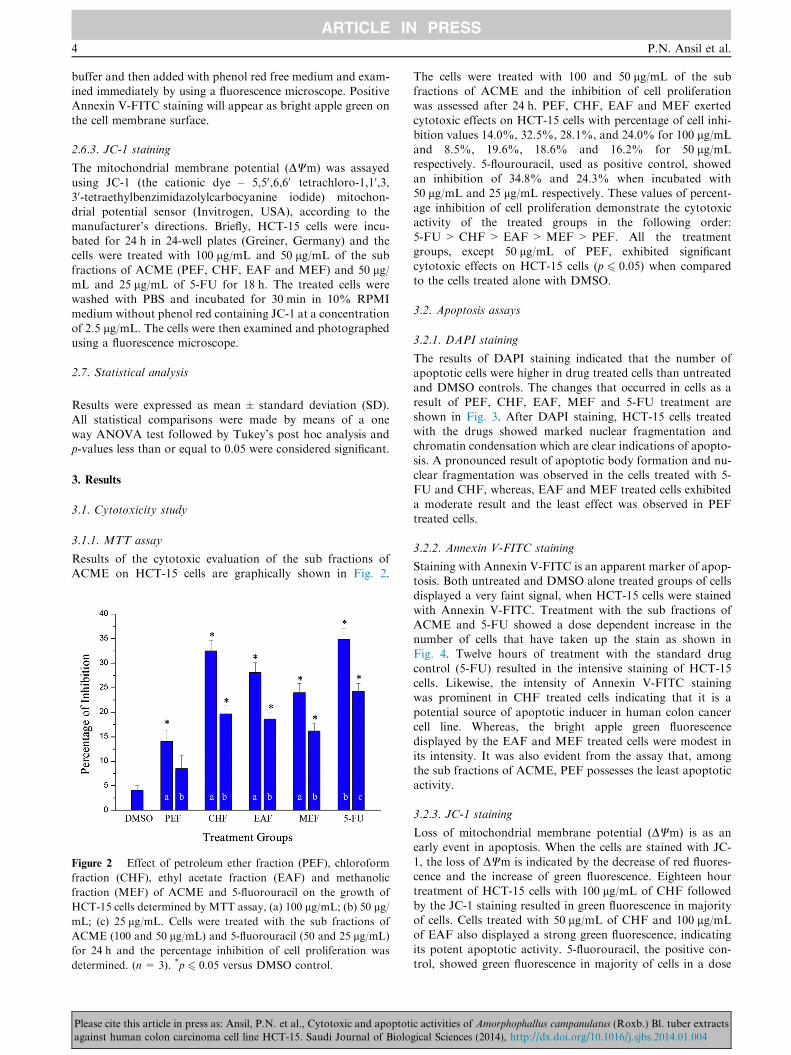
Figure 2 Effect of petroleum ether fraction (PEF), chloroform
fraction (CHF), ethyl acetate fraction (EAF) and methanolic
fraction (MEF) of ACME and 5-fluorouracil on the growth of
HCT-15 cells determined by MTT assay, (a) 100 lg/mL; (b) 50 lg/mL; (c) 25 lg/mL. Cells were treated with the sub fractions of
ACME (100 and 50 lg/mL) and 5-fluorouracil (50 and 25 lg/mL)
for 24 h and the percentage inhibition of cell proliferation was
determined. (n= 3). *p 6 0.05 versus DMSO control.
Please cite this article in press as: Ansil, P.N. et al., Cytotoxic and apoptotagainst human colon carcinoma cell line HCT-15. Saudi Journal of Biolo
The cells were treated with 100 and 50 lg/mL of the subfractions of ACME and the inhibition of cell proliferationwas assessed after 24 h. PEF, CHF, EAF and MEF exerted
cytotoxic effects on HCT-15 cells with percentage of cell inhi-bition values 14.0%, 32.5%, 28.1%, and 24.0% for 100 lg/mLand 8.5%, 19.6%, 18.6% and 16.2% for 50 lg/mL
respectively. 5-flourouracil, used as positive control, showedan inhibition of 34.8% and 24.3% when incubated with50 lg/mL and 25 lg/mL respectively. These values of percent-
age inhibition of cell proliferation demonstrate the cytotoxicactivity of the treated groups in the following order:5-FU> CHF > EAF >MEF > PEF. All the treatmentgroups, except 50 lg/mL of PEF, exhibited significant
cytotoxic effects on HCT-15 cells (p 6 0.05) when comparedto the cells treated alone with DMSO.
3.2. Apoptosis assays
3.2.1. DAPI staining
The results of DAPI staining indicated that the number ofapoptotic cells were higher in drug treated cells than untreatedand DMSO controls. The changes that occurred in cells as a
result of PEF, CHF, EAF, MEF and 5-FU treatment areshown in Fig. 3. After DAPI staining, HCT-15 cells treatedwith the drugs showed marked nuclear fragmentation andchromatin condensation which are clear indications of apopto-
sis. A pronounced result of apoptotic body formation and nu-clear fragmentation was observed in the cells treated with 5-FU and CHF, whereas, EAF and MEF treated cells exhibited
a moderate result and the least effect was observed in PEFtreated cells.
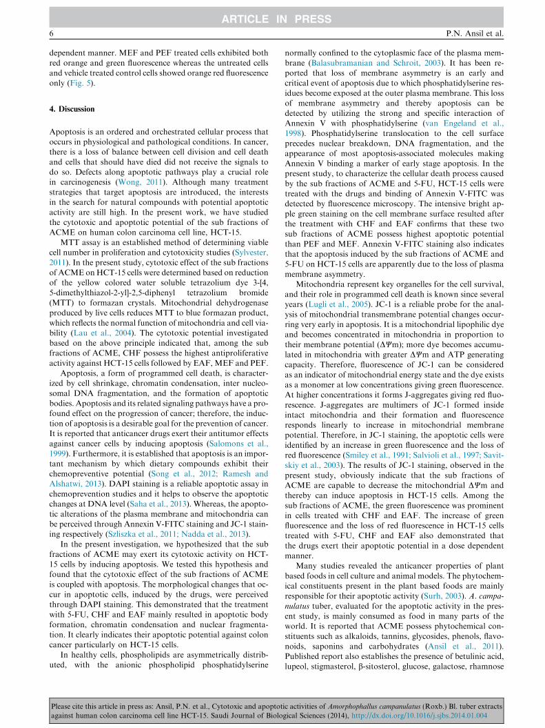
3.2.2. Annexin V-FITC staining
Staining with Annexin V-FITC is an apparent marker of apop-tosis. Both untreated and DMSO alone treated groups of cellsdisplayed a very faint signal, when HCT-15 cells were stained
with Annexin V-FITC. Treatment with the sub fractions ofACME and 5-FU showed a dose dependent increase in thenumber of cells that have taken up the stain as shown in
Fig. 4. Twelve hours of treatment with the standard drugcontrol (5-FU) resulted in the intensive staining of HCT-15cells. Likewise, the intensity of Annexin V-FITC stainingwas prominent in CHF treated cells indicating that it is a
potential source of apoptotic inducer in human colon cancercell line. Whereas, the bright apple green fluorescencedisplayed by the EAF and MEF treated cells were modest in
its intensity. It was also evident from the assay that, amongthe sub fractions of ACME, PEF possesses the least apoptoticactivity.
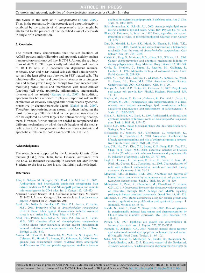
3.2.3. JC-1 staining
Loss of mitochondrial membrane potential (DWm) is as anearly event in apoptosis. When the cells are stained with JC-
1, the loss of DWm is indicated by the decrease of red fluores-cence and the increase of green fluorescence. Eighteen hourtreatment of HCT-15 cells with 100 lg/mL of CHF followed
by the JC-1 staining resulted in green fluorescence in majorityof cells. Cells treated with 50 lg/mL of CHF and 100 lg/mLof EAF also displayed a strong green fluorescence, indicating
its potent apoptotic activity. 5-fluorouracil, the positive con-trol, showed green fluorescence in majority of cells in a dose
ic activities of Amorphophallus campanulatus (Roxb.) Bl. tuber extractsgical Sciences (2014), http://dx.doi.org/10.1016/j.sjbs.2014.01.004
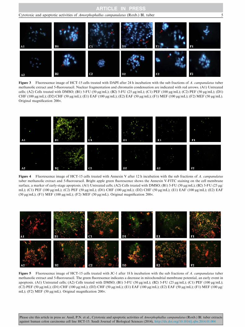
Figure 3 Fluorescence image of HCT-15 cells treated with DAPI after 24 h incubation with the sub fractions of A. campanulatus tuber
methanolic extract and 5-fluorouracil. Nuclear fragmentation and chromatin condensation are indicated with red arrows. (A1) Untreated
cells; (A2) Cells treated with DMSO; (B1) 5-FU (50 lg/mL); (B2) 5-FU (25 lg/mL); (C1) PEF (100 lg/mL); (C2) PEF (50 lg/mL); (D1)
CHF (100 lg/mL); (D2) CHF (50 lg/mL); (E1) EAF (100 lg/mL); (E2) EAF (50 lg/mL); (F1) MEF (100 lg/mL); (F2) MEF (50 lg/mL).
Original magnification 200·.
Figure 4 Fluorescence image of HCT-15 cells treated with Annexin V after 12 h incubation with the sub fractions of A. campanulatus
tuber methanolic extract and 5-fluorouracil. Bright apple green fluorescence shows the Annexin V-FITC staining on the cell membrane
surface, a marker of early-stage apoptosis. (A1) Untreated cells; (A2) Cells treated with DMSO; (B1) 5-FU (50 lg/mL); (B2) 5-FU (25 lg/mL); (C1) PEF (100 lg/mL); (C2) PEF (50 lg/mL); (D1) CHF (100 lg/mL); (D2) CHF (50 lg/mL); (E1) EAF (100 lg/mL); (E2) EAF
(50 lg/mL); (F1) MEF (100 lg/mL); (F2) MEF (50 lg/mL). Original magnification 200·.
Figure 5 Fluorescence image of HCT-15 cells treated with JC-1 after 18 h incubation with the sub fractions of A. campanulatus tuber
methanolic extract and 5-fluorouracil. The green fluorescence indicates a decrease in mitochondrial membrane potential, an early event in
apoptosis. (A1) Untreated cells; (A2) Cells treated with DMSO; (B1) 5-FU (50 lg/mL); (B2) 5-FU (25 lg/mL); (C1) PEF (100 lg/mL);
(C2) PEF (50 lg/mL); (D1) CHF (100 lg/mL); (D2) CHF (50 lg/mL); (E1) EAF (100 lg/mL); (E2) EAF (50 lg/mL); (F1) MEF (100 lg/mL); (F2) MEF (50 lg/mL). Original magnification 200·.
Cytotoxic and apoptotic activities of Amorphophallus campanulatus (Roxb.) Bl. tuber 5
Please cite this article in press as: Ansil, P.N. et al., Cytotoxic and apoptotic activities of Amorphophallus campanulatus (Roxb.) Bl. tuber extractsagainst human colon carcinoma cell line HCT-15. Saudi Journal of Biological Sciences (2014), http://dx.doi.org/10.1016/j.sjbs.2014.01.004
6 P.N. Ansil et al.
dependent manner. MEF and PEF treated cells exhibited bothred orange and green fluorescence whereas the untreated cellsand vehicle treated control cells showed orange red fluorescence
only (Fig. 5).
4. Discussion
Apoptosis is an ordered and orchestrated cellular process thatoccurs in physiological and pathological conditions. In cancer,there is a loss of balance between cell division and cell death
and cells that should have died did not receive the signals todo so. Defects along apoptotic pathways play a crucial rolein carcinogenesis (Wong, 2011). Although many treatment
strategies that target apoptosis are introduced, the interestsin the search for natural compounds with potential apoptoticactivity are still high. In the present work, we have studied
the cytotoxic and apoptotic potential of the sub fractions ofACME on human colon carcinoma cell line, HCT-15.
MTT assay is an established method of determining viablecell number in proliferation and cytotoxicity studies (Sylvester,
2011). In the present study, cytotoxic effect of the sub fractionsof ACME onHCT-15 cells were determined based on reductionof the yellow colored water soluble tetrazolium dye 3-[4,
5-dimethylthiazol-2-yl]-2,5-diphenyl tetrazolium bromide(MTT) to formazan crystals. Mitochondrial dehydrogenaseproduced by live cells reduces MTT to blue formazan product,
which reflects the normal function of mitochondria and cell via-bility (Lau et al., 2004). The cytotoxic potential investigatedbased on the above principle indicated that, among the subfractions of ACME, CHF possess the highest antiproliferative
activity against HCT-15 cells followed by EAF,MEF and PEF.Apoptosis, a form of programmed cell death, is character-
ized by cell shrinkage, chromatin condensation, inter nucleo-
somal DNA fragmentation, and the formation of apoptoticbodies. Apoptosis and its related signaling pathways have a pro-found effect on the progression of cancer; therefore, the induc-
tion of apoptosis is a desirable goal for the prevention of cancer.It is reported that anticancer drugs exert their antitumor effectsagainst cancer cells by inducing apoptosis (Salomons et al.,
1999). Furthermore, it is established that apoptosis is an impor-tant mechanism by which dietary compounds exhibit theirchemopreventive potential (Song et al., 2012; Ramesh andAlshatwi, 2013). DAPI staining is a reliable apoptotic assay in
chemoprevention studies and it helps to observe the apoptoticchanges at DNA level (Saha et al., 2013). Whereas, the apopto-tic alterations of the plasma membrane and mitochondria can
be perceived through Annexin V-FITC staining and JC-1 stain-ing respectively (Szliszka et al., 2011; Nadda et al., 2013).
In the present investigation, we hypothesized that the sub
fractions of ACME may exert its cytotoxic activity on HCT-15 cells by inducing apoptosis. We tested this hypothesis andfound that the cytotoxic effect of the sub fractions of ACMEis coupled with apoptosis. The morphological changes that oc-
cur in apoptotic cells, induced by the drugs, were perceivedthrough DAPI staining. This demonstrated that the treatmentwith 5-FU, CHF and EAF mainly resulted in apoptotic body
formation, chromatin condensation and nuclear fragmenta-tion. It clearly indicates their apoptotic potential against coloncancer particularly on HCT-15 cells.
In healthy cells, phospholipids are asymmetrically distrib-uted, with the anionic phospholipid phosphatidylserine
Please cite this article in press as: Ansil, P.N. et al., Cytotoxic and apoptotagainst human colon carcinoma cell line HCT-15. Saudi Journal of Biolo
normally confined to the cytoplasmic face of the plasma mem-brane (Balasubramanian and Schroit, 2003). It has been re-ported that loss of membrane asymmetry is an early and
critical event of apoptosis due to which phosphatidylserine res-idues become exposed at the outer plasma membrane. This lossof membrane asymmetry and thereby apoptosis can be
detected by utilizing the strong and specific interaction ofAnnexin V with phosphatidylserine (van Engeland et al.,1998). Phosphatidylserine translocation to the cell surface
precedes nuclear breakdown, DNA fragmentation, and theappearance of most apoptosis-associated molecules makingAnnexin V binding a marker of early stage apoptosis. In thepresent study, to characterize the cellular death process caused
by the sub fractions of ACME and 5-FU, HCT-15 cells weretreated with the drugs and binding of Annexin V-FITC wasdetected by fluorescence microscopy. The intensive bright ap-
ple green staining on the cell membrane surface resulted afterthe treatment with CHF and EAF confirms that these twosub fractions of ACME possess highest apoptotic potential
than PEF and MEF. Annexin V-FITC staining also indicatesthat the apoptosis induced by the sub fractions of ACME and5-FU on HCT-15 cells are apparently due to the loss of plasma
membrane asymmetry.Mitochondria represent key organelles for the cell survival,
and their role in programmed cell death is known since severalyears (Lugli et al., 2005). JC-1 is a reliable probe for the anal-
ysis of mitochondrial transmembrane potential changes occur-ring very early in apoptosis. It is a mitochondrial lipophilic dyeand becomes concentrated in mitochondria in proportion to
their membrane potential (DWm); more dye becomes accumu-lated in mitochondria with greater DWm and ATP generatingcapacity. Therefore, fluorescence of JC-1 can be considered
as an indicator of mitochondrial energy state and the dye existsas a monomer at low concentrations giving green fluorescence.At higher concentrations it forms J-aggregates giving red fluo-
rescence. J-aggregates are multimers of JC-1 formed insideintact mitochondria and their formation and fluorescenceresponds linearly to increase in mitochondrial membranepotential. Therefore, in JC-1 staining, the apoptotic cells were
identified by an increase in green fluorescence and the loss ofred fluorescence (Smiley et al., 1991; Salvioli et al., 1997; Savit-skiy et al., 2003). The results of JC-1 staining, observed in the
present study, obviously indicate that the sub fractions ofACME are capable to decrease the mitochondrial DWm andthereby can induce apoptosis in HCT-15 cells. Among the
sub fractions of ACME, the green fluorescence was prominentin cells treated with CHF and EAF. The increase of greenfluorescence and the loss of red fluorescence in HCT-15 cellstreated with 5-FU, CHF and EAF also demonstrated that
the drugs exert their apoptotic potential in a dose dependentmanner.
Many studies revealed the anticancer properties of plant
based foods in cell culture and animal models. The phytochem-ical constituents present in the plant based foods are mainlyresponsible for their apoptotic activity (Surh, 2003). A. campa-
nulatus tuber, evaluated for the apoptotic activity in the pres-ent study, is mainly consumed as food in many parts of theworld. It is reported that ACME possess phytochemical con-
stituents such as alkaloids, tannins, glycosides, phenols, flavo-noids, saponins and carbohydrates (Ansil et al., 2011).Published report also establishes the presence of betulinic acid,lupeol, stigmasterol, b-sitosterol, glucose, galactose, rhamnose
ic activities of Amorphophallus campanulatus (Roxb.) Bl. tuber extractsgical Sciences (2014), http://dx.doi.org/10.1016/j.sjbs.2014.01.004
Cytotoxic and apoptotic activities of Amorphophallus campanulatus (Roxb.) Bl. tuber 7
and xylose in the corm of A. campanulatus (Khare, 2007).Thus, in the present study, the cytotoxic and apoptotic activityexhibited by the extracts of A. campanulatus tuber might be
attributed to the presence of the identified class of chemicalsin single or in combination.
5. Conclusion
The present study demonstrates that the sub fractions ofACME possess antiproliferative and apoptotic activity against
human colon carcinoma cell line, HCT-15. Among the sub frac-tions of ACME, CHF significantly inhibited the proliferationof HCT-15 cells in a concentration dependent manner.
Whereas, EAF and MEF treated cells exhibited a moderate re-sult and the least effect was observed in PEF treated cells. Theinhibitory effect of natural bioactive substances in carcinogen-
esis and tumor growth may be through two main mechanisms:modifying redox status and interference with basic cellularfunctions (cell cycle, apoptosis, inflammation, angiogenesis,invasion and metastasis) (Kampa et al., 2007). In addition,
apoptosis has been reported to play an important role in theelimination of seriously damaged cells or tumor cells by chemo-preventive or chemotherapeutic agents (Galati et al., 2000).
Therefore, apoptosis-inducing natural products including themost promising CHF identified in the present investigationcan be explored as novel targets for anticancer drug develop-
ment. However, further studies are needed to comprehend thedifferent mechanisms by which the sub fractions of the metha-nolic extract of A. campanulatus tuber exert their cytotoxic andapoptotic effects on the colon cancer cell line, HCT-15.
Acknowledgments
The research was supported by the University Grants Com-mission (UGC), New Delhi, India. Financial assistance from
the UGC as Research Fellowship in Sciences for MeritoriousStudents to the first author is also thankfully acknowledged.
References
Afaq, F., Saleem, M., Krueger, C.G., Reed, J.D., Mukhtar, H., 2005.
Anthocyanin- and hydrolyzable tannin-rich pomegranate fruit
extract modulates MAPK and NF-kappaB pathways and inhibits
skin tumorigenesis in CD-1 mice. Int. J. Cancer 113, 423–433.
American Cancer Society, 2011. Colorectal cancer facts & Figures
2011–2013, Atlanta, Georgia, USA. Available at: http://www.can-
cer.org. Accessed on 24 December, 2011.
Ansil, P.N., Nitha, A., Prabha, S.P., Wills, P.J., Jazaira, V., Latha,
M.S., 2011. Protective effect of Amorphophallus campanulatus
(Roxb.) Blume tuber against thioacetamide induced oxidative
stress in rats. Asian Pac. J. Trop. Med. 4, 870–877.
Ansil, P.N., Prabha, S.P., Nitha, A., Wills, P.J., Jazaira, V., Latha,
M.S., 2012. Curative effect of Amorphophallus campanulatus
(Roxb.) Blume tuber methanolic extract against thioacetamide
induced oxidative stress in experimental rats. Asian Pac. J. Trop.
Biomed. 2, S83–S89.
Aviram, M., Dornfeld, L., Rosenblat, M., Volkova, N., Kaplan, M.,
Coleman, R., Hayek, T., Presser, D., Fuhrman, B., 2000. Pome-
granate juice consumption reduces oxidative stress, atherogenic
modifications to LDL, and platelet aggregation: studies in humans
Please cite this article in press as: Ansil, P.N. et al., Cytotoxic and apoptotagainst human colon carcinoma cell line HCT-15. Saudi Journal of Biolo
and in atherosclerotic apolipoprotein E-deficient mice. Am. J. Clin.
Nutr. 71, 1062–1076.
Balasubramanian, K., Schroit, A.J., 2003. Aminophospholipid asym-
metry: a matter of life and death. Annu. Rev. Physiol. 65, 701–734.
Block, G., Patterson, B., Subar, A., 1992. Fruit, vegetables, and cancer
prevention: a review of the epidemiological evidence. Nutr. Cancer
18, 1–29.
Das, D., Mondal, S., Roy, S.K., Maiti, D., Bhunia, B., Maiti, T.K.,
Islam, S.S., 2009. Isolation and characterization of a heteropoly-
saccharide from the corm of Amorphophallus campanulatus. Car-
bohydr. Res. 344, 2581–2585.
Galati, G., Teng, S., Moridani, M.Y., Chan, T.S., O’Brien, P.J., 2000.
Cancer chemoprevention and apoptosis mechanisms induced by
dietary polyphenolics. Drug. Metabol. Drug. Interact. 17, 311–349.
Gryfe, R., Swallow, C., Bapat, B., Redston, M., Gallinger, S.,
Couture, J., 1997. Molecular biology of colorectal cancer. Curr.
Probl. Cancer 21, 233–300.
Jemal, A., Tiwari, R.C., Murray, T., Ghafoor, A., Samuels, A., Ward,
E., Feuer, E.J., Thun, M.J., 2004. American Cancer Society.
Cancer statistics, 2004. CA Cancer J. Clin. 54, 8–29.
Kampa, M., Nifli, A.P., Notas, G., Castanas, E., 2007. Polyphenols
and cancer cell growth. Rev. Physiol. Biochem. Pharmacol. 159,
79–113.
Kaplan, M., Hayek, T., Raz, A., Coleman, R., Dornfeld, L., Vaya, J.,
Aviram, M., 2001. Pomegranate juice supplementation to athero-
sclerotic mice reduces macrophage lipid peroxidation, cellular
cholesterol accumulation and development of atherosclerosis. J.
Nutr. 131, 2082–2089.
Khan, A., Rahman, M., Islam, S., 2007. Antibacterial, antifungal and
cytotoxic activities of tuberous roots of Amorphophallus campanul-
atus. Turk. J. Biol. 31, 157–172.
Khare, C.P., 2007. Indian Medicinal Plants. An Illustrated Dictionary.
Springer, New York.
Kirkegaard, H., Johnsen, N.F., Christensen, J., Frederiksen, K.,
Overvad, K., Tjønneland, A., 2010. Association of adherence to
lifestyle recommendations and risk of colorectal cancer: a prospec-
tive Danish cohort study. BMJ 341, c5504.
Lau, C.B., Ho, C.Y., Kim, C.F., Leung, K.N., Fung, K.P., Tse, T.F.,
Chan, H.H., Chow, M.S., 2004. Cytotoxic activities of Coriolus
versicolor (Yunzhi) extract on human leukemia and lymphoma cells
by induction of apoptosis. Life Sci. 75, 797–808.
Lugli, E., Troiano, L., Ferraresi, R., Roat, E., Prada, N., Nasi, M.,
Pinti, M., Cooper, E.L., Cossarizza, A., 2005. Characterization of
cells with different mitochondrial membrane potential during
apoptosis. Cytometry A 68, 28–35.
Mahassni, S.H., Al-Reemi, R.M., 2013. Apoptosis and necrosis of
human breast cancer cells by an aqueous extract of garden cress
(Lepidium sativum) seeds. Saudi. J. Biol. Sci. 20, 131–139.
Mohapatra, P., Preet, R., Choudhuri, M., Choudhuri, T., Kundu,
C.N., 2011. 5-fluorouracil increases the chemopreventive potentials
of resveratrol through DNA damage and MAPK signaling
pathway in human colorectal cancer cells. Oncol. Res. 19, 311–321.
Mosmann, T., 1983. Rapid colorimetric assay for cellular growth and
survival: application to proliferation and cytotoxicity assays. J.
Immunol. Methods 65, 55–63.
Nadda, N., Setia, S., Vaish, V., Sanyal, S.N., 2013. Role of cytokines
in experimentally induced lung cancer and chemoprevention by
COX-2 selective inhibitor, etoricoxib. Mol. Cell. Biochem. 372,
101–112.
Potten, C.S., 1997. Epithelial cell growth and differentiation II.
Intestinal apoptosis. Am. J. Physiol. 273, G253–G257.
Ramesh, E., Alshatwi, A.A., 2013. Naringin induces death receptor
and mitochondria-mediated apoptosis in human cervical cancer
(SiHa) cells. Food Chem. Toxicol. 51, 97–105.
Saha, S.K., Sikdar, S., Mukherjee, A., Bhadra, K., Boujedaini, N.,
Khuda-Bukhsh, A.R., 2013. Ethanolic extract of the Goldenseal,
Hydrastis canadensis, has demonstrable chemopreventive effects on
ic activities of Amorphophallus campanulatus (Roxb.) Bl. tuber extractsgical Sciences (2014), http://dx.doi.org/10.1016/j.sjbs.2014.01.004
8 P.N. Ansil et al.
HeLa cells in vitro: Drug-DNA interaction with calf thymus DNA
as target. Environ. Toxicol. Pharmacol. 36, 202–214.
Salomons, G.S., Smets, L.A., Verwijs-Janssen, M., Hart, A.A.,
Haarman, E.G., Kaspers, G.J., et al, 1999. Bcl-2 family members
in childhood acute lymphoblastic leukemia: relationships with
features at presentation, in vitro and in vivo drug response and
long-term clinical outcome. Leukemia 13, 1574–1580.
Salvioli, S., Ardizzoni, A., Franceschi, C., Cossarizza, A., 1997. JC-1,
but not DiOC6(3) or rhodamine 123, is a reliable fluorescent probe
to assess delta psi changes in intact cells: implications for studies
on mitochondrial functionality during apoptosis. FEBS Lett. 411,
77–82.
Savitskiy, V.P., Shman, T.V., Potapnev, M.P., 2003. Comparative
measurement of spontaneous apoptosis in pediatric acute leukemia
by different techniques. Cytometry B Clin. Cytom. 56, 16–22.
Smiley, S.T., Reers, M., Mottola-Hartshorn, C., Lin, M., Chen, A.,
Smith, T.W., Steele Jr., G.D., Chen, L.B., 1991. Intracellular
heterogeneity in mitochondrial membrane potentials revealed by a
J-aggregate-forming lipophilic cation JC-1. Proc. Natl. Acad. Sci.
U.S.A. 88, 3671–3675.
Song, W., Shen, D.Y., Kang, J.H., Li, S.S., Zhan, H.W., Shi, Y.,
Xiong, Y.X., Liang, G., Chen, Q.X., 2012. Apoptosis of human
cholangiocarcinoma cells induced by ESC-3 from Crocodylus
siamensis bile. World J. Gastroenterol. 18, 704–711.
Surh, Y.J., 2003. Cancer chemoprevention with dietary phytochemi-
cals. Nat. Rev. Cancer 3, 768–780.
Sylvester, P.W., 2011. Optimization of the tetrazolium dye (MTT)
colorimetric assay for cellular growth and viability. Methods Mol.
Biol. 716, 157–168.
Please cite this article in press as: Ansil, P.N. et al., Cytotoxic and apoptotagainst human colon carcinoma cell line HCT-15. Saudi Journal of Biolo
Szliszka, E., Zydowicz, G., Janoszka, B., Dobosz, C., Kowalczyk-
Ziomek, G., Krol, W., 2011. Ethanolic extract of Brazilian green
propolis sensitizes prostate cancer cells to TRAIL-induced apop-
tosis. Int. J. Oncol. 38, 941–953.
Tian, Z., Pan, R., Chang, Q., Si, J., Xiao, P., Wu, E., 2007. Cimicifuga
foetida extract inhibits proliferation of hepatocellular cells via
induction of cell cycle arrest and apoptosis. J. Ethnopharmacol.
114, 227–233.
Tompkins, W.A., Watrach, A.M., Schmale, J.D., Schultz, R.M.,
Harris, J.A., 1974. Cultural and antigenic properties of newly
established cell strains derived from adenocarcinomas of the human
colon and rectum. J. Natl. Cancer Inst. 52, 1101–1110.
Tu, H., Jacobs, S.C., Borkowski, A., Kyprianou, N., 1996. Incidence
of apoptosis and cell proliferation in prostate cancer: relationship
with TGF-beta1 and bcl-2 expression. Int. J. Cancer 69, 357–363.
van Engeland, M., Nieland, L.J., Ramaekers, F.C., Schutte, B.,
Reutelingsperger, C.P., 1998. Annexin V-affinity assay: a review on
an apoptosis detection system based on phosphatidylserine expo-
sure. Cytometry 31, 1–9.
van Staveren, W.C., Solıs, D.Y., Hebrant, A., Detours, V., Dumont,
J.E., Maenhaut, C., 2009. Human cancer cell lines: Experimental
models for cancer cells in situ? For cancer stem cells? Biochim.
Biophys. Acta 1795, 92–103.
Vitale-Cross, L., Amornphimoltham, P., Fisher, G., Molinolo, A.A.,
Gutkind, J.S., 2004. Conditional expression of K-ras in an
epithelial compartment that includes the stem cells is sufficient to
promote squamous cell carcinogenesis. Cancer Res. 64, 8804–8807.
Wong, R.S., 2011. Apoptosis in cancer: from pathogenesis to
treatment. J. Exp. Clin. Cancer Res. 30, 87.
ic activities of Amorphophallus campanulatus (Roxb.) Bl. tuber extractsgical Sciences (2014), http://dx.doi.org/10.1016/j.sjbs.2014.01.004


