Embed Size (px)
Citation preview

Protective CD8 T Cell Memory Is Impaired during ChronicCD70-Driven Costimulation1
Klaas P. J. M. van Gisbergen,2* Ronald W. van Olffen,* Josine van Beek,‡
Koenraad F. van der Sluijs,† Ramon Arens,§ Martijn A. Nolte,* and Rene A. van Lier*
Chronic infection results in continuous formation and exhaustion of effector CD8 T cells and in failure of memory CD8 T celldevelopment. Expression of CD70 and other molecules that provide costimulation to T cells is maintained during chronic infection.To analyze the impact of constitutive CD70-driven costimulation, we generated transgenic mice expressing CD70 specifically onT cells. We show that CD70 promoted accumulation of CD8 T cells with characteristics strikingly similar to exhausted effectorCD8 T cells found during chronic infection. CD70 on T cells provided costimulation that enhanced primary CD8 T cell responsesagainst influenza. In contrast, memory CD8 T cell maintenance and protection against secondary challenge with influenza wasimpaired. Interestingly, we found no effect on the formation of either effector or memory CD4 T cells. We conclude that consti-tutive expression of CD70 is sufficient to deregulate the CD8 T cell differentiation pathway of acute infection reminiscent of eventsin chronic infection. The Journal of Immunology, 2009, 182: 5352–5362.
T he CD8 T cell population significantly contributes to im-mune responses that resolve acute viral infection. Activa-tion of naive CD8 T cells involves proliferation and dif-
ferentiation into Ag-specific effector CD8 T cells. These effectorCD8 T cells acquire effector functions such as the production ofimmune stimulatory IFN-� and cytolytic agents that enable themto eliminate virally infected cells. The effector CD8 T cell popu-lation contracts upon Ag clearance, and the remaining CD8 T cellsthat survive in the absence of Ag provide enhanced protectionagainst secondary challenge (1, 2).
CD8 T cell differentiation is perturbed during chronic infectionand this becomes apparent in exhaustion of effector function and inlack of memory development (3, 4). Upon restimulation, virus-specific CD8 T cells in chronic lymphocytic choriomeningitis vi-rus (LCMV)3 models display poor proliferation and cytotoxicityand low IL-2 and IFN-� production (4). CD8 T cell exhaustion hasbeen associated with up-regulated levels of inhibitory moleculessuch as programmed death protein 1 (PD-1) and IL-10 on CD8 Tcells of HIV and hepatitis C virus patients as well as on CD8 T
cells in experimental infection models with chronic LCMV (5–7).Blockade of PD-1 or IL-10R-mediated signals in chronic LCMVinfection establishes pathogen clearance, showing that these inhib-itory molecules are involved in the development of CD8 T cellexhaustion (8–10). The underlying mechanism why memory CD8T cells fail to develop during chronic infection, however, is lesswell understood. Low expression of IL-7R� and IL-2/15R� onCD8 T cells during chronic infection indicates inefficient mainte-nance on homeostatic cytokines in the absence of Ag (3, 11). In-deed, transfer of Ag-specific CD8 T cells of chronically infectedanimals to naive animals results in the disappearance of transferredCD8 T cells. This shows that removal of Ag is insufficient to re-store memory formation (3).
The persistence of pathogens during chronic infection results incontinual triggering of TLRs that constitutively up-regulate ex-pression of costimulatory molecules and production of proinflam-matory cytokines. One of the up-regulated pathways of costimu-lation is mediated through CD70 and CD27. CD70 is the uniqueligand of the TNFR superfamily member CD27 that is expressedon naive CD4 and CD8 T cells (12, 13). CD70-induced triggeringof CD27 enhances the proliferative capacity of T cells and theacquisition of effector functions, such as the production of IFN-�(14–16). Primary and secondary CD8 T cell responses against in-fluenza infection as well as secondary responses against acuteLCMV infection are impaired in the absence of CD27, demon-strating that CD70-driven costimulation is important for in vivoimmune responses (17, 18). Expression of CD70 is restricted underhomeostatic conditions but upon infection such as with influenzaor acute LCMV, it is found on mature dendritic cells (DCs) andactivated B and T lymphocytes (17, 19). In contrast to transientexpression of CD70 in acute infection, constitutive expression ofCD70 occurs in chronic HIV-1 infection and chronic autoimmunedisease (20–22). The constitutive CD70 expression is primarilydetected on T cells (20–22). To dissect the effects of constitutiveexpression of CD70 specifically on T cells, we generated trans-genic (Tg) mice expressing CD70 under a T cell-specific promoter.We found that constitutive expression of CD70 on T cells drivesAg-dependent formation of CD8 T cells that strikingly resemblesthe phenotype of effector CD8 T cells during chronic infection. In
*Department of Experimental Immunology and †Department of Pulmonology, Aca-demic Medical Center, Amsterdam, The Netherlands; ‡Department of Immunopathol-ogy, Sanquin Research at CLB and Landsteiner Laboratory, Amsterdam, The Neth-erlands; and §Division of Developmental Immunology, La Jolla Institute for Allergyand Immunology, La Jolla, CA 92037
Received for publication August 26, 2008. Accepted for publication February24, 2009.
The costs of publication of this article were defrayed in part by the payment of pagecharges. This article must therefore be hereby marked advertisement in accordancewith 18 U.S.C. Section 1734 solely to indicate this fact.1 This work was supported by Vidi and Vici grants of the Netherlands Organizationfor Scientific Research.2 Address correspondence and reprint requests to Dr. Klaas P. J. M. van Gisbergen,Department of Experimental Immunology, K0-132, Academic Medical Center, Uni-versity of Amsterdam, Meibergdreef 9, 1105 AZ, Amsterdam, The Netherlands.E-mail address: [email protected] Abbreviations used in this paper: LCMV, lymphocytic choriomeningitis virus; CM,central memory; EM, effector memory; pLN, peripheral lymph node; mLN, medias-tinal lymph node; MPEC, memory precursor effector cell; SLEC, short-lived effectorcell; Tg, transgenic; PD-1, programmed death protein 1; DC, dendritic cell; TCID50,50% tissue culture infective dose; BM, bone marrow; WT, wild type.
Copyright © 2009 by The American Association of Immunologists, Inc. 0022-1767/09/$2.00
The Journal of Immunology
www.jimmunol.org/cgi/doi/10.4049/jimmunol.0802809
particular, CD70-driven costimulation resulted in enhanced pri-mary CD8 T cell responses, but impaired memory CD8 T cellresponses against acute influenza infection. This demonstratesthat constitutive expression of CD70 deregulates CD8 T celldifferentiation compatible with events in chronic infection andidentifies CD70 as a target for intervention in HIV-1 and otherchronic diseases.
Materials and MethodsGeneration of Tg mice
To generate CD70 Tg mice that express CD70 specifically on T cells, aconstruct was created containing the cDNA of murine CD70 under thecontrol of the human CD2 promoter (Fig. 1A). The CD70 cDNA wasisolated by digestion with EcoRI from the pcDNA3 plasmid (23) and thencloned into the EcoRI site of the hCD2 promoter plasmid (provided by Dr.
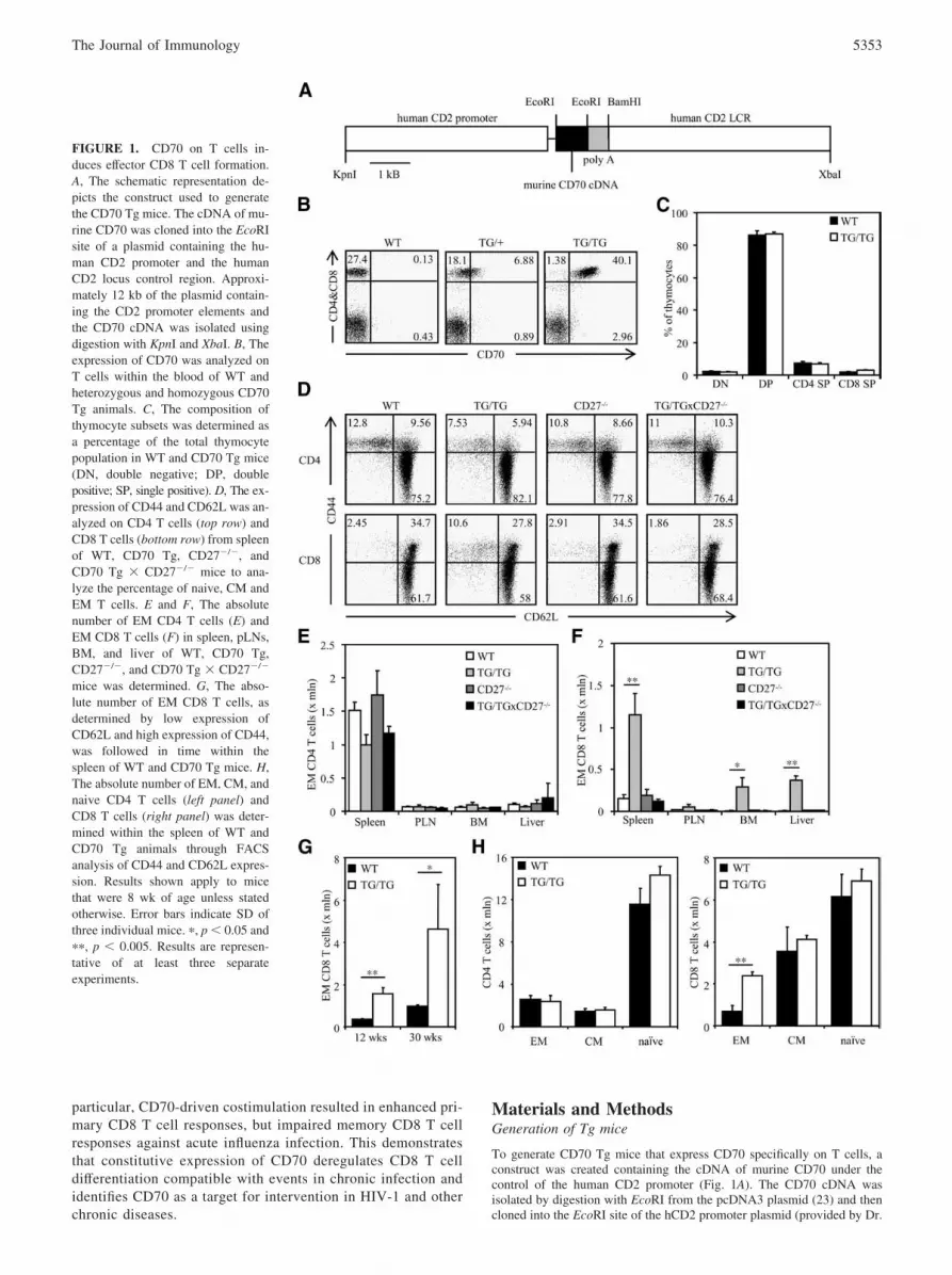
FIGURE 1. CD70 on T cells in-duces effector CD8 T cell formation.A, The schematic representation de-picts the construct used to generatethe CD70 Tg mice. The cDNA of mu-rine CD70 was cloned into the EcoRIsite of a plasmid containing the hu-man CD2 promoter and the humanCD2 locus control region. Approxi-mately 12 kb of the plasmid contain-ing the CD2 promoter elements andthe CD70 cDNA was isolated usingdigestion with KpnI and XbaI. B, Theexpression of CD70 was analyzed onT cells within the blood of WT andheterozygous and homozygous CD70Tg animals. C, The composition ofthymocyte subsets was determined asa percentage of the total thymocytepopulation in WT and CD70 Tg mice(DN, double negative; DP, doublepositive; SP, single positive). D, The ex-pression of CD44 and CD62L was an-alyzed on CD4 T cells (top row) andCD8 T cells (bottom row) from spleenof WT, CD70 Tg, CD27�/�, andCD70 Tg � CD27�/� mice to ana-lyze the percentage of naive, CM andEM T cells. E and F, The absolutenumber of EM CD4 T cells (E) andEM CD8 T cells (F) in spleen, pLNs,BM, and liver of WT, CD70 Tg,CD27�/�, and CD70 Tg � CD27�/�
mice was determined. G, The abso-lute number of EM CD8 T cells, asdetermined by low expression ofCD62L and high expression of CD44,was followed in time within thespleen of WT and CD70 Tg mice. H,The absolute number of EM, CM, andnaive CD4 T cells (left panel) andCD8 T cells (right panel) was deter-mined within the spleen of WT andCD70 Tg animals through FACSanalysis of CD44 and CD62L expres-sion. Results shown apply to micethat were 8 wk of age unless statedotherwise. Error bars indicate SD ofthree individual mice. �, p � 0.05 and��, p � 0.005. Results are represen-tative of at least three separateexperiments.
5353The Journal of Immunology
R. Meuwissen, Netherlands Cancer Institute, Amsterdam, The Nether-lands). The hCD2 promoter plasmid also contains the locus control regionof the human CD2 gene that confers T cell-specific, copy-dependent, andposition-independent gene expression in Tg mice. After linearization andremoval of plasmid sequences by digestion with KpnI and XbaI, the hCD2-mCD70 construct was injected into the pronuclei of fertilized oocytes ofC57BL/6 mice. A founder was identified by Southern blot analysis of tailDNA and mated with C57BL/6 mice to obtain heterozygous CD70 Tgmice. These mice were backcrossed to create homozygous CD70 Tg mice.
C57BL/6/J, OT-I Tg (The Jackson Laboratory), CD27�/� (18), and ho-mozygous CD70 Tg mice and homozygous CD70 Tg mice crossed withOT-I Tg or CD27�/� mice were maintained at specific pathogen-free con-ditions at the animal department of the Academic Medical Center (Am-sterdam, The Netherlands). Screening of the mice for CD70 Tg and OT-ITg expression was performed by flow cytometry of leukocytes from tailvein blood using anti-CD70 Abs (3B9) and anti-V�5 Abs (MR9-4), re-spectively. The CD27 genotype of the mice was screened using PCR ongenomic DNA as described previously (18). Mice were used at 8–12 wk ofage unless stated otherwise and within individual experiments mice werestrictly age matched. All animal experiments were performed according toinstitutional and national guidelines.
Antibodies
The following mAbs from eBioscience were used: anti-CD3 (145-2C11),anti-CD4 (RM4-5), anti-CD8 (53-6.7), anti-CD27 (LG.3A10), anti-CD44(IM7), anti-CD62L (MEL-14), anti-CD69 (H1.2F3), anti-CD70 (FR70),anti-B220 (RA3-6B2), anti-PD-1 (RMP1-30), anti-IL-7R� (A7R34), anti-KLRG1 (2F1), and anti-CD40L (MR1). Anti-IFN-� (XMG1.2), anti-IL-2(JES6-5H4), anti-IL-10 (JES5-16E3), anti-TNF-� (MP6-XT22), and anti-Ki-67 (B56) were purchased from BD Biosciences.
Flow cytometry
Single-cell suspensions were obtained from spleen, peripheral lymph nodes(pLNs), lungs, liver, and bone marrow (BM) by grinding tissue over nylonfilters (BD Biosciences). Contaminating RBC were removed from thesepreparations using erylysis buffer (155 mM NH4Cl, 10 mM KHCO3, and 1mM EDTA). Absolute cell counts were determined by an automated cellcounter (CasyCounter; Innovatis). Cells were stained with the indicatedfluorochrome-conjugated or biotinylated primary Abs in the presence ofanti-CD16/CD32 block (2.4G2, gift from L. Boon, Biosource BV, Utrecht,The Netherlands) for 30 min at 4°C in PBS containing 0.5% BSA. In thecase of biotinylated primary Abs, cells were incubated with fluorochrome-conjugated streptavidin (eBioscience) for 30 min at 4°C in PBS containing0.5% BSA. For staining of the nuclear Ag Ki-67, cells were fixed andpermeabilized for 30 min at 4°C with fixation and permeabilization buffer(eBioscience). Expression was measured using FACSCalibur or Canto flowcytometers (BD Biosciences).
Intracellular cytokine staining
Splenocytes were stimulated with 10 ng/ml PMA (Sigma-Aldrich) and 1�M ionomycin (Sigma-Aldrich) for 1 h. Then, 10 �g/ml brefeldin A (Sigma-Aldrich) was added to prevent cytokine release and after 4 h cells wereharvested and stained with Abs against CD4 and CD8. Next, cells werefixed and permeabilized using Cytofix/Cytoperm (BD Biosciences) andlabeled for intracellular cytokines using specified Abs.
Quantitative PCR
RNA was extracted using the Invisorb Spin Cell RNA Mini Kit (Invitek),cDNA was synthesized using Superscript Reverse Transcriptase II (Invitro-gen) and poly(T) oligonucleotides (Invitrogen), and quantitative real-timeRT-PCR was performed on a LightCycler (Roche). Transcription levelswere obtained using the LightCycler FastStart DNA Master SYBR Greenreagent kit (Roche) and the following primer sets for 18S (forward. 5�-TCAAGAACGAAAGTCGGAGG-3� and reverse, 5�-GGACATCTAAGGGCATCACA-3�); T-bet (forward, 5�-CAACAACCCCTTTGCCAAAG-3� and reverse, 5�-TCCCCCAAGCAGTTGACAGT-3�); Eomes(forward, 5�-TGGACTACCATGGACATCCAGAA-3� and reverse, 5�-TTCTCTTGCAAGCGCTGTTGT-3�); and Blimp-1 (forward, 5�-CCTCATCCCATGCTCAATCCA-3� and reverse, 5�-GGACTACTCTCGTCCTTCATGCT-3�). Values are represented relative to that of 18S, with thelowest experimental value standardized at 1.
Influenza infection
Mice were intranasally infected with 10� 50% tissue culture effective dose(TCID50) of the H1N1 influenza A virus A/PR8/34 for analysis of primaryimmune responses. Heterotypic infection with 100� TCID50 of the H3N2
influenza A virus HKx31 and 10� TCID50 of A/PR8/34 was performed toexamine secondary responses. At fixed time intervals, body weights of theinfected mice were obtained as a measure of disease and blood sampleswere drawn from the tail vein to determine levels of influenza-specific CD8T cells. At the indicated days after infection, mice were sacrificed andblood, spleen, mediastinal lymph nodes (mLNs), and lungs were collectedfor analysis. Viral loads within the lungs were quantified using quantitativePCR as previously described (24). Influenza-specific CD8 T cells wereenumerated using anti-CD8 Abs and PE- or allophycocyanin-conjugatedtetramers of H-2Db containing the influenza-derived peptide NP366–374
ASNENMETM.
Statistical analysis
Figures represent means and error bars denote SD. Student’s t test was usedto analyze for statistical significance. A value of p � 0.05 was consideredstatistically significant.
ResultsCD70 on T cells regulates peripheral T cell homeostasis
To study the impact of persistent CD70 expression on T cells asdescribed in chronic infections (22), we generated Tg mice thatexpress CD70 under control of the human CD2 promoter (Fig.1A). This resulted in expression of CD70 protein on T cells, butnot on other cell types (Fig. 1B). Because the transgene expres-sion of CD70 was low on heterozygous Tg T cells, we generatedhomozygous Tg mice, displaying T cell-specific CD70 expres-sion at higher levels than heterozygous Tg mice (Fig. 1B). Thelevels of CD70 on T cells of homozygous Tg mice were com-parable to expression of CD70 on T cells during chronic infec-tion (22). This prompted us to use homozygous Tg micethroughout the study.
The CD70 transgene was expressed early during development ofT cells within the thymus and was present on all thymocyte subsets(our unpublished data). In wild-type (WT) mice, CD27 is ex-pressed on thymocytes as well as on T cells (12). In contrast, CD70Tg mice did not have expression of CD27 on thymocytes, indi-cating that CD70 induced triggering and shedding of CD27 withinthe thymus (our unpublished data). Interestingly, this did not in-duce apparent changes in thymocyte development and thymocytesubsets were similar in size in WT and CD70 Tg mice (Fig. 1C).
The CD70 transgene was detected on T cells in spleen, pLNsand BM of CD70 Tg mice (our unpublished data). The levels ofCD27 were reduced on T cells within these tissues compared withWT mice (our unpublished data), indicating that the CD70 trans-gene has engaged its ligand. We analyzed whether CD70 on T cellsimpacted the formation of effector memory (EM) T cells similar toCD70 on DCs and B cells (14, 25). Therefore, the profile of CD44and CD62L expression was determined on CD4 and CD8 T cells.CD44 and CD62L characterize distinct T cell populations in mice,and CD44lowCD62Lhigh T cells are defined as naive T cells,CD44highCD62Lhigh T cells as central memory (CM) T cells, andCD44highCD62Llow T cells as EM T cells (26). CD70 Tg mice hadhigher percentages as well as absolute numbers of EM T cellswithin the CD8 compartment, but not within the CD4 compartmentof the spleen (Fig. 1, D–H). The absolute number of EM CD8 Tcells, in contrast to that of EM CD4 T cells, was also increasedwithin the liver and BM, but not the pLNs (Fig. 1, E and F). Weobserved that the absolute number of EM CD8 T cells within thespleen of CD70 Tg animals steadily increased with age, as oc-curred in WT animals (Fig. 1G). In contrast to EM CD8 T cells, thesize of all CD4 T cell populations and of naive and CM CD8 T cellpopulations were not changed in CD70 Tg mice compared withWT mice (Fig. 1H). Crossing the CD70 Tg mice onto theCD27�/� background completely reversed the memory T cell phe-notype to WT and CD27�/� levels (Fig. 1, D–F), showing that EMCD8 T cell formation is mediated by CD70-CD27 signaling. Thus,
5354 CONSTITUTIVE CD70 IMPAIRS CD8 T CELL MEMORY

CD70 expression is functional on T cells and induces increasedEM differentiation of CD8 T cells.
Constitutive CD70 induces CD8 T cells that resemble exhaustedeffector CD8 T cells
We analyzed the phenotype of EM CD8 T cells in CD70 Tg miceto examine how CD8 T cell differentiation under constitutiveCD70 costimulation related to that of chronic infection. We ob-served that EM CD8 T cells of CD70 Tg mice had reduced levelsof IL-7R� and enhanced levels of CD69 and PD-1 compared withthose of WT mice (Fig. 2, A and B). This was not observed onother CD4 and CD8 T cell populations (our unpublished data). Thelow expression of IL-7R� indicates maintenance of EM CD8 Tcells independent of the homeostatic cytokine IL-7 (27), whereashigh expression of CD69 and PD-1 indicates recent stimulation onAg (28). Ag-driven proliferation displays a much higher turnoveras compared with cytokine-driven homeostatic proliferation of
memory CD8 T cells (29). Therefore, to establish whether T cellproliferation was enhanced during constitutive CD70 costimula-tion, we analyzed the expression of Ki-67, which is expressed bycycling cells. The expression of Ki-67 was up-regulated in EMCD8 T cells of CD70 Tg mice compared with WT mice (Fig. 2C).EM CD4 T cells and naive and CM CD8 T cells of CD70 Tg micealso had elevated expression levels of Ki-67, but to a lesser extent(Fig. 2C). This indicates that naive, CM, and EM CD8 T cellpopulations may all contribute to the generation of EM phenotypeCD8 T cells in CD70 Tg mice. The unaltered Ki-67 levels of naiveand CM CD4 T cells and the marginally increased Ki-67 levelsof EM CD4 T cells in CD70 Tg mice correspond with the ab-sence of CD70-driven EM CD4 T cell formation. The expres-sion profile of EM CD8 T cells of CD70 Tg mice is reminiscentof that of pathogen-specific CD8 T cells in chronic LCMV orHIV-1 infection: i.e., low levels of IL-7R� (30), high levels ofKi-67 (31, 32), and high levels of activation-induced molecules
FIGURE 2. CD8 T cells under constitutive CD70 triggering display an exhausted phenotype. A, Histograms show the expression of CD69, PD-1, andIL-7R� on EM CD8 T cells of spleen of WT (top row) and CD70 Tg animals (bottom row). B, The percentage of EM CD8 T cells that express CD69,PD-1, and IL-7R� within spleen of WT and CD70 Tg animals was determined. C, The percentage of CD4 and CD8 T cell subsets of spleen of WT andCD70 Tg that undergo cell division was analyzed using intracellular staining for Ki-67. D, Dot plots show intracellular staining for IFN-� and IL-10 onsplenocytes gated for CD8 T cells of WT (top panel) and CD70 Tg mice (bottom panel) that had been stimulated for 5 h with PMA and ionomycin. E,The percentage of CD8 T cells of spleen of WT and CD70 Tg that coproduce IFN-� and IL-10 upon 5 h of PMA and ionomycin stimulation was determined.F, The expression of CD44 and CD62L was analyzed on CD8 T cells of spleen from WT, CD70 Tg, OT-I, and CD70 Tg � OT-I mice (left panel). Insetsin upper left corner represent average percentage of EM CD8 T cells � SD. The absolute number of CD44�CD62L� EM CD8 T cells in spleen, pLNs,BM, and liver of WT, CD70 Tg, OT-I, and CD70 Tg � OT-I mice was examined (right panel). G, CD8�CD44�CD62L� splenocytes were sorted to obtainEM CD8 T cells. The expression levels of T-bet, Eomes, and Blimp-1 were analyzed in EM CD8 T cells of WT and CD70 Tg mice using quantitative PCR.Results shown apply to mice that were 8 wk of age. Error bars indicate SD of three individual mice. �, p � 0.05 and ��, p � 0.005. Experiments wereperformed at least three times with identical results.
5355The Journal of Immunology

including CD69 (31) and PD-1 (5, 6). Thus, costimulationthrough CD70 enhances the formation of CD8 T cells that phe-notypically resemble Ag-dependent and rapidly proliferating ef-fector CD8 T cells in chronic infection.
Up-regulation of inhibitory molecules is another distinctive fea-ture of CD8 T cells of chronic infections (5–10). We observedup-regulation of the inhibitory molecule PD-1 on EM CD8 T cellsof CD70 Tg mice (Fig. 2, A and B). Therefore, we also analyzedWT and CD70 Tg CD8 T cells for the intracellular expression ofthe inhibitory cytokine IL-10 after short-term PMA and ionomycinstimulation (Fig. 2, D and E). CD8 T cells of both CD70 Tg andWT animals produced IFN-�, but CD8 T cells from CD70 Tganimals uniquely coproduced IL-10 (Fig. 2, D and E). AlthoughEM CD8 T cell numbers were increased, we did not observe en-hanced IFN-� production in CD8 T cells of CD70 Tg mice com-pared with WT mice (Fig. 2, D and E). This shows that CD8 Tcells that are continually stimulated through CD70 have up-regu-lated levels of inhibitory molecules that in chronic infection havebeen shown to induce CD8 T cell exhaustion (8–10).
To examine whether the constitutive CD70-driven activation ofCD8 T cells was indeed Ag dependent, we generated OT-I Tg micecoexpressing the CD70 transgene. The CD8 T cells of OT-I Tgmice contain a Tg TCR that specifically recognizes the MHC classI H-2Kb-restricted OVA peptide OVA257–264 SIINFEKL (33), anAg that they normally do not encounter. We found that the en-hanced EM phenotype of the CD8 T cell compartment of CD70 Tgmice was dependent on TCR triggering, as shown by comparablepercentages and absolute numbers of EM CD8 T cells of OT-I Tgmice and CD70 � OT-I Tg mice (Fig. 2F). Higher levels of EMCD8 T cells were present within spleen, BM, and liver but not thepLNs of CD70 Tg mice compared with CD70 � OT-I Tg mice(Fig. 2F). Thus, recognition of environmental Ags is required forCD70 to drive the formation of CD8 T cells with an EMphenotype.
We next analyzed whether transcription factors involved in CD8T cell development were differentially expressed in EM CD8 T
cells during constitutive CD70-driven costimulation. We observedthat, in contrast to T-bet, Eomes and Blimp-1 were strongly up-regulated in EM CD8 T cells of CD70 Tg mice compared withthose of WT mice (Fig. 2G), as has been previously described forexhausted effector CD8 T cells (34). Taken together, our data showthat constitutive signaling through CD70 and CD27 acceleratesAg-driven formation of CD8 T cells that acquire a phenotype sim-ilar to exhausted CD8 T cells.
Polyfunctional cytokine responses are impaired underCD70-driven costimulation
Exhausted CD8 T cells in chronic infection produce low levels ofcytokines such as IFN-�, IL-2, and TNF-� and display poor cy-totoxicity upon restimulation (4). Polyfunctional analysis of CD8T cells in HIV-1 patients has shown that, in particular, the abilityto produce multiple cytokines such as IFN-�, TNF-�, and IL-2simultaneously is impaired (35). Reduction of the polyfunctionalCD8 T cell response correlates with poorer effector function ofCD8 T cells on a per cell basis (35). Since we found up-regulationof the inhibitory molecules PD-1 and IL-10 on CD8 T cells ofCD70 Tg animals, we examined the polyfunctional T cell responsein WT and CD70 Tg mice. We observed that the IFN-�-producingCD4 and CD8 T cell populations of CD70 Tg mice were lesspolyfunctional than WT mice upon stimulation with PMA andionomycin (Fig. 3, A and B). In particular, the ability of CD4 andCD8 T cells to coproduce TNF-� and IL-2 along with IFN-� washampered (Fig. 3, A and B). The effect was more pronouncedwithin the CD4 T cell population and increased with age withinboth the CD4 and CD8 T cell population (Fig. 3, A and B). Thisshows that constitutive expression of CD70 on T cells inducesexhaustion in CD4 and CD8 T cells.
CD70 enhances effector CD8 T cell responses against influenza
Stimulation through CD70 on APCs quantitatively and qualita-tively enhances CD8 T cell responses against acute viral infection(18, 36). We were interested whether constitutive CD70 on T cells
FIGURE 3. Constitutive CD70-driven costimulationcompromises the polyfunctional cytokine response ofCD4 and CD8 T cells. The intracellular expression ofIFN-�, TNF-�, and IL-2 was analyzed in CD4 T cellsand CD8 T cells of spleen from WT and CD70 Tg an-imals upon 5-h PMA and ionomycin stimulation. A, Dotplots were gated on IFN-�-producing CD4 T cells orCD8 T cells and display the intracellular expression ofTNF-� and IL-2 of representative WT and CD70 Tgmice of 12 and 30 wk of age. B, Pie charts show theaverage percentage of IFN-�-producing CD4 or CD8 Tcells of WT and CD70 Tg mice that express onlyTNF-�, only IL-2, both TNF-� and IL-2, or neitherTNF-� and IL-2. Graphs display the results of two in-dependent experiments with three to five mice pergroup. The reduction of TNF-� and IL-2 expression inCD70 Tg compared with WT mice are significant forCD4 T cells at 12 wk (p � 0.005) and 30 wk (p �0.005) and for CD8 T cells at 30 wk (p � 0.05).
5356 CONSTITUTIVE CD70 IMPAIRS CD8 T CELL MEMORY

was also able to enhance CD8 T cell responses. For this purpose,WT and CD70 Tg mice were intranasally infected with the influ-enza virus A/PR8/34 and as a measure of disease the body weightof the mice was monitored. The decrease in body weight uponinfluenza infection was less severe and resolved at earlier timepoints in CD70 Tg mice than in WT mice (Fig. 4A). In addition,the viral loads of CD70 Tg mice were reduced compared withthose of WT mice at day 10 (Fig. 4B), but not at day 7 or 14 (ourunpublished data). To measure the magnitude of CD8 T cell re-sponses, we used tetramer staining of peripheral blood cells.Within the blood, influenza-specific CD8 T cells peak at higherlevels in CD70 Tg mice than in WT mice, but levels of influenza-specific CD8 T cells of CD70 Tg mice return to WT levels wheninfection is resolved (Fig. 4C). At the peak of the CD8 T cellresponse against influenza, CD70 Tg mice also contained highernumbers of tetramer� CD8 T cells than WT mice within the spleenand mLNs, but not within the lungs (Fig. 4D). Moreover, highernumbers of CD8 T cells in spleen of CD70 Tg mice than of WTmice produced IFN-� upon peptide restimulation (Fig. 4E). Pro-duction of granzyme B and IFN-� upon peptide restimulation wasnot different between lung-derived CD8 T cells of WT and CD70Tg animals, corroborating the tetramer analysis of the lungs (ourunpublished data). We have no direct evidence for involvement ofCD8 T cells in the increased antiviral response of CD70 Tg mice,although the enhanced CD8 T cell response in CD70 Tg micecorrelates with improved clinical performance and viral clearance.Since the influx of granulocytes, monocytes, or macrophageswithin the lungs was not enhanced and Ab responses were notimproved in CD70 Tg mice (our unpublished data), this stronglysuggests that the increased antiviral response is due to the en-hanced CD8 T cell response.
CD70 did not induce further up-regulation of PD-1 expressionon tetramer� CD8 T cells and did not trigger IL-10 production byCD8 T cells upon peptide restimulation (our unpublished data).Analysis of coproduction of IFN-�, TNF-�, and IL-2 upon peptiderestimulation did not reveal differences in the polyfunctional re-sponse between WT and CD70 Tg CD8 T cells of spleen and lungs(our unpublished data). Taken together, this shows that constitu-tive CD70 on T cells provides costimulation and does not induceT cell exhaustion in a setting of acute infection.
Constitutive CD70 induces waning of CD8 T cell memoryagainst influenza
Differentiation pathways of effector and memory CD8 T cellsseparate early after infection. At the peak of the CD8 T cell response,KLRG-1 identifies short-lived effector CD8 T cells (SLECs)and IL-7R� identifies memory precursor effector CD8 T cells(MPECs) (27, 37). Therefore, we analyzed these fractionswithin influenza-specific CD8 T cells at day 10 after primaryinfluenza infection within the blood. We found that constitutiveexpression of CD70 enhanced the generation of SLECs as wellas MPECs (Fig. 5A). This indicates that CD70 has a positiveeffect on memory formation through the generation of largernumbers of memory precursors.
The development of memory is a cardinal feature of CD8 T cellresponses against acute infection and transient CD70-driven co-stimulation has been shown to enhance memory CD8 T cell re-sponses against influenza and acute LCMV infection (17, 18). Toexamine the effect of constitutive CD70 costimulation on memoryCD8 T cell responses, we did a long-term follow-up of tetramer�
CD8 T cells after influenza infection. Remarkably, at day 30,CD70 Tg mice contained fewer total and fewer memory phenotype
FIGURE 4. Enhanced primary CD8 T cell responses develop against influenza in CD70 Tg mice. WT and CD70 Tg mice were intranasally infected withthe influenza virus A/PR8/34. A, The body weight of WT and CD70 Tg mice was followed in time after primary influenza infection. B, Viral loads withinthe lungs of WT and CD70 Tg mice were examined upon primary influenza infection at day 10. C, The percentage of tetramer� CD8 T cells within theblood of influenza-infected WT and CD70 Tg mice was followed in time. D, The absolute numbers of tetramer� CD8 T cells were determined within lungs,spleen, and mLNs of WT vs CD70 Tg mice at day 10 of primary influenza infection. E, The absolute numbers of IFN-�-producing CD8 T cells weredetermined in WT and CD70 Tg spleen of influenza-infected mice upon 5-h restimulation with influenza-specific peptide and IL-2. Results shown applyto mice that were 12 wk of age at the start of the experiment. Error bar indicates SD of eight individual mice. �, p � 0.05 and ��, p � 0.005. Comparableexperiments with similar results were performed three times.
5357The Journal of Immunology

influenza-specific CD8 T cells than WT mice within the blood(Fig. 5B). The number of SLECs at this time point is very low,which corresponds with viral clearance in both WT and CD70 Tg
mice (Fig. 5B). Follow-up of tetramer� CD8 T cells within theblood of WT and CD70 Tg mice beyond 30 days revealed a steadydecline in the number of influenza-specific memory CD8 T cells
FIGURE 5. Constitutive expression of CD70 induces waning of the memory CD8 T cell population over time. A, The absolute number of tetramer� CD8 T cells wasdetermined within the blood at day 10 of primary influenza infection. Based on expression of IL-7R� and KLRG1, the absolute number of IL-7R�lowKLRG1high SLECsand IL-7R�highKLRG1low MPECs was determined within the tetramer� CD8 T cell population. B, Absolute numbers of total, IL-7R�lowKLRG1high SLEC phenotype,and IL-7R�highKLRG1low memory phenotype tetramer� CD8 T cells are shown at day 30 after primary infection with HKx31. C, Long-term follow up is shown of thepercentage of tetramer� CD8 T cells within the blood after primary influenza infection with HKx31 in WT and CD70 Tg mice. D, Absolute numbers of tetramer� CD8T cells were determined within blood, mLNs, spleen, and lungs of WT and CD70 Tg mice at day 57 after primary infection with HKx31. E, Absolute numbers ofspleen-derived CD8 T cells that produce IFN-� upon 5-h restimulation with peptide and IL-2 were determined in WT and CD70 Tg mice that had been infected withHKx31 for 57 days. Results shown apply to mice that were 12 wk of age at the start of the experiment. Error bars indicate SD of five to eight individual mice. �, p � 0.05and ��, p � 0.005. Comparable experiments with similar results were performed three times.
FIGURE 6. Reduced memory CD8 T cell responses develop against influenza in CD70 Tg mice. WT and CD70 Tg mice were intranasally infected withinfluenza virus HKx31 and 51 or 61 days later with the serologically distinct influenza virus A/PR8/34. A, The body weight of WT and CD70 Tg mice was followedin time after the secondary influenza infection. B, Viral loads within the lungs of WT and CD70 Tg mice were examined at days 8 and 12 after the secondaryinfluenza infection. C, The percentage of tetramer� CD8 T cells was determined within the blood of WT and CD70 Tg mice at the indicated time points aftersecondary influenza infection. D, The absolute numbers of tetramer� CD8 T cells were determined within blood, spleen, and lungs of WT vs CD70 Tg mice atday 8 (left panel) and 12 (right panel) of the secondary influenza infection. E, The absolute numbers of IFN-�-producing CD8 T cells were determined upon 5-hrestimulation with influenza peptide and IL-2 in WT and CD70 TG spleen of mice that had undergone a secondary influenza infection for 8 days. Results shownapply to mice that were 12 wk of age at the start of the experiment. Error bars indicate SD of five to eight individual mice. �, p � 0.05 and ��, p � 0.005.Comparable experiments with similar results were performed three times.
5358 CONSTITUTIVE CD70 IMPAIRS CD8 T CELL MEMORY

that was much more pronounced in CD70 Tg mice (Fig. 5C).Around day 60, percentages and absolute numbers of influenza-specific memory CD8 T cells within the blood were �3- to5-fold lower in CD70 Tg mice compared with WT mice (Fig. 5,C and D). Within all other tissues examined such as the lungs,mLNs, and spleen, we detected only very low numbers of in-fluenza-specific memory CD8 T cells within CD70 Tg mice(Fig. 5D). WT animals contained significantly more influenza-specific CD8 T cells within the mLNs and in particular withinthe spleen than CD70 Tg animals (Fig. 5D). Also peptide re-stimulation revealed strongly decreased numbers of IFN-�-pro-ducing influenza-specific CD8 T cells in the spleen of CD70 Tgmice compared with WT mice (Fig. 5E). Thus, despite higherprimary effector CD8 T cell responses and higher levels ofMPECs, maintenance of memory CD8 T cells under constitu-tive CD70 costimulation was severely compromised.
Recall of influenza-specific CD8 T cells is reduced underconstitutive CD70
Reduced maintenance of influenza-specific memory under consti-tutive CD70-driven costimulation may result in compromised sec-ondary responses upon rechallenge with influenza virus. There-
fore, WT and CD70 Tg mice were sequentially infected with theinfluenza virus strains A/PR8/34 and HKx31. The use of serolog-ically distinct virus strains excludes interference by influenza-spe-cific Abs (38). CD70 Tg animals had more pronounced weight lossand higher viral loads at day 8 upon secondary influenza infectionthan WT animals (Fig. 6, A and B). Although CD70 Tg mice un-derwent more severe disease, similar to WT mice, they were ableto recover and cleared the influenza virus by day 12 (Fig. 6B). Thisindicates that in stark contrast to primary CD8 T cell responses,secondary CD8 T cell responses are impaired through constitutiveCD70 triggering. Indeed, percentages of influenza-specific CD8 Tcells within the blood were reduced in CD70 Tg mice comparedwith WT mice early but not late in the secondary response (Fig.6C). Enumeration of influenza-specific CD8 T cells by tetramerstaining at day 8 after rechallenge also showed a severe reductionin absolute numbers within blood and spleen, but not within thelungs of CD70 Tg mice compared with WT mice (Fig. 6D). Thisdifference was not apparent or strongly reduced in all compart-ments at day 12 after rechallenge (Fig. 6D). This shows that recallCD8 T cell responses are delayed in CD70 Tg mice, which reflectsthe reduced memory maintenance in these mice. The numbers ofIFN-�-producing CD8 T cells upon peptide restimulation were
FIGURE 7. Normal primary but reduced memoryCD4 T cell responses develop in CD70 Tg mice. A, Dotplots display intracellular IFN-� and IL-2 expression ofCD4 T cells upon 5-h restimulation of splenocytes withinfluenza virus at days 10 and 119 after primary influ-enza infection. Insets represent average percentage ofcells within quadrant � SD. B, The percentage of IFN-�-producing CD4 T cells of total CD4 T cells is shownupon 5-h restimulation of splenocytes of WT and CD70Tg animals with influenza virus. Splenocytes were iso-lated after 10 (left panel) or 119 days (right panel) ofprimary influenza infection. C, CD40L expression wasexamined on CD4 T cells of WT and CD70 Tg miceafter 5-h stimulation with PMA and ionomycin (leftpanel). Representative histograms of individual WT andCD70 Tg mice display CD40L expression under me-dium (gray line) and PMA and ionomycin conditions(black line) on CD4 T cells (right panel). D, IL-2 ex-pression was analyzed on CD4 T cells of WT and CD70Tg mice by intracellular cytokine staining after PMAand ionomycin stimulation (left panel). Representativehistograms of individual WT and CD70 Tg mice showIL-2 expression in CD4 T cells upon PMA and iono-mycin restimulation (right panel). E, IL-2 expression ofCD4 T cells was determined after 5-h restimulation withinfluenza virus of splenocytes from WT and CD70 Tgmice that had been infected with influenza virus for 10days. Results shown apply to mice that were 12 wk ofage at the start of the experiment. Error bars indicate SDof three to eight mice. �, p � 0.05. Experiments wererepeated at least once with similar results.
5359The Journal of Immunology

also reduced within the spleen of CD70 Tg mice at day 8 (Fig. 6E).Within the lungs, we did not detect differences in cytokine- orgranzyme B-producing CD8 T cells, reflecting the numbers oftetramer� CD8 T cells at this site (our unpublished data). Thus,maintenance of memory CD8 T cells was impaired and, therefore,secondary CD8 T cell responses were delayed but not abolishedunder constitutive CD70-driven costimulation.
Constitutive CD70 reduces CD4 T cell memory but does notimpair CD4 T cell help
In the absence of CD4 T cell help, primary CD8 T cell responseswere normal, but secondary CD8 T cell responses were severelycompromised (39). To examine whether CD70 impaired CD4 Tcell help, we analyzed the CD4 T cell response against influenza.Spleens of WT and CD70 Tg animals contained equal numbers ofinfluenza-specific CD4 T cells at the peak of the response (Fig. 7,A and B). At late time points after infection, the percentage ofinfluenza-specific CD4 T cells in spleens of CD70 Tg mice haddeclined �4-fold below those of WT mice (Fig. 7, A and B). Thisshows that CD4 and CD8 T cells respond similarly in that they areunable to maintain their memory population, but that CD70 co-stimulation does not induce helpless CD8 T cell responses throughelimination of CD4 T cells.
CD4 T cell help includes signaling through CD40L and IL-2(40–43). Therefore, we analyzed expression of these molecules onCD4 T cells of WT and CD70 Tg mice. CD4 T cells under con-stitutive CD70-driven costimulation up-regulated IL-2 and CD40Lupon restimulation with PMA and ionomycin, although levels ofCD40L were higher and levels of IL-2 were lower compared withWT mice (Fig. 7, C and D). The lower levels of IL-2 may representfunctional exhaustion of EM-type CD4 T cells in CD70 Tg mice.However, this was not observed in influenza-specific CD4 T cellsof CD70 Tg mice at the peak of the response (Fig. 7, A and E).Thus, we have no evidence that constitutive triggering throughCD70 impairs CD4 T cell help.
DiscussionIn the present study, we have investigated the impact of constitu-tive expression of the costimulatory molecule CD70 on T cells.CD70 on APCs acts as a costimulatory molecule and induces EMCD8 T cell formation with enhanced effector function (14, 15). Wehave observed that CD70 on T cells is functional as well andinduced costimulation that resulted in EM CD8 T cell differenti-ation and in enhanced primary CD8 T cell responses against in-fluenza. T cells are non-APCs that in mice do not present Ags toother T cells. This indicates that CD8 T cells acquire TCR stim-ulation separate from CD70-driven costimulation and that this sim-ilar to TCR and CD70 triggering by the same APC induces EMCD8 T cell differentiation. Indeed, in trans costimulation of CD8T cells by CD70 has been observed by others using soluble CD70Ig constructs (36). On APCs, codelivery of CD70 and MHC classII molecules to the immunological synapse with T cells occurs,underlining the hypothesis that coexpression of MHC class II andCD70 is required for CD4 T cell activation (44). We did not findthat CD70 on T cells significantly enhanced EM formation of CD4T cells. Constitutive expression of T cell-specific CD70 did notenhance the numbers of EM CD4 T cells within spleen, BM, andat peripheral sites such as the liver. Although CD70 on T cellsenhanced influenza-specific CD8 T cell responses, it did not en-hance virus-specific CD4 T cell responses against primary influ-enza infection. In contrast, CD70 on B cells and DCs mediatesdifferentiation of naive CD4 T cells into Th1-type T cells thatproduce IFN-� (14, 16). Possibly, CD4 T cell activation requires asingle APC to provide TCR stimulation and CD70 costimulation.
The size and activation state of other leukocyte populations areaffected in the CD70 Tg mice as has been described for CD70 Tgmice that express CD70 on B cells (14). Specifically, the levels ofB cells but not of other APCs such as monocytes and macrophagesare decreased in CD70 Tg mice. Remaining B cells, monocytes,and macrophages in CD70 Tg mice express enhanced levels ofMHC class II, indicating an elevated activation state (Ref. 14 andour unpublished data). This is the indirect consequence of en-hanced IFN-� signaling in CD70 Tg mice (Ref. 14 and our un-published data). Importantly, formation of EM CD8 T cells is notaltered in the absence of IFN-�, indicating that enhanced EM CD8T cell formation occurs directly through CD70-driven costimula-tion rather than indirectly through enhanced activation of APCs(Ref. 14 and our unpublished data).
Little is known on how costimulation through CD70 and CD27affects the differentiation pathway of naive CD8 T cells into ef-fector and memory cells. In this study, we showed that introduc-tion of CD70 enhances the formation of effector memory pheno-type CD8 T cells. Based on further analysis using molecules thatare expressed on effector cells such as CD69 and PD-1 and mol-ecules that are present on memory cells such as IL-7R�, it can beargued that these cells are effector rather than EM CD8 T cells. Agis required for the formation and maintenance of effector CD8 Tcells (29). We observed that CD70 required Ag to induce CD8 Tcell differentiation. Ag-driven proliferation of CD8 T cells istightly linked with differentiation into effector phenotype T cells.We do not know whether CD70 on T cells induces naive, CM, orEM CD8 T cells to differentiate into effector CD8 T cells. How-ever, analysis of Ki-67 expression showing elevated proliferationof naive, CM, and EM CD8 T cells may indicate that all of thesepopulations are stimulated to generate effector CD8 T cells underconstitutive expression of CD70.
We showed that EM CD8 T cells under constitutive CD70-driven costimulation have a phenotype remarkably similar to Ag-specific CD8 T cells in HIV-1 infection and other chronic infec-tions. EM phenotype CD8 T cells that develop during constitutiveCD70 triggering expressed inhibitory molecules that are involvedin the induction of T cell exhaustion in chronic infection such asPD-1 and IL-10. These molecules functionally impair effector CD8T cells in chronic infection, resulting in low production of IL-2 andIFN-� and poor cytotoxicity, and this prevents pathogen clearance(8–10). The level of cytokine production on a per cell basis and thenumber of cytokines, importantly IFN-�, TNF-�, and IL-2, cop-roduced by individual cells are major determinants of the strengthof CD8 T cell responses (45). Comparison of IFN-�-producingCD4 and CD8 T cells of CD70 Tg mice with those of WT micerevealed a reduced ability to coproduce TNF-� and IL-2 indicativeof functional exhaustion. The induction of functional impairmentof T cells likely requires the presence of persistent Ag. In theabsence of persistent Ag, such as during influenza responses, wedid not observe that constitutive CD70-driven costimulation re-sulted in CD8 T cell exhaustion. Introduction of CD70 in acuteinfluenza infection did not induce PD-1 and IL-10 up-regulation(our unpublished data). Moreover, polyfunctional analysis of thespleen and lung-resident influenza-specific CD8 T cells did notreveal functional exhaustion during primary and secondary re-sponses (our unpublished data).
Survival of CD8 T cells in chronic infection depends upon con-tinuous stimulation through persistent Ag (29). Ag-dependent sur-vival of CD8 T cells in chronic infection likely requires contribu-tion of signals from inflammatory cytokines and costimulatorymolecules. We hypothesize that CD70 provides such signals andenables Ag-dependent maintenance of CD8 T cells and that thisdevelopmental pathway may ultimately result in T cell exhaustion.
5360 CONSTITUTIVE CD70 IMPAIRS CD8 T CELL MEMORY

We reported previously that CD70 drives progressive effector Tcell formation that eventually results in depletion of T cells (46).Progressive accumulation of EM CD8 T cells also occurred inCD70 Tg mice with CD70 on T cells, although not as dramatic asin CD70 Tg mice with CD70 on B cells. This may reflect expres-sion levels of CD70, which are higher in B cell CD70 Tg mice orcell-specific expression of the CD70 transgene. Taken togetherwith our current results, this provides a strong argument that ex-haustion and depletion of T cells that prevent viral clearance inHIV-1 infection and other chronic infections result from CD70-driven immune activation.
We have shown that constitutive signaling through CD70 andCD27 is detrimental for long-term CD8 T cell-dependent immu-nity as it prevents formation of memory CD8 T cells. In strikingresemblance, the Ag-specific T cell population in chronic infectionsuch as with LCMV does not contain a memory T cell subset thatsurvives independently of Ag (3). The reason for the lack of mem-ory T cells in chronic infection is unknown and this may resultfrom impaired development or from elimination of memory CD8T cells. We did not observe that CD70 induced preferential effectorcell differentiation or impaired memory development during acuteinfection with influenza. Rather CD70 enhanced formation ofmemory precursors early in the primary response. We were alsounable to find evidence that CD70 impaired CD4 T cell responsesthat provide essential help for memory CD8 T cell responsesagainst viruses including influenza (39, 47). CD4 T cell removal inchronic LCMV infection results in exacerbation of disease (4),indicating that CD4 T cell help is functional during chronic infec-tion as well. This argues against defective development of memoryCD8 T cells through CD70 signaling and in chronic infection.Indeed, some evidence indicates that elimination of memory CD8T cells underlies the memory defect. Although FasL and Fas nor-mally do not mediate T cell apoptosis upon acute infection, thispathway of T cell apoptosis was observed upon acute influenzainfection in CD70 Tg mice (48), and, recently, it has been pro-posed that FasL and Fas contribute to apoptosis of Ag-specific Tcells in chronic infection (49–51). It will be interesting to analyzewhether FasL and Fas also remove memory T cells upon triggeringthrough CD70 and in chronic infection. However, it needs to bementioned that blockade of FasL- and Fas-dependent apoptosis hasalso been shown as a mechanism of CD70 to enhance memoryCD8 T cell responses (52).
Impairment of CD8 T cell memory through CD70 signaling is instriking contrast to earlier findings that demonstrated that CD70enhanced memory CD8 T cell responses. Ablation of CD70-drivencostimulation using CD27�/� mice resulted in reduced primaryand secondary CD8 T cell responses against influenza and reducedsecondary responses against acute LCMV (17, 18). A major dif-ference with these studies is the expression of CD70 that in acuteLCMV and influenza infection is found transiently on low per-centages of APCs and T cells (17, 19). Signaling through CD70and CD27 is regulated through expression of CD70 and, thus, dif-ferences in the expression level of CD70 and in the window ofCD70 expression may influence the outcome of immune re-sponses. Constitutive high levels of CD70 expression such as inCD70 Tg animals and in chronic infection may result in over-stimulation of CD4 and CD8 T cells and consequently immuno-pathology. Indeed, in striking contrast to acute LCMV, CD27�/�
mice are protected against chronic LCMV infection (53). Thesecretion of copious amounts of IFN-� and TNF-� by CD4 T cellsthat was attributed to CD70-driven costimulation resulted in dis-ruption of the production of neutralizing Abs (53). Also, memoryCD8 T cell development during chronic LCMV was restored in theabsence of CD27 signaling, but this was considered secondary to
the development of a neutralizing Ab response (53). Our experi-mental setup prevents neutralizing Abs from contributing to thesecondary response against influenza, demonstrating that CD70-induced immunopathology includes impairment of memory CD8 Tcell formation. Thus, under high and constitutive expression levelssuch as occur in chronic infection, CD70 impairs rather than en-hances memory CD8 T cell responses.
In conclusion, CD70 is functional as a costimulatory moleculeon T cells and enhances effector CD8 T cell mediated-immuneresponses, but abrogates long-term protection through impairmentof CD8 T cell memory. Peptide immunization in the presence ofsoluble CD70 results in strong primary and secondary responses(36). These adjuvant properties of CD70 have fueled the idea thatcostimulation through CD70 and CD27 can be harnessed as a strat-egy to break tolerance in the treatment of tumors (54). However,defective maintenance of long-term memory after constitutiveCD70-driven costimulation warrants caution regarding thestrength and duration of the therapeutic use of CD70 in vaccinationstrategies. Moreover, our results indicate that treatment of chronicinfection or chronic autoimmune disease may benefit from block-ade rather than activation of the costimulatory CD70-CD27 axis.
AcknowledgmentsWe thank Cathrien Beishuizen, Natasja Kragten, Felix Wensveen, Alex deBruin, Sten Libregts and Michiel van Oosterwijk for technical assistance,Gijs van Schijndel for making the influenza-specific tetramers and the staffof the animal facility of the AMC for excellent animal care. We thankLouis Boon, Mireille Toebes, and Ton Schumacher for providing essentialreagents. Finally, we thank Drs. Monika Wolkers and Hanneke Schuite-maker for critical reading of this manuscript and helpful discussions.
DisclosuresThe authors have no financial conflict of interest.
References1. Lefrancois, L. 2006. Development, trafficking, and function of memory T-cell
subsets. Immunol. Rev. 211: 93–103.2. Seder, R. A., and R. Ahmed. 2003. Similarities and differences in CD4� and
CD8� effector and memory T cell generation. Nat. Immunol. 4: 835–842.3. Wherry, E. J., D. L. Barber, S. M. Kaech, J. N. Blattman, and R. Ahmed. 2004.
Antigen-independent memory CD8 T cells do not develop during chronic viralinfection. Proc. Natl. Acad. Sci. USA 101: 16004–16009.
4. Zajac, A. J., J. N. Blattman, K. Murali-Krishna, D. J. Sourdive, M. Suresh,J. D. Altman, and R. Ahmed. 1998. Viral immune evasion due to persistence ofactivated T cells without effector function. J. Exp. Med. 188: 2205–2213.
5. Day, C. L., D. E. Kaufmann, P. Kiepiela, J. A. Brown, E. S. Moodley, S. Reddy,E. W. Mackey, J. D. Miller, A. J. Leslie, C. DePierres, et al. 2006. PD-1 expres-sion on HIV-specific T cells is associated with T-cell exhaustion and diseaseprogression. Nature 443: 350–354.
6. Trautmann, L., L. Janbazian, N. Chomont, E. A. Said, S. Gimmig, B. Bessette,M. R. Boulassel, E. Delwart, H. Sepulveda, R. S. Balderas, et al. 2006. Upregu-lation of PD-1 expression on HIV-specific CD8� T cells leads to reversibleimmune dysfunction. Nat. Med. 12: 1198–1202.
7. Graziosi, C., G. Pantaleo, K. R. Gantt, J. P. Fortin, J. F. Demarest, O. J. Cohen,R. P. Sekaly, and A. S. Fauci. 1994. Lack of evidence for the dichotomy of TH1and TH2 predominance in HIV-infected individuals. Science 265: 248–252.
8. Barber, D. L., E. J. Wherry, D. Masopust, B. Zhu, J. P. Allison, A. H. Sharpe,G. J. Freeman, and R. Ahmed. 2006. Restoring function in exhausted CD8 T cellsduring chronic viral infection. Nature 439: 682–687.
9. Brooks, D. G., M. J. Trifilo, K. H. Edelmann, L. Teyton, D. B. McGavern, andM. B. Oldstone. 2006. Interleukin-10 determines viral clearance or persistence invivo. Nat. Med. 12: 1301–1309.
10. Ejrnaes, M., C. M. Filippi, M. M. Martinic, E. M. Ling, L. M. Togher, S. Crotty,and M. G. von Herrath. 2006. Resolution of a chronic viral infection after inter-leukin-10 receptor blockade. J. Exp. Med. 203: 2461–2472.
11. Lang, K. S., M. Recher, A. A. Navarini, N. L. Harris, M. Lohning, T. Junt,H. C. Probst, H. Hengartner, and R. M. Zinkernagel. 2005. Inverse correlationbetween IL-7 receptor expression and CD8 T cell exhaustion during persistentantigen stimulation. Eur. J. Immunol. 35: 738–745.
12. Gravestein, L. A., J. D. Nieland, A. M. Kruisbeek, and J. Borst. 1995. NovelmAbs reveal potent co-stimulatory activity of murine CD27. Int. Immunol. 7:551–557.
13. van Lier, R. A., J. Borst, T. M. Vroom, H. Klein, P. van Mourik,W. P. Zeijlemaker, and C. J. Melief. 1987. Tissue distribution and biochemicaland functional properties of Tp55 (CD27), a novel T cell differentiation antigen.J. Immunol. 139: 1589–1596.
5361The Journal of Immunology

14. Arens, R., K. Tesselaar, P. A. Baars, G. M. van Schijndel, J. Hendriks, S. T. Pals,P. Krimpenfort, J. Borst, M. H. van Oers, and R. A. van Lier. 2001. ConstitutiveCD27/CD70 interaction induces expansion of effector-type T cells and results inIFN�-mediated B cell depletion. Immunity 15: 801–812.
15. Arens, R., K. Schepers, M. A. Nolte, M. F. van Oosterwijk, R. A. van Lier,T. N. Schumacher, and M. H. van Oers. 2004. Tumor rejection induced by CD70-mediated quantitative and qualitative effects on effector CD8� T cell formation.J. Exp. Med. 199: 1595–1605.
16. Soares, H., H. Waechter, N. Glaichenhaus, E. Mougneau, H. Yagita,O. Mizenina, D. Dudziak, M. C. Nussenzweig, and R. M. Steinman. 2007. Asubset of dendritic cells induces CD4� T cells to produce IFN-� by an IL-12-independent but CD70-dependent mechanism in vivo. J. Exp. Med. 204:1095–1106.
17. Matter, M., S. Mumprecht, D. D. Pinschewer, V. Pavelic, H. Yagita,S. Krautwald, J. Borst, and A. F. Ochsenbein. 2005. Virus-induced polyclonal Bcell activation improves protective CTL memory via retained CD27 expressionon memory CTL. Eur. J. Immunol. 35: 3229–3239.
18. Hendriks, J., L. A. Gravestein, K. Tesselaar, R. A. van Lier, T. N. Schumacher,and J. Borst. 2000. CD27 is required for generation and long-term maintenanceof T cell immunity. Nat. Immunol. 1: 433–440.
19. Hendriks, J., Y. Xiao, J. W. Rossen, K. F. van der Sluijs, K. Sugamura, N. Ishii,and J. Borst. 2005. During viral infection of the respiratory tract, CD27, 4-1BB,and OX40 collectively determine formation of CD8� memory T cells and theircapacity for secondary expansion. J. Immunol. 175: 1665–1676.
20. Lee, W. W., Z. Z. Yang, G. Li, C. M. Weyand, and J. J. Goronzy. 2007. Un-checked CD70 expression on T cells lowers threshold for T cell activation inrheumatoid arthritis. J. Immunol. 179: 2609–2615.
21. Oelke, K., Q. Lu, D. Richardson, A. Wu, C. Deng, S. Hanash, and B. Richardson.2004. Overexpression of CD70 and overstimulation of IgG synthesis by lupus Tcells and T cells treated with DNA methylation inhibitors. Arthritis Rheum. 50:1850–1860.
22. Wolthers, K. C., S. A. Otto, S. M. Lens, D. N. Kolbach, R. A. van Lier,F. Miedema, and L. Meyaard. 1996. Increased expression of CD80, CD86 andCD70 on T cells from HIV-infected individuals upon activation in vitro: regu-lation by CD4� T cells. Eur. J. Immunol. 26: 1700–1706.
23. Tesselaar, K., L. A. Gravestein, G. M. van Schijndel, J. Borst, and R. A. van Lier.1997. Characterization of murine CD70, the ligand of the TNF receptor familymember CD27. J. Immunol. 159: 4959–4965.
24. van der Sluijs, K. F., L. J. van Elden, M. Nijhuis, R. Schuurman, J. M. Pater,S. Florquin, M. Goldman, H. M. Jansen, R. Lutter, and P. T. van der Poll. 2004.IL-10 is an important mediator of the enhanced susceptibility to pneumococcalpneumonia after influenza infection. J. Immunol. 172: 7603–7609.
25. Keller, A. M., A. Schildknecht, Y. Xiao, M. van den Broek, and J. Borst. 2008.Expression of costimulatory ligand CD70 on steady-state dendritic cells breaksCD8� T cell tolerance and permits effective immunity. Immunity 29: 934–946.
26. Sprent, J., and D. F. Tough. 1994. Lymphocyte life-span and memory. Science265: 1395–1400.
27. Kaech, S. M., J. T. Tan, E. J. Wherry, B. T. Konieczny, C. D. Surh, andR. Ahmed. 2003. Selective expression of the interleukin 7 receptor identifieseffector CD8 T cells that give rise to long-lived memory cells. Nat. Immunol. 4:1191–1198.
28. Agata, Y., A. Kawasaki, H. Nishimura, Y. Ishida, T. Tsubata, H. Yagita, andT. Honjo. 1996. Expression of the PD-1 antigen on the surface of stimulatedmouse T and B lymphocytes. Int. Immunol. 8: 765–772.
29. Shin, H., S. D. Blackburn, J. N. Blattman, and E. J. Wherry. 2007. Viral antigenand extensive division maintain virus-specific CD8 T cells during chronic infec-tion. J. Exp. Med. 204: 941–949.
30. Paiardini, M., B. Cervasi, H. Albrecht, A. Muthukumar, R. Dunham, S. Gordon,H. Radziewicz, G. Piedimonte, M. Magnani, M. Montroni, et al. 2005. Loss ofCD127 expression defines an expansion of effector CD8� T cells in HIV-infectedindividuals. J. Immunol. 174: 2900–2909.
31. Orendi, J. M., A. C. Bloem, J. C. Borleffs, F. J. Wijnholds, N. M. de Vos,H. S. Nottet, M. R. Visser, H. Snippe, J. Verhoef, and C. A. Boucher. 1998.Activation and cell cycle antigens in CD4� and CD8� T cells correlate withplasma human immunodeficiency virus (HIV-1) RNA level in HIV-1 infection.J. Infect. Dis. 178: 1279–1287.
32. Sachsenberg, N., A. S. Perelson, S. Yerly, G. A. Schockmel, D. Leduc,B. Hirschel, and L. Perrin. 1998. Turnover of CD4� and CD8� T lymphocytesin HIV-1 infection as measured by Ki-67 antigen. J. Exp. Med. 187: 1295–1303.
33. Hogquist, K. A., S. C. Jameson, W. R. Heath, J. L. Howard, M. J. Bevan, andF. R. Carbone. 1994. T cell receptor antagonist peptides induce positive selection.Cell 76: 17–27.
34. Wherry, E. J., S. J. Ha, S. M. Kaech, W. N. Haining, S. Sarkar, V. Kalia,S. Subramaniam, J. N. Blattman, D. L. Barber, and R. Ahmed. 2007. Molecularsignature of CD8� T cell exhaustion during chronic viral infection. Immunity 27:670–684.
35. Betts, M. R., M. C. Nason, S. M. West, S. C. De Rosa, S. A. Migueles,J. Abraham, M. M. Lederman, J. M. Benito, P. A. Goepfert, M. Connors, et al.2006. HIV nonprogressors preferentially maintain highly functional HIV-specificCD8� T cells. Blood 107: 4781–4789.
36. Rowley, T. F., and A. Al-Shamkhani. 2004. Stimulation by soluble CD70 pro-motes strong primary and secondary CD8� cytotoxic T cell responses in vivo.J. Immunol. 172: 6039–6046.
37. Joshi, N. S., W. Cui, A. Chandele, H. K. Lee, D. R. Urso, J. Hagman, L. Gapin,and S. M. Kaech. 2007. Inflammation directs memory precursor and short-livedeffector CD8� T cell fates via the graded expression of T-bet transcription factor.Immunity 27: 281–295.
38. Flynn, K. J., G. T. Belz, J. D. Altman, R. Ahmed, D. L. Woodland, andP. C. Doherty. 1998. Virus-specific CD8� T cells in primary and secondaryinfluenza pneumonia. Immunity 8: 683–691.
39. Janssen, E. M., E. E. Lemmens, T. Wolfe, U. Christen, M. G. von Herrath, andS. P. Schoenberger. 2003. CD4� T cells are required for secondary expansion andmemory in CD8� T lymphocytes. Nature 421: 852–856.
40. Bennett, S. R., F. R. Carbone, F. Karamalis, R. A. Flavell, J. F. Miller, andW. R. Heath. 1998. Help for cytotoxic-T-cell responses is mediated by CD40signalling. Nature 393: 478–480.
41. Ridge, J. P., R. F. Di, and P. Matzinger. 1998. A conditioned dendritic cell canbe a temporal bridge between a CD4� T-helper and a T-killer cell. Nature 393:474–478.
42. Schoenberger, S. P., R. E. Toes, E. I. van der Voort, R. Offringa, and C. J. Melief.1998. T-cell help for cytotoxic T lymphocytes is mediated by CD40-CD40Linteractions. Nature 393: 480–483.
43. Williams, M. A., A. J. Tyznik, and M. J. Bevan. 2006. Interleukin-2 signalsduring priming are required for secondary expansion of CD8� memory T cells.Nature 441: 890–893.
44. Keller, A. M., T. A. Groothuis, E. A. Veraar, M. Marsman, L. M. de Buy Wenniger,H. Janssen, J. Neefjes, and J. Borst. 2007. Costimulatory ligand CD70 is delivered to theimmunological synapse by shared intracellular trafficking with MHC class II molecules.Proc. Natl. Acad. Sci. USA 104: 5989–5994.
45. Seder, R. A., P. A. Darrah, and M. Roederer. 2008. T-cell quality in memory andprotection: implications for vaccine design. Nat. Rev. Immunol. 8: 247–258.
46. Tesselaar, K., R. Arens, G. M. van Schijndel, P. A. Baars, M. A. van der Valk,J. Borst, M. H. van Oers, and R. A. van Lier. 2003. Lethal T cell immunodefi-ciency induced by chronic costimulation via CD27-CD70 interactions. Nat. Im-munol. 4: 49–54.
47. Belz, G. T., D. Wodarz, G. Diaz, M. A. Nowak, and P. C. Doherty. 2002. Com-promised influenza virus-specific CD8�-T-cell memory in CD4�-T-cell-deficientmice. J. Virol. 76: 12388–12393.
48. Arens, R., P. A. Baars, M. Jak, K. Tesselaar, E. I. van der Voort, M. H. van Oers,and R. A. van Lier. 2005. Cutting edge: CD95 maintains effector T cell ho-meostasis in chronic immune activation. J. Immunol. 174: 5915–5920.
49. Hughes, P. D., G. T. Belz, K. A. Fortner, R. C. Budd, A. Strasser, and P. Bouillet.2008. Apoptosis regulators Fas and Bim cooperate in shutdown of chronic im-mune responses and prevention of autoimmunity. Immunity 28: 197–205.
50. Hutcheson, J., J. C. Scatizzi, A. M. Siddiqui, G. K. Haines, III, T. Wu, Q. Z. Li,L. S. Davis, C. Mohan, and H. Perlman. 2008. Combined deficiency of proapop-totic regulators Bim and Fas results in the early onset of systemic autoimmunity.Immunity 28: 206–217.
51. Weant, A. E., R. D. Michalek, I. U. Khan, B. C. Holbrook, M. C. Willingham,and J. M. Grayson. 2008. Apoptosis regulators Bim and Fas function concurrentlyto control autoimmunity and CD8� T cell contraction. Immunity 28: 218–230.
52. Dolfi, D. V., A. C. Boesteanu, C. Petrovas, D. Xia, E. A. Butz, and P. D. Katsikis.2008. Late signals from CD27 prevent Fas-dependent apoptosis of primary CD8�
T cells. J. Immunol. 180: 2912–2921.53. Matter, M., B. Odermatt, H. Yagita, J. M. Nuoffer, and A. F. Ochsenbein. 2006.
Elimination of chronic viral infection by blocking CD27 signaling. J. Exp. Med.203: 2145–2155.
54. French, R. R., V. Y. Taraban, G. R. Crowther, T. F. Rowley, J. C. Gray,P. W. Johnson, A. L. Tutt, A. Al-Shamkhani, and M. J. Glennie. 2007. Erad-ication of lymphoma by CD8 T cells following anti-CD40 monoclonal anti-body therapy is critically dependent on CD27 costimulation. Blood 109:4810 – 4815.
5362 CONSTITUTIVE CD70 IMPAIRS CD8 T CELL MEMORY


