Embed Size (px)
Citation preview
Biochimica et Biophysica Acta 1793 (2009) 625–635
Contents lists available at ScienceDirect
Biochimica et Biophysica Acta
j ourna l homepage: www.e lsev ie r.com/ locate /bbamcr
Review
Proteomics of the lysosome
Torben Lübke a, Peter Lobel b,c, David E. Sleat b,c,⁎a Zentrum Biochemie und Molekulare Zellbiologie, Abteilung Biochemie II, Georg-August Universität Göttingen, 37073 Göttingen, Germanyb Center for Advanced Biotechnology and Medicine, Piscataway, NJ 08854, USAc Department of Pharmacology, University of Medicine and Dentistry of New Jersey - Robert Wood Johnson Medical School, Piscataway, NJ 08854, USA
⁎ Corresponding author. Tel.: +1 732 235 5028.E-mail address: [email protected] (D.E. Sleat).
0167-4889/$ – see front matter © 2008 Elsevier B.V. Adoi:10.1016/j.bbamcr.2008.09.018
a b s t r a c t
a r t i c l e i n f oArticle history:
Defects in lysosomal functi Received 16 May 2008Received in revised form 24 September 2008Accepted 30 September 2008Available online 15 October 2008Keywords:Lysosomal proteinProteomicMass spectrometryLysosomal storage diseaseMannose-6 phosphate receptorsubcellular fractionation
on have been associated with numerous monogenic human diseases typicallyclassified as lysosomal storage diseases. However, there is increasing evidence that lysosomal proteins arealso involved in more widespread human diseases including cancer and Alzheimer disease. Thus, there is acontinuing interest in understanding the cellular functions of the lysosome and an emerging approach to thisis the identification of its constituent proteins by proteomic analyses. To date, the mammalian lysosome hasbeen shown to contain ∼60 soluble luminal proteins and ∼25 transmembrane proteins. However, recentproteomic studies based upon affinity purification of soluble components or subcellular fractionation toobtain both soluble and membrane components suggest that there may be many more of both classes ofprotein resident within this organelle than previously appreciated. Discovery of such proteins has importantimplications for understanding the function and the dynamics of the lysosome but can also lead the waytowards the discovery of the genetic basis for human diseases of hitherto unknown etiology. Here, wedescribe current approaches to lysosomal proteomics and data interpretation and review the new lysosomalproteins that have recently emerged from such studies.
© 2008 Elsevier B.V. All rights reserved.
1. Introduction to the lysosome
Discovered by Christian de Duve over 50 years ago [1], thelysosome is a cytoplasmic cellular organelle that has risen toprominence because of its critical role in cellular function and tissuehomeostasis as well as its involvement in numerous human diseases(reviewed in [2,3]). Present in all nucleated eukaryotic cells, thelysosome is delimited by a single-layer lipid membrane and has anacidic internal pH (∼5) that is maintained by an ATP-dependentproton pump. The primary cellular function of the lysosome is thedegradation and recycling of macromolecules obtained by endocy-tosis, autophagy and other cellular trafficking pathways. Severalclasses of macromolecules are hydrolyzed including proteins, poly-saccharides, lipids and nucleic acids and this is achieved by theconcerted action of numerous soluble catabolic enzymes within thelumen of the lysosome, collectively termed acid hydrolases. Acidhydrolases have evolved to function in the low pH of this organelleand possess a wide variety of enzymatic properties. Over 60 of theseenzymes and soluble accessory proteins have been described to date.A number of studies have investigated the proteome of solublelysosomal proteins from a variety of mammalian tissues and cell types([4]; reviewed in [5]) revealing that, for the most part, these proteinstend to be fairly ubiquitous. However, some lysosomal proteins have
ll rights reserved.
very limited tissue distribution and perform specialized cellularfunctions e.g. the granzymes of immune cells [6].
In addition to the soluble luminal proteins, many integral andperipheral membrane proteins are associated with the lysosome andhave a variety of functions including catalysis, transmembranetransport of substrates and digestion products, establishment of pHgradients, vesicular transport and maintenance of lysosomal struc-tural integrity [3].
Classical biochemical and genetic analyses have resulted in thecharacterization of numerous components of the lysosome. Morerecently, the application of highly-sensitive proteomic approaches haslead to the identification of many new proteins that may function inthis compartment and for some, lysosomal localization has now beenverified. It is these new lysosomal proteins that form the focus of thisreview. Approaches to the discovery and validation of lysosomalcandidates have recently been reviewed in depth [5] and for the mostpart, will be outlined in brief here.
2. Lysosomes and human disease
The lysosomal system is of considerable biomedical importance asalterations in lysosomes and lysosomal proteins are associated withnumerous human diseases (Table 1) [3,7]. To date, over 50 monogenichuman genetic diseases have been identified that are primarilyassociated with lysosomal dysfunction and the majority of these areclassified as lysosomal storage diseases (LSDs). Here, deficiencies inlysosomal proteins, most commonly soluble luminal ones, result in an

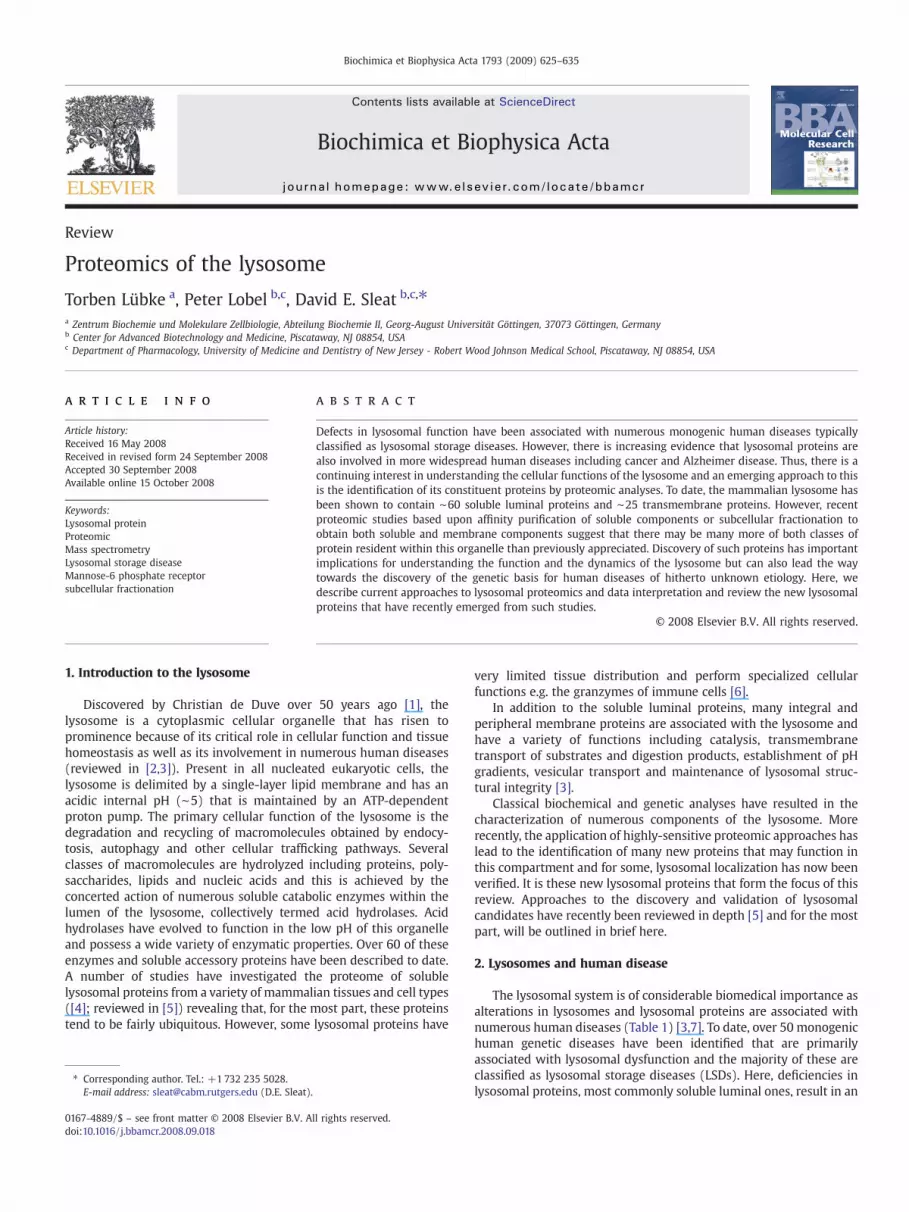
Table 1Established lysosomal proteins and associated human disorders
Protein Disease Protein type Protein function Man6-P
ABCA2 Membrane Lipid metabolism?ABCB9/TAP-like transporter Membrane Antigen processingATPase, V-H+ including ∼13 subunits Kufor–Rakeb syndrome/Parkinson disease 9 Membrane Membrane transporterCD68 MembraneCLC-7 Osteopetrosis Membrane TransporterCLN3 protein Ceroid lipofuscinosis, neuronal 3, juvenile Membrane UnknownCystinosin Cystinosis Membrane TransporterLALP70 MembraneLAPTM4 MembraneLIMP-1/CD63/LAMP-3 MembraneLIMP-2/LGP85 Membrane Lysosomal transportLipopolysaccharide-induced TNF factor
LITAFMembrane
LYAAT-1 Membrane TransporterLysosome-associated membrane
protein 1 (LAMP1)Membrane
Lysosome-associated membraneprotein 2 (LAMP2)
Danon disease Membrane Structural, protein import
Major facilitator superfamily domaincontaining 8
Ceroid lipofuscinosis, neuronal 7, late infantile,variant
Membrane Transporter (?)
Mucolipin Mucolipidosis IV (sialolipidosis) Membrane Membrane transporterNicastrin MembraneNPC1 protein Niemann-pick disease, type C1 Membrane Membrane transporter (?)Solute carrier family 17 Sialuria (Salla disease) Membrane Membrane transporter1-O-acylceramide synthase Soluble Catabolic enzyme +Acid ceramidase Farber disease Soluble Catabolic enzyme +Acid lipase Wolman disease Soluble Catabolic enzyme +Alpha-galactosidase A Fabry disease Soluble Catabolic enzyme +Alpha-L-iduronidase Mucopolysaccharidosis type I (Hurler and Scheie
syndromes)Soluble Catabolic enzyme +
Alpha-N-acetylgalactosaminidase Schindler disease, type I Soluble Catabolic enzyme +Alpha-N-acetylglucosaminidase Mucopolysaccharidosis type IIIb (Sanfilippo
syndrome B)Soluble Catabolic enzyme +
Arylsulfatase A Metachromatic leukodystrophy Soluble Catabolic enzyme +Arylsulfatase B Mucopolysaccharidosis type VI
(Maroteaux-Lamy syndrome)Soluble Catabolic enzyme +
Beta-galactosidase Mucopolysaccharidosis type IVb (Morquiosyndrome B)
Soluble Catabolic enzyme +
Beta-glucuronidase Mucopolysaccharidosis type VII (Sly syndrome) Soluble Catabolic enzyme +Beta-hexosaminidase alpha chain Tay–Sachs disease Soluble Catabolic enzyme +Beta-hexosaminidase beta chain Sandhoff disease Soluble Catabolic enzyme +Beta-mannosidase Mannosidosis, beta A, lysosomal Soluble Catabolic enzyme +Carboxypeptidase, vitellogenic-like Soluble Catabolic enzyme ? +Cathepsin B Soluble Catabolic enzyme +Cathepsin D Ceroid lipofuscinosis, neuronal 10, congenital Soluble Catabolic enzyme +Cathepsin F Soluble Catabolic enzyme +Cathepsin H Soluble Catabolic enzyme +Cathepsin K Pycnodysostosis Soluble Catabolic enzyme +Cathepsin L Soluble Catabolic enzyme +Cathepsin O Soluble Catabolic enzyme +Cathepsin S Soluble Catabolic enzyme +Cathepsin Z Soluble Catabolic enzyme +CLN5 protein Ceroid lipofuscinosis, neuronal 5, late infantile,
variantSoluble Unknown +
Deoxyribonuclease II Soluble Catabolic enzyme +Dipeptidyl-peptidase I Papillon-Lefevre syndrome Soluble Catabolic enzyme +Galactocerebrosidase Krabbe disease Soluble Catabolic enzyme +Gamma-glutamyl hydrolase Soluble Catabolic enzyme +Glycosylasparaginase Aspartylglucosaminuria Soluble Catabolic enzyme +GM2 activator Tay–Sachs disease, AB variant Soluble Accessory protein +Hyaluronidase Mucopolysaccharidosis type IX (hyaluronidase
deficiency)Soluble Catabolic enzyme +
Iduronate 2-sulfatase Mucopolysaccharidosis type II (Huntersyndrome)
Soluble Catabolic enzyme +
Interferon gamma inducible protein 30 Soluble Catabolic enzyme +Legumain Soluble Catabolic enzyme +Lysosomal alpha-glucosidase Glycogen storage disease II (Pompe disease) Soluble Catabolic enzyme +Lysosomal alpha-mannosidase Mannosidosis, alpha b, lysosomal Soluble Catabolic enzyme +Lysosomal protective protein/Cathepsin A Neuraminidase deficiency with beta-
galactosidase deficiencySoluble Catabolic enzyme/accessory protein +
Myeloperoxidase Myeloperoxidase deficiency Soluble Host defence +N-acetylgalactosamine-6-sulfatase Mucopolysaccharidosis type IVa (Morquio
syndrome a)Soluble Catabolic enzyme +
N-acetylglucosamine-6-sulfatase Mucopolysaccharidosis type IIId (Sanfilipposyndrome d)
Soluble Catabolic enzyme +
NPC2 protein Niemann-pick disease, type C2 Soluble Soluble transporter +
626 T. Lübke et al. / Biochimica et Biophysica Acta 1793 (2009) 625–635
Table 1 (continued)
Protein Disease Protein type Protein function Man6-P
N-sulphoglucosamine sulphohydrolase/heparan N-sulfatase
Mucopolysaccharidosis type IIIa (Sanfilipposyndrome a)
Soluble Catabolic enzyme +
Palmitoyl-protein thioesterase 1 Ceroid lipofuscinosis, neuronal 1, infantile Soluble Catabolic enzyme +Palmitoyl-protein thioesterase 2 Soluble Catabolic enzyme +Saposin Combined saposin deficiency; Krabbe disease,
atypical, due to saposin a deficiency;metachromatic leukodystrophy due to saposin Bdeficiency; Gaucher disease, atypical, due tosaposin C deficiency
Soluble Accessory protein +
Sialic acid 9-O-acetylesterase Soluble +Sialidase 1 Mucolipidosis I (sialidosis) Soluble Catabolic enzyme +Sialidase 4 Soluble Catabolic enzyme +Sphingomyelin phosphodiesterase Niemann-Pick disease, type a and b Soluble Catabolic enzyme +Tartrate-resistant acid phospahatase Soluble Catabolic enzyme +Tissue alpha-L-fucosidase Fucosidosis Soluble Catabolic enzyme +Tripeptidyl-peptidase I Ceroid lipofuscinosis, neuronal 2, late infantile Soluble Catabolic enzyme +Lysosomal acid phosphatase Soluble, membrane associated Catabolic enzyme –
Glucocerebrosidase Gaucher disease Soluble, membrane associated Catabolic enzyme –
Many membrane proteins involved in cellular trafficking have multiple cellular locations and some may be transiently associated with the lysosome (e.g., recycling receptors andadaptors involved in vesicular targeting).We have therefore limited this list tomembrane proteins that appear to be primarily located to the lysosome andwhich functionwithin thisorganelle.
627T. Lübke et al. / Biochimica et Biophysica Acta 1793 (2009) 625–635
accumulation of storage material within the lysosome. Storagematerial primarily represents undigested substrates and may resultin alterations in the ultrastructure of the lysosome that are oftenspecific to a given LSD and which may be diagnostically valuable.Defects in various classes of lysosomal proteins result in disease(reviewed in [7]) but most LSDs result from mutations in genesencoding lysosomal enzymes. Other LSDs result from defects insoluble accessory proteins that are involved in functions such aspresenting substrates to hydrolytic enzymes, membrane proteins thattransport degradation products out of the lysosome or components ofthe cellular machinery involved in the processing or trafficking oflysosomal proteins. Individually, LSDs are rare but as a group, theirfrequency is significant, with an incidence of ∼1:5000 live births [8].
In addition to these diseases, there is increasing evidence thatlysosomes and lysosomal activities may be involved in more wide-spread, polygenic diseases such as cancer [9], arthritis [10], athero-sclerosis [11] and Alzheimer's disease [12]. Of particular recentinterest is the function of the lysosome in cellular autophagicpathways that may play a protective role against infection, aging,neurodegeneration and cancer (reviewed in [13]).
3. Significance of lysosomal proteomic studies
As interest in the role of the lysosomal system under normalconditions and in human disease continues to increase, more studiesare being directed towards the elucidation of the mammalianlysosomal proteome and that of related organelles (reviewed in[3,5]). Understanding the full range of biological functions of thisorganelle provides essential information regarding the normalfunction of the cell but may also shed insights into how lysosomesmay influence general cellular activities in pathogenic states. In somecases, changes in lysosomal activities may be directly involved indisease and thus identifying such changes could potentially lead tonovel therapeutic targets. Other alterations in lysosomal activitiesmay not be directly involved in pathogenesis per se but may reflectdisease and could potentially be useful as prognostic or diagnosticmarkers. It is worth noting that while select lysosomal activities canbe individually measured in disease samples, the application ofquantitative proteomic approaches can provide a global picture of thelysosomal system in particular by revealing coordinate alterations inexpression.
Another rationale for the comprehensive characterization of themammalian lysosome is that there remain human diseases of
unknown genetic etiology that appear to be lysosomal in originbased upon morphological or other clinical criteria. For example, thepresence of membrane-delimited storage bodies, especially whenassociated with progressive neurodegeneration, would be highlysuggestive of a lysosomal storage disease. There are a number ofclinically defined diseases that fall into this category, notableexamples of which are the adult form of neuronal ceroid lipofuscinosisand geleophysic dysplasia (reviewed in [5]).
To date, proteomic approaches have led to the identification ofthree human lysosomal disease genes. In a comparative proteomicstudy [14], lysosomal proteins in late infantile neuronal ceroidlipofuscinosis, a fatal neurodegenerative disease of children, werecompared to those in normal controls. A previously uncharacterizedlysosomal protein was found to be absent in the patient samples andmutations were subsequently found in its respective gene, con-firming it to be the basis for disease. This protein was eventuallydemonstrated to be tripeptidyl peptidase I, a lysosomal protease[15,16].
Two other proteomic studies led to the discovery of disease genesnot by a comparative approach but by instead providing candidatesfor further genetic analysis. In a study of purified soluble lysosomalproteins, a previously characterized cholesterol binding protein wasfound and confirmed to reside within the lysosome [17] and thisinformation provided a rationale for a genetic screen of this protein inunsolved lysosomal storage diseases exhibiting cholesterol storage.Mutations were found in the gene encoding this protein in Niemann-Pick C Type 2, a neurovisceral disease of children that was of un-known basis at the time. Mucopolysaccharidosis IIIC (MPSIIIC) is alysosomal disease that was known to be caused by a loss of heparanacetyl-CoA:alpha-glucosaminide N-acetyltransferase (HGSNAT)activity and while the gene encoding this protein had been mappedby linkage analysis, it had not been identified [18]. In a proteomicstudy of purified lysosomal membrane proteins [19], a novelmembrane protein was identified whose gene mapped to the samelocus as that determined for the defect in MPSIIIC. Subsequentanalysis of this novel lysosomal protein revealed mutations in MPSIIICand confirmed that it encoded HGSNAT [20].
In addition to clinically-defined diseases of unknown basis, thereare numerous patients diagnosed with apparent lysosomal storagedisease in which the gene defect cannot be found. Many of thesecases may represent atypical clinical presentation of establishedLSDs, often with a mild or delayed onset resulting from partial loss offunction mutations [21]. However, some cases are likely to result

628 T. Lübke et al. / Biochimica et Biophysica Acta 1793 (2009) 625–635
from defects in lysosomal proteins that are not currently associatedwith human disease while others might be caused by mutations ingenes encoding lysosomal proteins that have not yet been identifiedor assigned to this organelle. In all of these cases, lysosomal pro-teomic analyses could play a pivotal role in the identification of genedefects.
4. Organellar proteomics, subcellular fractionation andapplication to the lysosome
Proteomic characterization of individual organelles [22] canprovide valuable information regarding their function in normal anddisease states. In addition, this type of approach circumventsanalytical issues associated with the complexity of the entireproteome and represents a tractable method for determining theproteome of a given tissue or cell type. The most widely appliedapproach towards organellar proteomics involves subcellular fractio-nation, typically by differential and gradient centrifugation, andprotein identification using liquid chromatography tandem massspectrometry (LC-MS/MS) and these methods can be applied towardsthe lysosomal proteome. However, because of the overlappingphysical properties of organelles, the resolution of subcellularfractionation by density centrifugation is limited, typically allowingfor the enrichment of organelles but not purification to homogeneity.As a result, these experiments are highly susceptible to false positiveerrors in assignment of location and datamust be interpreted carefullywith appropriate validation. This problem is compounded by the factthat some proteins may be present in multiple cellular locations. Forexample, proteins derived from mitochondria may be found in bothmitochondria and lysosomes involved in autophagic degradation ofmitochondria. Lysosomal proteins may be found in lysosomes as wellas their sites of synthesis (ER and Golgi) and in transport vesicles.
There are emerging methodological improvements that increasethe accuracy of analytical or preparative subcellular fractionationapproaches to organellar proteomics. In a density gradient, organellesmigrate with overlapping but distinct profiles thus, rather than simplycharacterizing the proteome of organelle-enriched preparations, oneapproach is to use quantitative mass spectrometric methods tomeasure the distribution of proteins across the gradient and assignlocation by reference to the distribution of marker proteins. Proteindistribution can be measured using quantitative isotopic labeling [23]or label-free methods [24].
Another approach for definitive localization is to use methods thatspecifically alter the biophysical properties of organelles, resulting in acharacteristic shift of their density as measured by gradient centrifu-gation. The distribution of marker enzymes for a given organelle isthus altered and other proteins with a similar shift in distribution canthereby be assigned to this compartment. For the lysosome, a selectiveshift in buoyant density can be achieved in a number of ways.Treatment of animals with Triton WR-1339 (tyloxapol) prior to tissueharvest and subcellular fractionation causes a lysosomal accumulationof lipids in the so-called “tritosomes” [25,26]. Loss of NPC2, alysosomal cholesterol binding protein, results in an accumulation ofcholesterol and other lipids within the lysosome with a concomitantdecrease in density [27]. In addition, treatment of cultured cells withprogesterone also results in a shift in the buoyant density of lysosomes[28], presumably by blocking cholesterol egress [29]. For each of thesemethods, the density of other organelles remains largely unchanged,thus the shift in distribution represents a specific test for lysosomallocalization. In the past, such approaches have been limited to theanalysis of proteins for which biochemical tests or immunologicalreagents are available. However, the use of mass spectrometry forprotein identification and quantitation in subcellular fractionationwillgreatly expand the application of these methods.
A number of studies have used subcellular fractionation toinvestigate the lysosomal proteome. In an early study, lysosomes
were prepared from human placenta by Percoll gradient centrifuga-tion and constituent proteins identified by N-terminal sequencing byEdman degradation after fractionation by two-dimensional gelelectrophoresis, resulting in the assignment of eight lysosomalproteins [30]. In another study, rat liver tritosomes prepared bysucrose density gradient centrifugation were analyzed using massspectrometric methods for protein identification and 215 proteinswere confidently identified [19]. Some of these proteins werepreviously designated to be lysosomal but the majority were assignedto other organelles. While some cellular proteins certainly havemultiple cellular locations, this does illustrate the problems of falsepositive localization (discussed earlier) that emerge in the analysis oforganelle-enriched fractions. Methods to address this problem arediscussed below (Section 6).
Protocols have also been established to isolate lysosomes byselectively loading with dextran [31], iron [32] or gold [33]. Thesedifferent approaches undoubtedly have individual merits but, to date,none have been applied to lysosomal proteomics studies.
5. Affinity purification of lysosomal proteins
A subgroup of lysosomal proteins representing the majority ofsoluble luminal components can be purified by affinity chromato-graphy for proteomic characterization. Newly synthesized lysosomalproteins contain a specific carbohydrate modification, mannose 6-phosphate (Man6-P) that is recognized and bound by two Man6-Preceptors (MPRs). MPRs direct the targeting of lysosomal proteinsfrom the trans-Golgi to an acidified prelysosomal compartment wheretheMan6-Pmodification is variably removed depending on tissue andcell type. Purified MPRs can be used for the visualization of Man6-Pglycoproteins in biological samples [34]. In addition, when immobi-lized on a solid support, purified MPRs can be used for the affinitypurification of Man6-P containing proteins which are specificallyeluted from the immobilized MPR by free Man6-P, an approach firstdescribed in an analysis of Man6-P glycoproteins purified from ratbrain [35].
Early proteomic analyses [35,36] of purified Man6-P glycoproteinsrelied upon N-terminal sequencing for protein identification butsubsequent studies have employedmore sensitivemass spectrometricmethods and have focussed on a wide variety of sources of theseproteins. Proteins containing Man6-P have now been affinity-purifiedfrom 17 different rat tissues [4,35], mouse brain [37] and plasma [38],human brain [37,39], plasma [40] and urine [36,41]. The sameapproach has also been used to purify Man6-P glycoproteins frommedia conditioned by mouse osteoclasts that naturally secretelysosomal proteins [42], human monocytes and breast cancer celllines that were chemically induced to secrete lysosomal proteins[43,44] or mouse embryonic fibroblasts that secrete lysosomalproteins in the absence of both MPRs [45]. Taken together, proteomicanalysis of purified Man6-P glycoproteins has resulted in theidentification of 60 known lysosomal proteins as well as numerouscandidates that may also have lysosomal function (reviewed in [5]).Other classes of proteins are also identified in these studies includingcontaminants that are not specifically eluted from the immobilizedMPR(i.e., “sticky” or abundant proteins). In addition, some non-lysosomalproteins, e.g., lectins and protease inhibitors, may bind and copurifywith true Man6-P glycoproteins. One approach that has proven usefulto differentiate between true Man6-P glycoproteins and other pro-teins has been to directly identify the site of the N-linked oligosaccha-ride that contains Man6-P using mass spectrometric methods afterdeglycosylation of purified Man6-P containing glycopeptides withEndoglycosidase H [37]. This method has verified the presence ofMan6-P in numerous proteins not previously thought to contain thismodification. However, based on the identification of proteins known tocontain Man6-P, a significant drawback of this approach is that not alldeglycosylated peptides that are known to be present are assigned and

629T. Lübke et al. / Biochimica et Biophysica Acta 1793 (2009) 625–635
therefore proteome coverage appears to be limited. Most likely, thissimply reflects the specific properties (e.g. sequence and size) of a givenpeptide that may cause it to be selectively lost during samplepreparation or to be missed by the mass spectrometer because it ispoorly ionized or it is outside the mass range constraints of theinstrument. Alternatively, failure to detect a given Man-6-phosphoryla-tion site may simply reflect low peptide abundance.
It is worth noting that while the majority of soluble lysosomalproteins are targeted to the lysosome via the Man6P-dependentpathway, other pathways do exist thus there may be multiple waysin which a given lysosomal protein can reach its final destination.Some soluble lysosomal proteins are targeted solely by Man6P-independent routes and will not be affinity purified on immobilizedMPR. An example is β-glucocerebrosidase which is targeted to thelysosome via interaction with the lysosomal transmembrane proteinLIMP2 [46].
6. Approaches to distinguish candidate novel lysosomal proteinsfrom contaminants
In the characterization of lysosomal proteomes prepared by bothsubcellular fractionation and affinity purification, distinguishingcandidates for novel lysosomal proteins from contaminants andother classes of protein remains problematic. To some extent,knowledge-based interpretation of data can be helpful; for example,homologs of known lysosomal proteins are particularly likely to residewithin this organelle. An example is acid-sphingomyelinase like 3A,which has been found in numerous subcellular fractionation andaffinity purification studies and which is almost certainly a truelysosomal protein. However, this sort of analysis fails with proteinswhere a known or predicted function remains to be determined. Anexample of such a protein is mammalian ependymin-related protein 1,which is lysosomal [27,45] but of completely unknown function. Giventhat many of the most interesting new lysosomal proteins will bethose of novel function, better methods to differentiate betweencandidates and other proteins are needed.
Methods based upon quantitative LC-MS/MS are now beginning toemerge that can help with this challenging problem. Schröder et al.[47] have recently reported an analysis of lysosomal membraneproteins isolated from human placenta and while the preparativeapproaches were conventional, this study differs from previousanalysis of similar samples in that a statistical analysis wasincorporated to exclude contaminants [47]. In this approach, alysosomal “dense pool” fraction which contained lysosomes, mito-chondria and other material, was first prepared from placenta byPercoll gradient centrifugation. Lysosomes were disrupted by incuba-tion with methionine methyl ester, which is hydrolyzed within thelysosome resulting in their selective disruption via osmotic stress.Mitochondria were then removed by sucrose density gradientcentrifugation and lipofuscin removed by a further Percoll gradient,resulting in an apparently homogeneous lysosomal membranepreparation. The lysosomal membrane and the dense pool sampleswere then analyzed by LC-MS/MS and enrichment in the formerdetermined by spectral counting, a semiquantitative method formeasuring relative protein abundance [48]. Based upon knownlysosomal proteins, a threshold for enrichment was established andthis was used to filter the data set together with a threshold for theprobability of enrichment. This resulted in exclusion of the majority ofknown mitochondrial, ER, plasma membrane and peroxisomalproteins identified and allowed for the most accurate description ofthe lysosomal membrane proteome to date. However, while this studycertainly represents a significant step forward in subcellular fractio-nation-based lysosomal proteomics, one limitation is that lysosomalconstituents cannot be distinguished from proteins undergoingdegradation within the lysosomal compartment. In order to achievethis, it will be necessary to follow the total distribution of candidate
proteins in all subcellular and extracellular fractions, not just thelysosome.
For preparations of affinity purifiedMan6-P glycoproteins, a recentapproach to exclude contaminants is based upon a comparisonbetween mock and specific eluates of the immobilized MPR affinitycolumns [4]. After loading and washing, columns are first eluted withbuffer containing mannose and glucose 6-phosphate (the “mock”eluate) then eluted with buffer containing Man6-P (the specificeluate). Spectral counting was then used to estimate the relativeabundance of each identified protein in both eluates with theprediction that true Man6-P glycoproteins (but possibly also proteinsthat do not contain Man6-P but which associate with Man6-Pglycoproteins, depending upon the strength of interaction) shouldbe enriched in the Man6-P eluate relative to the mannose/glucose 6-phosphate eluate. In contrast, non-specific contaminants (i.e., abun-dant or “sticky” proteins that leach from the column in a Man6-Pindependent manner) should be present at equal or greater levels inthe mock compared to specific eluate. Based upon known lysosomalproteins, this prediction proved to be the case, with 59/60 of theseproteins with the lower 95% confidence interval of the enrichmentratio being greater than 2.75 [4]. Spectral count analysis revealed 52proteins that are not currently assigned to the lysosome that weresimilarly enriched and thus represent primary candidates forlysosomal localization. In concept, the approach of comparing proteinlevels inmock versus specific affinity column eluates is similar to the I-DIRT procedure for identifying specific members of a protein complexthat are isolated by the affinity tagging of one of its constituents [49].However, one important difference is that estimation of protein levelsin the MPR affinity column eluates was achieved by spectral countingrather than isotopic labelling. This decreases the number of experi-mental manipulations and simplifies data analysis but there arelimitations to this approach.
Inherent to the use of spectral counting as a tool in assigninglysosomal localization in experiments based upon subcellular fractio-nation or affinity chromatography is that the confidence of theconclusions are highly dependent on the number of spectra observed.Statistical theory allows calculation of confidence intervals for therelative abundance ratio of a protein in two samples of interest [4,38].The 95% confidence intervals are quite wide for proteins with lowspectral counts and, as a result, significant conclusions are frequentlydifficult to make with respect to less abundant proteins in a mixture.Targetedmass spectrometric analyses that limit sampling of abundantspecies and promote sampling of minor species may help. However,measurement of protein abundance using isotope labeling methods ismuch less dependent on the number of spectra assigned to anindividual protein and may be especially useful in investigating lowabundance proteins. These approaches or others quantitative meth-ods, e.g. peptide peak integration combined with high-resolution MS,are likely to have increasingly significant application in the investiga-tion of lysosomal proteomes.
7. Approaches to the validation of lysosomal candidates
Novel lysosomal candidates can be identified by MPR affinitypurification or by subcellular fractionation but for either approach,there remains a need for the validation of cellular location. Mostproteins containing Man6-P reside within the lysosome but there aresome that contain this modification that do not reside within thisorganelle or which may do so only transiently (e.g., glycoproteins thatare aberrantly phosphorylated and thus targeted to the lysosomewhere they are rapidly degraded). Proteins assigned to the lysosomalcompartment by subcellular fractionation could represent newlysosomal proteins or alternatively, they could be contaminantsfrom other organelles, proteins undergoing degradation in thelysosome or proteins that associate with the lysosomal membraneafter cell lysis.

630 T. Lübke et al. / Biochimica et Biophysica Acta 1793 (2009) 625–635
There are two primary ways of achieving this, each with their ownparticular merits and limitations (reviewed in [5]). First, morpholo-gical approaches to compare the cellular distribution with lysosomalmarkers can provide valuable information. Endogenous or recombi-nant expressed proteins can be detected immunohistochemically orby tagging with a recombinant epitope or fluorescent protein butthere are drawbacks: generation and validation of antibodies can bedifficult and time-consuming for previously uncharacterized proteins;immuno- or fluorescent tags are prone to degradation in the proteaserich environment of the lysosome [45,50]; and expression oflysosomal proteins at supraphysiological levels can result in incorrectintracellular targeting or secretion from the cell [51,52], complicatingassignment of cellular location. Second, analytical subcellular fractio-nation can provide a powerful tool in determining the subcellularlocalization of a protein of interest, especially when combined withmethods that elicit a specific shift in lysosome density as discussedearlier. Again, however, antibody reagents may be required for thoseproteins that lack a functional assay.
8. Novel lysosomal M6P-containing proteins
Increasingly sensitive mass spectrometry techniques for proteinidentification have resulted in the identification of a large number ofproteins from numerous sources when purified by affinity chroma-tography on immobilized MPRs. These proteins can be classified intoa broad range of categories based upon their known or predictedproperties: 1) known lysosomal Man6-P glycoproteins; 2) poten-tially new lysosomal proteins; 3) proteins assigned to cellular com-partments other than the lysosome and 4); probable contaminants.Here, we will summarize the current state of knowledge regardingsome potentially new lysosomal proteins and also discuss a numberof proteins that have been demonstrated to contain Man6-P butwhich have been assigned to other cellular or extracellular loca-tions. For consistency, human gene nomenclature is used eventhough for some proteins, orthologs in mouse and rat were originallyidentified.
The following proteins have been identified in proteomic analysesof purified Man6-P glycoprotein preparation and may represent newlysosomal proteins. Table 2 summarizes the reports in which eachprotein was identified and summarizes evidence for lysosomallocalization including whether the presence of Man6-P has beendirectly verified using mass spectrometric methods [37].
8.1. Acid sphingomyelinase-like phosphodiesterase 3a (SMPLD3a)
SMPDL3awas first identified as an interacting partner of the tumorsuppressor gene DBC1 (formerly DBCCR1) in a yeast 2-hybrid screen[53]. The biological significance of this potential interaction is notknown and remains to be corroborated by other approaches. Thecellular function of SMPDL3a is not known. The eponym sphingo-myelinase-like is inferred from its homology (30% identical, 47%similar) to lysosomal acid sphingomyelinase but such an activity hasnot yet been demonstrated by biochemical means.
8.2. Cellular repressor of E1A-stimulated genes (CREG1)
CREG1was first described as a cellular proteinwith some sequencesimilarity to the adenoviral E1A oncogene that was proposed to play arole in transcriptional regulation of cell growth and differentiation[54]. CREG1 is a 220 amino acid glycoprotein that is secreted whenoverexpressed and which has been also suggested to functionintracellularly as a transcriptional repressor by counteracting E1A inhuman teratocarcinoma cells [55]. CREG1 has also been suggested topromote differentiation and cell growth arrest by the inhibition ofERK1/2 in cultured vascular smoothmuscle cells [56]. Secreted CREG1binds to the MPR300 (CI-MPR; M6P/IGF2R) in a glycosylation-
dependent manner and has been directly demonstrated to containMan6-P residues [37,45,55]. Endogenous CREG1 did not co-localizewith LAMP-1 [55] whereas other studies [45] could show that afterinternalization in human fibroblasts, a C-terminal tagged version ofCREG1 localizes to LAMP1-positive lysosomes. In addition, insubcellular fractionation analyses of rat liver, CREG1 was found tocodistribute with lysosomal markers in untreated rats and rats treatedwith Triton WR-1339 (M. Qian and Lobel, unpublished data) (seeSection 4). Recently, a lysosomal localization for CREG1 was unequi-vocally demonstrated by subcellular fractionation and immunoloca-lization [57] although its function within this organelle remains to beelucidated.
8.3. Arylsulfatase G (ARSG)
Initially identified by a bioinformatic search of EST databases[58], ARSG is a novel sulfatase gene with significant similarity tolysosomal arylsulfatase A (37% identical, 50% similar). Initial studiesof recombinant ARSG expressed in COS-7 cells suggested that it waslocalized to the ER [58] but the isolation of this protein by MPR-affinity purification and verification of Man6-phosphorylationstrongly suggested lysosomal localization. A subsequent study alsoshowed that this protein binds MPR and importantly, confirmed alysosomal localization by immunohistochemistry [59]. In the samestudy, ARSG was also demonstrated to have sulfatase activity atacidic pH.
8.4. Arylsulfatase K, telethon sulfatase (ARSK)
Another member of the arylsulfatase family, ARSK, was alsoidentified by bioinformatic analyses [60] and later identified byMPR-affinity purification and shown to contain Man6-P. ARSK is a 536amino acid protein with relatively little homology with othersulfatases (up to 22% identity) and such an activity has yet to bedemonstrated experimentally. The precise spatial orientation of keyamino acids are essential for sulfatase function and these areconserved in ARSK, consistent with a sulfatase activity [61].
8.5. N-acylsphingosine amidohydrolase (acid ceramidase)-like (NAAA)
Isolated by MPR-affinity purification from mouse brain, NAAA isa 359 amino acid N-acylethanolamine-hydrolyzing acid amidasethat is related (33% identical, 52% similar) to the establishedlysosomal acid ceramidase. NAAA was first purified from humanmegakaryoblastic cells [62] and subsequently found in various rattissues [63]. GFP-tagged NAAA derivatives were originally suggestedto localize within the lysosome [64,65] and recently, this has beendemonstrated for the endogenous protein [66]. NAAA was originallyfound to hydrolyze anadamide at acidic pH [62] and later shown tohydrolyze bioactive N-acylethanolamines into free fatty acids andethanolamine [65].
8.6. Mannose-6-phosphate protein P76 (P76, LOC196463)
P76 (formerly hypothetical protein LOC196463) was first identifiedas a relatively abundant constituent of preparations of Man6-Pglycoproteins from humans and rodents and has now been convin-cingly demonstrated to be located within the lysosome [45,67,68].Recombinant P76 was internalized and delivered to the lysosome ofcultured cells in a MPR-dependent manner [45] and, consistent withthis observation, carbohydrate residues containing Man6-P wereidentified at 5 of 6 potential N-linked glycosylation sites of P76isolated from human brain [37]. It is synthesized as single chainprecursor that is processed to multiple smaller chains [37,45,67,68]
The function of P76 is not known. This protein has significanthomology (36% identical, 52% similar) with phospholipase B

Table 2Potentially novel soluble lysosomal proteins that were identified in Man6-P glycoproteomic analyses
Protein Gene Ref. M6P sites verified Lysosomal validation
Acyloxyacyl hydrolase AOAH b, c YesArylsulfatase G ARSG h YesArylsulfatase K ARSK c, h, i YesN-acylsphingosine amidohydrolase 2 ASAH2 hN-acylethanolamine-hydrolyzing acid amidase NAAA h, iBiotinidase precursor BTD c, f, h, i YesCat eye syndrome critical region 1 CECR1 c, f, g YesClusterin CLU c, f, h, iCellular repressor of E1A-stimulated genes CREG1 a, b, c, d, f, i Yes Morphological, subcellular fractionation⁎Deoxyribonuclease 1 DNASE1 g, iMammalian ependymin related protein 1 EPDR1 c, d, f, h, i Yes Morphological, subcellular fractionationEpididymis-specific alpha-D-mannosidase MAN2B2 a, c, d, e, f, g, h, i YesER aminopeptidase 1 ERAP1 f, iHypothetical protein FLJ22662 FLJ22662 c, f, g, i YesPlasma alpha-L-2-fucosidase FUCA2 c, f, i YesInterleukin-4-induced gene 1 IL4I1 e, i MorphologicalMannose-6-phosphate protein P76 LOC196463 c, d, f, g, h, i Yes Morphological, subcellular fractionationPhospholipase D3 PLD3 c, h, iProcollagen-lysine 1,2-oxoglutarate 5-dioxygenase 1 PLOD1 b, e, f, g, iProtein O-fucosyltransferase 1 POFUT1 h, iProtein O-fucosyltransferase 2 POFUT2 c, f, h, i YesProstaglandin-H2 D-isomerase PTGDS c, f, h YesPancreatic ribonuclease RNASE1 g, fRibonuclease 6 RNASE6 d, f, g, iRibonuclease T2 RNASET2 c, e, f, g, h, i Subcellular fractionationSerine carboxypeptidase 1 SCPEP1 c, d, e, h, i Yes Morphological, subcellular fractionation⁎Neuroserpin SERPINI1 c, i YesAcid sphingomyelinase-like phosphodiesterase 3A SMPDL3A c, e, f, g, h, i YesMicrosomal stress 70 protein ATPase STCH c, f, g, i YesSulfatase modifying factor 2 SUMF2 c, f, i Yes
References are: a, Journet et al. [43], Electrophoresis 21, 3411–3419; b, Journet et al. [44], Proteomics 2, 1026–1040; c, Sleat et al. [39], Proteomics 5, 1520–1532; d, Kollmann et al. [45],Proteomics 5 3966–3978; e, Czupalla et al. [42], Mol Cell Proteomics 5, 134–143; f, Sleat et al. [37], Mol Cell Proteomics 5, 1942–1956.; g, Sleat et al. [5], Biochim Biophys Acta 1774,368–372 ; h, Qian et al. [35], Mol Cell Proteomics 7, 58–70; and i, Sleat et al. [4], J Prot Research 7, 3010–3021.
631T. Lübke et al. / Biochimica et Biophysica Acta 1793 (2009) 625–635
(lysophospholipase) from Dictyostelium discoideum, therefore a func-tion in lipid metabolism has been suggested but this remains to bedemonstrated. Interestingly, an apparent lysosomal acid phospholi-pase B activity was demonstrated using an electron microscopicapproach in liver and kidney from mice [69].
The highly glycosylated P67 from Trypanosoma brucei displays28% identity to human P76 but differs in an additional C-terminaltransmembrane domain and thus resembles the lysosomal integralmembrane proteins LAMP-1/-2 from a structural point of view [70].The depletion of P67 in trypanosomes by RNAi knockdown drama-tically alters lysosomal morphology and function and eventuallyleads to retarded growth and death of the parasite in the mam-malian bloodstream [71]. In RNAi-treated trypanosomes of blood-stream origin, lysosomes were extremely enlarged (4–6 times) andexhibited autophagosome-like membranes [71]. The observationthat ablation of the trypanosome ortholog of P76 results in a severelysosomal defect suggests that this protein may well be worthfurther investigation in human lysosomal storage diseases of un-known etiology.
8.7. FLJ22662
A second member of the phospholipase B-related family, FLJ22662is located on chromosome 12p13 and encodes a protein that ispredicted to be 552 amino acids long and which is 32% identical toP76. Like P76, a potential catalytic function for this protein remains tobe determined.
8.8. Mammalian ependymin-related protein 1 (EPDR1)
First purified from rat brain [35], human and rodent EPDR1 havesince been found in most proteomic analyses of M6P-glycoproteins.
EPDR1 contains at least one carbohydrate with Man6-P and has beenclearly demonstrated to reside within the lysosome [27,45]. EPDR1 isnamed after ependymin which is a predominant glycoprotein in thecerebrospinal fluid of teleost fishes [72]. Piscine ependymins havebeen suggested to be associated with neoplasticity and regenerationof the brain by modulating calcium homeostasis (reviewed in [73]).Mammalian EPDR1 was first described as a gene that was shown to beupregulated in human colorectal tumor specimens (hence designated“upregulated in colorectal cancer gene-1”) [74] and down-regulatedin hematopoetic cells [75]. While EPDR1 is clearly lysosomal,homology searching fails to any significant similarity to other proteinsof known function that might provide clues to its cellular role. Fromthis perspective, EPDR1 represents a particularly interesting newlysosomal protein that will require further investigation to ascertainits function.
8.9. Serine carboxypeptidase 1, retinoid-inducible serinecarboxypeptidase (SCPEP1)
SCPEP1 was originally identified in a screen for genes respondingto retinoic acid in rat vascular smooth muscle cells [76]. SCPEP1 isclassified as a member of the serine carboxypeptidase family S10which is characterized by a catalytic triad of Ser, Asp and His andwhich exhibit peptidase activity at acidic pH, which is consistentwith lysosomal function [77]. SCPEP1 is 23% identical to the lyso-somal serine carboxypeptidase protective protein/cathepsin A and20% identical to vacuolar carboxypeptidase Y from S. cerevisiae. The35 kDa mature SCPEP1 arises from a 51 kDa precursor and both a C-terminally fluorescent-tagged derivative and the endogenous pro-tein have been shown to localize to the lysosome [45,78]. However,to date, no enzymatic activity has been demonstrated for SCPEP1[78].

632 T. Lübke et al. / Biochimica et Biophysica Acta 1793 (2009) 625–635
8.10. Epididymis-specific alpha-D-mannosidase (MAN2B2)
In addition to thewell known lysosomal acid alpha-D-mannosidase(MAN2B1), MAN2B2 was purified as a second alpha-D-mannosidasein porcine epididymal fluid [79]. The 135 kDa MAN2B2 precursor isprocessed into subunits with apparent molecular weights of 63 kDaand 51 kDa, respectively [79]. The mouse ortholog is ubiquitouslyexpressed [80]. Identified in most studies of purified Man6-P glyco-proteins, human MAN2B2 has recently been shown to preferentiallycleave the core alpha 1,6-mannose residue from the Man3-GlcNAc butnot fromMan3-GlcNAc2 or larger highmannose oligosaccharides [81].A lysosomal alpha-mannosidase specific for alpha 1,6-linked coremannose residues, probably representing MAN2B2, was partiallypurified from human alpha-mannosidosis fibroblasts, human spleenand rat liver [82–84].
8.11. Ribonucleases 1, 6 and T2 (RNASE1, RNASE6, RNASET2)
Three ribonucleases have been purified by MPR affinity chromato-graphy including pancreatic ribonuclease (RNASE1), ribonuclease 6(RNASE6) and ribonuclease T2 (RNASET2) and these are promisinglysosomal candidates due to their respective hydrolytic activities. ForRNASET2, Man6-phosphorylation has been experimentally verified[37] and a lysosomal localization was recently demonstrated bysubcellular fractionation [85].
8.12. Cat eye syndrome critical region 1 (CECR1)
CECR1 is a human homolog of insect-derived growth factor andencodes an adenosine deaminase isoenzyme (ADA2) that was foundat low levels in human serum and may be active at sites ofinflammation during hypoxia and of tumor growth [86]. Interestingly,the pH optimum for adenosine deaminase activity is slightly acidic(∼pH 6.6) which could reflect a lysosomal function.
8.13. Acyloxyacyl hydrolase (AOAH)
AOAH specifically deacylates bacterial lipopolysaccharide endo-toxins and in doing so, is thought to play an important role in hostdefence. This protein has some sequence similarity with lysosomalsaposins, has been experimentally determined to contain Man6-P, canbe endocytosed in an MPR-dependent manner [87], has a pH optimafor catalytic activity between 5 and 6 depending on substrate [88] and,when expressed in BHK cells, it is present in vesicular cytoplasmicbodies [89]. All of these observations are consistent with a lysosomallocalization.
A number of proteins that are routinely identified in MPR-affinitypurified preparations are not currently assigned to the lysosome butare assigned to cellular compartments or thought to be secreted. Some
Table 3Novel lysosomal membrane proteins
Membrane proteins Gene Ref.
Lysosomal p40 C2orf18 a, e
Transmembrane protein 74 TMEM74 fHeparan-α-glucosaminide N-acetyl-transferase (TMEM76) HGSNAT b, c, eMajor facilitator superfamily domain containing 8 protein
(MFSD8 alias CLN7)MFSD8 d, e
LOC201931 alias FLJ38482 TMEM192 eC7orf28A alias LOC51622 C7orf28A e
References are: a, Boonen et al. [90], Biochem J 395, 39–47; b, Fan et al. [20], Am J Hum GeneAm J Hum Genet 81, 136–146; e, Schröder et al. [47], Traffic 8, 1676–1686; and f, Yu et al. [9Φ, large hydrophobic amino acid.
of these proteins may be incorrectly assigned or they may havemultiple cellular locations. A number of ER/Golgi proteins arefrequently seen, including procollagen-lysine 1,2-oxoglutarate 5-dioxygenase 1 and microsomal stress 70 protein ATPase [5]. Inaddition, numerous abundant plasma proteins have also been foundand surprisingly shown to contain Man6-P [40]. The significance ofMan6-P on these proteins is unclear but it is possible that they maysimply represent low affinity substrates for the lysosomal phospho-transferase that receive the Man6-P modification to some degree. ForER/Golgi proteins, this may simply reflect spatial proximity to thephosphotransferase. For abundant plasma proteins, this may reflecttheir high abundance within the biosynthetic secretory pathwaycombined with the fact that they are low affinity ligands for thelysosomal enzyme phosphotransferase, resulting in a proportion ofeach receiving the Man6-P modification. Thus, while a lysosomalfunction for some of these glycoproteins cannot be ruled out, furtherstudies are needed to understand the significance of Man6-phosphor-ylation in these cases.
9. Novel lysosomal membrane proteins
Subcellular fractionation, genetic and other approaches haverecently led to the discovery of several new lysosomal membraneproteins and provided many candidates for localization to thiscompartment (summarized in Table 3).
9.1. Lysosomal p40 (C2orf18)
Lysosomal p40 (gene name 4930471M23Rik) was originallyidentified in a preparation of lysosomal membranes derived from ratliver lysosomes [90]. Lysosomal localizationwas subsequently demon-strated by cosedimentation with lysosomal marker proteins aftersubcellular fraction and colocalization of GFP-tagged versions of p40with LAMP-1 [90]. Mouse p40 is a highly hydrophobic integralmembrane protein that has between seven to ten transmembranedomains that are linked to each other by relatively short loops. Unlikemost lysosomal membrane proteins, p40 does not contain any N-linked carbohydrate residues. The lysosomal half-life of p40 wasestimated to be ∼10 h [90] which is comparable with highly-glycosylated proteins such as LAMP-1 or LIMP-1, -2 or -3 for whichdeglycosylation results in an increased proteolysis [91,92]. Sequencehomologies suggest that p40 may function as a lysosomal nucleotide-sugar transporter.
9.2. Transmembrane protein 74 (TMEM74)
Initially, TMEM74 was identified in a genetic screen for humanORFs that could promote autophagy in cultured cells [93]. TMEM74 isa 305 amino acid protein with a predicted molecular weight of
Putative sorting signals (exxxll-like, yxxΦ-like) Lysosomal validation
Yes Morphological, subcellularfractionation
Yes, depending on orientation MorphologicalYes MorphologicalYes Morphological
Yes MorphologicalYes, depending on orientation Morphological
t 79, 738–744; c, Hrebicek et al. [96], Am J Hum Genet 79, 807–819; d, Siintola et al. [98],3], Biochem Biophys Res Commun 369, 622–629.

633T. Lübke et al. / Biochimica et Biophysica Acta 1793 (2009) 625–635
33.3 kDa and is conserved in mammals. It is of unknown function andlacks homology to other proteins that may provide clues to itsmolecular role. However, TMEM74-GFP fusion protein was shown tolocalize to lysosomes and autophagosomes and it appears importantin the regulation of autophagy [93] as its ablation by RNAi approachesstrongly inhibited starvation-induced autophagy.
9.3. Heparan-alpha-glucosaminide N-acetyltransferase(HGSNAT, TMEM76)
Heparan alpha-glucosaminide N-acetyltransferase activity has longbeen associated with the lysosomal compartment and with thelysosomal storage disorder mucopolysaccharidosis IIIC (MPS IIIC, orSanfilippo C) [94]. Prior to its discovery, a transmembrane acetylationmodel was hypothesized in which HGSNAT was proposed to be amembrane protein that transfers acetyl-residues from cytosolic acetyl-CoA onto luminal heparan sulfate [95]. Despite these insights into themechanism of HGSNAT action, its gene was not identified until 2006,when twogroups reported its discoveryusingdifferent approaches. Onestudy used a traditional genetic linkage analysis of 27 patients and 17unaffected relatives [96] while the other used a proteomic candidateapproach (see above) [20]. HGSNATconsists of 635 amino acids with 11predicted transmembrane domains and several tyrosine-based and di-leucine motifs in its cytosolic C-terminus for lysosomal targeting. Thelysosomal localisation of HGSNAT was confirmed by immunofluores-cence studies and wild type HGSNAT cDNA was shown to functionallyreconstitute N-acetyltransferase activity in fibroblasts of MPS IIICpatients. Subsequent studies of MPS IIIC patients have revealed arange of mutations in HGSNAT [20,96,97].
9.4. Major facilitator superfamily domain containing 8 protein(MFSD8, CLN7)
Mutations in the MFSD8 gene are the molecular cause of variantlate-infantile-onset neuronal ceroid lipofuscinosis (LINCL) in asubset of Turkish LINCL patients [98]. MFSD8 encodes a highlyconserved 518 amino acids protein with a calculated molecularweight of ∼58 kDa and 12 predicted transmembrane domains.Expression of a tagged version of MFSD8 in COS-1 and HeLa cellspredominantly resulted in lysosomal localization [98]. The mole-cular function ofMFSD8 is unknown but its affiliationwith themajorfacilitator superfamily in combination with homology to differenttransporter domains argues for a lysosomal transporter function.Proteomic analysis has also identified this protein as a component ofthe lysosome [47].
9.5. Other new lysosomal membrane proteins
The innovative recent study of human placental lysosome mem-branes by Schröder et al. [47] used a statistical analysis of massspectrometry data to differentiate lysosomal proteins from contami-nants (see Section 6) and has yielded numerous candidate lysosomalproteins for further evaluation. Enriched in the lysosomal fractionwere28 proteins not previously assigned to this location, of which 12were ofunknown function.As a test of this approach, the subcellular localizationof two of these proteins, TMEM92 and C7orf28A, was examined withYFP-tagged derivatives. Both localized to the lysosome indicating thatthe approach appears to provide reliable data. This study represents themost rigorous analysis of the lysosomal membrane proteome to dateand provides convincing evidence that there are numerous lysosomalmembrane proteins that remain to be characterized.
10. Concluding remarks
The multiple approaches towards the isolation and identification oflysosomal proteins described here have already generated a lengthy list
of potential novel candidates for residence in this organelle. The list ofpotentially new lysosomal proteins is likely to continue to growwith thediscovery of other proteins that are restricted to particular cells or tissuesthat have yet to be analyzed using proteomic approaches. This isillustrated with a recent analysis of multiple rat tissues [4] that hasyielded evenmore potential lysosomal candidates. In addition, improve-ments in the sensitivity and accuracy ofmass spectrometricmethods forprotein identificationwill increasingly facilitate the identification of lowabundance constituents of mixtures of lysosomal proteins.
Despite considerable progress, there remain significant obstacles toa complete characterization of the composition and function of thelysosome using proteomic approaches. First, as methods for proteinidentification become more sensitive, contaminants, as well as bonafide lysosomal proteins, will increasingly be identified, potentiallyresulting in more false positive assignments to the lysosome. To adegree, the problems associatedwith differentiating between proteinsof interest and contaminants is countered by an increasing generalawareness of the need for parallel control samples inproteomic studiestogether with robust biostatistical methods for data analysis [49].Second, in validating candidates identified by both subcellularfractionation and MPR-affinity chromatography, it is likely that infuture it may be insufficient to simply show that a given protein canlocalize to the lysosome as this will not differentiate functionallysosomal components from proteins undergoing lysosomal degrada-tion or the small proportion of some proteins that may aberrantlyreceive the Man6-P modification and be targeted to and stable in thelysosome. It is thus becoming clear that, in the validation of lysosomalcandidates, it is necessary to not only demonstrate a lysosomallocalization but also determine the proportion of the endogenousprotein that resideswithin this compartment. Thirdly,manyof the newlysosomal proteins that are beginning to emerge are of completelyunknown function, and while knowing that they are lysosomal isvaluable in its own right, a lack of functional information limits ourunderstanding of the global role of the lysosomal system. In manycases, as described earlier, homology with proteins of knownfunction can provide useful clues, but this is not always the case(e.g. EPDR1). Understanding the role of such proteins is of particularinterest as they could potentially facilitate completely new lysosomalfunctions. Future studies using genetic approaches in modelorganisms may be helpful in such cases and may also highlightpossible human diseases that might arise from their deficiency.
References
[1] C. De Duve, B.C. Pressman, R. Gianetto, R. Wattiaux, F. Appelmans, Tissuefractionation studies. 6. Intracellular distribution patterns of enzymes in rat-livertissue, Biochem. J. 60 (1955) 604–617.
[2] E. Holtzman, Lysosomes, Plenum Press, New York, 1989.[3] P. Saftig, Lysosomes, Landes Bioscience/Eurekah.com; Springer Science+Business
Media, Georgetown, Tex, New York, N.Y., 2005[4] D.E. Sleat, M.C. della Valle, H. Zheng, D.F. Moore, P. Lobel, The mannose 6-
phosphate glycoprotein proteome, J. Proteome Res. 7 (2008) 3010–3021.[5] D.E. Sleat, M. Jadot, P. Lobel, Lysosomal proteomics and disease, Proteomics
Clinical Applications 1 (2007) 1134–1146.[6] M. Bots, J.P. Medema, Granzymes at a glance, J. Cell Sci. 119 (2006) 5011–5014.[7] C.R. Scriver, The metabolic & molecular bases of inherited disease8th ed, McGraw-
Hill, New York, 2001.[8] P.J. Meikle, J.J. Hopwood, Lysosomal storage disorders: emerging therapeutic
options require early diagnosis, Eur. J. Pediatr. 162 (Suppl 1) (2003) S34–S37.[9] N. Fehrenbacher, M. Jaattela, Lysosomes as targets for cancer therapy, Cancer Res.
65 (2005) 2993–2995.[10] H.J. Salminen-Mankonen, J. Morko, E. Vuorio, Role of cathepsin K in normal joints
and in the development of arthritis, Curr. Drug Targets 8 (2007) 315–323.[11] O. Zschenker, T. Illies, D. Ameis, Overexpression of lysosomal acid lipase and other
proteins in atherosclerosis, J. Biochem. 140 (2006) 23–38.[12] R.A. Nixon, A.M. Cataldo, Lysosomal system pathways: genes to neurodegenera-
tion in Alzheimer's disease, J. Alzheimers. Dis. 9 (2006) 277–289.[13] B. Levine, G. Kroemer, Autophagy in the pathogenesis of disease, Cell 132 (2008)
27–42.[14] D.E. Sleat, R.J. Donnelly, H. Lackland, C.G. Liu, I. Sohar, R.K. Pullarkat, P. Lobel,
Association of mutations in a lysosomal protein with classical late-infantileneuronal ceroid lipofuscinosis, Science 277 (1997) 1802–1805.

634 T. Lübke et al. / Biochimica et Biophysica Acta 1793 (2009) 625–635
[15] N.D. Rawlings, A.J. Barrett, Tripeptidyl-peptidase I is apparently the CLN2 proteinabsent in classical late-infantile neuronal ceroid lipofuscinosis, Biochim. Biophys.Acta 1429 (1999) 496–500.
[16] M.J. Warburton, F. Bernardini, Tripeptidyl-peptidase I deficiency in classical late-infantile neuronal ceroid lipofuscinosis brain tissue. Evidence for defectivepeptidase rather than proteinase activity, J. Inherit. Metab. Dis. 23 (2000) 145–154.
[17] S. Naureckiene, D.E. Sleat, H. Lackland, A. Fensom, M.T. Vanier, R. Wattiaux, M.Jadot, P. Lobel, Identification of HE1 as the second gene of Niemann-Pick C disease,Science 290 (2000) 2298–2301.
[18] J. Ausseil, J.C. Loredo-Osti, A. Verner, C. Darmond-Zwaig, I. Maire, B. Poorthuis, O.P.van Diggelen, T.J. Hudson, T.M. Fujiwara, K. Morgan, A.V. Pshezhetsky, Localisationof a gene for mucopolysaccharidosis IIIC to the pericentromeric region ofchromosome 8, J. Med. Genet. 41 (2004) 941–945.
[19] R.D. Bagshaw, D.J. Mahuran, J.W. Callahan, A proteomic analysis of lysosomalintegral membrane proteins reveals the diverse composition of the organelle, Mol.Cell Proteomics 4 (2005) 133–143.
[20] X. Fan, H. Zhang, S. Zhang, R.D. Bagshaw, M.B. Tropak, J.W. Callahan, D.J.Mahuran, Identification of the gene encoding the enzyme deficient in muco-polysaccharidosis IIIC (Sanfilippo disease type C), Am. J. Hum. Genet. 79 (2006)738–744.
[21] I. Maire, Is genotype determination useful in predicting the clinical phenotype inlysosomal storage diseases? J. Inherit. Metab. Dis. 24 (Suppl 2) (2001) 57–61discussion 45–56.
[22] C.E. Au, A.W. Bell, A. Gilchrist, J. Hiding, T. Nilsson, J.J. Bergeron, Organellarproteomics to create the cell map, Curr. Opin. Cell Biol. 19 (2007) 376–385.
[23] T.P.J. Dunkley, R. Watson, J.L. Griffin, P. Dupree, K.S. Lilley, Localization of OrganelleProteins by Isotope Tagging (LOPIT), Mol. Cell Proteomics. 3 (2004) 1128–1134.
[24] L.J. Foster, C.L. de Hoog, Y. Zhang, Y. Zhang, X. Xie, V.K. Mootha, M. Mann, Amammalian organelle map by protein correlation profiling, Cell 125 (2006)187–199.
[25] R. Wattiaux, M. Wibo, P. Baudhuin, [Effect of the injection of Triton WR 1339on the hepatic lysosomes of the rat.], Arch. Int. Physiol. Biochim. 71 (1963)140–142.
[26] R. Wattiaux, M. Wibo, P. Baudhuin, Influence of the injection of Triton WR-1339on the properties of rat-liver lysosomes, in: A.V.S. de Reuck, M.P. Cameron (Eds.),Ciba Foundation Symposium Lysosomes, Little, Brown, and Company, Boston,1963, pp. 176–200.
[27] M.C. Della Valle, D.E. Sleat, I. Sohar, T. Wen, J.E. Pintar, M. Jadot, P. Lobel,Demonstration of lysosomal localization for the mammalian ependymin-relatedprotein using classical approaches combined with a novel density shift method,J. Biol. Chem. 281 (2006) 35436–35445.
[28] M.C. Gasingirwa, J. Thirion, C. Costa, B. Flamion, P. Lobel, M. Jadot, A method toassess the lysosomal residence of proteins in cultured cells, Anal. Biochem. 374(2008) 31–40.
[29] J.D. Butler, J. Blanchette-Mackie, E. Goldin, R.R. O'Neill, G. Carstea, C.F. Roff, M.C.Patterson, S. Patel, M.E. Comly, A. Cooney, et al., Progesterone blocks cholesteroltranslocation from lysosomes, J. Biol. Chem. 267 (1992) 23797–23805.
[30] T.K. Chataway, A.M. Whittle, M.D. Lewis, C.A. Bindloss, R.C. Davey, R.L. Moritz,R.J. Simpson, J.J. Hopwood, P.J. Meikle, Two-dimensional mapping and micro-sequencing of lysosomal proteins from human placenta, Placenta 19 (1998)643–654.
[31] K. Arai, T. Kanaseki, S. Ohkuma, Isolation of highly purified lysosomes from ratliver: identification of electron carrier components on lysosomal membranes,J. Biochem. 110 (1991) 541–547.
[32] B. Arborgh, J.L. Ericsson, H. Glaumann, Method for the isolation of iron-loadedlysosomes from rat liver, FEBS Lett. 32 (1973) 190–194.
[33] R. Henning, H. Plattner, Isolation of rat liver lysosomes by loading with colloidalgold, Biochim. Biophys. Acta 354 (1974) 114–120.
[34] K.J. Valenzano, L.M. Kallay, P. Lobel, An assay to detect glycoproteins that containmannose 6-phosphate, Anal. Biochem. 209 (1993) 156–162.
[35] D.E. Sleat, I. Sohar, H. Lackland, J. Majercak, P. Lobel, Rat brain contains high levelsof mannose-6-phosphorylated glycoproteins including lysosomal enzymes andpalmitoyl-protein thioesterase, an enzyme implicated in infantile neuronallipofuscinosis, J. Biol. Chem. 271 (1996) 19191–19198.
[36] D.E. Sleat, S.R. Kraus, I. Sohar, H. Lackland, P. Lobel, alpha-Glucosidase andN-acetylglucosamine-6-sulphatase are the major mannose-6-phosphate gly-coproteins in human urine, Biochem. J. 324 (Pt 1) (1997) 33–39.
[37] D.E. Sleat, H. Zheng, M. Qian, P. Lobel, Identification of sites of mannose 6-phosphorylation on lysosomal proteins, Mol. Cell Proteomics. 5 (2006) 686–701.
[38] M. Qian, D.E. Sleat, H. Zheng, D. Moore, P. Lobel, Proteomics analysis of serumfrom mutant mice reveals lysosomal proteins selectively transported by eachof the two mannose 6-phosphate receptors, Mol. Cell Proteomics. 7 (2008)58–70.
[39] D.E. Sleat, H. Lackland, Y. Wang, I. Sohar, G. Xiao, H. Li, P. Lobel, The human brainmannose 6-phosphate glycoproteome: a complex mixture composed of multipleisoforms of many soluble lysosomal proteins, Proteomics 5 (2005) 1520–1532.
[40] D.E. Sleat, Y.Wang, I. Sohar, H. Lackland, Y. Li, H. Li, H. Zheng, P. Lobel, Identificationand validation of mannose 6-phosphate glycoproteins in human plasma reveal awide range of lysosomal and non-lysosomal proteins, Mol. Cell Proteomics. 5(2006) 1942–1956.
[41] D.E. Sleat, H. Zheng, P. Lobel, The human urine mannose 6-phosphateglycoproteome, Biochim. Biophys. Acta 1774 (2007) 368–372.
[42] C. Czupalla, H. Mansukoski, T. Riedl, D. Thiel, E. Krause, B. Hoflack, Proteomicanalysis of lysosomal acid hydrolases secreted by osteoclasts: implications forlytic enzyme transport and bone metabolism, Mol. Cell Proteomics. 5 (2006)134–143.
[43] A. Journet, A. Chapel, S. Kieffer, M. Louwagie, S. Luche, J. Garin, Towards a humanrepertoire of monocytic lysosomal proteins, Electrophoresis 21 (2000) 3411–3419.
[44] A. Journet, A. Chapel, S. Kieffer, F. Roux, J. Garin, Proteomic analysis of humanlysosomes: application to monocytic and breast cancer cells, Proteomics 2 (2002)1026–1040.
[45] K. Kollmann, K.E. Mutenda, M. Balleininger, E. Eckermann, K. von Figura, B.Schmidt, T. Lubke, Identification of novel lysosomal matrix proteins by proteomeanalysis, Proteomics 5 (2005) 3966–3978.
[46] D. Reczek, M. Schwake, J. Schroder, H. Hughes, J. Blanz, X. Jin, W. Brondyk, S. VanPatten, T. Edmunds, P. Saftig, LIMP-2 is a receptor for lysosomal mannose-6-phosphate-independent targeting of beta-glucocerebrosidase, Cell 131 (2007)770–783.
[47] B. Schroder, C. Wrocklage, C. Pan, R. Jager, B. Kosters, H. Schafer, H.P. Elsasser, M.Mann, A. Hasilik, Integral and associated lysosomal membrane proteins, Traffic 8(2007) 1676–1686.
[48] H. Liu, R.G. Sadygov, J.R. Yates III, A model for random sampling and estimation ofrelative protein abundance in shotgun proteomics, Anal. Chem. 76 (2004)4193–4201.
[49] A.J. Tackett, J.A. DeGrasse, M.D. Sekedat, M. Oeffinger, M.P. Rout, B.T. Chait, I-DIRT, ageneral method for distinguishing between specific and nonspecific proteininteractions, J. Proteome. Res. 4 (2005) 1752–1756.
[50] H. Katayama, A. Yamamoto, N. Mizushima, T. Yoshimori, A. Miyawaki, GFP-likeproteins stably accumulate in lysosomes, Cell Struct. Funct. 33 (2008) 1–12.
[51] Y.A. Ioannou, D.F. Bishop, R.J. Desnick, Overexpression of human alpha-galactosidase A results in its intracellular aggregation, crystallization inlysosomes, and selective secretion, J. Cell Biol. 119 (1992) 1137–1150.
[52] E.D. Kakkis, A. Matynia, A.J. Jonas, E.F. Neufeld, Overexpression of the humanlysosomal enzyme alpha-L-iduronidase in Chinese hamster ovary cells, ProteinExpr. Purif. 5 (1994) 225–232.
[53] K.O. Wright, E.M. Messing, J.E. Reeder, Increased expression of the acidsphingomyelinase-like protein ASML3a in bladder tumors, J. Urol. 168 (2002)2645–2649.
[54] E. Veal, M. Eisenstein, Z.H. Tseng, G. Gill, A cellular repressor of E1A-stimulatedgenes that inhibits activation by E2F, Mol. Cell Biol. 18 (1998) 5032–5041.
[55] A. Di Bacco, G. Gill, The secreted glycoprotein CREG inhibits cell growth dependenton the mannose-6-phosphate/insulin-like growth factor II receptor, Oncogene 22(2003) 5436–5445.
[56] Y. Han, J. Deng, L. Guo, C. Yan, M. Liang, J. Kang, H. Liu, A.M. Graham, S. Li, CREGpromotes a mature smooth muscle cell phenotype and reduces neointimalformation in balloon-injured rat carotid artery, Cardiovasc. Res. (2008).
[57] P. Schahs, P. Weidinger, O.C. Probst, B. Svoboda, J. Stadlmann, H. Beug, T. Waerner,L. Mach, Cellular repressor of E1A-stimulated genes is a bona fide lysosomalprotein which undergoes proteolytic maturation during its biosynthesis, Exp. CellRes. 314 (2008) 3036–3047.
[58] P. Ferrante, S. Messali, G. Meroni, A. Ballabio, Molecular and biochemicalcharacterisation of a novel sulphatase gene: Arylsulfatase G (ARSG), Eur. J. Hum.Genet. 10 (2002) 813–818.
[59] M.A. Frese, S. Schulz, T. Dierks, Arylsulfatase g, a novel lysosomal sulfatase, J. Biol.Chem. 283 (2008) 11388–11395.
[60] M. Sardiello, I. Annunziata, G. Roma, A. Ballabio, Sulfatases and sulfatasemodifying factors: an exclusive and promiscuous relationship, Hum. Mol. Genet.14 (2005) 3203–3217.
[61] A.J. Obaya, Molecular cloning and initial characterization of three novel humansulfatases, Gene 372 (2006) 110–117.
[62] N. Ueda, K. Yamanaka, Y. Terasawa, S. Yamamoto, An acid amidase hydrolyzinganandamide as an endogenous ligand for cannabinoid receptors, FEBS Lett. 454(1999) 267–270.
[63] N. Ueda, K. Yamanaka, S. Yamamoto, Purification and characterization of an acidamidase selective for N-palmitoylethanolamine, a putative endogenous anti-inflammatory substance, J. Biol. Chem. 276 (2001) 35552–35557.
[64] S.B. Hong, C.M. Li, H.J. Rhee, J.H. Park, X. He, B. Levy, O.J. Yoo, E.H. Schuchman,Molecular cloning and characterization of a human cDNA and gene encoding anovel acid ceramidase-like protein, Genomics 62 (1999) 232–241.
[65] K. Tsuboi, Y.X. Sun, Y. Okamoto, N. Araki, T. Tonai, N. Ueda, Molecularcharacterization of N-acylethanolamine-hydrolyzing acid amidase, a novelmember of the choloylglycine hydrolase family with structural and functionalsimilarity to acid ceramidase, J. Biol. Chem. 280 (2005) 11082–11092.
[66] K. Tsuboi, N. Takezaki, N. Ueda, The N-acylethanolamine-hydrolyzing acidamidase (NAAA), Chem. Biodivers. 4 (2007) 1914–1925.
[67] F. Deuschl, K. Kollmann, K. von Figura, T. Lubke, Molecular characterization of thehypothetical 66.3-kDa protein in mouse: lysosomal targeting, glycosylation,processing and tissue distribution, FEBS Lett. 580 (2006) 5747–5752.
[68] A.G. Jensen, M. Chemali, A. Chapel, S. Kieffer-Jaquinod, M. Jadot, J. Garin, A. Journet,Biochemical characterisation and lysosomal localisation of the mannose-6-phosphate protein p76 (hypothetical protein LOC196463), Biochem. J. (2006).
[69] T. Nagata, N. Iwadare, Electron microscopic demonstration of phospholipase Bactivity in the liver and the kidney of themouse, Histochemistry 80 (1984) 149–152.
[70] D.L. Alexander, K.J. Schwartz, A.E. Balber, J.D. Bangs, Developmentally regulatedtrafficking of the lysosomal membrane protein p67 in Trypanosoma brucei, J. CellSci. 115 (2002) 3253–3263.
[71] R.F. Peck, A.M. Shiflett, K.J. Schwartz, A. McCann, S.L. Hajduk, J.D. Bangs, The LAMP-like protein p67 plays an essential role in the lysosome of African trypanosomes,Mol. Microbiol. 68 (2008) 933–946.
[72] V.E. Shashoua, Brain metabolism and the acquisition of new behaviors. III.Evidence for secretion of two proteins into the brain extracellular fluid aftertraining, Brain Res. 166 (1979) 349–358.

635T. Lübke et al. / Biochimica et Biophysica Acta 1793 (2009) 625–635
[73] R. Schmidt, Cell-adhesion molecules in memory formation, Behav. Brain Res. 66(1995) 65–72.
[74] I. Nimmrich, S. Erdmann, U. Melchers, S. Chtarbova, U. Finke, S. Hentsch, I.Hoffmann, M. Oertel, W. Hoffmann, O. Muller, The novel ependymin related geneUCC1 is highly expressed in colorectal tumor cells, Cancer Lett. 165 (2001) 71–79.
[75] C.C.Gregorio-King, J.L.McLeod, F.M. Collier, G.R. Collier, K.A. Bolton, G.J. VanDerMeer,J. Apostolopoulos, M.A. Kirkland, MERP1: a mammalian ependymin-related proteingene differentially expressed in hematopoietic cells, Gene 286 (2002) 249–257.
[76] J. Chen, J.W. Streb, K.M. Maltby, C.M. Kitchen, J.M. Miano, Cloning of a novelretinoid-inducible serine carboxypeptidase from vascular smooth muscle cells,J. Biol. Chem. 276 (2001) 34175–34181.
[77] G. Jung, H. Ueno, R. Hayashi, Carboxypeptidase Y: structural basis for proteinsorting and catalytic triad, J. Biochem. 126 (1999) 1–6.
[78] T.H. Lee, J.W. Streb, M.A. Georger, J.M. Miano, Tissue expression of the novel serinecarboxypeptidase Scpep1, J. Histochem. Cytochem. 54 (2006) 701–711.
[79] Y.Z. Jin, F. Dacheux, J.L. Dacheux, S. Bannai, Y. Sugita, N. Okamura, Purification andproperties of major alpha-D-mannosidase in the luminal fluid of porcineepididymis, Biochim. Biophys. Acta 1432 (1999) 382–392.
[80] S. Tascou, K. Nayernia, W. Engel, P. Burfeind, Refinement of the expression patternof a mouse homologue of the porcine 135-kDa alpha-d-mannosidase (MAN2B2),Biochem. Biophys. Res. Commun. 272 (2000) 951–952.
[81] C. Park, L.Meng, L.H. Stanton, R.E. Collins, S.W.Mast, X.Yi,H. Strachan,K.W.Moremen,Characterization of a human core-specific lysosomal {alpha}1,6-mannosidaseinvolved in N-glycan catabolism, J. Biol. Chem. 280 (2005) 37204–37216.
[82] P.F. Daniel, J.E. Evans, R. De Gasperi, B. Winchester, C.D. Warren, A humanlysosomal alpha(1-6)-mannosidase active on the branched trimannosyl core ofcomplex glycans, Glycobiology 2 (1992) 327–336.
[83] R. De Gasperi, P.F. Daniel, C.D. Warren, A human lysosomal alpha-mannosidasespecific for the core of complex glycans, J. Biol. Chem. 267 (1992) 9706–9712.
[84] J.F. Haeuw, T. Grard, C. Alonso, G. Strecker, J.C. Michalski, The core-specificlysosomal alpha(1–6)-mannosidase activity depends on aspartamidohydrolaseactivity, Biochem. J. 297 (Pt 3) (1994) 463–466.
[85] P. Campomenosi, S. Salis, C. Lindqvist, D. Mariani, T. Nordstrom, F. Acquati, R.Taramelli, Characterization of RNASET2, the first human member of the Rh/T2/Sfamily of glycoproteins, Arch. Biochem. Biophys. 449 (2006) 17–26.
[86] A.V. Zavialov, A. Engstrom, Human ADA2 belongs to a new family of growth factorswith adenosine deaminase activity, Biochem. J. 391 (2005) 51–57.
[87] J.A. Feulner, M. Lu, J.M. Shelton, M. Zhang, J.A. Richardson, R.S. Munford,Identification of acyloxyacyl hydrolase, a lipopolysaccharide-detoxifying enzyme,in the murine urinary tract, Infect. Immun. 72 (2004) 3171–3178.
[88] R.S. Munford, J.P. Hunter, Acyloxyacyl hydrolase, a leukocyte enzyme thatdeacylates bacterial lipopolysaccharides, has phospholipase, lysophospholipase,diacylglycerollipase, and acyltransferase activities in vitro, J. Biol. Chem. 267(1992) 10116–10121.
[89] J.F. Staab, D.L. Ginkel, G.B. Rosenberg, R.S. Munford, A saposin-like domaininfluences the intracellular localization, stability, and catalytic activity of humanacyloxyacyl hydrolase, J. Biol. Chem. 269 (1994) 23736–23742.
[90] M. Boonen, I. Hamer, M. Boussac, A.F. Delsaute, B. Flamion, J. Garin, M. Jadot,Intracellular localization of p40, a protein identified in a preparation of lysosomalmembranes, Biochem. J. 395 (2006) 39–47.
[91] J.G. Barriocanal, J.S. Bonifacino, L. Yuan, I.V. Sandoval, Biosynthesis, glycosylation,movement through the Golgi system, and transport to lysosomes by an N-linkedcarbohydrate-independent mechanism of three lysosomal integral membraneproteins, J. Biol. Chem. 261 (1986) 16755–16763.
[92] R. Kundra, S. Kornfeld, Asparagine-linked oligosaccharides protect Lamp-1 andLamp-2 from intracellular proteolysis, J. Biol. Chem. 274 (1999) 31039–31046.
[93] C. Yu, L. Wang, B. Lv, Y. Lu, L. Zeng, Y. Chen, D. Ma, T. Shi, TMEM74, a lysosome andautophagosome protein, regulates autophagy, Biochem. Biophys. Res. Commun.369 (2008) 622–629.
[94] U. Klein, H. Kresse, K. von Figura, Sanfilippo syndrome type C: deficiency of acetyl-CoA:alpha-glucosaminide N-acetyltransferase in skin fibroblasts, Proc. Natl. Acad.Sci. U. S. A. 75 (1978) 5185–5189.
[95] K.J. Bame, L.H. Rome, Acetyl coenzyme A: alpha-glucosaminide N-acetyltransfer-ase. Evidence for a transmembrane acetylation mechanism, J. Biol. Chem. 260(1985) 11293–11299.
[96] M. Hrebicek, L. Mrazova, V. Seyrantepe, S. Durand, N.M. Roslin, L. Nosk.ova, H.Hartmannova, R. Ivanek, A. Cizkova, H. Poupetova, J. Sikora, J. Urinovska, V.Stranecky, J. Zeman, P. Lepage, D. Roquis, A. Verner, J. Ausseil, C.E. Beesley, I. Maire,B.J. Poorthuis, J. van de Kamp, O.P. van Diggelen, R.A. Wevers, T.J. Hudson, T.M.Fujiwara, J. Majewski, K. Morgan, S. Kmoch, A.V. Pshezhetsky, Mutations inTMEM76⁎ cause mucopolysaccharidosis IIIC (Sanfilippo C syndrome), Am. J. Hum.Genet. 79 (2006) 807–819.
[97] A.O. Fedele, M. Filocamo, M. Di Rocco, G. Sersale, T. Lubke, P. di Natale, M.P. Cosma,A. Ballabio, Mutational analysis of the HGSNAT gene in Italian patients withmucopolysaccharidosis IIIC (Sanfilippo C syndrome). Mutation in brief #959.Online, Hum. Mutat. 28 (2007) 523.
[98] E. Siintola, M. Topcu, N. Aula, H. Lohi, B.A. Minassian, A.D. Paterson, X.Q. Liu, C.Wilson, U. Lahtinen, A.K. Anttonen, A.E. Lehesjoki, The Novel Neuronal CeroidLipofuscinosis Gene MFSD8 Encodes a Putative Lysosomal Transporter, Am. J.Hum. Genet. 81 (2007) 136–146.


