Embed Size (px)
Citation preview
Available online at www.sciencedirect.com
Placenta 29 (2008) 135e143
Proteomics of the Human Placenta: Promises and Realities
J.M. Robinson a,*, W.E. Ackerman IV b, D.A. Kniss b,c, T. Takizawa d, D.D. Vandre a
a Department of Physiology and Cell Biology, Ohio State University, 304 Hamilton Hall, 1645 Neil Avenue, Columbus, OH 43210, USAb Department of Obstetrics and Gynecology (Laboratory of Perinatal Research and Division of Maternal-Fetal Medicine),
Ohio State University, Columbus, OH 43210, USAc Department of Biomedical Engineering, Ohio State University, Columbus, OH 43210, USA
d Department of Molecular Anatomy, Nippon Medical School, Tokyo, Japan
Accepted 11 December 2007
Abstract
Proteomics is an area of study that sets as its ultimate goal the global analysis of all of the proteins expressed in a biological system ofinterest. However, technical limitations currently hamper proteome-wide analyses of complex systems. In a more practical sense, a desiredoutcome of proteomics research is the translation of large protein data sets into formats that provide meaningful information regarding clinicalconditions (e.g., biomarkers to serve as diagnostic and/or prognostic indicators of disease). Herein, we discuss placental proteomics by describ-ing existing studies, pointing out their strengths and weaknesses. In so doing, we strive to inform investigators interested in this area of researchabout the current gap between hyperbolic promises and realities. Additionally, we discuss the utility of proteomics in discovery-based research,particularly as regards the capacity to unearth novel insights into placental biology. Importantly, when considering under studied systems such asthe human placenta and diseases associated with abnormalities in placental function, proteomics can serve as a robust ‘shortcut’ to obtaininginformation unlikely to be garnered using traditional approaches.� 2008 Elsevier Ltd. All rights reserved.
Keywords: Placenta; Placental cell lines; Proteomics; Discovery-based research
1. Introduction
The human genome project was motivated by the notionthat delivering the dictionary of the human genetic code wouldenable biomedical scientists to solve the most deeply heldquestions regarding human disease. While the completion ofthis intellectually and technologically challenging projecthas provided an essential lexicon, it is abundantly clear thatthis information alone is insufficient to decipher gene functionat an organismal level. Ultimately, many important implica-tions of the human genome project for biology, pathobiology,and human healthcare will be realised only when coupled witha more complete understanding of the expression and functionof terminal gene products, the proteins.
* Corresponding author. Tel.: þ1 614 292 9663; fax: þ1 614 292 4888.
E-mail address: [email protected] (J.M. Robinson).
0143-4004/$ - see front matter � 2008 Elsevier Ltd. All rights reserved.
doi:10.1016/j.placenta.2007.12.005
Broadly speaking, proteomics refers to the identificationand quantification of all proteins derived from the genome.While the genome is relatively static at any point, the pro-teome is highly variable. This variability can be appreciatedby considering that protein expression, both quantitativelyand qualitatively, fluctuates markedly among different eukary-otic cell types. Moreover, protein expression in a given celltype also varies temporally during the course of differentia-tion, and in response to various environmental challenges.Adding to the complexity, many proteins can exist in multipleisoforms due to alternative splicing, RNA editing, alternativepromoters, and post-translational modifications [1]. Furthernuances can occur when comparing the normal proteome tothe same proteome in a disease state, a major goal of proteo-mics research [2e6]. Thus the 25,000 or so genes in thehuman genome can give rise to far greater numbers of proteinspecies than predicted solely by gene number (the number ofpossible protein species in the human has been estimated to be
136 J.M. Robinson et al. / Placenta 29 (2008) 135e143
as high as one million [7]). Therefore global analysis of a pro-teome from an intact tissue, organ, or organism is a dauntingproposition.
The prospect of high-throughput global proteome analysisis tantalising and highly desirable. However, traditionalproteomics methodologies fall well short of this objective. Asalient example lies in the technical challenges associatedwith identifying certain low abundance proteins that are ofimportance in disease processes. Considering the enormousdynamic range of protein expression (which can vary over7e8 orders of magnitude in cells and over 9 orders of magni-tude in serum [8,9]), this task is not trivial. Conventional two-dimensional gel electrophoresis (2-DGE) coupled with massspectrometry (MS) is inherently biased toward the identifica-tion of relatively abundant proteins; low-copy proteins, fre-quently the most interesting regulatory components of thecell, are susceptible to falling below the limits of detectionby 2-DGE [10,11]. Although the use of large amounts of start-ing material has enabled the identification of some low abun-dance proteins by 2-DGE [10], many remain difficult to detectdue to a ‘swamping out’ effect from more abundant structuraland housekeeping proteins [12]. Further technical issuesrelated to sample preparation, particularly the degree of pro-tein solubilization from a given biological sample, can alsolimit the repertoire of proteins available for analysis [11,13e22]. Beyond this, protein isoform usage under different con-ditions (e.g., differentiation state, healthy vs. disease statesand other variables) can also have profound physiologicconsequences [1], and must be suitably resolved if a given pro-teomics analysis is to be considered comprehensive. As sum-marised eloquently in the review by Rice and colleagues[23]: ‘‘Currently, there is no single analytical platform thatcan deliver proteome-wide coveragedthe desired holistic,non-reductionist approach.’’ Until technological advancesovercome these issues, it is crucial to bear these limitationsin mind when contemplating proteomics studies.
The placenta is certainly not immune to any of the compli-cations associated with proteomics analysis. How then are weto obtain meaningful and robust proteomics data from theplacenta? Rather than attempting to solve the entire placentalproteome en masse, two prevailing approaches have beenadopted to simplify the system to be analysed. In the first,sometimes referred to as parallel profiling, a comparative anal-ysis is conducted between two (or among several) treatmentgroups or clinical cohorts. Using a common fractionation strat-egy (typically, 2-DGE), candidate proteins can be targeted foridentification based upon criteria such as differential expres-sion. While throughput is emphasised over comprehensive-ness, this method presents an effective and practical meansto parse large protein data sets into formats that provide mean-ingful information regarding clinical conditions (e.g., noveltherapeutic targets, or biomarkers to serve as diagnostic and/or prognostic indicators of disease). A second strategy for sim-plification is to focus on a defined sub-proteome [24]. Forexample, proteomic analysis of specific sub-cellular compart-ments or multi-protein complexes, once isolated in enrichedform, could be conducted [25e27]. Alternatively, one could
choose to analyse proteins readily solubilized with a well-defined detergent-buffer system, such as in the analysis of lipidraft components [28,29]. Here, as opposed to the comparativeapproach, the intent is usually to define the normal subpro-teome in its entirety, for which a combination of complemen-tary methods for protein fractionation and identification maybe employed. Examples of both strategies to study placentalproteomes will be discussed in the following sections.
Placental proteomics is in its infancy since relatively fewstudies have been published to date. When placenta and pro-teomics were combined and used as the search terms to querythe PubMed database at the National Institutes of Health inSeptember, 2007 only 29 ‘hits’ were found, representinga combination of human and animal studies. Other proteomicsstudies related to pregnancy are found using other searchterms; for example, proteomics studies of vaginal fluids[30,31] and amniotic fluid [32e34] have been presented.The emerging role for proteomics analysis in pregnancyresearch has been reviewed [35]. Progress made in the useof proteomics methodologies in the study of pre-eclampsiahas been reviewed recently [36], and Rice and colleagues[23] have reviewed translational proteomics as applied toreproductive biology. In the present review we will discusssome of the studies found in our database search to illustratestrengths and weaknesses of the various approaches thathave been employed to obtain proteomics data from placentaand related cells or cell lines. We purposefully focus thisreview on proteomics studies of human placenta, placenta-derived cells, cell lines related to placenta, and sub-cellularcompartments derived from these cells or tissues.
2. Comparative studies of placentaand placental-derived cells
In an early study of its type, Hoang and colleagues usedcomparative proteomics to examine the effects of hypoxiaon cultured cytotrophoblasts from first-trimester placentas[37]. This in vitro model was chosen to mimic a number ofthe phenotypical characteristics of trophoblasts that can occurduring pregnancy associated with reduced placental perfusion,such as pre-eclampsia. These investigators employed 2-DGEand matrix assisted laser desorption ionizationdtime of flightmass spectrometry (MALDIeTOF MS) for their analysis.They detected w250 protein spots in the stained 2-DGEpreparations, 43 of which were identified by MALDIeTOFMS. Somewhat surprisingly, the intensity of only 6 spotschanged in abundance by two-fold or greater when the cellswere cultured under reduced oxygen tension. While this wasa well-executed study inasmuch as the experiments were con-ducted four times to assess interassay variation, these resultsillustrate certain limitations of this approach. In particular,these results highlight the detection limits of 2-DGE prepara-tions, given that the 250 protein spots observed is a clearunderestimate of the full protein repertoire of cytotrophoblasts(or any eukaryotic cell type for that matter). Indeed, a proteo-mics analysis of nucleoli isolated from human cells identified>250 proteins in that sub-cellular structure alone when a tandem
137J.M. Robinson et al. / Placenta 29 (2008) 135e143
mass spectrometry (MS/MS) analysis was applied to a complexmixture of peptides derived from protease digests of nucleoli[38]. The pros and cons of the 2-DGE approach have been dis-cussed by a number of investigators [10,14,20,39], and will notbe considered in this review. Nevertheless, it is worth noting thecomposition of the lysis buffer system used to extract thecytotrophoblasts [37]. They employed a commonly used lysissystem (containing urea, thiourea, and CHAPS detergent)that, while compatible with the isoelectric focusing stripsused in the first dimension of 2-DGE preparations, does not sol-ubilize all cellular proteins. For example, integral membraneproteins are poorly solubilized by this lysis system [40].Accordingly, none of the 43 proteins identified by Hoang andco-workers [37] were membrane proteins. Several studieshave been devoted to devising lysis buffers that improve theextraction of membrane proteins and are also compatible with2-DGE [13,16,17,22]. However, it has not been clearly estab-lished that any lysis buffer system compatible with 2-DGEyields a complete representation of all integral membraneproteins.
In another in vitro comparative study, Sawicki and col-leagues [41] examined the effects of neurokinin B, a tachykininfamily member implicated in the systemic manifestations ofpre-eclampsia [42], on placental cells. For this study, primarycultures of term cytotrophoblast cells were utilised [41]. Using2-DGE and LC/MS/MS, these authors found that in neurokininB-treated cytotrophoblasts, 20 proteins displayed significantdecreases in expression compared to control cells [41]. Nota-bly, four of these proteins were involved in antioxidantdefence pathways, while two inhibited intravascular anticoa-gulation. Interestingly, some of the targets of neurokinin Bsuppression were also found in the analysis of Hoang et al.[37] to be regulated in response to hypoxia.
In an analysis of clinical samples, Sun et al. [43] examinedprotein expression patterns in placental trophoblasts isolatedfrom normotensive and pre-eclamptic pregnancies. Using 2-DGE, 34 proteins were determined to be differentiallyexpressed between the two conditions. Seven of these proteinswere subsequently identified by MALDIeTOF-MS. Three ofthese proteins displayed reduced expression (peroxiredoxin2, disulfide isomerase ER-60, and Delta 3,5-Delta 2,4-dienoyl-CoA isomerase) while four proteins were up-regulated(TIM21-like protein, dihydrolipoyl dehydrogenase, endoplas-mic reticulum resident protein, and protein disulfide isomeraseprecursor) [42].
Considering the more complex scenario of whole tissues,Mine and colleagues compared placentas derived from normaland pre-eclamptic patients using 2-DGE and MS [44]. High-resolution 2-DGE maps were obtained using a series of narrowpH ranges for isoelectric focusing to improve spot resolution.This approach, which requires more separate gel runs to coverthe pH spectrum than using broad pH range isoelectric focus-ing, provides for greater coverage of the proteome. Theseauthors also found a larger number of spots detected when8M guanidine hydrochloride was substituted for 8M urea asa denaturing agent (450 spots vs. 314). This increased resolu-tion confounded their attempts to detect differences in the
proteomic maps using comparative techniques. To overcomethis problem, these investigators employed 2-dimensionalimmunoblotting to screen for circulating antibodies in thesera of pre-eclamptic women. From this analysis, antibodiesto dynactin p-50 were identified which, in a retrospective anal-ysis, were found to be elevated in pre-eclamptic patients andlevels correlated positively with disease severity.
Using a slightly different approach, Webster and colleaguescompared normal and pre-eclamptic placentas using immuno-blots probed with an anti-nitrotyrosine antibody [45]. In thisstudy, it was demonstrated that pre-eclamptic placentasshowed increased protein nitration [45]. Post-translationalmodifications for one of the nitrated proteins, mitogen-activated protein kinase (MAPK), were analysed by MS.MAPK peptides whose mass matched theoretical peptidesmodified by phosphorylation and nitration were identified.This study illustrates the potential for using a proteomicsapproach to study a specific protein and its posttranslationalmodifications.
In another study that considered both complex tissues andclinical context, Liu et al. [46] compared placental villous tis-sue from indicated terminations and spontaneous early preg-nancy loss by 2-DGE and MALDIeTOF MS. The gels fromearly pregnancy loss tissue contained 1735 � 121 proteinspots while the control group contained 1552 � 153 spots.Changes in apparent concentration were noted in 30 spots,among which 13 decreased and 5 increased. A total of 12 ofthese spots were subsequently identified by MALDIeTOFMS. The discrepancy in the total spot numbers in the two con-ditions was not addressed. Three principal antioxidantenzymes (copper/zinc-superoxide dismutase, peroxiredoxin3, and thioredoxin-like 1 protein) were down-regulated inthe early pregnancy loss cases, suggesting that depletion ofplacental antioxidant defences may contribute to this commonpregnancy complication. In addition, S100 calcium bindingprotein, galectin-1, chorionic somatomammotropin hormone1, transthyretin, fas inhibitory molecule, eukaryotic translationelongation factor, RNA-binding protein, ubiquitin-conjugatingenzyme E2N, and proteasome beta-subunit exhibited differen-tial expression.
A comparative proteomics study of similar complexity wasconducted using tissues from term and preterm deliveries [47].In this case, the placentas were dissected to include both fetaland maternal membranes. This study combined high-resolution 2-DGE and MS/MS to search for differencesbetween the two types of placenta in which only those proteinsmissing from one or the other condition were identified.Eleven proteins, absent from either term or preterm placentas,were identified. Fifty other proteins were found to vary signif-icantly between term and preterm placenta but these proteinswere not identified.
3. Comparative studies of placental surrogates
Cultured cell lines are often more convenient to work withthan cells derived from the placenta proper. In addition to con-venience, it is easier to obtain large quantities of these cultured
138 J.M. Robinson et al. / Placenta 29 (2008) 135e143
cells than to obtain equivalent numbers of primary cells fromthe placenta. The latter point may be crucial when proteins oflow abundance are of interest. Cells routinely used as surro-gates for trophoblasts are the BeWo, JEG-3, and JAR chorio-carcinoma cell lines.
To study cellular responses to supra-physiological oxygentension, Vorum and colleagues [48] performed a proteomicsanalysis (2-DGE and MALDIeTOF MS) using JEG-3 cells.Comparison of 2-DGE maps from control and hyperoxic cul-tures revealed 101 protein spots common to all gels. Whilevisual inspection of the figures in this paper reveals that thegels contain far more than 101 protein spots, presumably theseother proteins were not consistently detected from gel-to-gel.In response to high oxygen tension, 13 of the 101 proteinswere down-regulated. MALDIeTOF MS analysis of trypticdigests of those 13 protein spots resulted in the identificationof nine different proteins, including those involved in oxidantdefence (peroxiredoxin 1 and neuropolypeptide h3), glycolysis(glyceraldehyde-3-phosphate dehydrogenase and phospho-glycerate mutase), and cell structure/motility (villin 2, tubulinb, and profilin I). Notably, a number of the oxygen-responsiveproteins identified in this study were independently found inother systems to be hypoxia-inducible (glyceraldehyde-3-phosphate dehydrogenase, phosphoglycerate mutase, andpoly(rC)-binding protein 1).
Cellecell fusion to form syncytia is a key event in placentalbiology. BeWo cells, which can be induced to fuse in culture,have been employed in a number of studies to investigate var-ious aspects of syncytia formation. Hu and colleagues [49]conducted a proteomics analysis of the hypoxia-inducedresponse in syncytia formation in BeWo cells. They reportedthat hypoxia inhibits syncytialization and that 20 proteinsare either up-regulated or down-regulated as determined by2-DGE; those proteins were subsequently identified by MSanalysis. These hypoxia-responsive proteins included antioxi-dants (peroxiredoxin 1, peroxiredoxin 2 and peroxiredoxin 6),glycolysis pathway proteins (malate dehydrogenase and eno-lase), cytoskeletal components (keratin 1 and b-actin), annex-ins (annexin A2 and annexin A5), galectin-3, 14-3-3 tau, andseveral molecular chaperones.
In a second study of syncytialization, Nampoothiri et al.[50] performed proteomics profiling of BeWo cells grown inthe presence or absence of forskolin. In 2-DGE preparations,48 protein spots were observed in control cells and 63 spotsin forskolin-treated cells. From the latter set, four protein spotswere further analysed with MALDIeTOF-MS. These spotswere identified as retinoblastoma susceptibility protein 1, inte-grase interactor protein 1, and phosphorylated and dephos-phorylated forms of ras p21 [50]. While these results areinteresting, the low number of protein spots indicates under-sampling of the BeWo proteome; in the study of Hu et al.[49], >600 protein spots were resolved by 2-DGE. It is alsonoteworthy that plasma membrane proteins, which might beexpected to change dramatically during syncytialization,were under-represented in both this [50] and the prior [49]study, likely the result of the cell lysis buffer systems em-ployed (see previous discussion of this issue).
An example of a proteomics-type study that did not rely on2-DGE or MS analysis is the work of Ishioka and colleagues[51]. Using JAR cells to study protein alterations related tohypoxia, these investigators monitored a selected set of 40apoptosis-related proteins by immunoblotting methods withthe so-called Powerblot system. This approach has the advan-tage of monitoring a circumscribed set of proteins of interest;this same feature also sets the limits of this method, since onlythose selected proteins are monitored for change.
4. Studies of placental sub-proteomes
The studies discussed in the previous sections, whether withplacental tissue or cultured cells, were comparative in nature.Comparisons were made between normal tissue and tissuederived from diseased cases, or between treated cells andcontrols. This focus is to be expected; many investigators areinterested in finding clear cut differences in protein expressionpatterns, because such information may be of clinical signifi-cance. However, a common limitation in all of these parallelprofiling studies is the inherent, if not requisite, bias attendantin each simplification strategy. While both practical andinsightful, these early studies are limited in scope, focusingon selective differences in protein expression patterns (or pro-tein modifications) within complex samples. This is less a cri-tique of study design than an admonition of technologicalrestrictions: As current methodologies are insufficiently devel-oped to enable global protein profiling, much silent (but poten-tially valuable) information goes undetected, either the result ofphysico-chemical properties (e.g., insolubility in a definedbuffer), falling below the detection limits of a given platform,or failing to meet an arbitrary threshold for significance basedon differences in spot volume. As such, it remains essential thatcomparative profiling studies be interpreted in light of inherentconfines of a given biological system and proteomics platform.
Since the entire protein repertoire of the human placenta isnot presently known, detailed analyses of protein expression inhealthy placentas remains indispensable. However, the inher-ent complexity of placental tissue makes it currently impracti-cal to define the proteome of the tissue in its entirety. As statedpreviously, a major strategy used to address this problem is tofocus on a defined sub-proteome [24]. Using such a simplifiedsystem, a variety of complementary techniques for solubiliza-tion, fractionation, and identification can be used to take com-prehensive inventory of the full range of proteins therein.
Using such an approach, Paradela and colleagues examinedthe protein composition of a vesicular fraction enriched in pla-cental alkaline phosphatase (PLAP), termed microvillousmembranes (MVM) [52]. A total of 57 proteins were identifiedin this fraction from the assignment of 166 different peptides.Plasma membrane proteins accounted for 23% of the total,while 56% were cytoplasmic proteins. The number of proteinsidentified in this study seems low, and likely represents a sig-nificant underestimate of the MVM proteome (see below). Inaddition, the data presented by Paradela and colleagues [52]show that 38 of the 57 proteins (66%) were identified on thebasis of a single peptide. It has been noted that protein
139J.M. Robinson et al. / Placenta 29 (2008) 135e143
identification based on a single peptide may be problematic[53,54]. Ideally independent methods should be used to vali-date such observations.
Inasmuch as the mitochondrion plays a pivotal role in oxi-dative metabolism, attempting to define the proteome of thehuman mitochondrion is a laudable goal. Rabilloud and col-leagues have used placental mitochondria as the referencestandard to define the mitochondrial proteome [55,56]. Pla-centa was their choice of human tissue since large amountsof mitochondria can be isolated from this source and tissuesamples are generally available. While this analysis is ongo-ing, much progress has been made. The first of these studiesidentified a number of proteins, but none of them were mem-brane proteins. This was due to difficulties in identifyingmembrane proteins by 2-DGE using conventional samplepreparation techniques as previously noted. Improvements insample preparation were evident since 10 membrane proteinswere identified in their second paper in this series. There iscause for optimism that additional mitochondrial membraneproteins will be identified with 2-DGE and MS through contin-ued advances in technology. A total of 86 mitochondrial pro-teins were identified in this 2-DGE-based mitochondrialproteome analysis of the 311 known mitochondrial proteinsin the SWISS-PROT database at that time. Studies reportingon aspects of proteomics in the human placenta and relatedcells are summarised in Table 1.
5. Placental proteomics: a ‘shortcut’ to biological insight
A proteomics study can, in some cases, be likened to a fish-ing trip. Just as in a fishing trip where one sometimes catchesfish, even big ones, discovery-based science like proteomics ortranscriptomics (as in the use of DNA-microarray technologyto monitor mRNA expression) can also have big rewards. Ide-ally, this discovery-based approach can lead to new findingsthat enable hypothesis-based research that would not haveoccurred in the absence of such data. A cogent comparison
Table 1
Summary of human placenta-related proteomics studies reviewed
Cell or tissue Condition Methods of analysis Ref.
Placenta Pre-eclampsia 2-DGE and MS [44]
Placenta Pre-eclampsia Immunoblot and MS [45]
Placenta Early pregnancy loss 2-DGE and MALDIe
TOF MS
[46]
Placenta Preterm labor 2-DGE and MALDIe
TOF MS
[47]
Placenta Normal (microvilli) LC/MS/MS [52]
Placenta Normal (mitochondria) 2-DGE and MS [55,56]
Cytotrophoblast Hypoxia 2-DGE and MALDIe
TOF MS
[37]
Cytotrophoblast Neurokinin B-treated 2-DGE and LC/MS/MS [41]
Cytotrophoblast Pre-eclampsia 2-DGE MALDIeTOF MS
[43]
BeWo cells Hypoxia 2-DGE and MS [49]
BeWo cells Forskolin treated 2-DGE and MALDIe
TOF MS
[50]
JEG-3 cells Hyperoxia 2-DGE and MALDIe
TOF MS
[48]
JAR cells Hypoxia Immunoblot [51]
of discovery-based and hypothesis-based research, and theircomplementary roles in knowledge creation has been pre-sented by Kell and Oliver [57]. To illustrate these complemen-tary roles, in this section, we describe our experiences withproteomics that have led us to explore research questionsthat, until recently, we had never thought to ask.
The placenta is unusual in its reliance on a syncytial struc-ture, the syncytiotrophoblast (STB), as the key elementinvolved in the movement of metabolites, gases, and wasteproducts to and from the developing fetus. The apical plasmamembrane of the STB is the initial site where transport acrossthe placenta occurs. As important as this plasma membrane is,it is not well understood at the molecular level. Several groupshave developed methods for isolating membrane vesicle prep-arations enriched in markers for the STB apical plasma mem-brane, with an average enrichment of w20-fold (range 14e37fold) in 13 different studies [58]. However, the proteins of theSTB apical plasma membrane are generally both low in abun-dance and resistant to solubilization, rendering this sub-proteome especially difficult to resolve by standard 2-DGE[40]. To define the bona fide sub-proteome of the STB apicalplasma membrane, we reasoned that 20-fold enrichment maybe inadequate for high-quality proteomics; thus, we developedan alternative method to generate more highly enriched prep-arations of this membrane.
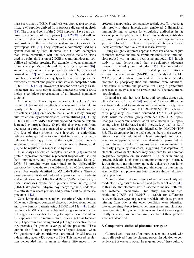
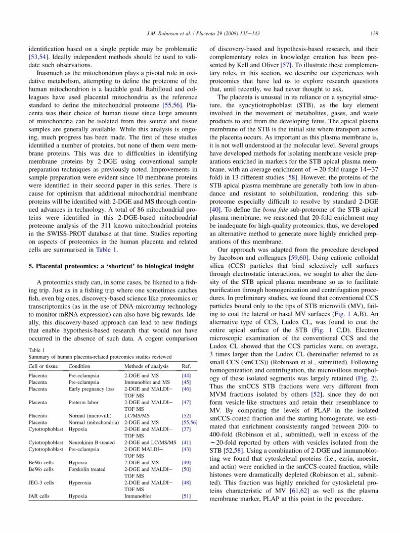
Our approach was adapted from the procedure developedby Jacobson and colleagues [59,60]. Using cationic colloidalsilica (CCS) particles that bind selectively cell surfacesthrough electrostatic interactions, we sought to alter the den-sity of the STB apical plasma membrane so as to facilitatepurification through homogenization and centrifugation proce-dures. In preliminary studies, we found that conventional CCSparticles bound only to the tips of STB microvilli (MV), fail-ing to coat the lateral or basal MV surfaces (Fig. 1 A,B). Analternative type of CCS, Ludox CL, was found to coat theentire apical surface of the STB (Fig. 1 C,D). Electronmicroscopic examination of the conventional CCS and theLudox CL showed that the CCS particles were, on average,3 times larger than the Ludox CL (hereinafter referred to assmall CCS (smCCS)) (Robinson et al., submitted). Followinghomogenization and centrifugation, the microvillous morphol-ogy of these isolated segments was largely retained (Fig. 2).Thus the smCCS STB fractions were very different fromMVM fractions isolated by others [52], since they do notform vesicle-like structures and retain their resemblance toMV. By comparing the levels of PLAP in the isolatedsmCCS-coated fraction and the starting homogenate, we esti-mated that enrichment consistently ranged between 200- to400-fold (Robinson et al., submitted), well in excess of thew20-fold reported by others with vesicles isolated from theSTB [52,58]. Using a combination of 2-DGE and immunoblot-ting we found that cytoskeletal proteins (i.e., ezrin, moesin,and actin) were enriched in the smCCS-coated fraction, whilehistones were dramatically depleted (Robinson et al., submit-ted). This fraction was highly enriched for cytoskeletal pro-teins characteristic of MV [61,62] as well as the plasmamembrane marker, PLAP at this point in the procedure.
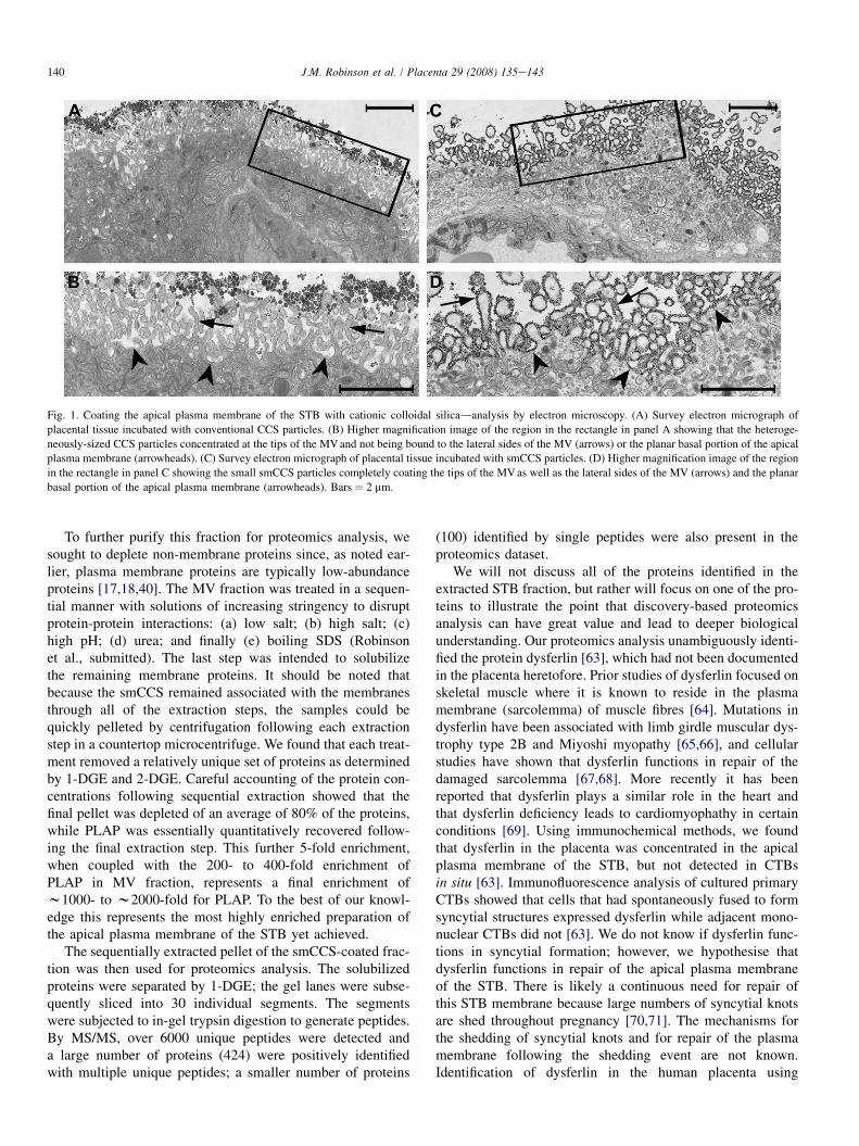
Fig. 1. Coating the apical plasma membrane of the STB with cationic colloidal silicadanalysis by electron microscopy. (A) Survey electron micrograph of
placental tissue incubated with conventional CCS particles. (B) Higher magnification image of the region in the rectangle in panel A showing that the heteroge-
neously-sized CCS particles concentrated at the tips of the MV and not being bound to the lateral sides of the MV (arrows) or the planar basal portion of the apical
plasma membrane (arrowheads). (C) Survey electron micrograph of placental tissue incubated with smCCS particles. (D) Higher magnification image of the region
in the rectangle in panel C showing the small smCCS particles completely coating the tips of the MV as well as the lateral sides of the MV (arrows) and the planar
basal portion of the apical plasma membrane (arrowheads). Bars ¼ 2 mm.
140 J.M. Robinson et al. / Placenta 29 (2008) 135e143
To further purify this fraction for proteomics analysis, wesought to deplete non-membrane proteins since, as noted ear-lier, plasma membrane proteins are typically low-abundanceproteins [17,18,40]. The MV fraction was treated in a sequen-tial manner with solutions of increasing stringency to disruptprotein-protein interactions: (a) low salt; (b) high salt; (c)high pH; (d) urea; and finally (e) boiling SDS (Robinsonet al., submitted). The last step was intended to solubilizethe remaining membrane proteins. It should be noted thatbecause the smCCS remained associated with the membranesthrough all of the extraction steps, the samples could bequickly pelleted by centrifugation following each extractionstep in a countertop microcentrifuge. We found that each treat-ment removed a relatively unique set of proteins as determinedby 1-DGE and 2-DGE. Careful accounting of the protein con-centrations following sequential extraction showed that thefinal pellet was depleted of an average of 80% of the proteins,while PLAP was essentially quantitatively recovered follow-ing the final extraction step. This further 5-fold enrichment,when coupled with the 200- to 400-fold enrichment ofPLAP in MV fraction, represents a final enrichment ofw1000- to w2000-fold for PLAP. To the best of our knowl-edge this represents the most highly enriched preparation ofthe apical plasma membrane of the STB yet achieved.
The sequentially extracted pellet of the smCCS-coated frac-tion was then used for proteomics analysis. The solubilizedproteins were separated by 1-DGE; the gel lanes were subse-quently sliced into 30 individual segments. The segmentswere subjected to in-gel trypsin digestion to generate peptides.By MS/MS, over 6000 unique peptides were detected anda large number of proteins (424) were positively identifiedwith multiple unique peptides; a smaller number of proteins
(100) identified by single peptides were also present in theproteomics dataset.
We will not discuss all of the proteins identified in theextracted STB fraction, but rather will focus on one of the pro-teins to illustrate the point that discovery-based proteomicsanalysis can have great value and lead to deeper biologicalunderstanding. Our proteomics analysis unambiguously identi-fied the protein dysferlin [63], which had not been documentedin the placenta heretofore. Prior studies of dysferlin focused onskeletal muscle where it is known to reside in the plasmamembrane (sarcolemma) of muscle fibres [64]. Mutations indysferlin have been associated with limb girdle muscular dys-trophy type 2B and Miyoshi myopathy [65,66], and cellularstudies have shown that dysferlin functions in repair of thedamaged sarcolemma [67,68]. More recently it has beenreported that dysferlin plays a similar role in the heart andthat dysferlin deficiency leads to cardiomyophathy in certainconditions [69]. Using immunochemical methods, we foundthat dysferlin in the placenta was concentrated in the apicalplasma membrane of the STB, but not detected in CTBsin situ [63]. Immunofluorescence analysis of cultured primaryCTBs showed that cells that had spontaneously fused to formsyncytial structures expressed dysferlin while adjacent mono-nuclear CTBs did not [63]. We do not know if dysferlin func-tions in syncytial formation; however, we hypothesise thatdysferlin functions in repair of the apical plasma membraneof the STB. There is likely a continuous need for repair ofthis STB membrane because large numbers of syncytial knotsare shed throughout pregnancy [70,71]. The mechanisms forthe shedding of syncytial knots and for repair of the plasmamembrane following the shedding event are not known.Identification of dysferlin in the human placenta using
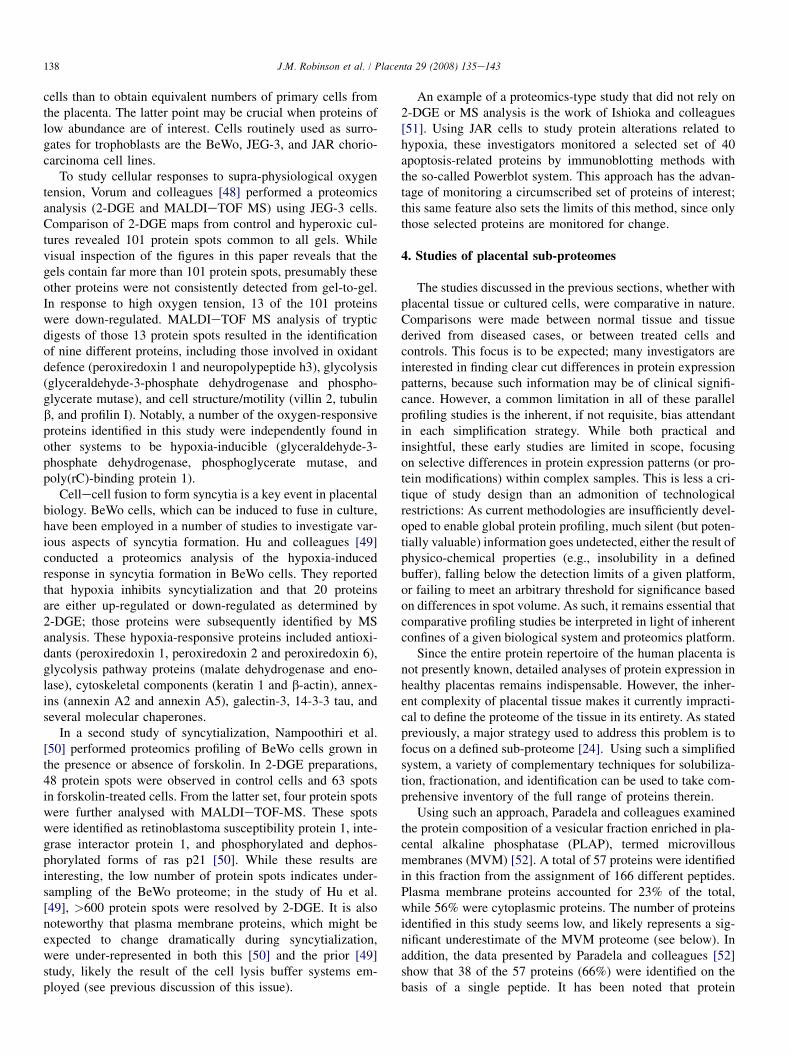
Fig. 2. Isolation of microvilli from the STBdanalysis by electron microscopy.
(A) Survey electron micrograph of the isolated STB MV coated with smCCS.
Note the microvillous appearance of the smCCS-coated membranes (arrows).
A relatively small number of uncoated membrane vesicles are also present (ar-
rowheads). (B) Higher magnification image of the region in the rectangle in
panel A. The extracellular face of the apical plasma membrane of the STB
is coated with smCCS particles (arrows). While most of the membrane surface
is coated with smCCS a low level of contaminating uncoated membranes are
present (arrowheads). Bars ¼ 1 mm.
141J.M. Robinson et al. / Placenta 29 (2008) 135e143
discovery-based proteomics has nevertheless permitted us tobegin addressing this novel hypothesis.
6. Conclusions
Placental proteomics research is in its early stages, butoffers great promise. Establishment of a comprehensive pro-teomics database for the placenta will be instrumental inallowing detailed comparisons between normal placentas and
those derived from disease states. In future studies, it will beimportant to move beyond protein identification to the analysisof isoform usage and more quantitative analysis of proteins innormal and diseased conditions. Although we now know thatproteomics analysis is far more difficult than was envisionedin the early days of this technology (owing, first and foremost,to the great complexity of the eukaryotic proteome), technol-ogies important for proteomics are continuously evolving,and there is reason to hope that these current limitations willultimately yield to new research.
Current limitations notwithstanding, discovery-based pro-teomics can serve as a ‘shortcut’ to the elucidation of biolog-ical problems. While this discovery-based approach may notappeal to all scientists, it may nonetheless provide importantinsights not obtained using more focused methods, therebyenabling hypothesis-based research that would not haveoccurred in the absence of such data. Our work on the proteo-mics-based discovery of dysferlin expression in the humanplacenta illustrates this point: This discovery and subsequentbiochemical and cell biological studies of dysferlin in the pla-centa and CTBs have provided insight into its potential role inplacental biology [63]. Indeed this work now positions us togenerate testable hypotheses concerning the function of dys-ferlin in the human placenta. The complementary roles ofdiscovery-based and hypothesis-based approaches to biomed-ical research in the creation of new knowledge/informationshould be appreciated. A knowledge reservoir, consisting ofboth information and understanding, concerning a given topicsuch as the placenta, can lead to hypotheses that are tested byacquisition of data that can lead to the acceptance or rejectionof the hypotheses and in so doing create new knowledge,primarily in the form of understanding. A complementary pro-cess is the acquisition of new data through discovery-basedprocesses (e.g., proteomics) leading to new knowledge,primarily in the form of information, thus enhancing theknowledge base. Both infusion of new information and under-standing in turn leads to additional testing of hypotheses.
Acknowledgements
Work from the author’s laboratories was supported in partby grants HD38764 and HD49628 from the National Institutesof Health.
References
[1] Godovac-Zimmermann J, Kleiner O, Brown LR, Drukier AK. Perspec-
tives in spicing up proteomics with splicing. Proteomics 2005;5:699e
709.
[2] Romero R, Espinoza J, Gotsch F, Kusanovic JP, Friel LA, Erez O, et al.
The use of high-dimensional biology (genomics, transcriptomics, proteo-
mics, and metabolomics) to understand the preterm parturition syndrome.
BJOG 2006;113:118e35.
[3] Archakov AI, Ivanov YD. Analytical nanobiology for medicine diagnos-
tics. Mol Biosyst 2007;3:336e42.
[4] Moseley FL, Bicknell KA, Marber MS, Brooks G. The use of proteomics
to identify novel therapeutic targets for treatment of disease. J Pharm
Pharmacol 2007;59:609e28.
142 J.M. Robinson et al. / Placenta 29 (2008) 135e143
[5] Moron JA, Devi LA. Use of proteomics for the identification of novel
targets in brain disease. J Neurochem 2007;102:306e15.
[6] Marko-Varga G, Ogiwara A, Nishimura T, Kawamaura T, Fujii K,
Kawakami Y, et al. Personalized medicine and proteomics: lessons
from non-small cell lung cancer. J Proteome Res 2007;6:2925e35.
[7] Huber LA. Is proteomics heading in the wrong direction? Nature Rev
Mol Cell Biol 2003;4:74e80.
[8] Anderson NL, Anderson NG. Proteome and proteomics: new technolo-
gies, new concepts, and new ideas. Electrophoresis 1998;19:1853e61.
[9] Corthals GL, Wasinger VC, Hochstrasser DF, Sanchez J-C. The dynamic
range of protein expression: a challenge for proteomics research. Electro-
phoresis 2000;21:1104e15.
[10] Gygi SP, Corthals GL, Zhang Y, Rochon Y, Aebersold R. Evaluation of
two-dimensional gel electrophoresis-based proteome analysis technol-
ogy. Proc Natl Acad Sci USA 2000;97:9390e5.
[11] Righetti PG, Castagna A, Herbert B, Candiano G. How to bring the
‘unseen’ proteome to the limelight via electrophoretic pre-fractionation
techniques. Biosci Rep 2005;25:3e17.
[12] Grabis S, Lubec G, Fountoulakis M. Limitations of current proteomics
technologies. J Chromatogr A 2005;1077:1e18.
[13] Luche S, Santoni V, Rabilloud T. Evaluation of nonionic and zwitterionic
detergents as membrane protein solubilizers in two-dimensional electro-
phoresis. Proteomics 2003;3:249e53.
[14] Hu Y, Huang X, Chen GYJ, Yao SQ. Recent advances in gel-based
proteome profiling techniques. Mol Biotechnol 2004;28:63e76.
[15] Ahmed N, Rice GE. Strategies for revealing lower abundance proteins in
two-dimensional protein maps. J Chromatogr B 2005;815:39e50.
[16] Zahedi R-P, Meisinger C, Sickmann A. Two-dimensional benzyldi-
methyl-n-hexadecylammonium chloride/DSD-PAGE for membrane
proteomics. Proteomics 2005;5:3581e8.
[17] Schindler J, Nothwag HG. Aqueous polymer two-phase systems: effective
tools for plasma membrane proteomics. Proteomics 2006;6:5409e17.
[18] Slomianny MC, Dupont A, Bouanou F, Beseme O, Guihot AL,
Amouyel P, et al. Profiling of membrane proteins from human macro-
phages: comparison of two approaches. Proteomics 2006;6:2365e75.
[19] Bodzon-Kulakowska A, Bierczynska-Krzysik A, Dylag T, Drabik A,
Suder P, Noga M, et al. Methods for sample preparation in proteomics
research. J Chromatogr B 2007;849:1e31.
[20] Lopez JL. Two-dimensional electrophoresis in proteome expression
analysis. J Chromatogr B 2007;849:190e202.
[21] Canas B, Pineiro C, Calvo E, Lopez-Ferrer D, Gallardo JM. Trends in
sample preparation for classical and second generation proteomics.
J Chromatogr A 2007;1153:235e58.
[22] Rabilloud T, Luche S, Santoni V, Chevallet M. Detergents and chaotropes
for protein solubilization before two-dimensional electrophoresis.
Methods Mol Biol 2007;355:111e9.
[23] Rice GE, Georgiou HM, Ahmed N, Shi G, Kruppa G. Translational
proteomics: Developing a predictive capacityda review. Placenta
2006;27(Suppl. A):S76e86.
[24] Stasyk LF, Huber LA. Zooming in: fractionation strategies in proteomics.
Proteomics 2004;4:3704e16.
[25] Dreger M. Subcellular proteomics. Mass Spectrometry Rev 2003;22:27e56.
[26] Righetti PG, Castagna A, Herbert B, Reymond F, Rossier JS. Pre-
fractionation techniques in proteome analysis. Proteomics 2003;3:
1397e407.
[27] Au CE, Bell AW, Gilchrist A, Hiding J, Nilsson T, Bergeron JJM. Cellular
proteomics to create the cell map. Curr Opin Cell Biol 2007;19:376e85.
[28] MacLellan DL, Steen H, Adam RM, Garlick M, Zurakowski D, Gygi SP,
et al. A quantitative proteomic analysis of growth factor-induced compo-
sitional changes in lipid rafts of human smooth muscle cells. Proteomics
2005;5:4733e42.
[29] Gupta N, Wollscheid B, Watts JD, Scheer B, Aebersold R, DeFranco AL.
Quantitative proteomic analysis of B cell lipid rafts reveals that ezrin
regulates antigen receptor-mediated lipid raft dynamics. Nat Immunol
2006;7:625e33.
[30] Dasari S, Pereira L, Reddy AP, Michaels J-E, Lu X, Jacob T, et al.
Comprehensive proteomics analysis of human cervical-vaginal fluid.
J Proteome Res 2007;6:1258e68.
[31] Pereira L, Reddy AP, Jacob T, Thomas A, Schneider KA, Dasari S, et al.
Identification of novel biomarkers of preterm birth in human cervicale
vaginal fluid. J Proteome Res 2007;6:1269e76.
[32] Buhimschi I, Christner R, Buhimschi CS. Proteomic biomarker analysis
of amniotic fluid for identification of intra-amniotic inflammation. BJOG
2005;112:173e81.
[33] Michel PE, Crettaz D, Morier P, Heller M, Gallot D, Tissot J-D, et al.
Proteome analysis of human plasma and amniotic fluid by Off-Gel�isoelectric focusing followed by nano-LC-MS/MS. Electrophoresis
2006;27:1169e81.
[34] Tsangaris GT, Karamessinis P, Kolialexi A, Garbis SD, Antsaklis A,
Mavrou A, et al. Proteomics analysis of amniotic fluid in pregnancies
with Down syndrome. Proteomics 2006;6:4410e9.
[35] Shankar R, Gude N, Cullinane F, Brennecke S, Purcell AW, Moses EK.
An emerging role for comprehensive proteome analysis in human preg-
nancy research. Reproduction 2005;129:685e96.
[36] Webster RP, Myatt L. Elucidation of the molecular mechanisms of
pre-eclampsia using proteomics technologies. Proteomics-Clin Appl
2007;1:1147e57.
[37] Hoang VM, Foulk R, Clauser K, Burlinggame A, Gibson BW, Fisher SJ.
Functional proteomics: examining the effects of hypoxia on the cytotro-
phoblast protein repertoire. Biochemistry 2001;40:4077e86.
[38] Anderson JS, Lam YW, Leung AK, Ong SE, Lyon CE, et al. Nucleolar
proteome dynamics. Nature 2005;433:77e83.
[39] Wittmann-Liebold B, Graack H-R, Pohl T. Two-dimensional gel electro-
phoresis as tool for proteomics studies in combination with protein iden-
tification by mass spectrometry. Proteomics 2006;6:4688e703.
[40] Santoni V, Molloy M, Rabilloud T. Membrane proteins and proteomics:
Un amour impossible? Electrophoresis 2000;21:1054e70.
[41] Sawicki G, Dakour J, Morrish DW. Functional proteomics of neurokinin
B in the placenta indicated a novel role in regulating cytotrophoblast
antioxidant defenses. Proteomics 2003;3:2044e51.
[42] Page NM, Woods RJ, Gardiner SM, Lomthaisong K, Gladwell RT,
Butlin DJ, et al. Excessive placental secretion of neurokinin B during
the third trimester causes pre-eclampsia. Nature 2000;405:797e800.
[43] Sun LZ, Yang NN, De W, Xiao YS. Proteomics analysis of proteins
differentially expressed in pre-eclamptic trophoblasts. Gynecol Obstet
Invest 2007;64:17e23.
[44] Mine K, Katayama A, Matsumura T, Nishino T, Kuwabara Y,
Ishikawa G, et al. Proteome analysis of human placentae: pre-eclampsia
versus normal pregnancy. Placenta 2007;28:676e87.
[45] Webster RP, Brockman D, Myatt L. Nitration of p38 MAPK in the
placenta: association of nitration with reduced catalytic activity of p38
MAPK in pre-eclampsia. Mol Human Reprod 2006;12:677e85.
[46] Liu A-X, Zhang W-W, Zhou T-H, Zhou C-Y, Yao W-M, Qian Y-L, et al.
Proteomics analysis on the alteration of protein expression in the placen-
tal villous tissue of early pregnancy loss. Biol Reprod 2006;75:414e20.
[47] Butt RH, Lee MWY, Pirshahid SA, Backlund PS, Wood S, Coorssen JR.
An initial proteomics analysis of human preterm labor: placental mem-
branes. J Proteome Res 2006;5:3161e72.
[48] Vorum H, Østergaard M, Hensechke P, Enghild JJ, Riazati M, Rice GE.
Proteomic analysis of hyperoxia-induced responses in the human chorio-
carcinoma cell line JEG-3. Proteomics 2004;4:861e7.
[49] Hu R, Jin H, Zhou S, Yang P, Li X. Proteomics analysis of hypoxia-
induced responses in the syncytialization of human placental cell line
BeWo. Placenta 2007;28:399e407.
[50] Nampoothiri LP, Neelima PS, Rao AJ. Proteomic profiling of forskolin-
induced differentiated BeWo cells: an in-vitro model of cytotrophoblast
differentiation. Reprod BioMed Online 2007;14:477e87.
[51] Ishioka S-I, Ezaka Y, Umemura K, Hayashi T, Endo T, Saito T. Proteo-
mic analysis of mechanisms of hypoxia-induced apotosis in trophoblastic
cells. Int J Med Sci 2007;4:36e44.
[52] Paradela A, Bravo SB, Henriquez M, Riquelme G, Gavilanes F, Gonzalez-
Ros JM, et al. Proteomic analysis of apical microvillous membranes of
syncytiotrophoblast cells reveals a high degree of similarity with lipid rafts.
J Proteome Res 2005;4:2435e41.
[53] Carr S, Abersold R, Baldwin M, Burlinggame A, Clauser K,
Nesvizskii A. The need for guidelines in publication of peptide and
143J.M. Robinson et al. / Placenta 29 (2008) 135e143
protein identification data. Working group on publication for peptide and
protein identification data. Mol Cell Proteomics 2004;3:531e3.
[54] Wilkins MR, Appel RD, Van Eyk JE, Chung MCM, Gorg A, Hecker M,
et al. Guidelines for the next 10 years of proteomics. Proteomics
2006;6:4e8.
[55] Rabilloud T, Kieffer S, Procaccio V, Louwagie M, Courchesne PL,
Patterson SD, et al. Two-dimensional electrophoresis of human placental
mitochondria and protein identification by mass spectrometry: toward
a human mitochondrial proteome. Electrophoresis 1998;19:1006e14.
[56] Lescuyer P, Strub J-M, Luche S, Diemer H, Martinez P, Van
Dorsselaer A, et al. Progress in the definition of a reference human
mitochondrial proteome. Proteomics 2003;3:157e67.
[57] Kell DB, Oliver SG. Here is the evidence, now what is the hypothesis?
The complementary roles of inductive and hypothesis-driven science in
the post-genomic era. BioEssay 2004;26:99e105.
[58] Jimenez V, Henriquez M, Llanos P, Riquelme G. Isolation and purifica-
tion of human plasma membrane from normal and pre-eclamptic preg-
nancies. Placenta 2004;25:422e37.
[59] Jacobson BS, Stolz DE, Schnitzer JE. Plasma membrane isolation using
the cationic colloidal silica isolation technique. In: Cells. A laboratory
manual. Cold Spring Harbor, NY: Cold Spring Harbor Laboratory Press;
1998. p. 35.1e35.14.
[60] Durr E, Yu J, Krasinska KM, Carver LA, Yates III JR, Testa JE, et al.
Direct proteome mapping of the lung microvascular endothelial cell
surface in vivo and in cell culture. Nature Biotech 2004;22:985e92.
[61] Bretscher A. Microfilament structure and function in the cortical
cytoskeleton. Annu Rev Cell Biol 1991;7:337e74.
[62] Fath KR, Burgess DR. Not actin alone. Curr Biol 1995;5:591e3.
[63] Vandre DD, Ackerman Jr WE, Kniss DA, Tewari AK, Mori M,
Takizawa T, et al. Dysferlin is expressed in human placenta but does
not associate with caveolin. Biol Reprod 2007;77:533e42.
[64] Anderson LVB, Davison K, Moss JA, Young C, Cullen MJ, Walsh J, et al.
Dysferlin is a plasma membrane protein and is expressed early in human
development. Hum Mol Genet 1999;8:855e61.
[65] Bashir R, Britton S, Strachan T, Keers S, Vafiadaki E, Lako M, et al.
A gene related to Caenorhabditis elegans spermatogenesis factor fer-1is mutated in limb-girdle muscular dystrophy type 2B. Nature Genet
1998;20:37e42.
[66] Liu J, Aoki M, Illa I, Chenyan W, Fardeau M, Angelini C, et al. Dysferlin
a novel skeletal muscle gene, is mutated in Miyoshi myopathy and limb
girdle muscular dystrophy. Nature Genet 1998;20:31e6.
[67] Bansal Dm Miyake K, Vogel SS, Groh S, Chen CC, Williamson R, et al.
Defective membrane repair in dysferlin-deficient muscular dystrophy.
Nature 2003;423:168e72.
[68] Lennon NJ, Kho A, Backai BJ, Perlmutter SL, Hyman BT, Brown Jr RH.
Dysferlin interacts with annexins A1 and A2 and mediates sarcolemmal
wound-healing. J Biol Chem 2003;278:50466e73.
[69] Han R, Bansal D, Miyake K, Muniz VP, Weiss RM, McNeil PL, et al.
Dysferlin-mediated membrane repair protects the heart from stress-
induced left ventricular injury. J Clin Invest 2007;117:1805e18.
[70] Huppertz V, Tews DS, Kaufmann P. Apotosis and syncytial fusion in
human placental trophoblast and skeletal muscle. Int Rev Cytol
2001;205:215e53.
[71] Abumaree MH, Stone PR, Chamley LW. An in vitro model of human
placental trophoblast deportation/shedding. Mol Hum Reprod 2006;
12:687e94.