Embed Size (px)
Citation preview

Introduction
Airborne ambient particulate matter (PM) is considered to play an important role in the adverse health effects asso-ciated with air pollution (Brunekreef and Holgate, 2002). Most epidemiological studies have focused on the effects of short-term exposure to air pollutants. In these short-term studies, a clear link was shown between levels of air pollut-ants and a tendency toward a hypercoagulable state. These associations are, for example, found with both PM
10 and
NO2 (Baccarelli et al., 2007) or only with the traffic-related
gaseous (NO2 and CO) instead of with PM mass (Rudez
et al., 2009). However, several epidemiological studies have associated long-term exposure to the fine fraction of PM (PM
2.5: PM with an aerodynamic diameter below 2.5 µm)
with an increase in pulmonary and cardiovascular morbid-ity and mortality (Pope et al., 2004; Schikowski et al., 2005). Notably, living close to a busy road over several years has been associated with increased cardiopulmonary mortality
(Received 07 July 2010; revised 06 October 2010; accepted 07 October 2010)
ISSN 0895-8378 print/ISSN 1091-7691 online © 2010 Informa Healthcare USA, Inc.DOI: 10.3109/08958378.2010.531062 http://www.informahealthcare.com/iht
R E S E A R C H A R T I C L E
Pulmonary and cardiovascular effects of traffic-related particulate matter: 4-week exposure of rats to roadside and diesel engine exhaust particles
Miriam E. Gerlofs-Nijland1, Annike I. Totlandsdal2, Evren Kilinç3, A. John F. Boere1, Paul H.B. Fokkens1, Daan L.A.C Leseman1, Constantinos Sioutas4, Per E. Schwarze2, Henri M. Spronk3, Patrick W.F. Hadoke5, Mark R. Miller5, and Flemming R. Cassee1
1Environment and Safety Division, Centre for Environmental Health, National Institute for Public Health and the Environment, Bilthoven, The Netherlands, 2Division of Environmental Medicine, Department of Air Pollution and Noise, Norwegian Institute of Public Health, Oslo, Norway, 3Cardiovascular Research Institute Maastricht, Maastricht University Medical Center, Maastricht, The Netherlands, 4Department of Civil and Environmental Engineering, University of Southern California, Los Angeles, CA, USA, and 5Centre for Cardiovascular Sciences, University of Edinburgh, Edinburgh, UK
AbstractTraffic-related particulate matter (PM) may play an important role in the development of adverse health effects, as documented extensively in acute toxicity studies. However, rather little is known about the impacts of prolonged exposure to PM. We hypothesized that long-term exposure to PM from traffic adversely affects the pulmonary and cardiovascular system through exacerbation of an inflammatory response. To examine this hypothesis, Fisher F344 rats, with a mild pulmonary inflammation at the onset of exposure, were exposed for 4 weeks, 5 days/week for 6 h a day to: (a) diluted diesel engine exhaust (PM
DEE), or: (b) near roadside PM (PM
2.5). Ultrafine particulates,
which are largely present in diesel soot, may enter the systemic circulation and directly or indirectly trigger car-diovascular effects. Hence, we assessed the effects of traffic-related PM on pulmonary inflammation and activity of procoagulants, vascular function in arteries, and cytokine levels in the heart 24 h after termination of the expo-sures. No major adverse health effects of prolonged exposure to traffic-related PM were detected. However, some systemic effects due to PM
DEE exposure occurred including decreased numbers of white blood cells and reduced
von Willebrand factor protein in the circulation. In addition, lung tissue factor activity is reduced in conjunction with reduced lung tissue thrombin generation. To what extent these alterations contribute to thrombotic effects and vascular diseases remains to be established. In conclusion, prolonged exposure to traffic-related PM in healthy animals may not be detrimental due to various biological adaptive response mechanisms.
Keywords: Traffic; particulate matter; PM; concentrated particles; pulmonary inflammation; diesel soot; vascular function; procoagulants; air pollution; toxicological
Inhalation Toxicology, 2010; 22(14): 1162–1173Inhalation Toxicology
2010
22
14
1162
1173
07 July 2010
06 October 2010
07 October 2010
0895-8378
1091-7691
© 2010 Informa Healthcare USA, Inc.
10.3109/08958378.2010.531062
Address for Correspondence: M.E. Gerlofs-Nijland, Environment and Safety Division, Centre for Environmental Health, National Institute for Public Health and the Environment, P.O. Box 1, 3720 BA Bilthoven, The Netherlands. E-mail: [email protected]
IHT
531062
UIHT
Inha
latio
n T
oxic
olog
y D
ownl
oade
d fr
om in
form
ahea
lthca
re.c
om b
y R
IVM
on
12/0
7/10
For
pers
onal
use
onl
y.

Pulmonary and cardiovascular effects of traffic-related PM 1163
(Hoek et al., 2002; Gehring et al., 2006). Hence, road traffic, which is a major source of PM
2.5 in urban areas, could be
particularly responsible for the impact of PM exposure on human health (Gauderman et al., 2005; Schikowski et al., 2005; Beelen et al., 2009; Hoffmann et al., 2009).
These epidemiological observations are supported by controlled toxicology studies performed with animals and human volunteers exposed to PM samples from different sites. Animal exposure studies attribute a greater toxicity of PM collected at locations that contain a high proportion of traffic emissions (Lai et al., 2005; Seagrave et al., 2006; Gerlofs-Nijland et al., 2007). Interestingly, it is becoming evident that exposure to traffic-related PM has marked actions on the cardiovascular system, as well as their more well-recognized pulmonary effects (Elder et al., 2007; McCreanor et al., 2007). Short-term exposures to diesel engine exhaust, an important source of PM
2.5, cause both vascular dysfunction and impaired
endogenous fibrinolysis in healthy and compromised volun-teers (Mills et al., 2005, 2007). In addition, elevated thrombus formation was shown ex vivo after inhalation of diesel engine exhaust (Lucking et al., 2008).
In these studies, mostly fresh generated emission particles were used as a surrogate of PM, which are not necessarily representative of the PM in ambient air. Inhalation of elevated concentrations of ambient air particles collected with dif-ferent size ranges at different sites by using concentrator technology (Sioutas et al., 1997; Kim et al., 2001a, 2001b) represents a more realistic PM exposure (Lippmann and Chen, 2009). A few hours exposure to PM
2.5 from urban traf-
fic sites caused an increase in cardiovascular symptoms and in lung toxicity and inflammation in rodents and volunteers (Gong et al., 2003; Cassee et al., 2005; Kleinman et al., 2005; Lippmann et al., 2005a; Kooter et al., 2006; Araujo et al., 2008; Ying et al., 2009). This also suggests a major contribution of traffic-related particles to the biological effects associated with PM. Therefore, these studies, in which relatively high PM exposure levels were applied, will be useful in understanding the impact of episodic PM exposure on human health.
At present, only a few publications, all from the same well-conducted study in New York, describe the impact of pro-longed exposure on normal and susceptible (i.e. mimicking a human disease) rodents at lower concentration, more envi-ronmentally relevant levels (Chen and Hwang, 2005; Chen and Nadziejko, 2005; Hwang et al., 2005; Lippmann et al., 2005b; Sun et al., 2005, 2008; Veronesi et al., 2005). The most striking results were seen on the cardiovascular system with altered vasomotor tone, induced vascular inflammation, and potenti-ated atherosclerosis both in Sterling Forest (Sun et al., 2005) and more traffic-influenced Manhattan (Ying et al., 2009).
In order to investigate the contribution of traffic to the long-term effect of particles, we performed a series of experi-ments in which we exposed rats to filtered air, to diluted diesel engine exhaust (rich in ultrafine particles), and to PM
2.5 derived from a nearby very busy freeway. Prior to the
PM exposures, a minor lung inflammation was induced by exposing the rats to ozone. We hypothesized that prolonged (i.e. 4-week) exposure to traffic-derived PM
2.5 exacerbates
the existing inflammatory reaction, which could result in an induction of oxidative stress with subsequent effects on the pulmonary and cardiovascular system. In order to verify this hypothesis, a comprehensive analysis of markers for pulmonary (oxidative stress, cytotoxicity, inflammation) and cardiovascular (coagulation, fibrinolysis, endothelial damage, thrombogenicity, heart inflammation, aorta con-tractibility) effects due to exposure to traffic-derived PM was performed.
Methods
AnimalsMale SPF F344 (DUCRL) rats were obtained from Charles River (Sulzfeld, Germany). The rats were housed in macro-lon type III cages with a room temperature maintained at 22 ± 2°C, relative humidity at 40–70%, and a 12-h light/dark cycle. Rats were allowed access to a cereal-based rodent diet (SMR-A; Hope Farms, Woerden, The Netherlands) and tap water via drinking bottles ad libitum during non-exposure periods. Exposure started after 7 days of acclimatization.
Experimental designA total of three experiments were conducted using different types of PM exposure (Gerlofs-Nijland et al., 2009a). At day 0, all rats were exposed (whole body) for 12 h to 0.4 ppm ozone (Marra and Rombout, 1990) to initiate a minor inflamma-tion in the lung (Cassee et al., 2005). After the initial ozone exposure, the animals were transferred to RIVM’s mobile exposure laboratory (MAPCEL) and subsequently exposed for 4 weeks (5 days per week, 6 h per day) to diesel engine exhaust (PM
DEE) or to concentrated ambient particles with an
aerodynamic diameter <2.5 µm (PM2.5
or also known as CAPs) near a busy roadside at Utrecht, The Netherlands (Figure 1). The PM
2.5 roadside study was repeated once due to the inher-
ent variability of the mass and composition of ambient PM.
PMDEE
Exposure was performed using a 35 KVA diesel generator (Bredenoord, Apeldoorn, The Netherlands) under idling conditions. The animals (n = 15/group) were exposed to 150 µg/m3 PM
DEE diluted with clean conditioned air.
PM2.5 roadsideRats were exposed to increased levels of PM
2.5 using the
Versatile Aerosol Concentration Enrichment Systems (VACES) (Kim et al., 2001a, 2001b) with a theoretical enrichment factor of 20 and at an output flow rate directed into the nose-only system of 20 LPM. The MAPCEL was placed close to (15 m), and east of a major roadside (A2; Utrecht–Amsterdam, The Netherlands), with prevailing westerly winds, used by 160,000 cars and trucks per day.
Control animals were exposed to filtered, purified air with the same temperature and relative humidity as the test atmos-pheres. All rats were nose-only exposed using novoplast tubes T (Münster AG, Muttenz, Switzerland) in nose-only exposure chambers. One week before exposure, animals were trained
Inha
latio
n T
oxic
olog
y D
ownl
oade
d fr
om in
form
ahea
lthca
re.c
om b
y R
IVM
on
12/0
7/10
For
pers
onal
use
onl
y.

1164 M. E. Gerlofs-Nijland et al.
in nose-only tubes to reduce the stress of the restraint (3 days, 1 h per day). Immediately after the exposures, the animals were returned to their housing facilities.
Blood (1 ml) was obtained after the first week of exposure (directly after the fifth exposure day; Figure 1) by orbital punc-ture under Brevimethal anaesthesia (50 mg/kg bodyweight, intramuscularly) to measure fibrinogen, von Willebrand factor (vWF), plasminogen activator inhibitor (PAI)-1, and CC16.
Based on the initial findings, additional parameters were investigated, i.e. vascular function, measurement of cytokines in cardiac tissue, and tissue factor (TF) activity and thrombin generation in lung tissue in one of the two roadside experiments to gain more insight in a possible biological mechanism.
Necropsy was performed on the day after the last exposure day (Figure 1). Experiments were approved by the Animal Ethics Committee (IUCAC) of the Dutch National Vaccine Institute (NVI, Bilthoven, The Netherlands).
Characterization of the test atmospheresA condensation particle counter (CPC model 3022A; TSI, St. Paul, MN) was used to determine the particle number con-centration in the inlet of the exposure chamber. The mass concentration was measured continuously in the inlet of the exposure chamber during the exposure with a nephelometer (DATARAM 2000; MIE, Billerica, MA). In the PM
2.5 roadside
experiments, the particle number and mass concentration were measured both before the VACES inlet and after the VACES. The time-integrated PM concentrations were also measured in the inlet of the exposure chamber by means of collection on three 47-mm filters placed in parallel, two poly-tetrafluoroethylene (PTFE; Teflon R2PJ047; Pall Corp., Ann Arbor, MI), and one quartz filter (QMA; Whatman Int Inc, Maidstone, England). A carbon sampler tube (Anasorb CSC Lot 2000; SKC Inc., Eighty Four, PA) was placed downstream of one of the PTFE filters at the outlet to collect the volatile organic components (VOCs). One set of PTFE filters and a carbon sampler tube were used for each exposure week. Carbon monoxide (ML 9830 CO; Lear Siegler, Englewood, CO), sulfur dioxide (model 43A; Thermo Environmental Instruments, Franklin, MA), and nitrogen oxides (model
42W; Thermo Environmental Instruments) were measured in the PM
DEE mixing chamber or at the inlet of the VACES.
In the PMDEE
experiment, a Scanning Mobility Particle Sizer (SMPS, DMA model 3071 + CPC model 3022A; TSI) was used to measure the particle size distribution (mean diameter and geometric standard deviation) every hour in the inlet of the exposure chamber. The weekly time-integrated particle size mass distribution was measured at the inlet of the VACES with an eight-stage Micro Orifice Impactor (model No. 100; MSP Corporation, Minneapolis, MN). Temperature and rela-tive humidity were recorded once every 5 min in the exposure chamber and control exposure chamber and recorded every 30 min in the inlet of the exposure chamber. The activated car-bon samplers were analyzed using GC-MS (RIVM, Bilthoven, The Netherlands) to determine the VOC concentrations.
NecropsyThe day after the final test atmosphere exposure, the rats were anesthetized with a mix of Ketamine and Rompun: 100 mg/kg of Ketamine (Aesculaap, Boxtel, The Netherlands) and 1 mg/kg Rompun (Bayer, Leverkusen, Germany). A cannula was inserted in the trachea. The abdomen was opened and a mini-mum of 6 ml blood was sampled through the abdominal aorta. The chest was opened and the lungs were perfused (pressure 30 cm H
2O) with saline to remove the blood from the lung using
a cannula placed through the right heart chamber into the pul-monary artery. The left bronchus was clamped and the left lung was cut just behind the clamp. The left lung was weighed and fixed for 1 h under a constant pressure of 20 cm H
2O using 4%
phosphate-buffered formaldehyde. The right lung was used for bronchoalveolar lavage fluid (BALF) collection by three lav-ages of sterile saline (27 ml/kg body weight). The heart was dis-sected, split into the right and left side and frozen in liquid N
2.
The descending thoracic aorta was dissected and immediately placed in Krebs buffer for organ bath measurements.
Bronchoalveolar lavage analysesThe collected BALF was centrifuged at 400g, 4°C, for 10 min. The cell-free fluid from the lavage was used for assess-ment of lactate dehydrogenase (marker for cytotoxicity),
6 h/day expo5 d/wk
Ozone exposure12 hour 0.4 ppm
Orbita punctionafter expo day 5
Weekend,no exposure
6 h/day expo5 d/wk
6 h/day expo5 d/wk
6 h/day expo4 d/wk
autopsy
Figure 1. Experimental exposure design.
Inha
latio
n T
oxic
olog
y D
ownl
oade
d fr
om in
form
ahea
lthca
re.c
om b
y R
IVM
on
12/0
7/10
For
pers
onal
use
onl
y.

Pulmonary and cardiovascular effects of traffic-related PM 1165
N-acetylglucosaminidase (macrophage activation), alkaline phosphatase (type II cell damage), and the levels of Clara-cell 16 protein (CC16, lung cell damages), reduced glutathione and oxidized glutathione (GSH and GSSG, respectively), albumin and total protein levels (increased permeability of the alveo-lar–capillary barrier), inflammatory mediators interleukin 6 (IL-6), and tumor necrosis factor (TNF)-α were determined as previously described (Cassee et al., 2005; Gerlofs-Nijland et al., 2005). Heme-oxygenase-1, a marker of oxidative stress, was determined using a commercially obtained reagent kit (Roche Nederland B.V, Mijdrecht, The Netherlands). The BALF pellet was resuspended in saline and used for total cell counts as well as preparation of cytospins for cell differential counts as previously described (Gerlofs-Nijland et al., 2005).
Hematological analysesPlasma levels of fibrinogen and CC16 were determined as pre-viously described (Cassee et al., 2005; Gerlofs-Nijland et al., 2009b). vWF was measured by enzyme-linked immunosorb-ent assay (ELISA; American Diagnostica Inc., Stamford, US). Levels of tissue plasminogen activator, total antigen, and active PAI-1 were measured in citrated plasma by ELISA (Innovative Research, Dearborn, M). Cell differentials were determined in EDTA (K3) (Terumo Europe N.V., Leuven, Belgium) anti-coagulated blood in an H1-E multispecies hematology ana-lyzer (Bayer B.V., Mijdrecht, The Netherlands). The following parameters were measured: white and red blood cell con-centrations (WBC and RBC, respectively), hemoglobin, and platelet (PLT) concentrations, the mean platelet volume, and the hematocrit value. In addition, mean corpuscular volume, mean cell hemoglobin, mean cell hemoglobin concentration, red blood cell distribution width, mean platelet component and hemoglobin distribution width were provided.
PathologyThe left lung was embedded in paraffin after fixation with for-maldehyde. Tissues were cut into 5-µm slices and slides were stained with hematoxylin and eosin before light microscopic examination. Slides were screened for pathological changes as a result of the exposure. The pathological lesions and inflammation were semi-quantitatively and blindly scored as absent, minimal, slight, moderate, marked, or strong.
Vascular functionEx vivo endothelial function and vascular responses were measured in isolated thoracic aortic rings by a modified method of Bagate et al. (2004) and Miller et al. (2009). Segments of thoracic aorta (~5 mm length) were cleaned of connective tissue and mounted in organ baths in Krebs buffer bubbled with 5% CO
2/95% O
2 at 37°C. A baseline tension of
14.7 mN was gradually applied over 10 min and vessels were allowed to equilibrate for a further 30 min.
Vessel viability was confirmed by a contractile response on addition of 80 mM KCl, repeated three times. Concentration–response curves to phenylephrine (PE; 1 nM to 10 μM) were obtained and a concentration that produced 80% maximum contraction (0.1–1 μM) was chosen for each individual rat
aortic ring. Following contraction, cumulative concentra-tion–response curves were obtained for acetylcholine (ACh; endothelium-dependent vasodilator; 1 nM to 10 μM), sodium nitroprusside (SNP; endothelium-independent nitric oxide donor; 0.1 nM to 1 μM), and isoprenaline (ISP) or verapamil (endothelium- and nitric oxide–independent vasodilators; 1 nM to 10 μM). At least 30-min washout was allowed before application of subsequent drugs.
Analyses of cardiac tissueCytokine mRNA expressionThe frozen right heart halves were homogenized in lysis buffer and total RNA isolated using a “Absolutely RNA™ RT-PCR Miniprep kit” (Stratagene, La Jolla, CA). mRNA in each sam-ple was reverse-transcribed into cDNA on a PCR system 2400 (Perkin Elmer, Groningen, The Netherlands) by using a High Capacity cDNA Archive Kit from Applied Biosystems (Life Technologies Corporation, Carlsbad, CA). Quantitative real-time (QRT) PCR was performed on triplicate samples, with 18S rRNA as an internal control, using the Applied Biosystems 7500 Real-Time PCR System, with pre-designed TaqMan Gene Expression Assays (IL-6, Rn00561420_m1; IL-1β, Rn00580432_m1; TNF-α, Rn00562055_m1; 18S, Hs99999901_s1) and TaqMan Universal PCR Master Mix. The expression of each gene within each sample was normalized against 18S rRNA and expressed relative to a heart tissue sample from one of the control rats using the formula 2-(ΔΔCt) in which ΔΔC
t = (C
t
mRNA – Ct 18S rRNA) sample – (C
t mRNA – C
t 18S rRNA) sam-
ple control rat.
Phosphorylation of mitogen-activated protein kinasesRight heart halves were homogenized in lysis buffer (20 mM Tris–HCl pH+7.5, 150 mM NaCl, 1 mM EDTA, 1 mM EGTA, 2.4 mM Na-pyrophosphate, 1.0 mM orthovanadate, 1 mM NaF, 21 µM leupeptin, 1.5 µM aprotinin, 15 µM pepstatin A, and 1% Triton-X) and examined by western analysis. Protein concentration in the samples was determined by using the Bio-Rad DC Protein Assay (Bio-Rad Laboratories B.V., Veenendaal, The Netherlands). Proteins (12.5–25 µg/ well) from the homog-enized heart tissue samples were separated by 10% sodium dodecyl sulfate–polyacrylamide gel electrophoresis and blot-ted onto nitrocellulose membranes. To ensure that the protein levels of each well were equal, Ponceau-staining was used for loading control. The membranes were then probed with anti-bodies for the respective phosphorylated kinases (p-ERK1/2, p-JNK1/2, p-p38) prior to incubation with horseradish peroxidase–conjugated secondary antibodies. The blots were developed using the Super-Signal® West Dura chemilumines-cence system (Perbio Science Nederland B.V., Etten-Leur, The Netherlands) according to the manufacturer’s instructions. Finally, the membranes were stripped by incubation for 15 min at room temperature with Mild Antibody Stripping Solution® from Chemicon International (Temecula, CA), and re-probed with antibodies against total mitogen-activated protein kinase (MAPK) proteins (ERK1/2, JNK, p38). Optical quantification of the protein bands were performed by using the KODAK 1D Image Analysis Software.
Inha
latio
n T
oxic
olog
y D
ownl
oade
d fr
om in
form
ahea
lthca
re.c
om b
y R
IVM
on
12/0
7/10
For
pers
onal
use
onl
y.

1166 M. E. Gerlofs-Nijland et al.
TF activity and thrombin generation in lung tissueTF activity and tissue-specific thrombin generation by means of the Calibrated Automated Thrombogram (Thrombinoscope BV, Maastricht, The Netherlands) were determined in lung tissue homogenates as described previously (Frederix et al., 2008). Briefly, thrombin generation was measured in the pres-ence of a final concentration of 5 pM TF and 4 µM phospholi-pids (PL, at 20:20:60 mol% PS:PE:PC) after addition of lung homogenates in human plasma and alternatively measure-ments were also implemented in the absence of both TF and PL. All TG results were normalized and expressed as percent-age of normal pooled, PLT-poor plasma which was prepared from at least 80 healthy volunteers (Spronk et al., 2008).
Statistical analysisData are expressed as mean ± standard deviation (SD) or standard error of mean (SEM). Vascular responses are expressed as percentage of the maximal contraction to PE, where positive values represent vasodilatation and 100% vasodilatation represents a complete abolition of PE-induced tone. The outcomes of the BALF, blood analyses, TF activity, and thrombin generation were compared using an unpaired Student’s t-test. Statistical comparisons of vasodilator curves were carried out using two-way analysis of variance (ANOVA), or unpaired Student’s t-test for comparisons of EC
50 and
maximum responses (estimated following linear regression of individual curves using GraphPad Prism V4.0b). P < 0.05 was accepted as statistically significant.
Results
Ozone exposureA separate group of 10 animals was used to confirm that ozone exposure induced a minor lung inflammation. At 24 h after the ozone exposure, there was a significant increase in lung permeability, as shown by elevated protein (487 ± 141
compared to control levels of 159 ± 49 mg/l; P < 0.001) and albumin (248 ± 99 mg/l versus 50 ± 14 mg/l in control group; P < 0.001) levels in BALF. Ozone exposure also increased the percentage of polymorphonuclear neutrophils in the alveolar region by approximately 2.5% (3.05 ± 2.55% versus 0.65 ± 0.95% for control), although this increase did not reach statistical significance (P > 0.05).
Exposures characteristicsDiesel engine exhaustThe PM
DEE exposures were performed at an overall average
particle mass of 174 ± 15 µg/m3 (Table 1). The average particle size (geometric median diameter) was 76 nm with a geomet-ric standard deviation of 1.95 nm as measured by SMPS, with an average particle number concentration of 434,000/ cm3. During the first exposure week, the carbon sampler was used only for 1 day and the amount of VOC measured was 564 µg/ m3. In addition, the concentrations of gaseous pollutants CO, NO, NO
2, and NOx were measured, with mean concentra-
tions of 3050, 1671, 918, and 2589 µg/m3, respectively. Levels of VOC during the last three exposure weeks could not be measured due to an overload in the carbon sampler tubes.
PM2.5 roadside #1During the first PM
2.5 roadside study, the overall average
particle mass was 485 ± 150 µg/m3 (Table 1). The average particle number concentration was 312,000/cm3 with a mean aerodynamic particle size of 1.04 µm and geometric standard deviation of 0.31 (measured by multiplicity of infection (MOI) before the VACES). The mean VOC content measured was 254 µg/m3, which was mainly driven by high levels of VOC (820 µg/m3) during the first week of exposure. These appeared to be caused by high amounts of heptane, most probably due to a two-stroke engine used for lawn mowing activities nearby. The concentrations of gaseous pollutants NO, NO
2, and NOx
were 56, 71, and 127 µg/m3, respectively.
Table 1 - Particle exposure characteristics of diesel engine exhaust and concentrated ambient particles near a roadside.
Experiment Week no Mass Number CO NO NO2
NOx VOC Inorganics MMAD†
µg/m3 # 105/cm3 µg/m3 µg/m3 µg/m3 µg/m3 µg/m3 µg/m3 µm
1 160 4.83 3515 2058 1128 3186 564 nd nd
2 162 4.44 3131 1671 937 2608 897‡ nd 0.29
PMDEE
3 191 4.18 2945 1571 841 2413 5416‡ nd 0.24
4 182 3.89 2689 1397 765 2162 4068‡ nd 0.17
Average 174 4.34 3050 1671 918 2589 564 0.23
1 484 3.72 nd 72 84 156 820 nd 1.18
2 284 3.51 nd 65 76 141 70 nd 0.99
PM2.5
3 528 1.71 nd 22 42 65 45 nd 1.01
roadside #1 4 643 3.53 nd 69 82 151 80 nd 0.97
Average 485 3.12 — 56 71 127 254 1.04
1 200 2.46 nd 82 48 130 6 44 1.46
2 199 2.63 nd 87 71 158 6 60 1.53
PM2.5
3 224 2.32 nd 85 59 144 49 46 2.13
roadside #2 4 232 1.84 nd 70 63 133 13 81 0.95
Average 214 2.31 — 81 61 142 19 58 1.52
PM, particulate matter; DEE, diesel engine exhaust; Inorganics, sum of sulfate, nitrate, chloride and sodium; VOC, volatile organic components; MMAD, Aerodynamic particle size measured by MOI; nd, not determined‡Unreliable outcomes due to overload carbon samplers, those values are not included in the average VOC content.
Inha
latio
n T
oxic
olog
y D
ownl
oade
d fr
om in
form
ahea
lthca
re.c
om b
y R
IVM
on
12/0
7/10
For
pers
onal
use
onl
y.

Pulmonary and cardiovascular effects of traffic-related PM 1167
PM2.5 roadside #2The overall average particle mass in the second PM
2.5 roadside
study was 214 ± 17 µg/m3. The average particle number con-centration was 231,000/cm3 with an associated aerodynamic mean particle size of 1.52 µm and geometric standard devia-tion of 0.23 (measured by MOI before the VACES). The VOC content was 19 µg/m3 with NO, NO
2, and NOx concentrations
of 81, 61, and 142 µg/m3, respectively.
BALF analysesProlonged exposure to PM
DEE or PM
2.5 near a roadside did not
induce a detectable inflammatory response in healthy rats. The number of MN in BALF was not significantly increased after 4 weeks of exposure to PM
2.5 roadside or PM
DEE; nor were
there any changes in the pro-inflammatory cytokines TNF-α and IL-6 (Table 2). Although some parameters (e.g. TNF-α, protein) showed strong differences with higher values after exposure to roadside PM
2.5, the only statistically significant
change was an increase in BALF CC16 after exposure to PM2.5
in the second roadside study (9.47 ± 1.14 versus 8.49 ± 1.21 in the control group; P < 0.05; Table 2). Notably, protein and albumin levels in BALF were significantly higher in all animals that were transported to our field location near the freeway compared to those that were exposed in our labo-ratory at the RIVM. Apart from the fact that batch-to-batch variation among the groups of animals that we received from the breeder cannot be excluded, the only other explanation is that the transport from the field location to the lab might have resulted in increased stress and increased baseline values of the noted parameters. Since most of the other parameters that we have assessed were not to be affected in a similar manner, and we performed the (statistical) comparisons only within
each of the three experiments, conclusions were not affected by this unexpected phenomenon.
Hematological analysesProlonged PM
DEE exposure resulted in significantly reduced
numbers of WBCs, lymphocytes, and basophilic granulo-cytes (Table 3). On the other hand, neither PM
2.5 roadside
exposure induced any significant changes in blood param-eters, although a small decrease of lymphocyte number was observed in the second PM
2.5 roadside study. A reduction in
the blood vWF levels was observed 4 weeks after exposure to PM
DEE (112.2 ± 34.2 mU/ml versus 132.5 ± 13.2 mU/ml in the
control group; P < 0.05; Table 3).
Lung pathologyThe lungs of the animals exposed to PM
DEE showed a number
of minor changes including perivascular and peribronchial inflammatory cell infiltrates and mononuclear inflamma-tory cells (lymphocytes). The number of alveolar macro-phages was generally low and there was no infiltration of neutrophilic or eosinophilic leukocytes. Although the inci-dence of a few changes was slightly increased, there were no changes that distinctly and convincingly could be related to PM
DEE exposure.
Roadside PM2.5
exposures resulted in a diffuse accumula-tion of alveolar macrophages in the lungs of all animals, albeit in low numbers. There was no infiltration of neutrophilic or eosinophilic leukocytes. Diffuse macrophage accumulation tended to be slightly more severe in PM
2.5-exposed rats com-
pared to rats exposed to filtered air (though not statistically significant). Alveolar macrophages of PM
2.5-exposed rats con-
tained small dark-stained phagocytized particles, which were
Table 2. Parameters in bronchoalveolar lavage fluid after exposure to PM from diesel engine exhaust or concentrated ambient particles near a roadside.
BALF Parameter Unit
PMDEE PM2.5 roadside #1 PM2.5 roadside #2
Control PM exposure Control PM exposure Control PM exposure
Mean ± SD Mean ± SD Mean ± SD Mean ± SD Mean ± SD Mean ± SD
Macrophages % 95.5 ± 1.5 95.0 ± 2.6 96.3 ± 1.2 96.4 ± 3.7 98.3 ± 0.8 98.6 ± 0.6
PMN % 3.18 ± 1.31 3.33 ± 1.88 2.10 ± 1.05 2.25 ± 2.93 0.98 ± 0.40 0.71 ± 0.37
Lymphocytes % 1.18 ± 0.62 1.45 ± 0.89 1.47 ± 0.87 1.23 ± 0.80 0.77 ± 0.47 0.63 ± 0.40
Total cells # × 106 0.91 ± 0.31 1.058 ± 0.3 0.63 ± 0.18 0.57 ± 0.17 0.67 ± 0.16 0.65 ± 0.23
Macrophages # × 106 0.87 ± 0.3 1.006 ± 0.28 0.61 ± 0.18 0.55 ± 0.17 0.66 ± 0.16 0.64 ± 0.23
PMN # × 106 0.03 ± 0.02 0.036 ± 0.03 0.01 ± 0.01 0.01 ± 0.02 0.006 ± 0.003 0.005 ± 0
Lymphocytes # × 106 0.01 ± 0 0.016 ± 0.01 0.01 ± 0.01 0.01 ± 0.01 0.005 ± 0.004 0.004 ± 0
HO-1 ng/mL 0.09 ± 0.04 0.11 ± 0.04 0.31 ± 0.08 0.32 ± 0.08 0.05 ± 0.05 0.03 ± 0.04
TNF-a ng/mL 16.3 ± 5.9 16.6 ± 10.7 29.1 ± 7.2 30.4 ± 6.9 137.0 ± 31.8 133.5 ± 32.5
IL-6 μg/mL 57.2 ± 12.3 61.5 ± 26.6 87.3 ± 19.2 90.0 ± 20.2 42.9 ± 13.3 45.5 ± 12.0
CC-16 μg/mL 5.08 ± 1.44 4.76 ± 2.05 7.02 ± 1.90 7.45 ± 2.84 8.49 ± 1.21 9.47* ± 1.14
ALP U/L 36.3 ± 11.1 35.5 ± 16.9 38.4 ± 13.0 40.8 ± 12.5 44.3 ± 10.1 44.3 ± 7.4
LDH U/L 108 ± 29 112 ± 40 180 ± 93 174 ± 57 106 ± 9 92 ± 10
Protein mg/L 183 ± 36 177 ± 40 282 ± 156 302 ± 103 359 ± 63 363 ± 85
Albumin mg/L 117 ± 24 115 ± 31 184 ± 108 209 ± 77 184 ± 38 185 ± 56
NAG-B U/L 1.06 ± 0.38 1.34 ± 0.43 2.91 ± 0.64 3.38 ± 0.57 n.d. ± n.d. n.d. ± n.d.
Total glutathion µmol/L 0.82 ± 0.44 1.27 ± 1.13 1.52 ± 0.93 1.48 ± 0.56 1.46 ± 0.90 1.70 ± 0.90
GSSG µmol/L 0.14 ± 0.13 0.13 ± 0.18 0.45 ± 0.26 0.41 ± 0.22 0.64 ± 0.42 0.55 ± 0.21
GSH µmol/L 0.55 ± 0.36 1.04 ± 0.96 0.74 ± 0.87 0.67 ± 0.52 0.37 ± 0.47 0.69 ± 0.67
*P < 0.05 compared to experimental control
Inha
latio
n T
oxic
olog
y D
ownl
oade
d fr
om in
form
ahea
lthca
re.c
om b
y R
IVM
on
12/0
7/10
For
pers
onal
use
onl
y.

1168 M. E. Gerlofs-Nijland et al.
not observed in controls and should be therefore considered as a result of the PM exposure.
Focal subpleural accumulations of alveolar macrophages accompanied by thickened alveolar septa occurred in ani-mals exposed to all three PM test atmospheres. However, the incidence was statistically significantly increased in roadside PM
2.5-exposed rats (P < 0.05, Fisher’s exact test).
Because no adverse, treatment-related effects were detected, no actual data on the pathological analysis are presented here.
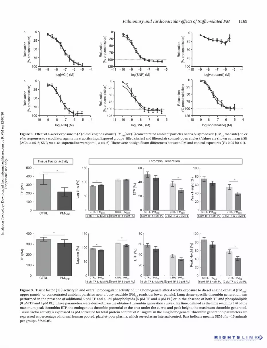
Vascular functionIn isolated rat aortic rings, the vasodilator PE caused a con-centration-dependent contraction (Figure 2). The response to PE was not different between control animals and PM
DEE-
exposed animals or control animals and animals exposed to roadside PM
2.5 (P > 0.05 for all, two-way ANOVA; n = 4–6). ACh,
SNP, and ISP all caused concentration-dependent relaxation of PE-contracted tissue. Responses in tissue from PM
DEE-
exposed animals or roadside PM2.5
-exposed animals were not different from their respective controls (P > 0.05 for all). In light of these results, organ bath analysis was not performed in the repetition of the PM
2.5 roadside study.
Cardiac tissueSamples of heart tissue from control and exposed rats were examined with regard to expression of IL-1β, TNF-α, and IL-6 mRNA as well as to phosphorylation of MAPKs.
Neither the expression of mRNA for these cytokines, nor the phosphorylation of the investigated MAPKs differed between control and PM-exposed animals (data not shown).
TF activity and thrombin generationLung TF activity was significantly decreased after exposure to PM
DEE (control 368 ± 61 pM versus PM
DEE 218 ± 51 pM;
P = 0.009; Figure 3) and slightly diminished after PM2.5
road-side exposure (control 348 ± 19 pM versus PM
2.5 310 ± 34 pM;
P = 0.047; Figure 3). Furthermore, partly coherent changes to lung TF activity were observed in thrombin generation, since the lag time is prolonged for exposure to PM
DEE (control 86%
± 3% versus PMDEE
90% ± 3%; P = 0.039; Figure 3) whereas the lag time is shortened by exposure to PM
2.5 (control 97%
± 2% versus PM2.5
roadside 91% ± 3%; P = 0.015; Figure 3) for the latter measured both with and without the addition of TF and phospholipids. Overall thrombin generation, as depicted by the ETP, was not altered upon long-term expo-sure to traffic-related PM: ETP control 51% ± 4% versus PM
DEE
46% ± 3% (P = 0.231; Figure 3) and control 58% ± 3% versus PM
2.5 roadside 51% ± 5% (P = 0.383; Figure 3). In addition,
analysis of lung tissue thrombogenicity in the absence of additional TF and phospholipids demonstrated an overall decreased lung-induced thrombin generation for long-term exposure to PM
DEE (ETP: control 38% ± 4% versus PM
DEE 28%
± 3%; P = 0.027), whereas no changes were observed after long-term exposure to PM
2.5 (38% ± 3% versus PM
2.5 roadside
32% ± 3%; P = 0.197). The attenuation of lung tissue–induced
Table 3. Parameters in blood after exposure to diesel engine exhaust or concentrated ambient particles near a busy roadside.
BALF Parameter Unit
PMDEE PM 2.5 roadside #1 PM 2.5 roadside #2
Control PM exposure Control PM exposure Control PM exposure
Mean ± SD Mean ± SD Mean ± SD Mean ± SD Mean ± SD Mean ± SD
4 Weeks
RBC x 1012/L 8.73 ± 0.36 8.65 ± 0.23 8.62 ± 0.23 8.59 ± 0.28 8.37 ± 0.20 8.41 ± 0.30
HGB mmol/L 9.20 ± 0.39 9.02 ± 0.23 8.90 ± 0.31 8.93 ± 0.32 8.81 ± 0.21 8.82 ± 0.30
HCT L/L 0.416 ± 0.016 0.412 ± 0.015 0.395 ± 0.015 0.396 ± 0.015 0.392 ± 0.010 0.393 ± 0.018
HDW mmol/L 1.832 ± 0.079 1.855 ± 0.052 1.954 ± 0.071 1.930 ± 0.111 1.795 ± 0.101 1.806 ± 0.111
PLT x 109/L 448 ± 57 431 ± 43 476 ± 89 462 ± 158 640 ± 60 671 ± 88
MPC g/dL 22.59 ± 0.79 22.07 ± 0.93 22.87 ± 0.83 22.97 ± 0.75 22.81 ± 0.75 23.02 ± 0.61
WBC x 109/L 3.69 ± 1.01 2.91*±0.5 3.34 ± 0.92 3.60 ± 1.11 4.23 ± 0.89 3.68 ± 0.82
PMN x 109/L 0.77 ± 0.26 0.67 ± 0.18 0.76 ± 0.25 0.86 ± 0.28 0.68 ± 0.18 0.67 ± 0.18
Lymphocytes x 109/L 2.78 ± 0.71 2.14* ± 0.43 2.47 ± 0.73 2.61 ± 0.85 3.36 ± 0.68 2.85† ± 0.64Basophils x 109/L 0.008 ± 0.005 0.002* ± 0.003 0.026 ± 0.013 0.028 ± 0.017 0.026 ± 0.019 0.022 ± 0.007
PMN % 20.6 ± 2.1 23.1 ± 5.6 23.1 ± 5.2 24.2 ± 5.7 16.2 ± 2.6 18.14 ± 2.74
Lymphocytes % 75.86 ± 2.55 73.37 ± 5.79 74.02 ± 5.11 72.44 ± 6.11 79.58 ± 3.31 77.49 ± 2.79
Basophils % 0.196 ± 0.069 0.132 ± 0.078 0.746 ± 0.263 0.778 ± 0.297 0.549 ± 0.307 0.583 ± 0.137
vWF mU/mL 133 ± 13 112* ± 34 136 ± 29 124 ± 41 163 ± 22 153 ± 26
PAI-1 ng/mL 0.15 ± 0.12 0.24 ± 0.15 0.16 ± 0.1 0.22 ± 0.11 0.16 ± 0.13 0.20 ± 0.19
tPA tot ng/mL 0.13 ± 0.04 0.12 ± 0.04 0.13 ± 0.05 0.13 ± 0.04
Fibrinogen mg/mL 1.74 ± 0.79 1.77 ± 0.67 1.52 ± 0.49 1.46 ± 0.65 2.63 ± 0.50 2.99 ± 1.16
CC16 ng/mL 30.3 ± 21.1 25.3 ± 14.1 19.5 ± 13.2 15.6 ± 10.1 25.9 ± 2.7 27.0 ± 4.5
Day 6
vWF mU/mL 193 ± 83 165 ± 78 198 ± 44 222 ± 21 137 ± 20 120 ± 37
PAI-1 ng/mL 0.27 ± 0.09 0.21 ± 0.12 0.37 ± 0.17 0.26 ± 0.19
CC16 ng/mL 25.1 ± 2.8 26.4 ± 4.3
Fibrinogen mg/mL 1.24 ± 0.48 1.27 ± 0.76 1.05 ± 0.33 0.99 ± 0.26 2.44 ± 0.45 2.69 ± 0.71
*P<0.05 compared to experimental control; †P=0.05 compared to experimental control.
Inha
latio
n T
oxic
olog
y D
ownl
oade
d fr
om in
form
ahea
lthca
re.c
om b
y R
IVM
on
12/0
7/10
For
pers
onal
use
onl
y.
Pulmonary and cardiovascular effects of traffic-related PM 1169
0a
25
50
Rel
axat
ion
(% p
reco
nstri
ctio
n)
75
100−10 −9 −8 −7
log[ACh] (M)
0
25
50
Rel
axat
ion
(% p
reco
nstri
ctio
n)
75
125
100
−11 −10 −9 −8 −7 −6 −5log[SNP] (M)
0
25
50
Rel
axat
ion
(% p
reco
nstri
ctio
n)
75
100−10 −9 −8 −7 −6 −5 −4
log[verapamil] (M)−6 −5 −4
0b
25
50
Rel
axat
ion
(% p
reco
nstri
ctio
n)
75
100−10 −9 −8 −7
log[ACh] (M)
0
25
50R
elax
atio
n(%
pre
cons
trict
ion)
75
125
100
0
25
50
75
125
100
−11 −10 −9 −8 −7 −6 −5log[SNP] (M)
Rel
axat
ion
(% p
reco
nstri
ctio
n)
−10 −9 −8 −7 −6 −5 −4log[isoprenaline] (M)
−6 −5 −4
Figure 2. Effect of 4-week exposure to (A) diesel engine exhaust (PMDEE
) or (B) concentrated ambient particles near a busy roadside (PM2.5
roadside) on ex vivo responses to vasodilator agents in rat aortic rings. Exposed groups (filled circles) and filtered air control (open circles). Values are shown as mean ± SE (ACh, n = 5–6; SNP, n = 4–6; isoprenaline/verapamil, n = 4–6). There were no significant differences between PM and control exposures (P > 0.05 for all).
Tissue Factor activity
*
** *
500
400
300
TF (p
M)
200
100
0CTRL PMDEE
**
* *
400
300
TF (p
M)
200
100
0CTRL PMDEE
Lag
time
(%)
0
50
100
150
CTRL5 pM TF & 4µM PL
PMDEE CTRL0 pM TF & 0 µM PL
PMDEE
0
50Lagt
ime
(%)
ETP
(%)100
150
CTRL
5 pM TF & 4µM PLPM2.5 CTRL
0 pM TF & 0 µM PLPM2.5
0
Pea
k H
eigh
t (%
)
0
20
40
60
80
100
20
40
60
80
CTRL
5 pM TF & 4µM PLPM2.5 CTRL
0 pM TF & 0 µM PLPM2.5 CTRL
5 pM TF & 4µM PLPM2.5 CTRL
0 pM TF & 0 µM PLPM2.5
0
20ETP
(%) 40
60
Thrombin Generation
CTRL5 pM TF & 4µM PL
PMDEE CTRL0 pM TF & 0 µM PL
PMDEE0
20
40
60
Pea
k H
eigh
t (%
) 80
100
CTRL5 pM TF & 4µM PL
PMDEE CTRL0 pM TF & 0 µM PL
PMDEE
Figure 3. Tissue factor (TF) activity in and overall procoagulant activity of lung homogenate after 4 weeks exposure to diesel engine exhaust (PMDEE
: upper panels) or concentrated ambient particles near a busy roadside (PM
2.5 roadside: lower panels). Lung tissue-specific thrombin generation was
performed in the presence of additional 5 pM TF and 4 µM phospholipids (5 pM TF and 4 µM PL) or in the absence of both TF and phospholipids (0 pM TF and 4 µM PL). Three parameters were derived from the obtained thrombin generation curves: lag time, defined as the time reaching 1/6 of the maximum peak thrombin; ETP, the endogenous thrombin potential or the area under the curve; and peak height, the maximum thrombin generated. Tissue factor activity is expressed as pM corrected for total protein content of 2.5 mg/ml in the lung homogenate. Thrombin generation parameters are expressed as percentage of normal human pooled, platelet-poor plasma, which served as an internal control. Bars indicate mean ± SEM of n = 15 animals per groups. *P < 0.05.
Inha
latio
n T
oxic
olog
y D
ownl
oade
d fr
om in
form
ahea
lthca
re.c
om b
y R
IVM
on
12/0
7/10
For
pers
onal
use
onl
y.

1170 M. E. Gerlofs-Nijland et al.
thrombin generation upon exposure to PMDEE
was confirmed by a decrease in peak height (control 55% ± 6% versus PM
DEE
39% ± 4%; P = 0.018; Figure 3). Furthermore, maximum thrombin generation given by the peak height was decreased after PM
2.5 exposure (control 57% ± 5% versus PM
2.5 road-
side 41% ± 5%; P = 0.020; Figure 3) confirming the trend in attenuation of the ETP.
Discussion
Prolonged exposure to traffic-related PM at levels approxi-mately 10 times higher than ambient levels, or exposure to specifically diesel engine exhaust, exerted only modest effects in relatively healthy rats. This was irrespective the fact that a mild inflammation was induced at the onset of exposure. Accumulation of particles within alveolar macro-phages was observed in both PM
2.5 roadside exposures dem-
onstrating that fine particulates are capable of reaching deep into the alveolar spaces. Biological changes were mainly of a cardiovascular nature, as shown by reduced WBC numbers, diminished levels of vWF protein, and reduced lung tissue thrombogenicity or procoagulant activity.
The fact that only very mild effects were detected in this study may be related to the adequately functioning host defense system of the rats. The animals were exposed to ozone (800 µg/m3 for 12 h) prior to prolonged exposure to traffic PM, which was intended to cause significant, yet non-severe, pulmonary inflammation to compromise the defense system at the beginning of exposure to PM. Ozone is known to provoke damage of type I epithelial cells and increased permeability of the alveolar walls (Bhalla, 1999; Dormans et al., 1990). Previous studies in our laboratory (van Bree et al., 2001, 2002) under similar conditions as in the cur-rent study (12–24 h; 800 µg/m3 ozone) resulted in a 2–3-fold increase protein levels in BALF, as well as a moderate influx of neutrophils (10–20% of total lavage cells). However, the inflammation induced in our study was rather mild, as only a slight (2.5%) increase in inflammatory cells was observed. On the other hand, a similar rise in lung permeability was found as reported previously. The difference in response might be caused by a difference in sensitivity between Fisher-344 rats used in the present study and the Wistar rats used previously. Nevertheless, ozone exposure was found to cause a similar degree of lung permeability to that found previously. This is important as an increase in permeability of the alveolar wall may assist in the translocation of particles from the lung into the circulation; one of the key mechanisms proposed to explain the systemic actions of inhaled particles (Geiser and Kreyling, 2010). Because accumulated particles within mac-rophages were observed in the present study, it seems plau-sible that translocation to the system circulation had taken place. Many epidemiological studies have claimed that, in particular, people with compromised airways are more likely to develop adverse health effects due to exposure to PM. This is in line with our observation that the rather healthy rats do not develop biological relevant adverse responses due to traffic-derived PM.
Using our diesel powered generator, a stable highly controlled test atmosphere was created that consisted of soot particles. The PM levels that were applied in this study can easily be detected in hot spots, such as road tunnels or at kerb sides of busy city streets. Ambient PM has been shown to have substantial spatial and temporal variation, both in terms of amount and physicochemical composi-tion and that the contribution of secondary inorganic components although to play a very limited role in induc-ing toxicity (Schlesinger and Cassee, 2003) contributed on average ~25%. These factors might very well explain the intra- and inter-experimental variability observed for the two roadside experiments. The PM mass concentrations were higher than those applied in the PM
DEE exposure. On
the basis of epidemiological associations that suggest a linear concentration–response relationship between PM and cardiorespiratory responses, it was assumed that PM mass concentrations would be predictive of the biological responses in the present study, however, this did not appear to be the case. Because the two roadside experiments led to higher, albeit distinctly different, average PM mass con-centrations, according to the general assumption, any effect seen for PM
DEE should also be observed in the PM
2.5 roadside
experiments. However, most parameters responding in the PM
DEE experiment were not affected by the PM roadside
exposures, which implies that other factors than PM mass (i.e. PM size and chemical composition) affect the in vivo responses. Indeed, previous studies by our group and others (Schwarze et al., 2006; Gerlofs-Nijland et al., 2007, 2009b) suggested that factors such as chemical composition are driving the toxicity. Another important difference between the PM
DEE exposure and the exposure to roadside PM
2.5 is the
higher gaseous pollutant concentrations for the exposure to diesel engine exhaust. As the PM
DEE gaseous components
were not exceeding limit values as defined by American Conference of Governmental Industrial Hygienists (ACGIH, 1991), we can assume that these could not explain the observed vascular responses.
One more variable between the PMDEE
and PM2.5
roadside exposures is the particle number concentration, with sub-stantially higher numbers for the PM
DEE experiment. Diesel
engine exhausts are dominated by particles of approximately <100 nm, which are also referred to as ultrafine particles. Several authors have suggested that ultrafine particles have adverse effects on the cardiovascular system (Delfino et al., 2005; Schulz et al., 2005; Knol et al., 2009). Therefore, it may very well be that in our PM
2.5 roadside experiments, in which
the numbers of ultrafine particles were lower than the PMDEE
, the number of ultrafines has played a more dominant role than PM mass.
Another explanation for the observed limited responses might be the development of adaptation caused by the long exposure duration. It is generally known that various biological markers have their optimal effect at different time points. Moreover, some markers like MAPKs may be activated over time in a multi-phasic way, i.e. even baseline levels vary from day to day (Thrane et al., 2001; Chen et al.,
Inha
latio
n T
oxic
olog
y D
ownl
oade
d fr
om in
form
ahea
lthca
re.c
om b
y R
IVM
on
12/0
7/10
For
pers
onal
use
onl
y.

Pulmonary and cardiovascular effects of traffic-related PM 1171
2003). Reduced vWF protein levels were already observed 6 days after exposure to PM
DEE and reached significance after
4 weeks. Measuring at the different time points might also implicate that changes in adaptative pathways are observed. This may explain some of the contradictory observations of other groups increased vWF levels to traffic-related PM (O’Neill et al., 2007; Yue et al., 2007), compared to a decrease in vWF in association with air pollutants (Carlsten et al., 2008; Hildebrandt et al., 2009). Elevated plasma vWF levels may imply an increased risk for thrombosis (Franchini and Mannucci, 2008), therefore, the time course of thrombotic responses may also vary between acute, subchronic, or prolonged exposure to air pollutants. Adaptation path-ways to chronic exposures present in healthy animals may be impaired in disease; therefore, experiments are cur-rently underway in our laboratory examining the actions of PM
DEE on the cardiovascular system in a model of
atherosclerosis.The impaired lung procoagulation activity after prolonged
exposure to traffic-related PM is supported by the reduced lung TF activity in conjunction with reduced lung tissue thrombin generation. In general, acute exposure to air pol-lution is associated to increased hypercoagulability shown by shorter prothrombin time (lag time) and elevated plasma thrombin generation (Baccarelli et al., 2007; Bonzini et al., 2010). However, these epidemiological studies provide insight in the plasma hypercoagulable state after acute exposure to air pollution, whereas impaired tissue procoagulant activity might also indicate an adaptive defense mechanism.
In our clinical studies, we have shown that a 2-h exposure of healthy volunteers to Edinburgh PM
2.5 had no effect on ves-
sel wall function as determined by forearm plethysmography (Mills et al., 2008). On the other hand, diluted diesel engine exhaust attenuated responses to the endothelium- dependent vasodilator ACh and the endothelium- independent vasodi-lator SNP, but not to the NO-independent vasodilator, verapamil. Previously, we have demonstrated in an animal experiment that similar responses occurred 4 h after acute exposure to various PM samples by intratracheal instilla-tion (Bagate et al., 2004). In addition, diesel engine exhaust particles directly inhibit vascular relaxation to endothelium-dependent vasodilators (Miller et al., 2009). Therefore, we assumed that prolonged exposure to PM
DEE and not road-
side PM2.5
was associated with cardiovascular impairment. However, no signs of impairment were observed after pro-longed exposures applied in the three experiments presented in this paper.
In the present study, we noted a decrease of WBC in the PM
DEE as well as in the second PM
2.5 roadside experi-
ment. Similar observations have been made in rats after acute exposure to traffic-related PM (Gerlofs-Nijland et al., 2005; Kooter et al., 2006). In human studies, Frampton and co-workers (2002) noted that NO
2 exposure resulted in
reduced lymphocytes that migrate to the lung, as increased lymphocyte numbers were found in the respiratory system. Recently, changes in differential WBC was reported in patients with chronic pulmonary disease related to ambient
air pollution exposure (Brüske et al., 2010). Although the biological significance and impact is still not clear, it seems that reduced circulating WBCs are related to increased exposure to air pollutants.
Freshly generated PMDEE
induced mild cardiovascular responses (impaired coagulation) but no respiratory effects were seen in relatively healthy rats. Also no biological rel-evant changes were detected after exposure to ambient roadside PM
2.5. The overall analysis of the results did not
support the hypothesis that PM mass concentrations are linear related to health effects. In contrast to common belief, prolonged exposure to traffic-related PM in healthy animals may not be detrimental due to various biological adaptive response mechanisms. It could be speculated that vulnerability of humans to acute or repeated exposure to PM may be primarily dependent on the presence of co-morbidity such as coronary heart disease. We conclude that prolonged although not chronic exposures in healthy animals have very limited impact on pulmonary and car-diovascular function. Further studies are needed in animals with established disease (such as more extensive pulmo-nary inflammation or developed cardiovascular disease) to reveal the influence of susceptibility on air pollution–induced toxicity.
Acknowledgements
We thank the technical team under the supervision of Ruud van Kinderen and Hans Strootman from the Animal Facility Department of the National Institute of Vaccines for their skillful biotechnical assistance. We also acknowledge the assistants, Liset de la Fonteyne-Blankestijn, Yvonne Wallbrink, Piet Beekhof, and colleagues under the super-vision of Henk van Loveren and Eugene Jansen, from the Laboratory for Health Protection Research of the RIVM’s Nutrition, Medicines and Consumer Safety Division for their excellent technical support.
Declaration of interest
The main part of this work was supported by a fund from the Dutch Ministry of Housing, Spatial Planning and the Environment (VROM) in the framework of research project Air Pollution and Health (M/630186 and M/630196) at the RIVM. Mark Miller and Patrick Hadoke are supported by a British Heart Foundation Programme Grant (RG/05/003). Evren Kilinç is supported by a grant of the the Netherlands Heart Foundation (grant number: 2006B064). The analy-ses of heart tissues conducted at the Norwegian Institute of Public Health were funded by the Research Council of Norway through the “Environment, Genetics and Health” program. This work has been presented in part at the Inhaled Particles X Conference 2008 and published in the accom-panying Journal of Physics Conference Series 2009. Evren Kilinç is supported by a grant of the the Netherlands Heart Foundation (grant number: 2006B064)
Inha
latio
n T
oxic
olog
y D
ownl
oade
d fr
om in
form
ahea
lthca
re.c
om b
y R
IVM
on
12/0
7/10
For
pers
onal
use
onl
y.

1172 M. E. Gerlofs-Nijland et al.
ReferencesACGIH. 1991. Documentation of the threshold limit values and biologi-
cal exposure indices. 6th ed. Cincinnati, OH: American Conference of Governmental Industrial Hygienists.
Araujo JA, Barajas B, Kleinman M, Wang X, Bennett BJ, Gong KW, Navab M, Harkema J, Sioutas C, Lusis AJ, Nel AE. 2008. Ambient particulate pol-lutants in the ultrafine range promote early atherosclerosis and systemic oxidative stress. Circ Res 102:589–596.
Baccarelli A, Zanobetti A, Martinelli I, Grillo P, Hou L, Giacomini S, Bonzini M, Lanzani G, Mannucci PM, Bertazzi PA, Schwartz J. 2007. Effects of exposure to air pollution on blood coagulation. J Thromb Haemost 5:252–260.
Bagate K, Meiring JJ, Gerlofs-Nijland ME, Vincent R, Cassee FR, Borm PJ. 2004. Vascular effects of ambient particulate matter instillation in spontaneous hypertensive rats. Toxicol Appl Pharmacol 197:29–39.
Beelen R, Hoek G, Houthuijs D, van den Brandt PA, Goldbohm RA, Fischer P, Schouten LJ, Armstrong B, Brunekreef B. 2009. The joint association of air pollution and noise from road traffic with cardiovascular mortality in a cohort study. Occup Environ Med 66:243–250.
Bhalla DK. 1999. Ozone-induced lung inflammation and mucosal barrier dis-ruption: toxicology, mechanisms, and implications. J Toxicol Environ Health B Crit Rev 2:31–86.
Bonzini M, Tripodi A, Artoni A, Tarantini L, Marinelli B, Bertazzi PA, Apostoli P, Baccarelli A. 2010. Effects of inhalable particulate matter on blood coagu-lation. J Thromb Haemost 8:662–668.
Brunekreef B, Holgate ST. 2002. Air pollution and health. Lancet 360:1233–1242.
Brüske I, Hampel R, Socher MM, Rückerl R, Schneider A, Heinrich J, Oberdörster G, Wichmann HE, Peters A. 2010. Impact of ambient air pol-lution on the differential white blood cell count in patients with chronic pulmonary disease. Inhal Toxicol 22:245–252.
Carlsten C, Kaufman JD, Trenga CA, Allen J, Peretz A, Sullivan JH. 2008. Thrombotic markers in metabolic syndrome subjects exposed to diesel exhaust. Inhal Toxicol 20:917–921.
Cassee FR, Boere AJ, Fokkens PH, Leseman DL, Sioutas C, Kooter IM, Dormans JA. 2005. Inhalation of concentrated particulate matter pro-duces pulmonary inflammation and systemic biological effects in com-promised rats. J Toxicol Environ Health Part A 68:773–796.
Chen JX, Berry LC, Christman BW, Meyrick B. 2003. Glutathione mediates LPS-stimulated COX-2 expression via early transient p42/44 MAPK activation. J Cell Physiol 197:86–93.
Chen LC, Hwang JS. 2005. Effects of subchronic exposures to concentrated ambient particles (CAPs) in mice. IV. Characterization of acute and chronic effects of ambient air fine particulate matter exposures on heart-rate variability. Inhal Toxicol 17:209–216.
Chen LC, Nadziejko C. 2005. Effects of subchronic exposures to concentrated ambient particles (CAPs) in mice. V. CAPs exacerbate aortic plaque devel-opment in hyperlipidemic mice. Inhal Toxicol 17:217–224.
Delfino RJ, Sioutas C, Malik S. 2005. Potential role of ultrafine particles in associations between airborne particle mass and cardiovascular health. Environ Health Perspect 113:934–946.
Dormans JA, Rombout PJ, van Loveren H. 1990. Surface morphology and mor-phometry of rat alveolar macrophages after ozone exposure. J Toxicol Environ Health 31:53–70.
Elder A, Couderc JP, Gelein R, Eberly S, Cox C, Xia X, Zareba W, Hopke P, Watts W, Kittelson D, Frampton M, Utell M, Oberdörster G. 2007. Effects of on-road highway aerosol exposures on autonomic responses in aged, spontaneously hypertensive rats. Inhal Toxicol 19:1–12.
Frampton MW, Boscia J, Roberts NJ Jr, Azadniv M, Torres A, Cox C, Morrow PE, Nichols J, Chalupa D, Frasier LM, Gibb FR, Speers DM, Tsai Y, Utell MJ. 2002. Nitrogen dioxide exposure: effects on airway and blood cells. Am J Physiol Lung Cell Mol Physiol 282:L155–L165.
Franchini M, Mannucci PM. 2008. Von Willebrand factor: another janus-faced hemostasis protein. Semin Thromb Hemost 34:663–669.
Frederix K, Kooter IM, van Oerle R, Fens D, Hamulyak K, Gerlofs-Nijland ME, Ten Cate H, Spronk HM. 2008. A new method to determine tissue specific tissue factor thrombomodulin activities: endotoxin and particulate air pollution induced disbalance. Thromb J 6:14.
Gauderman WJ, Avol E, Lurmann F, Kuenzli N, Gilliland F, Peters J, McConnell R. 2005. Childhood asthma and exposure to traffic and nitrogen dioxide. Epidemiology 16:737–743.
Gehring U, Heinrich J, Krämer U, Grote V, Hochadel M, Sugiri D, Kraft M, Rauchfuss K, Eberwein HG, Wichmann HE. 2006. Long-term exposure to ambient air pollution and cardiopulmonary mortality in women. Epidemiology 17:545–551.
Geiser M, Kreyling WG. 2010. Deposition and biokinetics of inhaled nanopar-ticles. Part Fibre Toxicol 7:2.
Gerlofs-Nijland ME, Boere AJ, Leseman DL, Dormans JA, Sandström T, Salonen RO, van Bree L, Cassee FR. 2005. Effects of particulate matter on the pulmonary and vascular system: time course in spontaneously hypertensive rats. Part Fibre Toxicol 2:2.
Gerlofs-Nijland ME, Dormans JA, Bloemen HJ, Leseman DL, John A, Boere F, Kelly FJ, Mudway IS, Jimenez AA, Donaldson K, Guastadisegni C, Janssen NA, Brunekreef B, Sandström T, van Bree L, Cassee FR. 2007. Toxicity of coarse and fine particulate matter from sites with contrasting traffic profiles. Inhal Toxicol 19:1055–1069.
Gerlofs-Nijland ME, Campbell A, Miller MR, Newby DE, Cassee FR. 2009a. Toxicity of inhaled traffic related particulate matter. J Phys Conf Ser 151:012049.
Gerlofs-Nijland ME, Rummelhard M, Boere AJ, Leseman DL, Duffin R, Schins RP, Borm PJ, Sillanpää M, Salonen RO, Cassee FR. 2009b. Particle induced toxicity in relation to transition metal and polycyclic aromatic hydrocarbon contents. Environ Sci Technol 43:4729–4736.
Gong H Jr, Sioutas C, Linn WS. 2003. Controlled exposures of healthy and asth-matic volunteers to concentrated ambient particles in metropolitan Los Angeles. Res Rep Health Eff Inst 1–36; discussion 37.
Hildebrandt K, Rückerl R, Koenig W, Schneider A, Pitz M, Heinrich J, Marder V, Frampton M, Oberdörster G, Wichmann HE, Peters A. 2009. Short-term effects of air pollution: a panel study of blood markers in patients with chronic pulmonary disease. Part Fibre Toxicol 6:25.
Hoek G, Brunekreef B, Goldbohm S, Fischer P, van den Brandt PA. 2002. Association between mortality and indicators of traffic-related air pollu-tion in the Netherlands: a cohort study. Lancet 360:1203–1209.
Hoffmann B, Moebus S, Dragano N, Möhlenkamp S, Memmesheimer M, Erbel R, Jöckel KH. Heinz Nixdorf Recall Investigative Group. 2009. Residential traffic exposure and coronary heart disease: results from the Heinz Nixdorf Recall Study. Biomarkers 14 Suppl 1:74–78.
Hwang JS, Nadziejko C, Chen LC. 2005. Effects of subchronic exposures to con-centrated ambient particles (CAPs) in mice. III. Acute and chronic effects of CAPs on heart rate, heart-rate fluctuation, and body temperature. Inhal Toxicol 17:199–207.
Kim S, Jaques PA, Chang M, Froines JR, Sioutas C. 2001a. Versatile aerosol concentration enrichment system (VACES) for simultaneous in vivo and in vitro evaluation of toxic effects of ultrafine, fine and coarse ambient particles Part I: Development and laboratory characterization. J Aerosol Sci 32:1281–1297.
Kim S, Jaques PA, Chang M, Barone T, Xiong C, Friedlander SK, Sioutas C. 2001b. Versatile aerosol concentration enrichment system (VACES) for simultaneous in vivo and in vitro evaluation of toxic effects of ultrafine, fine and coarse ambient particles Part II: Field Evaluation. J Aerosol Sci 32:1299–1314.
Kleinman MT, Hamade A, Meacher D, Oldham M, Sioutas C, Chakrabarti B, Stram D, Froines JR, Cho AK. 2005. Inhalation of concentrated ambient particulate matter near a heavily trafficked road stimulates antigen-in-duced airway responses in mice. J Air Waste Manag Assoc 55:1277–1288.
Knol AB, de Hartog JJ, Boogaard H, Slottje P, van der Sluijs JP, Lebret E, Cassee FR, Wardekker JA, Ayres JG, Borm PJ, Brunekreef B, Donaldson K, Forastiere F, Holgate ST, Kreyling WG, Nemery B, Pekkanen J, Stone V, Wichmann HE, Hoek G. 2009. Expert elicitation on ultrafine particles: likelihood of health effects and causal pathways. Part Fibre Toxicol 6:19.
Kooter IM, Boere AJ, Fokkens PH, Leseman DL, Dormans JA, Cassee FR. 2006. Response of spontaneously hypertensive rats to inhalation of fine and ultrafine particles from traffic: experimental controlled study. Part Fibre Toxicol 3:7.
Lai CH, Liou SH, Lin HC, Shih TS, Tsai PJ, Chen JS, Yang T, Jaakkola JJ, Strickland PT. 2005. Exposure to traffic exhausts and oxidative DNA dam-age. Occup Environ Med 62:216–222.
Lippmann M, Hwang JS, Maciejczyk P, Chen LC. 2005a. PM source apportion-ment for short-term cardiac function changes in ApoE-/- mice. Environ Health Perspect 113:1575–1579.
Lippmann M, Gordon T, Chen LC. 2005b. Effects of subchronic exposures to concentrated ambient particles in mice. IX. Integral assessment and human health implications of subchronic exposures of mice to CAPs. Inhal Toxicol 17:255–261.
Lippmann M, Chen LC. 2009. Health effects of concentrated ambient air par-ticulate matter (CAPs) and its components. Crit Rev Toxicol 39:865–913.
Lucking AJ, Lundback M, Mills NL, Faratian D, Barath SL, Pourazar J, Cassee FR, Donaldson K, Boon NA, Badimon JJ, Sandstrom T, Blomberg A, Newby DE. 2008. Diesel exhaust inhalation increases thrombus formation in man. Eur Heart J 29:3043–3051.
Inha
latio
n T
oxic
olog
y D
ownl
oade
d fr
om in
form
ahea
lthca
re.c
om b
y R
IVM
on
12/0
7/10
For
pers
onal
use
onl
y.

Pulmonary and cardiovascular effects of traffic-related PM 1173
Marra M, Rombout PJA. 1990. Design and performance of an inhalation cham-ber for exposing laboratory animals to oxidant air pollutants. Inhal Toxicol 2:187–204.
McCreanor J, Cullinan P, Nieuwenhuijsen MJ, Stewart-Evans J, Malliarou E, Jarup L, Harrington R, Svartengren M, Han IK, Ohman-Strickland P, Chung KF, Zhang J. 2007. Respiratory effects of exposure to diesel traffic in persons with asthma. N Engl J Med 357:2348–2358.
Miller MR, Borthwick SJ, Shaw CA, McLean SG, McClure D, Mills NL, Duffin R, Donaldson K, Megson IL, Hadoke PW, Newby DE. 2009. Direct impair-ment of vascular function by diesel exhaust particulate through reduced bioavailability of endothelium-derived nitric oxide induced by superoxide free radicals. Environ Health Perspect 117:611–616.
Mills NL, Törnqvist H, Robinson SD, Gonzalez M, Darnley K, MacNee W, Boon NA, Donaldson K, Blomberg A, Sandstrom T, Newby DE. 2005. Diesel exhaust inhalation causes vascular dysfunction and impaired endogenous fibrinolysis. Circulation 112:3930–3936.
Mills NL, Törnqvist H, Gonzalez MC, Vink E, Robinson SD, Söderberg S, Boon NA, Donaldson K, Sandström T, Blomberg A, Newby DE. 2007. Ischemic and thrombotic effects of dilute diesel-exhaust inhalation in men with coronary heart disease. N Engl J Med 357:1075–1082.
Mills NL, Robinson SD, Fokkens PH, Leseman DL, Miller MR, Anderson D, Freney EJ, Heal MR, Donovan RJ, Blomberg A, Sandström T, MacNee W, Boon NA, Donaldson K, Newby DE, Cassee FR. 2008. Exposure to concen-trated ambient particles does not affect vascular function in patients with coronary heart disease. Environ Health Perspect 116:709–715.
O’Neill MS, Veves A, Sarnat JA, Zanobetti A, Gold DR, Economides PA, Horton ES, Schwartz J. 2007. Air pollution and inflammation in type 2 diabetes: a mechanism for susceptibility. Occup Environ Med 64:373–379.
Pope CA 3rd, Burnett RT, Thurston GD, Thun MJ, Calle EE, Krewski D, Godleski JJ. 2004. Cardiovascular mortality and long-term exposure to particulate air pollution: epidemiological evidence of general pathophysi-ological pathways of disease. Circulation 109:71–77.
Rudez G, Janssen NA, Kilinc E, Leebeek FW, Gerlofs-Nijland ME, Spronk HM, ten Cate H, Cassee FR, de Maat MP. 2009. Effects of ambient air pollution on hemostasis and inflammation. Environ Health Perspect 117:995–1001.
Schikowski T, Sugiri D, Ranft U, Gehring U, Heinrich J, Wichmann HE, Krämer U. 2005. Long-term air pollution exposure and living close to busy roads are associated with COPD in women. Respir Res 6:152.
Schlesinger RB, Cassee F. 2003. Atmospheric secondary inorganic particulate matter: the toxicological perspective as a basis for health effects risk assessment. Inhal Toxicol 15:197–235.
Schulz H, Harder V, Ibald-Mulli A, Khandoga A, Koenig W, Krombach F, Radykewicz R, Stampfl A, Thorand B, Peters A. 2005. Cardiovascular effects of fine and ultrafine particles. J Aerosol Med 18:1–22.
Schwarze PE, Ovrevik J, Låg M, Refsnes M, Nafstad P, Hetland RB, Dybing E. 2006. Particulate matter properties and health effects: consistency of epi-demiological and toxicological studies. Hum Exp Toxicol 25:559–579.
Seagrave J, McDonald JD, Bedrick E, Edgerton ES, Gigliotti AP, Jansen JJ, Ke L, Naeher LP, Seilkop SK, Zheng M, Mauderly JL. 2006. Lung toxicity of ambi-ent particulate matter from southeastern U.S. sites with different contrib-uting sources: relationships between composition and effects. Environ Health Perspect 114:1387–1393.
Sioutas C, Koutrakis P, Godleski J, Ferguson S, Kim C, Burton RM. 1997. Harvard/EPA ambient fine particle concentrators for human and animal exposures. J Aerosol Sci 28:1057–1071.
Spronk HM, Dielis AW, De Smedt E, van Oerle R, Fens D, Prins MH, Hamulyák K, ten Cate H. 2008. Assessment of thrombin generation II: Validation of the Calibrated Automated Thrombogram in platelet-poor plasma in a clinical laboratory. Thromb Haemost 100:362–364.
Sun Q, Wang A, Jin X, Natanzon A, Duquaine D, Brook RD, Aguinaldo JG, Fayad ZA, Fuster V, Lippmann M, Chen LC, Rajagopalan S. 2005. Long-term air pollution exposure and acceleration of atherosclerosis and vascular inflammation in an animal model. JAMA 294:3003–3010.
Sun Q, Yue P, Kirk RI, Wang A, Moatti D, Jin X, Lu B, Schecter AD, Lippmann M, Gordon T, Chen LC, Rajagopalan S. 2008. Ambient air particulate matter exposure and tissue factor expression in atherosclerosis. Inhal Toxicol 20:127–137.
Thrane EV, Refsnes M, Thoresen GH, Låg M, Schwarze PE. 2001. Fluoride-induced apoptosis in epithelial lung cells involves activation of MAP kinases p38 and possibly JNK. Toxicol Sci 61:83–91.
van Bree L, Dormans JA, Boere AJ, Rombout PJ. 2001. Time study on develop-ment and repair of lung injury following ozone exposure in rats. Inhal Toxicol 13:703–718.
van Bree L, Dormans JA, Koren HS, Devlin RB, Rombout PJ. 2002. Attenuation and recovery of pulmonary injury in rats following short-term, repeated daily exposure to ozone. Inhal Toxicol 14:883–900.
Veronesi B, Makwana O, Pooler M, Chen LC. 2005. Effects of subchronic expo-sures to concentrated ambient particles. VII. Degeneration of dopamin-ergic neurons in Apo E-/- mice. Inhal Toxicol 17:235–241.
Ying Z, Kampfrath T, Thurston G, Farrar B, Lippmann M, Wang A, Sun Q, Chen LC, Rajagopalan S. 2009. Ambient particulates alter vascular func-tion through induction of reactive oxygen and nitrogen species. Toxicol Sci 111:80–88.
Yue W, Schneider A, Stölzel M, Rückerl R, Cyrys J, Pan X, Zareba W, Koenig W, Wichmann HE, Peters A. 2007 Aug 1. Ambient source-specific parti-cles are associated with prolonged repolarization and increased levels of inflammation in male coronary artery disease patients. Mutat Res 621(1–2):50–60
Inha
latio
n T
oxic
olog
y D
ownl
oade
d fr
om in
form
ahea
lthca
re.c
om b
y R
IVM
on
12/0
7/10
For
pers
onal
use
onl
y.


