Embed Size (px)
Citation preview
3 • AVIAN & EXOTICS | 5 • MEDICAL ONCOLOGY | 6 • PATHOLOGY
9 • WHAT’S YOUR DIAGNOSIS? | 10 • UPDATES & EDUCATION
rDVM QUARTERLYVETERINARY COMMUNIT Y NEWS FROM AMC SUMMER 2017
T H E E L M E R A N D M A M D O U H A B O B S T H O S P I TA L | C A S PA R Y R E S E A R C H I N S T I T U T ET H E I N S T I T U T E F O R P O S T G R A DUAT E E DU C AT ION | U S DA N I N S T I T U T E F O R A N I M A L H E A LT H E DU C AT ION
VETERINARY COMMUNIT Y NEWS FROM AMC | WINTER 201700 001
Dear Colleagues,
I am very pleased to present you with our Summer rDVM and alumni newsletter, the second one of 2017. In this edition, we are again highlighting what I feel to be some of our most unique and beloved services, such as our Avian & Exotics Service with our very own Dr. Kathy Quesenberry, and Oncolgy with Dr. Ann Hohenhaus. The exotics case is also special because it represents the kind of collaboration that happens here at AMC every day between services, in this case between Exotics and Interventional Radiology. The multitude and diver-sity of specialties truly is one of the most exciting things here. I always love (literally) bumping into amazing experienced specialists from 5-6 different services while just trying to make my way down the clinic hallway.
You would think things would quiet down in the summer—definitely not this year! Thanks to your amazing support, we are busier than we have been in many years; cages are full and things are hopping! Thank you! In addition, as always this time of year, we are in the midst of our house officer turnover, so I think it’s appropriate to devote a few words to our educational program.
The Animal Medical Center graduated its first intern class 53 years ago! How crazy is that? Since then, thousands of veterinarians have received their advanced postgraduate training at AMC; the majority in a one-year rotating internship, and beginning just a short time after, our two- and now three-year residencies. AMC alumni are everywhere in academia, private practice, research, government, and really anywhere where a highly qualified veteri-narian is needed. Even though I cannot really claim to be an AMC alum, it is a huge honor to be associated with this amazing group and I love nothing more than to meet with our alumni every day at AMC, in Manhattan, at national and international conferences —literally everywhere. Currently we have 22 rotating interns, five specialty interns, and about 30 residents, all focused on becoming smarter, better veterinarians to enhance the care of our current and their future clients.
Of course, none of this would be possible without your support. This is an amazing year for us and I am so grateful for the chance to work with all of you and to help you serve your client’s needs. Thank you again for your tremen-dous support of the AMC. As always, contact me any time with any questions, comments, or concerns. I look forward to hearing from you.
Richard
Richard E. Goldstein DVM, DACVIM, [email protected] 212-329-8824cell 347-733-7338
A LETTER FROM OUR CMO
VETERINARY COMMUNIT Y NEWS FROM AMC | SUMMER 2017 2
To Our Valued Partners In Care,
Summer at the Animal Medical Center means growth and change. We recently bid farewell to our 2017 class of graduating residents and interns and welcomed our incoming classes. We are so proud to see these doctors advancing in their careers, ensuring a bright future for the veterinary field.
In keeping with our commitment to the One Health Initiative, our third annual One Health Conference will take place here in New York City on November 4th. The One Health Initiative focuses on the collaboration between physicians and veterinarians in comparing similar, naturally occur-ring diseases that manifest in both people and animals – it will enable us all to better understand, diagnose, treat and even help prevent illness. This year’s conference will focus on cardiology and will feature veterinary speakers not only from AMC, but from NC State, UPenn and the University of Wisconsin. Our medical speakers will join us from Columbia, Cornell, NYU, MSKCC and Le Bonheur Children’s Hospital. I am so excited by the continued growth of this conference and look forward to seeing you there. Please check our website to register: www.amcny.org/onehealth.
In an effort to ensure we are meeting our referring veterinarian’s and client’s needs in the most effective manner, we are pleased to announce the appoint-ment of Liana Everaert as Executive Director of Client Relations. Liana brings strong strategic planning, marketing, and sales experience in human healthcare, business development, and customer relations to this newly created position. Liana will further our commitment to providing the highest quality service to you and the clients you refer.
I hope you continue to find this publication useful and informative. Our hope is that you see it as a valuable resource for your practice. Please feel free to let us know if you have suggestions on how we can keep you informed about what we are working on here at AMC.
As always, I would like to thank you for being a valued partner and for your continued support of AMC.
Sincerely,
Kate
Kathryn Coyne [email protected]
A LETTER FROM OUR CEO
00 VETERINARY COMMUNIT Y NEWS FROM AMC | SUMMER 2017 43
AVIAN & EXOTICS
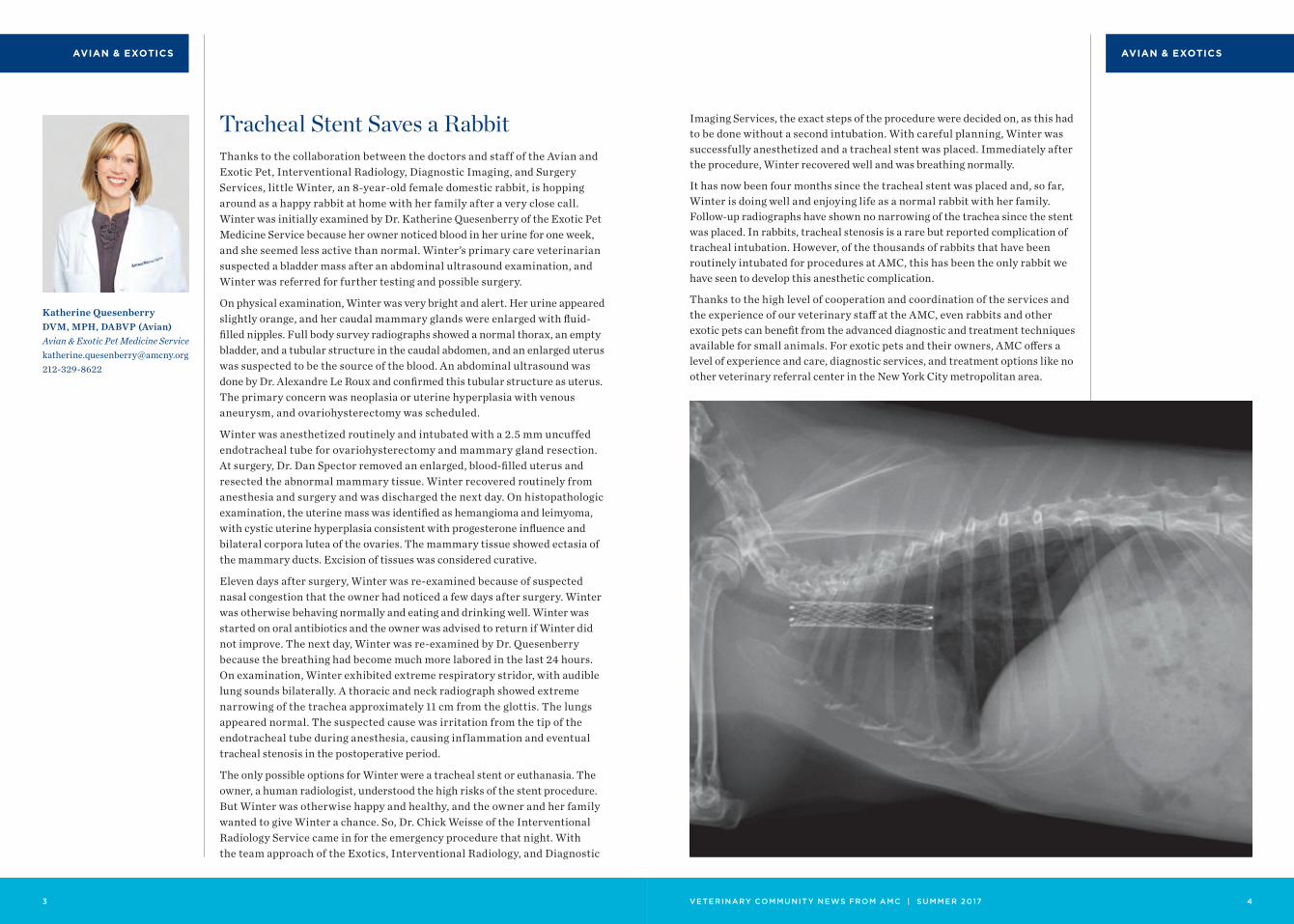
Thanks to the collaboration between the doctors and staff of the Avian and Exotic Pet, Interventional Radiology, Diagnostic Imaging, and Surgery Services, little Winter, an 8-year-old female domestic rabbit, is hopping around as a happy rabbit at home with her family after a very close call. Winter was initially examined by Dr. Katherine Quesenberry of the Exotic Pet Medicine Service because her owner noticed blood in her urine for one week, and she seemed less active than normal. Winter’s primary care veterinarian suspected a bladder mass after an abdominal ultrasound examination, and Winter was referred for further testing and possible surgery.
On physical examination, Winter was very bright and alert. Her urine appeared slightly orange, and her caudal mammary glands were enlarged with fluid-filled nipples. Full body survey radiographs showed a normal thorax, an empty bladder, and a tubular structure in the caudal abdomen, and an enlarged uterus was suspected to be the source of the blood. An abdominal ultrasound was done by Dr. Alexandre Le Roux and confirmed this tubular structure as uterus. The primary concern was neoplasia or uterine hyperplasia with venous aneurysm, and ovariohysterectomy was scheduled.
Winter was anesthetized routinely and intubated with a 2.5 mm uncuffed endotracheal tube for ovariohysterectomy and mammary gland resection. At surgery, Dr. Dan Spector removed an enlarged, blood-filled uterus and resected the abnormal mammary tissue. Winter recovered routinely from anesthesia and surgery and was discharged the next day. On histopathologic examination, the uterine mass was identified as hemangioma and leimyoma, with cystic uterine hyperplasia consistent with progesterone influence and bilateral corpora lutea of the ovaries. The mammary tissue showed ectasia of the mammary ducts. Excision of tissues was considered curative.
Eleven days after surgery, Winter was re-examined because of suspected nasal congestion that the owner had noticed a few days after surgery. Winter was otherwise behaving normally and eating and drinking well. Winter was started on oral antibiotics and the owner was advised to return if Winter did not improve. The next day, Winter was re-examined by Dr. Quesenberry because the breathing had become much more labored in the last 24 hours. On examination, Winter exhibited extreme respiratory stridor, with audible lung sounds bilaterally. A thoracic and neck radiograph showed extreme narrowing of the trachea approximately 11 cm from the glottis. The lungs appeared normal. The suspected cause was irritation from the tip of the endotracheal tube during anesthesia, causing inflammation and eventual tracheal stenosis in the postoperative period.
The only possible options for Winter were a tracheal stent or euthanasia. The owner, a human radiologist, understood the high risks of the stent procedure. But Winter was otherwise happy and healthy, and the owner and her family wanted to give Winter a chance. So, Dr. Chick Weisse of the Interventional Radiology Service came in for the emergency procedure that night. With the team approach of the Exotics, Interventional Radiology, and Diagnostic
Tracheal Stent Saves a Rabbit
Katherine Quesenberry DVM, MPH, DABVP (Avian) Avian & Exotic Pet Medicine [email protected]
AVIAN & EXOTICS
Imaging Services, the exact steps of the procedure were decided on, as this had to be done without a second intubation. With careful planning, Winter was successfully anesthetized and a tracheal stent was placed. Immediately after the procedure, Winter recovered well and was breathing normally.
It has now been four months since the tracheal stent was placed and, so far, Winter is doing well and enjoying life as a normal rabbit with her family. Follow-up radiographs have shown no narrowing of the trachea since the stent was placed. In rabbits, tracheal stenosis is a rare but reported complication of tracheal intubation. However, of the thousands of rabbits that have been routinely intubated for procedures at AMC, this has been the only rabbit we have seen to develop this anesthetic complication.
Thanks to the high level of cooperation and coordination of the services and the experience of our veterinary staff at the AMC, even rabbits and other exotic pets can benefit from the advanced diagnostic and treatment techniques available for small animals. For exotic pets and their owners, AMC offers a level of experience and care, diagnostic services, and treatment options like no other veterinary referral center in the New York City metropolitan area.
VETERINARY COMMUNIT Y NEWS FROM AMC | WINTER 2017 005
MEDICAL ONCOLOGY
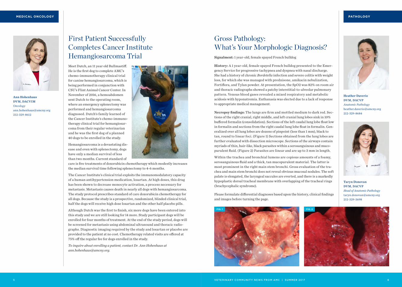
Meet Dutch, an 11 year old Bullmastiff. He is the first dog to complete AMC’s chemo-immunotherapy clinical trial for canine hemangiosarcoma, which is being performed in conjunction with CSU’s Flint Animal Cancer Center. In November of 2016, a hemoabdomen sent Dutch to the operating room, where an emergency splenectomy was performed and hemangiosarcoma diagnosed. Dutch’s family learned of the Cancer Institute’s chemo-immuno-therapy clinical trial for hemangiosar-coma from their regular veterinarian and he was the first dog of a planned 40 dogs to be enrolled in the study.
Hemangiosarcoma is a devastating dis-ease and even with splenectomy, dogs have only a median survival of less than two months. Current standard of care is five treatments of doxorubicin chemotherapy which modestly increases the median survival time following splenectomy to 4-6 months.
The Cancer Institute’s clinical trial exploits the immunomodulatory capacity of a human antihypertension medication, losartan. At high doses, this drug has been shown to decrease monocyte activation, a process necessary for metastasis. Metastasis causes death in nearly all dogs with hemangiosarcoma. The study protocol prescribes standard of care doxorubicin chemotherapy for all dogs. Because the study is a prospective, randomized, blinded clinical trial, half the dogs will receive high dose losartan and the other half placebo pills.
Although Dutch was the first to finish, six more dogs have been entered into this study and we are still looking for 14 more. Study participant dogs will be enrolled for four months of treatment. At the end of the study period, dogs will be screened for metastasis using abdominal ultrasound and thoracic radio-graphs. Diagnostic imaging required by the study and losartan or placebo are provided to the patient at no cost. Chemotherapy related visits are offered at 75% off the regular fee for dogs enrolled in the study.
To inquire about enrolling a patient, contact Dr. Ann Hohenhaus at [email protected].
First Patient Successfully Completes Cancer Institute Hemangiosarcoma Trial
VETERINARY COMMUNIT Y NEWS FROM AMC | SUMMER 2017 6
Taryn Donovan DVM, DACVPHead of Anatomic [email protected]
Heather Daverio DVM, DACVPAnatomic [email protected]
PATHOLOGY
Signalment: 1 year-old, female spayed French bulldog
History: A 1 year-old, female spayed French bulldog presented to the Emer-gency Service for progressive tachypnea and dyspnea with nasal discharge. She had a history of chronic Bordetella infection and severe colitis with weight loss, for which she was managed with prednisone, amikacin nebulization, Fortiflora, and Tylan powder. At presentation, the SpO2 was 82% on room air and thoracic radiographs showed a patchy interstitial-to-alveolar pulmonary pattern. Venous blood gases revealed a mixed respiratory and metabolic acidosis with hyponatremia. Euthanasia was elected due to a lack of response to appropriate medical management.
Necropsy findings: The lungs are firm and mottled medium to dark red. Sec-tions of the right cranial, right middle, and left cranial lung lobes sink in 10% buffered formalin (consolidation). Sections of the left caudal lung lobe float low in formalin and sections from the right caudal lung lobe float in formalin. Gen-eralized over all lung lobes are dozens of pinpoint (less than 1 mm), black to tan, round to linear foci. (Figure 1) Sections obtained from the lung lobes are further evaluated with dissection microscope. Sections of the airways contain myriads of thin, hair-like, black parasites within a serosanguinous and muco-purulent fluid. (Figure 2) Parasites are linear and are up to 3 mm in length.
Within the trachea and bronchial lumens are copious amounts of a foamy, serosanguinous fluid and a thick, tan mucopurulent material. The latter is most prominent in the right main stem bronchi. Gross evaluation of the tra-chea and main stem bronchi does not reveal obvious mucosal nodules. The soft palate is elongated, the laryngeal saccules are everted, and there is a markedly hypoplastic dorsal tracheal membrane with overlapping of the tracheal rings (brachycephalic syndrome).
Please formulate differential diagnoses based upon the history, clinical findings and images before turning the page.
Gross Pathology: What’s Your Morphologic Diagnosis?
FIG. 1 FIG. 2
Ann Hohenhaus DVM, [email protected]

VETERINARY COMMUNIT Y NEWS FROM AMC | WINTER 2017 00007
PATHOLOGY
VETERINARY COMMUNIT Y NEWS FROM AMC | SUMMER 2017 8
PATHOLOGY
References:
1. Bowman DD. Georgis’ Parasitol-ogy for Veterinarians. 10th ed. St. Louis, MO: Saunders Elsevier; 2014: 159-161, 189-190, 423.
2. Caro-Vadillo A, Martinez-Merlo E, Garcia-Real I, et al. Verminous pneumonia due to Filaroides hirthi in a Scottish terrier in Spain. Vet Rec. 2005; 157: 586-589.
3. Carrasco L, Hervas J, Gomez-Villamandos JC, et al. Massive Filaroides hirthi infestation asso-ciated with canine distemper in a puppy. Vet Rec. 1997; 140:72-73.
4. Caswell JL, Williams KJ. Respira-tory system. In: Maxie MG, ed. Jubb, Kennedy, and Palmer’s Pathology of Domestic Animals. 5th ed. St. Louis, MO; Elsevier; 2007: 645-8.
5. Gardiner CH, Poynton SL. An Atlas of Metazoan Parasites in Animal Tissues. Washington, DC: Armed Forces Institute of Pathol-ogy; 1999:22, 27.
6. Genta RM and Schad GA. Filaroides hirthi: Hyperinfective lungworm infection in immuno-suppressed dogs. Vet Pathol. 1984; 21:349-354.
Morphologic diagnosis: Pneumonia (bronchopneumonia and interstitial pneu-monia), neutrophilic, lymphoplasmacytic, histiocytic with numerous intral-esional metastrongyle nematodes (morphology consistent with Filaroides spp.), type II pneumocyte hyperplasia, fibroplasia, and multifocal fibrinous pleuritis.
Comments: The gross finding of miliary 1-5 mm wide, grey-white foci within the lungs with intraparenchymal nematodes is consistent with a verminous pneumonia. Differentials for verminous pneumonia in the canine include Eucoleus aerophilus (Capillaria aerophila), Oslerus osleri (Filaroides osleri), Crenosoma vulpis, Filaroides hirthi, and Andersonstrongylus milksi (Filaroides milksi), Dirofilaria immitis, and Angiostrongylus vasorum.4 In this case, the nematodes’ histologic features (i.e. type of musculature, strongyloid intes-tinal tract, and pseudocoelom) are typical of a metastrongylus nematode5, which include Oslerus osleri, Filaroides hirthi, Andersonstrongylus milksi, and Angiostrongylus vasorum. Based on the location of the nematodes within the alveolar spaces and bronchiole lumens, both O. osleri and A. vasorum are not considered the etiologic agent. O. osleri typically embed within the tracheal and bronchial mucosa where they form grossly and microscopically obvious inflammatory nodules.4 The adult nematodes of A. vasorum, also known as the French heartworm, are located within the pulmonary arteries where they incite a severe endarteritis.4 Filaroides hirthi and Andersonstrongylus milksi are both located within alveolar spaces and bronchioles and can have similar pathologic findings. Therefore, differentiation between the two is typically by evaluation of intact worms.1,4 However, given the relative prevalence of the two and the hyperinfection seen in this case, Filaroides hirthi is most likely.4
Filaroides hirthi, unlike most other metastrongyles, has a direct lifecycle and thus does not require an intermediate host for transmission.1–4 Transmission occurs via ingestion of infective L1 larvae from either ingestion of infected feces, regurgitated stomach material, or lung tissue; or during nursing.1–4 From the intestinal tract, the L1 larvae migrate to the lungs via hepatic portal circulation and/or mesenteric lymphatic drainage.1–4 Migrating larvae can be identified histologically in multiple tissue, most notably the mesenteric lymph nodes.4 Once at the lungs, the larvae develop through four additional larval stages (L1-L5) and adult female deposit larvated eggs or larvae. Larvae are then coughed up, swallowed, and leave the body via infected feces.1–4 F. hirthi has a wide spectrum of disease ranging from clinical silence to severe cough and dyspnea, with the most severe manifestations seen in immuno-compromised patients. 2–4,6 There are multiple reports of F. hirthi hyperinfec-tion within immunocompromised dogs. Immunocompromised patients are hypothesized to have a diminished stage specific immune response against the L1 larvae. This allows for unremitting migration of L1 larvae from their own gut allowing for continual autoinfection.3,6 Hyperinfection in the described patient is attributed chronic to steroid administration.
FIG. 3
FIG. 5
FIG. 4
FIG. 6
Histology: Filling and expanding 30-50% of the alveolar spaces and bronchiole lumens are numerous sections of larvae and adult nematodes. (Figures 3-6) Adult nematodes are 100 um wide with a smooth cuticle, a pseudocoelom, coelomyarian musculature, lateral cords, and reproductive and intestinal tracts. The intestinal tract is lined by few multinucleate cells and intestinal lining cells commonly contain hemosiderin. Uteri contain ova and developing larvae. Filling alveoli and bronchiole lumens are numerous macrophages, neutrophils, lymphocytes, plasma cells, and multinucleated giant cells admixed with necrotic cellular debris, erythrocytes, and fibrin. The alveolar septa are mildly expanded by similar inflammatory cells and fibroblasts and are multifocally lined by hyperplastic type II pneumocytes. The pleural surface is multifocally expanded by fibrin admixed by few macrophages, neutrophils, and lymphocytes.
VETERINARY COMMUNIT Y NEWS FROM AMC | WINTER 2017 009 VETERINARY COMMUNIT Y NEWS FROM AMC | SUMMER 2017 10
Cardiology Assessment of safety and effec-tiveness of Lasix administered by IV bolus compared with constant rate infusion to treat dogs with first time congestive heart failure
Dentistry Comparison of treating early canine periodontal disease with closed root planing alone versus concurrent use of doxycycline hyclate gel or clindamycin hydrochloride hydrogel
Internal Medicine Assessment of symmetric dimethylarginine (SDMA) and creatinine concentrations in cats with post-renal obstructions before and after decompression of the obstruction
Comparison of constant rate intravenous infusion and inter-mittent intramuscular adminis-tration of regular insulin in cats with diabetic ketoacidosis
Evaluation of the relationship between cobalamin and folate deficiencies and anemia in dogs
Interventional Radiology & Interventional Endoscopy Allogeneic stem cell delivery for cats with chronic kidney disease. Phase I: Safety
Autogenous stem cell delivery for chronic kidney disease. Phase II: Efficacy
Treatment of extrahepatic bili-ary duct obstruction (EHBDO) in dogs and cats by endoscopic retrograde cholangiopancreatog-raphy (ERCP) with biliary stent placement or the use of a rescue Subcutaneous Intestinal Biliary Bypass Device (SIBB)
Drug-eluting bead chemoem-bolization for non-resectable hepatocellular carcinoma (HCC) in dogs
Oncology Evaluation of efficacy and safety of feline interleukin-2 immu-nomodulator following surgical excision of feline fibrosarcoma
Leukocytes infiltrating canine solid tumors may harbor onco-genic mutations
Combination chemotherapy and immunotherapy for dogs with splenic hemangiosarcoma
Radiation Oncology Stereotactic Body Radiation Therapy (SBRT) for the treat-ment of nasal adenocarcinoma in the canine
For additional details and contact information for these studies, please visit amcny.org/clinicaltrials.
These events are open to all area veterinarians and technicians and are FREE of charge. All lectures are held at AMC from 8:00–9:00 am, unless otherwise noted. AMC lecture topics are subject to change. Please visit amcny.org/celectures or email [email protected] for up-to-date information. You may also sign up to receive email updates. All Partners In Practice (PIP) lectures are free and CE accred-ited, but require registration. Visit our Partners In Practice page to register today at amcny.org/pipseminars, as these events fill up quickly.
Upcoming Lectures AMC’s continuing education lecture series will resume in September. Please refer to our website for more information.
PIP COMPREHENSIVE CLINICAL CONFERENCES
Partners In Practice Compre-hensive Clinical Conferences are intended to provide several hours of comprehensive review and updates of important and contemporary topics in veteri-nary medicine. Upon comple-tion, participants should gain enhanced knowledge of the selected topic. Conferences are held at AMC on Sundays from 9:00 am–3:00 pm and are both RACE and NYSED approved.
Sunday, September 10 Behavior
Sunday, November 12 LVT Lecture – New and Practical Updates
PIP PRACTICAL CLINICAL WORKSHOPS
Partners In Practice Practical Clinical Workshops are designed to promote sound diagnosis and effective therapies. Bring and share case materials if you wish! Participate in our time-honored teaching rounds and small group, interactive workshops. Space is limited to 15 participants, so register today! These PIP Work-shops are held at The AMC on Tuesday evenings from 7:00– 8:30 pm and are NYSED approved.
Tuesday, September 12 Dentistry Presented by Dr. Django Martel
Tuesday, October 17 Cardiology Presented by Dr. Phil Fox
Tuesday, November 21 Common Emergency Toxicities Presented by Dr. Carly Fox
Tuesday, December 12 Exotic Medicine Presented by Dr. Katherine Quesenberry
CLINICAL TRIALS/ CURRENT STUDIES
CONTINUING EDUCATION LECTURE SERIES
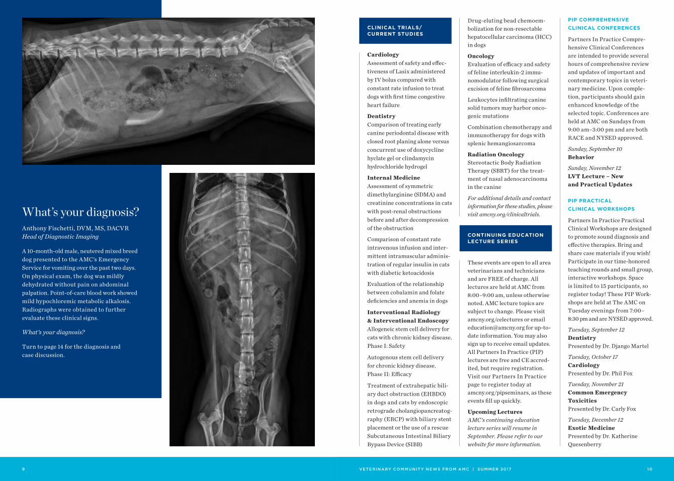
What’s your diagnosis?Anthony Fischetti, DVM, MS, DACVR Head of Diagnostic Imaging
A 10-month-old male, neutered mixed breed dog presented to the AMC’s Emergency Service for vomiting over the past two days. On physical exam, the dog was mildly dehydrated without pain on abdominal palpation. Point-of-care blood work showed mild hypochloremic metabolic alkalosis. Radiographs were obtained to further evaluate these clinical signs.
What’s your diagnosis?
Turn to page 14 for the diagnosis and case discussion.
1 1 VETERINARY COMMUNIT Y NEWS FROM AMC | SUMMER 2017 12
AMC doctors contributed to a number of research publications as well as national and interna-tional conference presentations during the first half of 2017.
Collaborative publications (AMC clinicians in bold font) reported a wide range of research initia-tives. These involved cardiovas-cular pathology, including a new form of feline cardiomyopathy, Bartonella infection of the heart and systemic organs, and right heart cardiomyopathy in Boxer dogs; investigation into causes of canine splenectomy; outcomes following urethral stenting in dogs; causes of retinal degen-eration, mycotic eye disease in canines; opportunities involving partnerships between academia and private practice; and emer-gency and critical care investi-gating markers of inflammation in anemia, micturition disor-ders, and effects of colloids on blood coagulation; and trauma. In addition, two competitive research grants were awarded to Dr. Dennis Trafny by ACVIM Cardiology specialty to study echocardiographic predictors of CHF in dogs and for a clinical trial to investigate efficacy of iso-sorbide dinitrate for CHF therapy.
AMC clinicians have been invited speakers at national con-ferences including NAVC (Drs. Goldstein and Fox); WSAVA (Dr. Goldstein); ACVIM (Drs. Fox, Weisse, Berent, and Goldstein), and at international conferences including India VMA (Dr. Fox), Italy VMA (Dr. Weisse), and BSAVA (Drs. Weisse and Berent).
SCIENTIFIC PUBLICATIONS
von Roedern M, Buriko Y, Prittie J, Lamb K. Investigation
of iron status and markers of inflammation in anaemic and non-anaemic hospitalised cats. J Small Anim Pract. 2017 Mar 6.
Langfitt E, Prittie JE, Buriko Y, Calabro JM. Disorders of micturition in small animal patients: clinical significance, etiologies, and management strategies. J Vet Emerg Crit Care (San Antonio). 2017 Mar;27(2):164-177. doi: 10.1111/vec.12564. Epub 2017 Jan 25.
Griego-Valles M, Buriko Y, Prittie JE, Fox PR. An in vitro comparison of the effects of voluven (6% hydroxyethyl starch 130/0.4) and hespan (6% hydroxyethyl starch 670/0.75) on measures of blood coagula-tion in canine blood. J Vet Emerg Crit Care (San Antonio). 2017 Jan;27(1):44-51.
Gottlieb DL, Prittie J, Buriko Y, Lamb KE. Evaluation of acute traumatic coagulopathy in dogs and cats following blunt force trauma. J Vet Emerg Crit Care (San Antonio). 2017 Jan;27(1):35-43.
Vachon C, Defarges A, Brisson B, Nykamp S, Weese JS, Denstedt J, Berent AC. Passive ureteral dilation and ureteroscopy after ureteral stent placement in five healthy Beagles. Am J Vet Res. 2017 Mar;78(3):381-392.
Pera J, Palma D, Donovan TA. Eosinophilic Esophagitis in a Kitten. J Am Anim Hosp Assoc. 2017 May 23. doi: 10.5326/JAAHA-MS-6367.
Corbin EE, Cavanaugh RP, Schwartz P, Zawadzki KI, Donovan T Splenomegaly in small-breed dogs: 45 cases(2005-2011). J Am Vet Med Assoc. 2017 May 15;250(10):1148-1154.
Enders A, van der Woerdt A, Donovan T. Endogenous mycotic endophthalmitis in a dog with candiduria and Evans
syndrome. Vet Ophthalmol. 2017 Jan;20(1):84-88.
Heller AR, van der Woerdt A, Gaarder JE, Sapienza JS, Hernandez-Merino E, Abrams K, Church ML, La Croix N. Sudden acquired retinal degeneration in dogs: breed distribution of 495 canines. Vet Ophthalmol. 2017 Mar;20(2):103-106.
Flesher K, Lam N, Donovan TA. Diagnosis and treatment of massive porcupine quill migra-tion in a dog. Can Vet J. 2017 Mar;58(3):280-28.
Fischetti AJ, Shiroma JT, Poteet BA. Academic and pri-vate practice partnerships in veterinary radiology residency training. Vet Radiol Ultrasound. 2017 Apr 23.
Nowak K, Wimberger K, Rich-ards SA, Hill RA, Le Roux A. Samango monkeys (Cercopithe-cus albogularis labiatus) manage risk in a highly seasonal, human-modified landscape in Amathole Mountains, South Africa. Int J Primatol. 2017;38(2):194-206.
Donovan TA, Fox PR, Balakrishnan N, Ericson M, Hooker V, Breitschwerdt EB. Pyogranulomatous pancarditis with intramyocardial Bartonella henselae San Antonio 2 (BhSA2) in a dog. J Vet Intern Med. 2017 Jan;31(1):142-148.
Vila J, Pariaut R, Moïse NS, Oxford EM, Fox PR, Reynolds CA, Saelinger C. Structural and molecular pathology of the atrium in boxer arrhythmo-genic right ventricular cardio-myopathy. J Vet Cardiol. 2017 Feb;19(1):57-67.
Oxford EM, Pariaut R, Tursi M, Fox PR, Santilli RA Immunoflu-orescent localization of plako-globin in endomyocardial biopsy samples to diagnose arrhyth-mogenic right ventricular
RESEARCH HIGHLIGHTS cardiomyopathy (ARVC) in the dog. Abst ACVIM. JVIM 2017.
Wallner M, Berretta RM, Eaton DM, Borghetti G, Wu J, Baker ST, Troupes CD, Sharp TE, Feldsott EA, Oyama MA, Fox PR, Wolfson MR, Houser SR. Cardiopulmo-nary characterization of a feline HFpEF model induced by slow progressive pressure overload. Eur Heart Failure J, presented at Fourth Heart Failure Congress, Paris, France. Abstr 2017.
This newsletter is distributed quarterly to AMC’s network of referring veterinarians, alumni and others who opt-in to receive this publication. To view past issues or to join our mailing list, please visit amcny.org/rdvm-quarterly. If you are an AMC alumnus who would like to sign up to receive periodic updates, please visit amcny.org/amc-alumni-registration.
To receive our current staff directory or if you have questions, email [email protected].
For access to the AMC Patient Referral Form, visit amcny.org/referralform.
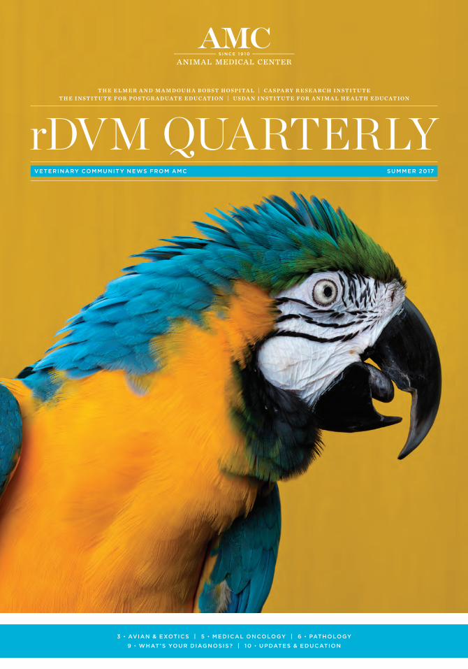
Cover photo courtesy of Corey Towers
Designed by Anthony Coombs
AVIAN & EXOTICS Dr. Kathy Quesenberry Dr. Cyndi Brown 212-329-8888 Sunday – Friday 9 am – 5 pm
CARDIOLOGY Dr. Philip Fox Dr. Betsy Bond Dr. Dennis Trafny 212-329-8701 Monday – Sunday 9 am – 5 pm
DENTISTRY Dr. Dan Carmichael Dr. Stephen Riback Dr. Django Martel 212-329-8678 Monday – Friday 9 am – 5 pm
DERMATOLOGY Dr. Mark Macina 212-329-8777 Tuesday – Saturday 9 am – 5 pm
INTERNAL MEDICINE A Dr. Beth Appleman Dr. Carly Bloom 212-329-8619 Monday – Sunday 9 am – 5 pm
INTERNAL MEDICINE B Dr. Douglas Palma Dr. Dennis Slade 212-329-8675 Monday – Sunday 9 am – 5 pm
INTERVENTIONAL RADIOLOGY & INTERVENTIONAL ENDOSCOPY Dr. Chick Weisse Dr. Allyson Berent 212-329-8700 Monday – Friday 9 am – 5 pm
NEUROLOGY Dr. Chad West Dr. John McCue Dr. Abbie Lebowitz 212-329-8770 Monday – Sunday 9 am – 5 pm
ONCOLOGY Dr. Nicole Leibman Dr. Ann Hohenhaus Dr. Maria Camps 212-329-8797 Monday – Saturday 9 am – 5 pm
OPHTHALMOLOGY Dr. Alexandra van der Woerdt 212-329-8813 Monday 10 am – 6 pm Tuesday 10 am – 5 pm Thursday 2 pm – 9 pm Friday 9 am – 3 pm
RADIATION ONCOLOGY Dr. Rachel St-Vincent 212-329-8821 Monday – Friday 9 am – 5 pm
REHABILITATION & INTEGRATIVE MEDICINE Dr. Leilani Alvarez Dr. Barry Cherno 212-329-8860 Monday – Saturday 9 am – 5 pm
SURGERY SERVICE 2 Dr. Dan Spector 212-329-8863 Wednesday – Saturday 9 am – 5 pm
SURGERY SERVICE 3 Dr. Pamela Schwartz 212-329-8867 Monday – Friday 9 am – 5 pm
SURGERY SERVICE 4 Dr. Rob Hart 212-329-8674 Monday – Friday 9 am – 5 pm
A.M.C. PORTAL amcny.org/ referral-portal-login
DR. RICHARD GOLDSTEIN, CMO 347-733-7338 richard.goldstein @amcny.org
PRIORITY EMERGENCY/ CRITICAL CARE HOTLINE 212-329-8616 or 646-556-6411 (fax)
ABOUT THIS NEWSLETTER
AMC Dedicated Phone Numbers for Referring Veterinarians
VETERINARY COMMUNIT Y NEWS FROM AMC | SUMMER 2017 14
REGISTER NOW
Saturday, November 4, 20178am – 4pm
Animal Medical Center’s 3rd Annual One Health Conference:
Connecting Human and Veterinary Medicine, A ComparativeApproach to Cardiology
Weill Cornell MedicineBelfer Research Building
413 East 69th StreetNew York, NY 10021
Register at: amcny.org/onehealth
This conference is a partnership between Animal Medical Center and Weill Cornell Medicine’s Clinical & Translational Science Center
TOPIC
ONE HEALTH PROGRAM SPEAKERS:
The Elmer and Mamdouha Bobst Hospital | Caspary Research InstituteThe Institute for Postgraduate Education | Usdan Institute for Animal Health Education
DVMKate Meuers NSCU
Taryn Donovan AMC
Mark Oyama Univ. of Penn.
Dennis Trafny AMC
Philip Fox AMC
Rebecca Stepien Univ. of Wisconsin
Cardiac Genomics
Endomyocardial Biopsy
Pathophysiology Mitral Valve Prolapse
Echocardiography
Hypertrophic Cardiomyopathy
Pulmonary Hypertension and Tracheomalacia
MDJeff Towbin Le Bonheur Children’s Hosp.
Navneet Narula Weill Cornell Medicine
Giovanni Ferrari Univ. of Penn.
Rebecca HahnColumbia Univ. Medical Ctr.
Mark SherridNYU Langone Medical Ctr.
Mohit Chawla MSKCC
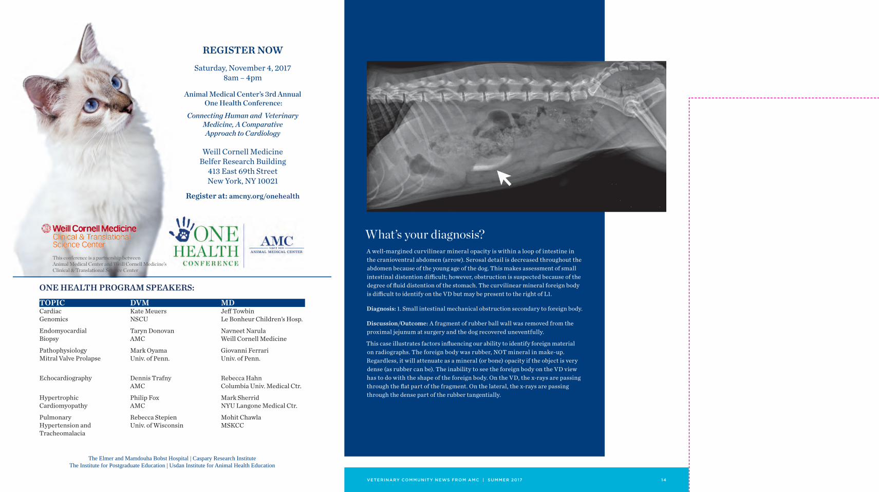
What’s your diagnosis?A well-margined curvilinear mineral opacity is within a loop of intestine in the cranioventral abdomen (arrow). Serosal detail is decreased throughout the abdomen because of the young age of the dog. This makes assessment of small intestinal distention difficult; however, obstruction is suspected because of the degree of fluid distention of the stomach. The curvilinear mineral foreign body is difficult to identify on the VD but may be present to the right of L1.
Diagnosis: 1. Small intestinal mechanical obstruction secondary to foreign body.
Discussion/Outcome: A fragment of rubber ball wall was removed from the proximal jejunum at surgery and the dog recovered uneventfully.
This case illustrates factors influencing our ability to identify foreign material on radiographs. The foreign body was rubber, NOT mineral in make-up. Regardless, it will attenuate as a mineral (or bone) opacity if the object is very dense (as rubber can be). The inability to see the foreign body on the VD view has to do with the shape of the foreign body. On the VD, the x-rays are passing through the flat part of the fragment. On the lateral, the x-rays are passing through the dense part of the rubber tangentially.
510 East 62nd Street New York, NY 10065
VETERINARY COMMUNIT Y NEWS FROM AMC | SUMMER 2017
Please make a contribution today.Please visit amcny.org/contribute for more information.
My employer will match this contribution.
My matching gift form is enclosed.
I have included the AMC in my will or trust.
Please send me Planned Giving information.
Yes, I want to help! Enclosed is a gift of $
NAME
ADDRESS
CITY STATE ZIP
TELEPHONE
This gift is in honor of:
This gift is in memory of:
Please acknowledge:
NAME
ADDRESS
CITY STATE ZIP
ACCOUNT NUMBER
EXP. DATE SEC. CODE
CARDHOLDER’S NAME
SIGNATURE
My check made payable to the Animal Medical Center is enclosed.
Please charge my:
AMERICAN EXPRESS DISCOVER MASTERCARD VISA


