Embed Size (px)
Citation preview

JACC Vol. 8, No.5November 1986:1105-12
PEDIATRIC CARDIOLOGY
Real-Time Doppler Color Flow Mapping for Detection of PatentDuctus Arteriosus
RICHARD E. SWENSSON, MD, FACC, LILLIAM M. VALDES-CRUZ, MD, FACC,
DAVID J. SAHN, MD, FACC, FREDERICK S. SHERMAN, MD, KYUNG J. CHUNG, MD, FACC,
SARAH SCA.GNELLI, ROMS, SANDRA HAGEN-ANSERT, ROMS
La Jolla , California
1105
In this study, ultrasound Doppler color flow mappingsystems were utilized to examine flow in the pulmonaryartery in 31 premature and term infants (aged 4 hoursto 9 months) with patent ductus arteriosus accompanying respiratory distress syndrome, as an isolated lesion, or with patent ductus in association with othercyanotic or acyanotic congenital heart disorders. Theflow mapping patterns were compared with those of acontrol population of IS infants who did not have patentductus arteriosus. In unconstricted ductus arteriosus,the flow from the aorta into the pulmonary artery wasdetected in late systole and early diastole and was distributed along the superior leftward lateral wall of themain pulmonary artery from the origin of the left pulmonary artery back in a proximal direction toward thepulmonary valve. In constricted patent ductus arterio-
The ductus arteriosus frequently remains patent in the preterm infant and can significantly complicate the clinicalcourse of respiratory distress syndrome (I). Likewise, inthe presence of cyanotic and acyanotic congenital heart disease in term and preterm infants , the contribution of patentductus arteriosus must often be elucidated. Because bothpharmacologic (2-4) and surgical (5) treatment in prematureinfants with respiratory distress syndrome have been shownto be effective in closing the ductus , early detection of patentductus arteriosus and methods for serial follow-up of ductalshunting are of substantial importance .
Advances in two-dimensional echocardiographic techniques have helped in identifying patent ductus arteriosus
From the Division of Pediatric Cardiology. University of California atSan Diego, La Jolla , California .
Manuscript received November 14, 1985; revised manuscript receivedApril 30. 1986, accepted June 6, 1986.
Address for reprints: Richard E. Swensson, MD, UCSD Medical Center , Division of Pediatric Cardiolog y, 225 Dickinson Street H814A, SanDiego, California 92103.
©1986 by the American College of Cardiology
sus, or especially in a ductus in association with cyanoticheart disease, the position of the ductal shunt in thepulmonary artery was more variable, often directed centrally or medially.
Waveform spectral Doppler sampling could be performed in specific positions guided by the Dopper flowmap to verify the phasic characteristics of the ductalshunt on spectral and audio outputs. Shunts through avery small patent ductus arteriosus were routinely detected in this group of infants, and right to left ductalshunts could also be verified by the Doppler flow mapping technique. This study suggests substantial promisefor real-time two-dimensional Doppler echocardiographic flow mapping for evaluation of patent ductusarteriosus in infants.
(J Am Coli CardioI1986;8 :1105-12)
at the bedside (6), and the addition of pulsed and continuouswave Doppler echocardiography has provided methods fordetection of ductal flow (7). These methods have proved tobe clinically useful for detection of patent ductus arteriosus;however , the determinants of the phasic characteristics andspatial distribution of ductal flow remain to be clarified inthe clinical setting. Although a laborious technique for pulmonary to systemic volume flow ratio determination hasbeen described (8) in an animal model with a " ductus-like"shunt, it has yet to be shown that such methodology canquantitate ductal shunting in small premature infants.
In this study , we explored the capabilities of new twodimensional Doppler echocardiographic flow mapping techniques for the detection and characterization of ductal shunting in preterm infants with simple patent ductus arteriosuscoexisting with respiratory distress syndrome and in otherterm and preterm infants with isolated patent ductus arteriosus or patent ductus arteriosus coexisting with congenitalheart disease.
Spatially directed spectral Doppler sampling under flowmapping guidance allowed us to take advantage of the higher
0735-1097/86/$3 .50

c
JACC Vol. 8, No.5November 1986:1105-12
SWENSSON ET AL.DOPPLER FLOW MAPPING IN DUCTUS ARTERIOSUS
1107
sampling rate and velocity quantitation of spectral Doppler.However, real-time Doppler flow imaging of the ductal shuntitself provided a better understanding of the spectral patternsobserved.
Methods
Study Patients
Group I. We studied 31 infants (Group 1) with patentductus arteriosus who had Doppler echocardiographic examinations performed in the intensive care nursery or thepediatric ward or as outpatients. Their ages ranged from 4hours to 9 months, and their weights ranged from 640 g to7.6 kg.
Eight of these (Group LA] (mean weight [± SO]1,085 ± 120 g) eventually underwent ductus ligation because of failure of indomethacin therapy for patent ductusarteriosus associated with prematurity (2-4). These infantswere respirator-dependent with radiographic and Dopplerevidence of patent ductus arteriosus (l,7,9). In six of theeight, imaging was performed at least twice by a flow mapping Doppler examination before and after indomethacinadministration.
Nine other term infants (Group I B) (mean weight 3,460g) had simple persistent ductus arteriosus diagnosed clinically between 4 weeks and 9 months of age. They also hadDoppler echocardiographic confirmation of its presence in
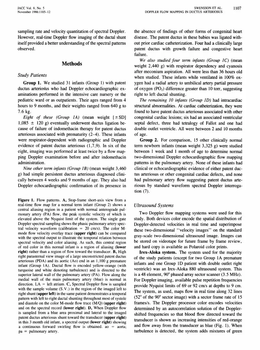
Figure 1. Flow patterns. A, Stop-frame short-axis view from areal-time flow map for a normal term infant (Group 2) shows acentral aliasing region in a patient with normal anterograde pulmonary artery (PA) flow, the peak systolic velocity of which iselevated above the Nyquist limit of the system. The single gateDopplerspectral samplingshowsthe phasicpulmonary arteryspectral velocity waveform (calibration = 20 cm/s). The color Mmode flow velocity overlay trace (upper right) can be comparedwith the spectral output to illustrate the temporal relation betweenspectral velocity and color aliasing. As such, this central regionof red color in this normal infant is a region of aliasing (lowerright) rather than a region of flow toward the transducer. B, Highright parasternal view image of a large unconstricted patent ductusarteriosus (PDA) and its aortic (Ao) end in an 1,100 g prematureinfant (Group lA). Ductal flow is encoded yellow-orange (withturquoise and white denoting turbulence) and is directed to thesuperior lateral wall of the pulmonary artery (PA). Flow along themedial wall of the main pulmonary artery (blue) is normal indirection. LA = left atrium. C, Spectral Doppler flow is sampledwith the sample volume (S.V.) in the region of the imaged left toright shunt (upper left) in the samepatientdemonstrates a temporalpattern with left to right ductal shuntingthroughout mostof systoleand diastole on the color M-mode flow trace (M/Q) (upper right)and on the spectral record (lower right). D, When Doppler flowis sampled from a blue area proximal and lateral to the imagedpatent ductus arteriosusshunt toward the transducer (upper right)in this 3 month old infant, a spectraloutput (lower right) showinga continuous forward swirling flow is obtained. ao = aorta;pa = pulmonary artery.
the absence of findings of other forms of congenital heartdisease. The patent ductus in these babies was ligated without prior cardiac catheterization. Four had a clinically largepatent ductus with growth failure and congestive heartfailure.
We also studied four term infants (Group IC) (meanweight 2,440 g) with respirator dependency and cyanosisafter meconium aspiration. All were less than 36 hours oldwhen studied. These infants while ventilated in 100% oxygen had a radial artery to umbilical artery partial pressureof oxygen (POz) difference greater than 10 torr, suggestingright to left ductal shunting.
The remaining 10 infants (Group ID) had intracardiacstructural abnormalities. At cardiac catheterization, they werefound to have patent ductus arteriosus associated with othercongenital cardiac lesions; six had an associated ventricularseptal defect, three had tetralogy of Fallot and one haddouble outlet ventricle. All were between 2 and 10 monthsof age.
Group 2. For comparison, 15 other clinically normalterm newborn infants (mean weight 3,325 g) were studiedbetween 1 week and I month of age to determine normaltwo-dimensional Doppler echocardiographic flow mappingpatterns in the pulmonary artery. None of these infants hadclinical or echocardiographic evidence of either patent ductus arteriosus or other congenital cardiac defects, and nonehad pulmonary artery flow suggesting patent ductus arteriosus by standard waveform spectral Doppler interrogation (7).
Ultrasound Systems
Two Doppler flow mapping systems were used for thisstudy. Both devices color encode the spatial distribution ofDoppler-detected velocities in real time and superimposethese two-dimensional "velocity images" on the standardgray-scale two-dimensional ultrasound image. Images canbe stored on videotape for future frame by frame review,and hard copy is available as Polaroid color prints.
Irex-Aloka system. The system used for the majorityof the study patients (except for two Group lA prematureinfants and one Group 10 patient with double outlet rightventricle) was an Irex-Aloka 880 ultrasound system. Thisis a 48 element, 90° phased array sector scanner (3.5 MHz).For Doppler imaging, available pulse repetition frequenciesprovide Nyquist limits of 69 or 92 cmls at depths to 9 em.The system, as used, maps flow in real time along 32 lines(52° of the 90° sector image) with a sector frame rate of 15frames/so The Doppler processor color encodes velocitiesdetermined by an autocorrelation solution of the Dopplershifted frequencies so that blood flow directed toward thetransducer is shown as increasing intensities of red-orangeand flow away from the transducer as blue (Fig. 1). Whenturbulence is detected, the system adds mixtures of green

1108 SWENSSON ET AL.DOPPLER FLOW MAPPING IN DUCTUS ARTERIOSUS
lACC Vol. S. No. 5November 19Sb:1105-1 2
to the basic colors at those loci (Fig. 2). The Irex-Alokasystem also allows simultaneous two-dimensional-derivedM-mode color flow overlay images (MQ display ) and singlegate fast Fourier transform spectral Doppler waveforms tobe obtained and displayed along with an audio output froma spatially directed single sample volume once the twodimensional flow image is frozen. As such , flow from specific location s can be interrogated for waveform pulsed Doppler spectral flow under two-dimensional flow imagingguidance.
Acoustec system. The second system used was a prototype Acoustec color flow mapping Doppler scanner. TheAcoustec system, as we used it, was a 3.3 MHz flow imaging prototype with a variable pulse repet ition frequencyand sector angle. The Acoustec system had no waveformspectral outputs available .
Examination Techniques
Ductal flow detection. All patients were examined froma standard parasternal short-axis view at the level of thegreat arteries or from a high right parasternal view or fromboth views, to image the pulmonary artery and aorta (Fig.I and 2). Flow images and derived spectral waveforms wereobtained along with a simultaneous electrocardiogram fortiming. The parasternal short-axis view was employed mostfrequently and was easiest to achieve (Fig. IA) Ductal flowfrom the aorta into the pulmonary artery in this view isexpected to be parallel to and toward the direction of interrogation , although the ductu s itself is imaged by usingthe lateral resolution of the system in this view. As such,the emphasis of this study was not in ductal imaging, butwas directed toward flow detection primarily for imaging 'ductal shunt flow in the main pulmonary artery.
Color flow mapping. The right ventricular outflow tract ,pulmonary valve , aortic valve and right and left pulmonaryarteries were first imaged in real time , and a search wasmade for visualization of the ductus . The patent ductusarteriosus, if imaged, was noted , and hard copy Polaroidstill photographs were obtained by manual freeze frame .Color flow mapping was then activated , and the pulmonaryartery and ductu s area were observed for the characteristicred-orange (Irex-Aloka) or red (Acoustec) pattern of left toright ductal flow from the aorta into the pulmonary arterythrough the patent ductus arteriosus. Slight changes in angulation were made, with special attention to the elevational(azimuthal) plane guided by the audio signal (in spectralmode) and the visual display. Next, a suprasternal or highright parasternal view was obtained and a similar searchmade . If visualized with the Irex-Aloka system , the area ofductal flow (Fig . IB) was then sampled with a single gateso that a spectral Doppler output could be recorded to determine the timing of flow (Fig . IC). For patients studiedwith the Acoustec system, single gate waveform spectralDoppler recordings were obtained from the selected areas
of the main pulmonary artery where ductal flow had beenimaged using a separate 5 MHz mechanical Doppler echocardiographic scanner (Biosound).
Videotape analysis. All studies were recorded on videotape, and photographs were obtained with a Kodak colorprint camera during playback of the videotaped records. Thegeneral size , direction and distribution of the imaged ductalshunt within the pulmonary artery were noted as well as itsphasic timing on spectral traces . Observations were relatedto nonflow imaging two-dimensional echocardiographicallyguided pulsed Doppler examinations that had been performed before the color flow examination in all of thesepatient s using the Biosound scanner at 5 MHz.
ResultsNormal flow patterns. In the normal infants (Group 2) ,
anterograde pulmonary artery flow as a uniform field ofblue-encoded velocities was seen filling the right ventricularoutflow tract, main pulmonary artery and right and leftbranches of the pulmonary artery uniforml y. If flow exceeded the Nyquist limit (as it sometimes did because normal( ± standard deviation) peak pulmonary artery flow velocityin children is 76.1 ± 12.7 cm/s) (10), a central area ofcolor-coded flow aliasing was seen just distal to the pulmonary valve. In this area, color velocity aliasing produceda central uniform nonturbulent red area just distal to thepulmon ary valve in early systole (Fig. IA). In none of theGroup 2 patients was reverse flow toward the transducerobserv ed near the pulmonary artery bifurcation . The samepattern was observed in open chest dogs when the ductalshunt was clamped closed .
Ductal Flow
Of the 31 Group I patients studied, all had ductal flowdetected by color Doppler flow mapping . In 23 patient s, theductal flow was easily seen in the left parasternal view . Theductal shunt was also visualized in high right parasternalviews in 10 of these patients . Four patients required visualization from the high right parasternal position for adequate characterization of a ductal shunt not well appreciatedon the left parasternal view. Three of these patients had atortuous constricted ductus seen at surgery for isolated patentductus arteriosus (n = I) or at cardiac catheterization forpatent ductus arteriosus associated with other cardiac abnormalities (n = 2).
Ductal flow patterns. Two basic flow patterns of leftto right shunting were observed for patent ductus arteriosus.In a large unconstricted ductus in premature infants (GroupIA), flow was toward the left pulmonary artery from theaorta and along the left superior wall of the main pulmonaryartery (Fig. IB) . Most commonly, the ductal flow was latesystolic and holodiastolic in a moderately broad jet (Fig.IC). Short periods of flow reversal in peak systole werealso seen in the premature infants (Group lA) (Fig. IC) . If

B
A
Figure 3. Turbulent flow. Late systolic flow away from the transducerrecord ed in the imaged lumen of a patent ductus arteriosus (PDA) isencoded light blue as turbulent flow, near the Nyquist lim it for velocityand going away from the transducer. right to left (R ~ L) through theductus from the pulmonary artery (PA) to the aorta (Ao). The image wasobtained from a term infant (Group IC) with meconium aspiration whohad a right to left ductal shunt documented by oximetry . See text fordetails .
•'igure 2. Detection of a small shunt. A, This patent ductus arteriosus (PDA) in a 7 month old infant is so small as tobe barely resolved by standard echocardiography (upper image). but the flow map (lower image) shows that it is thesource of a small centrally directed left to right shunt (arrow) . B, A tiny ductus (PDA) is imaged on the upper panel,but as seen in the middle panel; the shunt is directed quite medially (arrow) . Under flow image guidance. a diagnosticspectral waveform could be successfully obtained (lower panel). Ao = aorta; PA = pulmonary artery .
11 IO SWENSSON ET AL.DOPPLER FLOW MAPPING IN DUCTUS ARTERIOSUS
lACC Vol. 8, No.5November 1986:1105-12
sampling for spectral waveform Doppler flow was obtainedin areas other than where the reverse ductal flow was imaged(namely, areas visualized as blue), a forward swirling continuous pattern was obtained (Fig. lD).
The second pattern was noted in a small or constrictedductus in term infants (Group lB), premature infants (GroupIA) who had a good early response to intravenous indomethacin, or those infants who had associated cyanotic congenital defects. In such infants, flow was seen on the colorDoppler display as a narrow jet from the duct into the mainpulmonary artery, sometimes directed laterally, but mostcommonly directed centrally or even medially toward themedial rightward inferior wall of the main pulmonary artery(Fig. 2C). At times, flow could be observed in a ductus assmall as 1.5 mm, as measured from the two-dimensionalimages (Fig. 2).
Comparison with standard two-dimensional images.In Groups lA and lB, especially for the wide open patentductus arteriosus (Fig. IB), before indomethacin treatmentor in those infants in Group IB with congestive heart failure,the patent ductus was easily visualized on standard twodimensional images (5) and classic diagnostic flow patternswere detectable by nonimaging Doppler technique. Oncethe ductus had been partially constricted after indomethacinadministration, as in three of the eight Group lA prematureinfants after indomethacin and three of the nine Group IBinfants with a small tortuous patent ductus arteriosus, ductalimaging was not conclusive and the main pulmonary arteryspectral waveforms were not the classical to and fro waveforms for patent ductus arteriosus, even after prolongedsearching with the Doppler sample volume of the nonflowimaging scanner (Fig. lD). In all of these babies, however,the localized ductal shunt, though small (Fig. 2), was stillidentifiable, and diagnostic spectral waveforms could beobserved under flow imaging guidance (Fig. 2B).
Aliasing and turbulent flow. For both patterns, for largeand small ducts, reverse flow from the ductus (that is, thered-yellow color) was brighter than aliased flow. Reverseflow was also located more distally in the pulmonary arteryand had color mixtures with superimpositions of turquoise,white or orange to denote turbulence (Fig. IB). As such,reverse pulmonary artery ductal flow was distinguishablefrom aliasing in normal infants.
The aliased and sometimes turbulent patterns seen in theproximal pulmonary artery in Group ID infants with patentductus arteriosus and ventricular septal defect or tetralogyof Fallot were also distinguishable from the ductal flow,especially in real time. The turbulent flow from the ventricular septal defect or the right ventricular outflow tractobstruction moved anterograde down the pulmonary arteryin these infants and was not present in diastole. Ductal flowmoved through the imaged ductus as red colors mixed withturbulence and was observed moving retrograde toward thepulmonary valve in systole and diastole. In three of the sixpatients with ventricular septal defect and in one of the three
patients with tetralogy of Fallot in Group lD, the patentductus arteriosus shunt was not identified on the nonflowimaging Doppler examination, but was detectable on theflow map. All these ductuses were small.
Right to left shunting. Of the four Group IC infantswith clinical and oximetric evidence of a right to left ductalshunt, all had an open ductus imaged, and some left to rightshunting was visually apparent on the color flow map in theductal lumen during diastole. In all, however, in mid to latesystole, the patent ductus arteriosus itself showed increasedbrightness of blue velocity pixels, documenting the right toleft ductal shunt flow away from the transducer (Fig. 3).The right to left shunt was distinguishable by the nonimagingDoppler technique in only two of these four babies in whoma sample volume could be placed in the duct. Only in thesetwo could a clear signal be obtained showing an equivocalright to left flow away from the transducer throughout muchof the cardiac cycle.
The flow imaging examinations were generally easy toperform. Spectral sampling with audio and velocity waveform outputs characteristic for patent ductus arteriosus wereobtained rapidly and consistently under flow imaging guidance, even in an extremely small ductus with a localizedarea of shunting.
Discussion
Standard echocardiographic and Doppler diagnosisof patent ductus arteriosus. The ductus arteriosus frequently remains patent and often may dominate the clinicalcourse of the extremely premature infant during the firstseveral days of extrauterine life (I). The presence of patentductus arteriosus markedly affects cardiac output and cerebral blood flow and it can compromise respiratory, renaland gastrointestinal system functions in otherwise healthypremature infants. When patent ductus arteriosus is presentand associated with other congenital cardiac defects suchas tetralogy of Fallot or ventricular septal defect, the patentductus must also be identified and its contribution (eitherfavorable or deleterious) must be assessed. The combinationof high frequency ultrasound imaging of the ductus andDoppler detection of ductal shunting into the pulmonaryartery appears to provide a sensitive indicator when usedalong with clinical symptoms and radiologic signs to identifypatent ductus arteriosus in premature infants. In our ownexperience, we have found that two-dimensional echocardiographic imaging of the ductus from parasternal shortaxis views (6) or from suprasternal or high right parasternalimaging views of the pulmonary artery and descending aortahas provided a reliable method of detecting the usual contourof the unconstricted ductus. Likewise, in the detection ofleft to right shunting through a simple ductus in preterminfants with normal cardiac anatomy and respiratory distresssyndrome, using a combination of simple single pulsed Doppler examination guided by two-dimensional anatomic im-

JACC Vol. 8. No.5November 1986: 1105-12
SWENSSON ET AL.DOPPLER FLOW MAPPING IN DUCTUS ARTERIOSUS
III 1
aging, most pediatric echocardiographers have had substantial confidence in their ability to detect patent ductusarteriosus (7). Difficulties have arisen, however, in imagingthe constricted patent ductus arteriosus (and, at times, indistinguishing the ductus from the left pulmonary artery)and defining the presence and location of a small left toright shunt (or a right to left shunt). The constricted ductusarteriosus may have an echo-lucent lumen (6) and be indistinguishable from the newly closed patent ductus arteriosus. Detecting very small ductal shunts by the single gateDoppler technique may require an extensive search to findthe characteristic retrograde flow.
Advantages of color flow mapping. It was in the infantswith a constricted ductus, those with right to left ductal flowand those with a ductus in the presence of associated congenital heart disease, that the flow mapping method not onlyshortened examination time, but provided diagnostic observations not available from the standard pulsed Dopplerexamination, even after a prolonged search.
In this study, we successfully imaged patent ductus flowin 31 infants who had a patent ductus arteriosus with orwithout associated congenital heart disease, and comparedthose studies with those of 15 patients in whom no ductuswas present. In our study group with patent ductus arteriosus, two-dimensional Doppler color flow mapping addedto our understanding of flow patterns in the ductus, whichvaried from an unconstricted ductus in preterm infants to aconstricted ductus or a ductus associated with cyanotic heartdisease. A variety of patterns have been reported for phasicductal flow (9). Any particular sampling site for obtainingspectral Doppler flow in an area where a left to right shunthad been imaged showed the classic to and fro pattern ofductal flow in this study. By sampling areas adjacent-totheimaged ductal shunt on the opposite wall from where theshunt flow was imaged, we came to understand the forwardswirling, continuous flow patterns that have been described(7) and that could be obtained by sampling from pulmonaryartery areas imaged blue in the presence of patent ductusarteriosus (Fig. ID). We could also distinguish turbulentductal flow from other swirling and temporally short reverseflows sometimes present in patients with dilated pulmonaryarteries.
Using the flow mapping method, we also examined four(Group lC) infants with patent ductus arteriosus associatedwith persistent pulmonary hypertension of the newborn (persistent fetal circulation) who had radial artery to umbilicalartery P02 differences of greater than 10 torr and contrastechocardiographic results (10) suggesting a right to left ductal shunt. The right to left ductal flow was easily visualizedin each of these infants during systole (Fig. 3). Detectionof right to left ductal shunting with the regular spectralDoppler method is not routinely possible or easy to judgeand, in this regard, by localizing and directly imaging theright to left shunt flow in the patent ductus arteriosus, theflow imaging method was clearly advantageous.
Although the two-dimensional Doppler echocardiographic examination is noninvasive, the performance ofechocardiography can, nonetheless, be stressful for the smallpreterm infant. The success rate for echocardiography inachieving high quality imaging and Doppler informationabout the juxtaductal areas of the circulation is quite variable. The examination of a small ductus in critically ill,small preterm infants was particularly facilitated by flowmapping, and our impression was that in these infants, examination times were considerably shortened when compared with the usual nonflow imaging pulsed Dopplerexamination performed under two-dimensional echocardiographic guidance.
Patterns and directions of ductal flow. Localization ofductal flow by flow imaging with subsequent waveformsingle gate Doppler sampling increased the ease of detectionof patent ductus arteriosus shunting when compared withtraditional two-dimensional Doppler methods because of thedifficulty of placing the sample volume without imagingguidance into what can be a narrow stream of left to rightflow. As Daniels et al. (11,12) pointed out, flow patternsare complex in patent ductus arteriosus and difficult to elucidate by traditional Doppler techniques. Daniels et al. alsopointed out that the direction of the left to right ductal shuntis usually along the lateral wall of the pulmonary artery nearthe junction of the main pulmonary artery with the leftpulmonary artery, and also suggested that only continuousanterograde systolic and diastolic flow may be detectable inother areas of the pulmonary artery. Typically, the standardnonimaging Doppler examination requires a time-consuming search of the pulmonary artery bifurcation to detectshunts with narrow jets. In our study, jets directed alongthe center of the pulmonary artery or toward the medial wall-were usually associated with a tortuous or smaller partiallyconstricted ductus.
Differential diagDQsis of false positive ductal shunting.One source of false positive detection of ductal flow waseasily eliminated -by use of color flow mapping, namely,spurious detection of diastolic coronary artery flow in a planejust inferior to the pulmonary artery. We found no such,false positive results with the flow mapping method becauseductal flow was imaged more distally in the pulmonaryartery and was clearly superimposed on the two-dimensionalimage near the ductal orifice. Another confusing diagnosiswe experienced, which gives patterns of flow in the pulmonary artery cirulation that are turbulent and multidirectional, is left pulmonary artery peripheral pulmonary stenosis. In that disease, turbulent flow is propagated distally.In patent ductus arteriosus, the turbulent ductal flow propagates to some extent back toward the main pulmonaryartery and can be distinguished as flow of ductal origin.
Quantification of ductal shunting. Quantification byDoppler technique has been difficult in view of the varyingpatterns detected. Attempts to quantitate pulmonary to systemic blood flow ratio (Qp:Qs) by using Doppler-calculated

1112 SWENSSON ET AL.DOPPLER FLOW MAPPING IN DUCTUS ARTERIOSUS
lACC Vol. 8. No.5November 1986:1105-12
volume flows in two cardiac sites with methods describedin animal models (8) have proved too laborious and timeconsuming for application in the intensive care unit andhave also been limited by the major errors induced by estimating flow areas from echocardiographic dimensionsmeasured in a small heart. Premature infants and others withsimple ductus arteriosus do not undergo cardiac catheterization, and in babies with complex disease, ductal shuntingis hard to quantitate at catheterization except by angiography.
We recently performed flow imaging on three open chestdogs with a variably sized Dacron aortic to pulmonary arteryshunt in which ductal shunting could be quantitated with aprecalibrated electromagnetic flow meter on the shunt toinvestigate whether the spatial penetration of the reverseshunt flow into the pulmonary artery was a function of themagnitude of shunting. Our results suggest the possibilityfor using the maximal imaged area of shunt flow in thepulmonary artery as a direct semiquantitative indication ofshunt size (13). At present, ultrasound methods have offeredonly a semiquantitative approach for assessing ductal flowand require correlation of physical examination and radiologic and clinical data related to respiratory function andventilatory requirements for a complete profile of the significance of a ductus in each infant (14). Color flow mappingmay add new quantitation capabilities in this regard.
Conclusion. Our studies in this disease and other simpleshunt lesions (15) suggest that two-dimensional color Doppler flow mapping techniques promise to add to our capabilities to evaluate simple patent ductus arteriosus shuntsand may provide methods for simplified shunt quantitation.Our experience suggests that the technique is easy to understand and perform and that it is a high resolution technique that provides a unique understanding of the sometimescomplicated intracardiac and extracardiac flow patterns presentin patients with congenital heart disorders.
References1. Jacob J, Gluck L, DiSessa T, et al. The contribution of PDA in the
neonate with severe respiratory distress syndrome. J Pediatr1980;96:79-87.
2. Heymann MA, Rudolph AM, Silverman NH. Closure of the ductusarteriosus in premature infants by inhibition of prostaglandin synthesis.N Engl J Med 1976;295:530-3.
3. Friedman WF, Hirschklau SJ, Printz MP, Pitlick PT, Kirkpatrick SE.Pharmacologic closure of patent ductus arteriosus in the prematureinfant. N Engl J Med 1976;295:526-9.
4. Harris JP, Merritt TA, Alexson CG, Longfield L, Manning JA. Parenteral indomethacin for closure of the patent ductus arteriosus. AmJ Dis Child 1982;136:1005-8.
5. Mikhail M, Lee W, Toews W, et al. Surgical and medical experiencewith 734 premature infants with patent ductus arteriosus. J ThoracCardiovasc Surg 1982;83:349-57.
6. Sahn OJ, Allen HD. Real-time cross-sectional echocardiographic imaging of the patent ductus arteriosus in infants and children. Circulation1978;58:343-54.
7. Huhta JC, Cohen M, Gutgesell HP. Patency of the ductus arteriosusin normal neonates: two-dimensional echocardiography versus Doppler assessment. J Am Coli Cardiol 1984;4:561-4.
8. Meijboom EJ, Valdes-Cruz LM, Horowitz S, et al. A two-dimensionalDoppler echocardiographic method for calculation of pulmonary (Qp)and systemic (Qs) blood flow in a canine model with a variable-sizedleft-to-right extracardiac shunt. Circulation 1983;68:437-45.
9. Grenadier E, Oliveira Lima C, Allen HD, et al. Normal intracardiacand great vessel Doppler flow velocities in infants and children. J AmColi Cardiol 1984;4:343-50.
10. Sahn DJ, Allen HD, George W, Mason M, Goldberg SJ. The utilityof contrast echocardiographic techniques in the care of critically illinfants with cardiac and pulmonarydisease. Circulation 1977;56:959-68.
11. Daniels O. De Open Ductus Botalli en the Respiratory Distress Syndroom. Nijmegen, The Netherlands: KoninklijkeDrukkerijG. J. Thiemebv, 1982.
12. Daniels O. Hopman JC, Stoelinga GBA, Busch HJ, Peer PGM. Doppler flow characteristics in the main pulmonary artery and the La!Aoratio before and after ductal closure in healthy newborns. PediatrCardiol 1982;3:99-104.
13. Swensson RE, Sahn OJ, Valdes-Cruz L, Chung KJ, Sherman FS. Thecolor coded multigate M-mode Doppler display in a real-time flowmapping system: does it provide additional information? (abstr). J AmColi Cardiol 1986;7:229A.
14. Merritt TA, Harris JP, Roghmann K, et al. Early closure of the PDAin very low birth weight infants: a controlled trial. J Pediatr 1981;99:281-6.
15. Sahn OJ, Swensson RE, Valdes-Cruz LM, Scagnelli S, Main J. Twodimensional color flow mapping for evaluation of ventricular septaldefect shunts: a new diagnostic modality (abstr). Circulation1984;70(suppl I1):1I-364.


![18IB Doppler [21 marks] - IB Questionbank](https://img.pdfslide.net/doc/110x75/63186393cf65c6358f01f6bb/18ib-doppler-21-marks-ib-questionbank.jpg)


