Embed Size (px)
Citation preview

Seediscussions,stats,andauthorprofilesforthispublicationat:https://www.researchgate.net/publication/23193316
DevelopmentalchangesintheeffectsofprostaglandinE2inthechickenductusarteriosus
ARTICLEinJOURNALOFCOMPARATIVEPHYSIOLOGYB·SEPTEMBER2008
ImpactFactor:2.62·DOI:10.1007/s00360-008-0296-2·Source:PubMed
CITATIONS
19
READS
43
5AUTHORS,INCLUDING:
AngelLCogolludo
ComplutenseUniversityofMadrid
84PUBLICATIONS1,487CITATIONS
SEEPROFILE
EduardoVillamor
MaastrichtUniversitairMedischCentrum
87PUBLICATIONS1,128CITATIONS
SEEPROFILE
Availablefrom:AngelLCogolludo
Retrievedon:05February2016

THE CHICKEN EMBRYO AS A MODEL FOR DUCTUS ARTERIOSUS DEVELOPMENTAL
BIOLOGY
Pia Anna Elisabet Ågren

ISBN/EAN: 978-90-9025162-2

THE CHICKEN EMBRYO AS A MODEL FOR DUCTUS ARTERIOSUS DEVELOPMENTAL
BIOLOGY
PROEFSCHRIFT
ter verkrijging van de graad van doctor
aan de Universiteit Maastricht, op gezag van de Rector Magnificus,
prof. mr. G.P.M.F. Mols, volgens het besluit van het College van Decanen,
in het openbaar te verdedigen op donderdag 25 maart 2010 om 14.00 uur
door
Pia Anna Elisabet Ågren
Geboren op 8 augustus 1979
te Gårdsby, Sweden

Promotores Prof.dr. C.E. Blanco Prof.dr. L. Zimmermann Co-promotores Dr. E. Villamor Dr. A.L. Cogolludo-Torralba (Universidad Complutense, Madrid) Beoordelingscommissie Prof.dr. J.G. Nijhuis (voorzitter) Prof.dr. F. van Bel (Wilhelmina Kinderziekenhuis Utrecht) Prof.dr. H.A.J. Struijker Boudier Prof.dr. M. Post Prof.dr. J. Vazquez-Jimenez (Universitätsklinikum Aachen)

Table of contents Abbreviations 7
Chapter 1
INTRODUCTION 11
Chapter 2
ONTOGENY OF CHICKEN DUCTUS ARTERIOSUS RESPONSE TO
OXYGEN AND VASOCONSTRICTORS 75
Chapter 3
DEVELOPMENTAL CHANGES IN ENDOTHELIUM-DEPENDENT
RELAXATION OF THE CHICKEN DUCTUS ARTERIOSUS 89
Chapter 4
DEVELOPMENTAL CHANGES IN THE EFFECTS OF
PROSTAGLANDIN E2 IN THE CHICKEN DUCTUS ARTERIOSUS 113
Chapter 5
RESPONSE OF CHICKEN DUCTUS ARTERIOSUS TO HYPERCARBIC
AND NORMOCARBIC ACIDOSIS 127
Chapter 6
MORPHOLOGICAL AND FUNCTIONAL ALTERATIONS OF THE
DUCTUS ARTERIOSUS IN A CHICKEN MODEL OF HYPOXIA-INDUCED
FETAL GROWTH RETARDATION 149
Chapter 7
DISCUSSION 157
Chapter 8
SAMENVATTING 191
SUMMARY 193
ACKNOWLEDGEMENTS 195
CURRICULUM VITAE 199
5

6
Abbreviations
4-AP, 4-aminopyridine
AA, arachidonic acid
AC, adenylate cyclase
ACh, acetylcholine
ATP, adenosine-5'-triphosphate
BK, bradykinin
BKCa, large conductance Ca2+ activated K+ channel
Ca2+, calcium
[Ca2+]i, intracellular Ca2+ concentration
CaM, calmodulin
CAM, chorioallatoic membrane
cAMP, cyclic adenosine-3’, 5’-monophosphate
cGMP, cyclic guanosine-3’, 5’-monophosphate
ChTx, charybdotoxin
CO, carbon monoxide
CO2, carbon dioxide
COX, cyclooxygenase, two forms COX-1 and COX-2
DA, ductus arteriosus
DAF-2DA, 4,5-diaminoflourescein diacetate
DAF-2T, DAF-2 triazole (flouresecent product used to detect NO)
DAG, diacylglycerol
DASMC, ductus arteriosus smooth muscle cell
DMSO, dimethyl sulfoxide
DNTB, dithionitrobenzoic acid (oxidizing agent)
DTT, ditiotreitol (reducing agent)
ECE, endothelin-converting enzyme
EDHF, endothelium-derived hyperpolarizing factor
EDRF, endothelium derived relaxant factors
eNOS, endothelial nitric oxide synthase
Emax, calculated maximal response
ET-1, endothelin-1
ETA, endothelin receptor type A
ETB, endothelin receptor type B
7

ETC, electron transport chain
Gi, inhibitory G protein that inhibits cAMP production
Gq, G protein that enhance intracellular Ca2+ levels
Gs, stimulatory G protein that stimulates cAMP production
H2O2, hydrogen peroxide
HO, Heme oxygenase, two forms HO-1 and HO-2
IK, K+ current
IKCa, intermediate conductance Ca2+ activated K+ channel
iNOS, inducible nitric oxide synthase
IP3, inositol 1,4,5-triphosphate
IUGR, intrauterine growth restriction
K+, potassium
KATP, ATP sensitive K+ channels
KCa, Ca2+-activated K+ channels
KRB, Krebs-Ringer bicarbonate
KV, voltage gated K+ channels
L-NAME, Nω-Nitro-L-arginine methyl ester (NOS inhibitor)
M, muscarinic receptor
MAT, maximal active tension
MLC, myosin light chain
MLCK, myosin light chain kinase
MLCP, myosin light chain phosphatase
N2, nitrogen
NADPH, reduced form of nicotinamide adenine dinucleotide phosphate
NE, norepinephrine
nNOS, neuronal nitric oxide synthase
NO, nitric oxide
NOS, nitric oxide synthase
O2, oxygen
ODQ, 1H-[1,2,4]oxidiazolo[4,3a]quinoxalin-1-one (sGC inhibitor)
PA, pulmonary artery
PaCO2, partial pressure of CO2 in blood
PCO2, partial pressure of CO2
pD2, sensitivity
8

PDA, patent ductus arteriosus
PDE, phosphodiesterase
PG, prostaglandin
PGI2, prostacyclin
Phe, phenylephrine
PIP2, phosphatidylinositol 4,5- biphosphate
PKA, protein kinase A, a cAMP-dependent protein kinase
PKC, protein kinase C
PKG, protein kinase G, a cGMP-dependent protein kinase
PLC, phospholipase C
PO2, partial oxygen pressure
ROCK-1, Rho-associated coiled-coil containing protein kinase 1
ROK, Rho kinase
ROS, reactive oxygen species
SD, standard deviation
SE, standard error
SEM, standard error of mean
SERCA, sarco-endoplasmatic reticulum Ca2+-ATPase
sGC, soluble guanylate cyclase
SKCa, small conductance Ca2+ activated K+ channel
SM, smooth muscle
SMC, smooth muscle cells
SNP, sodium nitroprusside
SOC, store operated Ca2+ channels
TEA, tetraethylammonium
TP, thromboxane receptor
Txs, thromboxanes
VEGF, vascular endothelium growth factor
VSM, vascular smooth muscle
9

10

Chapter 1
INTRODUCTION
11

I General introduction
1 Foetal circulation and transition to extrauterine life
The foetus is an aquatic organism, obtaining oxygen (O2) and nutrients from the
placenta, whereas the newborn and adult is terrestrial, obtaining O2 through the lungs
(Bergwerff et al. 1999). To accommodate the different site of O2 uptake, the foetal
circulation is unique in several ways (Teitel and Cassidy 2001; Blackburn 2003):
shunts are present in the venous system (ductus venosus), in the heart (foramen ovale)
and in the arterial system (ductus arteriosus, DA). The presence of these foetal shunts
allows the circulation to be reasonably effective at distributing O2 and nutrients
(Teitel and Cassidy 2001). However, the presence of these shunts also allows for
mixing of oxygenated and deoxygenated blood. During fetal life the pulmonary circuit
exists as a high-resistance-low flow system while the systemic circuit has low-
resistance, due to presence of the placenta (Teitel and Cassidy 2001; Blackburn 2003).
As the blood returns from the placenta via the umbilical vein, it passes either
into the portal system of the liver or is shunted past the liver via the ductus venosus.
In the human fetus the amount of blood passing through the ductus venosus is 30% at
mid gestation and reduced to 20% after 30 weeks of gestation (Kiserud 2005). The
ductus venosus is connected with the inferior vena cava above the liver, where the
highly oxygenated blood from the umbilical vein joins the blood returning from the
lower part of the body. The blood returning from the umbilical vein has a higher
kinetic energy and stays in a stream separated along the left dorsal wall of the inferior
vena cava (Blackburn 2003). Around 50-60% of this highly oxygenated blood is then
shunted through the foramen ovale to the left atrium, where it is mixed with the
minimal blood flow returning from the lungs, and it continues into the left ventricle
(Blackburn 2003). Upon contraction of the heart, this blood is ejected into the
ascending aorta to feed the heart and brain (Figure 1).
The blood returning from the lower part of the body in the inferior vena cava
is mixed with blood returning from the upper body via the superior vena cava in the
right atrium (Teitel and Cassidy 2001; Blackburn 2003). This blood flow is directed
downward across the tricuspid valve and into the right ventricle. Upon contraction it
is ejected into the pulmonary artery (PA), where the high pulmonary vascular
resistance prevents more than 10-12% to enter the pulmonary bed and the majority of
this blood is shunted across the DA into the descending aorta (Blackburn 2003).
12

Figure 1. Blood flow through human fetal heart. Specific fetal fetures are the foramen ovale
and the ductus arteriosus, which accommodates the different oxygen uptake site of the fetus.
The changeover from foetal to neonatal circulation is directly linked with the
development and function of the pulmonary vasculature and changes in pulmonary
vascular resistance (Reynolds 1953; Smith 1998; Teitel and Cassidy 2001; Herrera et
al. 2004; Thebaud et al. 2004). At delivery, the low-resistance placental circulation is
removed, leading to an increase in systemic vascular resistance. The initiation of air
breathing leads to the expansion of the lungs, an increase in alveolar O2 concentration,
and vasodilation of the pulmonary bed (Teitel and Cassidy 2001). As vasodilatation
occurs, pulmonary vascular resistance falls rapidly, by almost 80%, resulting in a
dramatic increase in pulmonary blood flow (Teitel and Cassidy 2001). The decrease in
inferior vena cava return, due to the cessation of umbilical blood flow, leads to a
functional closure of the ductus venosus within minutes after birth, and together with
the increased blood return from the lungs, augments pressure in the heart leading to
foramen ovale closure (Reynolds 1953; Teitel and Cassidy 2001). At birth, the
simultaneous pulmonary vasodilatation and loss of the low-tension umbilico-placental
circulation increases the pressure in the systemic circuit, while there is a decrease in
the pulmonary circuit, leading to a reversed blood flow over the DA (Smith 1998).
Thereby, the DA is exposed to arterial blood, with high O2 tension (Smith 1998).
Closure of the normal DA is believed to occur in two steps: (1) a functional closure
due to constriction of the medial muscle layer that usually occurs within the first
hours after birth and (2) an anatomic closure that involves infolding of the
Left ventricleRight ventricle
Right atrium
Left atrium
Foramen ovale
Aorta Ductusarteriosus
From bodyand placenta
From body
To head
To lungs
From lungs
Left ventricleRight ventricle
Right atrium
Left atrium
Foramen ovale
Aorta Ductusarteriosus
From bodyand placenta
From body
To head
To lungs
From lungs
13

endothelium and disruption of the subintimal layers, which is usually completed by
the second week of life (Clyman et al. 1983).
2 Clinical relevance of the DA
2.1 Patency of the DA in the premature infant
In contrast with the full term DA, the premature DA is less likely to constrict after
birth. Therefore, preterm neonates, and more particularly those with gestational age
< 30 wk, and birth weight < 1500g, are at increased risk of having a persistent patent
(P) DA beyond 96 hours postnatally and absence of remodelling (Clyman 2006;
Koch et al. 2006). This may result in a left-to-right shunt and hyperperfusion of the
pulmonary vascular bed, often resulting in decreased lung compliance, increased need
for mechanical ventilation, and altered postnatal nutrition and growth. Thus, a PDA
may adversely affect morbidity and mortality in an already high-risk population
(Bancalari et al. 2005; Clyman 2006; Koch et al. 2006). In preterm infants, PDA
induces a higher risk of adverse outcomes such as chronic lung disease (Rojas et al.
1995), pulmonary hemorrhage (Kluckow et al. 1999), renal hypoperfusion
(Hammerman 1995) and decreased glomerular filtration rate, necrotizing enterocolitis
(Cotton et al. 1978), and if not managed, may lead to death (Cotton et al. 1978).
The incidence of PDA is inversely related to gestational age, such that it
affects up to 60% of infants less than 28 weeks gestation (Gersony et al. 1983; Van
Overmeire and Chemtob 2005). The ineffective ductal closure in preterm infants is
partially explained by decreased sensitivity to O2, diminished responsiveness to
prostaglandin (PG) withdrawal at birth, increased levels of PGs, nitric oxide (NO) and
other vasodilatory stimuli, and immaturity of the structural apparatus necessary for the
constriction and obliteration of the ductus lumen (Hammerman 1995; Smith 1998;
Kajino et al. 2000; Kajino et al. 2001; Van Overmeire and Chemtob 2005; Reese
2006). The premature DA has a much smaller intrinsic tone, and together with the
increased sensitivity to vasodilators, it might not be able to remain constricted during
the remodelling process, which might explain why extremely premature infants have a
high rate of reopening (Kajino et al. 2001).
The increased number of surviving preterm infants has led to a higher number
of infants requiring medical or surgical intervention for PDA. Standard therapies
include fluid restriction and use of cyclooxygenase (COX) inhibitors such as
indomethacin or ibuprofen (Van Overmeire and Chemtob 2005; Malviya et al. 2008).
14

Surgical ligation is used when medical treatment fails or is contraindicated (Mosalli
and Alfaleh 2008). There is no consensus as to what is the best time of treatment and
the optimal management strategy among this high-risk vulnerable population (Mosalli
and Alfaleh 2008). In addition, there has been considerable debate about the benefits
and risks of surgical ligation for PDA on subsequent neurodevelopmental outcome.
Surgery in the neonatal period is associated with a systemic inflammatory response; in
addition the use of sedative and/or anaesthetic drugs may adversely impact the
immature brain (Doyle 2001).
2.2 Persistent DA in term infants
In contrast to premature infants, in whom PDA generally is due to developmental
immaturity, patent ductus in term infants likely results from a significant structural
abnormality. Persistence of the DA has been defined as continued patency in term
newborns older than 3 months (Benson and Cowan 2002). There are several reasons
for a persistent ductus: 1) Failure of the normal constriction due to structural or
biochemical abnormalities. 2) Alternatively, the contractile apparatus might be
completely normal, but the stimulus, O2, may be lacking or ineffective. 3) Relaxant
mediators could also prevent closure. In the majority of cases, no clear cause is
identifiable, and both genetic and environmental factors may be involved (Benson and
Cowan 2002).
2.3 Congenital heart diseases
There are several congenital heart diseases that, as a consequence of the defect,
require a patent ductus for survival. The role of the ductus can either be to maintain
adequate pulmonary blood flow, to maintain adequate systemic flow or to improve
mixing of systemic and pulmonary circulations (Knight 2001). The behaviour of the
ductus in the immediate postnatal period may be crucial if the heart is congenitally
malformed. When either the pulmonary or the systemic circulation is entirely supplied
through the ductus, survival itself depends on the behaviour of the DA. The DA that
closes despite an obligatory need to remain patent has been named “the suicidal duct”
(Benson and Cowan 2002). If the DA is necessary for pulmonary blood flow, the
patients never experience the normal postnatal rise in blood oxygenation. Closure of
the DA usually occurs, however it is frequently delayed, but without medical and/or
surgical intervention, this can only lead to death (Benson and Cowan 2002). On the
15

other hand, if the DA is necessary for systemic blood flow, heart failure predominates,
which become rapidly more severe as the DA close.
2.4 Premature in utero closure
Contraction of the DA, with or without fetal heart failure, is a recognized side effect
of administration of COX inhibitors to the mother. This can lead to serious fetal and
neonatal complications including pulmonary hypertension, necrotizing enterocolitis
and intraventricular hemorrhage. Constriction of the DA causes an ischemic insult of
the vessel wall, making the vessel less responsive to PGE2, O2 and indomethacin
(Clyman et al. 1983). Thus a DA that has closed once but reopened again, is more
reluctant to close again. Exposure to indomethacin increases the thickness of the
avascular zone (i.e., the zone without vasa vasorum), associated with a decline in
distensability and maximal contractile capability due to smooth muscle cells (SMC)
loss and migration (Clyman et al. 2001). The excessive avascular zone thickness
causes a decrease in intravavascular blood flow, leading to hypoxia of the vessel wall,
which in turn makes the DA less responsive to O2 and remaining patent after birth
(Goldbarg et al. 2002).
II The mammalian DA
Significant progress in our understanding of the DA physiology and pathophysiology
has been achieved with the use of animal models, including the sheep, the rabbit, the
mouse, the rat or primates (Sutendra and Michelakis 2007). In this section of the
chapter, the embryology and the process of patency and closure of the mammalian
DA are analyzed. In section III, the vasoactive mediators and signalling pathways
involved in DA patency and closure are reviewed.
1 Embryology
During early fetal development, six arterial arches link the aortic sac with the paired
dorsal aortas, although all six arches are never present simultaneously (Benson and
Cowan 2002). In humans, the DA develops from the dorsal portion of the left sixth
arch. The sixth arch is associated early on with the developing lung buds, which
initially are supplied by a plexus of capillaries that develops from the arterial sac and
later connects to the dorsal aorta.
16

In normal cardiovascular development, the proximal portion of the sixth pair
of embryonic aortic arches persists as the proximal branch PAs, and the distal portion
of the left sixth arch persists as the DA, connecting the left PA with the left dorsal
aorta (Schneider and Moore 2006). Normally, the distal right sixth aortic arch loses its
connection to the dorsal aorta and degenerates. In humans this transformation is
complete by 8 weeks of fetal life (Schneider and Moore 2006).
2 Foetal patency
2.1 Foetal patency of the DA
Ductal patency in utero is an active state, dependent on the balance between dilating
and constricting forces. The ductus normally has a high level of intrinsic tone during
foetal life (Kajino et al. 2001), but the factors that promote ductus constriction in the
foetus have yet to be identified (Clyman 2006). The factors that oppose ductus
constriction are better understood. The elevated vascular pressure within the ductus
lumen, due to the constricted pulmonary bed, plays an important role in opposing
ductus constriction (Clyman et al. 1989). Circulating and locally produced PGs as
well as NO appear to be the major factors (Coceani et al. 1994 (lamb); Smith 1998;
Takizawa et al. 1999 (rat); Kajino et al. 2000; Seidner et al. 2001 (baboon)). Several
studies have shown developmental changes in the roles of PG and NO in different
mammalian species (see section III: 3.2 and 5).
3 Closure of the DA
3.1 Functional closure
Closure of the DA occurs in two steps, functional closure, due to vasoconstriction,
which precedes anatomical closure, due to remodelling. This closure occurs within
days and is crucial for the neonatal transition to an air-breathing organism (Smith
1998). In full-term newborn humans, the DA closes 24-48 h after delivery. The
simultaneous change in pulmonary vascular resistance and the loss of the placental
blood flow at birth, leads to a reversed blood flow over the DA (Smith 1998). After
birth, the DA is exposed to arterial blood, O2 tension rises rapidly, which leads to
constriction (Smith 1998).
At birth, the major source of PGE2, the placenta, is lost, and the increased
blood flow in the lungs reduces the amount of free circulating PGE2, since the lung is
the most important site for breakdown of PGs (Bouayad et al. 2001). During the first
17

hour after birth the amount of PGE2 decreases by around tenfold and within the first
three hours it is reduced by around twenty fold in foetal lambs (Clyman et al. 1980).
This loss of free circulating PGE2 has been postulated to be fundamental to the
closure of the DA.
3.2 Anatomical closure
The lumen is first occluded in the narrowest part, that is, in the proximal (pulmonary)
part of the DA, between opposed intimal cushions (Silver et al. 1981). Necrosis of the
inner DA wall, due to anoxia, is the first definitive morphological evidence of
sustained muscular contraction to appear (Silver et al. 1981). Based on studies in
baboons it was shown that regions of the DA where the lumen was most constricted
were associated with moderate/intense hypoxia; vascular endothelium growth factor
(VEGF) expression was increased in the hypoxic muscle media; and luminal
endothelial cells were proliferating (Clyman et al. 1999a). Cells in the most hypoxic
regions of the DA wall were undergoing DNA fragmentation.
The immature foetus has an increased glycolytic capacity, which may enable it
to tolerate episodes of hypoxia and nutrient shortage, thus making it more resistant to
postnatal cell death and permanent closure (Levin et al. 2005). In premature baboon
DA no evidence of moderate/intense hypoxia, no cell death, or VEGF expression was
found, events that all occurred in the term foetus (Clyman et al. 1999a). The preterm
DA must completely obliterate luminal blood flow to develop the same degree of
hypoxia as the term DA (Levin et al. 2006) and the preterm is capable of undergoing
anatomic remodelling if it can produce the same degree of intense hypoxia that is
observed at term (Seidner et al. 2001).
Waleh et al (2005) explain that the inflammatory response following postnatal
DA constriction may be necessary for ductus remodelling. Monocytes/ macrophages
and, to a lesser extent, T-lymphocytes adhere to the DA lumen after postnatal
constriction in baboon. The same inflammatory and remodelling changes are seen
whether or not the DA close spontaneously, close following PG inhibition, or close
after combined PG and NO inhibition. These changes are seen only in areas of the DA
that are tightly constricted (Waleh et al. 2005).
18

Table 1. Summary of the literature concerning closure and remodelling of the ductus
arteriosus.
Reference Species/ Age Experiments/Results Comments
(Eldridge et al. 1954)
Human/ Neonates 1-118 h
Blood oxygenation from the right foot and hand were compared, and up to 72 hours after birth, the saturation was significantly lower in the foot, indicating the presence of a veno-arterial shunt through the open DA.
(Silver et al. 1981)
Human An elastic network in the media of the SM was observed. Maturation of the contractile apparatus was also evident. Closure begins in the pulmonary half, between closely opposed intimal cushions. The vessel is probably too large to be closed merely by the contraction of circularly arranged muscles. Longitudinal muscles are present to shorten the DA. Necrosis of the inner DA wall, due to anoxia, is the first definite morphological evidence of sustained muscular contraction to appear, while central ischemic degeneration or necrosis is a result of strong sustained DA contraction.
The lumen is firstly occluded in the narrowest part, that is, in the proximal (pulmonary) part of the DA, between opposed intimal cushions.
(Tomita et al. 1996)
Human/ (1-66 mo)
The vascular (V) SM of the PDA remains capable of constriction beyond infancy, at least at the pulmonary end. In some cases of PDA, catecholamines induce late closure.
(Clyman et al. 1999a)
Baboon/ 125 and 175d (t 185d)
In full term, the DA was functionally closed on Doppler examination by 24h. Constriction was associated with moderate/intense hypoxia, increased VEGF and proliferating luminal endothelial cells. In the most hypoxic regions DNA fragmentation occurred. In contrast, in the premature, only 29% had closed their DA before 6d. None had evidence of moderate/intense hypoxia; no cell death, VEGF expression, endothelial proliferation nor neoimtima formation was found by day 6.
DA constriction is associated with hypoxia, cell death, VEGF expression etc, but not in the premature.
(Clyman et al. 2001)
Lamb/ 127-131d Infusion of indomethacin in utero increased VEGF and endothelial NO synthase (eNOS) expression, neoimtima formation, and muscle media cell death. Inhibition of NO production produces significantly greater contraction in the moderately constricted DA than in the control. A marked degree of DA constriction in utero, increases the avascular zone thickness due to tissue compaction (caused by circumferential and longitudinal muscle constriction), associated with SMC loss and migration into the neointima, leading to a significant decline in distensability and maximal contractile capability.
Exposure to indo-methacin in utero increases avascular zone thickness, associated with a decline in distensability and maximal contractile capability, due to SMC loss and migration.
(Seidner et al. 2001)
Baboon/ 125 and term 185d
Combined NOS and COX inhibition produced much greater degree of constriction in vivo than indomethacin alone. The effects of indomethacin wanes with increasing postnatal age. After delivery eNOS-expressing vasa vasorum invade the outer media, which does not express COX-1 or -2. Thus NOS inhibition could become useful. The preterm DA is capable of undergoing anatomic remodeling if it can produce the same degree of intense hypoxia that is observed at term.
The preterm DA is capable of undergoing anatomic remodeling if it can produce the same degree of intense hypoxia that is observed at term.
(Clyman et al. 2002)
Baboon/ 125d Indomethacin and L-NNA (NOS inhibitor) produce constriction of the preterm DA and VEGF expression, which corresponds exactly with the distribution and intensity of hypoxia. VEGF may play an essential role in vasa vasorum ingrowth and SM expansion of the neointima.
VEGF is an important mediator of the anatomic remodeling.
(Goldbarg et al. 2002)
Lamb/ 131-135d Exposure to indomethacin produced nuclear fragmentation and cell death in SMCs, directly related to the degree of DA constriction, and it occurs even at moderate degrees of constriction. It decreases vasa vasorum blood flow and increases wall thickness, thereby increasing the avascular zone thickness. These changes in cell viability lead to a significant decline in distensibility and contractile capacity, which explain why after indomethacin exposure in utero the DA remains patent and refuse to close.
Exposure of indomethacin in utero, decrease responsiveness to, among others, O2 and the DA remains patent after birth.
(Levin et al. 2005)
Lamb/ 101-107 and 138-144d
After birth the DA develops adenosine triphosphate (ATP), glucose and glycogen depletion in addition to hypoxia. Cell death correlates best with ATP depletion and is most marked when both glucose and O2 are severely depleted. Under hypoxic conditions, the immature DA is more capable of preserving its ATP supply as a result of increased glucose availability, glycogen stores and glucose utilization. The increased glycolytic capacity of the immature DA may enable it to tolerate episodes of hypoxia and nutrient shortage, making it more resistant to developing postnatal cell death and permanent closure.
The immature fetus can tolerate episodes of hypoxia and nutrient shortage due to the increased glycolytic capacity, making it more resistant to permanent closure.
(Waleh et al. 2005)
Baboon/ 125 and 175d
Similarities in gene expression between artherosclerotic- and DA -remodeling were found. Monocytes/macrophages and, to a lesser extent, T-lymphocytes adhere to the lumen. The same inflammatory and remodelling changes are seen whether or not the DA close spontaneously, close following PG inhibition or close after combined PG and NO inhibition. These changes are seen only in areas of the DA that are tightly constricted.
The inflammatory response following postnatal DA constriction may be necessary for ductus remodeling.
(Levin et al. 2006)
Baboon/ 123-127 and 175d
O2, glucose, glycogene and ATP depletion, and subsequent cell death in the muscle media occur even before complete loss of luminal blood flow in full term, while constriction of the preterm DA in vivo is associated with O2, glucose and ATP depletion. The preterm DA must completely obliterate luminal flow to develop the same degree of hypoxia, glucose depletion and cell death as found in the full-term DA.
The preterm DA must completely obliterate luminal blood flow to develop the same degree of hypoxia as the term DA.
19

3.3 Vasa vasorum and DA closure
The presence of vasa vasorum in the muscle media depends on the thickness of the
arterial wall (Clyman et al. 2001). If the vessel is thicker than 0.5 mm, the maximal
diffusion distance, there is a need for a vasa vasorum to provide the muscle media
with O2 and nutrients (Kajino et al. 2002). The muscle media of most vessels has a
region, adjacent to the lumen, which lacks vasa vasorum (Wolinsky and Glagov 1967;
Goldbarg et al. 2002). This avascular zone depends on diffusion from both the lumen
and vasa vasorum to meet its O2 needs and is therefore particularly vulnerable to
changes in O2 supply (Clyman et al. 1999a).
In the human foetus, before week 28, luminal blood flow is sufficient to meet
the O2 demands of the thin-walled ductus, while after the 28th week of gestation, vasa
vasorum usually enter the ductus wall (Clarke 1965; Goldbarg et al. 2002). The vasa
vasorum provide the DA with a unique mechanism for controlling the maximal
diffusion distance across its wall. By elimination of vasa vasorum blood flow to the
outer muscle media, in the newborn DA, the entire thickness of the muscle media
turns into an avascular zone, creating a diffusion distance that is approximately 3
times the maximally tolerated diffusion distance (Kajino et al. 2002; Levin et al.
2005). As a result, profound hypoxia (< 0.2%) develops even before luminal blood
flow is eliminated (Jurrus and Weiss 1977; Clyman et al. 1999a; Kajino et al. 2000;
Goldbarg et al. 2002; Kajino et al. 2002). In contrast, the extremely preterm DA has
no vasa vasorum, and with a reduction of luminal flow to the same degree as that
found at term, the preterm DA wall thickness is only reduced to 0.67 mm (Clyman et
al. 1999a). This diffusion distance is insufficient to produce the profound degree of
hypoxia needed for vessel remodelling and only mild to moderate hypoxia (2.0 - 0.7%
tissue O2 concentration) after postnatal constriction in vivo is developed (Clyman et
al. 1999a; Kajino et al. 2000). Without a drop in tissue O2 concentration to < 0.4%,
ductus remodelling does not occur. The mild to moderate degrees of hypoxia in the
preterm do not inhibit PG and NO production, whereas they do lead to loss of O2-
induced tension (Clyman et al. 1999a; Kajino et al. 2000). In the full-term newborn
DA, severe O2, glucose, glycogen and ATP depletion, cell death and tissue
remodelling occur even before there is complete loss of luminal blood flow. The
preterm ductus has to completely obstruct luminal blood flow before it develops the
20

same degree of in vivo hypoxia, glucose depletion, and cell death as found in the full-
term newborn ductus (Levin et al. 2006).
III Vasoactive mediators and signalling pathways involved in DA
patency and closure
1 Regulation of vascular tone, role of smooth muscle and endothelium,
differential maturation of smooth muscle in the DA
1.1 Regulation of vascular tone
Vascular tone is regulated by a balance between constricting and relaxing forces. The
primary signal responsible for activation of SM contractile proteins is calcium (Ca2+).
Thus, an increase in intracellular Ca2+ ([Ca2+]i) leads to contraction, while a decrease
leads to relaxation (Carvajal et al. 2000; Ratz et al. 2005). Cytosolic Ca2+ is increased
through Ca2+ release from intracellular stores (sarcoplasmic reticulum, SR) as well as
entry from the extracellular space through Ca2+ channels. The changes in [Ca2+]i lead
to a different degree of phosphorylation of the myosin light chain (MLC) kinase
(MLCK). Thus, an increase in [Ca2+]i activates the Ca2+/calmodulin (CaM)-dependent
MLCK which triggers a reversible phosphorylation at Ser19 of the MLC facilitating
the cross-bridge formation between actin and myosin filaments and contraction
(Somlyo and Somlyo 2003). However, Ca2+ does not directly activate SM motor
proteins (Ratz 2004). On the other hand, there is a Ca2+-independent mechanism, the
MLC phosphatase (MLCP), which dephosphorylates the MLC and counteracts the
contraction (Somlyo and Somlyo 2003; Kitazawa et al. 2004).
The MLCK mechanisms involves the membrane bound enzyme,
phospholipase C (PLC, Katzung 1995; Macrez-Lepretre et al. 1997; Kitazawa et al.
2004), which hydrolyses a component of the plasma membrane called
phosphatidylinositol 4,5- biphosphate (PIP2, Figure 2). Two second messengers are
formed, diacylglycerol (DAG) and inositol 1,4,5-triphosphate (IP3). DAG, being
positioned in the plasma membrane, activates the phospholipid- and Ca2+-sensitive
protein kinase C (PKC, Katzung 1995), which, in turn, stimulates Ca2+ influx through
voltage dependent Ca2+ channels (Macrez-Lepretre et al. 1997). IP3, the other
messenger, is water-soluble and diffuses through the cytoplasm, where it triggers
release of Ca2+ from the SR.
21

The MLCP mechanism involves an increase or a decrease in SM tension at a
constant Ca2+ concentration, which are correspondingly referred to as Ca2+
sensitization and desensitization of the SM (Somlyo and Somlyo 2003; Kitazawa et al.
2004; Ratz et al. 2005). Ca2+ sensitization/desensitization is an important component
of the constrictor/relaxant response of many agonists and may involve a number of
different mechanisms. The RhoA and Rho kinase (ROK) pathway is believed to be
the most important modulator of Ca2+ sensitivity in SM, and a large number of
vasoconstrictor agonists act by activating this pathway (Somlyo and Somlyo 2003). A
schematic drawing over vascular responses and actions of different agonists used in
this Thesis is depicted in Figure 4, on p. 39.
1.2 The role of the Endothelium
Endothelial cells control the tone of the underlying vascular smooth muscle (VSM)
cells by releasing various relaxing and contracting factors (Furchgott and Vanhoutte
1989). The main vasoconstrictors are endothelin-1 (ET-1) and thromboxane A2
(TxA2), while at least three endothelium derived relaxant factors (EDRF) have been
found. These include NO, PGs (mainly prostacyclin, PGI2) and a yet unidentified
factor called endothelium-derived hyperpolarizing factor (EDHF, Takizawa et al.
1999; Tare et al. 2000; Kamper et al. 2002; Sendao Oliveira and Bendhack 2004). NO
is produced by a family of enzymes called NOS (Arnal et al. 1999), while PGs are
synthesized by COX (Bos et al. 2004, see section III: 3 and 5). Endothelium-
dependent agonists such as acetylcholine (ACh) and bradykinin (BK), in addition to
physical vascular stimuli such as shear stress, stimulate endothelial cells to release
NO, PGs and EDHF (Scotland et al. 2005). The contribution of each of these factors
to endothelium-dependent vasodilatation varies across vascular beds and also with the
physiological or pharmacological stimuli used to stimulate the endothelium (Arnal et
al. 1999; Tare et al. 2000).
22

RR
GG
GTP GDP
PLCPLCPIP2
IP3
DAG
PKC
Ca2+Ca2+
Ca2+-CaM
MLCK(ínactive)
MLCK(active)
MLC(ínactive)
MLC-P(active)
Actin myosin interaction
Contraction
Phosphatase
Ca2+Ca2+Ca2+
SR
Figure 2. Schematic picture of the pathway of contraction. R, G protein coupled receptor; G,
G protein. For further explanation see section III: 1.1.
23

2 Vasoactive factors
2.1 Oxygen
The inflation of the lungs at birth produces a rapid increase in alveolar O2 tension,
leading to DA constriction and at the same time relaxes PAs and decreases pulmonary
vascular resistance (Waypa and Schumacker 2005). The importance of O2 in initiating
closure of the DA is underlined by the occurrence of six times as many cases of PDA
in babies born at high altitude (Alzamora-Castro et al. 1960).
2.2 Control of patency
The relatively low O2 tension to which the fetal DA is exposed helps maintain patency
(Smith 1998). At mean tissue O2 concentration between 0.2 and 0.4%, endogenous
PGs and NO play a significant role in regulating DA tone because they inhibit > 60%
of the active tension developed by the vessel (Kajino et al. 2000). As tissue O2
concentration increases above 0.4%, the vasoconstrictor effects of O2 outweigh the
inhibitory effects of PGs and NO (Kajino et al. 2000). Below 0.2% tissue O2
concentration, neither COX nor NOS inhibition had any effect on the tone of the DA,
suggesting that both PG and NO production are inhibited by hypoxia (Kajino et al.
2000).
2.3 Control of closure
That O2 is important in the closure of the DA is well known, however the mechanism
by which O2 produces contraction is less clear. There are four main theories about the
actual way that O2 causes contraction (Smith 1998). None of the models fit all of the
experimental observations and it is therefore likely that there is more than one O2
sensor. Furthermore O2 tension has a modulatory role on other vasoactive systems,
and therefore the contractility of locally released vasoactive agents varies with
alterations in O2 tension (Smith and McGrath 1993). It was early shown that O2 was
acting directly on the SM of the DA, and that the contractions to O2 were not due to
innervation (Born et al. 1956).
2.3.1 The cytochrome a3 hypothesis
The cytochrome a3 hypothesis is based on experiments performed by Fay et al (1971;
1972). They observed that cyanide and several other inhibitors of oxidative
phosphorylation inhibited O2-induced contraction (Fay 1971; Fay and Jobsis 1972)
24

and postulated that cytochrome a3 was the O2 sensor and that the contractile effect of
O2 was related to ATP levels in ductal SM (Fay and Jobsis 1972). Cyanide has been
shown to affect VSM contraction by several mechanisms, among them affecting
several potassium (K+) channels (Smith 1998). Thus, although cyanide affects the O2-
induced contraction, the mechanism of action remains unclear.
2.3.2 Arachidonate hypothesis
An attractive hypothesis, given that dilation of the ductus in utero is mediated by an
arachidonic acid (AA) metabolite (PGE2), is that an AA metabolite also might mediate
O2-induced contraction of the vessel with a shift in AA metabolism occurring at birth
(Smith 1998).
2.3.3 Endothelin/cytochrome P450 hypothesis
Coceani et al. (1991; 1992; 2000; 2002) suggested that cytochrome P450 acts as O2
sensor and that its interaction with O2 might increase ET-1 production by the ductal
endothelial cells, resulting in contraction. There is a large body of evidence obtained
from studies on lamb DA, mostly from a single laboratory, which supports the
cytochrome P450/ET-1 hypothesis (Coceani et al. 1994). However, the actual role of
ET-1 as a mediator of ductus constriction at birth is debated. Coceani et al (1999)
showed that ETA receptor knockout foetal mice displayed a markedly reduced O2-
induced constriction compared to wild type. Blockade of endothelin release in lamb
(Coceani et al. 1992) and rat (Shen et al. 2002) reduce O2-induced contraction by half,
while in rabbit the reduction is about one fifth (Shen et al. 2002). Thus, there is a
species-dependent difference in the degree of contribution of endothelin to the O2-
induced contraction.
2.3.4 Membrane depolarization
The nowadays main theory about O2 sensing and action is through membrane
depolarization. Vascular O2 sensing systems consists of a sensor, the function of
which is altered by changes in partial oxygen pressure (PO2), a mediator, produced
by this sensor, and an effector, which alters vascular tone in response to the mediator
(Archer and Michelakis 2002). Many aspects of the O2 sensor-effector pathway are
conserved among O2-sensitive mammalian tissues, the PAs, the DA, the
adrenomedullary cells, the neuroepithelial body, and the carotid body. In each tissue,
25

K+ channels have been implicated in the effector mechanism (Archer et al 2000),
whereas the O2 sensor has been proposed to involve a change in redox state, as
determined by mitochondria or NADPH oxidase. Attention has focused on the
mitochondria because electron transport chain (ETC) inhibitors mimic hypoxia,
constricting the PAs and activating the carotid body (Michelakis et al. 2002). The
mitochondria respond to changes in PO2 by altering their respiration and production
of ROS (Archer et al. 1993; Duchen 1999). This changes the cellular redox potential
and alters the function of many second messenger systems, and O2-sensitive K+
channels in the membrane (Michelakis et al. 2002).
Several studies have suggested that changes in O2 tension are signalled by
changes in redox status and that O2-induced contraction of the DA is, at least partly,
induced by membrane depolarization, which then results in entry of Ca2+ through L-
type voltage-gated Ca2+ channels (Roulet and Coburn 1981; Nakanishi et al. 1993;
Tristani-Firouzi et al. 1996; Smith 1998; Weir et al. 2005). Roulet and Coburn (1981)
first demonstrated that O2-induced DA constriction is associated with membrane
depolarization. Nakanishi et al. (1993) hypothesized that, in the rabbit DA, membrane
depolarization is related to O2- induced closure of an ATP sensitive K+ (KATP)
channel. In contrast, Tristani-Firouzi et al. (1996) suggested that a voltage gated K+
(KV) channel was responsible for O2-induced membrane depolarization of rabbit DA.
A patch clamp study of SMCs from the rabbit ductus showed that the K+
channels controlling membrane potential change from the O2-insensitive Ca2+-
activated (KCa) channels to the O2-sensitive KV channels with advancing gestational
age (Reeve et al. 1997). This suggests that the fundamental pathway involved in O2-
induced contraction changes with advancing gestational age. Moreover, ex vivo
transfer of the gene for KV1.5 or KV2.1 partially restores constriction to O2 in the
preterm rabbit DA (Thebaud et al. 2002). Accordingly, regulation by a mitochondrial
redox sensor of KV channels in human DASMCs has been demonstrated by
Michelakis et al. (2000; 2002).
In human DA it was shown that KV channels are the effectors of O2
constriction (Michelakis et al. 2002). Inhibiting these channels, either by 4-
aminopyridine (4-AP) or O2 leads to vasoconstriction. Although the KV channel in
both PAs and DA respond in opposite fashion when faced with similar O2 tensions,
the KV channel antagonist, 4-AP, constricts both the PAs and the DA, suggesting that
the O2 sensor mechanism is proximal to the channel (Tristani-Firouzi et al. 1996). It
26

was also shown that the proximal mitochondrial ETC serves as the O2 sensor
(Michelakis et al. 2002). Inhibition of complex I or III inhibits O2 constriction. The
mediator linking the sensor and effector in the human DA appears to be a ROS, in this
case hydrogen peroxide (H2O2, Figure 3). Michelakis et al (2002) proposed that the
increase in H2O2 levels that occurs with normoxia at the time of birth inhibits the KV
current in the SMCs of the DA, causing depolarization and vasoconstriction. Although
the K+ channel effector mechanism is widely conserved among O2 sensitive tissues,
the type of K+ channel and downstream response to channel inhibition may vary
among species, between tissues, and with maturation (Archer et al. 2000).
On the other hand it has been shown that ET-1 can have an inhibitory action
on K+ channels (Salter and Kozlowski 1998; Salter et al. 1998; Shimoda et al. 1998;
Coceani et al. 1999). Therefore, O2 may, either directly or mediated by ET-1 or some
other factor, cause membrane depolarization of the SMC, and increase [Ca2+]i through
the increase of Ca2+ influx, or the induction of Ca2+ release from intracellular store
sites, and finally cause DA contraction.
2.3.5 Rho-kinase and store operated calcium (SOC) channels.
A third phase (Figure 3) in the closure of the DA has been suggested to occur after the
initial electrical phase (KV channel inhibition, membrane depolarization, and
activation of the L-type, voltage-gated Ca2+-channel) and mediator phase (increased
endothelin synthesis); a Ca2+-sensitization phase (through activation of ROK,
Kajimoto et al. 2007). Hong et al. (2006), found a significant normoxic contraction
that occurred after blocking of KV channels with 4-AP, due to Ca2+ entry through
SOC channels and increased Ca2+-sensitization. 50% of the normoxic tension of sheep
DA, may be due to increased Ca2+-sensitization (Clyman et al. 2007). Both ROK and
tyrosine kinase appear to play a significant role in maintaining active tension (Clyman
et al. 2007). Kajimoto et al. (2007), found that ROK activation was necessary for
sustained O2 constriction and that Ca2+-sensitization accounts for approximately one
third of O2 constriction. They also showed that with prolonged exposure to O2, the
ROK pathway increases its contribution to DA constriction (Kajimoto et al. 2007).
Furthermore, increased expression of key components of the Rho-pathway was found
in DASMC and vasa vasorum (Kajimoto et al. 2007). Finally they also showed that
activation as well as increased expression occurred via an O2-dependent increase in
mitochondrial ROS production in human and rabbit DA (Kajimoto et al. 2007). In
27

preterm DAs, immaturity of mitochondrial ROS generation is associated with reduced
and delayed O2 constriction and lack of PO2-dependent upregulation of Rho-kinase
expression (Kajimoto et al. 2007). Inhibition of Rho-kinases (with Y-27632 or
fasudil) ablated the normoxic contraction of the DA regardless of whether they were
given before normoxic contraction or during contraction (Hong et al. 2006).
The preterm DA generates smaller tensions than the mature DA under both
normoxic and hypoxic conditions, even after endogenous PG and NO production have
been inhibited (Clyman et al. 2007). Hong et al. (2006) and Kajimoto et al. (2007),
suggested (in rabbit and human DA) that alterations in Ca2+-sensitization may
contribute to the increase in DA tension that occurs with advancing gestation, while
Clyman et al. (2007), found that the major factor responsible is the way that the
immature DA processes extracellular Ca2+. The mature DA has an increased
expression of L-type Ca2+ channels (Clyman et al. 2007).
Table 2. Summary of literature concering the effect of oxygen on the DA.
Reference Species/ Age Experiments/Results Comments
(Born et al. 1956) Lamb Increased arterial O2 saturation, suggested to act directly on the SMC, causes DA constriction. Inflation of the lungs with N2, with the placental circulation intact, did not constrict the DA. In contrast, asphyxia, through under-ventilation causes DA constriction, probably through release of catecholamines. After long exposure to high O2, fully reversed constriction is not seen at the return to fetal O2 saturation.
O2 causes constriction of the DA, and after some time with high O2, the vessel will not return to a fully dilated state.
(Kovalcik 1963) Lamb/ 75-144d O2 acts directly on the SMC to cause contraction. Atropine and phentolamine excluded a role for release of ACh or catecholamines in the response to O2.
(McMurphy et al. 1972)
Lamb/ 90-150d Higher PO2 levels are needed for constriction in younger fetuses. Maximal constriction is higher in older fetuses. BK and ACh augmented the constriction over different levels of PO2. Following exposure to BK or ACh, the DA of younger fetuses responded to a lower PO2 than before.
Threshold PO2 decreases and maximal constriction increases with age.
(Oberhansli-Weiss et al. 1972)
Lamb/ 98-147d Increasing or decreasing O2 tension from baseline (35-45 mm Hg) produced constriction. Lower PO2 was needed for older fetuses to constrict, and the maximal tension developed increased with age. After repeated exposure to high and low levels of PO2, baseline tension changed. ACh caused contraction at all O2 tensions, while BK was only effektive at high PO2. Atropin relaxed the DA in a dose-dependent way at both O2 tensions.
With repeated sequential exposure to high and low levels of PO2, the DA did not return to baseline.
(Knight et al. 1973)
Dog/ Near-term The DA constricted in response to high PO2, while hypoxia relaxed the DA. After repeated challenges with O2, the DA became refractory to O2 but not to ACh or norepinephrine (NE). Ducti from PDA-related fetuses tended to be more widely patent, and responded less to O2.
(Noel and Cassin 1976)
Guinea pigs/ 40d-term (60d)
Response to O2 matures with age. In near-term and term, O2 induce similar contractions as excess K+. K+-induced contractions did not differ between preterm and term. Over high (95%) O2-induced contraction, BK and ACh caused an augmentation, while neither epinephrine nor NE did.
(Bodach et al. 1980)
Guinea pig/ >60d & Lamb/ term (145-150d)
In both guinea pig and lamb O2 caused contractions.
(Roulet and Coburn 1981)
Guinea pig/ Neonates
O2-induced contractions were tonic, completely reversible and reproducible. Application of O2 resulted in stepwise development of tension coupled to action potentials and sustained membrane depolarization associated with tonic contraction. Mechanical sensitivity to O2 persisted at any K+ concentration up to 126 mM.
First to demonstrate that O2-induced constriction is associated with membrane depolarization.
28

(Nakanishi et al. 1993)
Rabbit/ 30d O2-induced contraction was associated with increases in [Ca2+]i and absence of extracellular Ca2+ eliminated the contraction. Ca channel blockers verapamil (L-type), diltiazem (L-type) and nickel (T-type) inhibited O2-induced contraction as well as K+-induced contraction. Infusion of glibencalmide (KATP blocker) caused contraction of the DA in hypoxic solution.
First to demonstrate that O2-induced DA constriction is associated with an increase in [Ca2+]i.
(Smith and McGrath 1993)
Rabbit/ 28d Increasing O2 tension from fetal to neonatal levels desensitizes the DA to a range of vasodilators including activation of AC directly (forskolin), GC (sodium nitroprusside, SNP), hyperpolarization through KATP (cromakalim) and adenosine.
Increasing O2 tension desensitizes the DA to a range of vasodilators.
(Coceani et al. 1994)
Lamb/ 133-143d
BK relaxed, indomethacin contracted DA, dose dependently with greater peak relaxation at high PO2.
(Tristani-Firouzi et al. 1996)
Rabbit/ 30-31d O2 reversibly inhibits a voltage- and 4-AP-sensitive K+ channel, causing membrane depolarization, an increase in [Ca2+]i through L-type voltage-gated Ca2+ channels, and constriction of the DA. Glibenclamide- and tetraethylammonium (TEA) did not inhibit O2-induced contractions, but 4-AP did. PA constricts to hypoxia, while DA relaxes; however 4-AP constricts both the PA and the DA, thus the O2-sensor is proximal to the KV channel.
PA constricts to hypoxia, while DA relaxes; however 4-AP constricts both, suggesting that the O2-sensor is proximal to the KV channel.
(Fineman et al. 1998)
Lamb/ 133d ETA receptor blockade by infusion of PD-156707 (ETA receptor antagonist) altered neither O2-induced constriction alone, nor the O2-induced constriction over PG and sGC blockade.
(Clyman et al. 1998)
Lamb/ 60-139d L-NAME, methylene blue and LY83583 constriction at neonatal (30%) O2 tension, but had no or small effects at fetal (0%) O2.
(Bateson et al. 1999)
Rabbit/ 29-30d Neonatal O2 tension enhanced the BK-induced contraction.
(Coceani et al. 1999)
Mouse/ 18d (t 19d)
Isolated DA from ETA+/+ fetuses contracted to O2, a TxA2 mimetic
(ONO-11113) and indomethacin. ETA-/- contracted marginally to O2
and ET-1. O2 response in ETA+/- was also reduced. However, postnatal
closure is not dependent on ET-1.
(Kajino et al. 2000)
Lamb/ 125-137d
In the presence of indomethacin and L-NAME a direct relationship between changes in tissue O2 concentration and ductus tone was observed. An intrinsic vasoconstrictor tone, independent of the O2 concentration was present, and most of the tension developed was independent of the O2 concentration. At mean tissue O2 concentration between 0.2 and 0.4%, endogenous PGs and NO play a significant role in regulating DA tone because they inhibit > 60% of the active tension. At tissue O2 concentration above 0.4%, the vasoconstrictor effects of O2 outweigh the inhibitory effects. Below 0.2% tissue O2 concentration, neither COX nor NOS inhibition had any effect on the tone, suggesting that hypoxia inhibit PG and NO production.
Both PG and NO production is inhibited by hypoxia, at a tissue O2 concentration < 0.2%.
(Michelakis et al. 2000)
Human/ Neonates (2d)
4-AP (KV), but not glyburide (KATP) or charybdotoxin (ChTx), constricts the DA. The magnitude of the response was equal to the response to O2, suggesting similar pathways. The O2 response was unaffected by blocking ET-1 formation and also by receptor blockade through BQ 123 (nonspecific). Inhibition of PG and NO synthesis enhanced the O2 constriction and decreased the threshold of PO2 needed for a response to occur. Nifedipine inhibited the response to both 4-AP and O2.
O2 cause constriction due to inhibition of KV channels, depolarizing the SMC, opening L-type Ca2+ channels and increasing [Ca2+]i.
(Reeve et al. 2001) Rabbit Redox changes in the cytosol of SMCs may act as the sensor to trigger changes in K+ channel activity in response to normoxia. ROS levels increase in normoxia in the DA, which could be completely reversed by catalase (H2O2 metabolizing enzyme).
Increased H2O2 lead to a cytosolic redox shift, K+ channel inhibition and constriction.
(Wu et al. 2001) Rat/ 19-21d In 19d fetus, the contractile system, including membrane depolarization, [Ca2+]i increases and Ca2+ activation of contractile proteins, is already functioning, but the O2-sensing mechanism is underdeveloped. Vitamin A administration accelerates the development of the O2-sensing mechanism. Endothelium removal did not alter the O2-induced response.
Vitamin A accelerates the development of the O2-sensing mechanism.
(Kajino et al. 2001)
Lamb/ 103-107 and 131-137d
95% O2 reduced PGE2 production, which may be explained by the depletion of reduced glutathione, which is necessary for PGE2 synthesis. NOS and COX inhibitors increased the sensitive to O2. A greater increment in contractile force was obtained in the preterm, probably due to increased sensitivity to PGs and NO. The intrinsic tone not sensitive to O2 was 64 ± 7% in late gestation, while only 34 ± 10% in the premature. Due to the poor intrinsic tone in the immature DA and its exaggerated response to NO, the effects of indomethacin may not be apparent in vivo, while it may appear to have a greater effect in late gestation, due to the increased intrinsic tone and decreased responsiveness to NO.
(Taniguchi et al. 2001)
Rat/ 21d In in vitro experiments, the ETA receptor antagonist BQ123 inhibited the O2-induced contractions. The postnatal closure of the DA is triggered by an increase in blood O2, which is mediated by ET-1-ETA.
Closure is triggered by an increase in blood O2, mediated by ET-1.
(Shen et al. 2002) Rat/ 21d & Rabbit/ 30d (t
Bosentan inhibited 13-14% of the O2-induced contraction in rabbit and 44-46% in rat, thus only partial inhibition, which makes it unlikely that
The contribution of endothelin in O2-induced
29

31d) endothelin is the sole mediator O2-induced contraction. In the rabbit, endothelin is present in both endothelium and VSM, but it is only released from the endothelium in response to O2.
contractions differs between species.
(Michelakis et al. 2002)
Human/ Neonates
KV channels are the effectors of O2 constriction. Inhibition of these channels, whether by 4-AP or O2, leads to vasoconstriction. The proximal mitochondrial ETC serves as the O2 sensor. Inhibition of complex I or III inhibits O2 constriction. The mediator, linking the sensor and effector seems to be a ROS (H2O2). The DASMCs express several O2- and 4-AP- sensitive K+ channels (KV1.5 and KV2.1). In tissue culture, normoxic PO2 downregulated several O2-sensitive KV channels, including KV1.5 and KV2.1. This was associated with membrane depolarization and impaired ability of the membrane to further depolarize to either O2 or 4-AP, a process termed ionic remodeling.
KV channels are the effectors of O2-induced constriction. The proximal mitochondrial ETC the sensor, and H2O2 the link. Downregulation of KV1.5 and KV2.1 occurred after O2 exposure.
(Thebaud et al. 2002)
Rabbit/ 30d Sildenafil reverses and prevents O2 constriction in the DA by increasing cGMP levels and activating DASMC large conductance Ca2+ activated K+ (BKCa) channels. Sildenafil hyperpolarizes DASMC by opening K+ channels, causing vasodilation.
(Olschewski et al. 2004)
Rabbit Both PA and systemic arteries (including the DA) respond in similar ways to vasoactive substances, with the exception of O2. Under normoxic conditions, a reducing agent, DTT, acted as a potent vasodilator in the DA through activation of IK, and hyperpolarization, while the opposite occurred in the PA. Under hypoxic conditions, an oxidizing agent, DNTB (dithionitrobenzoic acid), caused contraction through inactivation of IK and depolarization.
Redox changes could signal the opposite effects of O2 in DA and PA.
(Thebaud et al. 2004)
Rabbit/ 26 and 30d & Human/ term
The preterm DA constricts less to O2, even after COX and NOS inhibition, which enhanced the preterm response more. The weaker constriction to 4-AP and O2 is dependent on later K+ channel maturation and not on an immature contractile apparatus, indicated by the smaller maturational changes in phenylephrine (Phe)-induced constriction. Preterm DA is deficient in KV1.5 and KV2.1. Gene transfer restored channel expression and enhanced the O2 responsiveness in the preterm DA. In ionically remodeled human DA gene transfer of human KV1.5 restores 4-AP and O2-induced constriction.
Decreased O2 responsiveness of the preterm results from an immature KV channel effector mechanism in the SMCs.
(Keck et al. 2005) Lamb/ Near-term
Acute normoxia increases [Ca2+]i via release of Ca2+ from IP3-sensitive intracellular stores, and subsequent entry of extracellular Ca2+, thus the response to O2 is biphasic. Pharmacological inactivation of voltage-sensitive, but not ATP- or Ca2+-sensitive K+-channels mimics the effect of an acute increase in O2 tension.
Release of Ca2+ from IP3-sensitive stores accounts for the initial increase in DASMC [Ca2+]i in response to O2.
(Hong et al. 2006) Rabbit/Term A significant normoxic contraction can occur after blocking of KV channels with 4-AP. This contraction was due to Ca2+ entry through SOC channels and increased Ca2+-sensitization. In the presence of nifedipine and thapsigargin, a switch from 0 to 2 mmol/L Ca2+ during hypoxia results in an increase in tone. This indicates Ca2+ entry through SOC. Further contraction occur on switching from hypoxia to normoxia, suggesting that normoxia facilitates Ca2+ entry through SOC.
Much of the normoxic contraction is related to Ca2+ entry through SOC channels and to increased Ca2+-sensitization.
(Kajimoto et al. 2007)
Rabbit/26 and 30d & Human/ neonates
A third phase in O2 constriction, a Ca2+-sensitization, largely owing to activation of ROK, maintains constriction while reducing the requirement for Ca2+ influx. ROK activation accounts for 1/3 of O2 constriction. Prolonged exposure to O2 increases expression of RhoB and ROCK-1, key components in the Rho-pathway, a positive feedback loop, that is absent in preterm DA. Both ROK activation and increased expression occur via a redox mechanism that involves an O2-dependent increase in mitochondrial ROS production in human and rabbit DA.
O2 activates ROK and increases ROK expression in term DASMC by a redox-regulated positive-feedback mechanism that promotes sustained vasoconstriction.
(Clyman et al. 2007)
Lamb/ 102-108 and 132-142d
~60 % of MAT (maximal active tension) is maintained under hypoxic conditions compared to 70 % under normoxic conditions, in mature DA. The preterm DA generates smaller tensions than the mature in normoxic conditions and an even more marked difference is present under hypoxic conditions. This may be due to differences in ROK activity between mature and immature, but mainly developmental differences in the way the immature processes extracellular Ca2+, through a significant increase in the expression of L-type channels with advancing gestation.
Immature DA develop lower tensions than mature DA, primarily because of differences in the way they process Ca2+. Ca2+ L-type channel expression increases with advancing gestation.
30

I II III IV
mitochondria
H2O2[H2O2]
Kv
Ca2+
[Ca2+]
PO2 IP3
Ca2+
SR
Ca2+SOC
Ca2+CaM
MLCKMLCMLC
MLCPRho
Contraction
Smooth musclecellP
L-t
ype
P
1
2
34 5
6
78
9
I II III IV
mitochondria
H2O2[H2O2]
Kv
Ca2+
[Ca2+]
PO2 IP3
Ca2+
SR
Ca2+SOC
Ca2+CaM
MLCKMLCMLC
MLCPRho
Contraction
Smooth musclecellP
L-t
ype
P
1
2
34 5
6
78
9
Figure 3. Suggested pathway for the action of oxygen. Increased oxygen (1) stimulates the
mitochondria to release H2O2 (2). H2O2 antagonizes the KV channel (3), resulting in
depolarization of the membrane (4). This result in opening of L-type Ca2+ channels, release of
Ca2+ from sarcoplasmic reticulum (SR) through IP3-sensitive channels, and the opening of
store operated Ca2+ channels (SOCs), allowing influx of Ca2+ (5). The Ca2+ binds with
calmodulin (CaM, 6), and the Ca2+-CaM complex activates Myosin Light Chain (MLC)
Kinase (MLCK, 7), which phosphorylates the MLC leading to muscle contraction (8). MLC
phosphatase (MLCP) dephosphorylates MLC, allowing the cell to relax. The Rho associated
protein (Rho) inhibits the action of MLCP, thereby increasing Ca2+-sensitivity of the DA
contraction (9). The figure is a modified version of Greyner and Dzialowski 2008.
3 Prostanoids
PGs and thromboxanes (Txs), collectively named prostanoids, are mainly derived
from AA (Wright et al. 2001). The initial reaction is catalyzed by PG G/H synthase,
also known as COX, two isoforms of which have been identified. Both COX-1 and
COX-2 catalyze the synthesis of PGH2, a product required for the formation of the
various biologically active prostanoids (Clyman et al. 1999b; Coceani et al. 2001; Bos
et al. 2004; Coceani et al. 2005). Several PGs are synthesized from the primary
product PGH2 by individual enzymes (Wright et al. 2001).
31

PGs contain a cyclopentane ring while Txs contain a cyclohexane ring.
Modification of the pentane and hexane rings give rise to several different forms of
prostanoids (Bos et al. 2004). Naturally existing PGs can be subdivided into PGD,
PGE, PGF and PGI (Bos et al. 2004). The receptors for PGs have been named after
the native PG that is its most potent agonist (Smith 1998; Wright et al. 2001). Thus
the receptors are named DP, EP, FP and IP, and some of them are further subdivided
with numbers, as example the PGE receptor family consists of EP1-EP4 (Bos et al.
2004).
In general prostanoid receptors can be grouped into three categories based on
the cellular response (Wright et al. 2001). The first category consists of the relaxant
receptors, IP, EP2 and EP4 and DP receptors, that in general activate the G-protein GS,
which stimulates cAMP production by adenylate cyclase (AC). The second category
includes the contractile type of prostanoid receptors, TP, EP1 and FP that activates the
G-protein Gq, mediating enhanced intracellular Ca2+ levels by influencing the
phosphatidylinositol turnover. Finally the last group contains only EP3, which in
general inhibits AC via G-protein Gi.
3.1 Thromboxane A2
TxA2 induces SM contraction, mediated by the TP receptor, a G-protein-coupled
receptor, which typically triggers activation of PLC leading to an increase in [Ca2+]i
and Ca2+-sensitization of the contractile proteins (Wilson et al. 2005). TxA2 has been
shown to cause strong contractions of mammalian DA (Smith and McGrath 1995;
Coceani et al. 1999). As mentioned above, the possible roles of Txs and contractile
PGs in the O2-mediated ductal constriction have been suggested by some authors
(Starling and Elliott 1974; Friedman et al. 1978) and opposed by others (Coceani and
Olley 1973).
3.2 Prostaglandins
PGs and especially PGE2, play a major role in maintaining patency of mammalian
foetal DA. Both circulating and locally produced PGE2 and PGI2 have been showed to
be involved in the control of DA tone (Smith 1998; Kajino et al. 2000). As mentioned
before, the amount of circulating PGs decreases dramatically at birth (Clyman et al.
1980). This loss of free circulating PGE2 has been postulated to be fundamental to the
closure of the DA. A change in EP receptor expression is also seen during this time
32

period, a loss of EP3 and especially EP4 receptors in the newborn is consistent with its
decrease in responsiveness to PGE2 (Bouayad et al. 2002).
Table 3. Summary of literature concering the effect of prostaglandins on the DA.
Reference Species/ Age Experiments/Results Comments
(Clyman et al. 1983)
Lamb/ 138-140d
PGE2-infusion either dilated the DA (responders) or had no effect (nonresponders). Isolated DA of nonresponders showed a limited ability to relax to PGE2 and to contract to O2 and indomethacin, which was directly related to the amount of left-to-right shunting.
(Walsh and Mentzer 1987)
Lamb/ 125-135d
Both cAMP- and cGMP-induced relaxation exist in DASMC of fetal lambs. PGE1 induced relaxation through increase in cAMP, while nitrovasodilators stimulates increase in both cyclic nucleotides.
(Smith and McGrath 1995)
Rabbit/ 28d U46619 induced strong contractions, through Tx (TP) receptors. Picomolar concentrations of PGE2 could mask the effect of TxA2. Sulprostone was equipotent to U46619, and in other tissue it is 500-fold less potent at TP receptors, thus presence of a contractile EP receptor.
The fetal rabbit DA has at least two contractile prostanoid receptors, TP and EP.
(Momma et al. 1998)
Rat/ 20-21d Increased constriction to indomethacin after vitamin A infusion show accelerated maturation. Therapeutic doses of vitamin A can accelerate DA maturation in preterm and near-term fetal rats.
Fetal DA constriction increased after vitamin A pretreatment.
(Smith and McGrath 1988)
Rabbit/ Neonates
Indomethacin did not change O2-induced contraction, but the vessel is under tonic inhibition by PGs. Indomethacin increased DA sensitivity to NE tenfold, implying that intramurally PGs, decrease the sensitivity to NE. O2 tension did not change the sensitivity of the DA to NE.
COX products decrease the sensitivity of the DA to NE.
(Bateson et al. 1999)
Rabbit/ 29-30d
PG synthesis crucial in BK activation, in rabbit DA, whereas NO formation is secondarily involved and has both endothelial and nonendothelial sources. BK induced relaxation at low and contraction at high concentrations. Neonatal O2 tension enhanced the contraction. B2-receptors for relaxation, while B1-receptors for contraction and only in intact vessels.
PG synthesis the crucial mechanism in BK activation, in rabbit DA.
(Clyman et al. 1999b)
Lamb/ 125-137d
COX-1 is present in endothelial cells and SMC, while COX-2 is only present in endothelial cells lining the lumen. COX-2 exerts a significant contribution to PG formation and vasomotor tone.
(Bhattacharya et al. 1999)
Pig/ 78-90, 100-105d and term
EP2, EP3, and EP4 receptors were detected in the fetal DA, in equivalent proportions. In the neonatal pig only EP2 receptors were detected, although in a similar density as in the fetal.
(Michelakis et al. 2000)
Human/ Neonates (2d)
Inhibition of PG and NO synthesis enhanced the O2 constriction and decreased the threshold of PO2 needed for a response to occur.
(Reese et al. 2000)
Mouse Minimal expression of COX-1 and COX-2 during labor and postnatal closure. Selective COX-2 inhibition less effective on in utero DA constriction. COX double null mice are viable throughout development but die after birth with an open DA.
DA patency is dependent on circulating PGs.
(Takizawa et al. 2000a)
Rat/ 21d Inhibition with indomethacin and L-NAME or methylene blue causes a rapid increase in DA caliber followed by a subsequent decrease.
(Takahashi et al. 2000)
Lamb/ 125-137d
Selective COX-2 inhibitors (celecoxib and NS398) decreased PG production and contracted isolated DA rings. They decreased fetal plasma concentration of PGs and constricted fetal DA in vivo, but indomethacin tended to have a greater effect.
COX-2 inhibition decreased PG production and constricted the DA both in vivo and in vitro.
(Kajino et al. 2001)
Lamb/ 103-107 and 131-137d
95% O2 reduced PGE2 production, possibly through depletion of reduced glutathione, necessary for PGE2 synthesis. NOS and COX inhibitors increased the sensitive to O2. The intrinsic tone not sensitive to O2 was 64 ± 7% in late gestation, while only 34 ± 10% in the premature. Due to the poor intrinsic tone in the immature DA and its exaggerated response to NO, the effects of indomethacin may not be apparent in vivo, while it may appear to have a greater effect in late gestation, due to the increased intrinsic tone and decreased responsiveness to NO.
The smaller intrinsic tone and increased sensitivity to vasodilators can explain the high rate of reopening in extremely premature infants.
(Bouayad et al. 2002)
Lamb/ 125-140d
EP2, EP3D, and EP4 receptors were detected, in equivalent proportions, in the fetal, but decreased in the newborn to one third and only EP2 was detected, which is consistent with the decrease in responsiveness to PGE2. Stimulation of EP2 and EP4 increased cAMP formation, while EP3-induced relaxation acts through KATP channels and is blocked by glibenclamide.
The loss of EP3 and EP4 receptors in the newborn is consistent with its decrease in responsiveness to PGE2.
(Baragatti et al. 2003)
Mouse/ 19d and neonates
Both COX-1 and COX-2 are present. NO contributes to the patency, but less than PGs, implying a cooperative role between PGs and NO.
(Karadas et al. 2004)
Rat/ 16-21d Indomethacin constriction greater than DFU (COX-2 inhibitor), but both increased with gestational age.
(Thebaud et al. 2004)
Rabbit/ 26 and 30d & Human/ term
COX and NOS inhibition increase constriction to O2 and even more in the preterm, but the preterm DA still constricts less to O2.
(Waleh et al. 2004)
Lamb/ 96-107 and 135-144d
The immature DA is more sensitive to the relaxant effect of PGE2 than late gestation. EP receptor activation increases cAMP production and activation of KATP channels. Higher cAMP production in immature, but no change in
The immature fetus is more sensitive to the relaxant effect of PGE2
33

sensitivity to cAMP since forskolin induced equal response in both ages. than late gestation DA.
4 Endothelin-1
ET-1, which is synthesized by the vascular endothelium, is a very powerful
vasoconstrictor (Katzung 1995). An endothelin precursor, big ET-1 or pro-ET-1, is
cleaved to ET-1 (21 amino acids) by an endothelin-converting enzyme (ECE) found
on the endothelial cell membrane (Rubanyi and Botelho 1991). Two receptor
subtypes, termed ETA and ETB have been identified. The ETA receptor is coupled to a
G-protein linked to PLC and the formation of IP3 (Yoshida et al. 1994). ETB receptors
are located on vascular endothelial cells, where they mediate relaxation (Yoshida et
al. 1994). The complete sequence of the avian and mammalian ETA receptor displays
a high similarity with conserved binding affinity for the mammalian ET-1 (Kempf et
al. 1998).
ET-1 appears to play a role in producing the basal tone of the foetal ductus
(Coceani et al. 1999) and may be involved in the closure of the DA. However, the
actual role of endothelin as a mediator of ductus constriction at birth is debated.
Blocking ETA and ETB receptors inhibit the closure of the DA in foetal rats
(Taniguchi et al. 2001), but not in lambs (Fineman et al. 1998), nor in mice (Coceani
et al. 1999). The postnatal closure, which is triggered by an increase in blood O2, may
be mediated by ET-1 (Coceani et al. 1999; Taniguchi et al. 2001; Shen et al. 2002).
Table 4. Summary of literature concering the effect of endothelin on the DA.
Reference Species/ Age Experiments/Results Comments
(Fineman et al. 1998)
Lamb/ 133d ET-1 produces dose dependent contractions, but infusion of PD-156707 (ETA receptor antagonist) did not alter DA closure during the first 5 h after birth. ETA receptor blockade altered neither O2-induced constriction alone, nor the O2-induced constriction over PG and sGC blockade.
Endothelin-receptor blockade does not alter closure of DA.
(Coceani et al. 1999)
Mouse/ 18d (t 19d)
Isolated DA from ETA+/+ fetuses contracted to O2, a TxA2 mimetic (ONO-
11113) and indomethacin. ET-1 induced relaxation followed by contraction at higher concentrations. ETA
-/- contracted marginally to O2 and ET-1. ONO-11113 induced normal contractions. O2 response in ETA
+/- was also reduced. However, postnatal closure is not dependent on ET-1.
Even though mice lacking ETA receptors display smaller response to O2, DA closure still occurred.
(Coceani and Kelsey 2000)
Lamb/ 133-139d
The muscle layer is a prime source of ET-1 in the DA. Endogenous NO and CO does not seem to regulate ET-1 synthesis.
(Michelakis et al. 2000)
Human/ Neonates (2d)
O2 response was unaffected by blocking ET-1 formation and also by receptor blockade through BQ 123 (nonspecific).
(Taniguchi et al. 2001)
Rat/ 21d Inhibition of closure after ATZ (non-peptidic antagonist of ETA and ETB receptors) administration. In in vitro experiments, the ETA receptor antagonist BQ123 inhibited the O2-induced contractions. The postnatal closure is triggered by an increase in O2, which is mediated by ET-1-ETA.
Postnatal closure is triggered by an increase in blood O2, mediated by ET-1.
(Coceani et al. 2002)
Lamb/ 103-107 and 133-139d
ET-1 mimics O2 in becoming a more effective constrictor with gestation. Maturation of the contractile apparatus, could only account for part of this result, because K+ contraction is less affected than the ET-1 contraction by prematurity. ET-1 contraction is curtailed by ambient light through an undefined ETB receptor-linked mechanism.
ET-1 is a putative messenger of O2 in the DA.
(Shen et al. 2002)
Rat/ 21d & Rabbit/ 30d (t 31d)
Bosentan inhibited 13-14% of the O2-induced contraction in rabbit and 44-46% in rat, thus only partial inhibition, which makes it unlikely that endothelin is the sole mediator O2-induced contraction. In the rabbit,
The contribution of endothelin in O2-induced contractions
34

endothelin is present in both endothelium and VSM, but it is only released from the endothelium in response to O2.
differs between species.
(Momma et al. 2003)
Rat/ 19 and 21d Bosentan (ETA and ETB receptor antagonist) blocks fetal DA constriction, most effectively in combination with indomethacin.
5 NITRIC OXIDE
NO is derived from the amino acid L-arginine. The conversion of L-arginine to L-
citrulline and NO is catalyzed by a family of enzymes termed NOS (Arnal et al.
1999). Several NOS isoenzymes have been isolated and purified (Rang et al. 1995).
They can be divided into three main groups (Clyman et al. 1998): neuronal (nNOS),
endothelial (eNOS) and inducible (iNOS). There is an essential distinction between
the inducible form (iNOS, expressed in response to pathological stimuli) and the so-
called constitutive forms of the enzyme that are present under physiological
conditions (nNOS and eNOS, activated in the presence of Ca2+). Ca2+ catalyses the
binding of CaM to NOS, and thereby activates the enzyme. The most important
stimuli controlling endothelial NO synthesis under physiological conditions include
mechanical factors, pulsatile flow and shear stress (Rang et al. 1995).
NO readily permeates biological membrane barriers, and diffuses into the
cytosol, where it rapidly binds to the heme prosthetic group of soluble guanylate
cyclase (sGC), and increases intracellular levels of the second messenger, cyclic
guanosine monophosphate (cGMP, Takizawa et al. 2000b; Sendao Oliveira and
Bendhack 2004, for more information on the action of cGMP see section III 7).
Several studies indicate that NO contributes to the patency of the DA, but to a
lesser extent than dilator PGs, suggesting that NO is an accessory to PGE2 in the
regulation of ductal patency (Coceani et al. 1994 (lamb); Takizawa et al. 1999 (rat);
Seidner et al. 2001 (baboon)). Some experiments suggest a cooperative role of PGs
and NO, since NO production increases in mouse DA if either COX enzyme is deleted
(Baragatti et al. 2003).
Developmental changes in the role of PG and NO have been reported in
several species. Animal studies suggest that in the immature foetus (< 80% gestation)
the DA expresses abundant NOS (primarily the endothelial isoform) relative to the
near-term foetus, and that the products of this enzyme (i.e. NO) contribute
significantly to DA patency (Takizawa et al. 2000b; Richard et al. 2004).
Accordingly, inhibition of NOS was found to exert a greater effect in closing the DA
of the immature foetus, whereas COX inhibition exerted greater effect in the near-
term foetus (Momma and Toyono 1999; Takizawa et al. 2000b; Kajino et al. 2001;
35

Richard et al. 2004). The shift from NO to PGs in mediation of foetal ductal dilation
may be preparative for postnatal closure (Momma and Toyono 1999).
Table 5. Summary of literature concerning the effect of NO on the DA.
Reference Species/ Age Experiments/Results Comments
(Aronson et al. 1970)
Human/ 10-21 wk
The human DA contracted to ACh and BK. Dose-response curve to ACh was similar in the aortic and pulmonary ends.
(Knight et al. 1973)
Dog/ Near-term
The DA constricted in response to ACh. After repeated challenges with O2, the DA became refractory to O2 but not to ACh or NE. Ductus from PDA-related fetuses tended to be more widely patent, and responded less to ACh.
(Noel and Cassin 1976)
Guinea pigs/ 40d-term (60d)
Over high (95%) O2-induced contraction, BK and ACh caused an augmentation, while neither epinephrine nor NE did.
(Walsh and Mentzer 1987)
Lamb/ 125-135d
Both cAMP- and cGMP-induced relaxation exist in DASMC of fetal lambs. Nitrovasodilators stimulates increase in both cyclic nucleotides. Nitroglycerin and nitroprusside are more effective relaxants than PGE1.
Experiments without indomethacin to block basal PGs production.
(Coceani et al. 1994)
Lamb/ 133-143d
BK relaxed indomethacin contracted DA dose dependently with greater peak relaxation at high PO2. The relaxation was abolished in endothelium-denuded preparations. Pretreatment with NO inhibitors also prevented, in part (methylene blue) or in full (L-NAME), the relaxant effect. L-NAME and methylene blue contracted both intact and denuded.
BK-induced relaxation was abolished in endothelium-denuded preparations.
(Bustamante et al. 1996)
Rat/ 21d (t 21.5d)
NO, generated by iNOS, partly controls DA caliber in the rat fetus. L-NIL (iNOS inhibitor) induced greater DA constriction, than L-NAME.
iNOS inhibition caused larger contractions than L-NAME.
(Clyman et al. 1998)
Lamb/ 60-139d
L-NAME, methylene blue and LY83583 constriction at neonatal (30%) O2 tension, but had no or small effects at fetal (0%) O2. Relaxation to SNP and Ca ionophore (A23187) decreased with age, while 8Br cGMP response did not change. Both eNOS and iNOS were detected. eNOS was expressed in endothelial cells of the lumen and in the vasa vasorum. Endothelial removal partially blocks the effect of L-NAME, methylene blue and LY83583.
Developmental decrease in relaxations to SNP and a Ca inonophor, but no change in 8Br cGMP-induced relaxation with age.
(Momma and Toyono 1999)
Rat/ 19 and 21d
NO plays a major role in DA patency in fetal rats at the 19th day of gestation, while it plays only a minor role in near-term fetuses.
(Takizawa et al. 1999)
Rat/ 21d L-NAME caused slight constriction of the DA, whereas methylene blue and indomethacin caused marked constriction of the DA, suggesting that the NO-cGMP system as well as PGs contributes to the DA patency.
NO-cGMP as well as PGs contributes to the DA patency.
(Takizawa et al. 2000b)
Rat/ 17-21d The supplementary role of NO in determining the patency of the DA may be restricted to 19-day-old and more immature fetuses, while PGs may not play a leading role in the regulation of the DA at these stages.
NO contributes more to the regulation of DA patency in the premature.
(Takizawa et al. 2000a)
Rat/ 21d Inhibition with indomethacin and L-NAME or methylene blue causes a rapid increase in DA caliber followed by a subsequent decrease.
(Rairigh et al. 2000)
Lamb/ 128d nNOS was detected in DA media or adventitia. 7-NINA (nNOS- antagonist) caused mild contraction of DA.
(Michelakis et al. 2000)
Human/ Neonates
Inhibition of PG and NO synthesis enhanced the O2 constriction and decreased the threshold of PO2 needed for a response to occur.
(Kajino et al. 2001)
Lamb/ 103-107 and 131-137d
NOS and COX inhibitors increased sensitivity to O2. A greater increment in contractile force was obtained in the preterm, probably due to increased sensitivity to PGs and NO. The intrinsic tone not sensitive to O2 was 64 ± 7% in late gestation, while only 34 ± 10% in the premature. Due to the poor intrinsic tone in the immature DA and its exaggerated response to NO, the effects of indomethacin may not be apparent in vivo, while it may appear to have a greater effect in late gestation, due to the increased intrinsic tone and decreased responsiveness to NO.
The smaller intrinsic tone, and increased sensitivity to vasodilators can explain the high rate of reopening in extremely premature infants.
(Thebaud et al. 2002)
Rabbit/ 30d Sildenafil reverses and prevents O2 constriction in the DA by increasing cGMP levels and activating DASMC BKCa channels. Sildenafil hyperpolarizes DASMC by opening K+ channels, causing vasodilation.
Sildenafil hyperpolarizes DASMC, causing vasodilation.
(Baragatti et al. 2003)
Mouse/ 19d and neonates
NO contributes to the patency, but less than PGs, however deletion of either COX enzyme caused an increased contraction induced by L-NAME, implying a cooperative role between PGs and NO. eNOS appears to be the only NO-forming enzyme in the mouse DA.
Upregulation of NO production occurs if either COX enzyme is deleted.
(Richard et al. 2004)
Mouse/ 17-19d
NO more important in the premature than in the term, despite persistent eNOS expression even in the constricted postnatal DA. eNOS is the predominant NOS isoform. Ductal closure in the mouse occurs in the absence of vasa vasorum. Gestational maturity has significant, but opposite effects on NO and PG regulation of the mouse DA.
NO more important in DA patency in the premature than the term, while the opposite occurs with PGs.
(Thebaud et al. 2004)
Rabbit/ 26 and 30d & Human/ term
Preterm DA constricts less to O2, even after COX and NOS inhibition, which enhanced the preterm response more.
(Momma et al. 2005)
Rat/ 19 and 21d
Sildenafil, dilated fetal DA constricted with indomethacin very effectively, but only large doses induced a moderate dilation in neonatal DA.
Sildenafil more effective in fetal than neonatal.
36

6 Cathecholamines
Catecholamines are both neurotransmitters and hormones. They exert their effects by
activation of cell surface adrenoceptors on VSMC (Salomonsson and Arendshorst
2001). Adrenoceptors are G protein-coupled receptors that can be subdivided into
three main families α1, α2 and β (Wall et al. 2004) on the basis of both agonist and
antagonist selectivity (Katzung 1995). Generally α receptors cause contractions
through activation of the IP3 pathway, while β receptors cause relaxation by activation
of AC (Randall et al. 2001). VSM contains both α and β adrenoceptors, thus the net
response to an agonist depend not only on the agonist of use, but also on the relative
importance of each receptor in that specific tissue (Guimaraes and Moura 2001).
Catecholamines are important both early and late in gestation. They are
important in the formation of the aortic arches, and anomalies may be induced in
chicken embryos via β-adrenoceptor stimulation (Gilbert et al. 1977). Moreover,
activation of the β-adrenergic receptor mechanism is directly related to cardiovascular
anomalies produced in the chick embryos (Hodach et al. 1975). While at birth, a
marked increase in circulating plasma catecholamine concentration facilitates the
transition to extrauterine life (Eliot et al. 1980; Wittmann and Prechtl 1991).
The human DA, as well as other mammalian DA, has been shown to be
innervated with catecholamine containing nerves (Corbet 1998; Smith 1998). The
abundant nerve supply in the human DA is distributed throughout the SM media and
is present from an early stage of gestation (Boreus et al. 1969; Aronson et al. 1970).
Both lamb and guinea pig DA contract in response to electrical field stimulation
(Bodach et al. 1980) and the DA of several species contracted in response to
exogenous NE (Kovalcik 1963; Aronson et al. 1970; Smith and McGrath 1988).
The role of β-adrenoceptors in the control of mammalian DA tone also varies
among species (Smith 1998). Infusion of the β-adrenoceptor antagonist propanolol
had no effect on ductal tone in the in vivo lamb (Friedman et al. 1983). Furthermore,
in the presence of the α-adrenoceptor antagonist phenoxybenzamine, NE was able to
relax lamb DA preparations, but the effects were modest even at the highest doses
(Bodach et al. 1980). In the newborn rat, propanolol paradoxically delayed DA
closure (Arishima et al. 1995), but it has been suggested that this is secondary to the
effects of β-adrenergic blockade on the perinatal cardiovascular homeostasis and not
due to a direct effect on ductal tone (Smith 1998). On the other hand, in the guinea pig
37

DA, phenoxybenzamine converts the contractile effect of NE to a relaxation and
isoproterenol causes a marked relaxation (Bodach et al. 1980). These results together
with the lack of a PG-mediated relaxation could point to a special role for β-
adrenoceptors in prenatal patency in the guinea pig.
Table 6. Summary of literature concering the effect of catecholamines on the DA.
Reference Species/ Age Experiments/Results Comments
(Kovalcik 1963)
Lamb/ 75-144d NE and epinephrine have the same effect as O2. Atropine and phentolamine excluded a role for release of ACh or catecholamines in the response to O2.
(Aronson et al. 1970)
Human/ 10-21 wk The human DA is innervated with adrenergic nerves. NE, adrenaline (inhibited by phenoxybenzamine) and tyramine (direct neuronal release), but not isoproterenol, contracted the DA. Dose-response curves to ACh, histamine, 5-hydroxytryptamine and NE were similar in the aortic and pulmonary ends.
Human DA innervated
(Ikeda et al. 1972)
Lamb The pulmonary end contained more NE than the aortic end. A concentration gradient from the pulmonary trunk through to the aorta was observed. Catecholamine content and NE uptake was consistently higher in the pulmonary end.
Adrenergic nerve fibers are unevenly distributed in the lamb DA.
(Knight et al. 1973)
Dog/ Near-term The DA constricted in response to NE. Ductus from PDA-related fetuses tended to be more widely patent, and responded less to NE.
(Noel and Cassin 1976)
Guinea pigs/ 40d-term (60d)
Over high (95%) O2-induced contraction, neither epinephrine nor NE caused an augmentation.
(Bodach et al. 1980)
Guinea pig/ >60d & Lamb/ term (145-150d)
Guinea pig: NE produced relatively larger responses than high O2, electrical stimulation and ACh. Dibenzyline converted the NE-induced contractions to relaxations, but isoproterenol was more potent. Propanolol inhibited the relaxations. Lamb: The lamb was more sensitive to ACh than high O2, electrical stimulation and NE. β-induced relaxation was modest and only present in tissue with little intrinsic tone.
Both lamb and guinea pig DA are innervated. β-adrenoceptor mediated relaxation might play a special role in the guinea pig DA.
(Smith and McGrath 1988)
Rabbit/ Neonates Indomethacin increased DA sensitivity to NE tenfold, implying that intramurally PGs, decrease the sensitivity to NE. O2 tension did not change the sensitivity of the DA to NE.
COX products decrease the sensitivity of the DA to NE.
7 Cyclic nucleotides (cAMP, cGMP)
The second messengers, cyclic nucleotides (cAMP and cGMP) play a regulatory role
in SMCs (Matsumoto et al. 2003). Both cAMP and cGMP cause SM relaxation in
large part through their effects to lower intracellular Ca2+ or activate myosin
phosphatase (Rybalkin et al. 2003). The intracellular levels of cAMP and cGMP are
tightly controlled both by their rate of synthesis (by AC and GC, respectively) in
response to extracellular signals, and by their rate of hydrolysis by cyclic nucleotide
phosphodiesterases (PDEs Matsumoto et al. 2003). NO, and NO donors regulate SM
tone through activation of GC, elevation of cGMP, and activation of cGMP-dependent
protein kinase, known as PKG (Rybalkin et al. 2003). The most common relaxant
mechanism of PGs and β-adrenoceptor activation are induced through stimulation of a
G protein-coupled receptor that changes the activity of AC, leading to the production
of the second messenger cAMP, and thus activation of the cAMP-dependent protein
kinase, known as PKA (Katzung 1995; Bos et al. 2004). Both pathways facilitate
38
relaxation by reducing cytoplasmic Ca2+ and decreasing the sensitivity of the
contractile apparatus to Ca2+ (Sendao Oliveira and Bendhack 2004).
cGMP causes SM relaxation mainly via phosphorylation of several proteins by
a cGMP-dependent protein kinase, known as PKG (Sendao Oliveira and Bendhack
2004). Thus, the signal transduction mechanism involving NO is relatively simple
compared with that of PGE2, which requires a membrane-bound receptor to activate
AC.
Eleven distinct PDE gene families (PDE1-11) are defined based on sequence,
catalytic and regulatory considerations. PDE transforms cyclic nucleotides into
inactive 5’-nucleotide monophosphates (Carvajal et al. 2000). Although members of
the PDE1, PDE2, PDE3, PDE10 and PDE11 families can hydrolyze cAMP or cGMP,
PDE4, PDE7, and PDE8 enzymes selectively hydrolyze cAMP. In contrast, PDE5,
PDE6, and PDE9 family enzymes selectively hydrolyze cGMP (Maurice et al. 2003).
8 Others vasoactive mediators
8.1 Carbon monoxide (CO)
Heme oxygenase (HO) is the enzyme that forms CO. HO exists in two forms, HO-1
and HO-2. Both forms have been found in the DA within the endothelial and SMCs
(Coceani et al. 1997). Coceani et al. (1997), showed that CO is formed within the
sheep DA, and that it exerts relaxing influence on the SM. CO may therefore
contribute to the patency of the DA in vivo in combination with PGE2 and NO.
However, under physiological conditions the amount of CO made by the DA does not
seem to affect DA tone, but in situations where the synthesis is upregulated it may
exert a relaxing effect (Clyman 2006). Thus, the importance of CO in prenatal DA
patency is not clear.
8.2 Endothelium derived hyperpolarizing factor
EDHF causes hyperpolarization of the SM membrane, but the mechanism remains
unclear (Bolotina et al. 1994; Coleman et al. 2004; Sendao Oliveira and Bendhack
2004). There are several main theories about the nature of EDHF, most of them
involve the role of the endothelial KCa. One theory states that the endothelial
hyperpolarization following KCa channels activation spreads to the vascular myocytes
via myoendothelial gap junctions (Coleman et al. 2004). Some variations between
vascular beds and species in the density and relative importance of the different KCa
39

channels seem to occur. BKCa may be important in some vessels, while intermediate
and small KCa channels (IKCa and SKCa) are more important in other vascular beds
(Coleman et al. 2004). It is important to note that membrane hyperpolarization is
a phenomenon that is measured only in vitro. Thus, the existence of a hyperpolarizing
factor in vivo has been deduced to play a role in vasodilatation by the noted failure of
NOS inhibitors to completely block endothelium dependent vasodilatation (Sendao
Oliveira and Bendhack 2004). However, there are some evidence suggesting a role for
EDHF in controlling blood pressure in rats, from experiments with double knockouts
for both COX-1 and NOS (Scotland et al. 2005). Based on this study it seems to be a
gender-related difference in the role of EDHF and that this decreases the risk of
hypertension in females.
Only one study by Baragatti et al. (2007), have shown the presence of EDHF
in the DA (mouse). A relaxation to BK after suppression of NO, CO and PGE2
formation was found. Thus far this is the only study investigating the presences of
EDHF in the DA.
40

[Ca2+]i
PKA
PKG
cAMPATP
cGMPGTP
Crossactivation
PDE2 +
PDE3 –+
–
NO
Isoproterenol, NE
Forskolin
8-Br cAMP
8-Br cGMP
+
PDE5
5’-GMP
β
Ca2+
KV
ACh, NE, Phe, U46619, PGE2, ET-1
K+, O2,4-AP
AC
sGC
NO donors
Arginine + O2 Citrulline + NO
NOS
M
AChSKCa IKCa
IP3
R
G
PLC
DepolarizationDepolarization
PKCPKC
PIP2
DAG
Ca2+
SR
Figure 4. Schematic drawing over vascular responses, and action of different drugs. For
more information see section III.
41

IV The chicken embryo/fetus as a model
1 General introduction
1.1 Why the chick embryo?
The chicken embryo has a cardiovascular system that is similar to mammals. The
basic mechanisms of controlling vascular contractility also seem to be very similar in
chicken and mammalian species (Altimiras and Crossley 2000). However, even
though the chicken embryo is seen as one of the standard models for studies of
developmental biology (Wolpert et al. 2002), no one has ever studied the vascular
reactivity of the chicken DA before. Through a search in PubMed over the most
common animal models, it was found that the majority of studies of the DA were
performed in lamb (39%), dog (18%) and rat models (12%), while the amount of
studies in chickens was less than 1% (Figure 5.). The complexity of the available,
mammalian, models is causing a limitation of the knowledge (Sutendra and
Michelakis 2007). As the chicken develops outside the mother, effects of prenatal
stress can be studied without interference of maternal hormonal, metabolic or
hemodynamic effects (Ruijtenbeek et al. 2002a). Many mammals also have a
relatively long gestation period. The chicken embryo has even more advantages
compared to mammalians:
Eggs are widely available and cheap.
The incubation in breeding machines makes it easy to monitor and maintain
equal conditions.
Short incubation period of only 21 days.
The nutrient supply is separated from the respiration, which makes it easy to
perform experiments with hypoxia or malnutrition.
In the last years, our group has accumulated a broad experience in the study of
developmental circulation of chicken embryos and in the effects of adverse stimuli,
such as hypoxia or malnutrition, on pulmonary and systemic vascular function and
structure (le Noble et al. 2000; Ruijtenbeek et al. 2000; Ruijtenbeek et al. 2002a;
Ruijtenbeek et al. 2002b; Villamor et al. 2002; Ruijtenbeek et al. 2003a; Ruijtenbeek
et al. 2003b; Villamor et al. 2004; Villamor et al. 2005).
42

3% 4%4%
6%
7%
7%
12%
18%
39%
0%
chicken
cat
guinea-pig
mouse
bovine
rabbit
pig
rat
dog
lamb
Figure 5. The spread of studies on the ductus arteriosus in different animal models,
based on a search in PubMed (http://www.ncbi.nlm.nih.gov/pubmed/).
1.2 In ovo circulation compared to the mammalian foetal circulation
The avian embryo has a mammalian-like circulation with an extra-embryonic circuit
involved in the gas exchange, the chorioallatoic membrane or CAM (Metcalfe and
Stock 1993). The CAM comes about through a fusion of the allantois and the chorion,
at around 100 hours of incubation (Rahn et al. 1985). It is a very thin well-
vascularized membrane on the inside of the eggshell. With the CAM’s single
umbilical vein, and the meshwork of fine capillaries, it displays similar functions as
the homologue, the mammalian placenta, but it differs from the placenta anatomically
(Rahn et al. 1985).
The chicken embryo derives food through the viteline vein which carries
digested yolk material to the liver (White 1974). In the chicken embryo, unlike the
mammal, the umbilical blood flows directly to the inferior vena cava without entering
the liver (White 1974). Blood returning from the liver and from the CAM is mixed
before entering the heart. Thus, in both chickens and mammals the oxygenated blood
from the respiratory organ is diluted with blood from the posterior regions of the
embryo before it enters the heart (White 1974). The blood entering from the inferior
43

vena cava is shunted to the right atrium via the interatrial pores, which corresponds to
the mammalian foramen ovale (White 1974). A considerable part of the output from
the right ventricle bypasses the non-functional lungs by flowing through the two DA.
The first vessel diverting from the aorta are the brachiocephalic arteries, leading high-
oxygenated blood to the head and neck region. This flow of blood and O2 is greater in
the chicken embryo compared to the sheep foetus (White 1974). As a consequence,
little blood from the left ventricle enters the dorsal aorta to return to the posterior body
and the chorioallantois. For this reason the blood returning to the CAM has very low
O2 saturation in the chicken compared to the mammal (30% compared to 58%).
Another difference between mammals and chickens is the O2 gain in the respiratory
system; the chicken seems to gain more O2 via the CAM than the mammal gains from
the placenta. This might reflect the fact that the chicken derives its O2 directly from
the air, whereas the mammalian fetus has to derive it from the maternal blood flow
(White 1974). In the late stages of development the surface area of the CAM is not
great enough to maintain a complete saturation of the umbilical blood or to eliminate
the increasingly large amounts of carbon dioxide (CO2) produced by the growing
embryo (White 1974).
In summary, the main differences between the mammalian and chicken
embryo is the separation between the O2 and nutrient supply in birds. This leads to
some changes in the blood flow; especially there is no need for a ductus venosus in
birds. In birds both the left and right DA are maintained operational throughout
embryonic life (Bergwerff et al. 1999). Also in contrast to mammals, the aorta loops
right in birds, whereas it loops left in mammals (Bellaris and Osmond 1998). The
avian embryo has interatrial pores in the heart instead of the foramen ovale, however
the function remains the same.
1.3 Transition to ex ovo life
During the incubation, the embryo loses water that is replaced by air, which at the
blunt end of the egg creates the air sack. Because of the limited gas exchange capacity
of the eggshell, on the one hand, and the increasing metabolic rate of the embryo, on
the other hand, the partial pressure of O2 in the air sack decreases whereas the amount
of CO2 increases with age, leading to a hypoxic state towards the end of the
incubation (Wittmann and Prechtl 1991). The hypoxia reaches a maximum at around
day 19 of incubation. At this time, internal pipping occurs and pulmonary ventilation
44

is started, allowing the embryo to begin the transition from diffusive respiration to
convective breathing (Rahn et al. 1985). Thus exposure to O2 occurs more gradually
in the chicken embryo than in the mammalian foetus (Rahn et al. 1974). Internal
pipping marks the end of the prenatal period and start of the paranatal period, that
continues for about 24 hours. During the paranatal period the O2 uptake from the
CAM declines and a gradual transition occurs toward convective gas exchange by the
lungs. At the same time, the blood flow to the CAM declines and is routed towards
the lungs (Rahn et al. 1985). On average 12.3 h after internal pipping the embryo
penetrates the eggshell (Burton et al. 1989), a process called external pipping, and the
embryo gains access to atmospheric air. The paranatal period ends with the hatching
act (Rahn et al. 1985).
2 Ontogeny of vascular reactivity in the chicken embryo
Developmental changes in the vascular reactivity have been described in systemic and
pulmonary vessels of the chicken embryo (le Noble et al. 2000; Villamor et al. 2002).
The diameter of the vessels leading to an optimal contractile response (optimal
diameter) increased with incubation from ~360 to 450 μm in femoral arteries, from
~370 to 470 μm in carotid artery between 15 and 19 days. The amplitude of
contractions induced by high K+ solution increased by 7 fold in the femoral artery and
by 5 fold in the carotid artery in the same time period (le Noble et al. 2000). The
optimal diameter in the PAs increased from ~240 to 520 μm between 19 and 21 days
(15 days not studied) and an almost 2 fold increase in amplitude occurred during the
same time period (Villamor et al. 2002).
Adrenergic stimulation with NE and Phe induced strong contractions in the
femoral artery that increased significantly with development (le Noble et al. 2000).
The carotid artery responded to NE and Phe in the 19-day but not in the 15-day
embryos (le Noble et al. 2000). Electrical field stimulation induced frequency-
dependent contractions only in the 19-day femoral artery (le Noble et al. 2000). The
PAs displayed small contractions to NE and Phe, and the amplitude even decreased
between 19 and 21 days (Villamor et al. 2002).
Other contractile agents such as the TxA2 mimetic U46619 and ET-1 have
been studied in the PAs. They induced concentration-dependent contractions, which
increased in amplitude between 19 and 21 days (Villamor et al. 2002).
45

Endothelium-dependent relaxations have previously been studied, by using
ACh. Also relaxations through the NO donor SNP have been evaluated. Both induced
concentration-dependent relaxations in all three types of vessels (le Noble et al. 2000;
Villamor et al. 2002). The relaxations were almost complete in femoral and carotid
arteries, while in PAs the relaxation was around 60-75%. L-NAME halved the
response in the PAs, while only small but significant changes were obtained in carotid
and femoral arteries (le Noble et al. 2000; Villamor et al. 2002). Indomethacin did not
change the ACh-induced relaxations in the PAs and was not tested in the carotid and
PAs. The relaxations to both ACh and SNP were significantly higher after contraction
with ET-1 than with K+ in the PAs (Villamor et al. 2002).
3 Effects of prenatal insults on chicken embryo vascular reactivity
The effects of chronic hypoxia (15 % O2 instead of 21 %) during incubation have
been studied in the chicken embryo. Hypoxia from day 6 of incubation to day 19
reduced embryo survival and in the surviving embryos the body mass at day 19 was
significantly reduced (Ruijtenbeek et al. 2000; Villamor et al. 2004). Chronic hypoxia
tended to increase the NE content of the femoral artery and increased the density of
catecholamine-containing nerves (Ruijtenbeek et al. 2000). In vitro preparations of the
femoral arteries responded to high K+ and NE stimulation with contraction, where a
slight decrease in sensitivity to NE was observed. The β-adrencergic agonist
isoproterenol induced only marginal relaxation in K+ contracted vessel, but there were
no difference between hypoxic and control embryos (Ruijtenbeek et al. 2000).
PAs of hypoxic embryos showed reduced optimal diameter and amplitude to
high K+ solution (Villamor et al. 2004). Also the amplitudes of the contractions
induced by NE, U46619, ET-1 or electrical field stimulation was significantly smaller
in hypoxic embryos. However, the change in agonist-induced contraction was not
present after normalization for K+ response. Relaxations induced by ACh, SNP or
forskolin over K+ contraction were not significantly different between hypoxic and
normoxic embryos in either pulmonary or femoral arteries (Villamor et al. 2004).
Protein malnutrition by removal of 10 % of total albumen content at day 0
resulted in decreased body weight at day 19 (Ruijtenbeek et al. 2003b). Protein
reduction did not change optimal diameter but the amplitude of the response to high
K+ was significantly reduced. The relaxations induced by ACh and SNP were not
significantly modified.
46

4 Embryology and development of chicken DA
The pattern of development of the chicken embryonic arterial system is comparable
with that of mammalian embryos (Bellaris and Osmond 1998). The aortic arches are
blood vessels that supply the pharyngeal arches and serve as a communication
between the ventral and dorsal aortae. The aortic arches do not develop at once and
some are of a transitory nature, disappearing before others have even begun to form
(Bellaris and Osmond 1998). The DA is developed from the sixth aortic arch. The
sixth arch artery, or pulmonary arch artery, arises as a ventral outgrowth of the dorsal
aorta and blends into a ventral splanchnic plexus before it connects to the aortic sac
(de Ruiter et al. 1989).
Along the chicken DA, morphological heterogeneity can be directly observed
under the dissection microscope. This heterogeneity have been extensively studied by
Bergwerff et al. (1996), who characterized three morphological segments in the sixth
aortic arch artery of the chicken by the time of hatching. The first proximal segment
displays an elastic morphology, similar to that of the proximal aorta, until it bifurcates
into two typically muscular arteries: the DA and the post-ductal PA that show strong
actin expression in a small number of cell layers. Downstream from the muscular DA
portion, a third elastic segment can be identified (Bergwerff et al. 1996). It has been
suggested that this third segment results from the incorporation of dorsal aorta tissue
into the sixth arch artery (Hughes 1943; Harms 1967; Bergwerff et al. 1996).
Transition between the muscular (proximal) and the elastic (distal) segment of
chicken DA can be easily identified because the first one narrows considerably during
the last week of in ovo development (Harms 1967; Bergwerff et al. 1996).
Development of a specific phenotype compared to surrounding arteries provides the
structural and biomechanical basis for DA constriction after birth (Reese 2006). In the
case of the chicken DA, abrupt changes in morphological and functional vascular
phenotypes are present along the vessel, indicating a genetic basis for its unique status
(Bergwerff et al. 1996; Bergwerff et al. 1998; Bergwerff et al. 1999) and suggesting
local differences in the mechanisms leading to its closure.
47

V Aims and outline of the Thesis
As mentioned above, significant progress in our understanding of the DA physiology
and pathophysiology has been achieved with the use of animal models, including the
sheep, the rabbit, the mouse, the rat or primates (Smith 1998; Sutendra and
Michelakis 2007). However, mammalian models are technically complex and
experimental manipulations affect both the mother and the fetus (Sutendra and
Michelakis 2007). Therefore, there is a need for additional models, circumventing
these limitations (Sutendra and Michelakis 2007). The general aim of the present
Thesis was the implementation of the chicken embryo as an alternative and valid
model for the study of the DA developmental physiology and pathophysiology.
Therefore, we studied the developmental changes in the responsiveness of the chicken
DA to several vasoactive mediators that have been involved in the control of tone
and/or closure of the mammalian DA.
O2 is one of the most important factors for closure at birth and therefore we
started to study the role of O2 and other vasoconstrictors, which are described in
Chapter 2. As portrayed above O2 is not the only factor important for closure, but also
the removal of relaxant agents is crucial. Relaxant agents are also involved in keeping
the DA patent during the gestation. Many relaxant and contractile agents are produced
in the vascular endothelium. Therefore in Chapter 3, we studied the role of endothelial
relaxant factors and other agents in the NO-cGMP mediated pathway. PGs are the
most important factor in maintaining the mammalian DA patent. In Chapter 4, PGs
and other agents acting through the cAMP-mediated pathway are presented.
Respiratory and metabolic acidosis are common events in the clinical course
of very preterm infants(Bancalari et al. 2005). However, how acidocis affect the DA
has not been studied. In Chapter 5, we studied the influence of hypercarbic and
normocarbic acidosis on the DA.
One of the advantages with the chicken embryo as a model is the ease by
which chronic hypoxia can be studied. In the mammalian model, it is practically
impossible to separate the effect of hypoxia on the embryo from the effect of hypoxia
on the mother and the placenta. Since the chicken embryo develops outside the
mother, the role of hypoxia can be studied without interference. In Chapter 6 we
examined how hypoxia influences the properties and responses of the chicken DA.
48

General discussion and future perspectives on the chicken embryo as a model
for the study of the DA is presented in Chapter 7.
49
VI References
Aaronson, P. I., T. P. Robertson, et al. (2006). "Hypoxic pulmonary vasoconstriction:
mechanisms and controversies." J Physiol 570(Pt 1): 53-8.
Abman, S. H. (1999). "Abnormal vasoreactivity in the pathophysiology of persistent
pulmonary hypertension of the newborn." Pediatr Rev 20(11): e103-9.
Abman, S. H., B. A. Chatfield, et al. (1990). "Role of endothelium-derived relaxing
factor during transition of pulmonary circulation at birth." Am J Physiol 259(6
Pt 2): H1921-7.
Abman, S. H., B. A. Chatfield, et al. (1991). "Maturational changes in endothelium-
derived relaxing factor activity of ovine pulmonary arteries in vitro." Am J
Physiol 260(4 Pt 1): L280-5.
Agren, P., A. L. Cogolludo, et al. (2007). "Ontogeny of chicken ductus arteriosus
response to oxygen and vasoconstrictors." Am J Physiol Regul Integr Comp
Physiol 292(1): R485-96.
Ahola, T., V. Fellman, et al. (2004). "Plasma 8-isoprostane is increased in preterm
infants who develop bronchopulmonary dysplasia or periventricular
leukomalacia." Pediatr Res 56(1): 88-93.
Allegaert, K., C. Vanhole, et al. (2004). "Association of patent ductus arteriosus and
severe hypothyroidism in term newborns: coincidence or causal?" J Matern
Fetal Neonatal Med 16(6): 339-41.
Altimiras, J. and D. A. Crossley, 2nd (2000). "Control of blood pressure mediated by
baroreflex changes of heart rate in the chicken embryo (Gallus gallus)." Am J
Physiol Regul Integr Comp Physiol 278(4): R980-6.
Alzamora-Castro, V., G. Battilana, et al. (1960). "Patent ductus arteriosus and high
altitude." Am J Cardiol 5: 761-3.
Archer, S. and E. Michelakis (2002). "The mechanism(s) of hypoxic pulmonary
vasoconstriction: potassium channels, redox O(2) sensors, and controversies."
News Physiol Sci 17: 131-7.
Archer, S. L., J. Huang, et al. (1993). "A redox-based O2 sensor in rat pulmonary
vasculature." Circ Res 73(6): 1100-12.
Archer, S. L., E. K. Weir, et al. (2000). "Molecular identification of O2 sensors and
O2-sensitive potassium channels in the pulmonary circulation." Adv Exp Med
Biol 475: 219-40.
50

Arishima, K., T. Takizawa, et al. (1995). "Propranolol inhibits the spontaneous
closure of the ductus arteriosus in newborn rats." J Vet Med Sci 57(5): 943-4.
Arnal, J. F., A. T. Dinh-Xuan, et al. (1999). "Endothelium-derived nitric oxide and
vascular physiology and pathology." Cell Mol Life Sci 55(8-9): 1078-87.
Aronson, S., G. Gennser, et al. (1970). "Innervation and contractile response of the
human ductus arteriosus." Eur J Pharmacol 11(2): 178-86.
Bancalari, E., N. Claure, et al. (2005). "Patent ductus arteriosus and respiratory
outcome in premature infants." Biol Neonate 88(3): 192-201.
Baragatti, B., F. Brizzi, et al. (2003). "Cyclooxygenase-1 and cyclooxygenase-2 in the
mouse ductus arteriosus: individual activity and functional coupling with nitric
oxide synthase." Br J Pharmacol 139(8): 1505-15.
Baragatti, B., F. Brizzi, et al. (2007). "Interactions between NO, CO and an
endothelium-derived hyperpolarizing factor (EDHF) in maintaining patency of
the ductus arteriosus in the mouse." Br J Pharmacol 151(1): 54-62.
Bateson, E. A., R. Schulz, et al. (1999). "Response of fetal rabbit ductus arteriosus to
bradykinin: role of nitric oxide, prostaglandins, and bradykinin receptors."
Pediatr Res 45(4 Pt 1): 568-74.
Belanger, C., J. Copeland, et al. (2008). "Morphological changes in the chicken
ductus arteriosi during closure at hatching." Anat Rec (Hoboken) 291(8):
1007-15.
Bellaris, A. and M. Osmond (1998). The atlas of chick development, Academic Press.
Benson, L. N. and K. N. Cowan (2002). The arterial duct: its persistence and its
patency. Paediatric Cardiology. R. H. Anderson, E. J. Baker, F. J. Macartney,
M. L. Rigby, E. A. Shinebourne and M. Tynan, Churchill Livingstone: 1405-
1459.
Bergwerff, M., M. C. DeRuiter, et al. (1999). "Comparative anatomy and ontogeny of
the ductus arteriosus, a vascular outsider." Anat Embryol (Berl) 200(6): 559-
71.
Bergwerff, M., M. C. DeRuiter, et al. (1996). "Onset of elastogenesis and
downregulation of smooth muscle actin as distinguishing phenomena in artery
differentiation in the chick embryo." Anat Embryol (Berl) 194(6): 545-57.
Bergwerff, M., M. E. Verberne, et al. (1998). "Neural crest cell contribution to the
developing circulatory system: implications for vascular morphology?" Circ
Res 82(2): 221-31.
51

Bhattacharya, M., P. Asselin, et al. (1999). "Developmental changes in prostaglandin
E(2) receptor subtypes in porcine ductus arteriosus. Possible contribution in
altered responsiveness to prostaglandin E(2)." Circulation 100(16): 1751-6.
Biau, S., S. Bayle, et al. (2006). "The chick embryo: an animal model for detection of
the effects of hormonal compounds." Anal Bioanal Chem.
Blackburn, S. T. (2003). Maternal, fetal, and neonatal physiology : A clinical
perspective, Saunders, Elsevier Science.
Bodach, E., F. Coceani, et al. (1980). "The response of the isolated ductus arteriosus
to transmural stimulation and drugs." Br J Pharmacol 71(2): 419-27.
Boels, P. J., J. Deutsch, et al. (1999). "Maturation of the response to bradykinin in
resistance and conduit pulmonary arteries." Cardiovasc Res 44(2): 416-28.
Bolotina, V. M., S. Najibi, et al. (1994). "Nitric oxide directly activates calcium-
dependent potassium channels in vascular smooth muscle." Nature 368(6474):
850-3.
Boreus, L. O., T. Malmfors, et al. (1969). "Demonstration of adrenergic receptor
function and innervation in the ductus arteriosus of the human fetus." Acta
Physiol Scand 77(3): 316-21.
Born, G. V., G. S. Dawes, et al. (1956). "The constriction of the ductus arteriosus
caused by oxygen and by asphyxia in newborn lambs." J Physiol 132(2): 304-
42.
Bos, C. L., D. J. Richel, et al. (2004). "Prostanoids and prostanoid receptors in signal
transduction." Int J Biochem Cell Biol 36(7): 1187-205.
Bouayad, A., X. Hou, et al. (2002). "Cyclooxygenase isoforms and prostaglandin E2
receptors in the ductus arteriosus." Current Therapeutic Research 63(10): 669-
681.
Bouayad, A., H. Kajino, et al. (2001). "Characterization of PGE2 receptors in fetal
and newborn lamb ductus arteriosus." Am J Physiol Heart Circ Physiol 280(5):
H2342-9.
Bruggeman, V., P. Van As, et al. (2002). "Developmental endocrinology of the
reproductive axis in the chicken embryo." Comp Biochem Physiol A Mol
Integr Physiol 131(4): 839-46.
Burton, F. G., J. M. Stevensson, et al. (1989). "The relationship between eggshell
porosity and air space in gas tension measured before and during the
52

parafoetal period and their effects on the hatching process in the domestic
fowl." Respir. Physiology 77: 89-100.
Bustamante, S. A., Y. Pang, et al. (1996). "Inducible nitric oxide synthase and the
regulation of central vessel caliber in the fetal rat." Circulation 94(8): 1948-53.
Carlo, W. F., E. Villamor, et al. (2001). "Chronic exposure to cigarette smoke extract
impairs endothelium-dependent relaxation of chicken embryo pulmonary
arteries." Biol Neonate 80(3): 247-50.
Carvajal, J. A., A. M. Germain, et al. (2000). "Molecular mechanism of cGMP-
mediated smooth muscle relaxation." J Cell Physiol 184(3): 409-20.
Chand, N. and P. Eyre (1976). "Effects of prostaglandins E1, E2 and F2 alpha and
polyphloretin phosphate of domestic fowl." Arch Int Pharmacodyn Ther
221(2): 261-65.
Claeys, M., E. Wechsung, et al. (1981). "Prostaglandin E2 is the prevalent metabolite
of arachidonic acid formed by aortic tissue of the chicken." Arch Int
Pharmacodyn Ther 249(2): 312-5.
Clarke, J. A. (1965). "An X-ray microscopic study of the vasa vasorum of the human
ductus arteriosus." J Anat 99(Pt 3): 527-37.
Clyman, R. I. (2006). "Mechanisms regulating the ductus arteriosus." Biology of the
Neonate 89(4): 330-335.
Clyman, R. I., C. Y. Chan, et al. (1999a). "Permanent anatomic closure of the ductus
arteriosus in newborn baboons: the roles of postnatal constriction, hypoxia,
and gestation." Pediatr Res 45(1): 19-29.
Clyman, R. I., Y. Q. Chen, et al. (2001). "In utero remodeling of the fetal lamb ductus
arteriosus: the role of antenatal indomethacin and avascular zone thickness on
vasa vasorum proliferation, neointima formation, and cell death." Circulation
103(13): 1806-12.
Clyman, R. I., P. Hardy, et al. (1999b). "Cyclooxygenase-2 plays a significant role in
regulating the tone of the fetal lamb ductus arteriosus." Am J Physiol 276(3 Pt
2): R913-21.
Clyman, R. I., F. Mauray, et al. (1989). "Influence of increased pulmonary vascular
pressures on the closure of the ductus arteriosus in newborn lambs." Pediatr
Res 25(2): 136-42.
Clyman, R. I., F. Mauray, et al. (1983). "Factors determining the loss of ductus
arteriosus responsiveness to prostaglandin E." Circulation 68(2): 433-6.
53

Clyman, R. I., F. Mauray, et al. (1980). "Age-dependent sensitivity of the lamb ductus
arteriosus to indomethacin and prostaglandins." J Pediatr 96(1): 94-8.
Clyman, R. I., S. R. Seidner, et al. (2002). "VEGF regulates remodeling during
permanent anatomic closure of the ductus arteriosus." Am J Physiol Regul
Integr Comp Physiol 282(1): R199-206.
Clyman, R. I., N. Waleh, et al. (1998). "Regulation of ductus arteriosus patency by
nitric oxide in fetal lambs: the role of gestation, oxygen tension, and vasa
vasorum." Pediatr Res 43(5): 633-44.
Clyman, R. I., N. Waleh, et al. (2007). "Calcium-dependent and calcium-sensitizing
pathways in the mature and immature ductus arteriosus." Am J Physiol Regul
Integr Comp Physiol 293(4): R1650-6.
Coceani, F., C. Ackerley, et al. (2001). "Function of cyclo-oxygenase-1 and cyclo-
oxygenase-2 in the ductus arteriosus from foetal lamb: differential
development and change by oxygen and endotoxin." Br J Pharmacol 132(1):
241-51.
Coceani, F., S. Barogi, et al. (2005). "Cyclooxygenase isoenzymes and patency of
ductus arteriosus." Prostaglandins Leukot Essent Fatty Acids 72(2): 71-7.
Coceani, F. and L. Kelsey (1991). "Endothelin-1 release from lamb ductus arteriosus:
relevance to postnatal closure of the vessel." Can J Physiol Pharmacol 69(2):
218-21.
Coceani, F. and L. Kelsey (2000). "Endothelin-1 release from the lamb ductus
arteriosus: are carbon monoxide and nitric oxide regulatory agents?" Life Sci
66(26): 2613-23.
Coceani, F., L. Kelsey, et al. (1992). "Evidence for an effector role of endothelin in
closure of the ductus arteriosus at birth." Can J Physiol Pharmacol 70(7):
1061-4.
Coceani, F., L. Kelsey, et al. (1994). "Occurrence of endothelium-derived relaxing
factor--nitric oxide in the lamb ductus arteriosus." Can J Physiol Pharmacol
72(1): 82-8.
Coceani, F., L. Kelsey, et al. (2002). "The response of the lamb ductus arteriosus to
endothelin: developmental changes and influence of light." Life Sci 71(10):
1209-17.
54
Coceani, F., L. Kelsey, et al. (1997). "Carbon monoxide formation in the ductus
arteriosus in the lamb: implications for the regulation of muscle tone." Br J
Pharmacol 120(4): 599-608.
Coceani, F., Y. Liu, et al. (1999). "Endothelin A receptor is necessary for O(2)
constriction but not closure of ductus arteriosus." Am J Physiol 277(4 Pt 2):
H1521-31.
Coceani, F., Y. A. Liu, et al. (2000). "Deletion of the endothelin-A-receptor
suppresses oxygen-induced constriction but not postnatal closure of the ductus
arteriosus." J Cardiovasc Pharmacol 36(5 Suppl 1): S75-7.
Coceani, F. and P. M. Olley (1973). "The response of the ductus arteriosus to
prostaglandins." Can J Physiol Pharmacol 51(3): 220-5.
Cogolludo, A., L. Moreno, et al. (2009). "Activation of neutral sphingomyelinase is
involved in acute hypoxic pulmonary vasoconstriction." Cardiovasc Res 82(2):
296-302.
Colbert, M. C., M. L. Kirby, et al. (1996). "Endogenous retinoic acid signaling
colocalizes with advanced expression of the adult smooth muscle myosin
heavy chain isoform during development of the ductus arteriosus." Circ Res
78(5): 790-8.
Coleman, H. A., M. Tare, et al. (2004). "Endothelial potassium channels,
endothelium-dependent hyperpolarization and the regulation of vascular tone
in health and disease." Clin Exp Pharmacol Physiol 31(9): 641-9.
Copeland, J. and E. M. Dzialowski (2009). "Effects of hypoxic and hyperoxic
incubation on the reactivity of the chicken embryo (Gallus gallus) ductus
arteriosi in response to catecholamines and oxygen." Exp Physiol 94(1): 152-
61.
Corbet, A. J. (1998). Medical Manipulation of the Ductus Arteriosus. The Science and
Practice of Pediatric Cardiology. A. Garson, J. T. Bricker, D. J. Fisher and S.
R. Neish. Baltimore, Maryland, Williams & Wilkins. II.
Cotton, R. B., M. T. Stahlman, et al. (1978). "Medical management of small preterm
infants with symptomatic patent ductus arteriosus." J Pediatr 92(3): 467-73.
de Ruiter, M. C., A. C. Gittenberger-de Groot, et al. (1989). "The special status of the
pulmonary arch artery in the branchial arch system of the rat." Anat Embryol
(Berl) 179(4): 319-25.
55

Derzbach, L., A. Treszl, et al. (2005). "Gender dependent association between
perinatal morbidity and estrogen receptor-alpha Pvull polymorphism." J
Perinat Med 33(5): 461-2.
Doyle, L. W. (2001). "Outcome at 5 years of age of children 23 to 27 weeks'
gestation: refining the prognosis." Pediatrics 108(1): 134-41.
Duchen, M. R. (1999). "Contributions of mitochondria to animal physiology: from
homeostatic sensor to calcium signalling and cell death." J Physiol 516 ( Pt 1):
1-17.
Dunn, L. K., S. K. Gruenloh, et al. (2005). "Chick chorioallantoic membrane as an in
vivo model to study vasoreactivity: characterization of development-
dependent hyperemia induced by epoxyeicosatrienoic acids (EETs)." Anat Rec
A Discov Mol Cell Evol Biol 285(2): 771-80.
Earley, S. and T. C. Resta (2002). "Estradiol attenuates hypoxia-induced pulmonary
endothelin-1 gene expression." Am J Physiol Lung Cell Mol Physiol 283(1):
L86-93.
Eldridge, F. L., H. N. Hultgren, et al. (1954). "The physiologic closure of the ductus
arteriosus in newborn infants: a preliminary report." Science 119(3099): 731-
2.
Eliot, R. J., R. Lam, et al. (1980). "Plasma catecholamine concentrations in infants at
birth and during the first 48 hours of life." J Pediatr 96(2): 311-5.
Elsmen, E., I. Hansen Pupp, et al. (2004). "Preterm male infants need more initial
respiratory and circulatory support than female infants.[see comment]." Acta
Paediatrica 93(4): 529-33.
Fabres, J., W. A. Carlo, et al. (2007). "Both extremes of arterial carbon dioxide
pressure and the magnitude of fluctuations in arterial carbon dioxide pressure
are associated with severe intraventricular hemorrhage in preterm infants."
Pediatrics 119(2): 299-305.
Fay, F. S. (1971). "Guinea pig ductus arteriosus. I. Cellular and metabolic basis for
oxygen sensitivity." Am J Physiol 221(2): 470-9.
Fay, F. S. and F. F. Jobsis (1972). "Guinea pig ductus arteriosus. 3. Light absorption
changes during response to O 2." Am J Physiol 223(3): 588-95.
Fineman, J. R., Y. Takahashi, et al. (1998). "Endothelin-receptor blockade does not
alter closure of the ductus arteriosus." Am J Physiol 275(5 Pt 2): H1620-6.
56

Friedman, W. F., K. M. Fitzpatrick, et al. (1978). "The patent ductus arteriosus." Clin
Perinatol 5(2): 411-36.
Friedman, W. F., M. P. Printz, et al. (1983). "The vasoactivity of the fetal lamb ductus
arteriosus studied in utero." Pediatr Res 17(5): 331-7.
Furchgott, R. F. and P. M. Vanhoutte (1989). "Endothelium-derived relaxing and
contracting factors." Faseb J 3(9): 2007-18.
Gersony, W. M., G. J. Peckham, et al. (1983). "Effects of indomethacin in premature
infants with patent ductus arteriosus: results of a national collaborative study."
J Pediatr 102(6): 895-906.
Gilbert, E. F., H. J. Bruyere, Jr., et al. (1977). "The effect of practolol and butoxamine
on aortic arch malformation in beta adrenoreceptor stimulated chick embryos."
Teratology 15(3): 317-23.
Gittenberger-de Groot, A. C., J. L. Strengers, et al. (1985). "Histologic studies on
normal and persistent ductus arteriosus in the dog." J Am Coll Cardiol 6(2):
394-404.
Giuriato, L., M. Scatena, et al. (1993). "Rabbit ductus arteriosus during development:
anatomical structure and smooth muscle cell composition." Anat Rec 235(1):
95-110.
Goldbarg, S. H., Y. Takahashi, et al. (2002). "In utero indomethacin alters O2
delivery to the fetal ductus arteriosus: implications for postnatal patency." Am
J Physiol Regul Integr Comp Physiol 282(1): R184-90.
Gonzalez-Luis, G., F. Perez-Vizcaino, et al. (2005). "Age-related differences in
vasoconstrictor responses to isoprostanes in piglet pulmonary and mesenteric
vascular smooth muscle." Pediatr Res 57(6): 845-52.
Greyner, H. and E. M. Dzialowski (2008). "Mechanisms mediating the oxygen-
induced vasoreactivity of the ductus arteriosus in the chicken embryo." Am J
Physiol Regul Integr Comp Physiol 295(5): R1647-59.
Groenendijk, B. C., B. P. Hierck, et al. (2005). "Changes in shear stress-related gene
expression after experimentally altered venous return in the chicken embryo."
Circ Res 96(12): 1291-8.
Guarnieri, G. F., N. Laforgia, et al. (2008). "Delayed closure of ductus arteriosus in
term newborns with congenital hypothyroidism: effect of L-thyroxine
therapy." Pediatr Cardiol 29(2): 455-6.
57

Guimaraes, S. and D. Moura (2001). "Vascular adrenoceptors: an update." Pharmacol
Rev 53(2): 319-56.
Hammerman, C. (1995). "Patent ductus arteriosus. Clinical relevance of
prostaglandins and prostaglandin inhibitors in PDA pathophysiology and
treatment." Clin Perinatol 22(2): 457-79.
Hardy, P., I. Dumont, et al. (2000). "Oxidants, nitric oxide and prostanoids in the
developing ocular vasculature: a basis for ischemic retinopathy." Cardiovasc
Res 47(3): 489-509.
Harms, D. (1967). "[On the occlusion of the ductus arteriosus of Gallus domesticus]."
Z Zellforsch Mikrosk Anat 81(3): 433-44.
Hayashi, T., K. Yamada, et al. (1995). "Estrogen increases endothelial nitric oxide by
a receptor-mediated system." Biochem Biophys Res Commun 214(3): 847-55.
Herrera, C., J. Holberton, et al. (2004). "Prolonged versus short course of
indomethacin for the treatment of patent ductus arteriosus in preterm infants."
Cochrane Database Syst Rev(1): CD003480.
Hodach, R. J., A. E. Hodach, et al. (1975). "The role of beta-adrenergic activity in the
production of cardiac and aortic arch anomalies in chick embryos." Teratology
12(1): 33-45.
Hong, Z., F. Hong, et al. (2006). "Role of store-operated calcium channels and
calcium sensitization in normoxic contraction of the ductus arteriosus."
Circulation 114(13): 1372-9.
Hughes, A. F. W. (1943). "The histogenesis of the arteries of the chick embryo." J
Anat 77: 266-287.
Hyvelin, J. M., C. O'Connor, et al. (2004). "Effect of changes in pH on wall tension in
isolated rat pulmonary artery: role of the RhoA/Rho-kinase pathway." Am J
Physiol Lung Cell Mol Physiol 287(4): L673-84.
Ikeda, M., R. R. Sonnenschein, et al. (1972). "Catecholamine content and uptake of
the ductus arteriosus of the fetal lamb." Experientia 28(8): 914-5.
Ingemarsson, I. (2003). "Gender aspects of preterm birth." Bjog 110 Suppl 20: 34-8.
Ishikawa, T., T. Chijiwa, et al. (1989). "Thyroid hormones directly interact with
vascular smooth muscle strips." Mol Pharmacol 35(6): 760-5.
Janssen, L. J. (2008). "Isoprostanes and lung vascular pathology." Am J Respir Cell
Mol Biol 39(4): 383-9.
58

Janssen, L. J., A. Catalli, et al. (2005). "The pulmonary biology of isoprostanes."
Antioxid Redox Signal 7(1-2): 244-55.
Jourdan, K. B., T. W. Evans, et al. (1997). "Evidence for a dilator function of 8-iso
prostaglandin F2 alpha in rat pulmonary artery." Br J Pharmacol 120(7): 1280-
5.
Jurrus, E. R. and H. S. Weiss (1977). "In vitro tissue oxygen tensions in the rabbit
aortic arch." Atherosclerosis 28(3): 223-32.
Kajimoto, H., K. Hashimoto, et al. (2007). "Oxygen activates the Rho/Rho-kinase
pathway and induces RhoB and ROCK-1 expression in human and rabbit
ductus arteriosus by increasing mitochondria-derived reactive oxygen species:
a newly recognized mechanism for sustaining ductal constriction." Circulation
115(13): 1777-88.
Kajino, H., Y. Q. Chen, et al. (2000). "Tissue hypoxia inhibits prostaglandin and nitric
oxide production and prevents ductus arteriosus reopening." Am J Physiol
Regul Integr Comp Physiol 279(1): R278-86.
Kajino, H., Y. Q. Chen, et al. (2001). "Factors that increase the contractile tone of the
ductus arteriosus also regulate its anatomic remodeling." Am J Physiol Regul
Integr Comp Physiol 281(1): R291-301.
Kajino, H., S. Goldbarg, et al. (2002). "Vasa vasorum hypoperfusion is responsible
for medial hypoxia and anatomic remodeling in the newborn lamb ductus
arteriosus." Pediatr Res 51(2): 228-35.
Kajino, H., T. Taniguchi, et al. (2004). "An EP4 receptor agonist prevents
indomethacin-induced closure of rat ductus arteriosus in vivo." Pediatr Res
56(4): 586-90.
Kamper, A. M., L. C. Paul, et al. (2002). "Prostaglandins are involved in
acetylcholine- and 5-hydroxytryptamine-induced, nitric oxide-mediated
vasodilatation in human forearm." J Cardiovasc Pharmacol 40(6): 922-9.
Karadas, B., T. Kaya, et al. (2004). "Comparison of effects of cyclooxygenase
inhibitors on myometrial contraction and constriction of ductus arteriosus in
rats." Eur J Pharmacol 485(1-3): 289-98.
Katzung, B. G. (1995). Basic and clinical pharmacology. Norwalk, Connecticut,
Paramount Publishing Buiseness and Professional Group.
59

Keck, M., E. Resnik, et al. (2005). "Oxygen increases ductus arteriosus smooth
muscle cytosolic calcium via release of calcium from inositol triphosphate-
sensitive stores." Am J Physiol Lung Cell Mol Physiol 288(5): L917-23.
Kempf, H., C. Linares, et al. (1998). "Pharmacological inactivation of the endothelin
type A receptor in the early chick embryo: a model of mispatterning of the
branchial arch derivatives." Development 125(24): 4931-41.
Khalil, R. A. (2005). "Sex hormones as potential modulators of vascular function in
hypertension." Hypertension 46(2): 249-54.
Kim, H. S., M. Aikawa, et al. (1993). "Ductus arteriosus. Advanced differentiation of
smooth muscle cells demonstrated by myosin heavy chain isoform expression
in rabbits." Circulation 88(4 Pt 1): 1804-10.
Kiserud, T. (2005). "Physiology of the fetal circulation." Semin Fetal Neonatal Med
10(6): 493-503.
Kitazawa, T., A. N. Polzin, et al. (2004). "CPI-17-deficient smooth muscle of
chicken." J Physiol 557(Pt 2): 515-28.
Kluckow, M., N. Evans, et al. (1999). "Prostacyclin concentrations and transitional
circulation in preterm infants requiring mechanical ventilation." Arch Dis
Child Fetal Neonatal Ed 80(1): F34-7.
Knight, D. B. (2001). "The treatment of patent ductus arteriosus in preterm infants. A
review and overview of randomized trials." Semin Neonatol 6: 63-73.
Knight, D. H., D. F. Patterson, et al. (1973). "Constriction of the fetal ductus
arteriosus induced by oxygen, acteylcholine, and norepinephrine in normal
dogs and those genetically predisposed to persistent patency." Circulation
47(1): 127-32.
Koch, J., G. Hensley, et al. (2006). "Prevalence of spontaneous closure of the ductus
arteriosus in neonates at a birth weight of 1000 grams or less." Pediatrics
117(4): 1113-21.
Komuro, T., M. K. Borsody, et al. (2001). "The vasorelaxation of cerebral arteries by
carbon monoxide." Exp Biol Med (Maywood) 226(9): 860-5.
Kovalcik, V. (1963). "The Response of the Isolated Ductus Arteriosus to Oxygen and
Anoxia." J Physiol 169: 185-97.
le Noble, F. A., K. Ruijtenbeek, et al. (2000). "Contractile and relaxing reactivity in
carotid and femoral arteries of chicken embryos." Am J Physiol Heart Circ
Physiol 278(4): H1261-8.
60

Leo, M. D., Y. K. Siddegowda, et al. (2008). "Role of nitric oxide and carbon
monoxide in N(omega)-Nitro-L-arginine methyl ester-resistant acetylcholine-
induced relaxation in chicken carotid artery." Eur J Pharmacol 596(1-3): 111-
7.
Leonhardt, A., A. Glaser, et al. (2003). "Expression of prostanoid receptors in human
ductus arteriosus." Br J Pharmacol 138(4): 655-9.
Levin, M., S. Goldbarg, et al. (2005). "ATP depletion and cell death in the neonatal
lamb ductus arteriosus." Pediatr Res 57(6): 801-5.
Levin, M., D. McCurnin, et al. (2006). "Postnatal constriction, ATP depletion, and
cell death in the mature and immature ductus arteriosus." Am J Physiol Regul
Integr Comp Physiol 290(2): R359-64.
Lindgren, I., B. Zoer, et al. (2009). "Chronic hypoxia and incubation age affects ß-
adrenoceptor -mediated relaxation in systemic but not chorioallantoic arteries
of the broiler chicken fetus." Abstract, Book of the 35th Annual Meeting of
FNPS. Page 135.
Liu, L., C. E. Dean, et al. (2003). "Thyroid hormones interact with glucocorticoids to
affect somatotroph abundance in chicken embryonic pituitary cells in vitro."
Endocrinology 144(9): 3836-41.
Liu, L. and T. E. Porter (2004). "Endogenous thyroid hormones modulate pituitary
somatotroph differentiation during chicken embryonic development." J
Endocrinol 180(1): 45-53.
Lynch, F., M. Sweeney, et al. (2000). "Hypercapnia-induced contraction in isolated
pulmonary arteries is endothelium-dependent." Respir Physiol 121(1): 65-74.
Macrez-Lepretre, N., F. Kalkbrenner, et al. (1997). "Distinct functions of Gq and G11
proteins in coupling alpha1-adrenoreceptors to Ca2+ release and Ca2+ entry in
rat portal vein myocytes." J Biol Chem 272(8): 5261-8.
Malviya, M., A. Ohlsson, et al. (2008). "Surgical versus medical treatment with
cyclooxygenase inhibitors for symptomatic patent ductus arteriosus in preterm
infants." Cochrane Database Syst Rev(1): CD003951.
Mariani, G., J. Cifuentes, et al. (1999). "Randomized trial of permissive hypercapnia
in preterm infants." Pediatrics 104(5 Pt 1): 1082-8.
Martinez-Lemus, L. A., R. K. Hester, et al. (2003). "Pulmonary artery vasoactivity in
broiler and Leghorn chickens: an age profile." Poult Sci 82(12): 1957-64.
61

Matsumoto, T., T. Kobayashi, et al. (2003). "Phosphodiesterases in the vascular
system." J Smooth Muscle Res 39(4): 67-86.
Maurice, D. H., D. Palmer, et al. (2003). "Cyclic nucleotide phosphodiesterase
activity, expression, and targeting in cells of the cardiovascular system." Mol
Pharmacol 64(3): 533-46.
McMurphy, D. M., M. A. Heymann, et al. (1972). "Developmental changes in
constriction of the ductus arteriosus: responses to oxygen and vasoactive
agents in the isolated ductus arteriosus of the fetal lamb." Pediatr Res 6(4):
231-8.
Metcalfe, J. and M. K. Stock (1993). "Current topic: oxygen exchange in the
chorioallantoic membrane, avian homologue of the mammalian placenta."
Placenta 14(6): 605-13.
Michelakis, E., I. Rebeyka, et al. (2000). "Voltage-gated potassium channels in human
ductus arteriosus." Lancet 356(9224): 134-7.
Michelakis, E. D., I. Rebeyka, et al. (2002). "O2 sensing in the human ductus
arteriosus: regulation of voltage-gated K+ channels in smooth muscle cells by
a mitochondrial redox sensor." Circ Res 91(6): 478-86.
Michelakis, E. D., B. Thebaud, et al. (2004). "Hypoxic pulmonary vasoconstriction:
redox regulation of O2-sensitive K+ channels by a mitochondrial O2-sensor in
resistance artery smooth muscle cells." J Mol Cell Cardiol 37(6): 1119-36.
Miller, J. D. and W. A. Carlo (2007). "Safety and effectiveness of permissive
hypercapnia in the preterm infant." Curr Opin Pediatr 19(2): 142-4.
Momma, K., T. Nakanishi, et al. (2003). "Inhibition of in vivo constriction of fetal
ductus arteriosus by endothelin receptor blockade in rats." Pediatr Res 53(3):
479-85.
Momma, K. and M. Toyono (1999). "The role of nitric oxide in dilating the fetal
ductus arteriosus in rats." Pediatr Res 46(3): 311-5.
Momma, K., M. Toyono, et al. (1998). "Accelerated maturation of fetal ductus
arteriosus by maternally administered vitamin A in rats." Pediatr Res 43(5):
629-32.
Momma, K., K. Toyoshima, et al. (2005). "In vivo dilation of fetal and neonatal
ductus arteriosus by inhibition of phosphodiesterase-5 in rats." Pediatr Res
58(1): 42-5.
62

Mosalli, R. and K. Alfaleh (2008). "Prophylactic surgical ligation of patent ductus
arteriosus for prevention of mortality and morbidity in extremely low birth
weight infants." Cochrane Database Syst Rev(1): CD006181.
Moudgil, R., E. D. Michelakis, et al. (2005). "Hypoxic pulmonary vasoconstriction." J
Appl Physiol 98(1): 390-403.
Mueed, I., T. Tazzeo, et al. (2008). "Isoprostanes constrict human radial artery by
stimulation of thromboxane receptors, Ca2+ release, and RhoA activation." J
Thorac Cardiovasc Surg 135(1): 131-8.
Mulder, A. L., J. M. Golde, et al. (2000). "Developmental changes in plasma
catecholamine concentrations during normoxia and acute hypoxia in the chick
embryo." J Physiol 527 Pt 3: 593-9.
Mulder, A. L., A. Miedema, et al. (2002). "Sympathetic control of the cardiovascular
response to acute hypoxemia in the chick embryo." Am J Physiol Regul Integr
Comp Physiol 282(4): R1156-63.
Mulder, A. L., J. C. van Golde, et al. (1998). "Cardiac output distribution in response
to hypoxia in the chick embryo in the second half of the incubation time." J
Physiol 508 ( Pt 1): 281-7.
Mulder, A. L., C. A. van Goor, et al. (2001). "Alpha-adrenergic contribution to the
cardiovascular response to acute hypoxemia in the chick embryo." Am J
Physiol Regul Integr Comp Physiol 281(6): R2004-10.
Mulder, T. L., J. C. van Golde, et al. (1997). "Cardiac output distribution in the chick
embryo from stage 36 to 45." Cardiovasc Res 34(3): 525-8.
Nakagawa, T. (1993). "Delayed closure of ductus arteriosus in premature infants with
transient hypothyroidism." Lancet 341(8848): 839.
Nakanishi, T., H. Gu, et al. (1993). "Mechanisms of oxygen-induced contraction of
ductus arteriosus isolated from the fetal rabbit." Circ Res 72(6): 1218-28.
Nathanielsz, P. W. (2006). "Animal models that elucidate basic principles of the
developmental origins of adult diseases." Ilar J 47(1): 73-82.
Nguyen, M., T. Camenisch, et al. (1997). "The prostaglandin receptor EP4 triggers
remodelling of the cardiovascular system at birth." Nature 390(6655): 78-81.
Nishimura, H., Y. Yang, et al. (2003). "Maturation-dependent changes of angiotensin
receptor expression in fowl." Am J Physiol Regul Integr Comp Physiol 285(1):
R231-42.
63
Noel, S. and S. Cassin (1976). "Maturation of contractile response of ductus arteriosus
to oxygen and drugs." American journal of physiology 231(1): 240-243.
Oberhansli-Weiss, I., M. A. Heymann, et al. (1972). "The pattern and mechanisms of
response to oxygen by the ductus arteriosus and umbilical artery." Pediatr Res
6(9): 693-700.
Ojamaa, K., A. Kenessey, et al. (2000). "Thyroid hormone metabolism and cardiac
gene expression after acute myocardial infarction in the rat." Am J Physiol
Endocrinol Metab 279(6): E1319-24.
Oliveira, L., N. A. Stallwood, et al. (2000). "Effects of some isoprostanes on the
human umbilical artery in vitro." Br J Pharmacol 129(3): 509-14.
Olschewski, A., Z. Hong, et al. (2004). "Opposite effects of redox status on membrane
potential, cytosolic calcium, and tone in pulmonary arteries and ductus
arteriosus." Am J Physiol Lung Cell Mol Physiol 286(1): L15-22.
Olson, K. R., R. A. Dombkowski, et al. (2006). "Hydrogen sulfide as an oxygen
sensor/transducer in vertebrate hypoxic vasoconstriction and hypoxic
vasodilation." J Exp Biol 209(Pt 20): 4011-23.
Orshal, J. M. and R. A. Khalil (2004). "Gender, sex hormones, and vascular tone."
Am J Physiol Regul Integr Comp Physiol 286(2): R233-49.
Owen, P. J., R. Sabit, et al. (2007). "Thyroid disease and vascular function." Thyroid
17(6): 519-24.
Owens, G. K., M. S. Kumar, et al. (2004). "Molecular regulation of vascular smooth
muscle cell differentiation in development and disease." Physiol Rev 84(3):
767-801.
Polderman, K. H., C. D. Stehouwer, et al. (1993). "Influence of sex hormones on
plasma endothelin levels." Ann Intern Med 118(6): 429-32.
Rahn, H., S. Matalon, et al. (1985). Circulatory changes and oxygen delivery in the
chick embryo prior to hatching. Cardiovascular shunts. Alfred Benzon
Symposium 21. K. Johansen and W. W. Burggren. Copenhagen, Munksgaard:
199-215.
Rahn, H., C. V. Paganelli, et al. (1974). "The avian egg: air-cell gas tension,
metabolism and incubation time." Respir Physiol 22(3): 297-309.
Rairigh, R. L., L. Storme, et al. (2000). "Role of neuronal nitric oxide synthase in
regulation of vascular and ductus arteriosus tone in the ovine fetus." Am J
Physiol Lung Cell Mol Physiol 278(1): L105-10.
64
Randall, D., W. Burggren, et al. (2001). Eckert Animal Physiology - Mechanisms and
Adaptations. New York, W. H. Freeman and Company.
Rang, H. P., M. Dale, et al. (1995). Pharmacology. New York, Churchill Livingstone.
Ratz, P. H., K. M. Berg, et al. (2005). "Regulation of smooth muscle calcium
sensitivity: KCl as a calcium-sensitizing stimulus." Am J Physiol Cell Physiol
288(4): C769-83.
Reese, J. (2006). "Death, dying, and exhaustion in the ductus arteriosus: prerequisites
for permanent closure." Am J Physiol Regul Integr Comp Physiol 290(2):
R357-8.
Reese, J., B. C. Paria, et al. (2000). "Coordinated regulation of fetal and maternal
prostaglandins directs successful birth and postnatal adaptation in the mouse."
PNAS 97(17): 9759-9764.
Reeve, H. L., S. L. Archer, et al. (1997). "Ion channels in the pulmonary vasculature."
Pulm Pharmacol Ther 10(5-6): 243-52.
Reeve, H. L., S. Tolarova, et al. (2001). "Redox control of oxygen sensing in the
rabbit ductus arteriosus." J Physiol 533(Pt 1): 253-61.
Reynolds, S. R. M. (1953). "Circulatory adaptations to birth." The scientific
monthly(October).
Richard, C., J. Gao, et al. (2004). "Patency of the preterm fetal ductus arteriosus is
regulated by endothelial nitric oxide synthase and is independent of vasa
vasorum in the mouse." Am J Physiol Regul Integr Comp Physiol 287(3):
R652-60.
Rojas, M. A., A. Gonzalez, et al. (1995). "Changing trends in the epidemiology and
pathogenesis of neonatal chronic lung disease." J Pediatr 126(4): 605-10.
Roulet, M. J. and R. F. Coburn (1981). "Oxygen-induced contraction in the guinea pig
neonatal ductus arteriosus." Circ Res 49(4): 997-1002.
Rouwet, E. V., J. G. De Mey, et al. (2000). "Development of vasomotor responses in
fetal mesenteric arteries." Am J Physiol Heart Circ Physiol 279(3): H1097-
105.
Rowley, A. (1991). Eicosanoids: aspects of their structure, function, and evolution.
The Phylogenesis of Immune Functions. G. Warr and N. Cohen, CRC Press,
Boca Ration: 269-294.
Rubanyi, G. M. and L. H. Botelho (1991). "Endothelins." Faseb J 5(12): 2713-20.
65

Ruijtenbeek, K., J. G. De Mey, et al. (2002a). "The chicken embryo in developmental
physiology of the cardiovascular system: a traditional model with new
possibilities." Am J Physiol Regul Integr Comp Physiol 283(2): R549-50;
author reply R550-1.
Ruijtenbeek, K., C. G. Kessels, et al. (2003a). "Chronic moderate hypoxia during in
ovo development alters arterial reactivity in chickens." Pflugers Arch 447(2):
158-67.
Ruijtenbeek, K., C. G. Kessels, et al. (2002b). "Direct effects of acute hypoxia on the
reactivity of peripheral arteries of the chicken embryo." Am J Physiol Regul
Integr Comp Physiol 283(2): R331-8.
Ruijtenbeek, K., L. C. Kessels, et al. (2003b). "Chronic moderate hypoxia and protein
malnutrition both induce growth retardation, but have distinct effects on
arterial endothelium-dependent reactivity in the chicken embryo." Pediatr Res
53(4): 573-9.
Ruijtenbeek, K., F. A. le Noble, et al. (2000). "Chronic hypoxia stimulates periarterial
sympathetic nerve development in chicken embryo." Circulation 102(23):
2892-7.
Russell, M. J., R. A. Dombkowski, et al. (2008). "Effects of hypoxia on vertebrate
blood vessels." J Exp Zool Part A Ecol Genet Physiol 309(2): 55-63.
Rybalkin, S. D., C. Yan, et al. (2003). "Cyclic GMP phosphodiesterases and
regulation of smooth muscle function." Circ Res 93(4): 280-91.
Rzucidlo, E. M., K. A. Martin, et al. (2007). "Regulation of vascular smooth muscle
cell differentiation." J Vasc Surg 45 Suppl A: A25-32.
Sader, M. A. and D. S. Celermajer (2002). "Endothelial function, vascular reactivity
and gender differences in the cardiovascular system." Cardiovasc Res 53(3):
597-604.
Sakurai, H., R. Matsuoka, et al. (1996). "Expression of four myosin heavy chain genes
in developing blood vessels and other smooth muscle organs in rabbits." Eur J
Cell Biol 69(2): 166-72.
Salomonsson, M. and W. J. Arendshorst (2001). "Norepinephrine-induced calcium
signaling pathways in afferent arterioles of genetically hypertensive rats." Am
J Physiol Renal Physiol 281(2): F264-72.
66
Salter, K. J. and R. Z. Kozlowski (1998). "Differential electrophysiological actions of
endothelin-1 on Cl- and K+ currents in myocytes isolated from aorta, basilar
and pulmonary artery." J Pharmacol Exp Ther 284(3): 1122-31.
Salter, K. J., C. M. Wilson, et al. (1998). "Endothelin-1, delayed rectifier K channels,
and pulmonary arterial smooth muscle." J Cardiovasc Pharmacol 31 Suppl 1:
S81-3.
Schneider, D. J. and J. W. Moore (2006). "Patent ductus arteriosus." Circulation
114(17): 1873-82.
Schwertz, D. W. and S. Penckofer (2001). "Sex differences and the effects of sex
hormones on hemostasis and vascular reactivity." Heart Lung 30(6): 401-26;
quiz 427-8.
Scotland, R. S., M. Madhani, et al. (2005). "Investigation of vascular responses in
endothelial nitric oxide synthase/cyclooxygenase-1 double-knockout mice:
key role for endothelium-derived hyperpolarizing factor in the regulation of
blood pressure in vivo." Circulation 111(6): 796-803.
Seidner, S. R., Y. Q. Chen, et al. (2001). "Combined prostaglandin and nitric oxide
inhibition produces anatomic remodeling and closure of the ductus arteriosus
in the premature newborn baboon." Pediatr Res 50(3): 365-73.
Sendao Oliveira, A. P. and L. M. Bendhack (2004). "Relaxation induced by
acetylcholine involves endothelium-derived hyperpolarizing factor in 2-kidney
1-clip hypertensive rat carotid arteries." Pharmacology 72(4): 231-9.
Shen, J., T. Nakanishi, et al. (2002). "The role of endothelin in oxygen-induced
contraction of the ductus arteriosus in rabbit and rat fetuses." Heart Vessels
16(5): 181-8.
Shimoda, L. A., J. T. Sylvester, et al. (1998). "Inhibition of voltage-gated K+ current
in rat intrapulmonary arterial myocytes by endothelin-1." Am J Physiol 274(5
Pt 1): L842-53.
Silver, M. M., R. M. Freedom, et al. (1981). "The morphology of the human newborn
ductus arteriosus: a reappraisal of its structure and closure with special
reference to prostaglandin E1 therapy." Hum Pathol 12(12): 1123-36.
Smith, G. C. (1998). "The pharmacology of the ductus arteriosus." Pharmacol Rev
50(1): 35-58.
67

Smith, G. C., R. A. Coleman, et al. (1994). "Characterization of dilator prostanoid
receptors in the fetal rabbit ductus arteriosus." J Pharmacol Exp Ther 271(1):
390-6.
Smith, G. C. and J. C. McGrath (1988). "Indomethacin, but not oxygen tension,
affects the sensitivity of isolated neonatal rabbit ductus arteriosus, but not
aorta, to noradrenaline." Cardiovasc Res 22(12): 910-5.
Smith, G. C. and J. C. McGrath (1993). "Characterisation of the effect of oxygen
tension on response of fetal rabbit ductus arteriosus to vasodilators."
Cardiovasc Res 27(12): 2205-11.
Smith, G. C. and J. C. McGrath (1995). "Contractile effects of prostanoids on fetal
rabbit ductus arteriosus." J Cardiovasc Pharmacol 25(1): 113-8.
Somlyo, A. P. and A. V. Somlyo (2003). "Ca2+ sensitivity of smooth muscle and
nonmuscle myosin II: modulated by G proteins, kinases, and myosin
phosphatase." Physiol Rev 83(4): 1325-58.
Starling, M. B. and R. B. Elliott (1974). "The effects of prostaglandins, prostaglandin
inhibitors, and oxygen on the closure of the ductus arteriosus, pulmonary
arteries and umbilical vessels in vitro." Prostaglandins 8(3): 187-203.
Stevenson, D. K., J. Verter, et al. (2000). "Sex differences in outcomes of very low
birthweight infants: the newborn male disadvantage." Arch Dis Child Fetal
Neonatal Ed 83(3): F182-5.
Sutendra, G. and E. D. Michelakis (2007). "The chicken embryo as a model for ductus
arteriosus developmental biology: cracking into new territory." Am J Physiol
Regul Integr Comp Physiol 292(1): R481-4.
Sweeney, M., D. Beddy, et al. (1998). "Effects of changes in pH and CO2 on
pulmonary arterial wall tension are not endothelium dependent." J Appl
Physiol 85(6): 2040-6.
Sweeney, M., R. G. O'Regan, et al. (1999). "Effects of changes in pH and PCO2 on
wall tension in isolated rat intrapulmonary arteries." Exp Physiol 84(3): 529-
39.
Takahashi, T., Y. Sugishita, et al. (2001). "Ets-1 is involved in transcriptional
regulation of the chick inducible nitric oxide synthase gene in embryonic
ventricular myocytes." Mol Cell Biochem 226(1-2): 57-65.
68

Takahashi, Y., C. Roman, et al. (2000). "Cyclooxygenase-2 inhibitors constrict the
fetal lamb ductus arteriosus both in vitro and in vivo." Am J Physiol Regul
Integr Comp Physiol 278(6): R1496-505.
Takizawa, T., E. Horikoshi, et al. (1999). "Role of the nitric oxide-cGMP system in
the regulation of ductus arteriosus patency in fetal rats." J Vet Med Sci 61(12):
1277-80.
Takizawa, T., E. Horikoshi, et al. (2000a). "Biphasic response of the ductus arteriosus
to combined administration of indomethacin and L-NAME in fetal rats." Biol
Neonate 78(4): 300-3.
Takizawa, T., T. Kihara, et al. (2000b). "Role of nitric oxide in regulating the ductus
arteriosus caliber in fetal rats." J Vet Med Sci 62(7): 707-10.
Taniguchi, T., H. Azuma, et al. (2001). "Endothelin-1-endothelin receptor type A
mediates closure of rat ductus arteriosus at birth." J Physiol 537(Pt 2): 579-85.
Tare, M., H. C. Parkington, et al. (2000). "EDHF, NO and a prostanoid:
hyperpolarization-dependent and -independent relaxation in guinea-pig
arteries." Br J Pharmacol 130(3): 605-18.
Teitel, D. F. and S. C. Cassidy (2001). Fetal and postnatal circulations: Systemic.
Moss and Adams' Heart Disease in Infants, Children and Adolescents:
Including the Fetus and Young Adult. H. D. Allen, H. P. Gutgesell, E. B.
Clark and D. J. Driscoll. Philadelphia, Lippincott Williams & Wilkins. 1.
Thebaud, B., E. Michelakis, et al. (2002). "Sildenafil reverses O2 constriction of the
rabbit ductus arteriosus by inhibiting type 5 phosphodiesterase and activating
BK(Ca) channels." Pediatr Res 52(1): 19-24.
Thebaud, B., E. D. Michelakis, et al. (2004). "Oxygen-sensitive Kv channel gene
transfer confers oxygen responsiveness to preterm rabbit and remodeled
human ductus arteriosus: implications for infants with patent ductus
arteriosus." Circulation 110(11): 1372-9.
Thebaud, B., X. C. Wu, et al. (2008). "Developmental absence of the O2 sensitivity of
L-type calcium channels in preterm ductus arteriosus smooth muscle cells
impairs O2 constriction contributing to patent ductus arteriosus." Pediatr Res
63(2): 176-81.
Tomita, H., S. Fuse, et al. (1996). "Catecholamine-induced ductus arteriosus
constriction in children." Am J Cardiol 77(15): 1372-5.
69

Tristani-Firouzi, M., H. L. Reeve, et al. (1996). "Oxygen-induced constriction of
rabbit ductus arteriosus occurs via inhibition of a 4-aminopyridine-, voltage-
sensitive potassium channel." J Clin Invest 98(9): 1959-65.
Trotter, A., L. Maier, et al. (1999). "Effects of postnatal estradiol and progesterone
replacement in extremely preterm infants." J Clin Endocrinol Metab 84(12):
4531-5.
Tsang, S. Y., X. Yao, et al. (2004). "Differential regulation of K+ and Ca2+ channel
gene expression by chronic treatment with estrogen and tamoxifen in rat
aorta." Eur J Pharmacol 483(2-3): 155-62.
Ungureanu-Longrois, D., Y. Bezie, et al. (1997). "Effects of exogenous and
endogenous nitric oxide on the contractile function of cultured chick embryo
ventricular myocytes." J Mol Cell Cardiol 29(2): 677-87.
Waleh, N., H. Kajino, et al. (2004). "Prostaglandin E2--mediated relaxation of the
ductus arteriosus: effects of gestational age on g protein-coupled receptor
expression, signaling, and vasomotor control." Circulation 110(16): 2326-32.
Waleh, N., S. Seidner, et al. (2005). "The role of monocyte-derived cells and
inflammation in baboon ductus arteriosus remodeling." Pediatr Res 57(2):
254-62.
Wall, M. E., J. E. Faber, et al. (2004). "Norepinephrine-induced calcium signaling and
expression of adrenoceptors in avian tendon cells." Am J Physiol Cell Physiol
287(4): C912-8.
Walsh, R. S. and R. M. Mentzer, Jr. (1987). "Role of cyclic nucleotides in relaxation
of fetal lamb ductus arteriosus." Surgery 102(2): 313-8.
van Golde, J., T. Mulder, et al. (1997). "Changes in mean chorioallantoic artery blood
flow and heart rate produced by hypoxia in the developing chick embryo."
Pediatr Res 42(3): 293-8.
van Golde, J., T. Mulder, et al. (1996). "The chorioallantoic artery blood flow of the
chick embryo from stage 34 to 43." Pediatr Res 40(6): 867-71.
van Golde, J. M., T. A. Mulder, et al. (1999). "Hyperoxia and local organ blood flow
in the developing chick embryo." J Physiol 515 ( Pt 1): 243-8.
Van Overmeire, B. and S. Chemtob (2005). "The pharmacologic closure of the patent
ductus arteriosus." Semin Fetal Neonatal Med 10(2): 177-84.
70

van Wassenaer, A. G. and J. H. Kok (2008). "Trials with thyroid hormone in preterm
infants: clinical and neurodevelopmental effects." Semin Perinatol 32(6): 423-
30.
Wang, R., Z. Wang, et al. (1997). "Carbon monoxide-induced vasorelaxation and the
underlying mechanisms." Br J Pharmacol 121(5): 927-34.
Vatten, L. J. and R. Skjaerven (2004). "Offspring sex and pregnancy outcome by
length of gestation." Early Hum Dev 76(1): 47-54.
Waypa, G. B. and P. T. Schumacker (2005). "Hypoxic pulmonary vasoconstriction:
redox events in oxygen sensing." J Appl Physiol 98(1): 404-14.
Weinberger, B., S. Nisar, et al. (2006). "Lipid peroxidation in cord blood and neonatal
outcome." Pediatr Int 48(5): 479-83.
Weir, E. K., Z. Hong, et al. (2002). "Redox signaling in oxygen sensing by vessels."
Respir Physiol Neurobiol 132(1): 121-30.
Weir, E. K., J. Lopez-Barneo, et al. (2005). "Acute oxygen-sensing mechanisms." N
Engl J Med 353(19): 2042-55.
Weir, E. K., M. Obreztchikova, et al. (2008). "Mechanisms of oxygen sensing: a key
to therapy of pulmonary hypertension and patent ductus arteriosus." Br J
Pharmacol 155(3): 300-7.
Werkstrom, V., L. Ny, et al. (1997). "Carbon monoxide-induced relaxation and
distribution of haem oxygenase isoenzymes in the pig urethra and lower
oesophagogastric junction." Br J Pharmacol 120(2): 312-8.
White, P. T. (1974). "Experimental studies on the circulatory system of the late chick
embryo." J Exp Biol 61(3): 571-92.
Wideman, R. F., Jr., P. Maynard, et al. (1999). "Thromboxane mimics the pulmonary
but not systemic vascular responses to bolus HCl injections in broiler
chickens." Poult Sci 78(5): 714-21.
Villamor, E., C. G. Kessels, et al. (2003). "Role of superoxide anion on basal and
stimulated nitric oxide activity in neonatal piglet pulmonary vessels." Pediatr
Res 54(3): 372-81.
Villamor, E., C. G. Kessels, et al. (2004). "Chronic in ovo hypoxia decreases
pulmonary arterial contractile reactivity and induces biventricular cardiac
enlargement in the chicken embryo." Am J Physiol Regul Integr Comp Physiol
287(3): R642-51.
71

Villamor, E., C. G. Kessels, et al. (2005). "Cardiopulmonary effects of chronic
administration of the NO synthase inhibitor L-NAME in the chick embryo."
Biol Neonate 88(3): 156-63.
Villamor, E., F. Perez-Vizcaino, et al. (2000). "Relaxant effects of carbon monoxide
compared with nitric oxide in pulmonary and systemic vessels of newborn
piglets." Pediatr Res 48(4): 546-53.
Villamor, E., K. Ruijtenbeek, et al. (2002). "Vascular reactivity in intrapulmonary
arteries of chicken embryos during transition to ex ovo life." Am J Physiol
Regul Integr Comp Physiol 282(3): R917-27.
Villamor, E., T. Ruiz, et al. (1997). "Endothelium-derived nitric oxide-dependent
response to hypoxia in piglet intrapulmonary arteries." Biol Neonate 72(1):
62-70.
Williams, M. L., J. E. Coleman, et al. (2006). "Lentiviral expression of retinal
guanylate cyclase-1 (RetGC1) restores vision in an avian model of childhood
blindness." PLoS Med 3(6): e201.
Wilson, D. P., M. Susnjar, et al. (2005). "Thromboxane A2-induced contraction of rat
caudal arterial smooth muscle involves activation of Ca2+ entry and Ca2+
sensitization: Rho-associated kinase-mediated phosphorylation of MYPT1 at
Thr-855, but not Thr-697." Biochem J 389(Pt 3): 763-74.
Wittmann, J. and J. Prechtl (1991). "Respiratory function of catecholamines during
the late period of avian development." Respir Physiol 83(3): 375-86.
Wolinsky, H. and S. Glagov (1967). "Nature of species differences in the medial
distribution of aortic vasa vasorum in mammals." Circ Res 20(4): 409-21.
Wolpert, L., R. Beddington, et al. (2002). Principles of Development, Oxford
University Press.
Woodgate, P. G. and M. W. Davies (2001). "Permissive hypercapnia for the
prevention of morbidity and mortality in mechanically ventilated newborn
infants." Cochrane Database Syst Rev(2): CD002061.
Wray, S. and R. D. Smith (2004). "Mechanisms of action of pH-induced effects on
vascular smooth muscle." Mol Cell Biochem 263(1-2): 163-72.
Wright, D. H., D. Abran, et al. (2001). "Prostanoid receptors: ontogeny and
implications in vascular physiology." Am J Physiol Regul Integr Comp
Physiol 281(5): R1343-60.
72

Wu, G. R., S. Jing, et al. (2001). "The effect of vitamin A on contraction of the ductus
arteriosus in fetal rat." Pediatr Res 49(6): 747-54.
Yi, S. L., C. Kantores, et al. (2006). "8-Isoprostane-induced endothelin-1 production
by infant rat pulmonary artery smooth muscle cells is mediated by Rho-
kinase." Free Radic Biol Med 41(6): 942-9.
Yoshida, M., A. Suzuki, et al. (1994). "Mechanisms of vasoconstriction induced by
endothelin-1 in smooth muscle of rabbit mesenteric artery." J Physiol 477 ( Pt
2): 253-65.
Zhang, Y., E. Pertens, et al. (2005). "8-isoprostaglandin E(2) activates Ca(2+)-
dependent K(+) current via cyclic AMP signaling pathway in murine renal
artery." Eur J Pharmacol 520(1-3): 22-8.
Zhang, Y., T. Tazzeo, et al. (2003). "Vasodilatory and electrophysiological actions of
8-iso-prostaglandin E2 in porcine coronary artery." J Pharmacol Exp Ther
305(3): 1054-60.
Zoer, B., L. Kessels, et al. (2009). "Effects of prenatal hypoxia on pulmonary vascular
reactivity in chickens prone to pulmonary hypertension." J Physiol Pharmacol
60(1): 119-30.
73

74

Chapter 2
ONTOGENY OF CHICKEN DUCTUS ARTERIOSUS RESPONSE TO OXYGEN AND VASOCONSTRICTORS Pia Ågren, Angel L. Cogolludo, Carolina G. A. Kessels, Francisco Pérez-Viscaíno, Jo G. R. De Mey, Carlos E. Blanco and Eduardo Villamor Am J Physiol Regulatory Integrative Comp Physiol 292:485-496, 2007
75

76

Ontogeny of chicken ductus arteriosus response to oxygenand vasoconstrictors
Pia Agren,1 Angel L. Cogolludo,1,2 Carolina G. A. Kessels,1 Francisco Perez-Vizcaıno,2
Jo G. R. De Mey,3 Carlos E. Blanco,1 and Eduardo Villamor1
1Department of Pediatrics, Maastricht University Hospital, Research Institute Growth and Development (GROW),University of Maastricht, Maastricht; 2Department of Pharmacology, Institute of Pharmacology and Toxicology,School of Medicine, Universidad Complutense, Madrid, Spain; and 3Department of Pharmacology and Toxicology,Cardiovascular Research Institute Maastricht, University of Maastricht, Maastricht, The Netherlands
Submitted 21 March 2006; accepted in final form 7 August 2006
Agren P, Cogolludo AL, Kessels CG, Perez-Vizcaıno F, De MeyJG, Blanco CE, Villamor E. Ontogeny of chicken ductus arteriosusresponse to oxygen and vasoconstrictors. Am J Physiol Regul IntegrComp Physiol 292: R485–R495, 2007. First published August 17,2006; doi:10.1152/ajpregu.00204.2006.—The present study aimed tocharacterize the contractile reactivity of the chicken ductus arteriosus(DA) from the last stage of prenatal development and throughout theperinatal period. Isolated DA rings from 15-day, noninternally-pipped19-day, and externally-pipped 21-day embryos were studied usingmyograph techniques. On embryonic day 15, the chicken DA did notrespond to O2 (0 to 21%), norepinephrine (NE), or phenylephrine(Phe) but contracted in response to high-K� solution, the inhibitor ofvoltage-gated channels 4-aminopyridine, U-46619, and endothelin(ET)-1. These responses increased with advancing incubation age.Contractile responses to O2, NE, and Phe were present in the 19- and21-day embryo. Oxygen-induced contraction was restricted to thepulmonary side of the DA and was augmented by the nitric oxidesynthase inhibitor N�-nitro-L-arginine methyl ester and the solubleguanylate cyclase inhibitor 1H-[1,2,4]oxadiazolo[4,3-a]quinoxalin-1-one and reduced by the peptidic ETA and ETB-receptor antagonistPD-142,893. Transmural electrical stimulation of nerves, the nonse-lective cyclooxygenase (COX) inhibitor indomethacin, the COX-1inhibitor valeryl salicylate, the COX-2 inhibitor nimesulide, the in-hibitor of ATP-sensitive K� channels glibenclamide, and the inhibitorof Ca2�-activated K� channels tetraethylammonium did not causecontraction of the DA rings at any age. We conclude that transition toex ovo life is accompanied by dramatic changes in chicken DAreactivity. At 0.7 incubation, excitation-contraction and pharmacome-chanical coupling for several contractile agonists are already present,whereas the constrictor effects of O2 and cathecolamines appear laterin development and are located in the pulmonary side of the DA.
ductus arteriosus; chicken embryo; potassium channels; oxygen sens-ing; cathecolamines.
THE DUCTUS ARTERIOSUS (DA) is a vessel that connects thepulmonary artery to the aorta and provides, during the fetal life,a pulmonary-to-systemic diversion that shunts more than halfof the right ventricle output away from the nonventilated lungsinto the systemic circulation (43, 47). The main factors main-taining patency of the DA in utero are low O2 tension, highlevels of circulating PGE2, and locally produced PGE2 andPGI2 (24, 43). Failure of DA closure after birth is a commoncomplication of premature delivery that is still presenting
challenges in terms of diagnosis, assessment, and treatmentoptions (43).
Although the isolated DA is sensitive to a wide range ofcontractile agonists, the major factor actively stimulating con-traction at birth is increasing O2 tension, which has a profoundeffect on the DA, both directly and by modulating its responseto vasodilators and vasoconstrictors (43). To constrict properlyafter birth, the DA prepares itself for this specific task from aquite early onset during development (3). This preparation isreflected by changes in responsiveness with advancing gesta-tional age. These have been extensively characterized in nu-merous mammalian species including human, lamb, mouse,rat, guinea pig, dog, and rabbit (43). However, we are notaware of any reports dealing with the maturational changes inresponsiveness to O2 and other vasoactive agents of a non-mammalian DA.
The chicken (Gallus gallus) embryo represents an excellentmodel for investigating developmental physiology of the car-diovascular system. Chicken embryos have a mammalian-likecirculation, with an extraembryonic circuit involved in the gasexchange (the chorioallantois), analogous to the placenta, andthey maintain bilaterally developed DA (3). During the pastfew years, our group has analyzed developmental changes inreactivity of chicken embryo systemic and pulmonary arteries(26, 38, 51, 52). In the present study, we hypothesized that theresponsiveness of the chicken embryo DA to O2 and othervasoconstrictors is developmentally regulated. Therefore, ourgoal was to characterize the contractile properties of chickenembryo DA and to analyze how they are influenced by in ovodevelopment and by transition to ex ovo life.
METHODS
Incubation of Chicken Embryos and Vessel Isolation
Experiments were performed in accordance with Dutch law foranimal experimentation. Fertilized eggs of White Leghorn chickenswere incubated at 37.8°C, 45% humidity, and rotated once per hour(Incubator model 25HS; Masalles Comercial). Embryos incubated for15, 19, and 21 days of the 21-day incubation period were studied. The19-day embryos were defined as noninternally-pipped embryos, asverified by candling, while the 21-day embryos were defined asexternally-pipped when the beak of the embryo was observed in anopening of the eggshell. The embryos were taken out, immediately
Address for reprint requests and other correspondence: E. Villamor. Dept ofPediatrics, Univ. Hospital Maastricht, P. Debyelaan 25. P.O. Box 5800. 6202AZ Maastricht. The Netherlands (e-mail: [email protected]).
The costs of publication of this article were defrayed in part by thepayment of page charges. The article must therefore be hereby marked“advertisement” in accordance with 18 U.S.C. Section 1734 solely toindicate this fact.
Am J Physiol Regul Integr Comp Physiol 292: R485–R495, 2007.First published August 17, 2006; doi:10.1152/ajpregu.00204.2006.
0363-6119/07 $8.00 Copyright © 2007 the American Physiological Societyhttp://www.ajpregu.org R485
on Septem
ber 15, 2009 ajpregu.physiology.org
Dow
nloaded from
77

killed by decapitation, placed on the dorsal side on a petri dish coatedwith silicon and a midline laparotomy and sternotomy were per-formed. With the aid of a dissecting microscope, the right DA wascarefully dissected free from surrounding connective tissue and sev-ered distal to the takeoff of the right pulmonary artery and proximalto the insertion into the aorta (see Fig. 1). The in situ length (in mm)of right DA was 3.48 (SD 0.27), 4.71 (SD 0.40), and 5.58 (SD 0.40)at embryonic days 15, 19, and 21, respectively, whereas the maximalvascular segment length allowed in the myograph was 2 mm. There-fore, a similar amount of ductal tissue was severed from the pulmo-nary and aortic ends of the vessel to obtain vascular rings with alength of �2 mm. The right DA was selected because its shorterlength allowed a higher proportion of the vessel to be represented inthe myograph. In another set of experiments, and to analyze putativefunctional differences along the DA, rings obtained from the pulmo-nary and aortic side of the same right DA were compared (see Fig. 7).The boundary between pulmonary and aortic segments was deter-mined based on the marked differences of diameter observed, alongthe vessel in the 19- and 21-day embryos (see Fig. 1). In someexperiments, rings of the pre- and postductal pulmonary arteries werealso isolated.
Recording of Arterial Reactivity
Two stainless steel wires (diameter 40 �m) were inserted into thelumen of the endothelium-intact vessels, which were mounted as 1.5-
to 2-mm ring segments between an isometric force transducer and adisplacement device in a myograph (model 610M; Myo Technology,Aarhus, Denmark). The myograph organ bath (5 ml vol) was filledwith Krebs-Ringer bicarbonate (KRB) buffer maintained at 39°C andcontinuously aerated with one of the following gas mixtures: 95%N2-5% CO2 (PO2 � 2.6–3.3 kPa), 5% O2 -90%N2-5% CO2 (PO2 �6.8–7.2 kPa), 21% O2-74% N2-5% CO2 (PO2 � 16–20 kPa), or 95%O2-5% CO2 (PO2 � 72–76 kPa). The final pH was 7.38–7.42 andPCO2 was 4.6–5.6 kPa in all solutions. The PO2 of the KRB buffer wasmeasured with a blood gas analyzer (model 510 radiometer; ABL,Copenhagen, Denmark) and, in some experiments, was recordedcontinuously with an oxygen electrode (OXELP; World PrecisionInstruments, Berlin, Germany). Each DA ring was stretched to itsindividual optimal lumen diameter (i.e., the diameter at which itdeveloped the strongest contractile response to 62.5 mM KCl) usinga diameter-tension protocol as previously described (26, 52). Duringthe first phase of stabilization and determination of optimal diameter,the DA rings were maintained in KRB buffer aerated with 0% O2.
Afterward, and depending on the specific protocol, one of the above-described gas mixtures was used.
Contractile Responses
Concentration-response curves to KCl (4.75–125 mM), norepi-nephrine (NE; 10 nM-0.1 mM), phenylephrine (Phe; 10 nM-0.3 mM),the thromboxane A2 mimetic U-46619 (10 nM-10 �M), and endothe-
Fig. 1. Anatomy and histology of chickenductus arteriosus (DA). Note the differences indiameter between proximal and distal seg-ments of DA from a 15-day (A), a noninter-nally pipped 19-day (B), and an externally-pipped 21-day (C) chicken embryo. Black ar-rowheads in B indicate the takeoff ofpostductal pulmonary arteries. Ao, aorta;RPA, right pulmonary artery; LPA, left pul-monary artery; RDA, right DA; LDA, left DA.D: Elastin staining of a 19-day DA longitudi-nal section. P, pulmonary side; Ao, aortic side.E–G: light micrographs showing double stain-ing for elastin and �-actin of cross sections ofDA from a 19-day chicken embryo. Note thechanges from the aortic side (G) with lamellaractin expression to the central part (F) of theDA with a dense �-actin-positive media sub-jacent to the endothelium and few layers ofelastin around the muscular layer. In the pul-monary side (E), elastin fibers do not assembleinto the neatly organized lamellar structuresobserved in the aortic side. Bars 1 mm (A–C),200 �M (D), and 20 �M (E–G).
R486 CHICKEN DUCTUS ARTERIOSUS
AJP-Regul Integr Comp Physiol • VOL 292 • JANUARY 2007 • www.ajpregu.org
on Septem
ber 15, 2009 ajpregu.physiology.org
Dow
nloaded from
78

lin (ET)-1 (0.01 nM-0.1 �M) were constructed by increasing theorgan chamber concentration of the drug, by cumulative incrementsafter a steady-state response had been reached with each increment.When two or more agonists were studied in the same arterial prepa-ration, the vessels were repeatedly washed and allowed to equilibratefor at least 30 min. If the tone did not recover to resting level, thevessels were discarded for further experiments. Sympathetic neuroef-fector mechanisms were studied by using electrical field stimulation(0.25–16 Hz, 2 ms, 85 mA) via two platinum electrodes that wereplaced in the axial direction of the vessel. Constant-current pulseswere delivered by a stimulator (Technical Services, UniversiteitMaastricht, The Netherlands).
To determine the role of K� channels in DA contraction, weperformed concentration-response curves to 4-aminopyridine (4-AP;1 mM-10 mM), glibenclamide (1 �M-10 �M), and tetraethylammo-nium (TEA; 1 mM-10 mM), preferential inhibitors of voltage-gated(KV), ATP-sensitive (KATP), and Ca2�-activated (KCa) K� channels,respectively. To analyze the putative contractile effects of cyclooxy-genase (COX) inhibition, DA rings were incubated for 30 min in thepresence of the nonselective COX inhibitor indomethacin (10 �M),the COX-1 inhibitor valeryl salicylate (0.5 mM), or the COX-2inhibitor nimesulide (0.1 �M).
Response of chicken DA to O2. In a first set of experiments, DArings were incubated for (at least) 30 min with 95% N2-5% CO2. Thenthe gas mixture was switched to 21% O2-74% N2-5% CO2. This gaswas maintained for 10 min (a stable response to O2 was observed after2–3 min, see RESULTS). In a second set of experiments, and to analyzethe involvement of the nitric oxide (NO)/cGMP pathway and of ET-1in O2-induced contraction, DA rings were exposed to 21% O2-74%N2-5% CO2 for 10 min to obtain a first response to O2. Afterward, DArings were relaxed with 95% N2-5% CO2 and then incubated for 15min with vehicle (control), the NO synthase inhibitor N�-nitro-L-arginine methyl ester (L-NAME; 0.1 mM), the soluble guanylatecyclase inhibitor 1H-[1,2,4]oxadiazolo[4,3-a]quinoxalin-1-one (ODQ;10 �M), or the peptidic ETA and ETB-receptor antagonist PD-142,893(1 �M). After the incubation, a second stimulation with 21% O2 wasperformed in the presence of the above blockers. In a third set ofexperiments, DA were contracted (under 95% N2-5% CO2) with NE(1 �M) or KCl (62.5 mM), and when a stable response was achieved,the gas mixture was switched to 21% O2 -74% N2-5% CO2. Finally,in a fourth group of experiments, concentration-response curves toKCl and NE were performed during aeration of the KRB buffer with95% N2-5% CO2, 5% O2 -90%N2-5% CO2 or 95% O2-5% CO2.
Histological analysis. For evaluation of vascular structure, paraffincross and longitudinal sections (4 �m) from the DA were preparedand double staining was performed for elastin (Lawson solution;Klinipath, Duiven, The Netherlands), and a monoclonal antibodyagainst smooth muscle �-actin (Dako, Carpinteria, CA, diluted 1:300and using diaminobenzidine as the chromogen) (51). Perivascularadrenergic nerves were stained with glyoxylic acid in isolated wholemount preparations of DA segments, using femoral arteries as apositive control (26).
Data Analysis
Results are shown as means and SD of measurements in thenumber of embryos. Contractile responses are expressed in terms ofactive wall tension (N/m). Sensitivity to vasoconstrictors (expressedas pD2 � �log EC50) was determined for each artery by fittingindividual concentration-response data to a nonlinear sigmoid regres-sion curve and interpolating (Graphpad Prism version 2.01; GraphpadSoftware, San Diego, CA). Differences between mean values wereassessed by one-way ANOVA followed by Bonferroni’s post hoct-test. Differences were considered significant at a P � 0.05. Allanalyses were performed using a commercially available statisticspackage (GraphPad InStat).
Drugs and Solutions
KRB contained (in mmol/l): 118.5 NaCl, 4.75 KCl, 1.2MgSO4 �7H2O, 1.2 KH2PO4, 25.0 NaHCO3, 2.5 CaCl2, 5.5 glucose.Solutions containing different concentrations of K� were prepared byreplacing part of the NaCl by an equimolar amount of KCl. Arterenolbitartrate (NE), Phe, indomethacin, 4-AP, glibenclamide, TEA, andL-NAME were obtained from Sigma (St. Louis, MO); ET-1, valerylsalicylate, nimesulide, and PD-142,893 were from Alexis Biochemi-cals (Lausen, Switzerland); U-46619 was from Cayman Chemical(Ann Arbor, MI), and ODQ was from Tocris (Ballwin, MO). Alldrugs were dissolved initially in distilled deionized water (exceptindomethacin, valeryl salicylate, and nimesulide in ethanol andU-46619, glibenclamide, and ODQ in DMSO) to prepare adequatestock solutions, and further dilutions were made in KRB.
RESULTS
Chicken DA Anatomy and Histology
On its emergence from the right ventricle, the main pulmo-nary trunk is directly divided into right and left pulmonaryarteries that pass caudally and bifurcate into the DA and thepostductal pulmonary arteries (Fig. 1, A–C). The postductalpulmonary artery segments arise in an angle of 90° and enterimmediately the lungs, whereas the two DA appear as thenatural continuation of the preductal pulmonary arteries (Fig.1, A–C). The right DA runs caudally adhered to descendingaorta. As both DA pass caudally, they become progressivelybroader. Differences in diameter between proximal and distalsegments of DA become more prominent with advancingincubation age (Fig. 1, A–C). Finally, both DA join the dorsalaorta with an acute angle of insertion.
Double staining for elastin and smooth muscle �-actin dem-onstrated the presence of abrupt changes in vessel wall struc-ture along the DA. As shown in Fig. 1, D and G, in the aorticside of the DA, elastic fibers fill the vascular wall arranged inconcentric lamellae with �-actin-positive smooth muscle cellsembedded between the lamellae. From this elastic segment,and in aortopulmonary direction, a progressive reduction ofmedial thickness and elastin staining was observed (Fig. 1D).The narrow central segment of the DA displayed the structureof a muscular artery with a dense �-actin-positive mediasubjacent to the endothelium and few layers of elastic fibersaround the muscular layer (Fig. 1F). Finally, dense elasticfibers can again be observed in the vicinity of the pulmonaryartery (Fig. 1E). These fibers completely fill the media but donot assemble into the neatly organized lamellar structuresobserved in the aortic side.
Contractile Responses in Isolated DA Rings
Isolated DA obtained from 15-, 19- and 21-day chickenembryos responded to depolarizing high-K� solution with atonic contraction and the amplitude of this response signifi-cantly increased with age (Fig. 2A). The diameter at which amaximal response to 62.5 mM KCl was obtained did notsignificantly change between day 15 (761 �m, SD 76, n � 48)and day 19 (774 �m, SD 94, n � 81, P � 0.42 vs. 15-day), butdecreased at day 21 (549 �m, SD 192, n � 36, P � 0.001 vs.15- and 19-day).
To identify the K� channel types involved in the control ofDA tone, we tested the effects of glibenclamide, TEA, and4-AP, which preferentially block KATP, KCa, and KV channels,
R487CHICKEN DUCTUS ARTERIOSUS
AJP-Regul Integr Comp Physiol • VOL 292 • JANUARY 2007 • www.ajpregu.org
on Septem
ber 15, 2009 ajpregu.physiology.org
Dow
nloaded from
79

respectively. Neither glibenclamide nor TEA had any effect onbasal tone at any age (not shown). However, the KV channelinhibitor 4-AP caused a dose-dependent increase in tension,which increased significantly with age (Fig. 2B).
In another set of experiments, concentration-response curvesto the polypeptide ET-1 (Fig. 2D) and the thromboxane A2
receptor (TP) agonist U-46619 (Fig. 2C) were performed. Bothagonists contracted the 15-day DA and the responses werehigher in older animals. Weak but significant contractile re-sponses to ET-1 were observed at concentrations above 0.03nM in 19-day and 21-day DA, whereas concentrations �3 nMinduced a clear contractile response in the three age groups.The maximal response to ET-1 could not be reached at con-centrations up to 0.1 �M at any age. The responses induced byU-46619 were markedly increased in the 19-day embryos[maximum contraction (Emax) � 0.81 N/m, SD 0.18, n � 6]and the 21-day embryos (Emax� 0.86 N/m, SD 0.15, n � 5)compared with those obtained in the 15-day DA (Emax� 0.33N/m, SD 0.10, n � 8, P � 0.001 vs. 19- and 21-day), althoughno significant changes in pD2 values (15-day: 6.91, SD 0.21;19-day: 7.08, SD 0.25; 21-day: 7.17, SD 0.22) were observed.
Maturational responses to adrenoceptor activation. In con-trast to KCl, 4-AP, ET-1, and U-46619, the nonselectiveadrenergic receptor agonist NE (Fig. 3A) and the �1-adrenegicreceptor agonist Phe (Fig. 3B) failed to contract the 15-day DA.NE caused a concentration-dependent contraction of the 19-day and 21-day DA, reaching a higher maximal response in theolder animal. Thus, the Emax induced by NE averaged 0.55
N/m (SD 0.31) at day 19 (n � 18) and 0.94 N/m (SD 0.15) atday 21 (n � 8, P � 0.0029), but pD2 values were notsignificantly different at both ages (19-day: 6.22, SD 0.52;21-day: 6.21, SD 0.18). The Emax induced by Phe averaged0.78 N/m (SD 0.18) at day 19 (n � 6) and 1.17 N/m (SD 0.46)at day 21 (n � 8, P � 0.08) and mean pD2 were 5.58 (SD 0.28)and 5.56 (SD 0.16) at days 19 and 21, respectively. In contrastto the marked contractile responses to NE in the 19-day DA,we found no responses to NE in either pre- or postductalpulmonary arteries at this age (Fig. 3C). Electrical field stim-ulation did not consistently affect chicken DA tension (notshown). Furthermore, catecholamine-containing nerves weredetected with glyoxylic acid staining in femoral arteries but notin DA from the same embryos (Fig. 3D).
Lack of response to COX inhibitors. Incubation with thenonselective COX inhibitor indomethacin (10 �M), the COX-1inhibitor valeryl salicylate (0.5 mM), or the COX-2 inhibitornimesulide (0.1 �M) produced a weak contractile effect thatwas not significantly different from the contraction induced bythe vehicle (ethanol, 0.1% final chamber bath concentration,Fig. 4A). Moreover, none of the COX inhibitors affected thecontraction elicited by KCl (Fig. 4B) or NE (Fig. 4C).
Oxygen-induced contraction and modulation of responsesto vasoconstrictors. Switching the O2 mixture from 0 to 21%produced a progressive increase of the O2 concentration in theorgan chamber that reached a steady state after �3 min (Fig.5A). This switch in O2 concentration resulted in a contractileresponse in the DA from 19- and 21-day embryos, but not in
Fig. 2. Concentration-dependent contractile effects of KCl (A), 4-aminopyridine (4-AP; B), U-46619 (C), and endothelin-1 (ET-1; D) in 15-, 19- and 21-day DArings aerated with 5% O2-90% N2-5% CO2. Each point represents the mean and SD of the number of embryos. *P � 0.05 for difference from 19-day embryos,#P �0.05 for difference from 21-day embryos.
R488 CHICKEN DUCTUS ARTERIOSUS
AJP-Regul Integr Comp Physiol • VOL 292 • JANUARY 2007 • www.ajpregu.org
on Septem
ber 15, 2009 ajpregu.physiology.org
Dow
nloaded from
80

those from 15-days (Fig. 5A). Oxygen-induced contractionswere tonic, reversible, and reproducible. In the presence of NE(1 �M) the change from 0 to 21% O2 caused a further tonicconstriction, which was significantly higher in the 21-day thanin the 19-day DA (Fig. 5, B and D). In 62.5 mM KCl-contracted DA the contractile response to O2 was markedlyreduced compared with that obtained in NE-stimulated DA(Fig. 5, C and D).
The NO synthase inhibitor L-NAME (0.1 mM) and thesoluble guanylate cyclase inhibitor ODQ (10 �M) induced aslight contraction of the 19-day DA (L-NAME: 0.04 N/m, SD0.05, n � 8; ODQ: 0.07 N/m, SD 0.05, n � 8). Either thepresence of L-NAME or ODQ produced a marked increase inO2-evoked constriction (Fig. 5E). L-NAME did not produceany significant contractile effect and did not modify the (ab-sent) response to O2 of the 15-day DA (n � 4, not shown).Incubation with the peptidic ETA and ETB-receptor antagonistPD-142,893 (1 �M) did not significantly modify the basal walltension of the 19-day DA. The presence of PD-142,893 pro-duced a significant reduction (26.7%, SD 21.4, n � 12) ofO2-evoked constriction (Fig. 5E). When the 19-day DA wasincubated in the presence of both L-NAME and PD-142,893,no significant changes were observed in O2-induced constric-tion (Fig. 5E).
In another set of experiments, the contractions induced byNE or KCl were tested under different O2 concentrations (0, 5and 95%) in 15- and 19-day embryos (Fig. 6). KCl-inducedresponses were not affected by changing the O2 tension at anyage, whereas, in 19-day embryos, the maximal response to NEwas significantly increased under 95% O2 (Emax � 0.80 N/m,
SD 0.35, n � 10) compared with 5% O2 (Emax � 0.46 N/m, SD0.29, n � 18, P � 0.01) but not compared with 0% O2 (Emax �0.60 N/m, SD 0.30, n � 9, P � 0.2) (Fig. 6).
Heterogeneity of contractile responses along the DA. Thecentral part of the DA was used in the experiments describedabove (Figs. 2 to 6). In another set of experiments, the re-sponses to KCl, NE, and O2 were compared in the aortic vs. thepulmonary side of the DA from 19- and 21-day embryos. Thecontractile response to KCl was increased in the aortic com-pared with the pulmonary side of the DA (Fig. 7A). In contrast,NE-induced contractions were markedly reduced in the aorticvs. the pulmonary side of the DA (Fig. 7B). Either under basalconditions (Fig. 7, C and E) or following the stimulation withNE (Fig. 7, D and E), O2 induced contraction of the pulmonaryside, but relaxed the aortic side of chicken DA.
DISCUSSION
In air-breathing vertebrates varying from lungfish to mam-mals, the DA derives from the sixth pharyngeal arch artery and,in preparation for its specific task, undergoes a morphologicand functional differentiation program, starting early in devel-opment (3, 43). For the first time, developmental changes inDA responsiveness to O2 and other vasoconstrictors have beencharacterized in a nonmammalian species. Our results can besummarized as follows. First, on embryonic day 15, thechicken DA did not respond to O2, NE, or Phe but demon-strated depolarization-, thromboxane A2-, and ET-1-inducedconstriction that increased with age. Second, responsiveness toO2 and catecholamines were present in the 19- and 21-day
Fig. 3. Maturation in the contractile effects of adrenoceptors stimulation. Concentration-dependent curves for norepinephrine (NE; A) and phenylephrine (Phe;B) in 15-, 19-, and 21-day DA rings aerated with 5% O2-90% N2-5% CO2. C: lack of responsiveness to NE in 19-day pre- and postductal pulmonary arteriescompared with those obtained in DA from the same embryos. D: 19-day DA and femoral artery segments stained with glyoxylic acid. Bar � 100 �M. Each pointrepresents the mean and SD of the number of embryos. *P � 0.05 for difference from 19-day embryos. �
R489CHICKEN DUCTUS ARTERIOSUS
AJP-Regul Integr Comp Physiol • VOL 292 • JANUARY 2007 • www.ajpregu.org
on Septem
ber 15, 2009 ajpregu.physiology.org
Dow
nloaded from
81

embryo DA with some further developmental differences,particularly in the response to NE. Oxygen not only increasedtension of 19- and 21-day chicken DA but also potentiatedNE-induced contraction. Third, the response to O2 was aug-mented by NO synthase and soluble guanylate cyclase inhibi-tion and was reduced by the presence of an ETA and ETB-receptor antagonist. Fourth, oxygen-induced contraction was
located in the pulmonary side of the DA, whereas the aorticside demonstrated O2-evoked relaxation and a weaker NE-induced contraction. Finally, neither transmural stimulation ofnerves nor COX inhibition consistently constricted the chickenDA, suggesting lack of involvement of neural vasoconstrictionand locally formed dilatory PGs on chicken DA tone control,respectively.
Changes in Chicken DA Responsiveness with AdvancingGestational Age
We observed that the responses of chicken DA to O2 and toreceptor-independent (i.e., high-K� solution) and -dependent(i.e., NE, Phe, U-46619, and ET-1) vasoconstrictors increasedwith gestational age. Similar findings have been reported inalmost all mammalian species studied (28, 33, 43, 49). Thereduced contractile response of mammalian preterm DA to O2
and vasoconstrictors may reflect greater inhibition by locallyreleased vasodilators (i.e., PGs) (43). However, several studieshave demonstrated alterations of contractile pathways withadvancing gestational age (47), suggesting a maturation ofcontractility rather than merely a reduced degree of inhibitionby PGs (43).
Potassium-evoked depolarization induced significant con-traction in the DA of 15-day embryos. This indicates that atthis developmental stage DA smooth muscle cells are alreadyequipped with contractile proteins and an excitation-contrac-tion coupling. This receptor-independent contractile responseincreased, in the chicken DA, twofold between day 15 and day19, whereas in chicken embryo carotid and femoral arteriesdepolarization-evoked contraction increased five- to sevenfoldduring the same incubation period (26). Moreover, the pres-ence of responses to the thromboxane A2 mimetic U-46619 andET-1, in the 15-day DA, indicates that pharmacomechanicalcoupling is also developed rather early in this vessel. Accord-ingly, several studies in mammals indicate that the smoothmuscle cells in the DA are more differentiated than those inother fetal arteries (16, 25, 39). Expression of the adult-specificvascular smooth muscle myosin heavy chain isoform, SM2, isdevelopmentally regulated and not detected, before birth, in theaorta or pulmonary artery of the rabbit, but expressed in thefetal DA (16, 25, 39). Moreover, advanced differentiation hasalso been reported in the human DA that highly expressed theactin binding proteins calponin and caldesmon, when com-pared with the fetal aorta (42).
Responsiveness to Catecholamines
Secretion of catecholamines plays an important role inseveral of the adaptations that characterize the transition fromthe pre- to the neonatal period (31). In the present study, weobserved marked developmental differences in the response ofchicken DA to adrenergic agonists. Thus, NE and Phe had noeffects in 15-day DA but caused marked concentration-depen-dent contractions in 19- and 21-day DA. The lack of responsesto NE and Phe in the 15-day DA seems to reflect a specificimmaturity of the �-adrenergic pathway since both receptor-dependent [ET-1 and thromboxane A2 (TXA2)] and -indepen-dent contractions were present. Furthermore, developmentalincrease of responses to NE and Phe was observed in the DAbut not in either pre- or postductal pulmonary arteries (presentstudy and Ref. 52), indicating a specific maturation of
Fig. 4. A: contractile effects of 30 min of incubation with vehicle (ethanol0.1%), the nonselective COX inhibitor indomethacin (10 �M), the COX-1inhibitor valeryl salicylate (0.5 mM), or the COX-2 inhibitor nimesulide (0.1�M) in 15- and 19-day chicken embryos DA aerated with 5% O2-90% N2-5%CO2. The effects of the vehicle, indomethacin, valeryl salicylate, and nimesu-lide on the contractions induced by KCl (B; n � 6 in each group), and NE (C:n � 6 in each group) in 19-day DA are also represented. Each bar or pointrepresents the mean and SD of the number of embryos.
R490 CHICKEN DUCTUS ARTERIOSUS
AJP-Regul Integr Comp Physiol • VOL 292 • JANUARY 2007 • www.ajpregu.org
on Septem
ber 15, 2009 ajpregu.physiology.org
Dow
nloaded from
82

Fig. 5. Response of chicken DA to oxygen. The 21% O2 challenge was performed by switching the bubbling gas from 95% N2-5% CO2 to 21% O2-74% N2-5%CO2. A: representative traces demonstrating O2-evoked contraction in 19- and 21-day but not in 15-day DA. B–C: representative traces of the effects of O2 in19- and 21-day DA precontracted with NE (1 �M; B) and in 21-day DA precontracted with 62.5 mM KCl (C). D: averaged contractile effects of O2 over basaltone and in precontracted DA. *P � 0.05 for difference from 19-day. #P � 0.05 for difference from not precontracted DA. †P � 0.05 for difference fromNE-precontracted DA. E: effects of the NO synthase inhibitor L-NAME (0.1 mM), the soluble guanylate cyclase inhibitor 1H-[1,2,4]oxadiazolo[4,3-a]quinoxalin-1-one (ODQ; 10 �M), and the peptidic ETA and ETB-receptor antagonist PD-142,893 (1 �M) on the response of 19-day DA to O2. Results are expressed as thepercentage of the response to a first O2 challenge in the absence of the inhibitor. *P � 0.05 for difference from control. Each bar in D and E represents the meanand SD of the number of embryos.
R491CHICKEN DUCTUS ARTERIOSUS
AJP-Regul Integr Comp Physiol • VOL 292 • JANUARY 2007 • www.ajpregu.org
on Septem
ber 15, 2009 ajpregu.physiology.org
Dow
nloaded from
83

�-adrenergic pathway in the DA. Interestingly, the time courseof catecholamines in the plasma of chicken embryos is char-acterized by a maximum of epinephrine and NE at day 19,shortly before lung ventilation (i.e., internal pipping) is initi-ated (31, 54). In a previous study, developmental changes inthe response of chicken embryo femoral and carotid arteries toadrenergic agents were also observed (26). Similarly to theDA, the carotid arteries of 15-day embryos did not respond toNE or Phe, but a contraction was observed at embryonic day19. In contrast, the responses to NE and Phe were alreadypresent at day 15 in femoral arteries (26). Therefore, thepresence of high catecholamine concentrations in the chickenembryo during transition to ex ovo life, together with thedevelopmental increase of the responsiveness of DA and sys-temic arteries and the lack of pulmonary artery sensitivity tothese neurohormones, suggest a relevant role of cat-echolamines on the circulatory transition (i.e., closure of theDA, increase in systemic vascular resistance, and fall of pul-monary vascular resistance) of the chicken fetus.
Lack of response to electrical field stimulation and glyoxylicacid-induced histofluorescence indicate the absence of cate-cholamine-containing nerves in chicken DA. Accordingly, im-munohistochemical staining with anti-neurofilament antibodiesdid hardly reveal innervations of the DA in either mouse orchicken embryos (3, 50). In contrast, catecholamine containingnerves have been demonstrated in human (2), lamb (23), andguinea pig (6) DA, but nerve density appears very low com-pared with third and fourth pharyngeal arch-derived vessels,and there is no information on the extent to which innervationof the DA is involved in its peculiar reactivity (3, 43).
Responsiveness to Oxygen
The cellular pathways initiated by O2 and leading to mam-malian DA constriction are still controversial. Several studieshave suggested that changes in O2 tension are signaled bychanges in redox status and that O2-induced contraction of theDA is, at least partly, induced by membrane depolarization,which then results in entry of calcium through L-type voltage-operated Ca2� channels (32, 43, 49, 53). Nakanishi et al. (32)hypothesized that, in the rabbit DA, membrane depolarizationis related to O2-induced closure of a KATP channel. In contrast,Tristani-Firouzi et al. (49) suggested that a KV channel wasresponsible for the O2-induced membrane depolarization ofrabbit DA. Moreover, ex vivo transfer of the gene for Kv1.5 orKv2.1 partially restores constriction to O2 in the preterm rabbitDA (47). Accordingly, regulation by a mitochondrial redoxsensor of KV channels in human DA smooth muscle cells hasbeen demonstrated by Michelakis et al. (28, 29). On the otherhand, Coceani et al. (11–14) suggested that cytochrome P-450acts as O2 sensor and that its interaction with O2 might increaseET-1 production by the ductal smooth muscle cells, resulting incontraction. Therefore, O2 may, either directly or mediated byET-1, cause membrane depolarization of the smooth musclecells, and increase intracellular Ca2� concentration through theincrease of Ca2� influx, or the induction of Ca2� release fromintracellular store sites, and finally cause DA contraction.
In the present work, we observed that O2-induced contrac-tion of chicken DA was not affected or even augmented by NEpretreatment. However, in high K�-stimulated DA the re-sponse to O2 was reduced, suggesting that membrane depolar-
Fig. 6. Concentration-dependent contractile effects of KCl (A and B) and NE (C and D) in DA rings from 15-day (A and C) and 19-day (B and D) chickenembryos, aerated with one of the following gas mixtures: 95% N2-5% CO2 (PO2 � 2.6–3.3 kPa), 5% O2-90% N2-5% CO2 (PO2 � 6.8–7.2 kPa), or 95% O2-5%CO2 (PO2 � 72–76 kPa). Each point represents the mean and SD of the number of embryos. *P � 0.05 for difference from 5% O2.
R492 CHICKEN DUCTUS ARTERIOSUS
AJP-Regul Integr Comp Physiol • VOL 292 • JANUARY 2007 • www.ajpregu.org
on Septem
ber 15, 2009 ajpregu.physiology.org
Dow
nloaded from
84

ization may be a key mechanism mediating O2-induced con-traction in the chicken DA. From the variety of K� channelspresent in the vascular smooth muscle cells, KV channels seemto be the main determinants in controlling resting membranepotential in chicken DA smooth muscle cells, as demonstratedby the potent constriction induced by the preferential KV
channel inhibitor 4-AP and the lack of effect of the KATP
inhibitor glibenclamide and the KCa inhibitor TEA. Thebaud etal. (47) demonstrated a parallel maturation of O2- and 4-AP-induced contraction of rabbit DA. In our experiments, thecontractile responses to O2- and 4-AP also increased withgestational age in chicken DA. However, contractile responsesto 4-AP but not to O2 were already observed at day 15. Lackof this parallelism in the chicken DA does not preclude aputative role of KV channels in O2-induced contraction becauseKV channels have a remarkable diversity (53) and our exper-iments with 4-AP could have detected a population of 4-AP-
sensitive/O2-insensitive KV channels, whereas O2-sensitivechannels might appear later during development. Another pos-sibility is that, as discussed above, KV channels might not bedirectly regulated by O2 but by a mitochondrial-derived reac-tive oxygen species, specifically H2O2 (29, 53). Therefore,premature chicken DA may fail to constrict to O2 due to animmaturity of this or another O2 sensing system and notbecause its smooth muscle cells are deficient in KV channels.
Oxygen-induced contraction of chicken DA was reduced bythe presence of an ET-receptor blocker. Numerous findingsimplicate ET-1 as a regulator of mammalian DA tone and as amessenger in the constriction of the DA to O2 (11–14, 34, 41,43). Interference with ET-1 synthesis or action, whetherachieved pharmacologically or through genetic manipulation,curtails the constrictor effect of O2 (12, 14, 30, 41). However,there is a marked species difference in the degree of contribu-tion of ET-1 to the O2-induced ductal contraction. Thus,
Fig. 7. Heterogeneity of contractile responses along chicken DA. Response of segments from the pulmonary and aortic side of 21-day chicken DA to KCl (A),and NE (B). Representative traces (C and D) and averaged values (E) of O2-evoked contraction in the pulmonary side and O2-evoked relaxation in the aorticside of 19- and 21-day DA. Response to O2 was determined over basal tone in the 19-day DA and during contraction with NE (1 �M) in the 21-day DA. Eachbar or point represents the mean and SD of the number of embryos.
R493CHICKEN DUCTUS ARTERIOSUS
AJP-Regul Integr Comp Physiol • VOL 292 • JANUARY 2007 • www.ajpregu.org
on Septem
ber 15, 2009 ajpregu.physiology.org
Dow
nloaded from
85

reduction of O2-evoked contraction by ET-receptor antagonistsis more marked in the lamb (12) and rat (41) than in the rabbit(41) or chicken (present work) DA. Whether maturationaldifferences in the chicken DA response to O2 and ET-1 are dueto developmental changes in ET-1 receptor expression or intransductional pathways and the putative role of ET-1 inchicken DA closure warrant further investigation.
Compounds that inhibit NO synthesis constrict the DA ofseveral mammalian species, providing indirect evidence ofendogenous NO production and implying NO in DA regulation(37, 46). Moreover, endogenous NO appears to play a morerelevant role in regulating the patency of the mammalian DA inearlier fetal stages than in the near-term fetus (37, 46, 47). Incontrast, we observed that the constriction induced by the NOsynthase inhibitor L-NAME was absent in the DA of 15-daychicken embryos but present at day 19. In addition, L-NAMEand the soluble guanylate cyclase inhibitor ODQ produced amarked augmentation of O2-evoked constriction in the 19-dayDA, whereas the absence of response to O2 of the 15-day DAwas not modified by L-NAME. Interestingly, the DA of 15- and19-day chicken embryos demonstrated a similar relaxation toendothelium-derived NO and to NO donors (1), indicating thatthe mechanisms of NO production and relaxation are alreadyactive in the 15-day DA.
An increase in O2 tension produces not only constriction ofthe DA but also has a profound modulatory effect on othervasoactive systems (23, 43, 44). We have observed that theefficacy (but not the potency) of NE increased when 19-dayDA were bubbled with high O2 concentrations. In addition,when 21-day DA was precontracted with NE, the O2-evokedresponse was larger than under basal conditions. Therefore,regarding our results, one might speculate that the presence ofhigher O2 tension, higher catecholamines concentrations, thepositive interactions between O2- and NE-induced contrac-tions, and a developmental increase in the sensitivity of DA tothese vasoactive mediators are critical factors involved in theclosure of chicken DA during transition to ex ovo life.
Lack of Responsiveness to COX Inhibitors
In mammals, prenatal patency of the DA is critically depen-dent on PGs, with ample evidence implicating PGE2, bothlocally formed and blood-borne, as the prime effector (8, 24).This PGE2-based relaxing mechanism develops early in gesta-tion and in the premature embryo is potent enough to overridethe constrictor action of O2 (10, 43). The only mammalianspecies studied where PGs have not been shown to exert asignificant dilator effect is the guinea pig (6, 43). The initialreaction in the synthesis of all PGs is catalyzed by COX, twoisoforms of which (COX-1 and COX-2) have been identified(8, 9). Both COX-1 and COX-2 are present in the chicken andexpressed in the vascular endothelium (27, 35). Lamb, mouse,rat, rabbit, and piglet ductus preparations contracted upontreatment with indomethacin and other COX inhibitors (15, 19,20, 40, 43, 45). In contrast, we observed that, in the chickenDA, COX inhibitors did not produce significant contractileeffects. These results indicate that locally derived PGs do notexert a tonic relaxant effect on chicken DA but do not excludethat blood-borne PGs might regulate chicken DA tone in vivo.
Heterogeneity of Contractile Responses Along theChicken DA
In the chicken, the connection between the pulmonary arteryand the aorta (i.e., the anatomical DA) is a bilateral structurethat is proportionally longer than the unilateral DA of mam-malians. Along the chicken DA, morphological heterogeneitycan be directly observed under the dissection microscope (seeFig. 1). This heterogeneity have been extensively studied byBergwerff et al. (4), who characterized three morphologicalsegments in the sixth pharyngeal arch artery of the chicken bythe time of hatching. The first proximal segment displays anelastic morphology, similar to that of the proximal aorta, untilit bifurcates into two typically muscular arteries: the DA andthe postductal pulmonary artery that show strong actin expres-sion in a small number of cell layers. Downstream from themuscular DA portion, a third elastic segment can be identified(4). It has been suggested that this third segment results fromthe incorporation of dorsal aorta tissue into the sixth arch artery(4, 21, 22). Transition between the muscular (proximal) and theelastic (distal) segment of chicken DA can be easily identifiedbecause the first one narrows considerably during the last weekof in ovo development (4, 21).
In the present work, we confirmed the histological observa-tions of Bergwerff et al. (3, 4) and we found an interestingfunctional correlation. Oxygen-induced contraction was lo-cated in the proximal segment of the DA, whereas the distalsegment responded with relaxation. Moreover, a markedlyhigher contractile efficacy of NE was observed in the pulmo-nary side of the chicken DA. Similarly, higher contractileeffects of cathecolamines have been observed in the pulmonaryend of human DA (48). In the majority of mammals, normalDA closure takes place from the pulmonary artery to the aorticend (17, 18), but the presence of areas with so marked differ-ences in reactivity has not been described. Development of aspecific phenotype compared with surrounding arteries pro-vides the structural and biomechanical basis for DA constric-tion after birth (36). In the case of the chicken DA, abruptchanges in morphological and functional vascular phenotypesare present along the vessel, indicating a genetic basis for itsunique status (3–5) and suggesting local differences in themechanisms leading to its closure. Our results support the ideathat vascular smooth muscle cells from different embryologicalorigins and with a differential contribution of neural crestderivatives reside within the wall of chicken DA (3–5).
Perspectives
There is currently tremendous interest in understanding howthe physiology of individual animals changes and developsduring ontogeny and to what extent the timing of the onset ofa particular physiological regulatory system can be altered (7).Because avian embryos developed in a self-contained egg, theyhave long been favored animals for investigating how verte-brate cardiovascular physiological regulation unfolds duringdevelopment (7). In the present work, we describe the devel-opmental changes of the responses to O2 and other vasocon-strictors in the chicken DA. Our data support the concept thatincreasing O2 tension plays a critical role in the closure of DAalso in nonmammalian species. In addition, we document howthe chicken DA preparation for its specific task of postnatalclosure is reflected in critical maturational changes in reactiv-
R494 CHICKEN DUCTUS ARTERIOSUS
AJP-Regul Integr Comp Physiol • VOL 292 • JANUARY 2007 • www.ajpregu.org
on Septem
ber 15, 2009 ajpregu.physiology.org
Dow
nloaded from
86

ity. A patent DA complicates the clinical course of preterminfants, increasing their risks of developing chronic lung dis-ease, necrotizing enterocolitis, and intraventricular hemorrhage(36, 43). Understanding the basic mechanisms of either normalor altered functional and structural development of the DA, aswell as inter-species differences in DA homeostasis, mayprovide insights into human patent DA pathophysiology andtreatment.
REFERENCES
1. Agren P, Van der Weerden M, Kessels CG, Altimiras J, De Mey J,Blanco C, Villamor E. Response of chicken embryo ductus arteriosus toNO/cyclic GMP- and cyclic AMP-mediated relaxation (Abstract). PediatrRes 58: 354, 2005.
2. Aronson S, Gennser G, Owman C, Sjoberg NO. Innervation andcontractile response of the human ductus arteriosus. Eur J Pharmacol 11:178–186, 1970.
3. Bergwerff M, DeRuiter MC, Gittenberger-de Groot AC. Comparativeanatomy and ontogeny of the ductus arteriosus, a vascular outsider. AnatEmbryol (Berl) 200: 559–571, 1999.
4. Bergwerff M, DeRuiter MC, Poelmann RE, Gittenberger-de GrootAC. Onset of elastogenesis and downregulation of smooth muscle actin asdistinguishing phenomena in artery differentiation in the chick embryo.Anat Embryol (Berl) 194: 545–557, 1996.
5. Bergwerff M, Verberne ME, DeRuiter MC, Poelmann RE, Gitten-berger-de Groot AC. Neural crest cell contribution to the developingcirculatory system: implications for vascular morphology? Circ Res 82:221–231, 1998.
6. Bodach E, Coceani F, Dumbrille A, Okpako DT, Olley PM. Theresponse of the isolated ductus arteriosus to transmural stimulation anddrugs. Br J Pharmacol 71: 419–427, 1980.
7. Burggren W, Warburton S. Comparative developmental physiology: aninterdisciplinary convergence. Annu Rev Physiol 67: 203–223, 2005.
8. Clyman RI, Hardy P, Waleh N, Chen YQ, Mauray F, Fouron JC,Chemtob S. Cyclooxygenase-2 plays a significant role in regulating thetone of the fetal lamb ductus arteriosus. Am J Physiol Regul Integr CompPhysiol 276: R913–R921, 1999.
9. Coceani F, Ackerley C, Seidlitz E, Kelsey L. Function of cyclo-oxygenase-1 and cyclo-oxygenase-2 in the ductus arteriosus from foetallamb: differential development and change by oxygen and endotoxin. Br JPharmacol 132: 241–251, 2001.
10. Coceani F, Huhtanen D, Hamilton NC, Bishai I, Olley PM. Involve-ment of intramural prostaglandin E2 in prenatal patency of the lamb ductusarteriosus. Can J Physiol Pharmacol 64: 737–744, 1986.
11. Coceani F, Kelsey L. Endothelin-1 release from lamb ductus arteriosus:relevance to postnatal closure of the vessel. Can J Physiol Pharmacol 69:218–221, 1991.
12. Coceani F, Kelsey L, Seidlitz E. Evidence for an effector role ofendothelin in closure of the ductus arteriosus at birth. Can J PhysiolPharmacol 70: 1061–1064, 1992.
13. Coceani F, Kelsey L, Seidlitz E. The response of the lamb ductusarteriosus to endothelin: developmental changes and influence of light.Life Sci 71: 1209–1217, 2002.
14. Coceani F, Liu YA, Seidlitz E, Kelsey L, Kuwaki T, Ackerley C,Yanagisawa M. Deletion of the endothelin-A-receptor suppresses oxy-gen-induced constriction but not postnatal closure of the ductus arteriosus.J Cardiovasc Pharmacol 36, Suppl 1: S75–S77, 2000.
15. Coceani F, Olley PM, Bodach E. Lamb ductus arteriosus: effect ofprostaglandin synthesis inhibitors on the muscle tone and the response toprostaglandin E2. Prostaglandins 9: 299–308, 1975.
16. Colbert MC, Kirby ML, Robbins J. Endogenous retinoic acid signalingcolocalizes with advanced expression of the adult smooth muscle myosinheavy chain isoform during development of the ductus arteriosus. Circ Res78: 790–798, 1996.
17. Gittenberger-de Groot AC, Strengers JL, Mentink M, Poelmann RE,Patterson DF. Histologic studies on normal and persistent ductus arteri-osus in the dog. J Am Coll Cardiol 6: 394–404, 1985.
18. Giuriato L, Scatena M, Chiavegato A, Guidolin D, Pauletto P, SartoreS. Rabbit ductus arteriosus during development: anatomical structure andsmooth muscle cell composition. Anat Rec 235: 95–110, 1993.
19. Goldbarg SH, Takahashi Y, Cruz C, Kajino H, Roman C, Liu BM,Chen YQ, Mauray F, Clyman RI. In utero indomethacin alters O2
delivery to the fetal ductus arteriosus: implications for postnatal patency.Am J Physiol Regul Integr Comp Physiol 282: R184–R190, 2002.
20. Guerguerian AM, Hardy P, Bhattacharya M, Olley P, Clyman RI,Fouron JC, Chemtob S. Expression of cyclooxygenases in ductus arte-riosus of fetal and newborn pigs. Am J Obstet Gynecol 179: 1618–1626,1998.
21. Harms D. [On the occlusion of the ductus arteriosus of Gallus domesti-cus]. Z Zellforsch Mikrosk Anat 81: 433–444, 1967.
22. Hughes AFW. The histogenesis of the arteries of the chick embryo. J Anat77: 266–287, 1943.
23. Ikeda M, Sonnenschein RR, Masuoka DT. Catecholamine content anduptake of the ductus arteriosus of the fetal lamb. Experientia 28: 914–915,1972.
24. Kajino H, Chen YQ, Chemtob S, Waleh N, Koch CJ, Clyman RI.Tissue hypoxia inhibits prostaglandin and nitric oxide production andprevents ductus arteriosus reopening. Am J Physiol Regul Integr CompPhysiol 279: R278–R286, 2000.
25. Kim HS, Aikawa M, Kimura K, Kuro-o M, Nakahara K, Suzuki T,Katoh H, Okamoto E, Yazaki Y, Nagai R. Ductus arteriosus. Advanceddifferentiation of smooth muscle cells demonstrated by myosin heavychain isoform expression in rabbits. Circulation 88: 1804–1810, 1993.
26. le Noble FA, Ruijtenbeek K, Gommers S, de Mey JG, Blanco CE.Contractile and relaxing reactivity in carotid and femoral arteries ofchicken embryos. Am J Physiol Heart Circ Physiol 278: H1261–H1268,2000.
27. Mathonnet M, Lalloue F, Danty E, Comte I, Lievre CA. Cyclo-oxygenase 2 tissue distribution and developmental pattern of expression inthe chicken. Clin Exp Pharmacol Physiol 28: 425–432, 2001.
28. Michelakis E, Rebeyka I, Bateson J, Olley P, Puttagunta L, Archer S.Voltage-gated potassium channels in human ductus arteriosus. Lancet 356:134–137, 2000.
29. Michelakis ED, Rebeyka I, Wu X, Nsair A, Thebaud B, Hashimoto K,Dyck JR, Haromy A, Harry G, Barr A, Archer SL. O2 sensing in thehuman ductus arteriosus: regulation of voltage-gated K� channels insmooth muscle cells by a mitochondrial redox sensor. Circ Res 91:478–486, 2002.
30. Momma K, Nakanishi T, Imamura S. Inhibition of in vivo constrictionof fetal ductus arteriosus by endothelin receptor blockade in rats. PediatrRes 53: 479–485, 2003.
31. Mulder AL, Golde JM, Goor AA, Giussani DA, Blanco CE. Develop-mental changes in plasma catecholamine concentrations during normoxiaand acute hypoxia in the chick embryo. J Physiol 527: 593–599, 2000.
32. Nakanishi T, Gu H, Hagiwara N, Momma K. Mechanisms of oxygen-induced contraction of ductus arteriosus isolated from the fetal rabbit. CircRes 72: 1218–1228, 1993.
33. Noel S, Cassin S. Maturation of contractile response of ductus arteriosusto oxygen and drugs. Am J Physiol 231: 240–243, 1976.
34. Perreault T, Coceani F. Endothelin in the perinatal circulation. CanJ Physiol Pharmacol 81: 644–653, 2003.
35. Reed DW, Bradshaw WS, Xie W, Simmons DL. In vivo and in vitroexpression of a non-mammalian cyclooxygenase-1. Prostaglandins 52:269–284, 1996.
36. Reese J. Death, dying, and exhaustion in the ductus arteriosus: prerequi-sites for permanent closure. Am J Physiol Regul Integr Comp Physiol 290:R357–R358, 2006.
37. Richard C, Gao J, LaFleur B, Christman BW, Anderson J, Brown N,Reese J. Patency of the preterm fetal ductus arteriosus is regulated byendothelial nitric oxide synthase and is independent of vasa vasorum in themouse. Am J Physiol Regul Integr Comp Physiol 287: R652–R660, 2004.
38. Ruijtenbeek K, De Mey JG, Blanco CE. The chicken embryo indevelopmental physiology of the cardiovascular system: a traditionalmodel with new possibilities. Am J Physiol Regul Integr Comp Physiol283: R549–R550, 2002.
39. Sakurai H, Matsuoka R, Furutani Y, Imamura S, Takao A, MommaK. Expression of four myosin heavy chain genes in developing bloodvessels and other smooth muscle organs in rabbits. Eur J Cell Biol 69:166–172, 1996.
40. Sharpe GL, Larsson KS, Thalme B. Studies on closure of the ductusarteriosus. XII. In utero effect of indomethacin and sodium salicylate inrats and rabbits. Prostaglandins 9: 585–596, 1975.
41. Shen J, Nakanishi T, Gu H, Miyagawa-Tomita S, Wu GR, Momma K,Nakazawa M. The role of endothelin in oxygen-induced contraction ofthe ductus arteriosus in rabbit and rat fetuses. Heart Vessels 16: 181–188,2002.
R495CHICKEN DUCTUS ARTERIOSUS
AJP-Regul Integr Comp Physiol • VOL 292 • JANUARY 2007 • www.ajpregu.org
on Septem
ber 15, 2009 ajpregu.physiology.org
Dow
nloaded from
87

42. Slomp J, Gittenberger-de Groot AC, Glukhova MA, Conny vanMunsteren J, Kockx MM, Schwartz SM, Koteliansky VE. Differenti-ation, dedifferentiation, and apoptosis of smooth muscle cells during thedevelopment of the human ductus arteriosus. Arterioscler Thromb VascBiol 17: 1003–1009, 1997.
43. Smith GC. The pharmacology of the ductus arteriosus. Pharmacol Rev50: 35–58, 1998.
44. Smith GC, McGrath JC. Characterisation of the effect of oxygen tensionon response of fetal rabbit ductus arteriosus to vasodilators. CardiovascRes 27: 2205–2211, 1993.
45. Takahashi Y, Roman C, Chemtob S, Tse MM, Lin E, Heymann MA,Clyman RI. Cyclooxygenase-2 inhibitors constrict the fetal lamb ductusarteriosus both in vitro and in vivo. Am J Physiol Regul Integr CompPhysiol 278: R1496–R1505, 2000.
46. Takizawa T, Horikoshi E, Kamata A. Role of the nitric oxide-cGMPsystem in the regulation of ductus arteriosus patency in fetal rats. J VetMed Sci 61: 1277–1280, 1999.
47. Thebaud B, Michelakis ED, Wu XC, Moudgil R, Kuzyk M, Dyck JR,Harry G, Hashimoto K, Haromy A, Rebeyka I, Archer SL. Oxygen-sensitive Kv channel gene transfer confers oxygen responsiveness topreterm rabbit and remodeled human ductus arteriosus: implications forinfants with patent ductus arteriosus. Circulation 110: 1372–1379, 2004.
48. Tomita H, Fuse S, Chiba S. Catecholamine-induced ductus arteriosusconstriction in children. Am J Cardiol 77: 1372–1375, 1996.
49. Tristani-Firouzi M, Reeve HL, Tolarova S, Weir EK, Archer SL.Oxygen-induced constriction of rabbit ductus arteriosus occurs via inhi-bition of a 4-aminopyridine-, voltage-sensitive potassium channel. J ClinInvest 98: 1959–1965, 1996.
50. Verberne ME, Gittenberger-De Groot AC, Van Iperen L, PoelmannRE. Contribution of the cervical sympathetic ganglia to the innervation ofthe pharyngeal arch arteries and the heart in the chick embryo. Anat Rec255: 407–419, 1999.
51. Villamor E, Kessels CG, Ruijtenbeek K, van Suylen RJ, Belik J, deMey JG, Blanco CE. Chronic in ovo hypoxia decreases pulmonaryarterial contractile reactivity and induces biventricular cardiac enlarge-ment in the chicken embryo. Am J Physiol Regul Integr Comp Physiol287: R642–R651, 2004.
52. Villamor E, Ruijtenbeek K, Pulgar V, De Mey JG, Blanco CE.Vascular reactivity in intrapulmonary arteries of chicken embryos duringtransition to ex ovo life. Am J Physiol Regul Integr Comp Physiol 282:R917–R927, 2002.
53. Weir EK, Lopez-Barneo J, Buckler KJ, Archer SL. Acute oxygen-sensing mechanisms. N Engl J Med 353: 2042–2055, 2005.
54. Wittmann J, Prechtl J. Respiratory function of catecholamines duringthe late period of avian development. Respir Physiol 83: 375–386, 1991.
R496 CHICKEN DUCTUS ARTERIOSUS
AJP-Regul Integr Comp Physiol • VOL 292 • JANUARY 2007 • www.ajpregu.org
on Septem
ber 15, 2009 ajpregu.physiology.org
Dow
nloaded from
88

Chapter 3 DEVELOPMENTAL CHANGES IN ENDOTHELIUM-DEPENDENT RELAXATION OF THE CHICKEN DUCTUS ARTERIOSUS Pia Ågren, Saskia van der Sterren, Angel L. Cogolludo, Giovanna Frazziano, Jo G. R. De Mey, Carlos E. Blanco and Eduardo Villamor Journal of physiology and pharmacology, 2008, 59, 1, 55-76
89

90

P. ÅGREN1, S. VAN DER STERREN1, A.L. COGOLLUDO1,2, G. FRAZZIANO2, J.G.R. DE MEY3, C.E. BLANCO1, E. VILLAMOR1
DEVELOPMENTAL CHANGES IN ENDOTHELIUM-DEPENDENTRELAXATION OF THE CHICKEN DUCTUS ARTERIOSUS
1Department of Pediatrics, Maastricht University Hospital, Research Institute Growth andDevelopment (GROW), University of Maastricht, Maastricht, The Netherlands.
2Department of Pharmacology, Institute of Pharmacology and Toxicology, School of Medicine, Universidad Complutense, Madrid, Spain.
3Department of Pharmacology and Toxicology, Cardiovascular Research Institute Maastricht (CARIM), University of Maastricht, Maastricht, The Netherlands
We tested the hypothesis that endothelium-dependent relaxation in the chickenductus arteriosus (DA) is developmentally regulated. Isolated DA rings from 15-, 19-and 21-day-old (externally pipped) chicken embryos relaxed to acetylcholine (ACh).This relaxation was unaffected by indomethacin but impaired by endotheliumremoval, by the NO synthase inhibitor L-NAME, and by the soluble guanylatecyclase inhibitor ODQ, suggesting the involvement of NO. This NO production wasconfirmed with the fluorescent probe DAF-2DA. The combination of apamin andcharybdotoxin with L-NAME produced a further inhibition of ACh-inducedrelaxation, suggesting the participation of a putative EDHF. In the 21-day DA, therelaxations induced by ACh and sodium nitroprusside (SNP) were markedly reducedand scanning electron microscopy demonstrated an irregular endothelial lining withprotrusion and detachment of endothelial cells. The relaxations induced by BAY 41-2272 and 8-Br cGMP were not affected by age. When compared with 5%, lower(0%) and higher (21, 95%) O2 concentrations impaired ACh-induced relaxation. Insummary, we found that ACh induces endothelium-dependent relaxation of thechicken DA and that NO and EDHF are involved in this response. During chickenDA closure, endothelial cells undergo morphologic and functional alterations thatresult in the lack of endothelium-dependent relaxation.
K e y w o r d s : ductus arteriosus, chicken embryo, endothelium, EDHF, nitric oxide.
JOURNAL OF PHYSIOLOGY AND PHARMACOLOGY 2008, 59, 1, 55�76
www.jpp.krakow.pl
91

INTRODUCTION
Possibly, the most momentous change that has occurred in the field of vascularbiology in the past 50 years has been the discovery and elucidation of theendocrine/paracrine roles of the endothelium (1). In simple, but elegantexperiments, Furchgott and Zawadzki (2) found that relaxation with cholinergicagonists through muscarinic receptors in precontracted vessels was only possibleif endothelial cells were present. Several endothelium-derived relaxing factorshave been found, including nitric oxide (NO), prostaglandins (PGs, mainlyprostacyclin PGI2), carbon monoxide (CO) and a yet unidentified factor calledendothelium-derived hyperpolarizing factor (EDHF, 3, 4). The contribution ofeach of these factors to endothelium-dependent relaxation varies across vascularbeds and also with the physiological or pharmacological stimuli used to stimulatethe endothelium (1, 3).
The ductus arteriosus (DA) is a large fetal shunt connecting the pulmonaryartery to the aorta, allowing most right ventricular blood to bypass theunexpanded lungs. Patency of the DA in utero is an active state that occurs in anenvironment of low oxygen tension and through the vasorelaxant actions of bothcirculating and locally released PGs and to a lesser extent other mediatorsincluding NO and CO (4-10). The current information about the role of othercomponents of endothelium-dependent relaxation, such as the EDHF, in thecontrol of DA tone is scarce (4).
The normal vascular endothelium maintains a low vascular tone, suppressesvascular smooth muscle growth, inhibits platelet adherence and aggregation, andstems inflammation (11). A damaged endothelium is characterized by theincreased elaboration of vasoconstrictors, mitogens, and prothrombotic andproinflammatory mediators (11) that can be deleterious for other blood vesselsbut necessary for one, the DA, which requires a process of occlusion. In fact, aunique feature of DA development is the process of intimal thickening that thevessel undergoes and appears to be critical for its postnatal closure. Intimalthickening formation in the DA closely resembles pathological intimal thickeningand it starts with lifting of the endothelial cells that is followed by migration ofsmooth muscle cells through the fragmented elastic lamina into thesubendothelial region (12-14). Although numerous investigations have beenfocused on these endothelial structural alterations, fewer efforts have been madeto explore the subsequent changes on endothelial function.
The chicken embryo has been a mainstay of developmental biology studies. Inthe last years, our laboratory accumulated experience in the study ofdevelopmental circulatory changes in the chicken embryo and in the effects ofadverse stimuli, such as hypoxia or malnutrition, on pulmonary and systemicvascular function and structure (15-18). In these studies, a central role ofendothelium-dependent relaxation in vascular tone control was observed in bothpulmonary and systemic vessels. Very recently, we analyzed the ontogeny of the
56
92
chicken DA responses to oxygen and other vasoconstrictors. We found that at 0.7incubation excitation-contraction and pharmaco-mechanical coupling for severalcontractile agonists were already present, whereas the constrictor effects of O2
and cathecolamines appeared later in development (19). Interestingly, O2-inducedcontraction was augmented by NO synthase inhibition and reduced by endothelinreceptors blockade, indicating that DA responsiveness to O2 is modulated byendothelium-derived vasoactive agonists (19).
In the present study, we hypothesized that endothelium-dependent relaxationin the chicken DA is developmentally regulated. Therefore, our aims were toinvestigate this response at different incubation ages and during transition to exovo life, to analyze the participation of NO, PGs and EDHF, to analyze themodulatory role of O2, and to determine the presence of segmental differencesalong the DA in the sensitivity to endothelium-derived relaxation.
MATERIAL AND METHODS
Embryos incubation and vessel isolation
Experiments were performed in accordance with Dutch law for animal experimentation.Fertilized eggs of White Leghorn chickens were incubated at 37.8°C, 45% humidity and rotatedonce per hour (Incubator model 25HS, Masalles Comercial, Spain). Embryos incubated for 15, 19and 21 days of the 21-day incubation period were studied. The majority of the experiments wereperformed in 19 days (non-internally pipped) embryos, while for the study of developmentalchanges all three ages were compared (see below). On the experimental day the embryos werekilled by decapitation and the right DA was carefully dissected free and severed distal to the takeoffof the right pulmonary artery and proximal to the insertion into the dorsal aorta. The average in situlength (in mm) of the right DA was 3.48, 4.71, and 5.58 at embryonic day 15, 19, and 21respectively (19), whereas the maximal length allowed in the myograph was 2 mm. Therefore, asimilar length of ductal tissue was removed from the pulmonary and aortic ends of the vessel toobtain a segment of the central part of the DA with a length of approximately 2 mm. The right DAwas selected because its shorter length allowed having a higher proportion of the vessel representedin the myograph. The endothelium of some vessels was removed mechanically by rubbing theintimal surface with a metal rod. The absence of the endothelium was confirmed by scanningelectron microscopy in a subset of 10 vessels.
Recording of arterial reactivity
Two stainless steel wires (diameter 40 µm) were inserted into the lumen of the DA, which wasmounted as a ring segment between an isometric force transducer and a displacement device in amyograph (Danish Myo Technology A/S model 610M, Aarhus, Denmark). The myograph organbath (5 ml vol) was filled with Krebs-Ringer bicarbonate (KRB) buffer maintained at 39°C andcontinuously aerated with one of the following gas mixtures: 0% O2/95% N2/5% CO2 (pO2 = 2.6-3.3 kPa), 5% O2 /90 %N2/5% CO2 (pO2 = 6.8-7.2 kPa), 21% O2 /74 %N2/5% CO2 (pO2 = 16-20 kPa),or 95% O2/5% CO2 (pO2 = 72-76 kPa). The final pH was 7.38�7.42 and pCO2 was 4.6�5.6 kPa inall solutions. Each DA was stretched to its individual optimal lumen diameter, i.e., the diameter atwhich it developed the strongest contractile response to 62.5 mM K+, using a diameter-tensionprotocol as previously described (16, 19). During the first phase of stabilization and determination
57
93

of optimal diameter, DA was maintained in KRB buffer aerated with 0% O2. Afterwards, oxygenconcentration was switched to 5%, unless the effect of O2 on the relaxant responses was evaluated.When two or more agonists were studied in the same arterial preparation, the vessels wererepeatedly washed and allowed to equilibrate for at least 30 min. If the tone did not recover toresting level or 62.5 mM K+ failed to induce an adequate level of contraction, the vessels werediscarded for further experiments.
Relaxant responses
Relaxations induced by ACh (10 nM- 0.1 mM) were studied in vessels contracted withnorepinephrine (NE, 1 µM) or K+ (62.5 mM). When stable contractions were obtained, ACh wasadded cumulatively to the bath until a maximal response was achieved. In some experiments and inorder to analyze the involvement of NO, PGs, and EDHF, ACh-induced relaxations were studied inthe presence of the NOS inhibitor Nω-Nitro-L-arginine methyl ester (L-NAME, 0.1 mM), thesoluble guanylate cyclase (sGC) inhibitor 1H-[1,2,4]oxadiazolo[4,3-a]quinoxalin-1-one (ODQ, 10µM), the cyclooxygenase (COX) inhibitor indomethacin (10 µM), or the combination of the specificinhibitor of small conductance calcium-activated potassium channels (SKCa) apamin (1 µM) and thenonselective inhibitor of intermediate, (IKCa) and large (BKCa) KCa-channels and some voltage-dependent K+-channels charybdotoxin (0.1 µM) (3). In other experiments, relaxations induced bybradykinin (10 nM- 0.1 mM), the NO donor sodium nitroprusside (SNP, 10 nM- 0.1 mM), the cyclicGMP-specific phosphodiesterase (PDE5) inhibitor sildenafil (1 nM- 10 µM), the NO-independentstimulator of sGC BAY 41-2272 (10 nM- 1 µM) or the non-hydrolysable cGMP analog 8-Br cGMP(0.1 µM - 100 µM), were analyzed. To evaluate basal production of NO, cumulative concentration-response curves to L-NAME (10 µM - 1 mM) were performed in DA rings of 15-, 19- and 21-dayembryos, under basal tone. Contractile responses were taken as an indication that there wasattenuation of tone by endogenous NO.
Changes in relaxation depending on development, O2, and region of the DA.
To analyze developmental changes on relaxation, the responses to ACh, SNP, BAY 41-2272,and 8-Br cGMP were studied in the central part of DAs from 15-, 19-, and 21-day-old embryos. The21-day embryos were externally pipped.
To evaluate the modulatory role of O2 in DA relaxation, the responses to ACh, SNP, BAY 41-2272, and 8 Br-cGMP were studied under 0%, 5%, 21%, and 95% O2. In these experiments, optimaldiameter was determined at 0% O2. Thereafter the O2 concentration was switched to the appropriatelevel and the vessels were allowed to equilibrate for at least 30 min before the concentration-response curve for the agonists was obtained. In another group of experiments, and in order toanalyze the involvement of endogenously produced NO in O2-induced contraction of the DA, DArings of 19- and 21-day embryos were exposed to a change from 0% to 21% O2, as previouslydescribed (19). In these experiments, DA rings were incubated for (at least) 30 min with 0% O2/95%N2/5% CO2. Then the gas mixture was switched to 21% O2/74% N2/5% CO2. This gas wasmaintained for 10 min (a stable response to O2 was observed after 2-3 min). Afterwards, DA ringswere relaxed with 0% O2/95% N2/5% CO2 and then incubated for 15 min with vehicle (control) orL-NAME (0.1 mM). After the incubation, a second stimulation with 21% O2 was performed. Thissecond response to O2 is expressed as a percentage of the first response.
In order to analyze the differences along the DA in the relaxations induced by ACh, SNP, andBAY 41-2272, we performed experiments in which the pulmonary and the aortic parts of the vesselwere compared in 19- and 21-day old embryos. The boundary between pulmonary and aortic
58
94

segments was determined during the dissection based on the marked differences of diameterobserved along the chicken DA (see 19).
In situ assessment of NO production
ACh-induced NO release was assessed in DA using a membrane-permeable 4,5-diaminofluorescein diacetate (DAF-2DA) probe that hydrolyzes inside the cells, releasing DAF-2(20). In the presence of NO, DAF-2 converts into a detectable fluorescent product, DAF-2 triazole(DAF-2T, 20, 21). Endothelium intact segments of the DA from 19-day and 21-day embryos wereincubated with DAF-2DA (10 µM) for 30 min at 37°C in HEPES buffer of composition (inmmol/L): NaCl 130, KCl 5, MgCl2 1.2, CaCl2 1.5, glucose 10, HEPES 10 (pH 7.3 with NaOH). DArings were placed in the stage of a fluorescent inverted microscope (Leica DM IRB, Wetzlar,Germany), superfused with HEPES buffer solution (2 ml·min-1) and illuminated through the luminalsurface using a 450-490 nm band-pass filter. The emitted fluorescence of the whole vessel wasfiltered using a 515 nm long-pass emission filters. Images were taken at 1 min intervals with a LeicaDC300F colour digital camera and saved for off-line analysis. Fluorescence was quantified usingImageJ (version 1.32j, NIH, http://rsb.info.nih/ij/). Intensity values are reported as a percent of theinitial values after subtracting background. Preparations were allowed to equilibrate for 30 minbefore the application of ACh (1 µM). In additional experiments, vessels were incubated with L-NAME (0.1 mM) for 15 min before and during the addition of ACh.
Scanning Electron Microscopy
The endothelial surface of DA rings was examined by scanning electron microscopy. HarvestedDA rings were fixed overnight at 4°C in 2.5% glutaraldehyde in 0.13 mol/L cacodylate buffer, pH7.4. After being rinsed twice in buffer, specimens were postfixed in 1% ostium tetroxide at 4°C for1.5 h, rinsed three times in cacodylate buffer, and dehydrated through graded concentrations ofethanol. The specimens were critical point-dried from 100% ethanol with liquid CO2. After thespecimens were dried, they were bisected to expose the luminal surface, mounted on stubs, andsputter-coated with gold. The samples were viewed and photographed at 10 kV with a scanningelectron microscope (Philips XL30, Eindhoven, The Netherlands).
Data analysis
Results are shown as means (SD) of measurements in n embryos. For clarity, results are shownin the figures as means ± SE. Contractions are expressed in terms of active wall tension (N/m),calculated as the force divided by twice the length of the segment, while the relaxant responses areexpressed as the percentage of reduction of the contraction induced by NE or K+. Sensitivity(expressed as pD2 = -log EC50) and maximal relaxation (Emax) to agonists was determined for eachartery by fitting individual concentration-response data to a non-linear sigmoidal regression curveand interpolating (Graphpad Prism version 2.01; GraphPad Software Inc, San Diego, CA, U.S.A.).When a drug produced a biphasic response (e.g. ACh, which produces relaxation at lowconcentrations and contraction at high concentrations) only the relaxant concentrations were takeninto account for the regression curve. When a maximal response was not achieved with a givenagonist, the regression curve could not be calculated and the responses for each concentration of theagonist were used for comparison. Differences between mean values were assessed by one-wayANOVA followed by Bonferroni post hoc t-test. Non-paired t-tests were used if only two groupswere compared. Differences were considered significant at a P<0.05. All analyses were performedusing a commercially available statistics package (GraphPad InStat version 3.00, GraphPadSoftware Inc, San Diego, CA, U.S.A.).
59
95

Drugs and solutions
Krebs-Ringer bicarbonate buffer (KRB) contained (in mmol L-1): NaCl, 118.5; KCl, 4.75;MgSO4 �7H2O, 1.2; KH2PO4, 1.2; NaHCO3, 25.0; CaCl2, 2.5; glucose, 5.5. Solutions containingdifferent concentrations of K+ were prepared by replacing part of the NaCl by an equimolar amountof KCl. Arterenol bitartrate (NE), SNP, apamin, L-NAME, bradykinin, indomethacin, 8 Br-cGMP,DAF-2DA, and ACh were obtained from Sigma Chemical Co (St. Louis, MO). ODQ was obtainedfrom Tocris (Ballwin, MO) and charybdotoxin and BAY 41-2272 from Alexis Biochemicals(Uithoorn, The Netherlands). Sildenafil was a kind gift from Pfizer (Sandwich, Kent, UK). All thedrugs were dissolved initially in distilled deionised water (except ODQ, sildenafil, and BAY 41-2272 that were dissolved in DMSO and indomethacin that was dissolved in ethanol) to prepare a10-1 M, 10-2 M or 10-3 M stock solution and further dilutions were made in KRB.
RESULTS
Isolated DA obtained from 15-, 19- and 21-day chicken embryos responded todepolarizing high-K+ solution with a tonic contraction and this responsesignificantly increased with age (15-day: 0.90 N/m, SD 0.27, n = 16, P< 0.05 fromboth 19 and 21-day; 19-day: 2.01 N/m, SD 0.7, n = 25; 21-day 2.99 N/m, SD 0.7,n = 12, P< 0.05 from 19-day old). The diameter at which a maximal response to62.5 mM KCl was obtained increased between day 15 (764 µm, SD 88, n = 35)and 19 (846 µm, SD 113, n = 70, P<0.001 vs.15-day), but decreased at day 21(505 µm, SD 211, n = 27, P<0.001 vs.15- and 19-day). NE (1 µM) induced a toniccontraction of the DA that was not significantly different (P = 0.2669) betweenthe 19-day (1.45 N/m, SD 0.5, n = 9) and the 21-day (1.79 N/m, SD 0.73, n = 9)and that was absent in the 15-day DA (not shown).
Endothelium-dependent relaxation
Fig. 1 illustrates the effect of ACh on 19-day DA contracted with NE or 62.5mM K+. In endothelium intact DA, ACh evoked a biphasic response: lowconcentrations induced relaxation, while concentrations above 10 µM inducedcontraction. The relaxant efficacy of ACh was significantly higher (P<0.01) inDAs contracted with NE (Emax = 96.1 %, SD 21; pD2: 6.37, SD 0.22, n = 9) thanin DAs contracted with high K+ solution (Emax = 42.6 %, SD 16.6; pD2: 6.18, SD0.24, n = 19). In DA without endothelium ACh-induced relaxation was markedlyreduced (Emax = 24.0 %, SD 12.9, P<0.0001 vs. endothelium intact, Fig. 1A). Theeffective removal of the endothelium was verified by scanning electronmicroscopy (Fig. 3E). The relaxation induced by the NO donor SNP was notaffected by endothelium removal (Fig. 1B) but the relaxation evoked by the PDE5inhibitor sildenafil was significantly reduced in the endothelium-denuded DA(Fig. 1C). No relaxation to bradykinin was observed (not shown).
As shown in Fig. 1D, ACh-induced relaxation of K+-contracted DAs wasabolished by the the sGC inhibitor ODQ and significantly reduced by the NOSinhibitor L-NAME (Emax = 11.7 %, SD 6.1; pD2: 5.47, SD 0.14, n = 8, P<0.05 vs
60
96

control). ACh-induced relaxation of NE-contracted DAs was also significantlyreduced by L-NAME (Emax = 69.5 %, SD 19.8, P<0.05 vs control; pD2: 6.5, SD0.23, n = 7) and ODQ (Emax = 58.4 %, SD 0.36, P<0.05 vs control; pD2: 6.85, SD0.08 , n = 3, P<0.05 vs. control, Fig. 1E). The combination of L-NAME withapamin and charybdotoxin produced a further inhibition of ACh-inducedrelaxation (Emax = 46.74 %, SD 17; pD2: 6.77, SD 0.16, n = 8) than that producedby L-NAME alone (P<0.05, Fig. 1E). The COX inhibitor indomethacin did notaffect ACh-induced relaxation of the chicken DA (not shown).
61
-9 -8 -7 -6 -5 -4
0
50
100
150
control
L-NAME
ODQ
Log [ACh] (M)
Rela
xati
on
(%
)
-9 -8 -7 -6 -5 -4 -3
0
50
100
150
control
L-NAME
ODQ
Log [ACh] (M)
Rela
xati
on
(%
)
-9 -8 -7 -6 -5 -4 -3
0
50
100
150
+E
-E
Log [ACh] (M)
Rela
xati
on
(%
)
(n=8)
(n=6)
(n=8)
(n=6)*
A B
C(n=6)
(n=4)*
** * *
(n=17)
(n=8)
(n=4)
(n=8)
(n=8)
(n=3)
(n=8)
D
E
L-NAME
+ Apamin
+ ChTx
-9 -8 -7 -6 -5 -4 -3
0
50
100
150
+E
-E
Log [SNP] (M)
Rela
xati
on
(%
)
-10 -9 -8 -7 -6 -5 -4
0
50
100
150
+E
-E
Log [Sildenafil] (M)
Rela
xati
on
(%
)
**
*
**
Fig. 1. Concentration-dependent relaxant effects of acetylcholine (ACh, A, D and E), sodiumnitroprusside (SNP, B) and sildenafil (C) in 19-day DA rings with (+E) and without (-E) endothelium.The vessels were contracted with norepinephrine (1 µM, A, B and E) or K+ (62.5 mM, C and D) andaerated with 5% O2 /90 %N2/5% CO2. The effects of the nitric oxide synthase inhibitor L-NAME (0.1mM), the soluble guanylate cyclase inhibitor ODQ (10 µM) and the combination of L-NAME, apamin(1 µM) and charybdotoxin (0.1 µM) are also shown. Each point represents the mean and SE of nembryos. * P <0.05 for difference (in Emax or in a given concentration) from control (+E in A, B and C).
97

Changes during transition to ex ovo life
We studied changes in the relaxations induced by ACh and several agonistsacting at different steps of the NO-sGC-cGMP pathway in DA from 15-, 19- and21-day embryos. As shown in Figs 2A, 2B and Table 1, the relaxant efficacy ofACh was markedly reduced in the 21-day DA. No marked differences in ACh-induced relaxation were observed between 15- and 19-day DAs, but ACh-
62
-9 -8 -7 -6 -5 -4 -3
0
50
100
150
15
19
21
Log [SNP] (M)
Rela
xati
on
(%
)
19+ODQ
A
-9 -8 -7 -6 -5 -4 -3
0
50
100
150
19
21
Log [ACh] (M)
Rela
xati
on
(%
)
(n=8)
(n=9)*
B
-9 -8 -7 -6 -5 -4 -3
0
50
100
150
15
19
21
Log [ACh] (M)
Rela
xati
on
(%
)
(n=16)
(n=17)
(n=12)
*
-8 -7 -6 -5 -4 -3
0
50
100
150
15
19
21
Log [8-Br cGMP] (M)
Rela
xati
on
(%
)
(n=8)
(n=6)
(n=4)
(n=13)
(n=14)
(n=12)*
(n=4)
(n=3)
(n=9)
(n=8)
(n=4)
-5 -4 -30
5
10 15
19
21
Log [L-NAME] (M)
% o
f K
+-c
on
tracti
on (n=4)
(n=4)
(n=4)
*
*
C D
E F
-10 -9 -8 -7 -6 -5
0
50
100
150
15
19
21
19+ODQ
Log [BAY 41-2272] (M)
Rela
xati
on
(%
)
Fig. 2. Developmental changes of relaxations induced by acetylcholine (ACh, A and B), the NOdonor sodium nitroprusside (SNP, C) the NO-independent stimulator of sGC BAY 41-2272 (D), andthe cGMP analog 8-Br cGMP (E) and on the contractions induced by the NO synthase inhibitor L-NAME (F) in 15-, 19- and 21-day DA rings aerated with 5% O2 /90 %N2/5% CO2. The effects ofthe soluble guanylate cyclase inhibitor ODQ (10 µM) on SNP- and BAY 41-2272-inducedrelaxations are also shown (only in the 19-day DA). For relaxation experiments DAs werecontracted with K+ (62.5 mM, A, C, D, E) or norepinephrine (1 µM, B). * P <0.05 for difference(in Emax or in a given concentration) from 15- and 19-day DA.
98

induced contraction started at lower concentrations in the 19-day vessels (Fig.2A). The NO donor SNP also caused a concentration-dependent relaxation withdevelopmental differences (Fig. 2C and Table 1). Thus, the maximal relaxationwas markedly attenuated in vessels from 21-day embryos as compared to theother ages (Fig. 2C and Table 1). However, the sensitivity to SNP did not changewith age (Table 1). The sGC inhibitor ODQ completely abolished the response toSNP (Fig. 2C), indicating that the drug was essentially acting through theactivation of sGC. Both, BAY 41-2272 and 8-Br cGMP relaxed K+-inducedcontractions with much more efficacy (full relaxation) than SNP at all ages (Fig.2 and Table 1). Furthermore, no age-related differences were found for eitherBAY 41-2272 or 8-Br cGMP (Fig. 2D, 2E and Table 1).
The 19-day DA exhibited a marked increase in DAF-2T fluorescence duringincubation with ACh (1 µM) indicating increased NO generation. The increase inDAF-2T fluorescence with ACh was completely blocked by L-NAME (Figs 4Aand 4B), indicating specificity of the signal for NO. In contrast, ACh did notevoke significant changes in DAF-2T fluorescence in the 21-day DA. L-NAMEproduced a concentration-dependent contraction that was reduced in the 21-daywhen compared with the 15- and the 19-day DA (Fig. 2F). However, maximalresponse to L-NAME only represented ~ 3-8% of KCl-induced contraction. Inaddition, the presence of L-NAME produced an increase of O2-inducedcontraction in the 19-day DA (173.5% of the first O2-induced contraction, SD72.4) but no significant changes in the 21-day DA (89.1% of the first O2-inducedcontraction, SD 19.6).
When examined by scanning electron microscopy, the endothelium of the 19-day DA showed a smooth and continuous surface with small cytoplasmicstructures (similar to microvilli) protruding toward the lumen (Figs 3A and 3B).By contrast, the intimal surface of segments harvested from the pulmonary side ofthe 21-d DA, demonstrated an irregular endothelial lining with protrusion and
63
Table 1. Concentration-response parameters for acetylcholine (ACh), sodium nitroprusside (SNP),8-Br cGMP and BAY 41-2272 in ductus arteriosus from 15-, 19- and 21-day chicken embryos.
Values are means (SD); n = no. of embryos (arteries); pD2 = -log EC50; Emax, maximal relaxant effect(% of contraction). * P < 0.05 from both 15 and 19-day old. (5% O2 and precontraction with62.5 mM K+). As maximal effect was not achieved with BAY 41-2272, pD2 was not calculated andEmax represents the response to 1 µM.
15-day-old 19-day-old 21-day-old
Emax pD2 n Emax pD2 n Emax pD2 n
ACh 51.84 (14.8) 5.86 (0.42) 16 45.34 (17.3) 6.17 (0.32) 19 13.57 (14.7)* 6.57 (0.32)
*12
SNP 73.44 (19.4) 5.47 (0.52) 13 72.06 (22.9) 5.92 (0.40) 14 23.81 (12.4)* 5.42 (0.39) 12
8-Br cGMP 129.51(26.9) 5.06 (0.16) 8 123.78 (18.2) 5.32 (0.34) 6 118.00 (17.7) 4.94 (0.09) 4
BAY 41-2272 97.98 (11.2) - 3 122.17 (18.7) - 9 123.31 (42.1) - 8
99

detachment of endothelial cells, leaving large areas of exposed subendothelialtissue (Fig. 3C). These irregularities of the endothelial surface were markedly less
64
19 d pulmonary side
21 d pulmonary side
19 d aortic side
21 d aortic side
19 d endothelium denuded
A B
C D
E
Fig. 3. Scanning electronic micrographs of the endothelial surface of DA rings from 19-day (A andB) and 21-day (C and D) chicken embryos. In the 19-day DA, the endothelium showed a smooth andcontinuous surface with small cytoplasmic structures (similar to microvilli) protruding toward thelumen. By contrast, the intimal surface of segments harvested from the pulmonary side of the 21-dDA (C), demonstrated an irregular endothelial lining with protrusion and detachment of endothelialcells, leaving large areas of exposed subendothelial tissue. These irregularities of the endothelialsurface were markedly less apparent in the aortic side of the 21-day DA (D) in which only small areasof exposed subendothelial tissue were observed between endothelial cells. E: Exposed subendothelialfibers and absence of endothelial cells in a 19-day chicken DA segment after mechanical removal ofthe endothelium by gently rubbing the intimal surface with a metal rod. Bars 20 µm.
100

apparent in the aortic side of the 21-day DA in which only small areas of exposedsubendothelial tissue were observed between endothelial cells (Fig. 3D).
65
21 d control (n=4)
19 d control (n=4)
19 d + L-NAME (n=4)
0 2 4 6 8 10 12
Time (min)
0
25
50
75
100
DA
F-e
mitte
d f
luore
scen
ce
(% o
f b
asa
l)
* **
**
**
*
** *
B
- ACh + ACh
19 d
contr
ol
21 d
contr
ol
19
d +
L-N
AM
EA
Fig. 4. Acetylcholine(ACh, 1µM) -stimulatedNO productionvisualized with DAF-2DA. A: representativemicroscopic imagesshowing the ACh-evoked changes of NOlevels measured byDAF-2T fluorescenceintensity in the DA(pulmonary side) of 19-and 21-day chickenembryos in the absenceor the presence of L-NAME (0.1 mM). B:time courses of theincrease in DAF-2Tfluorescence after theaddition of ACh in theabsence or in thepresence of L-NAME.Each point representsthe mean and SE of nembryos. * P <0.05 fordifference from 21-dayDA.
101

Modulatory role of oxygen
To analyze the influence of external O2 concentration on the relaxation inducedby ACh, SNP, BAY 41-2272, and 8Br-cGMP, concentration-response curves to
66
-9 -8 -7 -6 -5 -4 -3
-50
0
50
100
0% (n=7)
5% (n=14)
21% (n=6)
95% (n=6)
Log [SNP] (M)R
ela
xati
on
(%
)
-10 -9 -8 -7 -6 -5
-50
0
50
100
0% (n=4)
5% (n=9)
21% (n=6)
95% (n=8)
Log [BAY41-2272] (M)
Rela
xati
on
(%
)
-9 -8 -7 -6 -5 -4
-50
0
50
100
0% (n=12)
5% (n=19)
21% (n=6)
95% (n=6)
Log [ACh] (M)
Rela
xati
on
(%
)
D
A B
C
*�
*
*
*��
��
*
-8 -7 -6 -5 -4 -3
-50
0
50
100
0% (n=4)
5% (n=6)
21% (n=6)
95% (n=8)
Log [8Br-cGMP] (M)
Rela
xati
on
(%
)
-9 -8 -7 -6 -5 -4 -3
-50
0
50
100
aortic
pulmonary
Log [ACh] (M)
Rela
xati
on
(%
)
-9 -8 -7 -6 -5 -4 -3
-50
0
50
100
Aortic
Pulmonary
Log [SNP] (M)
Rela
xati
on
(%
)
(n=6)
(n=6)
^(n=6)
(n=6)
FE
^
Fig. 5. Effects of oxygen. Concentration-dependent relaxant effects of acetylcholine (ACh, A), theNO donor sodium nitroprusside (SNP, B) the NO-independent stimulator of sGC BAY 41-2272 (C),and the cGMP analog 8-Br cGMP (D) in DA rings (central parts) from 19-day chicken embryos,aerated with one of the following gas mixtures: 0% O2/95% N2/5% CO2 (PO2 = 2.6-3.3 kPa), 5%O2 /90 %N2/5% CO2 (PO2 = 6.8-7.2 kPa), 21% O2 /74 %N2/5% CO2 (pO2 = 16-20 kPa), or 95%O2/5% CO2 (PO2 = 72-76 kPa). Panels E and F: concentration-dependent relaxant effects of ACh(E) and SNP (F) in the aortic and the pulmonary sides of DA rings from 19-day chicken embryos,aerated with 5% O2 /90 %N2/5% CO2. Vessels were contracted with K+ (62.5 mM). * P < 0.05 forEmax difference from 5% O2. � P < 0.05 for Emax difference from 21% O2. � P < 0.05 for Emax
difference from 95% O2. ^ P <0.05 for difference (in Emax) from aortic side.
102

these agonists were performed in the 19-day DA under four different O2
concentrations (0%, 5%, 21%, and 95%). The purpose of selecting 95% O2 was toprovide a wide spectrum of in vitro oxygenation and not to attempt to mimic levelsof oxygenation that yield cardiovascular effects in vivo (16). As shown in Fig. 5Aand Table 2, bubbling the organ chamber with 0% O2 completely inhibited ACh-induced relaxation and the relaxation obtained under 21 and 95% O2 wassignificantly reduced when compared with 5% O2 (Table 2). Also, when comparedwith 5% O2 (Table 2), the responses to SNP (Fig. 5B) and BAY 41-2272 (Fig. 5C)were significantly reduced by 21 and 95% O2 (Table 2) but unaffected by 0% O2
(Table 2). O2 tension did not affect 8Br-cGMP-induced relaxation (Fig. 5D).
Changes in relaxation along the DA
We compared the relaxations induced by ACh, SNP, and BAY 41-2272 in thepulmonary and the aortic side of the DA. The contraction induced by 62.5 mMK+ in the pulmonary side (2.37 N/m, SD 0.52, n = 20) was not significantlydifferent from that induced in the aortic side (2.48 N/m, SD 0.55, n = 20, P =0.5229). As shown in Figure 5E, the relaxant efficacy of ACh was significantlyhigher in the aortic (19-day aortic DA: Emax = 61.97%, SD 23.6; pD2 6.46, SD 0.33n = 6; 21-day aortic DA: Emax = 28.87%, SD 7.6; pD2 6.49, SD 0.15, n = 8, 21-day DA not shown in figure) than in the pulmonary part of the chicken DA (19-day pulmonary DA: Emax = 34.36%, SD 10.7; pD2 6.35, SD 0.18, n = 6; 21-daypulmonary DA: Emax = 18.09%, SD 16; pD2 6.77, SD 0.2, n = 8, 21-day DA notshown in figure). Moreover, the contraction induced by high doses of ACh wasmore marked in the pulmonary side. Relaxations induced by BAY41-2272 (notshown) and SNP (Fig. 5F) were also markedly smaller in the pulmonary (19-day
67
Table 2. Concentration-response parameters for acetylcholine (ACh), sodium nitroprusside (SNP),8-Br cGMP and BAY 41-2272 in 19-day chicken ductus arteriosus at different oxygenconcentrations.
Values are means (SD); n = no. of embryos (arteries); pD2 = -log EC50; Emax, maximal relaxant effect(% of contraction). * P< 0.05 from 5% oxygen (in Table 3) � P < 0.05 from 21% oxygen. � P < 0.05from 95% oxygen. Precontraction with 62.5 mM K+. The gas mixtures were composed of 0%O2/95% N2/5% CO2 (PO2 = 2.6-3.3 kPa), 5% O2 /90 %N2/5% CO2 (PO2 = 6.8-7.2 kPa), 21% O2 /74%N2/5% CO2 (pO2 = 16-20 kPa), or 95% O2/5% CO2 (PO2 = 72-76 kPa). As maximal effect wasnot achieved with BAY 41-2272, pD2 was not calculated and Emax represents the response to 1 µM.
0% oxygen 21% oxygen 95% oxygen
Emax pD2 n Emax pD2 n Emax pD2 n
ACh 0*� � - 12 21.11 (14.2)
*6.44 (0.36) 6 19.04 (13.9)
* 6.20 (0.66) 13
SNP 82.57 (14.5)�� 5.62 (0.25) 7 51.78 (13.9) 5.27 (0.29)
*6 52.47 (10.1) 5.31 (0.25)
*6
8-Br cGMP 131.0 (28.0) 5.18 (0.29) 4 122.6 (19.3) 5.17 (0.30) 6 124.8 (15.5) 5.10 (0.15) 8
BAY41-2272 108.7 (31.0)� - 4 88.21 (22.4)
*- 6 61.64 (12.2)
* - 8
103

pulmonary DA: Emax = 30%, SD 9.7; pD2 5.12, SD 0.33, n = 6; 21-day pulmonaryDA: Emax = 16.65%, SD 11.5; pD2 5.24, SD 0.32, n = 8, 21-day DA not shown infigure) than in the aortic side (19-day aortic DA: Emax = 63.08%, SD 9.5; pD2 5.94,SD 0.25 n = 6; 21-day aortic DA: Emax = 46.98%, SD 7.6; pD2 5.86, SD 0.11, n =8, 21-day DA not shown in figure).
DISCUSSION
Surprisingly, the classical experiment of Furchgott and Zawadzki (2) on theloss of ACh-induced relaxation upon mechanical removal of the endothelium hadnot been performed in the DA of any species. Therefore, we report for the firsttime that (i) ACh produces endothelium-dependent relaxation of the isolated DAof the chicken; (ii) that NO and a putative EDHF, but not PGs, are involved in thisresponse; (iii) that ACh-induced relaxation varies along the DA length, is affectedby environmental O2 tension, and undergoes during transition to ex ovo life,particularly in the pulmonary side of the DA, a marked decrease that isaccompanied by dramatic alterations in endothelial morphology; (iv) that DAendothelium-independent relaxations mediated by agonists acting through theNO-sGC signalling pathway, such as the NO donor SNP or the NO-independentstimulator of sGC BAY 41-2272, are also altered by development and O2
concentration and vary along the DA length.
Endothelium-dependent relaxation in chicken DA
Numerous earlier studies failed to demonstrate ACh-induced relaxation in theDA of several mammalian species (22-27). However, in these studiesendothelium integrity was not taken into account and in the majority of them theeffects of ACh were evaluated on the basal tone of the vessels (i.e. withoutprecontraction). In addition, it should be noted that the absence of ACh-inducedrelaxation has been reported in several vascular preparations including the humanumbilical vessels and bovine intrapulmonary veins (28, 29). Contrary to ACh, thepresence of bradykinin-induced endothelium-dependent relaxation in lamb andmouse DA has been previously demonstrated (6). NO appeared to be the onlyagent responsible for that relaxation, as suggested by its complete prevention inthe presence of L-NAME. In contrast, we have not observed bradykinin-inducedrelaxation of chicken DA. However, it should be noted that bradykinin shows low,if any, affinity for the ornithokinin (the �avian bradyikinin�) receptor whencompared with the authentic ligand ornithokinin ([Thr6,Leu8]bradykinin) (30).
Nitric oxide and chicken DA
Although the lack of molecular evidence on NOS expression is an importantlimitation of the present study, our functional experiments indicate that
68
104

endothelium-derived NO is a central component of ACh-induced relaxation in thechicken DA, as demonstrated by its complete (when vessels were pre-contractedwith high K+ solution) or partial (when vessels were pre-contracted with NE)prevention in the presence of the NOS inhibitor L-NAME or the sGC inhibitorODQ. In addition, we demonstrated that ACh induced ductal NO production, asassessed by DAF-2T fluorescence, which was blocked by the presence of L-NAME. The COX products PGs (and more particularly PGE2) are classicallyconsidered as the major relaxant agents of the mammalian DA (5) but, morerecently, a role of NO in opposing DA constriction has also been postulated. Infact, the combined use of a NOS inhibitor and a COX inhibitor (indomethacin)produced a much greater degree of DA constriction than indomethacin aloneeither in vivo or in vitro (6, 7, 31). Moreover, the three isoforms of NOS (i.e.eNOS, nNOS and iNOS) have been detected in the lamb DA and the contractileeffects induced by NOS inhibitors have been described in several species (4, 31 -35). However, endothelium-derived NO appears to be not critical for ductuspatency, as demonstrated in mice lacking the eNOS gene (4).
Interestingly, it has been suggested that NO contributes to regulation of DAtone in the premature more than the term fetus (35). Among species, there is ahigh degree of variability concerning the extent of DA maturation beforephysiological constriction is necessary (13). A unique feature of DA developmentin some mammalian species, including humans, is the formation of intimalcushions during the last third of gestation (13, 33). This process is accompaniedby phenotypic changes in endothelial and vascular smooth muscle cells and startswith lifting of the endothelium from the internal elastic lamina, accumulation ofglycosaminoglycans and migration of endothelial and smooth muscle cells intothe subendothelial region (12-14, 33). A mechanistic role for NO in smoothmuscle cell migration into the ductal neointima has been suggested (33). Otherspecies, including chicken, rat, mouse and rabbit show no intimal cushionformation prior to DA constriction (13). However, during ductal closure in thesespecies, apposition of the endothelial lining due to media constriction is alsoaccompanied by fragmentation of the internal elastic lamina, migration of smoothmuscle cells into the subendothelial space, and protrusion and detachment ofendothelial cells (13, 36). In the present study, scanning electron microscopydemonstrated that chicken DA constriction was accompanied by dramaticalterations in endothelial cells shape and attachment. Our results also show a clearcorrelation between these morphologic alterations and endothelial function. Thus,ACh-induced relaxation and NO production were markedly impaired in the 21-day DA. In addition, the presence of L-NAME produced a modest but significantincrease of O2-induced contraction in the 19-day but not in the 21-day DA. Thesedata indicate that during chicken DA closure endothelial cells undergo a processof morphologic and functional impairment that results in the lack of endothelium-dependent relaxation.
69
105

NO activates smooth muscle sGC, which increases intracellular cGMP levels,causing vasodilatation. cGMP in turn, is rapidly degraded by the cGMP-specificPDE5. We observed that the PDE5 inhibitor sildenafil induced endothelium-dependent relaxation in the chicken DA. In combination with the L-NAME-induced contractions, our observations with sildenafil suggest basal NOproduction in the chicken DA. Relaxant effects of sildenafil in the rabbit DA havebeen previously characterized by Thebaud et al. (37). Interestingly, they found thatsildenafil was indirectly acting as a BKCa channel opener via a cGMP-dependentmechanism (37). Our experiments with sildenafil were performed duringdepolarization-induced contraction with high K+ solution. Under this condition,sildenafil was still able to reduce vascular tone, suggesting that mechanisms notrelated to hyperpolarization were responsible for its action in the chicken DA.
Prostaglandins and chicken DA
Vasodilating PGs are important regulators of mammalian DA tone and have acentral role in the development of pathways that control DA response tovasoactive stimulus (5, 10). Lamb, mouse, rat, rabbit, and piglet DA preparationscontracted upon treatment with indomethacin and other COX inhibitors (5). Incontrast, we observed that, in the isolated DA of the chicken, COX inhibitors didnot produce significant contractile effects and did not affect receptor-dependentand-independent DA contraction (19). In the present work, we observed that thepresence of indomethacin did not affect endothelium-dependent relaxation of theisolated DA of the chicken. Therefore, our data suggest that locally produced PGsare not important mediators in the control of chicken ductal tone. Accordingly, ithas been speculated that the DA O2-sensing mechanism might have preceded thePG mechanisms in evolution (38). Interestingly, we have observed that PGE2, themost significant PG in regulating the patency of the mammalian DA (5), failed torelax and even produced contraction of the chicken DA (39).
EDHF and chicken DA
The existence of an additional (partially or totally resistant to inhibitors of COXand NOS) endothelial relaxing pathway that involves smooth musclehyperpolarization has been attributed to a non-characterized endothelial factorcalled EDHF (see 3 for a review). The molecular identity of a putative EDHF is notresolved, but potential candidates range from endothelium-derived K+ toarachidonic acid metabolites (3). A hallmark of the EDHF-mediated response is itsabolition by the combination of apamin plus charybdotoxin (3). In the present study,we observed that ACh-induced relaxation was markedly reduced when chicken DAwas contracted by high K+ depolarizing solution and that under this condition L-NAME and ODQ produced a complete inhibition of ACh-induced relaxation. Incontrast, the larger relaxation induced by ACh in NE-precontracted DA was onlypartially reduced by L-NAME or ODQ and a higher degree of inhibition was
70
106
observed when L-NAME was combined with apamin and charybdotoxin. Thesedata suggest that a putative EDHF plays a role in the endothelial control of chickenDA tone. However, our results are limited by the lack of assessment of ACh-evokedchanges on membrane potential in DA smooth muscle cells. Very recently,Baragatti et al. (5) observed that a putative EDHF was responsible for thebradykinin-induced relaxations of mouse DA, when NO, PGs and CO formationwere inhibited. Interestingly, the proposed mechanism for O2-evoked DAcontraction includes the closure of O2- and redox-sensitive KV channels that causessmooth muscle cells depolarisation, opening of the voltage-gated Ca2+ channels,increase in intracellular Ca2+ concentration, and vasoconstriction (19, 40, 41).Therefore, the hyperpolarisation mediated by a putative EDHF might partiallycounteract the effects of O2 in the DA. This issue remains to be investigated and thechicken DA appears the ideal model for that purpose.
Changes in endothelium -independent relaxation during transition to ex ovo life
As mentioned above, ACh-induced relaxation and NO production weremarkedly decreased in the DA of the externally-pipped 21-day chicken. In contrast,the relaxation induced by the cell-permeable analog of cGMP 8-Br-cGMP was notaffected by age, suggesting that the mechanisms mediating cGMP-inducedrelaxation are preserved in the vascular smooth muscle of the closing DA. We alsoobserved that the response to the NO donor SNP was impaired by age, which mighthave suggested that transition to ex ovo life is accompanied by a reduction in sGCactivity. However, the response to BAY 41-2272 remained unchanged in the 21-dayDA. The recent discoveries of compounds that stimulate or activate sGCindependently of NO allow this pharmacological target to be approached from anew perspective. NO-independent but haem-dependent stimulators of sGC (such asYC-1 and BAY 41-2272, 42, 43), as well as NO- and haem-independent sGCactivators, are emerging as valuable pharmacological tools (42). Oxidation by ODQof the prosthetic haem moiety of sGC (42) impaired both SNP- and BAY 41-2272-induced relaxation, suggesting that both agonists share a common haem-dependentmechanism of sGC stimulation (42). To explain the differences between SNP andBAY 41-2272, it should be taken into account that the release of NO from SNP iscoupled to a process of bioactivation in the vascular tissue (44) and this processmight be altered by age in the DA. In addition, BAY 41-2272 shows a strongsynergism when combined with NO (42). Although we have observed a markeddecrease in NO production in the 21-day DA, we cannot discard the possibility thatpart of the BAY 41-2272-evoked relaxation might be the consequence of anincrease in the relaxant effects of the residual endogenous NO (42).
The modulatory role of oxygen on DA relaxation
An increase in O2 tension produces not only constriction of the DA but alsohas a profound modulatory effect on other vasoactive systems (5, 45, 46). We
71
107

observed that low (0%) O2 concentrations abolished ACh- induced relaxation butdid not significantly affect SNP-, BAY 41-2272-, or 8-Br-cGMP-inducedrelaxations. Interestingly, when the DAs were bubbled with 21 or 95% O2 (insteadof 5% O2) the responses to ACh, SNP, and BAY 41-2272 were reduced but theresponses to 8-Br-cGMP remained unchanged. Our results with 8-Br-cGMPindicate that signaling events downstream of sGC activation are not affected byO2 tension in the chicken DA. Other investigators have shown that increasing O2
tension desensitized the mammalian DA to a range of vasodilators includingbradykinin (6) and SNP (5). In contrast, the response of ovine DA to NOSinhibitors was reduced at low O2 concentrations, suggesting a reduced productionof NO under that condition (31). Moreover, severe hypoxia produced increasedtension in the ovine DA by inhibiting PG- and NO-mediated relaxation (47).Interestingly, we have previously observed that, in contrast to the DA, in chickenembryo pulmonary arteries ACh- and SNP-evoked relaxation were higher under95% than under 5% O2 (16). Therefore, the modulatory effects of O2 on NO-evoked relaxation appear to vary depending on the vessel studied. Themechanisms that regulate these differences between vessels warrant furtherinvestigation.
Heterogeneity along the DA
The chicken DA presents morphological and functional heterogeneity alongits path between the pulmonary artery and the aorta (13, 19, 48). Thus, thepulmonary side shows the structure of a muscular artery and responds to O2 withcontraction, whereas the aortic part shows the morphology of an elastic artery andrelaxes in response to O2 (13, 19, 48). The contractile effect of cathecolamines isalso more marked in the pulmonary side of the chicken DA (19). In the presentwork, we observed that ACh-induced relaxation was higher in the aortic side ofthe vessel. Similarly SNP- and BAY 41-2272-induced relaxations were also moremarked in the aortic side. In addition, the endothelial alterations that we observedby scanning electron microscopy in the 21-day DA were dramatically moremarked in the pulmonary side. Our data support the suggestion of Bergwerff et al.(48) that the distal part of the chicken DA is not derived from the sixth aortic archbut is the result of the incorporation of dorsal aorta tissue. Therefore, the chickenDA is composed of cell populations with different embryological origin andphenotypes (13) and this is reflected in the response to vasoconstrictors (19) andvasodilators (present work) and in the morphologic alterations that the vesselundergoes during its occlusion.
Concluding remarks and perspectives
In summary, we have demonstrated that endothelium-dependent ACh-inducedrelaxation is present in the chicken DA and that NO and a putative EDHF areinvolved in it. The relaxations induced by ACh, as well as the relaxations
72
108

mediated by agonists acting through the NO-sGC signalling pathway, are alteredby development and O2 concentration, and vary along the DA length.
With our previous publication (19) and the present work, we havedemonstrated that, as occurs in the mammalian DA (5, 10), the effects ofvasoactive mediators on chicken DA tone are developmentally regulated withloss of responsiveness to vasodilators and increase of responsiveness tovasoconstrictors with advancing age. When human premature birth occurs, DAmaturation has not been completed, which is reflected in the high incidence of apatent DA, a clinical condition that is still presenting challenges in terms ofdiagnosis, assessment and treatment options (5, 38). Previous studies havedemonstrated that the endothelium of the DA undergoes extensive changes duringthe perinatal period (14, 48, 49). The processes of separation of endothelial cellsfrom their basal lamina and neointimal formation are required for closure of theDA when it constricts in newborns (14, 48, 49). Our results indicate that thisprocess of endothelial detachment is accompanied by a marked impairment in NOproduction and endothelium-mediated relaxation. Endothelial damage is commonto vascular diseases but, interestingly, occurs as a normal developmental processin the DA. Therefore, a better understanding of the mechanisms governing thisphysiologic endothelial damage in the perinatal DA might not only providedeeper insights into the pathophysiology of patent DA, but also of other vascularpathologies.
Acknowledgements: This work was supported by a grant from �Fundación de InvestigaciónMédica Mutua Madrileña�. We would like to thank Hans Duimel, Paul Bomans and Peter Frederikfrom the Department of Electron Microscopy of the Maastricht University for their collaboration inprocessing and visualizing the scanning images of the DA.
REFERENCES
1. Ignarro LJ. Nitric oxide as a unique signaling molecule in the vascular system: a historicaloverview. J Physiol Pharmacol 2002; 53: 503-514.
2. Furchgott RF, Zawadzki JV. The obligatory role of endothelial cells in the relaxation of arterialsmooth muscle by acetylcholine. Nature 1980; 288: 373-376.
3. Busse R, Edwards G, Feletou M, et al. EDHF: bringing the concepts together. TrendsPharmacol Sci 2002; 23: 374-380.
4. Baragatti B, Brizzi F, Barogi S, et al. Interactions between NO, CO and an endothelium-derivedhyperpolarizing factor (EDHF) in maintaining patency of the ductus arteriosus in the mouse. BrJ Pharmacol 2007; 151: 54-62.
5. Smith GC. The pharmacology of the ductus arteriosus. Pharmacol Rev 1998; 50: 35-58.6. Coceani F, Kelsey L, Seidlitz E. Occurrence of endothelium-derived relaxing factor-nitric oxide
in the lamb ductus arteriosus. Can J Physiol Pharmacol 1994; 72: 82-88.7. Seidner SR, Chen YQ, Oprysko PR, et al. Combined prostaglandin and nitric oxide inhibition
produces anatomic remodeling and closure of the ductus arteriosus in the premature newbornbaboon. Pediatr Res 2001; 50: 365-373.
73
109

8. Momma K, Toyono M. The role of nitric oxide in dilating the fetal ductus arteriosus in rats.Pediatr Res 1999; 46: 311-315.
9. Takizawa T, Kihara T, Kamata A, Yamamoto M, Arishima K. Role of nitric oxide in regulatingthe ductus arteriosus caliber in fetal rats. J Vet Med Sci 2000; 62: 707-710.
10. Reese J, Anderson JD, Brown N, Roman C, Clyman RI. Inhibition of Cyclooxygenase Isoformsin Late- but not Mid-Gestation Decreases Contractility of the Ductus Arteriosus and PreventsPostnatal Closure in Mice. Am J Physiol Regul Integr Comp Physiol 2006 (In press).
11. Cooke JP. A novel mechanism for pulmonary arterial hypertension? Circulation 2003; 108:1420-1421.
12. de Reeder EG, Poelmann RE, van Munsteren JC, Patterson DF, Gittenberger-de Groot AC.Ultrastructural and immunohistochemical changes of the extracellular matrix during intimalcushion formation in the ductus arteriosus of the dog. Atherosclerosis 1989; 79: 29-40.
13. Bergwerff M, DeRuiter MC, Gittenberger-de Groot AC. Comparative anatomy and ontogeny ofthe ductus arteriosus, a vascular outsider. Anat Embryol (Berl) 1999; 200: 559-571.
14. Slomp J, van Munsteren JC, Poelmann RE, et al. Formation of intimal cushions in the ductusarteriosus as a model for vascular intimal thickening. An immunohistochemical study ofchanges in extracellular matrix components. Atherosclerosis 1992; 93: 25-39.
15. le Noble FA, Ruijtenbeek K, Gommers S, de Mey JG, Blanco CE. Contractile and relaxingreactivity in carotid and femoral arteries of chicken embryos. Am J Physiol Heart Circ Physiol2000; 278: H1261-1268.
16. Villamor E, Ruijtenbeek K, Pulgar V, De Mey JG, Blanco CE. Vascular reactivity inintrapulmonary arteries of chicken embryos during transition to ex ovo life. Am J Physiol RegulIntegr Comp Physiol 2002; 282: R917-927.
17. Villamor E, Kessels CG, Ruijtenbeek K, et al. Chronic in ovo hypoxia decreases pulmonaryarterial contractile reactivity and induces biventricular cardiac enlargement in the chickenembryo. Am J Physiol Regul Integr Comp Physiol 2004; 287: R642-651.
18. Ruijtenbeek K, le Noble FA, Janssen GM, et al. Chronic hypoxia stimulates periarterialsympathetic nerve development in chicken embryo. Circulation 2000; 102: 2892-2897.
19. Agren P, Cogolludo AL, Kessels CG, et al. Ontogeny of chicken ductus arteriosus response tooxygen and vasoconstrictors. Am J Physiol Regul Integr Comp Physiol 2007; 292: R485-496.
20. Kojima H, Nakatsubo N, Kikuchi K, et al. Detection and imaging of nitric oxide with novelfluorescent indicators: diaminofluoresceins. Anal Chem 1998; 70: 2446-2453.
21. Sylvester FA, Stepp DW, Frisbee JC, Lombard JH. High-salt diet depresses acetylcholinereactivity proximal to NOS activation in cerebral arteries. Am J Physiol Heart Circ Physiol2002; 283: H353-363.
22. Bodach E, Coceani F, Dumbrille A, Okpako DT, Olley PM. The response of the isolated ductusarteriosus to transmural stimulation and drugs. Br J Pharmacol 1980; 71: 419-427.
23. Boreus LO, Malmfors T, McMurphy DM, Olson L. Demonstration of adrenergic receptorfunction and innervation in the ductus arteriosus of the human fetus. Acta Physiol Scand 1969;77: 316-321.
24. Aronson S, Gennser G, Owman C, Sjoberg NO. Innervation and contractile response of thehuman ductus arteriosus. Eur J Pharmacol 1970; 11: 178-186.
25. Knight DH, Patterson DF, Melbin J. Constriction of the fetal ductus arteriosus induced byoxygen, acteylcholine, and norepinephrine in normal dogs and those genetically predisposed topersistent patency. Circulation 1973; 47: 127-132.
26. McMurphy DM, Heymann MA, Rudolph AM, Melmon KL. Developmental changes inconstriction of the ductus arteriosus: responses to oxygen and vasoactive agents in the isolatedductus arteriosus of the fetal lamb. Pediatr Res 1972; 6: 231-238.
74
110

27. Oberhansli-Weiss I, Heymann MA, Rudolph AM, Melmon KL. The pattern and mechanisms ofresponse to oxygen by the ductus arteriosus and umbilical artery. Pediatr Res 1972; 6: 693-700.
28. Ignarro LJ, Byrns RE, Wood KS. Endothelium-dependent modulation of cGMP levels andintrinsic smooth muscle tone in isolated bovine intrapulmonary artery and vein. Circ Res 1987;60: 82-92.
29. Van de Voorde J, Vanderstichele H, Leusen I. Release of endothelium-derived relaxing factorfrom human umbilical vessels. Circ Res 1987; 60: 517-522.
30. Schroeder C, Beug H, Muller-Esterl W. Cloning and functional characterization of theornithokinin receptor. Recognition of the major kinin receptor antagonist, HOE140, as a fullagonist. J Biol Chem 1997; 272: 12475-12481.
31. Clyman RI, Waleh N, Black SM, et al. Regulation of ductus arteriosus patency by nitric oxidein fetal lambs: the role of gestation, oxygen tension, and vasa vasorum. Pediatr Res 1998; 43:633-644.
32. Fox JJ, Ziegler JW, Ivy DD, et al. Role of nitric oxide and cGMP system in regulation of ductusarteriosus tone in ovine fetus. Am J Physiol 1996; 271: H2638-2645.
33. Mason CA, Chang P, Fallery C, Rabinovitch M. Nitric oxide mediates LC-3-dependentregulation of fibronectin in ductus arteriosus intimal cushion formation. FASEB J 1999; 13:1423-1434.
34. Rairigh RL, Storme L, Parker TA, et al. Role of neuronal nitric oxide synthase in regulation ofvascular and ductus arteriosus tone in the ovine fetus. Am J Physiol Lung Cell Mol Physiol2000; 278: L105-110.
35. Richard C, Gao J, LaFleur B, et al. Patency of the preterm fetal ductus arteriosus is regulatedby endothelial nitric oxide synthase and is independent of vasa vasorum in the mouse. Am JPhysiol Regul Integr Comp Physiol 2004; 287: R652-660.
36. Yoder MJ, Baumann FG, Goodyear JI, Imparato AM. Endothelial alterations in the constrictingrabbit ductus arteriosus: relationship to smooth muscle cell bleb formation. Scan ElectronMicrosc 1980; 3: 271- 276.
37. Thebaud B, Michelakis E, Wu XC, et al. Sildenafil reverses O2 constriction of the rabbit ductusarteriosus by inhibiting type 5 phosphodiesterase and activating BK(Ca) channels. Pediatr Res2002; 52: 19-24.
38. Sutendra G, Michelakis ED. The chicken embryo as a model for Ductus Arteriosusdevelopmental biology: cracking into new territory. Am J Physiol Regul Integr Comp Physiol2007; 292: R481-484.
39. Agren P, Van Der Weerden M, Kessels CG, et al. Response of chicken embryo ductus arteriosusto NO/cyclic GMP- and cyclic AMP-mediated relaxation (abstract). Pediatric Research 2005;58: 354.
40. Michelakis ED, Rebeyka I, Wu X, et al. O2 sensing in the human ductus arteriosus: regulationof voltage-gated K+ channels in smooth muscle cells by a mitochondrial redox sensor. Circ Res2002; 91: 478-486.
41. Weir EK, Lopez-Barneo J, Buckler KJ, Archer SL. Acute oxygen-sensing mechanisms. N EnglJ Med 2005; 353: 2042-2055.
42. Evgenov OV, Pacher P, Schmidt PM, et al. NO-independent stimulators and activators ofsoluble guanylate cyclase: discovery and therapeutic potential. Nat Rev Drug Discov 2006; 5:755-768.
43. Gonzalez-Luis G, Cogolludo A, Moreno L, et al. Relaxant effects of the soluble guanylatecyclase activator and NO sensitizer YC-1 in piglet pulmonary arteries. Biol Neonate 2006; 90:66-72.
44. Feelisch M. The use of nitric oxide donors in pharmacological studies. Naunyn SchmiedebergsArch Pharmacol 1998; 358: 113-122.
75
111

45. Ikeda M, Sonnenschein RR, Masuoka DT. Catecholamine content and uptake of the ductusarteriosus of the fetal lamb. Experientia 1972; 28: 914-915.
46. Smith GC, McGrath JC. Characterisation of the effect of oxygen tension on response of fetalrabbit ductus arteriosus to vasodilators. Cardiovasc Res 1993; 27: 2205-2211.
47. Kajino H, Chen YQ, Chemtob S, et al. Tissue hypoxia inhibits prostaglandin and nitric oxideproduction and prevents ductus arteriosus reopening. Am J Physiol Regul Integr Comp Physiol2000; 279: R278-286.
48. Bergwerff M, DeRuiter MC, Poelmann RE, Gittenberger-de Groot AC. Onset of elastogenesisand downregulation of smooth muscle actin as distinguishing phenomena in arterydifferentiation in the chick embryo. Anat Embryol (Berl) 1996; 194: 545-557.
49. de Reeder EG, van Munsteren CJ, Poelmann RE, Patterson DF, Gittenberger-de Groot AC.Changes in distribution of elastin and elastin receptor during intimal cushion formation in theductus arteriosus. Anat Embryol (Berl) 1990; 182: 473-480.
R e c e i v e d : September 4, 2007A c c e p t e d : January 22, 2008
Author�s address: E. Villamor. Dept of Pediatrics, University Hospital Maastricht. P. Debyelaan25. P.O. Box 5800. 6202 AZ Maastricht. The Netherlands.Phone +31 43-3877246, fax +31 43-3875246; e-mail: [email protected]
76
112

Chapter 4 DEVELOPMENTAL CHANGES IN THE EFFECTS OF PROSTAGLANDIN E2 IN THE CHICKEN DUCTUS ARTERIOSUS Pia Ågren, Saskia van der Sterren, Angel L. Cogolludo, Carlos E. Blanco and Eduardo Villamor J Comp Physiol B (2009) 179: 133-143
113

114

J Comp Physiol B (2009) 179:133–143
DOI 10.1007/s00360-008-0296-2ORIGINAL PAPER
Developmental changes in the eVects of prostaglandin E2 in the chicken ductus arteriosus
Pia Ågren · Saskia van der Sterren · Angel L. Cogolludo · Carlos E. Blanco · Eduardo Villamor
Received: 1 April 2008 / Revised: 10 July 2008 / Accepted: 3 August 2008 / Published online: 26 August 2008© The Author(s) 2008. This article is published with open access at Springerlink.com
Abstract Prostaglandin E2 (PGE2) is the major vasodila-tor prostanoid of the mammalian ductus arteriosus (DA). Inthe present study we analyzed the response of isolated DArings from 15-, 19- and 21-day-old chicken embryos toPGE2 and other vascular smooth muscle relaxing agentsacting through the cyclic AMP signaling pathway. PGE2
exhibited a relaxant response in the 15-day DA, but not inthe 19- and 21-day DA. Moreover, high concentrations ofPGE2 (¸3 �M in 15-day and ¸1 �M in 19-day and 21-dayDA) induced contraction of the chicken DA. The presenceof the TP receptor antagonist SQ29,548, unmasked a relax-ant eVect of PGE2 in the 19- and 21-day DA and increasedthe relaxation induced by PGE2 in the 15-day DA. Thepresence of the EP receptor antagonist AH6809 abolishedPGE2-mediated relaxation. The relaxant responses inducedby PGE2 and the �-adrenoceptor agonist isoproterenol, butnot those elicited by the adenylate cyclase activator for-skolin or the phosphodiesterase 3 inhibitor milrinone,decreased with maturation. High oxygen concentrations(95%) decreased the relaxation to PGE2. The relaxingpotency and eYcacy of isoproterenol and milrinone were
higher in the pulmonary than in the aortic side of the DA,whereas no regional diVerences were found in the responseto PGE2. We conclude that, in contrast to the mammaliansituation, PGE2 is a weak relaxant agent of the chicken DAand, with advancing incubation, it even stimulates TP vaso-constrictive receptors.
Keywords Ductus arteriosus · Chicken embryo · Prostaglandins · Adenylate cyclase · cAMP
Introduction
The ductus arteriosus (DA), a fetal arterial connectionbetween the pulmonary artery and the descending aorta, isindispensable for fetal life. Considerable evidence supports amajor role for the cyclooxygenase (COX) product prosta-glandin (PG) E2 in maintaining mammalian fetal patency andfor the rapid decline in PGE2 levels as trigger of DA closureafter birth (see Clyman 2006; Smith 1998 for reviews).
In blood vessels, PGE2 acts on four diVerent prostanoidreceptors: EP1, EP2, EP3 and EP4. Both EP2- and EP4-receptorsubtypes mediate PGE2 relaxation through a cyclic AMP-dependent mechanism (Narumiya and FitzGerald 2001). EP1 isa constrictive receptor and EP3 induces a decline in cAMP,inhibiting smooth muscle relaxation (Narumiya and FitzGerald2001). The main receptor responsible for PGE2-induced DArelaxation varies among mammalian species and was reportedas EP4 in rabbits (Smith 1998; Smith et al. 1994; Smith andMcGrath 1995), mice (Nguyen et al. 1997), rats (Kajino et al.2004), lambs (Waleh et al. 2004), baboon (Waleh et al. 2004),and humans (Leonhardt et al. 2003), and as EP2 in pigs(Bhattacharya et al. 1999) and lambs (Bouayad et al. 2001).The cAMP-inhibiting EP3 receptor is also present in the rabbitand piglet DA, modulating the dilating eVect of PGE2
Communicated by H.V. Carey.
P. Ågren · S. van der Sterren · C. E. Blanco · E. Villamor (&)Department of Pediatrics, Maastricht University Medical Center (MUMC+), Research Institute Growth and Development (GROW), University Hospital Maastricht, P. Debyelaan 25, P.O. Box 5800, 6202 AZ Maastricht, The Netherlandse-mail: [email protected]
A. L. CogolludoDepartment of Pharmacology, School of Medicine, Universidad Complutense, CIBER Enfermedades Respiratorias (CibeRes), 28040 Madrid, Spain
123
115

134 J Comp Physiol B (2009) 179:133–143
(Bhattacharya et al. 1999; Smith 1998; Smith et al. 1994;Smith and McGrath 1995). In contrast, stimulation of EP3
receptor in the lamb DA produces not contraction but relaxa-tion via a cAMP-independent pathway (Bouayad et al. 2001).
SigniWcant progress in our understanding of the ductaleVects of PGE2, as well as other aspects of DA physiologyand pathophysiology, has been achieved with the use ofmammalian models (Sutendra and Michelakis 2007). How-ever, these models are technically complex and experimen-tal manipulations aVect both the mother and the fetus.Therefore, there is a need for additional models, addressingthese limitations (Sutendra and Michelakis 2007).Recently, we analyzed the ontogeny of the chicken DAresponse to O2 and other vasoconstrictors (Agren et al.2007), as well as the relaxation induced by agonists actingthrough the nitric oxide (NO)-soluble guanylate cyclase(sGC)-cyclic GMP (cGMP) pathway (Agren et al. 2008).We found that the eVects of vasoactive mediators onchicken DA tone are developmentally regulated with lossof responsiveness to vasodilators and increase of respon-siveness to vasoconstrictors with advancing incubation age.
Most of the physiological functions that prostanoidsexhibit in mammals seem to be present in avian and othernon-mammalian vertebrates (Rowley 1991). Prostanoidsare produced in chicken blood vessels and participate in theregulation of vascular tone (Chand and Eyre 1976; Claeyset al. 1981; Wideman et al. 1999). However, we haveobserved that in the chicken DA COX inhibitors did notproduce signiWcant contractile eVects (Agren et al. 2007)and did not aVect endothelium-dependent relaxation (Agrenet al. 2008), suggesting that locally produced PGs are notimportant mediators in the control of chicken ductal tone.Accordingly, Dzialowski and Greyner (2008) demonstratedthat COX inhibition did not produce any change in thebasal tension of DA of another avian species, the emu.However, they also observed that the emu DA was relaxedby exogenous PGE2 (Dzialowski and Greyner 2008). Thisindicates that PGE2, the most potent vasodilator in the regu-lation of mammalian DA tone, is also active in the avianDA. In the present study we hypothesized that the chickenDA is responsive to PGE2 and that its responsiveness isdevelopmentally regulated. Therefore, we analyzed, atdiVerent ages, the ex vivo response of chicken DA to PGE2
and other vascular smooth muscle relaxing agents actingthrough the cAMP signaling pathway.
Material and methods
Embryos incubation and vessel isolation
Experiments were performed in accordance with Dutch lawfor animal experimentation. Fertilized eggs of White
Leghorn chickens were incubated at 37.8°C, 45% humidityand rotated once per hour (Incubator model 25HS, MasallesComercial, Spain). Embryos incubated for 15, 19 and21 days of the 21-day incubation period were studied. Themajority of the experiments were performed in 19-day(non-internally pipped) embryos, while for the study ofdevelopmental changes all three ages were compared (seebelow). On the experimental day the embryos were killedby decapitation and the right DA was carefully dissectedfree and severed distal to the takeoV of the right pulmonaryartery and proximal to the insertion into the dorsal aorta.The average in situ length (in mm) of the right DA was 3.48(SD 0.27), 4.71 (SD 0.40), and 5.58 (SD 0.40) at embryonicday 15, 19, and 21, respectively, (Agren et al. 2007),whereas the maximal length allowed in the myograph was2 mm. Therefore, a similar length of ductal tissue wasremoved from the pulmonary and aortic ends of the vesselto obtain a segment of the central part of the DA with alength of approximately 2 mm. The right DA was selectedbecause its shorter length allowed having a higher propor-tion of the vessel represented in the myograph.
Recording of arterial reactivity
Two stainless steel wires (diameter 40 �m) were insertedinto the lumen of the DA, which was mounted as a ring seg-ment between an isometric force transducer and a displace-ment device in a myograph (Danish Myo Technology A/Smodel 610 M, Aarhus, Denmark). The myograph organbath (5 mL vol) was Wlled with Krebs–Ringer bicarbonate(KRB) buVer maintained at 39°C and continuouslyaerated with one of the following gas mixtures: 0% O2/95%N2/5% CO2 (pO2 = 2.6–3.3 kPa), 5% O2/90%N2/5% CO2
(pO2 = 6.8–7.2 kPa), or 95% O2/5% CO2 (pO2 = 72–76 kPa). The Wnal pH was 7.38–7.42 and pCO2 was 4.6–5.6 kPa in all solutions. Each DA was stretched to itsindividual optimal lumen diameter, i.e., the diameter atwhich it developed the strongest contractile response to62.5 mM K+, using a diameter-tension protocol as previ-ously described (Agren et al. 2007; Villamor et al. 2002).During the Wrst phase of stabilization and determination ofoptimal diameter, DA rings were maintained in KRB buVerwith a pO2 of »3 kPa. Afterward, oxygen concentrationwas switched to 5% (pO2 »7 kPa), unless the eVect of O2
on the relaxant responses was evaluated. When two or moreagonists were studied in the same arterial preparation, thevessels were repeatedly washed and allowed to equilibratefor at least 30 min. If the tone did not recover to restinglevel or 62.5 mM K+ failed to induce an adequate level ofcontraction, the vessels were discarded for further experi-ments.
The eVects of PGE2 (0.1 nM–10 �M), the direct activator ofadenylate cyclase forskolin (0.1–10 �M), the �-adrenoceptor
123
116

J Comp Physiol B (2009) 179:133–143 135
agonist isoproterenol (1 nM–10 �M) and the phosphodies-terase 3 (PDE3) inhibitor milrinone (0.1 �M–0.1 mM) wereevaluated in DA rings contracted with K+ (62.5 mM), or thethromboxane (Tx)A2-mimetic (selective TP-receptor ago-nist) U46619 (0.1 �M). When stable contractions wereobtained, agonists were added cumulatively to the bathuntil a maximal response was achieved. In some experi-ments, the relaxation evoked by PGE2 was studied in thepresence of the TP-receptor antagonist SQ29,548 (10 �M),or the non-selective EP receptor antagonist AH6809 (6-iso-propoxy-9-oxoxanthene-2-carboxylic acid; 10 �M) and therelaxations evoked by isoproterenol and forskolin werestudied in the presence of the non-selective COX inhibitorindomethacin (10 �M), the COX-1 inhibitor valerylsalicylate (0.5 mM), or the COX-2 inhibitor nimesulide(0.1 �M).
In order to analyze developmental changes on relaxa-tion, the responses to PGE2, forskolin, isoproterenol, andmilrinone were studied in the central part of DA from 15-,19-, and 21-day-old embryos. The 21-day embryos wereexternally pipped for about 1-3 h. In another set of experi-ments, aimed to evaluate the modulatory role of O2 in DArelaxation, the responses to PGE2, were studied under threediVerent conditions of pO2: »3, 7, and 74 kPa. In theseexperiments, optimal diameter was determined at 3 kPa.Thereafter the O2 concentration was switched to the appro-priate level and the vessels were allowed to equilibrate forat least 30 min before the concentration- response curve forthe agonists was obtained. Finally and in order to analyzethe diVerences along the DA in the responses to PGE2, for-skolin, isoproterenol, milrinone and U46619, we performeda third set of experiments in which the pulmonary and theaortic parts of the vessel were compared in 19-dayembryos. The responses to U46619 and milrinone were alsoanalyzed in the pulmonary and the aortic parts of the DAfrom 15-day embryos. The boundary between pulmonaryand aortic segments was determined during the dissectionbased on the marked diVerences of diameter observed alongthe chicken DA (see Agren et al. 2007).
Data analysis
Results are shown as mean (SD) of measurements in nembryos. For clarity, results are shown in the Wgures asmean § SE. Contractions are expressed in terms of activewall tension (N/m), calculated as the force divided by twicethe length of the segment, while the relaxant responses areexpressed as the percentage of reduction of the contractioninduced by K+, or U46619. Sensitivity (expressed aspD2 = ¡log EC50) and maximal relaxation (Emax) to ago-nists was determined for each artery by Wtting individualconcentration–response data to a non-linear sigmoidalregression curve and interpolating (Graphpad Prism version
2.01; GraphPad Software Inc, San Diego, CA, USA). Whena maximal response was not achieved with a given agonist,the regression curve could not be calculated and theresponses for each concentration of the agonist were usedfor comparison. DiVerences between mean values wereassessed by one-way ANOVA followed by Bonferroni posthoc t test. Non-paired t tests were used if only two groupswere compared. DiVerences were considered signiWcant ata P < 0.05. All analyses were performed using a commer-cially available statistics package (GraphPad InStat version3.00; GraphPad Software Inc, San Diego, CA, USA).
Drugs and solutions
PGE2, U46619, SQ29,548, and AH6809 were obtainedfrom Cayman Chemical (Ann Arbor, MI, USA). Valerylsalicylate, nimesulide, and milrinone were obtained fromAlexis Biochemicals (Lausen, Switzerland). All the otherdrugs were obtained from Sigma Chemical Co (St Louis,MO, USA). PGE2, U46619, and AH6809 were dissolved inDMSO and SQ29,548 indomethacin, valeryl salicylate,nimesulide and milrinone in ethanol to prepare adequatestock solutions and further dilutions were made in KRB.The Wnal bath concentration of DMSO and ethanol did notexceed 0.1%, which we have found elsewhere to have littleor no eVect on mechanical activity (Agren et al. 2007). Allthe other drugs were dissolved initially in distilled deion-ised water.
Results
Potassium-induced contraction
Isolated DA obtained from 15-, 19- and 21-day chickenembryos responded to depolarizing high-K+ (62.5 mM)solution with a tonic contraction and this response signiW-cantly increased with age (15-day: 0.21 N/m, SD 0.05,n = 30, P < 0.001 from both 19 and 21-day; 19-day: 0.61 N/m, SD 0.19, n = 69; 21-day: 0.99 N/m, SD 0.37, n = 21,P < 0.001 from 19-day old.). The diameter at which a max-imal response to 62.5 mM KCl was obtained increasedbetween day 15 (787 �m, SD 89, n = 30) and 19 (830 �m,SD 121, n = 69, P < 0.001 vs.15-day), but decreased at day21 (483 �m, SD 227, n = 21, P < 0.001 vs.15- and 19-day).
EVects of PGE2 in chicken DA
In the Wrst set of experiments we tested the relaxant eVectsinduced by PGE2 in chicken DA stimulated with KCl62.5 mM. We found that PGE2 exhibited a relaxantresponse in 15-day DA (Figs. 1a, 2a; Table 1), but no sig-niWcant relaxation was observed in 19- and 21-day DA
123
117

136 J Comp Physiol B (2009) 179:133–143
(Figs. 1b, 2a; Table 1). Moreover, high concentrations ofPGE2 (¸3 �M in 15-day and ¸1 �M in 19- and 21-day DA)induced contraction of the chicken DA (Figs. 1a, b, 2a).The presence of the TP receptor antagonist SQ29,548,unmasked a relaxant eVect of PGE2 in the 19- (Figs. 1c, 2b;Table 1) and 21-day DA (Fig. 2b; Table 1) and increasedthe relaxation induced by PGE2 in the 15-day DA (Fig. 2b;Table 1). Under these conditions (i.e., presence ofSQ29,548), the maximal relaxant response induced byPGE2 was reduced in term (externally pipped 21-day) ver-
sus preterm (non-internally pipped 15-day and 19-day)chicken DA, while the sensitivity to PGE2 was not aVectedby age (Table 1). When the 19-day DA was contracted withthe TP receptor agonist U46619 (0.1 �M), PGE2 induced aconcentration-dependent relaxation with a threshold con-centration of 1 nM (Figs. 1d, 2d) and without achieving themaximal eVect with the highest concentration tested(10 �M). The presence of the EP receptor antagonistAH6809 reduced the contraction induced by U46619 from0.8 N/m (SD 0.11, n = 6) to 0.58 N/m (SD 0.12, n = 6,P < 0.01 vs. control) and abolished PGE2-mediated relaxa-tion of U46619-contracted DA (Fig. 2d). As shown inFig. 2c, bubbling the organ chamber with 95% O2 (pO2
»74 kPa) produced a marked reduction of the relaxationevoked by PGE2 in 19-day DA (contracted with62.5 mM K+ and in the presence of SQ29,548).
Relaxant eVects of isoproterenol, forskolin, and milrinone in chicken DA
In K+-stimulated DA, the addition of the �-adrenoceptoragonist isoproterenol, the direct activator of adenylatecyclase forskolin, or the PDE3 inhibitor milrinone caused aconcentration-dependent relaxation (Fig. 3a, c, e; Table 1).As expected the �-adrenergic receptor blocker propanololdrastically reduced the relaxation response induced by iso-proterenol (Fig. 3a). Similar to PGE2, the relaxationinduced by isoproterenol was markedly diminished in 21-day versus 15- or 19-day chicken DA (Fig. 3a). In contrast,the relaxant eVect of forskolin did not change with age(Fig. 3c; Table 1) and milrinone was less potent (but simi-larly eYcacious) in relaxing the 15-day than the 19-, or 21-day chicken DA (Fig. 3e; Table 1). Finally, as shown inFig. 3b and d, the relaxation evoked by isoproterenol andforskolin were not signiWcantly aVected by the presence ofthe COX inhibitors indomethacin, valeryl salicylate, ornimesulide, compared to control vessels (incubated withvehicle, ethanol 0.1%).
Heterogeneity of relaxant responses along the DA
In the last set of experiments the responses to the drugsstudied above were compared in the pulmonary versus theaortic side of the DA. The contractions induced by KClwere not signiWcantly diVerent when the pulmonary and theaortic side of the DA from the 19-day (pulmonary: 0.72 N/m, SD 0.16, n = 14; aortic: 0.67 N/m, SD 0.2, n = 14) and15-day embryos (pulmonary: 0.21 N/m, SD 0.03, n = 6;aortic: 0.24 N/m, SD 0.05, n = 6) were compared. Simi-larly, the optimal diameters were not signiWcantly diVerentbetween the pulmonary and the aortic side of the 19-dayDA (pulmonary: 889 �m, SD 88, n = 14; aortic: 936 �m,SD 87, n = 14) and 15-day DA (pulmonary: 742 �m, SD
Fig. 1 Typical tracings illustrating the vascular eVects of prostaglan-din E2 (PGE2) in chicken DA. During contraction induced by K+
(62.5 mM), PGE2 induced a concentration-dependent relaxation of the15-day DA (a) but a contraction of the 19-day DA (b). In the presenceof the TP receptor antagonist SQ 29,548 (10 �M, c) or when the vesselwas contracted with the TP receptor agonist U46619 (0.1 �M, d), arelaxant eVect of PGE2 was observed in the 19-day DA
KCl -10 -9 -8 -7 -6 -5.5 -5KCl -10 -9 -8 -7 -6 -5.5 -50
1
2
3
4
)N
m( ecroF
U46619 -10 -9 -8 -7 -6 -5.5 -50
1
2
3
4
)N
m( ecroF
KCl -10 -9 -8 -7 -6.5 -6 -5.5 -5KCl -10 -9 -8 -7 -6.5 -6 -5.5 -5
0.0
0.3
0.6
)N
m( ecroF
0.0
0.5
1.0
1.5
2.0
KCl + SQ29,548
-8 -7.5 -7 -6.5 -6 -5.5 -5
)N
m( ecro
F
5 min
5 min
5 min
5 min
A
B
C
D
123
118

J Comp Physiol B (2009) 179:133–143 137
38, n = 6; aortic: 792 �m, SD 49, n = 6). As shown inFig. 4a, the relaxation induced by PGE2 (vessels were con-tracted with 62.5 mM K+ in the presence of SQ29,548) wassimilar in both sides of the DA. In contrast, the relaxingpotency and eYcacy of isoproterenol (Fig. 4b) was signiW-cantly higher (P < 0.05) in the pulmonary (pD2 7.71, SD0.26, Emax 117.95%, SD 8.3, n = 6) than in the aortic side(pD2 7.37, SD 0.15; Emax 72.77%, SD 3.1, n = 6). Forskolin(Fig. 4c) also induced a higher relaxation in the pulmonarythan in the aortic side of the 19-day DA, but signiWcantdiVerences were only observed with the highest concentra-
tion tested (10 �M). Similarly to isoproterenol, the relaxingpotency and eYcacy of milrinone (Fig. 4d) was signiW-cantly (P < 0.05) higher in the pulmonary (pD2 5.96, SD0.16, Emax 80.2%, SD 9.8, n = 6) than in the aortic side (pD2
5.0, SD 0.16; Emax 59.2%, SD 14, n = 4). The eVects of mil-rinone were also studied in the 15-day DA and a similarsigniWcantly higher relaxation was observed in the pulmo-nary (pD2 5.4, SD 0.18, Emax 77.1%, SD 9.4, n = 6) than inthe aortic side (pD2 4.9, SD 0.17, Emax 47.2%, SD 11,n = 6). Finally, as shown in Fig. 4e, the contractile potencyand eYcacy of U46619 was also signiWcantly higher
Fig. 2 Concentration-dependent relaxant eVects of prostaglandin E2(PGE2,) in chicken DA during contraction induced by K+ (62.5 mM,a), K+ in the presence of the TP receptor antagonist SQ 29,548 (b andc), or the TP receptor agonist U46619 (0.1 �M, d). The organ cham-bers were areated with 5% O2/90%N2/5% CO2 (a, b, d). The eVect ofdiVerent oxygen concentrations on the relaxations induced by PGE2 inthe 19-day DA is depicted in c, in which the DA rings were aerated
with 0% O2/95% N2/5% CO2, or 95% O2/5% CO2. For clarity, thecurve to PGE2 in 5% O2 from panel B is repeated in c (dashed line).The eVects of the EP receptor antagonist AH6809 (10 �M) on PGE2-induced relaxation is depicted d (19-day DA). * P < 0.05 for diVerencefrom the other ages (a, b) and for diVerence from other oxygen concen-trations (c)
* *
-10 -9 -8 -7 -6 -5
0
50
100
Control (n=7)AH6809 (n=6)
Log [PGE2] (M))
%( n
oitaxaleR
-9 -8 -7 -6 -5
0
50
100
3 kPa (n=4)7 kPa (n=8)74 kPa (n=4)
Log [PGE2] (M)
)%(
noitaxale
R
-9 -8 -7 -6 -5
0
50
100
15 (n=4)19 (n=8)21 (n=4)
Log [PGE2] (M)
)%(
noitaxale
R
*
Precontraction: KCl+ SQ
Precontraction: KCl
-9 -8 -7 -6 -5
0
50
100
15 (n=10)19 (n=9)21 (n=7)
Log [PGE2] (M)
)%(
noitaxale
R
Precontraction: KCl+ SQ
Precontraction: U46619
A B
C D
Table 1 Concentration-response parameters for relaxant substances in chicken ductus arteriosus
Values are means (SD); n = no. of embryos (arteries), e.p. externally pipped embryos; pD2 = ¡log EC50; Emax, maximal relaxant eVect. The pD2was not calculated when a non-signiWcant relaxation was observed
* P < 0.05 from both 15 and 19-day old; 9 P < 0.05 from 19-day old; ‡ P < 0.05 for 19 and 21-day old (5% O2 and precontraction with 62.5 mMKCl)
15-day-old 19-day-old 21-day-old, e.p.
Emax pD2 n Emax pD2 n Emax pD2 n
PGE2 29.79 (20.1) 7.15 (0.43) 10 5.06 (24.0) – 9 4.7 (8.1) – 7
PGE2 (SQ) 64.14 (11.5) 6.45 (0.21) 4 46.89 (24.5) 6.36 (0.06) 8 18.27 (3.7)* 6.19 (0.15) 4
Isoproterenol 120.7 (25.2) 6.63 (0.31)9 8 107.3 (23.8) 7.03 (0.46) 29 64.0 (15.3)* 6.99 (0.26) 12
Forskolin 97.2 (9.5) 5.59 (0.09) 8 124.6 (37.5) 5.58 (0.28) 7 111.7 (21.3) 5.44 (0.05) 8
Milrinone 75.12 (10.1) 5.4 (0.14)‡ 6 86.31 (9.8) 6.0 (0.15) 8 86.16 (19.4) 6.06 (0.18) 8
123
119

138 J Comp Physiol B (2009) 179:133–143
(P < 0.05) in the pulmonary than in the aortic side of the19-day (pulmonary: pD2 6.52, SD 0.15, Emax 1.58 N/m, SD0.61, n = 6; aortic: pD2 6.1, SD 0.13, Emax 0.34 N/m,SD 0.18, n = 6) and the 15-day (pulmonary: pD2 6.84, SD0.185, Emax 0.33 N/m, SD 0.24, n = 6; aortic: pD2 5.3,SD 0.16, Emax 0.08 N/m, SD 0.06, n = 6) chicken DA.
Discussion
PGE2 plays a major role in prenatal patency and postnatalclosure of the mammalian DA, namely during fetal life byexerting a potent relaxant eVect (Clyman 2006; Smith1998), possibly in conjunction with nitric oxide (NO, Coce-ani et al. 1994; Momma and Toyono 1999; Seidner et al.2001; Takizawa et al. 2000), and after birth by abruptlywithdrawing its action. We have previously shown that thetransition to ex ovo life is accompanied by dramaticchanges in chicken DA reactivity (Agren et al. 2007, 2008).In the present paper, we have characterized, for the Wrsttime, the eVects of PGE2 and other drugs acting through theadenylate cyclase/cAMP pathway on chicken ductal tone.Our main Wndings are: (1) PGE2 induced an EP receptor-mediated relaxation of the chicken DA that was partially orwholly masked by a simultaneous contractile eVect medi-
ated through TP receptors; (2) the relaxant responsesinduced by PGE2 and the �-adrenoceptor agonist isoprote-renol, but not those elicited by the adenylate cyclase activa-tor forskolin or the PDE3 inhibitor milrinone, decreasedwith maturation, (3) relaxations to PGE2 are decreased withincreasing oxygen concentrations and (4) the eVects ofPGE2 were similar in the pulmonary and aortic sides of theDA, whereas isoproterenol and milrinone caused a moreeYcient relaxation in the pulmonary side of the chickenDA.
As mentioned elsewhere, COX inhibitors did not inducesigniWcant changes in the basal tension of the chicken(Agren et al. 2007) or the emu (Dzialowski and Greyner2008) DA and did not alter the vasoactive eVects of otheragonists in the chicken DA (Agren et al. 2007, 2008), sug-gesting that locally produced PGs are not important media-tors in the control of ductal tone in these avian species.However, Dzialowski and Greyner (2008) observed thatexogenous PGE2 relaxed the oxygen-constricted emu DAwith a threshold concentration of 0.1–1 �M and a full relax-ation at 10 �M, the highest concentration tested. In thepresent paper, we demonstrate that exogenous PGE2 isindeed a DA vasodilator in the chicken but its relaxantresponse is masked by its ability to activate TP receptors.We found that TP receptor occupancy either by an agonist
Fig. 3 Concentration-depen-dent relaxant eVects of isoprote-renol (a, b), forskolin (c, d) and milrinone (e) in 15, 19 and 21-day DA rings aerated with 5% O2/90%N2/5% CO2. The eVects of the �-adrenergic receptor blocker propanolol (prop) on isoproterenol-evoked relaxation are shown in a. b, d Depict the eVects of the nonselective COX inhibitor indomethacin (10 �M), the COX-1 inhibitor valeryl salicylate (Val. salic., 0.5 mM), or the COX-2 inhibitor nimesu-lide (0.1 �M) on the relaxations induced by isoproterenol (b) and forskolin (d) in 19-day DA. Potassium (62.5 mM) was used as precontractile agent. * P < 0.05 for diVerence in Emax from the other ages. ‡ P < 0.05 for diVerence in Emax from 19 day. 9 P < 0.05 for diVerence in pD2 from the other ages
-10 -9 -8 -7 -6 -5 -4
0
50
100
150
151921
Log [Isoproterenol] (M)
)%(
noitaxaleR
19 prop
-8 -7 -6 -5 -4
0
50
100
150
151921
Log [Forskolin] (M)
)%(
noitaxaleR
(n=8)
(n=29)(n=12)
(n=8)
(n=8)
(n=7)(n=8)
*
-9 -8 -7 -6 -5 -4 -3
0
50
100
150
15 (n=6)19 (n=8)21 (n=8)
Log [Milrinone] (M)
)%(
noitaxaleR
†
‡
-8 -7 -6 -5 -4
0
50
100
150
Control (n=5)Indomethacin (n=5)Nimesulide (n=5)Val. salic. (n=5)
Log [Forskolin] (M)
)%(
noitaxale
R
-10 -9 -8 -7 -6 -5 -4
0
50
100
150
Control (n=6)Indomethacin (n=6)Nimesulide (n=6)Val.salic.(n=6)
Log [Isoproterenol] (M)
)%(
noitaxale
R
A B
D
E
C
123
120

J Comp Physiol B (2009) 179:133–143 139
(U46619) or an antagonist (SQ29,548) results in theappearance or the increase of the relaxant response toPGE2. This is consistent with a stimulatory action of PGE2
on TP receptors. The PGE2 promiscuous interaction withTP receptors, especially at concentrations higher than1 �M, has also been observed in several vascular tissuesincluding human umbilical arteries (Boersma et al. 1999)and veins (Daray et al. 2003), human uterine arteries(Baxter et al. 1995) and aorta of the spontaneously hyper-tensive rat (Tang et al. 2008). An alternative explanationfor the eVects of SQ29,548 on PGE2-induced relaxationmight be the blockade of a baseline TxA2- or isoprostane-induced tone (Gonzalez-Luis et al. 2005), allowing greaterrelaxatory actions of PGE2 on its dilatory receptors. How-ever, this possibility is unlikely, since SQ29,548 have noeVect on DA mechanical activity, suggesting the lack ofbasal release of a TP receptor stimulator.
PGE2-induced relaxation of the chicken DA was abol-ished by AH6809 suggesting the involvement of EP recep-tors. However, AH6809 is a non selective EP receptorantagonist, which may also block DP and TP receptors(Tang et al. 2008). Accordingly, we observed that AH6809impaired the contraction induced by U46619. Future func-
tional experiments with more selective EP antagonists, aswell as investigations addressing the expression and bind-ing activity of prostanoid receptors in the chicken DA are,therefore, warranted. Nevertheless, we observed that therelaxant potency and eYcacy of PGE2 in the chicken DA,even after blocking its eVect on the TP receptor, was verylow. Dzialowski and Greyner (2008) also described a lowrelaxant potency of PGE2 in the emu DA. In contrast, PGE1
and PGE2 in the picomolar and low nanomolar rangerelaxed the rat, rabbit, pig and lamb DA (Bhattacharya et al.1999; Clyman et al. 1983, 1980; Smith 1998; Waleh et al.2004), while the only mammalian species studied wherePGs have not been shown to exert a signiWcant DA relaxa-tion is the guinea pig (Bodach et al. 1980; Smith 1998).With our present results we can only speculate about thefunctional consequences of the low relaxant potency ofPGE2 in the chicken DA. In the mammalian fetus, circulat-ing concentrations of PGE2 appear to be of placental origin(Clyman 2006). To the best of our knowledge, there is nostudy in the literature examining prostanoids levels in avianembryos or the role of the choriallantoic membrane, theanalogous of mammalian placenta, in the production ofprostaglandins. These issues remain to be investigated.
Fig. 4 Heterogeneity of relax-ant and contractile responses along chicken ductus arteriosus (DA). Response of segments from the pulmonary and aortic side of 19-day chicken DA to PGE2 (a), isoproterenol (b), and forskolin (c), milrinone (d), and U46619 (e). The responses to milrinone and U46619 in the pulmonary and aortic side of 15-day chicken DA are also shown. Relaxations were studied during contraction induced by K+ (62.5 mM). The TP receptor antagonist SQ 29,548 (10 �M) was present in the experiments involving PGE2. The vessels were aerated with 5% O2/90%N2/5% CO2 (pO2 »7 kPa). * P < 0.05 for diVerence in Emax aortic versus pulmonary side. ‡ P < 0.05 for diVerence in the response to a given concentra-tion. 9 P < 0.05 for diVerence in pD2 aortic versus pulmonary side
* †
‡
* †
* †
-8 -7 -6 -5
0
50
100
150
Aortic 19d (n=7)Pulmonary 19d (n=8)
Log [PGE2] (M)
)%(
noitaxale
R
-9 -8 -7 -6 -5
0.0
0.5
1.0
1.5
2.0
Aortic 19d (n=6)Pulmonary 19d (n=6)
Pulmonary 15d (n=6)Aortic 15d (n=6)
Log [U46619] (M)
)m/
N( n
oisnet lla
w evitcA
-8 -7 -6 -5 -4
0
50
100
150
Aortic 15 d (n=6)Pulmonary 15d (n=6)Aortic 19d (n=4)Pulmonary 19d (n=6)
Log [Milrinone] (M)
)%(
noitaxale
R
-8 -7 -6 -5
0
50
100
150
Aortic 19d (n=6)Pulmonary 19d (n=6)
Log [Forskolin] (M)
)%(
noitaxale
R
-9 -8 -7 -6 -5
0
50
100
150
Aortic 19d (n=6)Pulmonary 19d (n=6)
Log [Isoproterenol] (M)
)%( n
oitaxaleR
* †
* †
A B
C D
E
123
121

140 J Comp Physiol B (2009) 179:133–143
In mammals, the immature fetal DA is more sensitivethan the late-gestation fetal DA to the relaxing eVects ofPGE2 (Bhattacharya et al. 1999; Clyman et al. 1983, 1980;Waleh et al. 2004). Herein, we have found that chicken DAis more sensitive to PGE2 in preterm than in term. The acti-vation of TP receptors by PGE2 is, at least partially, respon-sible for these changes with age. Thus, in the absence of TPantagonist a relaxant response is observed in the 15-dayDA, whereas no relaxation, but contraction, is found in the19-day DA. However, in the presence of the TP antagonistthe relaxant responses to PGE2 are similar in 15 and19 days. These results indicate that the contribution of TPreceptor activation in the response to PGE2 increases duringmaturation. Accordingly, we have previously demonstrateda developmental increase in the sensitivity of chicken DAto the speciWc TP agonist U46619 (Agren et al. 2007). Inaddition, from 19- to 21-day there is an additional reductionin the relaxant responses to PGE2 when SQ29,548 is pres-ent (i.e., independent of TP receptor activation). Similar toPGE2, the �-adrenoceptor agonist isoproterenol inducedless relaxation in 21-day when compared with 15- or 19-day DA. PGE2 and isoproterenol induced relaxation, atleast partially, through the adenylate cyclase/cAMP path-way. Therefore, our results suggest a developmentaldecrease in the relaxant eYcacy of this pathway during thetransition to ex ovo life. Nevertheless, direct activation ofadenylate cyclase with forskolin induced a similar relaxa-tion at all stages of incubation. Thus, the developmentaldiVerences in the responses to PGE2 and isoproterenol from19- to 21-day seem to be due to changes in the couplingbetween their receptors and adenylate cyclase. Accord-ingly, Waleh et al. (2004) found that the EP receptor ago-nists PGE2 (nonselective), butaprost (EP2 selective) andM&B 28767 (EP3 selective) produced a greater increase incAMP production in the immature than in the mature lambDA. However, there were no diVerences between theimmature and mature ductus in the relaxation or cAMP pro-duction induced by forskolin (Waleh et al. 2004). In theemu, the other avian DA that has been studied, Dzialowskiand Greyner found similar PGE2-relaxations in the day 45and day 49 DA (total incubation in the emu: 50 days)(Dzialowski and Greyner 2008). Unfortunately, they didnot analyze the response to PGE2 of the DA of less matureemu ductus.
The role of �-adrenoceptors in the control of mammalianDA tone also varies among species (Smith 1998). Infusionof the �-adrenoceptor antagonist propanolol had no eVecton ductal tone in the in vivo lamb (Friedman et al. 1983).Furthermore, in the presence of the �-adrenoceptor antago-nist phenoxybenzamine, norepinephrine was able to relaxlamb DA preparations, but the eVects were modest even atthe highest doses (Bodach et al. 1980). In the newborn rat,propanolol paradoxically delayed DA closure (Arishima
et al. 1995), but it has been suggested that this is secondaryto the eVects of �-adrenergic blockade on the perinatal car-diovascular homeostasis and not due to a direct eVect onductal tone (Smith 1998). On the other hand, in the guineapig DA, phenoxybenzamine converts the contractile eVectof norepinephrine to a relaxation and isoproterenol causes amarked relaxation (Bodach et al. 1980). These resultstogether with the lack of a prostaglandin-mediated relaxa-tion could point to a special role for �-adrenoceptors in pre-natal patency in the guinea pig. Herein, we have observed asimilar pattern in the chicken DA. Previously, we demon-strated that the nonselective adrenergic receptor agonistnorepinephrine and the �1-adrenegic receptor agonist phen-ylephrine induced a developmentally increased contractionof the chicken DA. Secretion of catecholamines plays animportant role in several of the adaptations that characterizethe transition of the chicken to ex ovo life (Mulder et al.2000; Wittmann and Prechtl 1991). The augmentation ofthe �-adrenergic-mediated contraction and the concomitantreduction of the �-adrenergic-mediated relaxation suggestan active participation of catecholamines in the closure ofthe chicken DA.
The cyclic nucleotides cAMP and cGMP are consideredthe main intracellular secondary messengers involved insmooth muscle relaxation (Omori and Kotera 2007; Rybal-kin et al. 2003). These molecules are degraded by a familyof enzymes known as phosphodiesterases (PDEs). Thereare now known to be 11 diVerent PDE families expressed inmammalian tissues. Depending on the species, the majorPDEs present in vascular smooth muscle are PDE1, PDE3and PDE5 (Moreno et al. 2004; Omori and Kotera 2007;Rybalkin et al. 2003). PDE3 s are termed cGMP-inhibitedcAMP PDEs, whereas PDE5 speciWcally hydrolyzes cGMP(Moreno et al. 2004; Omori and Kotera 2007; Rybalkinet al. 2003). We have demonstrated that the chicken DA isrelaxed by the PDE5 inhibitor sildenaWl (Agren et al. 2008)and by the PDE3 inhibitor milrinone (present work). Whenclinically used in premature infants, it appeared that PDE3inhibitors inhibited closure of the DA (Toyoshima et al.2006). Toyoshima et al. (2006) demonstrated that the PDE3inhibitors milrinone and amrinone exhibited dose-depen-dent DA-dilating eVects in rat fetuses and pups and weremore eVective in the preterm than in the near-term period.Liu et al. (2008) observed, in the ovine DA, a developmen-tal increase in the activity and expression of PDE1, 3, 4,and 5 and reported that the mature ductus required higherconcentrations of PDE1, PDE3, and PDE4 inhibitors toinhibit its tension to the same extent as in the immatureductus. In contrast, we have observed a reduced relaxanteVect of milrinone in the 15-day chicken DA when com-pared with the 19- or 21-day DA. This reduced response tomilrinone in the 15-day DA was homogeneously presentwhen segments of the central part, the pulmonary end or the
123
122

J Comp Physiol B (2009) 179:133–143 141
aortic end of the DA were analyzed (see below). Our resultsmight suggest that an increase in PDE3-mediated cAMPhydrolysis is involved in the age-related decrease of PGE2-and isoproterenol-induced relaxation observed in thechicken DA. However, if that was the case, the relaxationinduced by forskolin should also have exhibited a reductionwith maturation.
Another observation of the present work was that hyper-oxia markedly impaired PGE2-induced relaxation. Previ-ously, we reported that high oxygen concentrationsdecreased the endothelium- dependent and -independentrelaxation induced by, respectively, acetylcholine and thenitric oxide donor, sodium nitroprusside and increased thecontraction mediated by adrenergic agonists (Agren et al.2007, 2008). Accordingly, it has been demonstrated thatincreasing oxygen tension from fetal to neonatal levelsdesensitizes the rabbit DA to a range of vasodilators,including PGE2 (Smith and McGrath 1991, 1993). Thus thepostnatal increase in O2 is not only one of the main triggersfor constriction of the DA but also has a profound modula-tory eVect on other vasoactive systems favoring the actionof vasoconstrictors and decreasing the action of vasodila-tors (Smith 1998).
The chicken DA presents morphological and functionalheterogeneity along its path between the pulmonary arteryand the aorta (Agren et al. 2007; Belanger et al. 2008;BergwerV et al. 1999, 1996). Thus, the pulmonary sideshows the structure of a muscular artery and responds to O2
with contraction, whereas the aortic part shows the mor-phology of an elastic artery and relaxes in response to O2
(Agren et al. 2007; Belanger et al. 2008; BergwerV et al.1999, 1996). In addition, acetylcholine, SNP and the NO-independent stimulator of sGC BAY 41-2272 inducedlarger relaxations in the aortic side of the vessel (Agrenet al. 2008). In the present work, we observed that isoprote-renol, forskolin, and milrinone evoked signiWcantly largerrelaxations in the pulmonary than in the aortic side of thechicken DA. This might indicate that the pulmonary side ofthe vessel is more sensitive to the vasodilators actingthrough cAMP, whereas the aortic side would be more sen-sitive to cGMP-mediated relaxation. However, and surpris-ingly, the relaxation evoked by PGE2 was similar in bothregions of the DA. We can only speculate about the natureof this Wnding. As the contraction induced by the TP recep-tor agonist U46619 was signiWcantly higher in the pulmo-nary side, the masking eVect of PGE2-induced TPstimulation might be smaller in the aortic than in the pul-monary side. However, the experiments were performed inthe presence of TP antagonism. Another speculation mightbe that cAMP-mediated mechanisms are less involved inthe relaxation evoked by PGE2 in the aortic side. In fact, ithas been demonstrated that PGE2 dilates the late gestationfetal lamb DA through pathways that involve either cAMP
(via EP2 and EP4 receptors) or ATP-dependent potassiumchannels (via EP3, Bouayad et al. 2001). However, in theexperiments that compared the pulmonary and the aorticside of the chicken DA, the vessels were contracted with ahigh potassium solution which precluded the relaxationinduced by potassium channels activation.
The heterogeneity of the chicken DA makes the vesselparticularly attractive for the study of its developmentalvascular biology but also poses a methodological problem.As we mentioned elsewhere, the maximal length of therings allowed in the myographs that we used is 2 mm,whereas the length of the chicken DA is »4–6 mm.Although the pulmonary and the aortic side of the ductuscan be clearly diVerentiated during the dissection (Agrenet al. 2007), when mounting the central portion of the DA itis diYcult to control for the exact amount of tissue of eachpart that is Wnally present in the myograph. In addition,putative developmental changes in the proportion betweenthe two parts of the DA, might inXuence developmentalchanges in the responsiveness to the diVerent agonists. Thislimitation of our experimental setting was particularly rele-vant when the response to milrinone was analyzed. Weobserved that milrinone-induced relaxation increased withdevelopment but also was higher in the pulmonary than inthe aortic side of the DA. Therefore the reduced relaxationin the 15-day DA could be related to the presence of higheramounts of tissue from the aortic side of the vessel. Wecontrolled for this possibility by analyzing the response tomilrinone in the pulmonary and aortic sides of the 15-dayDA and we observed that the developmental increase waspresent in both sides of the vessel.
In summary, with the present and our previous studies(Agren et al. 2007, 2008), we have completed an extensivecharacterization of the pharmacology of the chicken DAand demonstrated its sensitivity to a wide range of vasoac-tive agonists including oxygen, prostanoids, potassiumchannels blockers, NO, catecholamines, endothelin-1,adenylate cyclase activators, guanylate cyclase activatorsand PDE inhibitors. As occurs in the mammalian, the multi-plicity of these vasoactive systems seems at odds with therelatively simple physiological role of the DA (Smith1998). The main vasoconstrictor of the mammalian DA, thepostnatal increase in oxygen tension, appears to play also arelevant role in the closure of the chicken DA (Agren et al.2007; Belanger et al. 2008), whereas the main vasodilatorof the mammalian DA, PGE2, is a weak relaxant agent ofthe chicken DA which even stimulates vasoconstrictivereceptors. As the mammalian DA, the chicken DA under-goes a process of maturation to prepare the task of postnatalclosure. Due to the above similarities and taking intoaccount the diVerences, the chicken embryo appears as anextremely interesting model for translational developmen-tal vascular biology (Sutendra and Michelakis 2007).
123
123

142 J Comp Physiol B (2009) 179:133–143
Ackowledgments This work was partially supported by a Grantfrom “Fundación de Investigación Médica Mutua Madrileña”.
Open Access This article is distributed under the terms of the Crea-tive Commons Attribution Noncommercial License which permits anynoncommercial use, distribution, and reproduction in any medium,provided the original author(s) and source are credited.
References
Agren P, Cogolludo AL, Kessels CG, Perez-Vizcaino F, De Mey JG,Blanco CE, Villamor E (2007) Ontogeny of chicken ductus arte-riosus response to oxygen and vasoconstrictors. Am J Physiol Re-gul Integr Comp Physiol 292:R485–R496
Agren P, van der Sterren S, Cogolludo AL, Frazziano G, de Mey JG,Blanco CE, Villamor E (2008) Developmental changes in endo-thelium-dependent relaxation of the chicken ductus arteriosus. JPhysiol Pharmacol 59:55–76
Arishima K, Takizawa T, Oda T, Yamamoto M, Togashi H, Somiya H,Eguchi Y (1995) Propranolol inhibits the spontaneous closure ofthe ductus arteriosus in newborn rats. J Vet Med Sci 57:943–944
Baxter GS, Clayton JK, Coleman RA, Marshall K, Sangha R, Senior J(1995) Characterization of the prostanoid receptors mediatingconstriction and relaxation of human isolated uterine artery. Br JPharmacol 116:1692–1696
Belanger C, Copeland J, Muirhead D, Heinz D, Dzialowski EM (2008)Morphological changes in the chicken ductus arteriosi during clo-sure at hatching. Anat Rec (Hoboken)
BergwerV M, DeRuiter MC, Gittenberger-de Groot AC (1999) Com-parative anatomy and ontogeny of the ductus arteriosus, a vascu-lar outsider. Anat Embryol (Berl) 200:559–571
BergwerV M, DeRuiter MC, Poelmann RE, Gittenberger-de Groot AC(1996) Onset of elastogenesis and downregulation of smoothmuscle actin as distinguishing phenomena in artery diVerentiationin the chick embryo. Anat Embryol (Berl) 194:545–557
Bhattacharya M, Asselin P, Hardy P, Guerguerian AM, Shichi H, HouX, Varma DR, Bouayad A, Fouron JC, Clyman RI, Chemtob S(1999) Developmental changes in prostaglandin E(2) receptorsubtypes in porcine ductus arteriosus. Possible contribution in al-tered responsiveness to prostaglandin E(2). Circulation100:1751–1756
Bodach E, Coceani F, Dumbrille A, Okpako DT, Olley PM (1980) Theresponse of the isolated ductus arteriosus to transmural stimula-tion and drugs. Br J Pharmacol 71:419–427
Boersma JI, Janzen KM, Oliveira L, Crankshaw DJ (1999) Character-ization of excitatory prostanoid receptors in the human umbilicalartery in vitro. Br J Pharmacol 128:1505–1512
Bouayad A, Kajino H, Waleh N, Fouron JC, AndelWnger G, VarmaDR, Skoll A, Vazquez A, Gobeil F Jr, Clyman RI, Chemtob S(2001) Characterization of PGE2 receptors in fetal and newbornlamb ductus arteriosus. Am J Physiol Heart Circ Physiol280:H2342–H2349
Chand N, Eyre P (1976) EVects of prostaglandins E1, E2 and F2 alphaand polyphloretin phosphate of domestic fowl. Arch Int Pharmac-odyn Ther 221:261–265
Claeys M, Wechsung E, Herman AG, Nugteren DH (1981) Prostaglan-din E2 is the prevalent metabolite of arachidonic acid formed byaortic tissue of the chicken. Arch Int Pharmacodyn Ther 249:312–315
Clyman RI (2006) Mechanisms regulating the ductus arteriosus. BiolNeonate 89:330–335
Clyman RI, Mauray F, Roman C, Heymann MA, Payne B (1983)EVect of gestational age on ductus arteriosus response to circulat-ing prostaglandin E2. J Pediatr 102:907–911
Clyman RI, Mauray F, Rudolph AM, Heymann MA (1980) Age-dependent sensitivity of the lamb ductus arteriosus to indometha-cin and prostaglandins. J Pediatr 96:94–98
Coceani F, Kelsey L, Seidlitz E (1994) Occurrence of endothelium-de-rived relaxing factor–nitric oxide in the lamb ductus arteriosus.Can J Physiol Pharmacol 72:82–88
Daray FM, Minvielle AI, Puppo S, Rothlin RP (2003) Pharmacologicalcharacterization of prostanoid receptors mediating vasoconstric-tion in human umbilical vein. Br J Pharmacol 139:1409–1416
Dzialowski EM, Greyner H (2008) Maturation of the contractile responseof the Emu ductus arteriosus. J Comp Physiol 178:401–412
Friedman WF, Printz MP, Kirkpatrick SE, Hoskins EJ (1983) Thevasoactivity of the fetal lamb ductus arteriosus studied in utero.Pediatr Res 17:331–337
Gonzalez-Luis G, Perez-Vizcaino F, Garcia-Munoz F, de Mey JG,Blanco CE, Villamor E (2005) Age-related diVerences in vaso-constrictor responses to isoprostanes in piglet pulmonary andmesenteric vascular smooth muscle. Pediatr Res 57:845–852
Kajino H, Taniguchi T, Fujieda K, Ushikubi F, Muramatsu I (2004) AnEP4 receptor agonist prevents indomethacin-induced closure ofrat ductus arteriosus in vivo. Pediatr Res 56:586–590
Leonhardt A, Glaser A, Wegmann M, Schranz D, Seyberth H, NusingR (2003) Expression of prostanoid receptors in human ductusarteriosus. Br J Pharmacol 138:655–659
Liu H, Manganiello V, Waleh N, Clyman RI (2008) Expression, activ-ity and function of phosphodiesterases in the mature and imma-ture ductus arteriosus. Pediatric research
Momma K, Toyono M (1999) The role of nitric oxide in dilating thefetal ductus arteriosus in rats. Pediatr Res 46:311–315
Moreno L, Losada B, Cogolludo A, Lodi F, Lugnier C, Villamor E,Moro M, Tamargo J, Perez-Vizcaino F (2004) Postnatal matura-tion of phosphodiesterase 5 (PDE5) in piglet pulmonary arteries:activity, expression, eVects of PDE5 inhibitors, and role of the ni-tric oxide/cyclic GMP pathway. Pediatr Res 56:563–570
Mulder AL, Golde JM, Goor AA, Giussani DA, Blanco CE (2000)Developmental changes in plasma catecholamine concentrationsduring normoxia and acute hypoxia in the chick embryo. J Physiol527(Pt 3):593–599
Narumiya S, FitzGerald GA (2001) Genetic and pharmacological anal-ysis of prostanoid receptor function. J Clin Invest 108:25–30
Nguyen M, Camenisch T, Snouwaert JN, Hicks E, CoVman TM,Anderson PA, Malouf NN, Koller BH (1997) The prostaglandinreceptor EP4 triggers remodelling of the cardiovascular system atbirth. Nature 390:78–81
Omori K, Kotera J (2007) Overview of PDEs and their regulation. CircRes 100:309–327
Rowley A (1991) Eicosanoids: aspects of their structure, function, andevolution. In: Warr G, Cohen N (eds) The phylogenesis of im-mune functions. CRC Press, Boca Ration, pp 269–294
Rybalkin SD, Yan C, Bornfeldt KE, Beavo JA (2003) Cyclic GMPphosphodiesterases and regulation of smooth muscle function.Circ Res 93:280–291
Seidner SR, Chen YQ, Oprysko PR, Mauray F, Tse MM, Lin E, KochC, Clyman RI (2001) Combined prostaglandin and nitric oxideinhibition produces anatomic remodeling and closure of the duc-tus arteriosus in the premature newborn baboon. Pediatr Res50:365–373
Smith GC (1998) The pharmacology of the ductus arteriosus. Pharma-col Rev 50:35–58
Smith GC, Coleman RA, McGrath JC (1994) Characterization of dila-tor prostanoid receptors in the fetal rabbit ductus arteriosus. JPharmacol Exp Ther 271:390–396
Smith GC, McGrath JC (1991) Prostaglandin E2 and fetal oxygen ten-sion synergistically inhibit response of isolated fetal rabbit ductusarteriosus to norepinephrine. J Cardiovasc Pharmacol 17:861–866
123
124

J Comp Physiol B (2009) 179:133–143 143
Smith GC, McGrath JC (1993) Characterisation of the eVect of oxygentension on response of fetal rabbit ductus arteriosus to vasodila-tors. Cardiovasc Res 27:2205–2211
Smith GC, McGrath JC (1995) Contractile eVects of prostanoids on fe-tal rabbit ductus arteriosus. J Cardiovasc Pharmacol 25:113–118
Sutendra G, Michelakis ED (2007) The chicken embryo as a model forductus arteriosus developmental biology: cracking into new terri-tory. Am J Physiol Regul Integr Comp Physiol 292:R481–R484
Takizawa T, Kihara T, Kamata A, Yamamoto M, Arishima K (2000)Role of nitric oxide in regulating the ductus arteriosus caliber infetal rats. J Vet Med Sci 62:707–710
Tang EH, Jensen BL, Skott O, Leung GP, Feletou M, Man RY, Van-houtte PM (2008) The role of prostaglandin E and thromboxane-prostanoid receptors in the response to prostaglandin E2 in theaorta of Wistar Kyoto rats and spontaneously hypertensive rats.Cardiovasc Res 78:130–138
Toyoshima K, Momma K, Imamura S, Nakanishi T (2006) In vivodilatation of the fetal and postnatal ductus arteriosus by inhibitionof phosphodiesterase 3 in rats. Biol Neonate 89:251–256
Villamor E, Ruijtenbeek K, Pulgar V, De Mey JG, Blanco CE (2002)Vascular reactivity in intrapulmonary arteries of chicken embryosduring transition to ex ovo life. Am J Physiol Regul Integr CompPhysiol 282:R917–R927
Waleh N, Kajino H, Marrache AM, Ginzinger D, Roman C, SeidnerSR, Moss TJ, Fouron JC, Vazquez-Tello A, Chemtob S, ClymanRI (2004) Prostaglandin E2–mediated relaxation of the ductusarteriosus: eVects of gestational age on g protein-coupled receptorexpression, signaling, and vasomotor control. Circulation110:2326–2332
Wideman RF Jr, Maynard P, Bottje WG (1999) Thromboxane mimicsthe pulmonary but not systemic vascular responses to bolus HClinjections in broiler chickens. Poult Sci 78:714–721
Wittmann J, Prechtl J (1991) Respiratory function of catecholaminesduring the late period of avian development. Respir Physiol83:375–386
123
125

126

Chapter 5
RESPONSE OF CHICKEN DUCTUS ARTERIOSUS TO HYPERCARBIC AND NORMOCARBIC ACIDOSIS Rob M. Moonen, Pia Ågren, Angel L. Cogolludo, Francisco Pérez-Vizcaíno and Eduardo Villamor Neonatology (in press)
127

128
Neonatology (in press)
R.M.J Moonen1, P. Ågren1, A.L. Cogolludo2, F. Pérez-Viscaíno2, E. Villamor1
RESPONSE OF CHICKEN DUCTUS ARTERIOSUS TO HYPERCARBIC AND
NORMOCARBIS ACIDOSIS
1Department of Pediatrics, Maastricht University Medical Center (MUMC+), School for
Oncology and Developmental Biology (GROW), Maastricht, 6202 AZ, the Netherlands.
2Department of Pharmacology, School of Medicine, Universidad Complutense, CIBER
Enfermedades Respiratorias (CibeRes), Madrid, 28040, Spain.
Background: Changes in pH can have profound effects on vascular tone and reactivity but its influence on the
ductus arteriosus (DA) remains unknown.
Objective: To analyze the effects of hypercarbic and normocarbic acidosis on the reactivity of the chicken DA.
Methods: DA rings from 19-d chicken fetuses (total incubation time, 21-d) were mounted in a wire myograph for
isometric tension recording.
Results: In DA rings (pulmonary side) stimulated with O2, norepinephrine (NE), KCl, or U46619 changes from
control conditions (5% CO2, 24 mM NaHCO3, pH 7.40) to 7.5% CO2 (pH 7.25) or 10% CO2 (pH 7.14) induced a
concentration-dependent relaxation that reached 43.0% (SD 21.3) of the O2-, 28.6% (SD 23.1) of the NE-, 10.4%
(SD 18.7) of the KCl-, and 6.8% (SD 12.6) of the U46619- induced contraction. Hypercarbic acidosis-induced
relaxation was impaired by the non selective K+ channel blocker tetraethylammonium or the BKCa channel inhibitor
iberiotoxin. Normocarbic acidosis (5% CO2, 12 mM NaHCO3, pH 7.13) induced transient relaxation of the DA that
was not affected by the presence of tetraethylammonium or iberiotoxin. Euhydric hypercarbia (10% CO2, 48 mM
NaHCO3, pH 7.46) induced a transient contraction of the DA.
Conclusions: Our results indicate that the chicken DA is very sensitive to changes in extracellular pH and that
stimulation of BKCa channels may account for the ductal relaxing effects of hypercarbic acidosis.
Keywords: ductus arteriosus, acidosis, permissive hypercapnia, potassium channels.
129

INTRODUCTION
Respiratory and metabolic acidosis are frequent events in the clinical course of very preterm
infants (Woodgate and Davies 2001; Bancalari et al. 2005; Thome et al. 2006; Fabres et al. 2007;
Jadhav et al. 2007; Miller and Carlo 2007; Sutendra and Michelakis 2007; Bouissou et al. 2008).
Changes in pH can have profound influence on pulmonary and systemic vascular resistance and
on the reactivity of the vessels to agonists. It is generally accepted that extracellular acidosis
causes pulmonary arterial vasoconstriction, whereas it induces vasodilation in the systemic
circulation. However, the experimental data on these effects are conflicting and extracellular
acidosis has been reported to induce contraction, relaxation, or no effect in pulmonary and
systemic vessels (Sweeney et al. 1998; Sweeney et al. 1999; Lynch et al. 2000; Hyvelin et al.
2004; Wray and Smith 2004). The effects of acidosis on the reactivity of the vessel that connects
the pulmonary and the systemic circulation during fetal life, i.e. the ductus arteriosus (DA), have
been scarcely studied. In one of the earliest studies on DA reactivity, Kovalcik (1963) reported
that changing the pH of the Tyrode solution over the range pH 6 to pH 8 did not alter the
response to O2 of DA rings from fetal guinea-pigs. Also, increasing the CO2 content of the gas
passing through the Tyrode solution had no direct contractile effect but increased the ductal
relaxation induced by hypoxia (Kovalcik 1963).
In the last years the chicken fetus has emerged as an excellent model for the study of DA
vascular biology (Agren et al. 2007; Sutendra and Michelakis 2007; Agren et al. 2008; Belanger
et al. 2008; Greyner and Dzialowski 2008; Agren et al. 2009; Cogolludo et al. 2009; Van der
Sterren et al. 2009). Our group and another laboratory have characterized the developmental
changes in the responsiveness of the chicken DA to O2 as well as other vasoactive agonists
including prostaglandins, NO and catecholamines. In the present study we hypothesized that pH
alterations over a clinically significant range would affect DA tone and responsiveness.
Therefore, we analyzed the effects of in vitro hypercarbic and normocarbic acidosis on the
reactivity of the chicken DA.
METHODS
Incubation of chicken embryos and vessel isolation.
Experiments were performed in accordance with Dutch law for animal experimentation.
Fertilized eggs of White Leghorn chickens were incubated at 37.8°C, 45% humidity and rotated
130

once per hour (Incubator model 25HS, Masalles Comercial, Spain). Embryos incubated for 19
days of the 21-day incubation period were studied. The 19-day were defined as non-internally-
pipped embryos, as verified by candling. The embryos were taken out, immediately killed by
decapitation and a midline laparotomy and sternotomy were performed. With the aid of a
dissecting microscope, the right and the left DA were carefully dissected free from surrounding
tissue, severed distal to the takeoff of the right or left pulmonary artery and proximal to the
insertion into the aorta, and divided in two segments referred to as pulmonary side and aortic
side. The boundary between pulmonary and aortic side was determined based on the marked
differences in diameter observed along the chicken DA (Agren et al. 2007; Van der Sterren et al.
2009).
Recording of DA reactivity
Two stainless steel wires (diameter 40 µm) were inserted into the lumen of the DA, which was
mounted as a ring segment between an isometric force transducer and a displacement device in a
myograph (Danish Myo Technology A/S model 610M, Aarhus, Denmark). The myograph organ
bath (5 mL vol) was filled with Krebs-Ringer bicarbonate (KRB, composition in mmol L-1: NaCl,
118.5; KCl, 4.75; MgSO4 •7H2O, 1.2; KH2PO4, 1.2; NaHCO3, 25.0; CaCl2, 2.5; glucose, 5.5.)
buffer maintained at 39°C. DA rings were normalized to a resting pretension corresponding to an
intraluminal pressure of 20 mmHg. During the first phase of mounting and stabilization, DA
rings were maintained in KRB buffer aerated with 0% O2/95% N2/5% CO2 (PO2 = 2.6-3.3 kPa).
Afterwards, the gas mixture was switched to 21% O2/74% N2/5% CO2 (PO2 = 16-20 kPa). This
change in oxygenation induced contraction in the pulmonary side and relaxation in the aortic side
of the DA. In some experiments, the effects of hypercarbic and normocarbic acidosis were
studied on the tone induced by 21% O2. In other experiments a second contractile agonist was
added in vessels already exposed to 21% O2. KCl (62.5 mM), norepinephrine (NE; 10 M), and
the thromboxane A2 mimetic U46619 (1 M) were the agonist used in these experiments.
Hypercarbic acidosis, normocarbic acidosis and euhydric hypercarbia
Hypercarbic acidosis was produced by switching the gas mixture from 21% O2/74% N2/5% CO2
(pH 7.40, SD 0.05, n=12) to 21% O2/71.5% N2/7.5% CO2 (pH 7.25, SD 0.05, n=10) When 7.5%
CO2 induced a stable response (or after 5 min when no response was observed) a second switch
131

to 21% O2/69% N2/10% CO2 (pH 7.14, SD 0.04, n=10) was performed. Ten minutes later, the
bath solution was returned to control conditions (5% CO2). Normocarbic acidosis was induced by
exchanging the standard KRB buffer to a pH 7.13 (SD 0.05, n=10) solution in which the
concentration of NaHCO3 was reduced to 12 mmol L-1 (isosmotically replaced by NaCl).
Aeration with 21% O2/74% N2/5% CO2 was maintained during these experiments. Ten minutes
later, the bath solution was returned to control conditions. Euhydric hypercarbia was induced by
exchanging the standard KRB buffer for one containing 48 mmol L-1 NaHCO3 (replacing NaCl
isosmotically). This solution was gassed with 21% O2/74% N2 /10% CO2, (pH 7.46, SD 0.07,
n=10).
In order to asses the mechanisms involved in the response of the chicken DA to
hypercarbic and normocarbic acidosis, some experiments were performed in the presence of the
following pharmacological tools: the NO synthase (NOS) inhibitor N (G)-nitro-L- arginine
methyl ester (L-NAME; 0.1mM), the soluble guanylate cyclase (sGC) inhibitor 1H-
[1,2,4]oxadiazolo[4,3-a]quinoxalin-1-one (ODQ, 10 M), the sarco-endoplasmatic reticulum
Ca2+-ATPase (SERCA) inhibitor thapsigargin (2 μM), non selective K+ channel inhibitor
tetraethylammonium (5 mM), the voltage-gated K+ channel (KV) inhibitor 4-aminopyridine (4-
AP, 10 mM), the ATP-sensitive K+ channel (KATP) inhibitor glibenclamide (10 μM), the large-
conductance Ca2+-activated K+ channel (BKCa ) inhibitor iberiotoxin (100 nM), the intermediate-
conductance KCa (IKCa ) channel inhibitor TRAM-34 (1 μM), and the small-conductance KCa
(SKCa ) channel inhibitor apamin (50 nM).
In order to evaluate the effects of prolonged incubation under normocarbic acidosis on the
responsiveness of the chicken DA to contractile and relaxing agonists, DA rings were incubated
for 1 h in control or low bicarbonate (12 mM NaHCO3) KRB (both solutions aerated with
21% O2/5% CO2) and concentration-response curves to KCl (31.25 mM - 125 mM), NE (1 nM -
10 μM), the NO donor sodium nitroprusside (SNP, 10 nM - 0.1 mM) and the adenylate cyclase
activator forskolin (10 nM - 10 μM) were constructed. The relaxing effects of SNP and forskolin
were evaluated during the contraction induced by KCl (62.5 mM).
Data analysis
Results are shown as mean (SD) of measurements in n animals. For clarity, results are shown in
the figures as mean SE. Contractions are expressed in terms of active wall tension (N/m),
132

calculated as the force divided by twice the length of the segment, while the relaxant responses
are expressed as the percentage of reduction of the contraction induced by O2, NE, KCl or
U46619. Differences between mean values were compared by ANOVA for repeated measures or
by one-way ANOVA followed by Bonferroni post hoc test. Differences were considered
significant at a P <0.05. All analyses were performed using a commercially available statistics
package (GraphPad InStat version 3.00; GraphPad Software Inc, San Diego, CA, U.S.A.).
Drugs and solutions
Solutions containing different concentrations of K+ were prepared by replacing part of the NaCl
of the KRB buffer by an equimolar amount of KCl. U46619 was obtained from Cayman
Chemical (Ann Arbor, MI), and ODQ was from Tocris (Ballwin, MO). All the other drugs were
obtained from Sigma (St. Louis, MO). All drugs were dissolved initially in distilled deionized
water (except U46619, ODQ, and TRAM-34 that were dissolved in DMSO) to prepare adequate
stock solutions and further dilutions were also made in deionized water. The final bath
concentration of DMSO did not exceed 0.1%, which we have found elsewhere to have little or no
effect on mechanical activity (Agren et al. 2007).
RESULTS
The mean body-mass of the 19-day chicken embryos was 29.06 g (SD 2.23, n=98). The mean
diameter of the DA rings, under passive tension equivalent to 20 mmHg transmural pressure, was
861 m (SD 193, n=94) for the pulmonary side and 915 m (SD 111, n=24) for the aortic side.
Mean active wall tensions in response to 21% O2, NE (10 M), KCl (62.5 mM), and U46619
(1 M) are summarized in Table 1.
133

A
B
C
D
0.5 N/m
10 min
O2
NE
KCl
U46619
0.5 N/m
10 min
0.5 N/m
10 min
0.5 N/m
10 min
O2
CO2
pH
0% 21%
5% 7.5% 10% 5%
7.4 7.47.25 7.14
O2
CO2
pH
21%
5% 7.5% 10% 5%
7.4 7.47.25 7.14
O2
CO2
pH
21%
5% 7.5% 10% 5%
7.4 7.47.25 7.14
O2
CO2
pH
21%
5% 7.5% 10% 5%
7.4 7.47.25 7.14
A
B
C
D
0.5 N/m
10 min
O2
NE
KCl
U46619
0.5 N/m
10 min
0.5 N/m
10 min
0.5 N/m
10 min
O2
CO2
pH
0% 21%
5% 7.5% 10% 5%
7.4 7.47.25 7.14
O2
CO2
pH
21%
5% 7.5% 10% 5%
7.4 7.47.25 7.14
O2
CO2
pH
21%
5% 7.5% 10% 5%
7.4 7.47.25 7.14
O2
CO2
pH
21%
5% 7.5% 10% 5%
7.4 7.47.25 7.14
Figure 1. Hypercarbic acidosis-induced relaxation of the chicken DA (pulmonary side). Typical original
recordings of tension development over time in DA rings from 19-d chicken fetuses. Vessels were
contracted with O2 (A), norepinephrine (NE, 10 M, B), KCl (62.5 mM, C), or U46619 (1 M, D). Down-
headed arrows indicate adding contractile agonists and up-headed arrows indicate changes in CO2/pH
conditions.
134

Hypercarbic acidosis relaxed chicken DA.
As shown in Figures 1 and 2, increasing the CO2 in the KRB solution reduced, in a
concentration-dependent manner, the tone induced by O2, NE and U46619 in the pulmonary side
of the chicken DA. The relaxation induced by hypercarbic acidosis was significantly higher in the
DA rings contracted with O2 (7.5% CO2: 26.3% relaxation, SD 15.6; 10% CO2: 43.0% relaxation,
SD 21.3, n=23) than in the rings contracted with KCl (7.5% CO2: 9.8%, SD 15.8; 10% CO2:
10.45%, SD 18.7, n=16, p<0.01 vs. O2-contracted vessels), or U46619 (7.5% CO2: 4.6 %, SD
2.0; 10% CO2: 6.8%, SD 12.6, n=14, p<0.001 vs. O2-contracted vessels). In contrast, the
relaxation induced by hypercarbic acidosis in NE-contracted DA rings (7.5% CO2: 18.0%, SD
15.9; 10% CO2: 28.6%, SD 23.1, n=23) was not significantly different than in O2-contracted
rings. Hypercarbic acidosis-induced relaxation was fully reversed when CO2 in the bubbling gas
mixture was returned to 5% (Figure 1).
As shown in Figure 2 B and C, the presence of the NOS inhibitor L-NAME, the sGC
inhibitor ODQ, the SERCA inhibitor thapsigargin, the Kv channel inhibitor 4-AP, the KATP
channel inhibitor glibenclamide, the IKCa channel inhibitor TRAM-34, and the SKCa channel
inhibitor apamin did not affect the relaxation induced by hypercarbic acidosis. In contrast, in the
presence of the non selective K+ channel inhibitor tetraethylammonium or the BKCa channel
inhibitor iberiotoxin the relaxation induced by hypercarbic acidosis was significantly (p<0.05)
impaired (Figure 2 C).
As shown in Figure 3, hypercarbic acidosis also relaxed the aortic side of the chicken DA.
These DA rings, responded to O2 with relaxation (Figure 3 A), as previously described .
Hypercarbic acidosis did not significantly change the tone of O2 relaxed aortic side DA rings but
induced significant relaxations when the vessels were pre-contracted with NE (7.5% CO2: 10.3%
relaxation, SD 11.4; 10% CO2: 18.2% relaxation, SD 7.1, n=6) or U46619 (7.5% CO2: 14.9%
relaxation, SD 11.3; 10% CO2: 31.2% relaxation, SD 19.3, n=7). In contrast, hypercarbic acidosis
did not induce a significant relaxation of the aortic side rings pre-contracted with KCl (Figure 3
C).
135

O2 (n
=23)
NE (n=2
3)
KCl (n=1
6)
U4661
9 (n
=14)
0
20
40
60
* **
*
7.5% CO2
10% CO2
% r
elax
atio
n
contro
l (n=2
3)
ODQ (n=7
)
L-NAM
E (n=6
)0
20
40
607.5% CO2
10% CO2
% r
elax
atio
n
contro
l (n=2
3)
4-AP (n
=10)
Gli (n
=8)
TEA (n=8
)
IbTX (n
=9)
TRAM-3
4 (n
=6)
Apa (n
=8)
Thaps
(n=4
)0
20
40
60
#
#
#
#
7.5% CO2
10% CO2
% r
elax
atio
n
A B
C
Figure 2. Mechanisms involved in hypercarbic acidosis-induced relaxation of the chicken DA (pulmonary
side). A: Summary (mean + SE) of the effects of 7.5% CO2 (pH 7.25, black columns) and 10% CO2 (pH
7.14, white columns) on the contraction induced by O2, KCl, NE, and U46619. * p<0.05 for difference
from O2-contracted DA. B and C: Effects of L-NAME, ODQ, 4-AP, glibenclamide (Gli),
tetraethylammonium (TEA), iberiotoxin (IbTX), TRAM-34, apamin (Apa), and thapsigargin (Thaps) on
the relaxation induced by hypercarbic acidosis in DA rings contracted with NE (B), or O2 (C) . # p<0.05
for difference with the relaxation induced by a similar CO2 concentration in the control group.
136

A
B
C
D
KCl
0.25 N/m
5 min
O2
NE
U46619
0.75 N/m
10 min
0.5 N/m
5 min
0.25 N/m
5 min
O2
CO2
pH
0% 21%
5% 7.5% 10% 5%
7.4 7.47.25 7.14
O2
CO2
pH
21%
5% 7.5% 10% 5%
7.4 7.47.25 7.14
O2
CO2
pH
21%
5% 7.5% 10% 5%
7.4 7.47.25 7.14
O2
CO2
pH
21%
5% 7.5% 10% 5%
7.4 7.47.25 7.14
Figure 3. Hypercarbic acidosis-induced relaxation of the chicken DA (aortic side). Typical original
recordings of tension development over time in DA rings from 19-d chicken fetuses. Vessels were
stimulated with O2 (A), norepinephrine (NE, 10 M, B), KCl (62.5 mM, C), or U46619 (1 M, D). Down-
headed arrows indicate adding contractile agonists and up-headed arrows indicate changes in CO2/pH
conditions.
137

Normocarbic acidosis induced transient relaxation of the chicken DA.
Normocarbic acidosis (CO2 5%, NaHCO3 12 mmol L-1, pH 7.13) induced relaxation of chicken
DA rings (pulmonary side) contracted by O2 (Figure 4 A) or NE (Figure 4 B, and C)). When the
CO2 concentration was reduced to 2.5% (euhydric hypocarbia, pH 7.40) the effect of
normocarbic acidosis was reverted (Figure 4 A, and B). However, as shown in Figure 4 C, this
return of tone was also observed when the conditions of normocarbic acidosis were maintained,
indicating the transient nature of the relaxation induced by this type of acidosis. The peak of this
transient relaxation (Figure 4 E) was significantly lower in the DA rings contracted with KCl
(22.9% SD 11.8, n=9) than in rings contracted with O2 (61.1% SD 15.9, n=9) or NE (53.9.1%,
SD 25.1, n=9). The transient relaxation induced by normocarbic acidosis was not affected by the
presence of tetraethylammonium, iberiotoxin, TRAM-34, or apamin (data not shown).
Euhydric hypercarbia induced a transient contraction of the chicken DA.
To analyze whether hypercarbia induced vascular effects in the absence of acidosis, DA rings
(pulmonary side) contracted with O2 or NE were exposed to euhydric hypercarbia (10% CO2,
NaHCO3 48 mmol L-1, pH 7.46). As shown in Figure 4 D, euhydric hypercarbia induced a
transient increase of the previous tone induced by O2 (59.2% increase, SD 22.3, n=7) and NE
(8.7% increase, SD 3.3, n=8, Figure 4 F), which returned to the previous level after 3-5 min. Ten
minutes after the start of euhydric hypercapnia the tone of the DA rings showed a 1.2% (SD 12.0,
n=7) and a 6.7% (SD 4.3, n=8) reduction of the previous tone induced by O2 and NE,
respectively.
Table 1. Mean active wall tensions. Mean (SD) active wall tensions (N/m) in response to O2, NE
(10 M), KCl, and U46619 (1 M) in the pulmonary and aortic side of the chicken DA. * p<0.05 vs.
pulmonary side, NS = not significant.
Pulmonary side n Aortic side n P
21% O2 0.26 (0.13) 50 -0.09 (0.08) 8 <0.05
NE (10 M) 0.51 (0.26) 56 0.29 (0.11) 6 < 0.05
KCl (62.5 mM) 0.41 (0.12) 33 0.54 (0.25) 13 NS
U46619 (1 M) 0.83 (0.29) 17 0.41 (0.43) 7 < 0.05
138

A
E F
O2 (n=9
)
NE (n=9
)
KCl (n=
9)0
20
40
60
80
*
% relax
atio
n
O2 (n=7
)
NE (n=8
)0.0
0.1
0.2
0.3
0.4
W
all te
nsi
on (N/m
)
*
B
C
D
0.25 N/m4 min
O2
NE
NE
0.5 N/m
3 min
0.5 N/m
3 min
O2
CO2
pH
0% 21%
5% 2.5%
7.4 7.47.13
O2
CO2
pH
21%
5% 2.5%
7.4 7.47.13
O2
CO2
pH
21%
5%
7.4 7.13
O20% 21%
5% 10%
7.4 7.46
O2
5 min
0.5 N/m
CO2
pH
Figure 4. Effects of normocarbic acidosis and euhydric hypercarbia in chicken DA (pulmonary side). A-
D: Typical original recordings of tension development over time in DA rings from 19-d chicken fetuses.
Vessels were contracted with O2 or NE (10 μM). Down-headed arrows indicate adding contractile
agonists and up-headed arrows indicate changes in CO2/pH conditions. The relaxation induced by
normocarbic acidosis (CO2 5%, NaHCO3 12 mmol L-1, pH 7.13) was reverted by euhydric hypocarbia (A,
B) but also when normocarbic acidosis was maintained (C). Euhydric hypercarbia (10% CO2, NaHCO3 48
mmol L-1, pH 7.46) induced a transient contraction of the pulmonary side of chicken DA (D). E: Summary
(mean + SE) of the relaxant effects of normocarbic acidosis in the pulmonary side. F: Summary (mean +
SE) of the contractile effects of euhydric hypercarbia in the pulmonary side. * p<0.05 for difference from
O2-contracted DA.
139

-9 -8 -7 -6 -5
0.00
0.25
0.50
0.75
1.00
log [NE] (M)
Ac
tiv
e w
all
ten
sio
n (
N/m
)
-8 -7 -6 -5 -4
0
25
50
75
100
log [SNP] (M)
% R
ela
xa
tio
n
-8 -7 -6 -5
0
25
50
75
100
log [Forskolin] (M)
% R
ela
xa
tio
n
25 50 75 100 1250.10
0.35
0.60
0.85
[KCl] (mM)
Ac
tiv
e w
all
ten
sio
n (
N/m
)
Figure 5. Changes in tension in chicken DA rings (pulmonary side) in response to cumulative
concentrations of KCl (A), norepinephrine (NE, B), sodium nitroprusside (SNP, C) and forskolin (D) in
control (squares) and normocarbic acidotic (pH 7.13, triangles) conditions. For the study of SNP- and
forskolin-induced relaxation, vessels were pre-contracted with KCl (62.5 mM). Each point represents the
mean SE of 6-8 animals.
Prolonged incubation under normocarbic acidosis did not affect DA responsiveness to agonists.
As shown in Figure 5, 1 h of incubation under normocarbic acidosis (CO2 5%, NaHCO3 12 mmol
L-1, pH 7.14) had no effect on either the potency or the efficacy of KCl and NE in causing
contraction, and of SNP and forskolin in causing relaxation of the chicken DA (pulmonary side).
DISCUSSION
The effects of acidosis on DA reactivity have been scarcely studied (Kovalcik 1963). In the
present work, we observed that in vitro hypercarbic and normocarbic acidosis induced relaxation
140

of the chicken DA. This relaxation varied depending on the agonist used to contract the vessel
but, interestingly, it was maximal when DA contraction was induced by O2. Moreover, a marked
reduction in DA tone was achieved at clinically significant levels of acidosis (i.e. pH 7.25). When
the mechanisms putatively involved in acidosis-induced ductal relaxation were analyzed, we
observed that the presence of the non selective K+ channel blocker tetraethylammonium or the
selective BKCa channel inhibitor iberiotoxin abolished the relaxation evoked by hypercarbic
acidosis but not by normocarbic acidosis. Therefore, our data suggest that stimulation of BKCa
channels may account for the relaxing effects of hypercarbic acidosis in the chicken DA.
The chicken fetus appears as an interesting model for the study of DA developmental
biology. Recently, an extensive characterization of the pharmacology of the chicken DA has been
performed by our group and other investigators. The chicken DA is sensitive to a wide range of
vasoactive agonists including O2, prostanoids, potassium channel blockers, NO, catecholamines,
and endothelin-1 (Agren et al. 2007; Agren et al. 2008; Belanger et al. 2008; Greyner and
Dzialowski 2008; Agren et al. 2009; Cogolludo et al. 2009; Van der Sterren et al. 2009). The
main vasoconstrictor of the mammalian DA, the postnatal increase in O2 tension, appears to play
also a relevant role in the closure of the chicken DA whereas the main vasodilator of the
mammalian DA, PGE2, is a weak relaxant agent of the chicken DA which even stimulates
vasoconstrictive receptors (Agren et al. 2007; Greyner and Dzialowski 2008; Agren et al. 2009;
Cogolludo et al. 2009). Another important difference with the mammalian ductus, is the absence
of response of the avian DA to cyclooxygenase (COX) inhibitors (Agren et al. 2007). Therefore,
and although only a few studies suggest that the vascular effects of acidosis are mediated by
COX-associated vasoactive mediators (Wray and Smith 2004), our present findings about the
ductal effects of acidosis warrant confirmation in mammalian species.
Numerous studies demonstrated that the response and the specific mechanisms
responsible for the vascular effects of acidosis are species, vascular bed, and acidosis type (i.e.
metabolic or respiratory) dependent (Wray and Smith 2004). Increase in the endothelial release of
NO (Carr et al. 1993; Fukuda et al. 1993; Aalkjaer and Poston 1996), decrease of Ca2+ influx
through blockade of L-type channels (Austin and Wray 2000), increase in Ca2+ sequestration
through SERCA activation (Loutzenhiser et al. 1990; Elmoselhi et al. 1995; Elmoselhi et al.
1996), and hyperpolarization through activation of KATP, KV, or KCa channels (Hayabuchi et al.
1998; Lindauer et al. 2003) have been demonstrated to account for the relaxation induced by the
141

decrease in extra or intracellular pH in different blood vessels. By using pharmacological tools,
we evaluated the participation of these mechanisms in the ductal effects of acidosis. We observed
that the relaxation induced by hypercarbic acidosis was not affected by the blockade of NOS,
sGC, SERCA, KATP, KV, SKCa or IKCa channels. In contrast, hypercarbic acidosis-induced
relaxation was impaired when the DA rings were stimulated with high K+ depolarizing solution
or by the blockade of BKCa channels. As mentioned above, this suggests that the mechanism
responsible for hypercarbic acidosis-induced relaxation involves membrane hyperpolarization
due to activation of BKCa channel activity.
Membrane hyperpolarization appeared also to play a role in the transient relaxation
evoked by normocarbic acidosis, which was also impaired by high K+ solution. However, none of
blockers tested affected normocarbic acidosis-induced relaxation. Therefore, the underlying
mechanism could not be clarified in this study. The transduction of the extracellular pH
alterations into intracellular pH changes varies depending on the type of acidosis (Wray and
Smith 2004) and this might explain the differences between hypercarbic and normocarbic
acidosis. To exclude the possibility that a direct effect of molecular CO2 might be responsible for
the ductal relaxation evoked by hypercarbic acidosis, we performed experiments in which CO2
concentration was raised while pH was maintained (euhydric hypercarbia). Under these
conditions only a transient contraction was observed. In other vascular beds, the extracellular pH-
independent action of elevated CO2 has been variously reported as vasodilator, vasoconstrictor, or
without effect (Sweeney et al. 1998; Sweeney et al. 1999; Vankova et al. 2005). Euhydric
hypercarbia causes intracellular acidosis due to diffusion of CO2 into the cell and, in fact, the
difference in degree of intracellular acidosis observed in vascular tissues during hypercarbic
acidosis and euhydric hypercarbia was relatively small (Vankova et al. 2005). Therefore, our data
suggest that the decrease in the extracellular pH is the stimulus that relaxes the chicken DA.
However, further research is warranted to clarify the differential effects of hypercarbic and
normocarbic extracellular acidosis not only on DA reactivity but also on intracellular pH,
membrane potential, and Ca2+ influx.
An extremely interesting feature of the chicken DA is the presence of morphological and
functional heterogeneity along its path between the pulmonary artery and the aorta. Thus, the
pulmonary side shows the structure of a muscular artery and responds to O2 with contraction,
whereas the aortic part shows the morphology of an elastic artery and relaxes in response to O2
142

(Agren et al. 2007; Belanger et al. 2008; Copeland and Dzialowski 2009). In addition, the two
parts of the DA show marked differences in their responsiveness to contractile and relaxing
agonists (Agren et al. 2008; Agren et al. 2009). Bergwerff et al (1996) demonstrated that the
distal part of the chicken DA is mesodermal in origin and is the result of the incorporation of
dorsal aorta tissue, whereas the muscular pulmonary side was shown to consist almost
exclusively of neural crest derived cells. In the present work, we observed that acidosis-induced
relaxation was uniformly present in both segments of the chicken DA. Therefore, the O2-sensitive
smooth muscle cells of the pulmonary side of the DA did not present a specific response to
hypercarbic acidosis.
In several vascular tissues from different species acidosis produces not only direct
changes in tone but also has a modulatory effect on other vasoactive systems (Farrukh et al.
1989; Farrukh et al. 1996; Dvoretsky et al. 2000; Modin et al. 2001; Hattori et al. 2002; Yartsev
et al. 2002). As the ductal relaxation induced by normocarbic acidosis was transient (Figure 4),
we aimed to analyze whether, apart from this direct effect, a more prolonged incubation under
normocarbic acidosis altered the DA responsiveness to contractile and relaxant agonists. We
observed that this degree of normocarbic acidosis had no effect on either the potency or the
efficacy of KCl and NE in causing contraction, and of SNP and forskolin in causing relaxation of
the chicken DA. Therefore, neither receptor-dependent nor -independent contraction and neither
cGMP- nor cAMP-mediated relaxation appear to be affected by prolonged exposure of the DA to
acidosis.
Permissive hypercarbia in which higher levels of PaCO2 are tolerated (provided the
arterial pH does not fall below a preset minimal value) is often used in the ventilatory
management of extremely premature infants, in an attempt to reduce ventilator-induced lung
injury (Mariani et al. 1999; Woodgate and Davies 2001; Thome et al. 2006; Miller and Carlo
2007). The main concerns about the vascular effects of permissive hypercarbia addressed the
possibility of alterations in the cerebral and pulmonary circulation (Thome and Carlo 2002).
Moreover, the incidence of patent DA does not appear to be augmented in the preterm newborns
exposed to permissive hypercarbia (Mariani et al. 1999). On the other side, respiratory acidosis
requiring increased ventilator rates and peak airway pressures is frequently observed in infants
with patent DA (Bancalari et al. 2005; Bouissou et al. 2008) and it has been reported a correlation
between metabolic acidosis and large patent DA (Jadhav et al. 2007). In the present study, we
143

demonstrated that the clinically accepted acidosis produces significant relaxation of the DA. Our
data, although in vitro and in a non-mammalian species, suggest a role for pH in the control of
ductal tone.
144

REFERENCES
Aalkjaer, C. and L. Poston (1996). "Effects of pH on vascular tension: which are the important
mechanisms?" J Vasc Res 33(5): 347-59.
Agren, P., A. L. Cogolludo, et al. (2007). "Ontogeny of chicken ductus arteriosus response to
oxygen and vasoconstrictors." Am J Physiol Regul Integr Comp Physiol 292(1): R485-96.
Agren, P., S. van der Sterren, et al. (2009). "Developmental changes in the effects of
prostaglandin E2 in the chicken ductus arteriosus." J Comp Physiol B 179(2): 133-43.
Agren, P., S. van der Sterren, et al. (2008). "Developmental changes in endothelium-dependent
relaxation of the chicken ductus arteriosus." J Physiol Pharmacol 59(1): 55-76.
Austin, C. and S. Wray (2000). "Interactions between Ca(2+) and H(+) and functional
consequences in vascular smooth muscle." Circ Res 86(3): 355-63.
Bancalari, E., N. Claure, et al. (2005). "Patent ductus arteriosus and respiratory outcome in
premature infants." Biol Neonate 88(3): 192-201.
Belanger, C., J. Copeland, et al. (2008). "Morphological changes in the chicken ductus arteriosi
during closure at hatching." Anat Rec (Hoboken) 291(8): 1007-15.
Bergwerff, M., M. C. DeRuiter, et al. (1996). "Onset of elastogenesis and downregulation of
smooth muscle actin as distinguishing phenomena in artery differentiation in the chick
embryo." Anat Embryol (Berl) 194(6): 545-57.
Bouissou, A., T. Rakza, et al. (2008). "Hypotension in preterm infants with significant patent
ductus arteriosus: effects of dopamine." J Pediatr 153(6): 790-4.
Carr, P., J. E. Graves, et al. (1993). "Carbon dioxide induced vasorelaxation in rat mesenteric
small arteries precontracted with noradrenaline is endothelium dependent and mediated
by nitric oxide." Pflugers Arch 423(3-4): 343-5.
Cogolludo, A. L., J. Moral-Sanz, et al. (2009). "Maturation of O2 sensing and signalling in the
chicken ductus arteriosus." Am J Physiol Lung Cell Mol Physiol.
Copeland, J. and E. M. Dzialowski (2009). "Effects of hypoxic and hyperoxic incubation on the
reactivity of the chicken embryo (Gallus gallus) ductus arteriosi in response to
catecholamines and oxygen." Exp Physiol 94(1): 152-61.
Dvoretsky, D. P., O. V. Karachentseva, et al. (2000). "Effects of extracellular pH on the response
of the isolated rat mesenteric artery to electrical field stimulation." Acta Physiol Hung
87(2): 145-52.
145

Elmoselhi, A. B., M. Blennerhassett, et al. (1995). "Properties of the sarcoplasmic reticulum
Ca(2+)-pump in coronary artery skinned smooth muscle." Mol Cell Biochem 151(2): 149-
55.
Elmoselhi, A. B., S. E. Samson, et al. (1996). "SR Ca2+ pump heterogeneity in coronary artery:
free radicals and IP3-sensitive and -insensitive pools." Am J Physiol 271(5 Pt 1): C1652-
9.
Fabres, J., W. A. Carlo, et al. (2007). "Both extremes of arterial carbon dioxide pressure and the
magnitude of fluctuations in arterial carbon dioxide pressure are associated with severe
intraventricular hemorrhage in preterm infants." Pediatrics 119(2): 299-305.
Farrukh, I. S., G. H. Gurtner, et al. (1989). "Effect of pH on pulmonary vascular tone, reactivity,
and arachidonate metabolism." J Appl Physiol 67(1): 445-52.
Farrukh, I. S., J. R. Hoidal, et al. (1996). "Effect of intracellular pH on ferret pulmonary arterial
smooth muscle cell calcium homeostasis and pressure." J Appl Physiol 80(2): 496-505.
Fukuda, S., M. Morioka, et al. (1993). "Endothelium dependence of effects of high PCO2 on
agonist-induced contractility of rat aorta." Am J Physiol 264(2 Pt 2): H512-9.
Greyner, H. and E. M. Dzialowski (2008). "Mechanisms mediating the oxygen-induced
vasoreactivity of the ductus arteriosus in the chicken embryo." Am J Physiol Regul Integr
Comp Physiol 295(5): R1647-59.
Hattori, K., S. Tsuchida, et al. (2002). "Augmentation of NO-mediated vasodilation in metabolic
acidosis." Life Sci 71(12): 1439-47.
Hayabuchi, Y., Y. Nakaya, et al. (1998). "Effect of acidosis on Ca2+-activated K+ channels in
cultured porcine coronary artery smooth muscle cells." Pflugers Arch 436(4): 509-14.
Hyvelin, J. M., C. O'Connor, et al. (2004). "Effect of changes in pH on wall tension in isolated rat
pulmonary artery: role of the RhoA/Rho-kinase pathway." Am J Physiol Lung Cell Mol
Physiol 287(4): L673-84.
Jadhav, P., P. S. Parimi, et al. (2007). "Parenteral amino acid and metabolic acidosis in premature
infants." JPEN J Parenter Enteral Nutr 31(4): 278-83.
Kovalcik, V. (1963). "The Response of the Isolated Ductus Arteriosus to Oxygen and Anoxia." J
Physiol 169: 185-97.
146

Lindauer, U., J. Vogt, et al. (2003). "Cerebrovascular vasodilation to extraluminal acidosis occurs
via combined activation of ATP-sensitive and Ca2+-activated potassium channels." J
Cereb Blood Flow Metab 23(10): 1227-38.
Loutzenhiser, R., Y. Matsumoto, et al. (1990). "H(+)-induced vasodilation of rat aorta is
mediated by alterations in intracellular calcium sequestration." Circ Res 67(2): 426-39.
Lynch, F., M. Sweeney, et al. (2000). "Hypercapnia-induced contraction in isolated pulmonary
arteries is endothelium-dependent." Respir Physiol 121(1): 65-74.
Mariani, G., J. Cifuentes, et al. (1999). "Randomized trial of permissive hypercapnia in preterm
infants." Pediatrics 104(5 Pt 1): 1082-8.
Miller, J. D. and W. A. Carlo (2007). "Safety and effectiveness of permissive hypercapnia in the
preterm infant." Curr Opin Pediatr 19(2): 142-4.
Modin, A., H. Bjorne, et al. (2001). "Nitrite-derived nitric oxide: a possible mediator of 'acidic-
metabolic' vasodilation." Acta Physiol Scand 171(1): 9-16.
Sutendra, G. and E. D. Michelakis (2007). "The chicken embryo as a model for ductus arteriosus
developmental biology: cracking into new territory." Am J Physiol Regul Integr Comp
Physiol 292(1): R481-4.
Sweeney, M., D. Beddy, et al. (1998). "Effects of changes in pH and CO2 on pulmonary arterial
wall tension are not endothelium dependent." J Appl Physiol 85(6): 2040-6.
Sweeney, M., R. G. O'Regan, et al. (1999). "Effects of changes in pH and PCO2 on wall tension
in isolated rat intrapulmonary arteries." Exp Physiol 84(3): 529-39.
Thome, U. H. and W. A. Carlo (2002). "Permissive hypercapnia." Semin Neonatol 7(5): 409-19.
Thome, U. H., W. Carroll, et al. (2006). "Outcome of extremely preterm infants randomized at
birth to different PaCO2 targets during the first seven days of life." Biol Neonate 90(4):
218-25.
Van der Sterren, S., P. Agren, et al. (2009). "Morphological and functional alterations of the
ductus arteriosus in a chicken model of hypoxia-induced fetal growth retardation." Pediatr
Res 65(3): 279-84.
Vankova, M., V. A. Snetkov, et al. (2005). "Euhydric hypercapnia increases vasoreactivity of rat
pulmonary arteries via HCO3- transport and depolarisation." Cardiovasc Res 65(2): 505-
12.
147

Woodgate, P. G. and M. W. Davies (2001). "Permissive hypercapnia for the prevention of
morbidity and mortality in mechanically ventilated newborn infants." Cochrane Database
Syst Rev(2): CD002061.
Wray, S. and R. D. Smith (2004). "Mechanisms of action of pH-induced effects on vascular
smooth muscle." Mol Cell Biochem 263(1-2): 163-72.
Yartsev, V. N., O. V. Karachentseva, et al. (2002). "Effect of pH changes on reactivity of rat
mesenteric artery segments at different magnitude of stretch." Acta Physiol Scand 174(1):
1-7.
Authors address: E. Villamor, MD, PhD. Dept of Pediatrics, Maastricht University Medical
Center (MUMC+). P. Debyelaan 25. P.O. Box 5800. 6202 AZ Maastricht. The Netherlands.
Phone +31 43-3877246, fax +31 43-3875246 e-mail: [email protected]
148

Chapter 6 MORPHOLOGICAL AND FUNCTIONAL ALTERATIONS OF THE DUCTUS ARTERIOSUS IN A CHICKEN MODEL OF HYPOXIA-INDUCED FETAL GROWTH RETARDATION Saskia van der Sterren, Pia Ågren, Bea Zoer, Carolina L. Kessels, Carlos E. Blanco and Eduardo Villamor Pediatric Res 2009, 65 (3): 279-284
149

150

Morphological and Functional Alterations of the DuctusArteriosus in a Chicken Model of Hypoxia-Induced Fetal
Growth RetardationSASKIA VAN DER STERREN, PIA ÅGREN, BEA ZOER, LILIAN KESSELS, CARLOS E. BLANCO, AND EDUARDO VILLAMOR
Department of Pediatrics, Maastricht University Medical Centre, GROW School for Oncology and Developmental Biology, 6202 AZMaastricht, The Netherlands
ABSTRACT: The hypoxic conditions in which children with intrauter-ine growth retardation (IUGR) develop are hypothesized to alter thedevelopment of the ductus arteriosus (DA). We aimed to evaluate theeffects of in ovo hypoxia on chicken DA morphometry and reactivity.Hypoxia (15% O2 from day 6 to 19 of the 21-d incubation period)produced a reduction in the body mass of the 19-d fetuses and ashortening of right and left DAs. However, ductal lumen and mediacross-sectional areas were not affected by hypoxia. The ductal contrac-tions induced by oxygen, KCl, H2O2, 4-aminopyridine, and endothelin-1were similar in control and hypoxic fetuses. In contrast, the DAs fromthe hypoxic fetuses showed increased contractile responses to norepi-nephrine and phenylephrine and impaired relaxations to acetylcholine,sodium nitroprusside, and isoproterenol. The relaxations induced by8-Br-cGMP, forskolin, Y-27632, and hydroxyfasudil were not altered bychronic hypoxia. In conclusion, chronic in ovo hypoxia-induced growthretardation in fetal chickens and altered the response of the DA toadrenergic agonists and to endothelium-dependent and -independent relax-ing agents. Our observations support the concept that prolonged patency ofthe DA in infants with IUGR may be partially related with hypoxia-inducedchanges in local vascular mechanisms. (Pediatr Res 65: 279–284, 2009)
Intrauterine growth retardation (IUGR) is most frequentlythe consequence of placental insufficiency resulting in de-
creased availability of nutrients and oxygen (1). AlthoughIUGR is considered as an important cause of pre- and post-natal morbidity, its impact on the several areas of morbidityassociated with prematurity remains controversial. Reportsvary from higher to similar rates of respiratory distress syn-drome, chronic lung disease, necrotizing enterocolitis, retinop-athy of prematurity, and patent ductus arteriosus (DA) inpreterm infants with and without IUGR (1,2).
The putative alterations produced by hypoxia and otherprenatal insults in the normal development of DA areunclear. Histologic evidence of accelerated maturation ofthe DA was described in the autopsy of preterm infantsexposed to chronic intrauterine stress, leading to the hy-pothesis that this may have resulted clinically in effectivepostnatal DA closure (3). In contrast, other authors havedescribed in the DA of premature infants with IUGR,histologic changes, such as fragmentation, coagulation,necrosis of the internal elastic lamina, hemorrhage with
necrosis, and loosening of elastic fibers and muscles in thetunica media that may have produced subsequent patent DA (4).
The chicken fetus is an excellent model for the study of thecardiovascular consequences of prenatal growth retardation(5–7). Recently, we have studied the chicken DA reactivityand demonstrated that the effects of vasoactive mediators onits tone are developmentally regulated with loss of respon-siveness to vasodilators and increase of responsiveness tovasoconstrictors with advancing age (8–10).
Hypoxia has profound effects on endothelial and vascularsmooth muscle cellular physiology affecting the transcription-ally regulated expression of vasoactive substances, the mod-ulation of receptor populations, the density and activities ofion channels, and the signal transduction pathways involved inmodulating vascular tone (5–7,11–14). Postnatal constrictionof the full-term DA produces hypoxia of the muscle media,and this stimulus appears as essential for the anatomic remod-eling that prevents subsequent ductal reopening (15). In thepresent study, we hypothesize that prolonged hypoxic expo-sure alters the development of the DA contractile apparatusthat is essential for rapid constriction after birth. Therefore,our aim was to evaluate the effects of in ovo hypoxia on thechicken DA reactivity and morphology.
METHODS
Incubation and hypoxia protocol. Experiments were performed in accor-dance with Dutch law for animal experimentation and were approved by theEthical Committee of the University of Maastricht. Fertile Lohman-selectedWhite Leghorn eggs were incubated at 37.8°C and 45% humidity (Incubatormodel 25HS, Masalles Comercial, Spain). Control embryos were incubatedunder normoxic conditions (21% O2, 0.03% CO2). Experimental embryoswere incubated under normoxic conditions until day 6 of incubation wheneggs were transferred to a second incubator where hypoxia (15.0 � 0.3%atmospheric O2, 0.03% CO2) was maintained by providing a constant flow ofN2 and compressed air with a flow meter. The O2 and CO2 concentrations inthe incubator were monitored with a DrDAQ O2 sensor (Pico Technology,United Kingdom) and an infrared CO2 analyzer (Beckman Instruments, Inc.,Fullerton, CA).
Morphometric analysis of the DA. On day 19 of incubation, the embryoswere taken out and immediately killed by decapitation. With the aid of adissecting microscope (Mild M8; Leica, Cambridge, United Kingdom), the
Received August 4, 2008; accepted September 25, 2008.Correspondence: Eduardo Villamor, M.D., Ph.D., Department of Pediatrics, Maas-
tricht University Medical Centre (MUMC�), P. Debyelaan 25, P.O. Box 5800, 6202 AZMaastricht, The Netherlands; e-mail: [email protected]
Abbreviations: 4-AP, 4-aminopyridine; ACh, acetylcholine; cGMP, cy-clic GMP; CSA, cross-sectional area; ET-1, endothelin-1; KRB, Krebs-Ringer bicarbonate; KV, voltage-gated K� channels; NE, norepinephrine;pD2, �log EC50; Phe, phenylephrine; sGC, soluble guanylate cyclase; SNP,sodium nitroprusside
0031-3998/09/6503-0279PEDIATRIC RESEARCH
Vol. 65, No. 3, 2009
Copyright © 2009 International Pediatric Research Foundation, Inc.Printed in U.S.A.
279 151

heart, the esophagus and the crop were removed, the right and the left DAwere exposed, and the preparation was photographed (Fig. 1) with a digitalcamera (Leica DFC 280). Pictures were subsequently analyzed with a com-puterized morphometric system (Quantimet 570C; Leica,). Analysis wasperformed by a single observer (P.A.), who was unaware of the experimentalconditions. The total length of the DAs, the length of the aortic and thepulmonary segments, and the maximal and minimal diameters were deter-mined. Each measurement was repeated three times and the mean valuerecorded.
Microscopic morphometric analysis was assessed in DAs fixed in situ withthe whole-body freezing technique. Embryos were frozen in 1% carboxym-ethylcellulose in liquid nitrogen. Serial, transversal sections (8 �m thick)through the whole length of the DAs, were obtained on a Leica freezingmicrotome (CM 3050S) and double staining was performed for elastin(Lawson solution; Klinipath, Duiven, The Netherlands) and a MAb againstsmooth muscle �-actin (Dako, Carpinteria, CA). Microscopic images wereanalyzed with the Quantimet 570C to determine media layer cross-sectionalarea (CSA), media wall thickness, and intraluminal CSA. This analysis wasonly performed in the right DA because its orientation allowed the obtentionof transversal sections of the vessel (Fig. 2). From each specimen, 3–5sections with the morphology of the pulmonary side and 8–10 with themorphology of the aortic side of the DA were analyzed. Sections with atransitional morphology between the two parts were discarded for analysis.Analysis was performed by a single observer (L.K.), who was unaware of theexperimental conditions.
Recording of DA reactivity
Reactivity was analyzed only in the proximal (pulmonary) side of thevessel. Rings from hypoxic and normoxic embryos were studied in parallel.The myograph organ bath was filled with Krebs-Ringer bicarbonate (KRB)buffer maintained at 39°C and continuously aerated with one of the followinggas mixtures: 95% N2/5% CO2 (PO2 � 2.6–3.3 kPa), 5% O2/90% N2/5% CO2
(PO2 � 6.8–7.2 kPa), or 21% O2/74% N2/5% CO2 (PO2 � 16–20 kPa). Thefinal pH was 7.38–7.42 and PCO2 was 4.6–5.6 kPa in all solutions (8). EachDA ring was stretched to its individual optimal lumen diameter, i.e., thediameter at which it developed the strongest contractile response to 62.5 mMKCl (8). During the first phase of stabilization and determination of optimaldiameter, KRB buffer was aerated with 95% N2/5% CO2. Afterward, anddepending on the specific protocol, one of the above-described gas mixtureswas used.
To assess the responsiveness of the DA to oxygen, vessels were incubatedfor 30 min with 95% N2/5% CO2. Then the gas mixture was switched to 21%O2/74% N2/5% CO2. This gas was maintained for 10 min. In another set ofexperiments, the organ chamber was bubbled with 5% O2/90% N2/5% CO2
and concentration-response curves to KCl, the inhibitor of voltage-gated (KV)K� channels 4-aminopyridine (4-AP), H2O2, endothelin-1 (ET-1), norepi-nephrine (NE), and phenylephrine (Phe) were constructed. Relaxing agonistswere evaluated during contraction induced by 62.5 mM K�. Concentration-response curves for acetylcholine (ACh), the nitric oxide (NO) donor sodiumnitroprusside (SNP), the cGMP (cGMP) analog 8-Br-cGMP, the �-adreno-ceptor agonist isoproterenol, the adenylyl cyclase activator forskolin, and theRho-kinase inhibitors Y-27632 and hydroxyfasudil were constructed.
Drugs and solutions. KRB contained (in mmol/L): NaCl, 118.5; KCl,4.75; MgSO4�7H2O, 1.2; KH2PO4, 1.2; NaHCO3, 25.0; CaCl2, 2.5; glucose,5.5. Solutions containing different concentrations of K� were prepared byreplacing part of the NaCl by an equimolar amount of KCl. All the drugs wereobtained from Sigma Chemical Co (St. Louis, MO) and dissolved initially indistilled deionized water (except forskolin in ethanol).
Data analysis. Results are shown as mean (SD) of measurements in nanimals. For clarity, results in the figures are shown as mean � SEM.Contractions are expressed in Newton/meter and relaxations as percentageof reduction of the contraction induced by K�. Sensitivity (expressed aspD2 � �log EC50) and maximal contraction or relaxation (Emax) toagonists was determined by fitting individual concentration-response datato a nonlinear sigmoidal regression curve. Comparisons were made by anunpaired t test. Differences were considered significant at a p � 0.05.
RESULTS
Fetal growth and DA morphometry. Exposure of chickenfetuses to 15% instead of 21% O2 from day 6 to day 19 ofincubation induced growth retardation (body mass normoxic:29.9 g, SD 5.6, n � 34; hypoxic: 25.3, SD 5.5, n � 32, p �0.01 versus normoxic).
Both the right and the left DAs were shorter in the hypoxicfetuses (Fig. 1B). When the lengths of pulmonary and theaortic sides of the DA were analyzed, it was observed that theductal shortening induced by hypoxia was due to a shorteningof the pulmonary segment (Fig. 1B). The external diameter ofthe DA was not significantly different between normoxic andhypoxic fetuses (Fig. 1C).
As shown in Fig. 2, the chicken DAs exhibited two distinctphenotypes along their lengths. The proximal (pulmonary) DAdisplayed the structure of a muscular artery with a dense�-actin-positive media subjacent to the endothelium and fewlayers of elastic fibers around the muscular layer. The distal(aortic) DA contains many elastic fibers that fill the vascularwall arranged in concentric lamellae with �-actin-positivesmooth muscle cells embedded between the lamellae. Themuscular phenotype was present in 49.4% (SD 8.7, n � 5) ofthe length of the right DA from the normoxic animals and in33.0% (SD 11.5, n � 5, p � 0.05 versus control) of the vessellength from the hypoxic group. The lumen CSA was signifi-cantly greater in the pulmonary than the aortic side of the DAand the media CSA was significantly greater in the aortic than
Figure 1. Anatomy of the chicken DA. The method used to determine the length and diameter of the pulmonary (solid line) and the aortic (dashed line) of theDAs is also depicted. Ao, aorta; PA, pulmonary artery. Lengths (B) and diameters (C) of the DAs of 19-d chicken fetuses exposed to hypoxia (f) or normoxia(�) during incubation. Pulm, pulmonary side; Aor, aortic side. Each bar represents the mean � SEM of eight animals. *p � 0.05 for difference from normoxia.
280 VAN DER STERREN ET AL.
152

in the pulmonary side of the vessel. The media and lumenCSAs of the right DA were not significantly different betweennormoxic and hypoxic fetuses (Fig. 2C).
DA reactivity. Switching the O2 mixture from 0 to 21% (3to 18 kPa) produced a progressive increase of the O2 concen-tration in the organ chamber that reached a steady state after�3 min (8). This increase in O2 concentration resulted in acontractile response of the DA that was not significantlydifferent between hypoxic and normoxic fetuses (Fig. 3A). In
addition, the contractions induced by 4-AP (Fig. 3A), KCl(Fig. 3B; pD2 normoxic: 1.61, SD 0.12; hypoxic: 1.69, SD0.14; p � 0.05), H2O2 (Fig. 3C), and ET-1 (Fig. 3D) did notvary between the experimental groups. The diameter at whichmaximal responses to depolarizing high-K� solution wereobtained was significantly higher in the DAs from the hypoxicfetuses (843.1 �m, SD 92.31, n � 32 versus 775.8 �m, SD92.3, n � 32; p � 0.05), but the passive wall stretch inducedby this optimal diameter was not significantly different (nor-moxia: 0.89 N/m, SD 0.19; hypoxia: 0.92 N/m, SD 0.24).
The adrenergic agonists NE (Fig. 3E) and Phe (Fig. 3F)induced significantly (p � 0.05) larger contractions in the DAsfrom the hypoxic (NE: 0.91 N/m, SD 0.22, n � 11; Phe:0.87N/m, SD 0.20, n � 11) than in those from the normoxicfetuses (NE: 0.72 N/m, SD 0.23, n � 11; Phe:0.69 N/m, SD0.18, n � 11). However, the sensitivity (pD2) to NE (nor-moxic: 6.67, SD 0.22; hypoxic: 6.65, SD 0.23) and Phe(normoxic: 5.42, SD 0.15; hypoxic: 5.45, SD 0.17) did notvary between the experimental groups.
Acetylcholine (Fig. 4A), SNP (Fig. 4B), 8-Br-cGMP (Fig.4C), isoproterenol (Fig. 5A), forskolin (Fig. 5B), Y-27632(Fig. 5C), and hydroxyfasudil (Fig. 5D) relaxed DA rings(precontracted with 62.5 mM KCl) in a concentration-dependent manner. The relaxations induced by ACh (Emax
normoxic: 39.49%, SD 16.2, n � 8; Emax hypoxic: 15.02%,SD 10.2, n � 8, p � 0.05 versus normoxic), SNP (Emax
normoxic: 59.9%, SD 27.7, n � 8; Emax hypoxic: 33.1%, SD16.4, n � 8, p � 0.05 versus normoxic), and isoproterenol(Emax normoxic: 168.1%, SD 19.4, n � 8; Emax hypoxic:112%, SD 16.4, n � 8, p � 0.05 versus normoxic) weresignificantly impaired in the DAs from the fetuses incubatedunder hypoxic conditions. The sensitivity to SNP was signif-icantly greater in normoxic than in hypoxic fetuses (pD2
normoxic: 5.84, SD 0.4; pD2 hypoxic 4.95, SD 0.41, p � 0.01versus normoxic). The sensitivities to ACh and isoproteronoldid not differ significantly between groups. The relaxationsinduced by 8-Br-cGMP, forskolin, Y-27632, and hydroxyfa-sudil were not significantly different when normoxic andhypoxic DAs were compared.
DISCUSSION
We have characterized the morphologic and functionalstatus of the DA in a chicken model of hypoxia-induced fetalgrowth retardation (5–7). We did not find significant evi-dences for an accelerated or delayed anatomical remodeling ofthe DA in the growth-retarded chicken fetuses. In contrast, theDA of the hypoxic fetuses showed an enhanced contractileresponse to �-adrenergic agonists and an impaired relaxationto the �-adrenoceptor agonist isoproterenol, the endothelium-dependent vasodilator acetylcholine and the endothelium-independent vasodilator SNP.
The DA belongs to a specialized system of O2-sensitiveorgans and tissues in the body that includes, among others, thepulmonary arteries, the carotid body, and the neuroepithelialbody, which share striking similarities in the ways they re-spond to changes in O2 tension (16,17). The mechanismproposed to explain O2-induced ductal contraction includes a
Figure 2. Histology of the chicken DA. Light micrographs showing doublestaining for elastin and �-actin of cross-sections of snap-frozen 19-d chickenfetuses exposed to normoxia (A) or hypoxia (B) during incubation. Bars 200�M. Ao, aorta; PA, pulmonary artery; Eso, esophagus; Br, bronchus; R, right;L, left. (C) The media and lumen cross-sectional areas of the right DA werenot significantly different between normoxic (�) and hypoxic (f) fetuses.Each bar represents the mean � SEM of five animals.
281HYPOXIA AND DUCTUS ARTERIOSUS
153

sensor: the electron transport chain of the mitochondria thatincreases production of reactive oxygen species, particularlyH2O2, in response to changes in O2 levels. H2O2 can reach thecell membrane and decrease the opening of KV channels. Thiscauses DA depolarization, opening of the voltage-gated Ca2�
channels, increase in intracellular Ca2�, and vasoconstriction(16,17). Preliminary results of our laboratory indicate that this“mitochondria-H2O2-KV channels” axis is also involved in theresponse of chicken DA to O2 (8, and unpublished results).Although chronic hypoxia could putatively alter each compo-nent of the above axis (18), we have not observed anyhypoxia-induced alteration of the chicken DA response tooxygen. Moreover, the contractions induced by high-K� de-polarizing solution, the KV channel blocker 4-AP, and H2O2
were not significantly affected by chronic hypoxia.Within the vasculature in general, chronic hypoxia produces a
broad spectrum of structural and functional changes that aretypically organ specific (18). As reported elsewhere, chronicmoderate hypoxia in the chicken fetus led, in the femoral artery,to impairment of endothelium-dependent relaxation (6,7) andincreased periarterial sympathetic innervation (5), whereas in thepulmonary arteries led to an impairment of the responsiveness to
several vasoconstrictors but did not affect endothelium-dependentor -independent relaxations (7). In the present work, we observedthat chronic hypoxia impaired ACh- and SNP-mediated relax-ations of the DA. Previously, we demonstrated that ACh inducesendothelium-dependent relaxation of the chicken DA and thatNO and endothelium derived hyperpolarizing factor are involvedin this response (9). On the other hand, SNP evoked endotheli-um-independent relaxation of the chicken DA through the acti-vation of soluble guanylate cyclase (sGC) (9). Therefore, theNO/sGC/cGMP axis is active in the chicken DA and our presentresults indicate that chronic hypoxia induced an impairment ofthis vasodilatory pathway. It should be noted that, in the presentwork, we studied the effects of relaxant agonists in DA ringscontracted with high-K� depolarizing solution. Thus, the contri-bution of hyperpolarizing mechanisms to relaxation could not beanalyzed in our experiments. In relation to the vascular effects ofchronic hypoxia, numerous investigations have examined theinvolvement of the NO/sGC/cGMP axis in pulmonary and sys-temic vessels. The majority of these studies report hypoxia-induced endothelial impairment with normal response to NOdonors (19,20). However, other authors report alterations in bothendothelium-dependent and -independent relaxation after expo-
Figure 3. Contractile effects of oxygen, 4-AP, KCl, H2O2, ET-1, norepinephrine, and phenylephrine in DA rings of 19-d chicken fetuses exposed to hypoxia (f)or normoxia (�) during incubation. Each point (or bar) represents the mean � SEM of 8–11 animals. *p � 0.05 for difference in Emax from normoxia.
Figure 4. Concentration-dependent relaxant effects of acetylcholine, SNP, and 8-Br cGMP in DA rings of 19-d chicken fetuses incubated under hypoxia (f) ornormoxia (�). Each point represents the mean � SEM of 6–11 animals. *p � 0.05 for difference in Emax from normoxia. §p � 0.05 for difference in pD2 fromnormoxia.
282 VAN DER STERREN ET AL.
154

sure to chronic hypoxia (21). In turn, chronic hypoxia has beenreported to elevate vascular sGC abundance and activity in somestudies (12), but decrease it in others (22). In the present study,we observed that the relaxation induced by the cell-permeableanalog of cGMP, 8-Br-cGMP, was similar in the DA fromnormoxic and hypoxic fetuses, indicating that signaling eventscoupling cGMP to relaxation were not affected by chronic hyp-oxia. Taking together, our results suggest that chronic hypoxia-induced impairment of NO-evoked relaxation is due to a reducedsGC activity. However, an additional impairment the endothelialNO synthase or an increase in phosphodiesterase-mediated ca-talysis of cGMP cannot be discarded.
Our study also reports that adrenergic agonist-induced con-tractions to norepinephrine and phenylephrine were enhancedin the DA from chicken fetuses exposed to chronic hypoxiaduring incubation. On the other hand, the DA relaxationsinduced by the �-adrenoceptor agonist isoproterenol wereimpaired in the hypoxic fetuses. Accordingly, other investi-gators reported that femoral artery constriction in response toadrenergic agonists was significantly enhanced in femoralarteries from rats (23) and lambs (14) gestated under hypoxia.In contrast, chronic hypoxia impaired the contraction evokedby �-adrenergic agonists in pulmonary arteries (14). Previ-ously, we demonstrated that norepinephrine and phenyleph-rine induced a developmentally increased contraction of thechicken DA (8). Interestingly, the relaxation induced by iso-proterenol decreased with development (10). Therefore, thechanges induced by hypoxia in DA adrenergic responsivenessmimic the changes induced by development, suggesting thatthe effect of hypoxia might be consequence of a developmen-tal acceleration. In addition, the impairment of ACh- andSNP-mediated relaxation, which is a characteristic feature ofthe mature chicken DA (9), was also observed in the hypoxicfetuses. However, other features of the mature chicken DA,
such as the enhanced responsiveness to O2, KCl, H2O2, ET-1,or 4-AP (8,24) were not present in the DA of the hypoxicanimals.
Activation of the RhoA/Rho-kinase pathway can induce Ca2�
sensitization, a phenomenon in which sustained vasoconstrictionoccurs, independent of ongoing increases in cytosolic Ca2�
(25,26). Recently, Rho kinase activation has been implicated inDA constriction in several mammalian species (25,26). Ourobservation of the Rho-kinase inhibitors Y-27632 and hydroxy-fasudil concentration dependently decreasing tone of KCl-precontracted chicken DA suggests that this pathway also playsa role in the regulation of DA contraction in this species. Nu-merous experimental evidences indicate that chronic hypoxiaaugments Rho kinase-induced vascular smooth muscle Ca2�
sensitization, particularly in the pulmonary vasculature (13,27)McNamara et al. (13) reported that pulmonary vascular RhoA isactivated and Rho-kinase activity is increased in rat pups byexposure to hypoxia from birth for 14 d. Moreover, hypoxia-induced RhoA expression in the lung is age dependent and foundto be greatest in the fetus (27). In the present study, we observedthat the relaxation evoked by Y-27632 and hydroxyfasudil wassimilar in DAs from normoxic and hypoxic fetuses. This indi-rectly suggests that Rho-kinase activity in the chicken DA wasunaffected by chronic prenatal hypoxia.
The chicken DA presents morphologic and functional hetero-geneity along its path between the pulmonary artery and the aorta(8,24). Thus, the pulmonary side shows the structure of a mus-cular artery and responds to O2 with contraction, whereas theaortic part shows the morphology of an elastic artery and relaxesin response to O2 (8,24). In addition, ACh, SNP, and the NO-independent stimulator of sGC BAY 41-2272 induced largerrelaxations in the aortic side of the vessel (9), whereas isoproter-enol, forskolin, and the phosphodiesterase 3 inhibitor milrinoneevoked larger relaxations in the pulmonary side (10). The reac-
Figure 5. Concentration-dependent relax-ant effects of isoproterenol, forskolin,Y-27632, and hydroxyfasudil in DA ringsof 19-d chicken fetuses incubated underhypoxia (f) or normoxia (�). Each pointrepresents the mean � SEM of 6–11 ani-mals. *p � 0.05 for difference in Emax
from normoxia.
283HYPOXIA AND DUCTUS ARTERIOSUS
155

tivity studies of the present work were exclusively focused on thepulmonary side, which appears to be the “real” DA in thechicken. Bergwerff et al. (28) demonstrated that the distal elasticpart of the chicken DA is mesodermal in origin and is the resultof the incorporation of dorsal aorta tissue, whereas the muscularpulmonary side was shown to consist almost exclusively ofneural crest derived cells. In the present work, we observed thatthe proportion of DA with muscular morphology was signifi-cantly reduced in the hypoxic animals. In addition, and althoughthe in situ diameter of the DA was not affected by hypoxia, thefunctional diameter (i.e., the one at which the vessels showedmaximal K�-induced contraction) was higher in the hypoxicgroup. With our present results, we can only speculate aboutthese findings. However, and interestingly, numerous studiesdemonstrated that exposing animals to chronic hypoxia results inmorphologic and functional changes in the neural crest-derived,oxygen-sensing cells of the carotid body (29). The specific effectsof chronic hypoxia in the neural crest-derived cells of the DA andin the vascular elastic properties warrant further investigation.
As mentioned in the introduction, there have been reportedsigns of accelerated (3) or abnormal (4) DA development inpreterm infants with IUGR. Recently, Rakza et al. (2) describedthat the DA is larger in infants with IUGR than in eutrophicpreterm infants, as soon as 6 h after birth. In our chicken model,and with the exception of the above discussed shortening of thepulmonary segment, no other signs of morphologic alterationswere found and the CSAs of the DA lumen and wall werecomparable between normoxic and hypoxic 19-d fetuses. Thissuggests that at this stage of development the DA maintains asimilar degree of patency in both groups. The 19-d fetuses usedin the present study were noninternally pipped. On day 19 ofincubation, the fetus internally pips by piercing the air cell innermembrane with its beak and begins lung ventilation. During theinternal pipping, the relative blood flow to the chorioallantoicmembrane progressively declines, whereas blood flow to thelungs increases (24). However, the presence of the ductal shunt isstill necessary during internal pipping. In fact, Belanger et al.(24), reported that internal pipping did not induce significantchanges in chicken DA diameter but these changes were ob-served during external pipping. At this stage, ductal closure wasassociated with a breakdown of the internal elastic lamina, mi-gration of smooth muscle cells into the neointimal zone, andswelling and detachment of the endothelial layer (9,24). By day2 posthatching the lumen of the proximal portion of the DA iscompletely occluded (24). Whether the structural and functionalalterations that we described in the present work accelerate ordelay this process remains to be investigated.
REFERENCES
1. Fang S 2005 Management of preterm infants with intrauterine growth restriction.Early Hum Dev 81:889–900
2. Rakza T, Magnenant E, Klosowski S, Tourneux P, Bachiri A, Storme L 2007 Earlyhemodynamic consequences of patent ductus arteriosus in preterm infants withintrauterine growth restriction. J Pediatr 151:624–628
3. King DT, Emmanouilides GC, Andrews JC, Hirose FM 1980 Morphologic evidence ofaccelerated closure of the ductus arteriosus in preterm infants. Pediatrics 65:872–880
4. Ibara S, Tokunaga M, Ikenoue T, Murata Y, Hirano T, Asano H, Uemura Y 1994Histologic observation of the ductus arteriosus in premature infants with intrauterinegrowth retardation. J Perinatol 14:411–416
5. Ruijtenbeek K, le Noble FA, Janssen GM, Kessels CG, Fazzi GE, Blanco CE, DeMey JG 2000 Chronic hypoxia stimulates periarterial sympathetic nerve develop-ment in chicken embryo. Circulation 102:2892–2897
6. Ruijtenbeek K, Kessels LC, De Mey JG, Blanco CE 2003 Chronic moderate hypoxiaand protein malnutrition both induce growth retardation, but have distinct effects onarterial endothelium-dependent reactivity in the chicken embryo. Pediatr Res 53:573–579
7. Villamor E, Kessels CG, Ruijtenbeek K, van Suylen RJ, Belik J, de Mey JG, BlancoCE 2004 Chronic in ovo hypoxia decreases pulmonary arterial contractile reactivityand induces biventricular cardiac enlargement in the chicken embryo. Am J Physiol RegulIntegr Comp Physiol 287:R642–R651
8. Agren P, Cogolludo AL, Kessels CG, Perez-Vizcaino F, De Mey JG, Blanco CE,Villamor E 2007 Ontogeny of chicken ductus arteriosus response to oxygen andvasoconstrictors. Am J Physiol Regul Integr Comp Physiol 292:R485–R496
9. Agren P, van der Sterren S, Cogolludo AL, Frazziano G, de Mey JG, Blanco CE,Villamor E 2008 Developmental changes in endothelium-dependent relaxation of thechicken ductus arteriosus. J Physiol Pharmacol 59:55–76
10. Agren P, van der Sterren S, Cogolludo AL, Blanco CE, Villamor E 2008 Develop-mental changes in the effects of prostaglandin E(2) in the chicken ductus arteriosus.J Comp Physiol [B] [Epub ahead of print]
11. Marino M, Beny JL, Peyter AC, Bychkov R, Diaceri G, Tolsa JF 2007 Perinatalhypoxia triggers alterations in K� channels of adult pulmonary artery smoothmuscle cells. Am J Physiol Lung Cell Mol Physiol 293:L1171–L1182
12. Li D, Laubach VE, Johns RA 2001 Upregulation of lung soluble guanylate cyclaseduring chronic hypoxia is prevented by deletion of eNOS. Am J Physiol Lung CellMol Physiol 281:L369–L376
13. McNamara PJ, Murthy P, Kantores C, Teixeira L, Engelberts D, van Vliet T,Kavanagh BP, Jankov RP 2008 Acute vasodilator effects of Rho-kinase inhibitors inneonatal rats with pulmonary hypertension unresponsive to nitric oxide. Am JPhysiol Lung Cell Mol Physiol 294:L205–L213
14. Herrera EA, Pulgar VM, Riquelme RA, Sanhueza EM, Reyes RV, Ebensperger G,Parer JT, Valdez EA, Giussani DA, Blanco CE, Hanson MA, Llanos AJ 2007High-altitude chronic hypoxia during gestation and after birth modifies cardiovas-cular responses in newborn sheep. Am J Physiol Regul Integr Comp Physiol292:R2234–R2240
15. Kajino H, Goldbarg S, Roman C, Liu BM, Mauray F, Chen YQ, Takahashi Y, Koch CJ,Clyman RI 2002 Vasa vasorum hypoperfusion is responsible for medial hypoxia andanatomic remodeling in the newborn lamb ductus arteriosus. Pediatr Res 51:228–235
16. Sutendra G, Michelakis ED 2007 The chicken embryo as a model for ductusarteriosus developmental biology: cracking into new territory. Am J Physiol RegulIntegr Comp Physiol 292:R481–R484
17. Thebaud B, Wu XC, Kajimoto H, Bonnet S, Hashimoto K, Michelakis ED, ArcherSL 2008 Developmental absence of the O2 sensitivity of L-type calcium channels inpreterm ductus arteriosus smooth muscle cells impairs O2 constriction contributingto patent ductus arteriosus. Pediatr Res 63:176–181
18. Paffett ML, Walker BR 2007 Vascular adaptations to hypoxia: molecular andcellular mechanisms regulating vascular tone. Essays Biochem 43:105–119
19. Peyter AC, Muehlethaler V, Liaudet L, Marino M, Di Bernardo S, Diaceri G, TolsaJF 2008 Muscarinic receptor M1 and phosphodiesterase 1 are key determinants inpulmonary vascular dysfunction following perinatal hypoxia in mice. Am J PhysiolLung Cell Mol Physiol 295:L201–L213
20. Phillips SA, Olson EB, Morgan BJ, Lombard JH 2004 Chronic intermittent hypoxiaimpairs endothelium-dependent dilation in rat cerebral and skeletal muscle resistancearteries. Am J Physiol Heart Circ Physiol 286:H388–H393
21. Xue Q, Ducsay CA, Longo LD, Zhang L 2008 Effect of long-term high-altitudehypoxia on fetal pulmonary vascular contractility. J Appl Physiol 104:1786–1792
22. Hassoun PM, Filippov G, Fogel M, Donaldson C, Kayyali US, Shimoda LA, BlochKD 2004 Hypoxia decreases expression of soluble guanylate cyclase in cultured ratpulmonary artery smooth muscle cells. Am J Respir Cell Mol Biol 30:908–913
23. Williams SJ, Campbell ME, McMillen IC, Davidge ST 2005 Differential effects ofmaternal hypoxia or nutrient restriction on carotid and femoral vascular function inneonatal rats. Am J Physiol Regul Integr Comp Physiol 288:R360–R367
24. Belanger C, Copeland J, Muirhead D, Heinz D, Dzialowski EM 2008 Morphologicalchanges in the chicken ductus arteriosi during closure at hatching. Anat Rec(Hoboken) 291:1007–1015
25. Kajimoto H, Hashimoto K, Bonnet SN, Haromy A, Harry G, Moudgil R, NakanishiT, Rebeyka I, Thebaud B, Michelakis ED, Archer SL 2007 Oxygen activates theRho/Rho-kinase pathway and induces RhoB and ROCK-1 expression in human andrabbit ductus arteriosus by increasing mitochondria-derived reactive oxygen species:a newly recognized mechanism for sustaining ductal constriction. Circulation115:1777–1788
26. Clyman RI, Waleh N, Kajino H, Roman C, Mauray F 2007 Calcium-dependent andcalcium-sensitizing pathways in the mature and immature ductus arteriosus. Am JPhysiol Regul Integr Comp Physiol 293:R1650–R1656
27. Bailly K, Ridley AJ, Hall SM, Haworth SG 2004 RhoA activation by hypoxia inpulmonary arterial smooth muscle cells is age and site specific. Circ Res 94:1383–1391
28. Bergwerff M, Verberne ME, DeRuiter MC, Poelmann RE, Gittenberger-de GrootAC 1998 Neural crest cell contribution to the developing circulatory system:implications for vascular morphology? Circ Res 82:221–231
29. Wang ZY, Bisgard GE 2005 Postnatal growth of the carotid body. Respir PhysiolNeurobiol 149:181–190
284 VAN DER STERREN ET AL.
156

Chapter 7
DISCUSSION
157

I General discussion and future perspectives
Failure of DA closure after birth is a common complication of premature delivery that
is still presenting challenges in terms of diagnosis, assessment and treatment options
(Smith 1998; Bancalari et al. 2005; Clyman 2006; Reese 2006). Significant progress
in our understanding of the DA physiology and pathophysiology has been achieved
with the use of animal models, including the sheep, the rabbit, the mouse, the rat or
primates (Sutendra and Michelakis 2007). However, the complexity of the available
mammalian models is an important limitation (Sutendra and Michelakis 2007).
Effects of external stresses, such as hypoxia or malnutrition (as seen in placental
insufficiency) cannot be studied without maternal and placental influences
(Ruijtenbeek et al. 2002a; Sutendra and Michelakis 2007). Intervention of the
fetal/placental circulation through complex surgery or manipulation of the ambient O2
levels affects both the mother and the fetus (Ruijtenbeek et al. 2002a; Sutendra and
Michelakis 2007). Many mammals also have a relatively long gestation period,
thereby resulting in time consuming and expensive studies (Ruijtenbeek et al. 2002a).
A new model dealing with these difficulties needs to be developed.
During the last years, our group has analyzed the developmental changes in
the reactivity of chicken embryo systemic and PAs and the influence of prenatal
insults, such as chronic hypoxia or malnutrition, on chicken cardiovascular
development (van Golde et al. 1996; Mulder et al. 1997; van Golde et al. 1997;
Mulder et al. 1998; van Golde et al. 1999; le Noble et al. 2000; Mulder et al. 2000;
Ruijtenbeek et al. 2000; Carlo et al. 2001; Mulder et al. 2001; Mulder et al. 2002;
Ruijtenbeek et al. 2002a; Ruijtenbeek et al. 2002b; Villamor et al. 2002; Ruijtenbeek
et al. 2003a; Ruijtenbeek et al. 2003b; Villamor et al. 2004; Villamor et al. 2005).
However, and although probably more is known about the descriptive embryology,
teratology, and experimental embryology of the chicken embryo than of any other
model organism, developmental changes in the reactivity of the chicken DA had not
been studied. In the present Thesis we have completed an extensive characterization
of the developmental pharmacology of the chicken DA and demonstrated its
sensitivity to a wide range of vasoactive agonists including O2, prostanoids, K+
channel blockers, NO, CO2, catecholamines, ET-1, AC activators, GC activators, PDE
inhibitors, and ROK blockers. At the same time, the group of Dzialowski (University
of North Texas) confirmed some of our results and added crucial new information for
158

the understanding of chicken DA developmental vascular biology (Belanger et al.
2008; Greyner and Dzialowski 2008; Copeland and Dzialowski 2009). The main
findings of the present Thesis can be summarized as follows:
1. The main vasoconstrictor of the mammalian DA, the postnatal increase in O2
tension, appears also to play a relevant role in the closure of the chicken DA
(Chapter 2). O2 also has a profound modulatory effect on other vasoactive
systems favoring the action of vasoconstrictors (Chapter 2) and decreasing the
action of vasodilators (Chapters 3 and 4)
2. The main vasodilator of the mammalian DA, PGE2, is a weak relaxant agent of
the chicken DA which even stimulates vasoconstrictive receptors (Chapter 4).
3. The chicken DA undergoes a developmental increase in its contractile
responsiveness (Chapter 2) and a developmental decrease in its responsiveness
to several relaxant agonists (Chapters 3 and 4).
4. The chicken DA presents morphological and functional heterogeneity along its
path between the PA and the aorta. Thus, the pulmonary side shows the
structure of a muscular artery and responds to O2 with contraction, whereas the
aortic part shows the morphology of an elastic artery and relaxes in response
to O2 (Chapter 2). Apart from O2, there is a marked difference between the
pulmonary and the aortic side in the responsiveness to contractile (Chapter 2)
and relaxant agonists (Chapters 3 and 4).
5. Moderate changes in extracellular pH have profound effects on chicken DA
tone. Hypercarbic and normocarbic acidosis induced ductal relaxation
(Chapter 5).
6. Chronic hypoxia in ovo did not induce significant remodeling of chicken DA
but altered ductal reactivity (Chapter 6).
1 Developmental changes in chicken DA responsiveness to oxygen and other
vasoconstrictors
The fetal circulation is designed to meet the requirements of a rapidly growing
organism existing at a low PO2 relative to postnatal life. Therefore, the assembly of a
blood vessel into a well-organized and functional structure is essential for organ
growth and development. In mature animals, VSM and endothelial cells, both highly
specialized cells, have the principal function of regulating blood vessel tone, blood
pressure, and blood flow distribution (Owens et al. 2004; Rzucidlo et al. 2007).
159
During development, however, endothelial and VSM cells play a key role in blood
vessel morphogenesis. These cells exhibit high rates of proliferation, migration, and
production of extracellular matrix components that make up a major portion of the
vessel wall. These processes occur while the new forming vessels are simultaneously
acquiring the capacity to regulate vascular tone (Owens et al. 2004; Rzucidlo et al.
2007).
To ensure proper constriction after birth, the DA acquires vasoactive
competence early in the development and changes in responsiveness with advancing
gestational age (Smith 1998; Bergwerff et al. 1999; Clyman 2006). In Chapter 2, we
show that a significant contraction to high- K+ depolarizing solution is present in the
15-day DA (0.7 incubation), indicating that at this developmental stage DASMCs are
already equipped with contractile proteins and a mature excitation-contraction
coupling. The relative small increase in the response to depolarization between 15 and
19 days (2-fold), compared to the increase in carotid and femoral arteries (5-7 fold, le
Noble et al. 2000), implies an early maturation of the SM in the DA.
As presented in Chapter 2, the 15-day DA did not respond to O2 or α-
adrenoceptor agonists, but the presence of responses to the TxA2 mimetic U46619 and
ET-1, in the 15-day DA, indicates that pharmacomechanical coupling is also
developed rather early in this vessel. Accordingly, several studies in mammals
indicate that the SMCs in the DA are more differentiated than those in other fetal
arteries (Kim et al. 1993; Colbert et al. 1996; Sakurai et al. 1996).
As mentioned above, the immature (15-day) chicken DA was unresponsive to
O2 and α-adrenoceptor agonists. Interestingly, the time course of catecholamines in
the plasma of chicken embryos is characterized by a maximum of epinephrine and NE
at day 19, shortly before lung ventilation (i.e. internal pipping) is initiated (Wittmann
and Prechtl 1991; Mulder et al. 2000). This process of internal pipping is also
accompanied by an increase in O2 concentration. We have observed that the efficacy
(but not the potency) of NE increased when 19-day DA was bubbled with high O2
concentrations. In addition, when 21-day DA was precontracted with NE, the O2-
evoked response was larger than under basal conditions. Therefore, regarding our
results, one might speculate that the presence of higher O2 tension, higher
catecholamine concentrations, the positive interactions between O2- and NE-induced
contractions, and a developmental increase in the sensitivity of the DA to these
160

vasoactive mediators are critical factors involved in the closure of the chicken DA
during transition to ex ovo life.
2 Developmental changes in chicken DA responsiveness to relaxant agonists
2.1 Prostaglandins
PGE2 plays a major role in prenatal patency and postnatal closure of the mammalian
DA, namely during fetal life by exerting a potent relaxant effect (Smith 1998; Clyman
2006), and after birth by abruptly withdrawing its action. In the mammalian ductus,
inhibition of PGs production by COX inhibitors induces a tonic contraction of the
vessel (Smith et al. 1994; Smith and McGrath 1995; Nguyen et al. 1997; Smith 1998;
Bhattacharya et al. 1999; Bouayad et al. 2001; Leonhardt et al. 2003; Kajino et al.
2004; Waleh et al. 2004). Most of the physiological functions that prostanoids exhibit
in mammals seem to be present in avian and other non-mammalian vertebrates
(Rowley 1991). Prostanoids are produced in chicken blood vessels and participate in
the regulation of vascular tone (Chand and Eyre 1976; Claeys et al. 1981; Wideman et
al. 1999). However, we observed that in the chicken DA COX inhibitors did not
produce significant contractile effects (Chapter 2) and did not affect the
responsiveness to other agonists (Chapter 3). This suggests that locally produced PGs
are not important mediators in the control of chicken ductal tone. Accordingly,
Dzialowski and Greyner (2008) demonstrated that COX inhibition did not produce
any change in the basal tension of DA of another avian species, the emu.
When we analyzed the direct effects of PGE2 in chicken DA tone (Chapter 4),
we observed that PGE2 is indeed a vasodilator but its relaxant response is masked by
its ability to activate TP receptors. We found that TP receptor occupancy either by an
agonist (U46619) or an antagonist (SQ29,548) results in the appearance or the
increase of the relaxant response to PGE2. This is consistent with a stimulatory action
of PGE2 on TP receptors. Nevertheless, the relaxant potency and efficacy of PGE2 in
the chicken DA, even after blocking its effect on the TP receptor, was very low. Very
recently, Greyner & Dzialowski (2008) confirmed the contractile effects of PGE2,
PGD2, and PGF2α in the chicken DA.
161
2.2 Endothelium-dependent relaxation
Stimulation of muscarinic receptors by ACh evokes an endothelium-dependent
relaxation in systemic (aorta, femoral, carotid, mesenteric) and PAs of the chicken
fetus (le Noble et al. 2000; Rouwet et al. 2000; Villamor et al. 2002; Martinez-Lemus
et al. 2003; Nishimura et al. 2003). Endothelium-derived NO appears as the main
mediator of this relaxation but EDHF might also be involved (le Noble et al. 2000;
Villamor et al. 2002). Inhibition of the production of PGs (i.e. COX blockade) does
not affect ACh-evoked relaxation of systemic or PAs (le Noble et al. 2000; Villamor
et al. 2002). Although technical limitations have restricted the studies of endothelium-
dependent relaxation to vessels from more mature fetal chickens, NO appears as a
critical regulator of fetal circulation during earlier stages of development as well
(Ungureanu-Longrois et al. 1997; Takahashi et al. 2001; Dunn et al. 2005;
Groenendijk et al. 2005). In several mammalian species endothelium-dependent
relaxation, particularly in the pulmonary circulation, is reduced during fetal life and
transiently compromised immediately after birth (Abman et al. 1991; Boels et al.
1999; Villamor et al. 2003). Paradoxically, increased release of endogenous NO
seems to be necessary for a smooth transition of the pulmonary circulation at birth
(Abman et al. 1990; Abman 1999). In the chicken, endothelium-dependent relaxation
of pulmonary and systemic arteries remain unchanged during the last phase of
incubation which includes the gradual transition to postnatal life (i.e. during the
processes of internal and external pipping, le Noble et al. 2000; Villamor et al. 2002).
Therefore, the transient impairment of pulmonary endothelial function described in
mammalian neonates is absent in the chicken.
The endothelium is also an important modulator of the vascular tone of the
chicken DA during in ovo life and during its closure at hatching (Chapter 3). ACh
induces a concentration-dependent response in DA in fetal chickens. Low
concentrations induce endothelium-dependent relaxation of the chicken DA mediated
via NO and EDHF. High concentrations induce an endothelium-dependent
contraction. O2-induced contraction of the DA is also modulated by the endothelium:
increases with inhibition of NOS or sGC and decreases in the presence of ET-1
receptor blockers. Endothelial damage is common to numerous vascular diseases but,
interestingly, occurs as a normal developmental process in the DA. When examined
by scanning electron microscopy, the endothelium of the DA from the fetus prior to
internal pipping shows a smooth and continuous surface. In contrast, the intimal
162

surface of DAs harvested from externally-pipped embryos has an irregular endothelial
lining with protrusion and detachment of endothelial cells, leaving large areas of
exposed subendothelial tissue. This process of endothelial detachment has been
recently confirmed by other investigators (Belanger et al. 2008) and is accompanied
by a marked impairment in NO production and endothelium-mediated relaxation
(Chapter 3).
2.3 Cyclic GMP-mediated relaxation
As mentioned above, ACh-induced relaxation and NO production were markedly
decreased in the DA of the externally-pipped 21-day chicken. In contrast, the
relaxation induced by the cell-permeable analog of cGMP 8-Br-cGMP was not
affected by age, suggesting that the mechanisms mediating cGMP-induced relaxation
are preserved in the VSM of the closing DA (Chapter 3). We also observed that the
response to the NO donor SNP was impaired by age, which might suggest that
transition to ex ovo life is accompanied by a reduction in sGC activity. However, the
response to the NO-independent sGC activator BAY 41-2272 remained unchanged in
the 21-day DA. To explain the differences between SNP and BAY 41-2272, it should
be taken into account that the release of NO from SNP is coupled to a process of
bioactivation in the vascular tissue (Feelisch 1998) and this process might be altered
by age in the DA.
2.4 Cyclic AMP-mediated relaxation
In Chapter 4 we show that the DA from 15-day chicken fetuses is more sensitive to
PGE2 than the DA from 19-day fetuses. However, in the presence of a TP antagonist
the relaxant responses to PGE2 are similar in 15 and 19 days, a result that indicate that
the contribution of TP receptor activation in the response to PGE2 increases during
maturation. In addition, from 19- to 21-day there is an additional reduction in the
relaxant responses to PGE2 when SQ29,548 is present (i.e., independent of TP
receptor activation). The relaxant response by the β-adrenoceptor agonist
isoproterenol, but not those elicited by the AC activator forskolin or the PDE3
inhibitor milrinone, decreased with maturation. PGE2 and isoproterenol induce
relaxation, at least partially, through the AC/cAMP pathway. Our results, therefore,
suggest a developmental decrease in the relaxant efficacy of the AC/cAMP pathway
during the transition to ex ovo life. Nevertheless, direct activation of AC with
163

forskolin induced a similar relaxation at all stages of incubation. Thus, the
developmental differences in the responses to PGE2 and isoproterenol from 19- to 21-
day seem to be due to changes in the coupling between their receptors and AC.
3 The two sides of the chicken DA
The chicken DA is the result of the fusion of two vessels with different embryological
origin, morphology and functionality (Chapters 2, 3, 4, 5, 6 and Bergwerff et al. 1996;
Bergwerff et al. 1999; Belanger et al. 2008; Greyner and Dzialowski 2008). The
pulmonary side shows the structure of a muscular artery and responds to O2 with
contraction, whereas the aortic part shows the morphology of an elastic artery and
relaxes in response to O2 (Chapter 2). A markedly higher contractile efficacy of NE
and U46619 was observed in the pulmonary side of the chicken DA (Chapter 2 and
4), while ACh, SNP and the NO-independent stimulator of sGC BAY 41-2272
induced larger relaxations in the aortic side of the vessel (Chapter 3). In addition, we
show in Chapter 4 that isoproterenol, forskolin, and milrinone evoked significantly
larger relaxations in the pulmonary side. This might indicate that the pulmonary side
of the vessel is more sensitive to the vasodilators acting through cAMP-mediated
pathway, whereas the aortic side would be more sensitive to cGMP-mediated
relaxation. However, and surprisingly, the relaxation evoked by PGE2 was similar in
both regions of the DA.
The pulmonary side appears to be the “real” DA in the chicken. Bergwerff et
al (1996; 1998; 1999) demonstrated that the distal elastic part of the chicken DA is of
mesodermal origin and is the result of the incorporation of dorsal aorta tissue, whereas
the muscular pulmonary side was shown to consist almost exclusively of neural crest
derived cells. In the majority of mammals, normal DA closure takes place from the
pulmonary to the aortic end (Gittenberger-de Groot et al. 1985; Giuriato et al. 1993),
but the presence of areas with so marked differences in reactivity have not previously
been described. The heterogeneity of the chicken DA makes the vessel particularly
attractive for the study of its developmental vascular biology but also poses a
methodological problem. As mentioned in Chapter 2, the maximal length of the rings
allowed in the myographs that we used is 2 mm, whereas the length of the chicken
DA is ~4-6 mm. Although the pulmonary and the aortic side of the ductus can be
clearly differentiated during the dissection, when mounting the central portion of the
DA it is difficult to control for the exact amount of tissue of each part that is finally
164

present in the myograph. In addition, putative developmental changes in the
proportion between the two parts of the DA might influence developmental changes
in the responsiveness to the different agonists.
4 Changes in pH and DA reactivity
Respiratory and metabolic acidosis are frequent events in the clinical course of very
preterm infants (Woodgate and Davies 2001; Bancalari et al. 2005; Fabres et al. 2007;
Miller and Carlo 2007). Moreover, permissive hypercarbia in which higher levels of
PaCO2 are tolerated (provided the arterial pH does not fall below a preset minimal
value) is often used in the ventilatory management of extremely premature infants, in
an attempt to reduce ventilator-induced lung injury (Mariani et al. 1999; Woodgate
and Davies 2001; Miller and Carlo 2007). Changes in pH can have profound influence
on pulmonary and systemic vascular resistance and on the reactivity of the vessels to
agonists (Sweeney et al. 1998; Sweeney et al. 1999; Lynch et al. 2000; Hyvelin et al.
2004; Wray and Smith 2004). Surprisingly, the effects of pH on the reactivity of DA
had not been previously studied.
We have observed (Chapter 5) that in vitro hypercarbic and normocarbic
acidosis induced relaxation of the chicken DA. This relaxation varied depending on
the agonist used to contract the vessel but, interestingly, it was maximal when DA
contraction was induced by O2. Moreover, a marked reduction in DA tone was
achieved at clinically significant levels of acidosis (i.e. pH 7.25). When the
mechanisms putatively involved in acidosis-induced ductal relaxation were analyzed,
we observed that the presence of the non selective K+ channel blocker TEA or the
selective BKCa channels inhibitor iberiotoxin abolished the relaxation evoked by
hypercarbic acidosis but not by normocarbic acidosis. Therefore, our data suggest that
stimulation of BKCa channels may account for the relaxing effects of hypercarbic
acidosis in the chicken DA. Our data, although in vitro and in a non-mammalian
species, suggest a role for pH in the control of ductal tone and warrant confirmation in
the mammalian DA.
5 Chronic prenatal hypoxia and DA morphology and function
An adverse intrauterine environment is most frequently the consequence of placental
insufficiency resulting in decreased availability of nutrients and O2. Chronic exposure
to hypoxia or undernutrition or a combination of both these stressors during prenatal
165

life may interfere with fetal growth, which may result in changes in arterial function
and structure. The chicken embryo is an excellent model for the study of the
consequences of hypoxia-induced prenatal growth retardation (Ruijtenbeek et al.
2000; Ruijtenbeek et al. 2002a; Ruijtenbeek et al. 2003a; Ruijtenbeek et al. 2003b;
Villamor et al. 2004). In Chapter 6 we analyzed the effects of in ovo hypoxia (15% O2
from day 6 to 19 of the 21-day incubation period) on chicken DA morphometry and
reactivity. We did not find significant evidences for an accelerated or delayed
anatomical remodeling of the DA in the growth-retarded chicken fetuses. However,
the proportion of DA with muscular morphology was significantly reduced in the
hypoxic animals and the vessels showed an enhanced contractile response to α-
adrenergic agonists and an impaired relaxation to the β-adrenoceptor agonist
isoproterenol, the endothelium-dependent vasodilator ACh and the endothelium-
independent vasodilator SNP. Our observations support the concept that prolonged
patency of the DA in infants with IUGR may be partially related with hypoxia-
induced changes in local vascular mechanisms.
In contrast to our results, Copeland & Dzialowski (2009) reported that 15% O2
from day 0 of incubation induced a delayed response to catecholamines and O2.
Critical time windows exist during development and if environmental changes are
experienced in the window of vulnerability, then the trajectory of development of the
responding organ may be changed in ways that result in transient or persistent
alterations (Nathanielsz 2006). The difference between our results and the ones of
Copeland & Dzialowski (2009), suggest that exposure of the chicken embryo to
hypoxia during very early development is critical for the development of an impaired
contractile ability of the DA.
6 Future Perspectives
6.1 Further characterization of the maturation of O2 sensing and signalling in the DA
Although modulated by the endothelium, the DA constriction in response to O2
persists in vitro and after endothelial denudation, indicating that the core mechanism
resides within the SMCs (Tristani-Firouzi et al. 1996). In the DA from rabbit
(Tristani-Firouzi et al. 1996) and human term fetuses (Michelakis et al. 2000), O2-
induced constriction is thought to be mediated by the inhibition of KV channels,
resulting in membrane depolarization, and Ca2+ entry through voltage operated Ca2+
channels. Michelakis et al. (2000) proposed a model in which a rise in O2 modulates
166
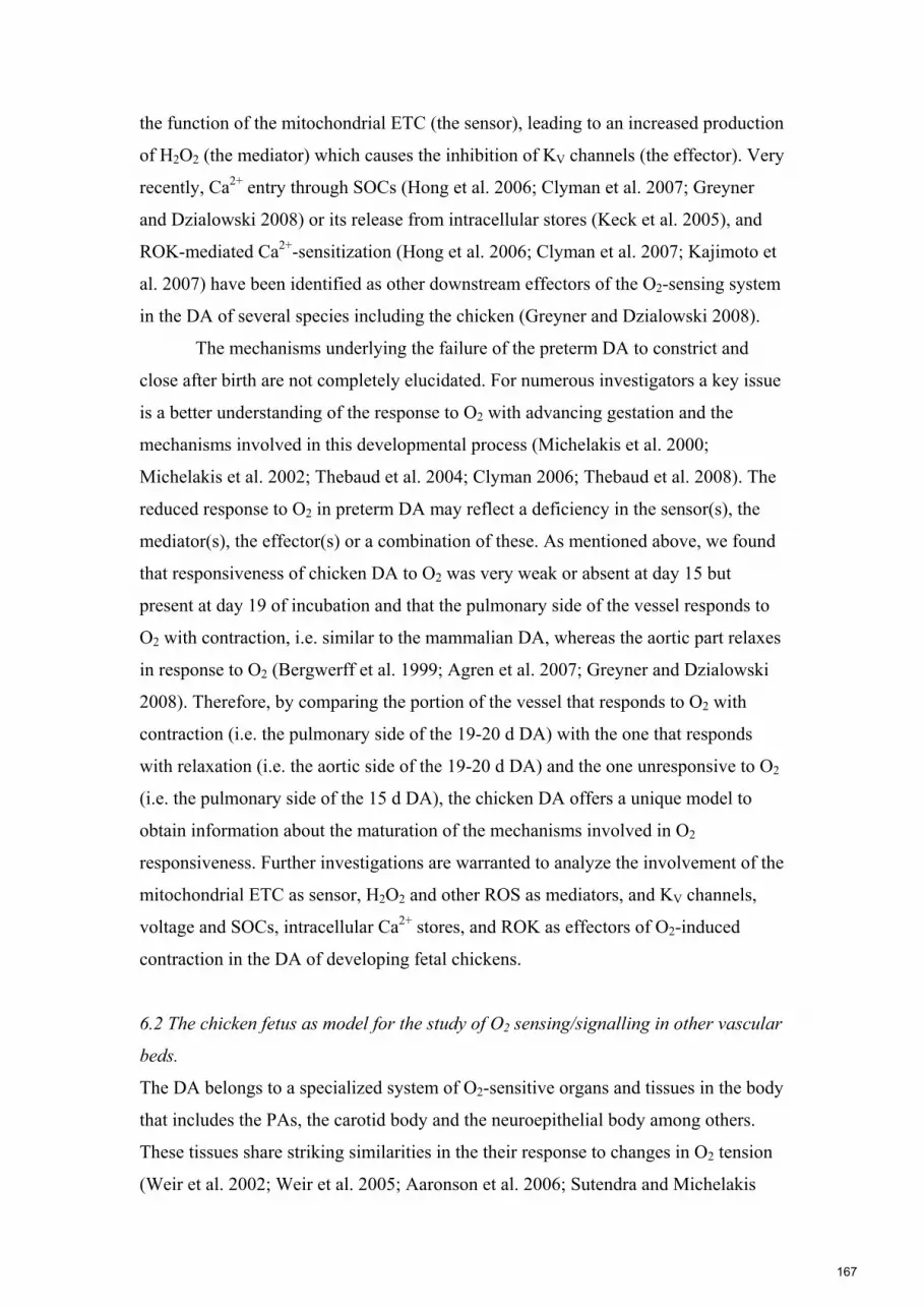
the function of the mitochondrial ETC (the sensor), leading to an increased production
of H2O2 (the mediator) which causes the inhibition of KV channels (the effector). Very
recently, Ca2+ entry through SOCs (Hong et al. 2006; Clyman et al. 2007; Greyner
and Dzialowski 2008) or its release from intracellular stores (Keck et al. 2005), and
ROK-mediated Ca2+-sensitization (Hong et al. 2006; Clyman et al. 2007; Kajimoto et
al. 2007) have been identified as other downstream effectors of the O2-sensing system
in the DA of several species including the chicken (Greyner and Dzialowski 2008).
The mechanisms underlying the failure of the preterm DA to constrict and
close after birth are not completely elucidated. For numerous investigators a key issue
is a better understanding of the response to O2 with advancing gestation and the
mechanisms involved in this developmental process (Michelakis et al. 2000;
Michelakis et al. 2002; Thebaud et al. 2004; Clyman 2006; Thebaud et al. 2008). The
reduced response to O2 in preterm DA may reflect a deficiency in the sensor(s), the
mediator(s), the effector(s) or a combination of these. As mentioned above, we found
that responsiveness of chicken DA to O2 was very weak or absent at day 15 but
present at day 19 of incubation and that the pulmonary side of the vessel responds to
O2 with contraction, i.e. similar to the mammalian DA, whereas the aortic part relaxes
in response to O2 (Bergwerff et al. 1999; Agren et al. 2007; Greyner and Dzialowski
2008). Therefore, by comparing the portion of the vessel that responds to O2 with
contraction (i.e. the pulmonary side of the 19-20 d DA) with the one that responds
with relaxation (i.e. the aortic side of the 19-20 d DA) and the one unresponsive to O2
(i.e. the pulmonary side of the 15 d DA), the chicken DA offers a unique model to
obtain information about the maturation of the mechanisms involved in O2
responsiveness. Further investigations are warranted to analyze the involvement of the
mitochondrial ETC as sensor, H2O2 and other ROS as mediators, and KV channels,
voltage and SOCs, intracellular Ca2+ stores, and ROK as effectors of O2-induced
contraction in the DA of developing fetal chickens.
6.2 The chicken fetus as model for the study of O2 sensing/signalling in other vascular
beds.
The DA belongs to a specialized system of O2-sensitive organs and tissues in the body
that includes the PAs, the carotid body and the neuroepithelial body among others.
These tissues share striking similarities in the their response to changes in O2 tension
(Weir et al. 2002; Weir et al. 2005; Aaronson et al. 2006; Sutendra and Michelakis
167

2007; Weir et al. 2008). However, if the mechanisms responsible for SM contraction
in hypoxic pulmonary vasoconstricition and normoxic contraction of the DA are
virtually the same, why is the response to a change in O2 diametrically opposite?
Numerous investigators agree that a change in the redox status of the SMCs in the PA
and DA initiates the change in vascular tone (Weir et al. 2002; Weir et al. 2005;
Aaronson et al. 2006; Sutendra and Michelakis 2007; Weir et al. 2008). Exactly what
the redox signal may be and the subsequent signalling sequence remains to be
determined. The hypoxic vasoconstriction of pulmonary vessels is a highly conserved
adaptive physiological mechanism that optimizes O2 saturation of pulmonary venous
blood by increasing pulmonary vascular resistance in poorly aerated lung regions
(Villamor et al. 1997; Michelakis et al. 2004; Moudgil et al. 2005; Weir et al. 2005;
Aaronson et al. 2006; Russell et al. 2008). In contrast, the systemic vasculature
frequently responds to hypoxia with vasodilation in an effort to maintain adequate
tissue oxygenation (Russell et al. 2008). In fetal chickens, the DA (pulmonary side)
and the femoral arteries respond to hypoxia with relaxation (Ruijtenbeek et al. 2002b)
and the aortic side of the DA, the intrapulmonary arteries (Zoer et al. 2009) and the
chorioallantoic arteries (Lindgren et al. 2009) with contraction. The mechanisms that
initiate, differentiate and regulate this variety of vascular responses to O2 need to be
elucidated. In addition, the O2- sensitive vessels of the chicken fetus offer a unique
model for the analysis of new mediators, such as sphingomyelinase (Cogolludo et al.
2009) or hydrogen sulfide (Olson et al. 2006), which have been suggested to
participate in the process of vascular O2 sensing/signalling.
6.3 Effects of prenatal hormonal environment on the development of the DA
Among prematurely born infants, females have an advantage compared to males, with
a significantly greater chance of surviving, and a better outcome with less morbidity
than male infants with similar birth weights and gestational ages (Ingemarsson 2003;
Elsmen et al. 2004; Vatten and Skjaerven 2004). The biological mechanisms
contributing to the male disadvantage or female advantage have not been elucidated.
However, speculations involve estrogen and androgens. The fetus is supplied from the
placenta with estrogen in increasing amounts during the gestation and premature
cessation has been shown to contribute to perinatal morbidity, an effect that may be
higher in males since they have no own production of estrogen (Trotter et al. 1999;
Derzbach et al. 2005).
168

One of the neonatal complications that is more common in preterm males than
females is PDA (Stevenson et al. 2000). Differences in vascular reactivity depending
on gender have been shown in many vascular responses in both systemic and PAs
(Polderman et al. 1993; Hayashi et al. 1995; Earley and Resta 2002; Sader and
Celermajer 2002). Both endothelium-dependent and -independent responses differ
between males and females. Sex hormones seem to play a role in regulating the
vascular response, both in long term and short term (Schwertz and Penckofer 2001;
Sader and Celermajer 2002; Khalil 2005). Estrogens have been shown to regulate the
gene expression of K+ and Ca2+ channels and to play a role in regulation of Ca2+
handling (Orshal and Khalil 2004; Tsang et al. 2004).
Sexual differentiation occurs in opposite ways in birds and mammals. In
mammals, the phenotype of homogametic sex (female) develops in the (relative)
absence of steroids and in the heterogametic sex (male) the presence of testosterone
and other testicular hormones is a preliminary condition for development of male
characteristics (Biau et al. 2006). In the absence of testicular hormones development
takes the feminine course. In contrast, birds develop masculine characteristics in the
absence of estrogens, while in females (the heterogametic sex), extensive expression
of the aromatase gene (around day 5-6 of incubation) leads to estrogen synthesis
(Bruggeman et al. 2002; Biau et al. 2006). Already at 7.5 day of incubation, plasma
levels of steroids differ between males and females (Bruggeman et al. 2002).
Another hormone with a possible role regulating DA development is thyroid
hormone. Several plasma-membrane ion transporters including, KV channels, are
regulated at both the transcriptional and post-transcriptional levels by thyroid hormone
(Ishikawa et al. 1989; Ojamaa et al. 2000; Owen et al. 2007) and an association
between PDA and hypothyroidism, in term as well as in preterm infants, has also been
found (Nakagawa 1993; Allegaert et al. 2004; Guarnieri et al. 2008; van Wassenaer
and Kok 2008).
Manipulation of the hormonal environment of the chicken embryo can be
easily performed by injection, into the albumen of fertilized eggs, of aromatase
inhibitors (which blocks the synthesis of estrogen from testosterone, Bruggeman et al.
2002) or anti-thyroid drugs (Liu et al. 2003; Liu and Porter 2004). Therefore the
chicken offers a unique model to obtain information about the influence of hormones
in the maturation of the DA. Also the direct effect of the hormones in DA reactivity
remains to be studied.
169

6.4 Responsiveness of chicken DA to isoprostanes
Isoprostanes are PG isomers that are produced in vivo primarily by a free radical-
catalyzed peroxidation of polyunsaturated fatty acids (Janssen et al. 2005; Janssen
2008). Initially, isoprostanes were recognized as being valuable markers of oxidative
stress and numerous pathological conditions have been shown to be associated with
increases in urinary, plasma, and tissue levels of isoprostanes. Moreover, few
members of the isoprostane family are biologically active and could contribute to the
functional consequences of oxidant injury and mediate many of the features of the
disease states for which they are used as indicators (Janssen et al. 2005; Janssen
2008).
Newborns and particularly preterm infants are very susceptible to oxidative
stress. A large growing body of literature is addressing the possible role of
isoprostanes in the pathophysiology of several neonatal conditions, such as asphyxia,
intraventricular hemorrhage, periventricular leukomalacia, bronchopulmonary
dysplasia, pulmonary hypertension and retinopathy of prematurity (Hardy et al. 2000;
Ahola et al. 2004; Weinberger et al. 2006; Yi et al. 2006). However, the possible
contribution of isoprostanes to the normal transition to extra-uterine life remains
largely unknown. There is increasing evidence that isoprostanes exert a wide variety
of actions on VSM and endothelial cells. Many have described contractile responses
to isoprostanes in a wide variety of vascular tissues, generally via stimulation of TP
receptors (Oliveira et al. 2000; Gonzalez-Luis et al. 2005; Janssen et al. 2005; Janssen
2008; Mueed et al. 2008).
In addition, and depending on the compound and the vascular bed vasodilatory
actions of isoprostanes have been also identified (Jourdan et al. 1997; Zhang et al.
2003; Gonzalez-Luis et al. 2005; Zhang et al. 2005). These actions were frequently
masked by the vasoconstrictive effects of isoprostanes on TP receptors and were only
observed when these receptors were blocked by antagonists (such as SQ 29,548 or ICI
192605), or saturated by an agonist (such as U46619, Jourdan et al. 1997; Zhang et al.
2003; Zhang et al. 2005). However, the effects of isoprostanes have not been studied
in the DA of any species. Our preliminary results suggest that the isoprostanes 8-iso-
PGE2 and 8-iso-PGF2α induce TP-mediated contraction of the chicken DA. Similarly
to PGE2, 8-iso-PGE2 induced ductal relaxation when the TP receptor was blocked or
170

saturated. The developmental changes in the DA responsiveness to isoprostanes
warrant future investigation.
6.5 Responsiveness of chicken DA to CO
Another mediator that plays a relevant role in the control of the mammalian DA tone
and whose study is warranted in the non-mammalian DA is CO (Smith 1998;
Baragatti et al. 2007). CO is produced by the degradation of heme by intracellular HO.
Different research groups have provided evidence of the ability of CO to relax
vascular tone similar to that of NO (i.e. through activation of sGC, Wang et al. 1997;
Werkstrom et al. 1997; Villamor et al. 2000; Komuro et al. 2001; Leo et al. 2008).
The expression and activity of HO as well as the developmental changes in the
responsiveness of the chicken DA to CO are currently under investigation in our
laboratory.
6.6 Effects of prenatal/perinatal insults on DA development.
As mentioned elsewhere, the egg-chicken model offers the possibility of manipulating
the environment of development without interferences of maternal hormonal,
metabolic, or hemodynamic alterations. In the present Thesis we analyzed the effects
of chronic prenatal hypoxia on chicken DA development. Very recently, Copeland &
Dzialowski (2009) demonstrated that incubation in hyperoxia accelerates chicken DA
development. Preliminary results of our laboratory also indicate that premature
exposure to hyperoxia induces significant alterations in the developmental trajectory
of the chicken DA. Other prenatal insults that we demonstrated to induce alterations
in chicken perinatal vascular homeostasis are malnutrition (Ruijtenbeek et al. 2003b),
exposure to cigarette smoke extract (Carlo et al. 2001) or inhibition of NOS (Villamor
et al. 2005). Injection of drugs (Carlo et al. 2001; Villamor et al. 2005) or gene
transfer (Williams et al. 2006) during incubation open a new territory for the
investigation of the pathways involved in the developmental vascular biology of the
DA.
171

Aaronson, P. I., T. P. Robertson, et al. (2006). "Hypoxic pulmonary vasoconstriction: mechanisms and controversies." J Physiol 570(Pt 1): 53-8.
Abman, S. H. (1999). "Abnormal vasoreactivity in the pathophysiology of persistent pulmonary hypertension of the newborn." Pediatr Rev 20(11): e103-9.
Abman, S. H., B. A. Chatfield, et al. (1990). "Role of endothelium-derived relaxing factor during transition of pulmonary circulation at birth." Am J Physiol 259(6 Pt 2): H1921-7.
Abman, S. H., B. A. Chatfield, et al. (1991). "Maturational changes in endothelium-derived relaxing factor activity of ovine pulmonary arteries in vitro." Am J Physiol 260(4 Pt 1): L280-5.
Agren, P., A. L. Cogolludo, et al. (2007). "Ontogeny of chicken ductus arteriosus response to oxygen and vasoconstrictors." Am J Physiol Regul Integr Comp Physiol 292(1): R485-96.
Ahola, T., V. Fellman, et al. (2004). "Plasma 8-isoprostane is increased in preterm infants who develop bronchopulmonary dysplasia or periventricular leukomalacia." Pediatr Res 56(1): 88-93.
Allegaert, K., C. Vanhole, et al. (2004). "Association of patent ductus arteriosus and severe hypothyroidism in term newborns: coincidence or causal?" J Matern Fetal Neonatal Med 16(6): 339-41.
Altimiras, J. and D. A. Crossley, 2nd (2000). "Control of blood pressure mediated by baroreflex changes of heart rate in the chicken embryo (Gallus gallus)." Am J Physiol Regul Integr Comp Physiol 278(4): R980-6.
Alzamora-Castro, V., G. Battilana, et al. (1960). "Patent ductus arteriosus and high altitude." Am J Cardiol 5: 761-3.
Archer, S. and E. Michelakis (2002). "The mechanism(s) of hypoxic pulmonary vasoconstriction: potassium channels, redox O(2) sensors, and controversies." News Physiol Sci 17: 131-7.
Archer, S. L., J. Huang, et al. (1993). "A redox-based O2 sensor in rat pulmonary vasculature." Circ Res 73(6): 1100-12.
Archer, S. L., E. K. Weir, et al. (2000). "Molecular identification of O2 sensors and O2-sensitive potassium channels in the pulmonary circulation." Adv Exp Med Biol 475: 219-40.
Arishima, K., T. Takizawa, et al. (1995). "Propranolol inhibits the spontaneous closure of the ductus arteriosus in newborn rats." J Vet Med Sci 57(5): 943-4.
Arnal, J. F., A. T. Dinh-Xuan, et al. (1999). "Endothelium-derived nitric oxide and vascular physiology and pathology." Cell Mol Life Sci 55(8-9): 1078-87.
Aronson, S., G. Gennser, et al. (1970). "Innervation and contractile response of the human ductus arteriosus." Eur J Pharmacol 11(2): 178-86.
Bancalari, E., N. Claure, et al. (2005). "Patent ductus arteriosus and respiratory outcome in premature infants." Biol Neonate 88(3): 192-201.
Baragatti, B., F. Brizzi, et al. (2003). "Cyclooxygenase-1 and cyclooxygenase-2 in the mouse ductus arteriosus: individual activity and functional coupling with nitric oxide synthase." Br J Pharmacol 139(8): 1505-15.
Baragatti, B., F. Brizzi, et al. (2007). "Interactions between NO, CO and an endothelium-derived hyperpolarizing factor (EDHF) in maintaining patency of the ductus arteriosus in the mouse." Br J Pharmacol 151(1): 54-62.
Bateson, E. A., R. Schulz, et al. (1999). "Response of fetal rabbit ductus arteriosus to bradykinin: role of nitric oxide, prostaglandins, and bradykinin receptors." Pediatr Res 45(4 Pt 1): 568-74.
172
Belanger, C., J. Copeland, et al. (2008). "Morphological changes in the chicken ductus arteriosi during closure at hatching." Anat Rec (Hoboken) 291(8): 1007-15.
Bellaris, A. and M. Osmond (1998). The atlas of chick development, Academic Press. Benson, L. N. and K. N. Cowan (2002). The arterial duct: its persistence and its
patency. Paediatric Cardiology. R. H. Anderson, E. J. Baker, F. J. Macartney, M. L. Rigby, E. A. Shinebourne and M. Tynan, Churchill Livingstone: 1405-1459.
Bergwerff, M., M. C. DeRuiter, et al. (1999). "Comparative anatomy and ontogeny of the ductus arteriosus, a vascular outsider." Anat Embryol (Berl) 200(6): 559-71.
Bergwerff, M., M. C. DeRuiter, et al. (1996). "Onset of elastogenesis and downregulation of smooth muscle actin as distinguishing phenomena in artery differentiation in the chick embryo." Anat Embryol (Berl) 194(6): 545-57.
Bergwerff, M., M. E. Verberne, et al. (1998). "Neural crest cell contribution to the developing circulatory system: implications for vascular morphology?" Circ Res 82(2): 221-31.
Bhattacharya, M., P. Asselin, et al. (1999). "Developmental changes in prostaglandin E(2) receptor subtypes in porcine ductus arteriosus. Possible contribution in altered responsiveness to prostaglandin E(2)." Circulation 100(16): 1751-6.
Biau, S., S. Bayle, et al. (2006). "The chick embryo: an animal model for detection of the effects of hormonal compounds." Anal Bioanal Chem.
Blackburn, S. T. (2003). Maternal, fetal, and neonatal physiology : A clinical perspective, Saunders, Elsevier Science.
Bodach, E., F. Coceani, et al. (1980). "The response of the isolated ductus arteriosus to transmural stimulation and drugs." Br J Pharmacol 71(2): 419-27.
Boels, P. J., J. Deutsch, et al. (1999). "Maturation of the response to bradykinin in resistance and conduit pulmonary arteries." Cardiovasc Res 44(2): 416-28.
Bolotina, V. M., S. Najibi, et al. (1994). "Nitric oxide directly activates calcium-dependent potassium channels in vascular smooth muscle." Nature 368(6474): 850-3.
Boreus, L. O., T. Malmfors, et al. (1969). "Demonstration of adrenergic receptor function and innervation in the ductus arteriosus of the human fetus." Acta Physiol Scand 77(3): 316-21.
Born, G. V., G. S. Dawes, et al. (1956). "The constriction of the ductus arteriosus caused by oxygen and by asphyxia in newborn lambs." J Physiol 132(2): 304-42.
Bos, C. L., D. J. Richel, et al. (2004). "Prostanoids and prostanoid receptors in signal transduction." Int J Biochem Cell Biol 36(7): 1187-205.
Bouayad, A., X. Hou, et al. (2002). "Cyclooxygenase isoforms and prostaglandin E2 receptors in the ductus arteriosus." Current Therapeutic Research 63(10): 669-681.
Bouayad, A., H. Kajino, et al. (2001). "Characterization of PGE2 receptors in fetal and newborn lamb ductus arteriosus." Am J Physiol Heart Circ Physiol 280(5): H2342-9.
Bruggeman, V., P. Van As, et al. (2002). "Developmental endocrinology of the reproductive axis in the chicken embryo." Comp Biochem Physiol A Mol Integr Physiol 131(4): 839-46.
Burton, F. G., J. M. Stevensson, et al. (1989). "The relationship between eggshell porosity and air space in gas tension measured before and during the
173

parafoetal period and their effects on the hatching process in the domestic fowl." Respir. Physiology 77: 89-100.
Bustamante, S. A., Y. Pang, et al. (1996). "Inducible nitric oxide synthase and the regulation of central vessel caliber in the fetal rat." Circulation 94(8): 1948-53.
Carlo, W. F., E. Villamor, et al. (2001). "Chronic exposure to cigarette smoke extract impairs endothelium-dependent relaxation of chicken embryo pulmonary arteries." Biol Neonate 80(3): 247-50.
Carvajal, J. A., A. M. Germain, et al. (2000). "Molecular mechanism of cGMP-mediated smooth muscle relaxation." J Cell Physiol 184(3): 409-20.
Chand, N. and P. Eyre (1976). "Effects of prostaglandins E1, E2 and F2 alpha and polyphloretin phosphate of domestic fowl." Arch Int Pharmacodyn Ther 221(2): 261-65.
Claeys, M., E. Wechsung, et al. (1981). "Prostaglandin E2 is the prevalent metabolite of arachidonic acid formed by aortic tissue of the chicken." Arch Int Pharmacodyn Ther 249(2): 312-5.
Clarke, J. A. (1965). "An X-ray microscopic study of the vasa vasorum of the human ductus arteriosus." J Anat 99(Pt 3): 527-37.
Clyman, R. I. (2006). "Mechanisms regulating the ductus arteriosus." Biology of the Neonate 89(4): 330-335.
Clyman, R. I., C. Y. Chan, et al. (1999a). "Permanent anatomic closure of the ductus arteriosus in newborn baboons: the roles of postnatal constriction, hypoxia, and gestation." Pediatr Res 45(1): 19-29.
Clyman, R. I., Y. Q. Chen, et al. (2001). "In utero remodeling of the fetal lamb ductus arteriosus: the role of antenatal indomethacin and avascular zone thickness on vasa vasorum proliferation, neointima formation, and cell death." Circulation 103(13): 1806-12.
Clyman, R. I., P. Hardy, et al. (1999b). "Cyclooxygenase-2 plays a significant role in regulating the tone of the fetal lamb ductus arteriosus." Am J Physiol 276(3 Pt 2): R913-21.
Clyman, R. I., F. Mauray, et al. (1989). "Influence of increased pulmonary vascular pressures on the closure of the ductus arteriosus in newborn lambs." Pediatr Res 25(2): 136-42.
Clyman, R. I., F. Mauray, et al. (1983). "Factors determining the loss of ductus arteriosus responsiveness to prostaglandin E." Circulation 68(2): 433-6.
Clyman, R. I., F. Mauray, et al. (1980). "Age-dependent sensitivity of the lamb ductus arteriosus to indomethacin and prostaglandins." J Pediatr 96(1): 94-8.
Clyman, R. I., S. R. Seidner, et al. (2002). "VEGF regulates remodeling during permanent anatomic closure of the ductus arteriosus." Am J Physiol Regul Integr Comp Physiol 282(1): R199-206.
Clyman, R. I., N. Waleh, et al. (1998). "Regulation of ductus arteriosus patency by nitric oxide in fetal lambs: the role of gestation, oxygen tension, and vasa vasorum." Pediatr Res 43(5): 633-44.
Clyman, R. I., N. Waleh, et al. (2007). "Calcium-dependent and calcium-sensitizing pathways in the mature and immature ductus arteriosus." Am J Physiol Regul Integr Comp Physiol 293(4): R1650-6.
Coceani, F., C. Ackerley, et al. (2001). "Function of cyclo-oxygenase-1 and cyclo-oxygenase-2 in the ductus arteriosus from foetal lamb: differential development and change by oxygen and endotoxin." Br J Pharmacol 132(1): 241-51.
174

Coceani, F., S. Barogi, et al. (2005). "Cyclooxygenase isoenzymes and patency of ductus arteriosus." Prostaglandins Leukot Essent Fatty Acids 72(2): 71-7.
Coceani, F. and L. Kelsey (1991). "Endothelin-1 release from lamb ductus arteriosus: relevance to postnatal closure of the vessel." Can J Physiol Pharmacol 69(2): 218-21.
Coceani, F. and L. Kelsey (2000). "Endothelin-1 release from the lamb ductus arteriosus: are carbon monoxide and nitric oxide regulatory agents?" Life Sci 66(26): 2613-23.
Coceani, F., L. Kelsey, et al. (1992). "Evidence for an effector role of endothelin in closure of the ductus arteriosus at birth." Can J Physiol Pharmacol 70(7): 1061-4.
Coceani, F., L. Kelsey, et al. (1994). "Occurrence of endothelium-derived relaxing factor--nitric oxide in the lamb ductus arteriosus." Can J Physiol Pharmacol 72(1): 82-8.
Coceani, F., L. Kelsey, et al. (2002). "The response of the lamb ductus arteriosus to endothelin: developmental changes and influence of light." Life Sci 71(10): 1209-17.
Coceani, F., L. Kelsey, et al. (1997). "Carbon monoxide formation in the ductus arteriosus in the lamb: implications for the regulation of muscle tone." Br J Pharmacol 120(4): 599-608.
Coceani, F., Y. Liu, et al. (1999). "Endothelin A receptor is necessary for O(2) constriction but not closure of ductus arteriosus." Am J Physiol 277(4 Pt 2): H1521-31.
Coceani, F., Y. A. Liu, et al. (2000). "Deletion of the endothelin-A-receptor suppresses oxygen-induced constriction but not postnatal closure of the ductus arteriosus." J Cardiovasc Pharmacol 36(5 Suppl 1): S75-7.
Coceani, F. and P. M. Olley (1973). "The response of the ductus arteriosus to prostaglandins." Can J Physiol Pharmacol 51(3): 220-5.
Cogolludo, A., L. Moreno, et al. (2009). "Activation of neutral sphingomyelinase is involved in acute hypoxic pulmonary vasoconstriction." Cardiovasc Res 82(2): 296-302.
Colbert, M. C., M. L. Kirby, et al. (1996). "Endogenous retinoic acid signaling colocalizes with advanced expression of the adult smooth muscle myosin heavy chain isoform during development of the ductus arteriosus." Circ Res 78(5): 790-8.
Coleman, H. A., M. Tare, et al. (2004). "Endothelial potassium channels, endothelium-dependent hyperpolarization and the regulation of vascular tone in health and disease." Clin Exp Pharmacol Physiol 31(9): 641-9.
Copeland, J. and E. M. Dzialowski (2009). "Effects of hypoxic and hyperoxic incubation on the reactivity of the chicken embryo (Gallus gallus) ductus arteriosi in response to catecholamines and oxygen." Exp Physiol 94(1): 152-61.
Corbet, A. J. (1998). Medical Manipulation of the Ductus Arteriosus. The Science and Practice of Pediatric Cardiology. A. Garson, J. T. Bricker, D. J. Fisher and S. R. Neish. Baltimore, Maryland, Williams & Wilkins. II.
Cotton, R. B., M. T. Stahlman, et al. (1978). "Medical management of small preterm infants with symptomatic patent ductus arteriosus." J Pediatr 92(3): 467-73.
de Ruiter, M. C., A. C. Gittenberger-de Groot, et al. (1989). "The special status of the pulmonary arch artery in the branchial arch system of the rat." Anat Embryol (Berl) 179(4): 319-25.
175

Derzbach, L., A. Treszl, et al. (2005). "Gender dependent association between perinatal morbidity and estrogen receptor-alpha Pvull polymorphism." J Perinat Med 33(5): 461-2.
Doyle, L. W. (2001). "Outcome at 5 years of age of children 23 to 27 weeks' gestation: refining the prognosis." Pediatrics 108(1): 134-41.
Duchen, M. R. (1999). "Contributions of mitochondria to animal physiology: from homeostatic sensor to calcium signalling and cell death." J Physiol 516 ( Pt 1): 1-17.
Dunn, L. K., S. K. Gruenloh, et al. (2005). "Chick chorioallantoic membrane as an in vivo model to study vasoreactivity: characterization of development-dependent hyperemia induced by epoxyeicosatrienoic acids (EETs)." Anat Rec A Discov Mol Cell Evol Biol 285(2): 771-80.
Earley, S. and T. C. Resta (2002). "Estradiol attenuates hypoxia-induced pulmonary endothelin-1 gene expression." Am J Physiol Lung Cell Mol Physiol 283(1): L86-93.
Eldridge, F. L., H. N. Hultgren, et al. (1954). "The physiologic closure of the ductus arteriosus in newborn infants: a preliminary report." Science 119(3099): 731-2.
Eliot, R. J., R. Lam, et al. (1980). "Plasma catecholamine concentrations in infants at birth and during the first 48 hours of life." J Pediatr 96(2): 311-5.
Elsmen, E., I. Hansen Pupp, et al. (2004). "Preterm male infants need more initial respiratory and circulatory support than female infants.[see comment]." Acta Paediatrica 93(4): 529-33.
Fabres, J., W. A. Carlo, et al. (2007). "Both extremes of arterial carbon dioxide pressure and the magnitude of fluctuations in arterial carbon dioxide pressure are associated with severe intraventricular hemorrhage in preterm infants." Pediatrics 119(2): 299-305.
Fay, F. S. (1971). "Guinea pig ductus arteriosus. I. Cellular and metabolic basis for oxygen sensitivity." Am J Physiol 221(2): 470-9.
Fay, F. S. and F. F. Jobsis (1972). "Guinea pig ductus arteriosus. 3. Light absorption changes during response to O 2." Am J Physiol 223(3): 588-95.
Fineman, J. R., Y. Takahashi, et al. (1998). "Endothelin-receptor blockade does not alter closure of the ductus arteriosus." Am J Physiol 275(5 Pt 2): H1620-6.
Friedman, W. F., K. M. Fitzpatrick, et al. (1978). "The patent ductus arteriosus." Clin Perinatol 5(2): 411-36.
Friedman, W. F., M. P. Printz, et al. (1983). "The vasoactivity of the fetal lamb ductus arteriosus studied in utero." Pediatr Res 17(5): 331-7.
Furchgott, R. F. and P. M. Vanhoutte (1989). "Endothelium-derived relaxing and contracting factors." Faseb J 3(9): 2007-18.
Gersony, W. M., G. J. Peckham, et al. (1983). "Effects of indomethacin in premature infants with patent ductus arteriosus: results of a national collaborative study." J Pediatr 102(6): 895-906.
Gilbert, E. F., H. J. Bruyere, Jr., et al. (1977). "The effect of practolol and butoxamine on aortic arch malformation in beta adrenoreceptor stimulated chick embryos." Teratology 15(3): 317-23.
Gittenberger-de Groot, A. C., J. L. Strengers, et al. (1985). "Histologic studies on normal and persistent ductus arteriosus in the dog." J Am Coll Cardiol 6(2): 394-404.
176

Giuriato, L., M. Scatena, et al. (1993). "Rabbit ductus arteriosus during development: anatomical structure and smooth muscle cell composition." Anat Rec 235(1): 95-110.
Goldbarg, S. H., Y. Takahashi, et al. (2002). "In utero indomethacin alters O2 delivery to the fetal ductus arteriosus: implications for postnatal patency." Am J Physiol Regul Integr Comp Physiol 282(1): R184-90.
Gonzalez-Luis, G., F. Perez-Vizcaino, et al. (2005). "Age-related differences in vasoconstrictor responses to isoprostanes in piglet pulmonary and mesenteric vascular smooth muscle." Pediatr Res 57(6): 845-52.
Greyner, H. and E. M. Dzialowski (2008). "Mechanisms mediating the oxygen-induced vasoreactivity of the ductus arteriosus in the chicken embryo." Am J Physiol Regul Integr Comp Physiol 295(5): R1647-59.
Groenendijk, B. C., B. P. Hierck, et al. (2005). "Changes in shear stress-related gene expression after experimentally altered venous return in the chicken embryo." Circ Res 96(12): 1291-8.
Guarnieri, G. F., N. Laforgia, et al. (2008). "Delayed closure of ductus arteriosus in term newborns with congenital hypothyroidism: effect of L-thyroxine therapy." Pediatr Cardiol 29(2): 455-6.
Guimaraes, S. and D. Moura (2001). "Vascular adrenoceptors: an update." Pharmacol Rev 53(2): 319-56.
Hammerman, C. (1995). "Patent ductus arteriosus. Clinical relevance of prostaglandins and prostaglandin inhibitors in PDA pathophysiology and treatment." Clin Perinatol 22(2): 457-79.
Hardy, P., I. Dumont, et al. (2000). "Oxidants, nitric oxide and prostanoids in the developing ocular vasculature: a basis for ischemic retinopathy." Cardiovasc Res 47(3): 489-509.
Harms, D. (1967). "[On the occlusion of the ductus arteriosus of Gallus domesticus]." Z Zellforsch Mikrosk Anat 81(3): 433-44.
Hayashi, T., K. Yamada, et al. (1995). "Estrogen increases endothelial nitric oxide by a receptor-mediated system." Biochem Biophys Res Commun 214(3): 847-55.
Herrera, C., J. Holberton, et al. (2004). "Prolonged versus short course of indomethacin for the treatment of patent ductus arteriosus in preterm infants." Cochrane Database Syst Rev(1): CD003480.
Hodach, R. J., A. E. Hodach, et al. (1975). "The role of beta-adrenergic activity in the production of cardiac and aortic arch anomalies in chick embryos." Teratology 12(1): 33-45.
Hong, Z., F. Hong, et al. (2006). "Role of store-operated calcium channels and calcium sensitization in normoxic contraction of the ductus arteriosus." Circulation 114(13): 1372-9.
Hughes, A. F. W. (1943). "The histogenesis of the arteries of the chick embryo." J Anat 77: 266-287.
Hyvelin, J. M., C. O'Connor, et al. (2004). "Effect of changes in pH on wall tension in isolated rat pulmonary artery: role of the RhoA/Rho-kinase pathway." Am J Physiol Lung Cell Mol Physiol 287(4): L673-84.
Ikeda, M., R. R. Sonnenschein, et al. (1972). "Catecholamine content and uptake of the ductus arteriosus of the fetal lamb." Experientia 28(8): 914-5.
Ingemarsson, I. (2003). "Gender aspects of preterm birth." Bjog 110 Suppl 20: 34-8. Ishikawa, T., T. Chijiwa, et al. (1989). "Thyroid hormones directly interact with
vascular smooth muscle strips." Mol Pharmacol 35(6): 760-5.
177

Janssen, L. J. (2008). "Isoprostanes and lung vascular pathology." Am J Respir Cell Mol Biol 39(4): 383-9.
Janssen, L. J., A. Catalli, et al. (2005). "The pulmonary biology of isoprostanes." Antioxid Redox Signal 7(1-2): 244-55.
Jourdan, K. B., T. W. Evans, et al. (1997). "Evidence for a dilator function of 8-iso prostaglandin F2 alpha in rat pulmonary artery." Br J Pharmacol 120(7): 1280-5.
Jurrus, E. R. and H. S. Weiss (1977). "In vitro tissue oxygen tensions in the rabbit aortic arch." Atherosclerosis 28(3): 223-32.
Kajimoto, H., K. Hashimoto, et al. (2007). "Oxygen activates the Rho/Rho-kinase pathway and induces RhoB and ROCK-1 expression in human and rabbit ductus arteriosus by increasing mitochondria-derived reactive oxygen species: a newly recognized mechanism for sustaining ductal constriction." Circulation 115(13): 1777-88.
Kajino, H., Y. Q. Chen, et al. (2000). "Tissue hypoxia inhibits prostaglandin and nitric oxide production and prevents ductus arteriosus reopening." Am J Physiol Regul Integr Comp Physiol 279(1): R278-86.
Kajino, H., Y. Q. Chen, et al. (2001). "Factors that increase the contractile tone of the ductus arteriosus also regulate its anatomic remodeling." Am J Physiol Regul Integr Comp Physiol 281(1): R291-301.
Kajino, H., S. Goldbarg, et al. (2002). "Vasa vasorum hypoperfusion is responsible for medial hypoxia and anatomic remodeling in the newborn lamb ductus arteriosus." Pediatr Res 51(2): 228-35.
Kajino, H., T. Taniguchi, et al. (2004). "An EP4 receptor agonist prevents indomethacin-induced closure of rat ductus arteriosus in vivo." Pediatr Res 56(4): 586-90.
Kamper, A. M., L. C. Paul, et al. (2002). "Prostaglandins are involved in acetylcholine- and 5-hydroxytryptamine-induced, nitric oxide-mediated vasodilatation in human forearm." J Cardiovasc Pharmacol 40(6): 922-9.
Karadas, B., T. Kaya, et al. (2004). "Comparison of effects of cyclooxygenase inhibitors on myometrial contraction and constriction of ductus arteriosus in rats." Eur J Pharmacol 485(1-3): 289-98.
Katzung, B. G. (1995). Basic and clinical pharmacology. Norwalk, Connecticut, Paramount Publishing Buiseness and Professional Group.
Keck, M., E. Resnik, et al. (2005). "Oxygen increases ductus arteriosus smooth muscle cytosolic calcium via release of calcium from inositol triphosphate-sensitive stores." Am J Physiol Lung Cell Mol Physiol 288(5): L917-23.
Kempf, H., C. Linares, et al. (1998). "Pharmacological inactivation of the endothelin type A receptor in the early chick embryo: a model of mispatterning of the branchial arch derivatives." Development 125(24): 4931-41.
Khalil, R. A. (2005). "Sex hormones as potential modulators of vascular function in hypertension." Hypertension 46(2): 249-54.
Kim, H. S., M. Aikawa, et al. (1993). "Ductus arteriosus. Advanced differentiation of smooth muscle cells demonstrated by myosin heavy chain isoform expression in rabbits." Circulation 88(4 Pt 1): 1804-10.
Kiserud, T. (2005). "Physiology of the fetal circulation." Semin Fetal Neonatal Med 10(6): 493-503.
Kitazawa, T., A. N. Polzin, et al. (2004). "CPI-17-deficient smooth muscle of chicken." J Physiol 557(Pt 2): 515-28.
178

Kluckow, M., N. Evans, et al. (1999). "Prostacyclin concentrations and transitional circulation in preterm infants requiring mechanical ventilation." Arch Dis Child Fetal Neonatal Ed 80(1): F34-7.
Knight, D. B. (2001). "The treatment of patent ductus arteriosus in preterm infants. A review and overview of randomized trials." Semin Neonatol 6: 63-73.
Knight, D. H., D. F. Patterson, et al. (1973). "Constriction of the fetal ductus arteriosus induced by oxygen, acteylcholine, and norepinephrine in normal dogs and those genetically predisposed to persistent patency." Circulation 47(1): 127-32.
Koch, J., G. Hensley, et al. (2006). "Prevalence of spontaneous closure of the ductus arteriosus in neonates at a birth weight of 1000 grams or less." Pediatrics 117(4): 1113-21.
Komuro, T., M. K. Borsody, et al. (2001). "The vasorelaxation of cerebral arteries by carbon monoxide." Exp Biol Med (Maywood) 226(9): 860-5.
Kovalcik, V. (1963). "The Response of the Isolated Ductus Arteriosus to Oxygen and Anoxia." J Physiol 169: 185-97.
le Noble, F. A., K. Ruijtenbeek, et al. (2000). "Contractile and relaxing reactivity in carotid and femoral arteries of chicken embryos." Am J Physiol Heart Circ Physiol 278(4): H1261-8.
Leo, M. D., Y. K. Siddegowda, et al. (2008). "Role of nitric oxide and carbon monoxide in N(omega)-Nitro-L-arginine methyl ester-resistant acetylcholine-induced relaxation in chicken carotid artery." Eur J Pharmacol 596(1-3): 111-7.
Leonhardt, A., A. Glaser, et al. (2003). "Expression of prostanoid receptors in human ductus arteriosus." Br J Pharmacol 138(4): 655-9.
Levin, M., S. Goldbarg, et al. (2005). "ATP depletion and cell death in the neonatal lamb ductus arteriosus." Pediatr Res 57(6): 801-5.
Levin, M., D. McCurnin, et al. (2006). "Postnatal constriction, ATP depletion, and cell death in the mature and immature ductus arteriosus." Am J Physiol Regul Integr Comp Physiol 290(2): R359-64.
Lindgren, I., B. Zoer, et al. (2009). "Chronic hypoxia and incubation age affects ß-adrenoceptor -mediated relaxation in systemic but not chorioallantoic arteries of the broiler chicken fetus." Abstract, Book of the 35th Annual Meeting of FNPS. Page 135.
Liu, L., C. E. Dean, et al. (2003). "Thyroid hormones interact with glucocorticoids to affect somatotroph abundance in chicken embryonic pituitary cells in vitro." Endocrinology 144(9): 3836-41.
Liu, L. and T. E. Porter (2004). "Endogenous thyroid hormones modulate pituitary somatotroph differentiation during chicken embryonic development." J Endocrinol 180(1): 45-53.
Lynch, F., M. Sweeney, et al. (2000). "Hypercapnia-induced contraction in isolated pulmonary arteries is endothelium-dependent." Respir Physiol 121(1): 65-74.
Macrez-Lepretre, N., F. Kalkbrenner, et al. (1997). "Distinct functions of Gq and G11 proteins in coupling alpha1-adrenoreceptors to Ca2+ release and Ca2+ entry in rat portal vein myocytes." J Biol Chem 272(8): 5261-8.
Malviya, M., A. Ohlsson, et al. (2008). "Surgical versus medical treatment with cyclooxygenase inhibitors for symptomatic patent ductus arteriosus in preterm infants." Cochrane Database Syst Rev(1): CD003951.
Mariani, G., J. Cifuentes, et al. (1999). "Randomized trial of permissive hypercapnia in preterm infants." Pediatrics 104(5 Pt 1): 1082-8.
179

Martinez-Lemus, L. A., R. K. Hester, et al. (2003). "Pulmonary artery vasoactivity in broiler and Leghorn chickens: an age profile." Poult Sci 82(12): 1957-64.
Matsumoto, T., T. Kobayashi, et al. (2003). "Phosphodiesterases in the vascular system." J Smooth Muscle Res 39(4): 67-86.
Maurice, D. H., D. Palmer, et al. (2003). "Cyclic nucleotide phosphodiesterase activity, expression, and targeting in cells of the cardiovascular system." Mol Pharmacol 64(3): 533-46.
McMurphy, D. M., M. A. Heymann, et al. (1972). "Developmental changes in constriction of the ductus arteriosus: responses to oxygen and vasoactive agents in the isolated ductus arteriosus of the fetal lamb." Pediatr Res 6(4): 231-8.
Metcalfe, J. and M. K. Stock (1993). "Current topic: oxygen exchange in the chorioallantoic membrane, avian homologue of the mammalian placenta." Placenta 14(6): 605-13.
Michelakis, E., I. Rebeyka, et al. (2000). "Voltage-gated potassium channels in human ductus arteriosus." Lancet 356(9224): 134-7.
Michelakis, E. D., I. Rebeyka, et al. (2002). "O2 sensing in the human ductus arteriosus: regulation of voltage-gated K+ channels in smooth muscle cells by a mitochondrial redox sensor." Circ Res 91(6): 478-86.
Michelakis, E. D., B. Thebaud, et al. (2004). "Hypoxic pulmonary vasoconstriction: redox regulation of O2-sensitive K+ channels by a mitochondrial O2-sensor in resistance artery smooth muscle cells." J Mol Cell Cardiol 37(6): 1119-36.
Miller, J. D. and W. A. Carlo (2007). "Safety and effectiveness of permissive hypercapnia in the preterm infant." Curr Opin Pediatr 19(2): 142-4.
Momma, K., T. Nakanishi, et al. (2003). "Inhibition of in vivo constriction of fetal ductus arteriosus by endothelin receptor blockade in rats." Pediatr Res 53(3): 479-85.
Momma, K. and M. Toyono (1999). "The role of nitric oxide in dilating the fetal ductus arteriosus in rats." Pediatr Res 46(3): 311-5.
Momma, K., M. Toyono, et al. (1998). "Accelerated maturation of fetal ductus arteriosus by maternally administered vitamin A in rats." Pediatr Res 43(5): 629-32.
Momma, K., K. Toyoshima, et al. (2005). "In vivo dilation of fetal and neonatal ductus arteriosus by inhibition of phosphodiesterase-5 in rats." Pediatr Res 58(1): 42-5.
Mosalli, R. and K. Alfaleh (2008). "Prophylactic surgical ligation of patent ductus arteriosus for prevention of mortality and morbidity in extremely low birth weight infants." Cochrane Database Syst Rev(1): CD006181.
Moudgil, R., E. D. Michelakis, et al. (2005). "Hypoxic pulmonary vasoconstriction." J Appl Physiol 98(1): 390-403.
Mueed, I., T. Tazzeo, et al. (2008). "Isoprostanes constrict human radial artery by stimulation of thromboxane receptors, Ca2+ release, and RhoA activation." J Thorac Cardiovasc Surg 135(1): 131-8.
Mulder, A. L., J. M. Golde, et al. (2000). "Developmental changes in plasma catecholamine concentrations during normoxia and acute hypoxia in the chick embryo." J Physiol 527 Pt 3: 593-9.
Mulder, A. L., A. Miedema, et al. (2002). "Sympathetic control of the cardiovascular response to acute hypoxemia in the chick embryo." Am J Physiol Regul Integr Comp Physiol 282(4): R1156-63.
180

Mulder, A. L., J. C. van Golde, et al. (1998). "Cardiac output distribution in response to hypoxia in the chick embryo in the second half of the incubation time." J Physiol 508 ( Pt 1): 281-7.
Mulder, A. L., C. A. van Goor, et al. (2001). "Alpha-adrenergic contribution to the cardiovascular response to acute hypoxemia in the chick embryo." Am J Physiol Regul Integr Comp Physiol 281(6): R2004-10.
Mulder, T. L., J. C. van Golde, et al. (1997). "Cardiac output distribution in the chick embryo from stage 36 to 45." Cardiovasc Res 34(3): 525-8.
Nakagawa, T. (1993). "Delayed closure of ductus arteriosus in premature infants with transient hypothyroidism." Lancet 341(8848): 839.
Nakanishi, T., H. Gu, et al. (1993). "Mechanisms of oxygen-induced contraction of ductus arteriosus isolated from the fetal rabbit." Circ Res 72(6): 1218-28.
Nathanielsz, P. W. (2006). "Animal models that elucidate basic principles of the developmental origins of adult diseases." Ilar J 47(1): 73-82.
Nguyen, M., T. Camenisch, et al. (1997). "The prostaglandin receptor EP4 triggers remodelling of the cardiovascular system at birth." Nature 390(6655): 78-81.
Nishimura, H., Y. Yang, et al. (2003). "Maturation-dependent changes of angiotensin receptor expression in fowl." Am J Physiol Regul Integr Comp Physiol 285(1): R231-42.
Noel, S. and S. Cassin (1976). "Maturation of contractile response of ductus arteriosus to oxygen and drugs." American journal of physiology 231(1): 240-243.
Oberhansli-Weiss, I., M. A. Heymann, et al. (1972). "The pattern and mechanisms of response to oxygen by the ductus arteriosus and umbilical artery." Pediatr Res 6(9): 693-700.
Ojamaa, K., A. Kenessey, et al. (2000). "Thyroid hormone metabolism and cardiac gene expression after acute myocardial infarction in the rat." Am J Physiol Endocrinol Metab 279(6): E1319-24.
Oliveira, L., N. A. Stallwood, et al. (2000). "Effects of some isoprostanes on the human umbilical artery in vitro." Br J Pharmacol 129(3): 509-14.
Olschewski, A., Z. Hong, et al. (2004). "Opposite effects of redox status on membrane potential, cytosolic calcium, and tone in pulmonary arteries and ductus arteriosus." Am J Physiol Lung Cell Mol Physiol 286(1): L15-22.
Olson, K. R., R. A. Dombkowski, et al. (2006). "Hydrogen sulfide as an oxygen sensor/transducer in vertebrate hypoxic vasoconstriction and hypoxic vasodilation." J Exp Biol 209(Pt 20): 4011-23.
Orshal, J. M. and R. A. Khalil (2004). "Gender, sex hormones, and vascular tone." Am J Physiol Regul Integr Comp Physiol 286(2): R233-49.
Owen, P. J., R. Sabit, et al. (2007). "Thyroid disease and vascular function." Thyroid 17(6): 519-24.
Owens, G. K., M. S. Kumar, et al. (2004). "Molecular regulation of vascular smooth muscle cell differentiation in development and disease." Physiol Rev 84(3): 767-801.
Polderman, K. H., C. D. Stehouwer, et al. (1993). "Influence of sex hormones on plasma endothelin levels." Ann Intern Med 118(6): 429-32.
Rahn, H., S. Matalon, et al. (1985). Circulatory changes and oxygen delivery in the chick embryo prior to hatching. Cardiovascular shunts. Alfred Benzon Symposium 21. K. Johansen and W. W. Burggren. Copenhagen, Munksgaard: 199-215.
Rahn, H., C. V. Paganelli, et al. (1974). "The avian egg: air-cell gas tension, metabolism and incubation time." Respir Physiol 22(3): 297-309.
181

Rairigh, R. L., L. Storme, et al. (2000). "Role of neuronal nitric oxide synthase in regulation of vascular and ductus arteriosus tone in the ovine fetus." Am J Physiol Lung Cell Mol Physiol 278(1): L105-10.
Randall, D., W. Burggren, et al. (2001). Eckert Animal Physiology - Mechanisms and Adaptations. New York, W. H. Freeman and Company.
Rang, H. P., M. Dale, et al. (1995). Pharmacology. New York, Churchill Livingstone. Ratz, P. H., K. M. Berg, et al. (2005). "Regulation of smooth muscle calcium
sensitivity: KCl as a calcium-sensitizing stimulus." Am J Physiol Cell Physiol 288(4): C769-83.
Reese, J. (2006). "Death, dying, and exhaustion in the ductus arteriosus: prerequisites for permanent closure." Am J Physiol Regul Integr Comp Physiol 290(2): R357-8.
Reese, J., B. C. Paria, et al. (2000). "Coordinated regulation of fetal and maternal prostaglandins directs successful birth and postnatal adaptation in the mouse." PNAS 97(17): 9759-9764.
Reeve, H. L., S. L. Archer, et al. (1997). "Ion channels in the pulmonary vasculature." Pulm Pharmacol Ther 10(5-6): 243-52.
Reeve, H. L., S. Tolarova, et al. (2001). "Redox control of oxygen sensing in the rabbit ductus arteriosus." J Physiol 533(Pt 1): 253-61.
Reynolds, S. R. M. (1953). "Circulatory adaptations to birth." The scientific monthly(October).
Richard, C., J. Gao, et al. (2004). "Patency of the preterm fetal ductus arteriosus is regulated by endothelial nitric oxide synthase and is independent of vasa vasorum in the mouse." Am J Physiol Regul Integr Comp Physiol 287(3): R652-60.
Rojas, M. A., A. Gonzalez, et al. (1995). "Changing trends in the epidemiology and pathogenesis of neonatal chronic lung disease." J Pediatr 126(4): 605-10.
Roulet, M. J. and R. F. Coburn (1981). "Oxygen-induced contraction in the guinea pig neonatal ductus arteriosus." Circ Res 49(4): 997-1002.
Rouwet, E. V., J. G. De Mey, et al. (2000). "Development of vasomotor responses in fetal mesenteric arteries." Am J Physiol Heart Circ Physiol 279(3): H1097-105.
Rowley, A. (1991). Eicosanoids: aspects of their structure, function, and evolution. The Phylogenesis of Immune Functions. G. Warr and N. Cohen, CRC Press, Boca Ration: 269-294.
Rubanyi, G. M. and L. H. Botelho (1991). "Endothelins." Faseb J 5(12): 2713-20. Ruijtenbeek, K., J. G. De Mey, et al. (2002a). "The chicken embryo in developmental
physiology of the cardiovascular system: a traditional model with new possibilities." Am J Physiol Regul Integr Comp Physiol 283(2): R549-50; author reply R550-1.
Ruijtenbeek, K., C. G. Kessels, et al. (2003a). "Chronic moderate hypoxia during in ovo development alters arterial reactivity in chickens." Pflugers Arch 447(2): 158-67.
Ruijtenbeek, K., C. G. Kessels, et al. (2002b). "Direct effects of acute hypoxia on the reactivity of peripheral arteries of the chicken embryo." Am J Physiol Regul Integr Comp Physiol 283(2): R331-8.
Ruijtenbeek, K., L. C. Kessels, et al. (2003b). "Chronic moderate hypoxia and protein malnutrition both induce growth retardation, but have distinct effects on arterial endothelium-dependent reactivity in the chicken embryo." Pediatr Res 53(4): 573-9.
182
Ruijtenbeek, K., F. A. le Noble, et al. (2000). "Chronic hypoxia stimulates periarterial sympathetic nerve development in chicken embryo." Circulation 102(23): 2892-7.
Russell, M. J., R. A. Dombkowski, et al. (2008). "Effects of hypoxia on vertebrate blood vessels." J Exp Zool Part A Ecol Genet Physiol 309(2): 55-63.
Rybalkin, S. D., C. Yan, et al. (2003). "Cyclic GMP phosphodiesterases and regulation of smooth muscle function." Circ Res 93(4): 280-91.
Rzucidlo, E. M., K. A. Martin, et al. (2007). "Regulation of vascular smooth muscle cell differentiation." J Vasc Surg 45 Suppl A: A25-32.
Sader, M. A. and D. S. Celermajer (2002). "Endothelial function, vascular reactivity and gender differences in the cardiovascular system." Cardiovasc Res 53(3): 597-604.
Sakurai, H., R. Matsuoka, et al. (1996). "Expression of four myosin heavy chain genes in developing blood vessels and other smooth muscle organs in rabbits." Eur J Cell Biol 69(2): 166-72.
Salomonsson, M. and W. J. Arendshorst (2001). "Norepinephrine-induced calcium signaling pathways in afferent arterioles of genetically hypertensive rats." Am J Physiol Renal Physiol 281(2): F264-72.
Salter, K. J. and R. Z. Kozlowski (1998). "Differential electrophysiological actions of endothelin-1 on Cl- and K+ currents in myocytes isolated from aorta, basilar and pulmonary artery." J Pharmacol Exp Ther 284(3): 1122-31.
Salter, K. J., C. M. Wilson, et al. (1998). "Endothelin-1, delayed rectifier K channels, and pulmonary arterial smooth muscle." J Cardiovasc Pharmacol 31 Suppl 1: S81-3.
Schneider, D. J. and J. W. Moore (2006). "Patent ductus arteriosus." Circulation 114(17): 1873-82.
Schwertz, D. W. and S. Penckofer (2001). "Sex differences and the effects of sex hormones on hemostasis and vascular reactivity." Heart Lung 30(6): 401-26; quiz 427-8.
Scotland, R. S., M. Madhani, et al. (2005). "Investigation of vascular responses in endothelial nitric oxide synthase/cyclooxygenase-1 double-knockout mice: key role for endothelium-derived hyperpolarizing factor in the regulation of blood pressure in vivo." Circulation 111(6): 796-803.
Seidner, S. R., Y. Q. Chen, et al. (2001). "Combined prostaglandin and nitric oxide inhibition produces anatomic remodeling and closure of the ductus arteriosus in the premature newborn baboon." Pediatr Res 50(3): 365-73.
Sendao Oliveira, A. P. and L. M. Bendhack (2004). "Relaxation induced by acetylcholine involves endothelium-derived hyperpolarizing factor in 2-kidney 1-clip hypertensive rat carotid arteries." Pharmacology 72(4): 231-9.
Shen, J., T. Nakanishi, et al. (2002). "The role of endothelin in oxygen-induced contraction of the ductus arteriosus in rabbit and rat fetuses." Heart Vessels 16(5): 181-8.
Shimoda, L. A., J. T. Sylvester, et al. (1998). "Inhibition of voltage-gated K+ current in rat intrapulmonary arterial myocytes by endothelin-1." Am J Physiol 274(5 Pt 1): L842-53.
Silver, M. M., R. M. Freedom, et al. (1981). "The morphology of the human newborn ductus arteriosus: a reappraisal of its structure and closure with special reference to prostaglandin E1 therapy." Hum Pathol 12(12): 1123-36.
Smith, G. C. (1998). "The pharmacology of the ductus arteriosus." Pharmacol Rev 50(1): 35-58.
183

Smith, G. C., R. A. Coleman, et al. (1994). "Characterization of dilator prostanoid receptors in the fetal rabbit ductus arteriosus." J Pharmacol Exp Ther 271(1): 390-6.
Smith, G. C. and J. C. McGrath (1988). "Indomethacin, but not oxygen tension, affects the sensitivity of isolated neonatal rabbit ductus arteriosus, but not aorta, to noradrenaline." Cardiovasc Res 22(12): 910-5.
Smith, G. C. and J. C. McGrath (1993). "Characterisation of the effect of oxygen tension on response of fetal rabbit ductus arteriosus to vasodilators." Cardiovasc Res 27(12): 2205-11.
Smith, G. C. and J. C. McGrath (1995). "Contractile effects of prostanoids on fetal rabbit ductus arteriosus." J Cardiovasc Pharmacol 25(1): 113-8.
Somlyo, A. P. and A. V. Somlyo (2003). "Ca2+ sensitivity of smooth muscle and nonmuscle myosin II: modulated by G proteins, kinases, and myosin phosphatase." Physiol Rev 83(4): 1325-58.
Starling, M. B. and R. B. Elliott (1974). "The effects of prostaglandins, prostaglandin inhibitors, and oxygen on the closure of the ductus arteriosus, pulmonary arteries and umbilical vessels in vitro." Prostaglandins 8(3): 187-203.
Stevenson, D. K., J. Verter, et al. (2000). "Sex differences in outcomes of very low birthweight infants: the newborn male disadvantage." Arch Dis Child Fetal Neonatal Ed 83(3): F182-5.
Sutendra, G. and E. D. Michelakis (2007). "The chicken embryo as a model for ductus arteriosus developmental biology: cracking into new territory." Am J Physiol Regul Integr Comp Physiol 292(1): R481-4.
Sweeney, M., D. Beddy, et al. (1998). "Effects of changes in pH and CO2 on pulmonary arterial wall tension are not endothelium dependent." J Appl Physiol 85(6): 2040-6.
Sweeney, M., R. G. O'Regan, et al. (1999). "Effects of changes in pH and PCO2 on wall tension in isolated rat intrapulmonary arteries." Exp Physiol 84(3): 529-39.
Takahashi, T., Y. Sugishita, et al. (2001). "Ets-1 is involved in transcriptional regulation of the chick inducible nitric oxide synthase gene in embryonic ventricular myocytes." Mol Cell Biochem 226(1-2): 57-65.
Takahashi, Y., C. Roman, et al. (2000). "Cyclooxygenase-2 inhibitors constrict the fetal lamb ductus arteriosus both in vitro and in vivo." Am J Physiol Regul Integr Comp Physiol 278(6): R1496-505.
Takizawa, T., E. Horikoshi, et al. (1999). "Role of the nitric oxide-cGMP system in the regulation of ductus arteriosus patency in fetal rats." J Vet Med Sci 61(12): 1277-80.
Takizawa, T., E. Horikoshi, et al. (2000a). "Biphasic response of the ductus arteriosus to combined administration of indomethacin and L-NAME in fetal rats." Biol Neonate 78(4): 300-3.
Takizawa, T., T. Kihara, et al. (2000b). "Role of nitric oxide in regulating the ductus arteriosus caliber in fetal rats." J Vet Med Sci 62(7): 707-10.
Taniguchi, T., H. Azuma, et al. (2001). "Endothelin-1-endothelin receptor type A mediates closure of rat ductus arteriosus at birth." J Physiol 537(Pt 2): 579-85.
Tare, M., H. C. Parkington, et al. (2000). "EDHF, NO and a prostanoid: hyperpolarization-dependent and -independent relaxation in guinea-pig arteries." Br J Pharmacol 130(3): 605-18.
Teitel, D. F. and S. C. Cassidy (2001). Fetal and postnatal circulations: Systemic. Moss and Adams' Heart Disease in Infants, Children and Adolescents:
184

Including the Fetus and Young Adult. H. D. Allen, H. P. Gutgesell, E. B. Clark and D. J. Driscoll. Philadelphia, Lippincott Williams & Wilkins. 1.
Thebaud, B., E. Michelakis, et al. (2002). "Sildenafil reverses O2 constriction of the rabbit ductus arteriosus by inhibiting type 5 phosphodiesterase and activating BK(Ca) channels." Pediatr Res 52(1): 19-24.
Thebaud, B., E. D. Michelakis, et al. (2004). "Oxygen-sensitive Kv channel gene transfer confers oxygen responsiveness to preterm rabbit and remodeled human ductus arteriosus: implications for infants with patent ductus arteriosus." Circulation 110(11): 1372-9.
Thebaud, B., X. C. Wu, et al. (2008). "Developmental absence of the O2 sensitivity of L-type calcium channels in preterm ductus arteriosus smooth muscle cells impairs O2 constriction contributing to patent ductus arteriosus." Pediatr Res 63(2): 176-81.
Tomita, H., S. Fuse, et al. (1996). "Catecholamine-induced ductus arteriosus constriction in children." Am J Cardiol 77(15): 1372-5.
Tristani-Firouzi, M., H. L. Reeve, et al. (1996). "Oxygen-induced constriction of rabbit ductus arteriosus occurs via inhibition of a 4-aminopyridine-, voltage-sensitive potassium channel." J Clin Invest 98(9): 1959-65.
Trotter, A., L. Maier, et al. (1999). "Effects of postnatal estradiol and progesterone replacement in extremely preterm infants." J Clin Endocrinol Metab 84(12): 4531-5.
Tsang, S. Y., X. Yao, et al. (2004). "Differential regulation of K+ and Ca2+ channel gene expression by chronic treatment with estrogen and tamoxifen in rat aorta." Eur J Pharmacol 483(2-3): 155-62.
Ungureanu-Longrois, D., Y. Bezie, et al. (1997). "Effects of exogenous and endogenous nitric oxide on the contractile function of cultured chick embryo ventricular myocytes." J Mol Cell Cardiol 29(2): 677-87.
Waleh, N., H. Kajino, et al. (2004). "Prostaglandin E2--mediated relaxation of the ductus arteriosus: effects of gestational age on g protein-coupled receptor expression, signaling, and vasomotor control." Circulation 110(16): 2326-32.
Waleh, N., S. Seidner, et al. (2005). "The role of monocyte-derived cells and inflammation in baboon ductus arteriosus remodeling." Pediatr Res 57(2): 254-62.
Wall, M. E., J. E. Faber, et al. (2004). "Norepinephrine-induced calcium signaling and expression of adrenoceptors in avian tendon cells." Am J Physiol Cell Physiol 287(4): C912-8.
Walsh, R. S. and R. M. Mentzer, Jr. (1987). "Role of cyclic nucleotides in relaxation of fetal lamb ductus arteriosus." Surgery 102(2): 313-8.
van Golde, J., T. Mulder, et al. (1997). "Changes in mean chorioallantoic artery blood flow and heart rate produced by hypoxia in the developing chick embryo." Pediatr Res 42(3): 293-8.
van Golde, J., T. Mulder, et al. (1996). "The chorioallantoic artery blood flow of the chick embryo from stage 34 to 43." Pediatr Res 40(6): 867-71.
van Golde, J. M., T. A. Mulder, et al. (1999). "Hyperoxia and local organ blood flow in the developing chick embryo." J Physiol 515 ( Pt 1): 243-8.
Van Overmeire, B. and S. Chemtob (2005). "The pharmacologic closure of the patent ductus arteriosus." Semin Fetal Neonatal Med 10(2): 177-84.
van Wassenaer, A. G. and J. H. Kok (2008). "Trials with thyroid hormone in preterm infants: clinical and neurodevelopmental effects." Semin Perinatol 32(6): 423-30.
185

Wang, R., Z. Wang, et al. (1997). "Carbon monoxide-induced vasorelaxation and the underlying mechanisms." Br J Pharmacol 121(5): 927-34.
Vatten, L. J. and R. Skjaerven (2004). "Offspring sex and pregnancy outcome by length of gestation." Early Hum Dev 76(1): 47-54.
Waypa, G. B. and P. T. Schumacker (2005). "Hypoxic pulmonary vasoconstriction: redox events in oxygen sensing." J Appl Physiol 98(1): 404-14.
Weinberger, B., S. Nisar, et al. (2006). "Lipid peroxidation in cord blood and neonatal outcome." Pediatr Int 48(5): 479-83.
Weir, E. K., Z. Hong, et al. (2002). "Redox signaling in oxygen sensing by vessels." Respir Physiol Neurobiol 132(1): 121-30.
Weir, E. K., J. Lopez-Barneo, et al. (2005). "Acute oxygen-sensing mechanisms." N Engl J Med 353(19): 2042-55.
Weir, E. K., M. Obreztchikova, et al. (2008). "Mechanisms of oxygen sensing: a key to therapy of pulmonary hypertension and patent ductus arteriosus." Br J Pharmacol 155(3): 300-7.
Werkstrom, V., L. Ny, et al. (1997). "Carbon monoxide-induced relaxation and distribution of haem oxygenase isoenzymes in the pig urethra and lower oesophagogastric junction." Br J Pharmacol 120(2): 312-8.
White, P. T. (1974). "Experimental studies on the circulatory system of the late chick embryo." J Exp Biol 61(3): 571-92.
Wideman, R. F., Jr., P. Maynard, et al. (1999). "Thromboxane mimics the pulmonary but not systemic vascular responses to bolus HCl injections in broiler chickens." Poult Sci 78(5): 714-21.
Villamor, E., C. G. Kessels, et al. (2003). "Role of superoxide anion on basal and stimulated nitric oxide activity in neonatal piglet pulmonary vessels." Pediatr Res 54(3): 372-81.
Villamor, E., C. G. Kessels, et al. (2004). "Chronic in ovo hypoxia decreases pulmonary arterial contractile reactivity and induces biventricular cardiac enlargement in the chicken embryo." Am J Physiol Regul Integr Comp Physiol 287(3): R642-51.
Villamor, E., C. G. Kessels, et al. (2005). "Cardiopulmonary effects of chronic administration of the NO synthase inhibitor L-NAME in the chick embryo." Biol Neonate 88(3): 156-63.
Villamor, E., F. Perez-Vizcaino, et al. (2000). "Relaxant effects of carbon monoxide compared with nitric oxide in pulmonary and systemic vessels of newborn piglets." Pediatr Res 48(4): 546-53.
Villamor, E., K. Ruijtenbeek, et al. (2002). "Vascular reactivity in intrapulmonary arteries of chicken embryos during transition to ex ovo life." Am J Physiol Regul Integr Comp Physiol 282(3): R917-27.
Villamor, E., T. Ruiz, et al. (1997). "Endothelium-derived nitric oxide-dependent response to hypoxia in piglet intrapulmonary arteries." Biol Neonate 72(1): 62-70.
Williams, M. L., J. E. Coleman, et al. (2006). "Lentiviral expression of retinal guanylate cyclase-1 (RetGC1) restores vision in an avian model of childhood blindness." PLoS Med 3(6): e201.
Wilson, D. P., M. Susnjar, et al. (2005). "Thromboxane A2-induced contraction of rat caudal arterial smooth muscle involves activation of Ca2+ entry and Ca2+ sensitization: Rho-associated kinase-mediated phosphorylation of MYPT1 at Thr-855, but not Thr-697." Biochem J 389(Pt 3): 763-74.
186

Wittmann, J. and J. Prechtl (1991). "Respiratory function of catecholamines during the late period of avian development." Respir Physiol 83(3): 375-86.
Wolinsky, H. and S. Glagov (1967). "Nature of species differences in the medial distribution of aortic vasa vasorum in mammals." Circ Res 20(4): 409-21.
Wolpert, L., R. Beddington, et al. (2002). Principles of Development, Oxford University Press.
Woodgate, P. G. and M. W. Davies (2001). "Permissive hypercapnia for the prevention of morbidity and mortality in mechanically ventilated newborn infants." Cochrane Database Syst Rev(2): CD002061.
Wray, S. and R. D. Smith (2004). "Mechanisms of action of pH-induced effects on vascular smooth muscle." Mol Cell Biochem 263(1-2): 163-72.
Wright, D. H., D. Abran, et al. (2001). "Prostanoid receptors: ontogeny and implications in vascular physiology." Am J Physiol Regul Integr Comp Physiol 281(5): R1343-60.
Wu, G. R., S. Jing, et al. (2001). "The effect of vitamin A on contraction of the ductus arteriosus in fetal rat." Pediatr Res 49(6): 747-54.
Yi, S. L., C. Kantores, et al. (2006). "8-Isoprostane-induced endothelin-1 production by infant rat pulmonary artery smooth muscle cells is mediated by Rho-kinase." Free Radic Biol Med 41(6): 942-9.
Yoshida, M., A. Suzuki, et al. (1994). "Mechanisms of vasoconstriction induced by endothelin-1 in smooth muscle of rabbit mesenteric artery." J Physiol 477 ( Pt 2): 253-65.
Zhang, Y., E. Pertens, et al. (2005). "8-isoprostaglandin E(2) activates Ca(2+)-dependent K(+) current via cyclic AMP signaling pathway in murine renal artery." Eur J Pharmacol 520(1-3): 22-8.
Zhang, Y., T. Tazzeo, et al. (2003). "Vasodilatory and electrophysiological actions of 8-iso-prostaglandin E2 in porcine coronary artery." J Pharmacol Exp Ther 305(3): 1054-60.
Zoer, B., L. Kessels, et al. (2009). "Effects of prenatal hypoxia on pulmonary vascular reactivity in chickens prone to pulmonary hypertension." J Physiol Pharmacol 60(1): 119-30.
187

188

Chapter 8
SAMENVATTING SUMMARY
ACKNOWLEDGEMENTS CURRICULUM VITAE
189

190

Samenvatting
Het persisteren van de ductus arteriosus (DA) bij prematuren na de geboorte is nog
steeds een uitdaging wat betreft diagnose, benadering en behandelingsmogelijkheden.
Er is een significante vooruitgang in de kennis over fysiologie en pathosfysiologie van
de DA geboekt door het gebruik van diermodellen, zoals schapen, konijnen, muizen,
raten en primaten. Echter de complexiteit van de beschikbare diermodellen is een
belangrijke limiterende factor. Effecten van externe factoren als hypoxie en
malnutritie (zoals gezien wordt bij placentaire insufficiëntie) kunnen niet bestudeerd
worden zonder te worden beïnvloedt door maternale en placentaire factoren.
Interventies van de foetale en placentaire circulatie door middel van complexe
chirurgie of manipulatie van het zuurstofgehalte in de omgeving zullen zowel effecten
hebben op de moeder als de foetus. Veel zoogdieren kennen daarnaast een relatief
lange zwangerschapsperiode, resulterend in langdurige en kostbare experimenten. Er
is noodzaak aan een nieuw model die deze beperkingen niet kent.
Nog meer dan in andere diermodellen is bij het kippenembryo de
beschrijvende embryologie, teratologie en experimentele embryologie bestudeerd.
Echter veranderingen in DA reactiviteit bij het kippenembryo gedurende de foetale
ontwikkeling waren nog niet bestudeerd. Met deze thesis vervolledigen we een
uitgebreide beschrijving van farmacologie tijdens de foetale ontwikkeling van de DA
bij het kippenembryo en beschrijven we de gevoeligheid van de DA aan verschillende
vasoactieve agonisten. De belangrijkste bevindingen van deze thesis kunnen als volgt
worden samengevat:
1. De postnatale stijging in zuurstofspanning, de belangrijkste vasoconstictor van
de DA bij zoogdieren, blijkt ook een relevante rol te spelen in het sluiten van
de DA in de kip (hoofdstuk 2). Zuurstof heeft ingrijpende modulerende
effecten op andere vasoactieve systemen zoals bevordering van de effecten
van vasoconstrictoren (hoofdstuk 2) en vermindering van de effecten van
vasodilatoren (hoofdstuk 3 en 4).
2. De belangrijkste vasodilator van de DA bij zoogdieren, PGE2, heeft slechts
een zwak relaxerend effect op de DA bij de kip en stimuleert zelfs receptoren
betrokken bij de vasoconstrictie (hoofdstuk 4).
191

3. Er is een toename in contractiele responsiviteit (hoofdstuk 2) en een afname
van responsiviteit voor verschillende relaxerende agonisten (hoofdstuk 3 en 4)
gedurende de foetale ontwikkeling van de DA bij de kip.
4. Er is een duidelijke morfologische en functionele heterogeniciteit van de DA
bij de kip. De pulmonale kant heeft de structuur van een musculeuze arterie en
contraheert als reactie op zuurstof. De aortale kant heeft de structuur van een
elastische arterie die relaxeert als reactie op zuurstof (hoofdstuk II). Daarnaast
is er ook een duidelijk verschil in responsiviteit voor vasocontrictoren
(hoofdstuk 2) en vasodilatoren (hoofdstuk 3 en 4).
5. Veranderingen in extracellulaire pH hebben ingrijpende effceten op de tonus
van de DA bij de kip. Hypercapnische en normocapnische acidose veroorzaakt
relaxatie van de DA (hoofdstuk 5).
6. Chronisch hypoxische incubatie van de kippeneieren veroorzaakte geen
significante remodelling van de DA bij de kip maar veranderde wel de
reactiviteit van de DA (hoofdstuk 6).
192

Summary
Failure of ductus arteriosus (DA) closure after birth is a common complication of
premature delivery that is still presenting challenges in terms of diagnosis, assessment
and treatment options. Significant progress in our understanding of the DA
physiology and pathophysiology has been achieved with the use of animal models
including sheep, rabbits, mice, rats or primates. However, the complexity of available
models is an important limitation. The effects of external stress, such as hypoxia and
malnutrition (as seen in placental insufficiency) can not be studied without
interference from the mother as well as the placenta. Complex surgery to interfere
with foetal or placental circulation, as well as modifications of ambient oxygen level,
will influence both the mother and the foetus. Many mammals also have a long
gestation, leading to long and expensive experiments. There is a need to develop a
new model, without these limitations.
However, and even though probably more is known about the descriptive
embryology, teratology, and experimental embryology of the chicken embryo than of
any other model organism, developmental changes in the reactivity of the chicken DA
had not previously been studied. With this Thesis we present a complete extensive
characterization of the developmental pharmacology of the chicken DA. The main
findings of the present Thesis can be summarized as follows:
1. The main vasoconstrictor of the mammalian DA, the postnatal increase in
oxygen tension, appears to play also a relevant role in the closure of the
chicken DA (chapter 2). O2 has also a profound modulatory effect on other
vasoactive systems favoring the action of vasoconstrictors (chapter 2) and
decreasing the action of vasodilators (chapter 3 and 4)
2. The main vasodilator of the mammalian DA, PGE2, is a weak relaxant agent of
the chicken DA which even stimulates vasoconstrictive receptors (Chapter 4).
3. The chicken DA undergoes a developmental increase in its contractile
responsiveness (chapter 2) and a developmental decrease in its responsiveness
to several relaxant agonists (chapters 3 and 4).
4. The chicken DA presents morphological and functional heterogeneity along its
path between the pulmonary artery and the aorta. Thus, the pulmonary side
shows the structure of a muscular artery and responds to O2 with contraction,
193

whereas the aortic part shows the morphology of an elastic artery and relaxes
in response to O2 (chapter 2). Apart from O2, there is a marked difference
between the pulmonary and the aortic side in the responsiveness to contractile
(chapter 2) and relaxant agonists (chapters 3 and 4).
5. Moderate changes in extracellular pH have profound effects on chicken DA
tone. Hypercarbic and normocarbic acidosis induced ductal relaxation (chapter
5).
6. Chronic hypoxia in ovo did not induce significant remodeling of chicken DA
but altered ductal reactivity (chapter 6).
194

Acknowledgements So I have reached the end of the project, and also the end of this book. I would like to
take this opportunity to thank some people that have helped me with the progress of
the Thesis and during my stay in the Netherlands.
My supervisors:
Eduardo, my main supervisor for this Thesis. We have had our discussions and
arguments about things, but in the end we did a good job and we finished the project.
Thank you for all the time and energy that you have put into the project. Also thank
you for mending Björn’s leg when he was bitten by that dog on our biking excursion
around Maastricht. Carlos for your comments and ideas and Luc for supporting the
chicken ductus project. Angel, you joined the lab during a time when a lot of things
were going on. It was lovely to have you around to discuss problems and finding
solutions. You taught me a lot about pharmacology and I deeply appreciate your help
with the development of my Thesis.
The department of Pharmacology:
Jo de Mey, your door was always open, for discussion and advice. You always took
the time and I really enjoyed our discussions. I want to thank you for your
commitment and energy and for creating a positive and creative sphere in the
myograph lab.
I would like to thank my colleagues Fazzi and Ger for sharing their room with me.
You guys are just lovely, frequently putting a smile on my face, arguing with each
other like a married couple. Both of you were always there to help and assist me,
answer questions and to keep me company. A special thank you to Fazzi for being
such a good guy, and for providing me with all the chicken pictures. Our room had a
tendency to turn into a meeting point, with Marijke and Paul quite often coming in
for a chat. Marijke, we shared some great laughs in the lab, and to be honest quite
often on the cost of others. You are the myograph expert in the lab, and anytime I had
a question you were there to help me.
195

The chicken group:
It was an interesting experience working with the chicken group (Lilian, Bea, Trudie,
Saskia and Rob). I think we learned a lot from each other. Lilian has performed a lot
of the histological work for my Thesis. Saskia, you came in as a talented student and
worked hard with the experiments. It resulted in more than one article and the first
one, on hypoxia, became my 6th chapter. Trudie was only with our group for a short
period of time. You were a good friend. I remember when we got together to bake
Swedish ginger thins shortly before Christmas. That was really fun. Rob also joined
the group, and performed experiments on the ductus. You also worked hard and it
resulted in an article that became my 5th chapter in this Thesis. Thank you also for
writing the Dutch summary for my Thesis. Thanks to all of you.
I also want to mention Natascha and Tamara, who took care of a lot of the
bureaucratic steps for my thesis. Thank you for all your help. Thanks to “Fundación
de Investigación Médica Mutua Madrileña” for the financial support.
My Family:
Björn, my best friend, my companion that moved with me to Maastricht. For always
being there to support me, and help me through difficult times. Thank you for all the
good times that we already have had and for the ones that I’m sure we will have in the
future. During our stay in Maastricht, we took the opportunity to visit some special
places, among them Luxemburg. In the winter we visited Vianden, with a great castle
overlooking the small town. Everything was lit up, and the snow was lying thick. We
just had a great time there. In the summer we went to Bourscheid. We spent the days
hiking around in the surrounding mountains. With millions of ticks (not so great), and
great views. I really hope we will go back there some day.
Nike, you always greeted me with your wagging tail. Just to take you out on a walk,
even if I were in a bad mood you lifted my spirit, just by running around being a bit
crazy. Our Sunday excursions into the walking area between Maastricht and Cadier en
Keer always gave me new energy. We usually started early in the morning, bringing
breakfast with us. Even though it wasn’t allowed you did get to run around a bit. I
miss you like crazy, but I know you have it better now.
196

My current distractions, Vessla (our hovawart) and Panda (our really furry cat). Even
if you probably didn’t help me that much with my Thesis, at least you have distracted
me from it.
Thanks to my parents, that even learned how to Skype, to be able to stay in contact
with us during our stay in the Netherlands. You have always supported me in my
choices.
And Friends:
A very special occasion during our stay in Maastricht was my boyfriends (now
husband) 30th birthday, when our friends (Karin, Ola, their daughter Emma, Björn,
Jenny, Micke, Karin, Annelie and Fredrik) visited us from Sweden. It was just
lovely to have our friends around us, but there is one thing I remember more than
anything else. We had just come back from picking them up at the airport and
Annelie and Fredrik started unpacking their bags. As they know how it is to be stuck
in another country, longing home, they brought us some special gifts, several bags
with CHEEZ DOODLES, CHEEZ BALLZ and special Swedish candy. That was just
so special. We also had a lovely dinner together all of us, and we got to visit the
Casemates (Maastricht’s underground maze).
Last but no least, of course, I must thank IKEA. That is a friend you can always trust.
It reminds you of Sweden, with Swedish books in the bookshelves, and Swedish food
and candy. We used to go there for a visit every now and then, just to feel a bit like
home.
During this time I have learned a lot and also realized some things about myself.
Among other things I have realized that I really enjoy my home country.
197

198

C U R R I C U L U M V I T A E NAME Pia Anna Elisabet Ågren DATE OF BIRTH August 8, 1979 PLACE OF BIRTH Gårdsby, Sweden EDUCATION
1998-2003 Master in Biology, Linköping University, Sweden Thesis: A descriptive study of behavioural repertoire and frequency of a European lynx family (Lynx lynx) in Zoo.
2003-2005 Master in Applied Biology, Linköping University, Sweden Thesis: Ontogeny of the Vascular Reactivity of the Ductus Arteriosus in the Chicken Embryo.
RESEARCH
2004-2010 PhD Pharmacology/Vascular biology, Department of Pediatrics, Maastricht University Medical Center (MUMC+), Research Institute Growth and Development (GROW), University of Maastricht, Maastricht, The Netherlands
Title: The Chicken Embryo as a Model for Ductus Arteriosus Developmental Biology
2009-2010 Trainee, Wallenberg Laboratory for Cardiovascular Research,
Sahlgrenska Akademy, Göteborg University, Sweden
LIST OF PUBLICATIONS
Ontogeny of chicken ductus arteriosus response to oxygen and vasoconstrictors. Pia Ågren, Angel L. Cogolludo, Carolina G. A. Kessels, Francisco Pérez-Viscaíno, Jo G. R. De Mey, Carlos E. Blanco and Eduardo Villamor, Am J Physiol Regul Integr Comp Physiol. 2007; 292:485-496. Developmental changes in endothelium-dependent relaxation of the chicken ductus arteriosus. Pia Ågren, Saskia van der Sterren, Angel L. Cogolludo, Giovanna Frazziano, Jo G. R. De Mey, Carlos E. Blanco and Eduardo Villamor, Journal of Physiology and Pharmacology. 2008; 59(1):55-76. Developmental changes in the effects of prostaglandin E2 in the chicken ductus arteriosus. Pia Ågren, Saskia van der Sterren, Angel
199

L. Cogolludo, Carlos E. Blanco and Eduardo Villamor, Journal of Comparative Physiology. 2009 Feb; 179(2):133-43. Morphological and functional alterations of the ductus arteriosus in a chicken model of hypoxia-induced fetal growth retardation. Saskia van der Sterren, Pia Ågren, Bea Zoer, Carolina L. Kessels, Carlos E. Blanco and Eduardo Villamor, Pediatric Research. 2009 Mar; 65(3):279-84. Response of chicken ductus arteriosus to hypercarbic and normocarbic acidosis. Rob M. Moonen, Pia Ågren, Angel L. Cogolludo, Francisco Peréz-Viscaíno and Eduardo Villamor, Neonatology. 2009 Dec 10; 98(1):47-56
200


