Embed Size (px)
Citation preview

Images Paediatr Cardiol. 2007 Apr-Jun; 9(2): 27–36. PMCID: PMC3232578
Surgical closure of patent ductus arteriosus in pre-term babiesP Valentík, IC Omeje, R Poruban, M Šagát, and M Nosál
Department of Cardiac Surgery, The Slovak Children's Heart center, Limbova 1, 833 51 Bratislava, Slovakia.Contact information: Matej Nosál, Department of Cardiac Surgery, The Slovak Children's Heart center, Limbova 1, 833 51 Bratislava,Slovakia. Tel: +421 (2) 59371327 Fax: +421 (2) 54775766 ; Email: [email protected]
Copyright : © Images in Paediatric Cardiology
This is an open-access article distributed under the terms of the Creative Commons Attribution-Noncommercial-Share Alike 3.0 Unported,which permits unrestricted use, distribution, and reproduction in any medium, provided the original work is properly cited.
Abstract
Objectives:
To present by illustration the surgical options in neonatal PDA closure with emphasis on clip application.
Methods:
Photo/video-documentation of surgical closure of PDA in a neonate by clip application coupled withfree-hand drawings showing PDA closure by ligation and division. Review of 38 neonates undergoingsurgical PDA closure in our institution between 1998 and 2006.
Results:
Overall survival following surgery was 100%. There was one case of residual PDA and threepostoperative complications – 2 cases of pneumothorax and one chylothorax.
Conclusion:
The outcome of surgical closure of PDA in neonates is very good with zero mortality in our series andonly few postoperative complications.
MeSH: patent arterial duct, surgery
Introduction
Patent ductus arteriosus (PDA) accounts for about 10% of all congenital heart anomalies. Its incidenceis highest in premature babies and twice more frequent in females than in males. The clinicalmanifestation of PDA depends on the volume of blood shunt through it, which in turn is determined bythe diameter and length of the ductus and the pulmonary vascular resistance. Untreated PDA can lead toobstructive pulmonary diseases and heart failure. In pre-term babies initial treatment is usuallypharmacological. Surgical options are considered when the duct fails to close following treatment withdrugs. In older infants treatment options also include percutaneous closure using coil embolization or bydevices. Video assisted closure (VATS) of PDA - a less invasive alternative to posterolateral thoracotomyhas been used in some centres. Here we present by illustration the surgical closure of PDA in a pre-termbaby through a left lateral thoracotomy.
Surgical approach
The patient is in a lateral position with the left side up (Fig. 1). Surgical approach is through a leftthoracotomy performed in the third or fourth intercostal space, as in the case of surgery for isolatedcoarctation of the aorta.
A curved incision is made starting at the anterior axillary line and extending posteriorly (Fig. 2). The
1
2
3
Surgical closure of patent ductus arteriosus in pre-term babies http://www.ncbi.nlm.nih.gov/pmc/articles/PMC3232578/?report=printable
1 z 13 5. 4. 2013 14:19

serratus anterior is mostly preserved while the fourth intercostal space is identified and opened.
The rib spreader is inserted and opened in stages to avoid rib fractures (Fig. 3). The lung is retractedanteriorly and the mediastinal pleura opened over the aorta. Stay sutures may be placed along each sideof the pleural incision.
The ductus is partially mobilised with the aid of a dissector (Fig. 4, 5).
Surgical Techniques
1. PDA closure by clip - single or double
After the ductus has been dissected, a clip of appropriate size is placed around it and closed by applyinggentle pressure on the clip holder (Fig. 6, 7).
Care should be taken to avoid injury to the recurrent laryngeal nerve. The position of the clip is reviewedbefore closing the chest (Fig. 8).
A second clip can be placed to ensure the ductus is completely closed. The entire procedure is shown onthe attached video (Fig. 9).
2. PDA ligation: The ductus is mobilized; a ligature is passed around it and gently tied down.
Double ligation technique involving ligatures on the pulmonary and aortic ends of the ductus has alsobeen widely used (Fig. 10). Purse-string sutures may be used in place of simple ligatures. A metal clipmay be placed for additional security.
3. Ligation and Division: This method is rarely used for the closure of isolated PDA. It entails placingtwo 6/0 prolene purse-string sutures around the ductus, tying and dividing the ductus as shown inFig. 11. In coarctation patients a single purse-string suture is used, hence the aortic end of the ductus iscompletely excised prior to coarctation repair.
Surgical Closure of PDA in Pre-term Babies – Our Experience
We conducted a retrospective study of 38 pre-term neonates who underwent surgical PDA closure in ourcentre between 1998 and 2006. Of these, 24 were females and 14 males. The average age of gestation was30 weeks (range: 24 – 37 wks), the median birth weight was 1200g (range: 670 – 3580g). The medianage at operation was 34 days (range: 10 – 130days). 66% of patients weighed less than 1500g at the timeof surgery; median weight at operation was 1430g (range: 730 – 4170g). 84% of all procedures wereperformed in the operating room; the remaining cases were done in the neonatal intensive care unit.
In 71% of cases, PDA closure was achieved by use of single or double clip. One patient had PDA ligationonly, while 10 patients (26%) had both ligation and clip. The mean time of surgical procedure was 58min± 20 (27-101).
There was zero mortality in our series and only few procedure-related complications. 2 patients (5%) hadpneumothorax, while one patient (2.6%) had chylothorax requiring surgical revision. There was only onecase of residual PDA on immediate postoperative echo, which on follow-up examination was discoveredto have spontaneously closed.
Comments
Despite the advance on pharmacological and other less invasive procedures, surgery still plays a vital rolein the treatment of patients with persistent arterial duct. The surgical procedures are quite straight-forward, postoperative complications are few with near-zero mortality in recent years. Our preferredsurgical technique for ductal closure in pre-term neonates is clip application. A single clip will alwayssuffice, but where in doubt, the surgeon may place a second clip to achieve complete closure. Thismethod is relatively simple and entails minimal dissection around the friable ductal tissue unlikeligation. Hence, it is a safer procedure with very low risk of ductal tissue tear and consequentlife-threatening bleeding.
4,5
6
6
Surgical closure of patent ductus arteriosus in pre-term babies http://www.ncbi.nlm.nih.gov/pmc/articles/PMC3232578/?report=printable
2 z 13 5. 4. 2013 14:19

References
1. Hillman ND, Mavroudis C, Backer CL. Patent ductus arteriosus. In: Mavroudis C, Backer CL, editors.In: Pediatric Cardiac Surgery. Philadelphia: Mosby; 2003. pp. 223–233.
2. Haas G. Patent ductus arteriosus and aortopulmonary window. In: Baue AE, editor. In: Glenn'sthoracic and Cardiovascular Surgery. Appleton and Lange, Stamford; 1996. pp. 1137–1161.
3. Villa E, Eynden FV, Le Bret E, Folliguet T, Laborde F. Paediatric video-assisted thoracoscopic clippingof patent ductus arteriosus: experience in more than 700 cases. Eur J Cardiothorac Surg.2004;25:387–393. [PubMed: 15019665]
4. Brandt B, Marvin WJ, Ehrenhaft JL, # Heintz S, Doty DB. Ligation of patent ductus arteriosus inpremature infants. Ann Thorac Surg. 1981;32:166–72. [PubMed: 7259356]
5. Ghosh PK, Lubliner J, Mogilnar M, Yakirevich V, Vidne BA. Ligation of patent ductus arteriosus invery low birthweight premature neonates. Thorax. 1985;40:533–537. [PMCID: PMC460127][PubMed: 4035621]
6. Mandhan PL, Samarakkody U, Brown S, Kukkady A, Maoate K, Blakelock R, Beasley S. Comparison ofsuture ligation and clip application for the treatment of patent ductus arteriosus in preterm neonates. JThorac Cardiovasc Surg. 2006;132:672–674. [PubMed: 16935125]
7. Jonas RA. In: Comprehensive Surgical Management of Congenital Heart Disease. London: Arnold;2004. Patent ductus arteriosus, aortopulmonary window, sinus of Valsalva fistula, aortoventriculartunnel; pp. 187–206.
Figures and Tables
Surgical closure of patent ductus arteriosus in pre-term babies http://www.ncbi.nlm.nih.gov/pmc/articles/PMC3232578/?report=printable
3 z 13 5. 4. 2013 14:19

Figure 1
Patient position (All images are shown from the surgeon's view with the patient's head to the right).
Surgical closure of patent ductus arteriosus in pre-term babies http://www.ncbi.nlm.nih.gov/pmc/articles/PMC3232578/?report=printable
4 z 13 5. 4. 2013 14:19
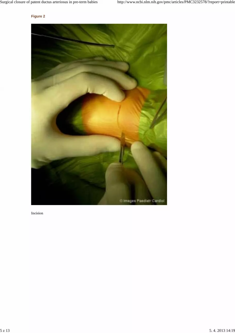
Figure 2
Incision
Surgical closure of patent ductus arteriosus in pre-term babies http://www.ncbi.nlm.nih.gov/pmc/articles/PMC3232578/?report=printable
5 z 13 5. 4. 2013 14:19

Figure 3
Left thoracotomy
Surgical closure of patent ductus arteriosus in pre-term babies http://www.ncbi.nlm.nih.gov/pmc/articles/PMC3232578/?report=printable
6 z 13 5. 4. 2013 14:19

Figure 4
Dissection of the ductus
Surgical closure of patent ductus arteriosus in pre-term babies http://www.ncbi.nlm.nih.gov/pmc/articles/PMC3232578/?report=printable
7 z 13 5. 4. 2013 14:19

Figure 5
Anatomy
Surgical closure of patent ductus arteriosus in pre-term babies http://www.ncbi.nlm.nih.gov/pmc/articles/PMC3232578/?report=printable
8 z 13 5. 4. 2013 14:19

Figure 6
Position of the clip
Surgical closure of patent ductus arteriosus in pre-term babies http://www.ncbi.nlm.nih.gov/pmc/articles/PMC3232578/?report=printable
9 z 13 5. 4. 2013 14:19
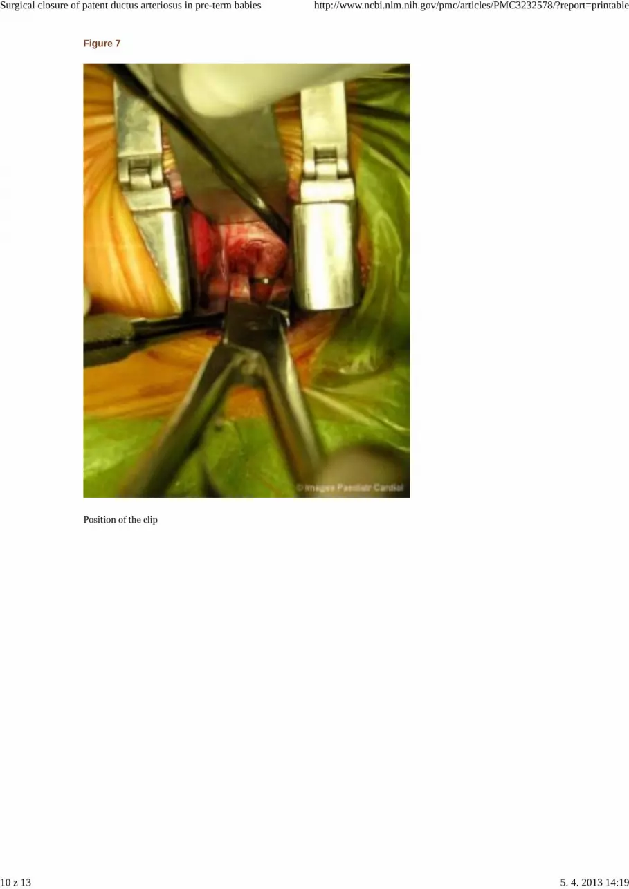
Figure 7
Position of the clip
Surgical closure of patent ductus arteriosus in pre-term babies http://www.ncbi.nlm.nih.gov/pmc/articles/PMC3232578/?report=printable
10 z 13 5. 4. 2013 14:19
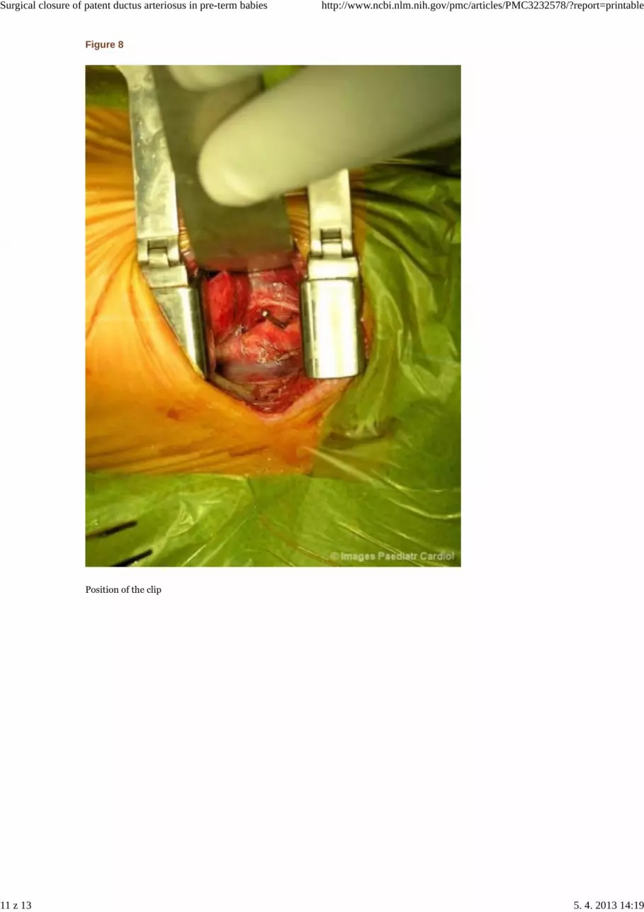
Figure 8
Position of the clip
Surgical closure of patent ductus arteriosus in pre-term babies http://www.ncbi.nlm.nih.gov/pmc/articles/PMC3232578/?report=printable
11 z 13 5. 4. 2013 14:19

Figure 10
Ligation
Surgical closure of patent ductus arteriosus in pre-term babies http://www.ncbi.nlm.nih.gov/pmc/articles/PMC3232578/?report=printable
12 z 13 5. 4. 2013 14:19

Figure 11
Ligation and division
Articles from Images in Paediatric Cardiology are provided here courtesy of Medknow Publications
Surgical closure of patent ductus arteriosus in pre-term babies http://www.ncbi.nlm.nih.gov/pmc/articles/PMC3232578/?report=printable
13 z 13 5. 4. 2013 14:19


