Embed Size (px)
Citation preview
Receptor for advanced glycation end products and its ligandhigh-mobility group box-1 mediate allergic airwaysensitization and airway inflammation
Md Ashik Ullah, M Pharm,a,b Zhixuan Loh, B Biomed Sci,b Wan Jun Gan, B Biomed Sci,b Vivian Zhang, PhD,b
Huan Yang, PhD,c Jian Hua Li, PhD,c Yasuhiko Yamamoto, PhD,d Ann Marie Schmidt, PhD,e Carol L. Armour, PhD,a
J. Margaret Hughes, PhD,a,f Simon Phipps, PhD,b* andMaria B. Sukkar, PhDa,g* Sydney and Brisbane, Australia, New York,
NY, and Kanazawa, Japan
Background: The receptor for advanced glycation endproducts (RAGE) shares common ligands and signalingpathways with TLR4, a key mediator of house dust mite(Dermatophagoides pteronyssinus) (HDM) sensitization. Wehypothesized that RAGE and its ligand high-mobility groupbox-1 (HMGB1) cooperate with TLR4 to mediate HDMsensitization.Objectives: To determine the requirement for HMGB1 andRAGE, and their relationship with TLR4, in airwaysensitization.Methods: TLR42/2, RAGE2/2, and RAGE-TLR42/2 micewere intranasally exposed to HDM or cockroach (Blatellagermanica) extracts, and features of allergic inflammationwere measured during the sensitization or challenge phase.Anti-HMGB1 antibody and the IL-1 receptor antagonistAnakinra were used to inhibit HMGB1 and the IL-1 receptor,respectively.Results: The magnitude of allergic airway inflammation inresponse to either HDM or cockroach sensitization and/orchallenge was significantly reduced in the absence of RAGE butnot further diminished in the absence of both RAGE and TLR4.HDM sensitization induced the release of HMGB1 from the
From the athe Woolcock Institute of Medical Research, Sydney Medical School,
University of Sydney; bthe Laboratory for Respiratory Neuroscience and Mucosal
Immunity, School of Biomedical Sciences, University of Queensland, Brisbane; cthe
Laboratory of Biomedical Science, Feinstein Institute for Medical Research, New
York; dthe Department of Biochemistry and Molecular Vascular Biology, Graduate
School of Medical Science, Kanazawa University; ethe Department of Pathology,
Langone Medical Centre, New York University; fthe Faculty of Pharmacy, University
of Sydney; and gthe School of Pharmacy, Graduate School of Health, University of
Technology, Sydney.
*These authors contributed equally to this work.
This work was funded by a Deputy Vice Chancellor Bridging Grant (University of
Sydney) and seed funding (University of Technology, Sydney) awarded to M.B.S.,
and an NHMRC Australia Project Grant (1023756) awarded to S.P.
Conflicts of interest: J. M. Hughes is employed by the National Health and Medical
Research Council (NHMRC) of Australia Grant Review Panel Membership 2013; has
received research support from the NHMRC of Australia (Project Grant APP 632830
2010-2012); and has received lecture fees from Learning Solutions, The University of
Sydney Early Career Researcher Biomedical Program 2011-2012. The rest of the
authors declare that they have no relevant conflicts of interest.
Received for publication August 2, 2013; revised November 23, 2013; accepted for pub-
lication December 3, 2013.
Corresponding author: Maria B. Sukkar, PhD, School of Pharmacy, Graduate School of
Health, The University of Technology, Sydney, Broadway, NSW, 2007, Australia.
E-mail: [email protected]. Or: Simon Phipps, PhD, School of Biomedical
Sciences, University of Queensland, St Lucia, QLD, 4072, Australia. E-mail:
0091-6749/$36.00
� 2014 American Academy of Allergy, Asthma & Immunology
http://dx.doi.org/10.1016/j.jaci.2013.12.1035
airway epithelium in a biphasic manner, which corresponded tothe sequential activation of TLR4 then RAGE. Release ofHMGB1 in response to cockroach sensitization also was RAGEdependent. Significantly, HMGB1 release occurred downstreamof TLR4-induced IL-1a, and upstream of IL-25 and IL-33production. Adoptive transfer of HDM-pulsedRAGE1/1dendritic cells to RAGE2/2 mice recapitulated theallergic responses after HDM challenge. Immunoneutralizationof HMGB1 attenuated HDM-induced allergic airwayinflammation.Conclusion: The HMGB1-RAGE axis mediates allergic airwaysensitization and airway inflammation. Activation of this axis inresponse to different allergens acts to amplify the allergicinflammatory response, which exposes it as an attractive targetfor therapeutic intervention. (J Allergy Clin Immunol2014;nnn:nnn-nnn.)
Key words: Asthma, allergic sensitization, epithelium, TLR4,RAGE, IL-1a, HMGB1, IL-25, IL-33, TSLP
Activation of pattern-recognition receptors (PRRs) at the airwaymucosa is fundamental to the process of allergic sensitization,the strongest identifiable risk factor for the development ofasthma.1 Genetic analyses are revealing that single nucleotidepolymorphisms in or near loci that encode for PRRs such asToll-like receptor (TLR) family members and downstreamsignaling molecules are strongly associated with the etiology ofasthma.1,2 We and other researchers have used experimentalmodels to show that the absence of TLR4 is protective againsthouse dust mite (Dermatophagoides pteronyssinus) (HDM)induced allergic airway inflammation.3-6 By using chimericmice, the laboratory of Hammad and Lambrecht has shown thatHDM-induced activation of TLR4 in airway epithelial cells(AEC), rather than immune cells, is sufficient to promote allergicsensitization via the release of innate cytokines such as IL-25,IL-33, and thymic stromal lymphopoietin, which are critical tothe development of type2 inflammation in asthma.3,7 Furthermore,they demonstrated that type 2 innate cytokines and chemokines areproduced downstream of the endogenous damage–associated mo-lecular patterns (DAMP), uric acid, and IL-1a, the latter of whichacts in an autocrine manner in AECs to amplify type 2 inflamma-tion7,8 However, it remains undetermined whether IL-1a actsdirectly upstream of type 2 innate cytokines or additional DAMPsand/or other mediators are involved in this cascade.
Although there is consensus that TLR4 is important in theairway response to HDM, it is increasingly evident that differentPRRs dominate in the response to different allergens.9 This callsfor greater understanding of the mechanisms through which the
1
J ALLERGY CLIN IMMUNOL
nnn 2014
2 ULLAH ET AL
Abbreviations used
AEC: A
irway epithelial cellBALF: B
ronchoalveolar lavage fluidCCL: C
hemokine ligandCR: C
ockroachDAMP: D
amage-associated molecular patternDC: D
endritic cellHMGB1: H
igh mobility group box-1HDM: H
ouse dust miteIL-1R: In
terleukin-1 receptorPRR: P
attern-recognition receptorRAGE: R
eceptor for advanced glycation end productsTLR4: T
oll-like receptor 4WT: W
ild typeactivation of one or more PRRs are amplified, integrated, orcoordinated in the immune response to allow strategic targeting ofmultiple PRRs or their common downstream pathways fortherapeutic intervention. We hypothesized that the receptor foradvanced glycation end products (RAGE) serves an integral rolein the airway response to inhaled allergens via its remarkablecapacity to interact with a diverse repertoire of DAMPs.10 RAGEshares common ligands with TLR4 and signals via intracellularadaptor proteins that also are used by TLR4.11 There is someevidence that RAGE and TLR4 cooperate as essential partnersto augment the inflammatory response.10,12-14 Recently, Miluti-novic et al15 demonstrated that RAGE-deficient mice areprotected against HDM-induced allergic airway inflammationin a mouse model of chronic asthma but did not investigate itsrole in the initiation of the allergic response (ie, sensitizationphase). Thus, whether RAGE is involved in the process of allergicsensitization or cooperates with TLR4 to amplify the immuneresponse during allergen sensitization is an important questionthat remains to be addressed. Because AECs respond to inhaledallergens through the release of DAMPs, it is conceivable thatRAGEmight also serve as a common mechanism for the recogni-tion of multiple DAMPs released downstream of PRR activa-tion.10 One such DAMP may be HMGB1, a well-characterizedRAGE ligand reported to be associated with lung function impair-ment and more severe disease in subjects with asthma.16-20 A rolefor HMGB1 in the effector phase of the allergic inflammatoryresponse has recently been demonstrated,19,21 but whether it isinvolved in the process of allergic sensitization is unknown.The objectives of this study were to investigate whether RAGEis involved in the process of allergic airway sensitization, whetherit collaborates with TLR4 in this response and whether its ligandHMGB1 is an effector molecule of allergic sensitization.
METHODSFor detailed methods including mice and reagents, experimental protocols
and procedures and statistical analysis, please see this article’s Methods
section in the Online Repository available at www.jacionline.org.
RESULTS
RAGE does not synergize with TLR4 to augment the
effector phase of allergic airway inflammationTo investigate whether TLR4 and RAGE cooperate in the
development of allergic airway inflammation, we performed anacute HDM model in wild type (WT), RAGE-deficient (2/2),
TLR42/2, and RAGE-TLR42/2 mice as per the study design(Fig 1, A). TLR42/2 mice were significantly protected againstall features of allergic inflammation, including eosinophilia andneutrophilia, chemokine and TH2 cytokine production, mucusproduction, increases in lung mast cell numbers, total serumIgE, and HDM-specific IgG1 (Fig 1, B-K). With the exceptionof neutrophilia and expression of the neutrophil-activechemokine CXCL1, RAGE2/2 mice were significantly protectedagainst all features of allergic inflammation measured. Impor-tantly, RAGE2/2 and TLR42/2 mice were similarly protected,and, with the exception of IL-5 production, RAGE-TLR42/2
mice were not afforded additional protection (Fig 1, B-K).The lack of additional protection against HDM-induced
inflammation in the RAGE-TLR42/2 mice may reflect thedominance of TLR4 in the response to this allergen.3-6 Thus,we next performed an acute model of cockroach (Blatellagermanica) (CR) induced allergic airway inflammation inwhich TLR4 plays a less important role22 (study design, sameas Fig 1, A). Interestingly, TLR42/2 mice were significantlyprotected against neutrophilia but not eosinophilia, whereasRAGE2/2 mice were protected against eosinophilia but notneutrophilia (Fig 2, A and B). Further, although both TLR42/2
andRAGE2/2micewere similarly protected against IL-5 produc-tion; TLR42/2 mice were not protected against increases in totalserum IgE and CR-specific IgG1 and mucus production, whereasRAGE2/2 mice were protected against all these aspects of theinflammatory response (Fig 2, C-F). Similar to the findingswith HDM, the absence of both RAGE and TLR4 did not furtherdiminish the inflammatory-immune response (Fig 2, A-F).
RAGE contributes to airway sensitization but does
not synergize with TLR4Next, we determined whether RAGE is involved, initially, in the
process of allergic sensitizationandwhether it cooperateswithTLR4in this response. We isolated lymph node cells 5 days after HDMsensitization in WT, TLR42/2, RAGE2/2, and RAGE-TLR42/2
mice to study TH2 cell priming ex vivo (study design, Fig 3, A).HDM-stimulated IL-13 production was significantly reduced inlymph node cells isolated from TLR42/2 and RAGE2/2 mice,with no further reduction in RAGE-TLR42/2 mice (Fig 3, B).
Because dendritic cells (DCs) play a critical role in allergicsensitization and adaptive immune responses to inhaledallergens,23 we determined whether protection against allergicsensitization in RAGE2/2 mice is due to a defect in DC function(study design, Fig 3, A). Compared with WT controls, RAGE2/2
mice had significantly reduced numbers of DCs in the lung anddraining lymph nodes at day 3 after HDM sensitization (Fig 3,C andD). In view of these results, we investigated whether recon-stitution of RAGE2/2 mice with HDM-pulsed RAGE1/1 DCs(ie, replacing the HDM exposure during sensitization) wassufficient to promote allergic airway inflammation after HDMchallenge (study design, Fig 4, A). Importantly, the magnitudeof allergic airway inflammation was the same after the adoptivetransfer of HDM-pulsed RAGE1/1 DCs, irrespective of whetherthe recipient mouse was RAGE2/2 or RAGE1/1 (Fig 4, B-G).
HMGB1 is secreted by AECs during allergen
sensitizationBecause we demonstrated that RAGE signaling in DCs is
important for the process of allergic sensitization and the later
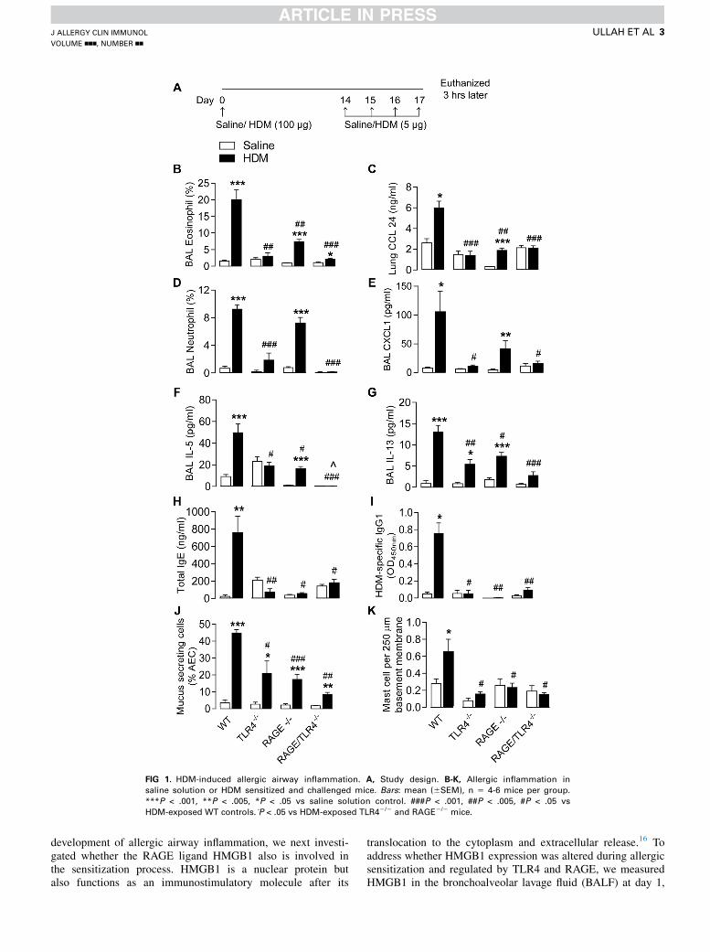
FIG 1. HDM-induced allergic airway inflammation. A, Study design. B-K, Allergic inflammation in
saline solution or HDM sensitized and challenged mice. Bars: mean (6SEM), n 5 4-6 mice per group.
***P < .001, **P < .005, *P < .05 vs saline solution control. ###P < .001, ##P < .005, #P < .05 vs
HDM-exposed WT controls. P̂ < .05 vs HDM-exposed TLR42/2 and RAGE2/2 mice.
J ALLERGY CLIN IMMUNOL
VOLUME nnn, NUMBER nn
ULLAH ET AL 3
development of allergic airway inflammation, we next investi-gated whether the RAGE ligand HMGB1 also is involved inthe sensitization process. HMGB1 is a nuclear protein butalso functions as an immunostimulatory molecule after its
translocation to the cytoplasm and extracellular release.16 Toaddress whether HMGB1 expression was altered during allergicsensitization and regulated by TLR4 and RAGE, we measuredHMGB1 in the bronchoalveolar lavage fluid (BALF) at day 1,
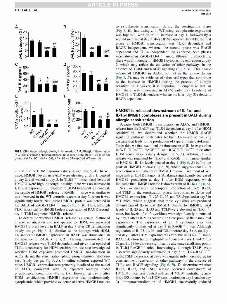
FIG 2. CR-induced allergic airway inflammation. A-F, Allergic inflammation
in CR sensitized and challenged mice. Bars: mean (6SEM), n5 5-6 mice per
group. ###P < .001, ##P < .005, #P < .05 vs CR-exposed WT controls.
J ALLERGY CLIN IMMUNOL
nnn 2014
4 ULLAH ET AL
2, and 3 after HDM exposure (study design, Fig 5, A). In WTmice, HMGB1 levels in BALF were elevated at day 1, peakedat day 2, and waned at day 3. In TLR42/2 mice, basal levels ofHMGB1 were high, although, notably, there was no increase inHMGB1 expression in response to HDM treatment. In contrast,the profile of HMGB1 release in RAGE2/2 mice was similar tothat observed in the WT controls, except at day 3, when it wassignificantly lower. Negligible HMGB1 protein was detected inthe BALF of RAGE-TLR42/2 mice (Fig 5, B). Thus, althoughTLR4 is critical for HMGB1 release, activation of RAGE second-ary to TLR4 augments HMGB1 release.
To determine whether HMGB1 release is a general feature ofairway sensitization and not specific to HDM, we measuredHMGB1 protein levels in BALF at day 3 after CR sensitization(study design, Fig 5, A). Similar to the findings with HDM,CR-induced HMGB1 expression in BALF was diminished inthe absence of RAGE (Fig 5, C). Having demonstrated thatHMGB1 release was TLR4 dependent and given that epithelialTLR4 is necessary for HDM sensitization, we next investigatedwhether HDM exposure promoted HMGB1 translocation inAECs during the sensitization phase using immunohistochem-istry (study design, Fig 5, A). In saline solution–exposed WTmice, HMGB1 expression was primarily localized to the nucleiof AECs, consistent with its expected location underphysiological conditions (Fig 5, D). However, at day 3 afterHDM sensitization, HMGB1 expression was predominantlycytoplasmic, which provided evidence of active HMGB1 nuclear
to cytoplasmic translocation during the sensitization phase(Fig 5, E). Interestingly, in WT mice, cytoplasmic expressionwas biphasic, with an initial increase at day 1, followed by asecond increase at day 3 after HDM exposure. Notably, the firstphase of HMGB1 translocation was TLR4 dependent andRAGE independent, whereas the second phase was RAGEdependent and TLR4 independent. As expected, both phaseswere absent in RAGE-TLR42/2 mice, although, unexpectedly,there was an increase in HMGB1 cytoplasmic expression at day2, which may reflect the activation of other pathways in theabsence of TLR4 and RAGE signaling (Fig 5, F). This phasicrelease of HMGB1 in AECs, but not in the airway lumen(Fig 5, B), may be evidence of other cell types that contributeto the increase in HMGB1 during the process of allergicsensitization. However, it is important to emphasize that, inboth the airway lumen and in AECs, early (day 1) release ofHMGB1 is TLR4 dependent, whereas its later (day 3) release isRAGE dependent.
HMGB1 is released downstream of IL-1a, andIL-1a-HMGB1 complexes are present in BALF during
allergic sensitizationBecause both HMGB1 translocation in AECs, and HMGB1
release into the BALF was TLR4 dependent at day 1 after HDMsensitization, we determined whether the HMGB1-RAGEsignaling pathway contributes to the TLR4–uric acid–IL-1acascade that leads to the production of type 2 innate cytokines.7
To do this, we first examined the time course of IL-1a expressionin WT, TLR42/2, RAGE2/2, and RAGE-TLR42/2 mice afterHDM sensitization (study design, Fig 5, A). Although IL-1arelease was regulated by TLR4 and RAGE in a manner similarto HMGB1, IL-1a levels peaked at day 1 (Fig 6, A) before thepeak of HMGB1 release (Fig 5, B), which suggests that IL-1aproduction was upstream of HMGB1 release. Treatment of WTmicewith an IL-1R antagonist (Anakinra) significantly decreasedHMGB1 production at day 1 after HDM exposure, whichindicated that HMGB1 release is downstream of IL-1a (Fig 6, A).
Next, we measured the temporal production of IL-25, IL-33,and TSLP in the sensitization phase. In contrast to IL-1a andHMGB1, expression of IL-25, IL-33, and TSLP peaked at day 3 inWT mice, which suggests that these cytokines are produceddownstream of IL-1a and HMGB1. Similar to HMGB1, basallevels of IL-25 and IL-33 and TSLP were elevated in TLR42/2
mice, but levels of all 3 cytokines were significantly attenuatedby day 3 after HDM exposure (the time point of their maximalexpression). The expression of all 3 cytokines also wassignificantly diminished at day 3 in RAGE2/2mice. Althoughregulation of IL-25, IL-33, and TSLP before day 3 (ie, on day 1and day 2 after HDM exposure) was variable in TLR42/2 mice,RAGE deletion had a negligible influence at days 1 and 2. IL-25 and IL-33 levels were significantly attenuated at all time pointsin TLR4-RAGE2/2 mice. Interestingly, although TSLP levelsalso were significantly attenuated at day 3 in TLR4-RAGE2/2
mice, TSLP expression at day 2 was significantly increased, againconsistent with activation of other pathways in the absence ofTLR4 and RAGE signaling (Fig 6, B). To determine whetherIL-25, IL-33, and TSLP release occurred downstream ofHMGB1, mice were treated with anti-HMGB1 neutralizing anti-body (30 minutes before HDM sensitization, on day 1, and on day2). Immunoneutralization of HMGB1 successfully reduced
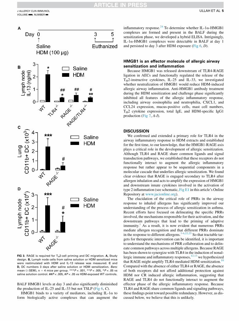
FIG 3. RAGE is required for TH2-cell priming and DC migration. A, Study
design. B, Lymph node cells from saline solution or HDM sensitized mice
were restimulated with HDM and IL-13 release was measured. C and
D, DC numbers 3 days after saline solution or HDM sensitization. Bars:mean (6SEM), n 5 4 mice per group. ***P < .001, **P < .005, *P < .05 vs
saline solution control. ##P < .005, #P < .05 vs HDM-exposed WT controls.
J ALLERGY CLIN IMMUNOL
VOLUME nnn, NUMBER nn
ULLAH ET AL 5
BALF HMGB1 levels at day 3 and also significantly diminishedthe production of IL-25 and IL-33 but not TSLP (Fig 6, C).
HMGB1 binds to a variety of mediators, including IL-1a toform biologically active complexes that can augment the
inflammatory response.24 To determine whether IL-1a-HMGB1complexes are formed and present in the BALF during thesensitization phase, we developed a hybrid ELISA. Intriguingly,IL-1a-HMGB1 complexes were detectable in BALF at day 1and persisted to day 3 after HDM exposure (Fig 6, D).
HMGB1 is an effector molecule of allergic airway
sensitization and inflammationBecause HMGB1 was released downstream of TLR4-RAGE
ligation in AECs and functionally regulated the release of theTH2-instructive cytokines, IL-25 and IL-33, we investigatedwhether neutralization of HMGB1 would reduce HDM-inducedallergic airway inflammation. Anti-HMGB1 antibody treatmentduring the HDM sensitization and challenge phase significantlyinhibited all features of the allergic inflammatory response,including airway eosinophilia and neutrophilia, CXCL1, andCCL24 expression, mucus-positive cells, mast cell numbers,TH2 cytokine expression, total IgE, and HDM-specific IgG1production (Fig 7, A-I).
DISCUSSIONWe confirmed and extended a primary role for TLR4 in the
airway inflammatory response to HDM extracts and establishedfor the first time, to our knowledge, that the HMGB1-RAGE axisplays a critical role in the development of allergic sensitization.Although TLR4 and RAGE share common ligands and signaltransduction pathways, we established that these receptors do notfunctionally interact to augment the allergic inflammatoryresponse but rather appear to be sequential components in amolecular cascade that underlies allergic sensitization. We foundclear evidence that RAGE is engaged secondary to TLR4 afterallergen inhalation and acts to amplify the expression of HMGB1and downstream innate cytokines involved in the activation oftype 2 inflammation (see schematic, Fig E1 in this article’s OnlineRepository at www.jacionline.org).
The elucidation of the critical role of PRRs in the airwayresponse to inhaled allergens has significantly improved ourunderstanding of the process of allergic sensitization in asthma.Recent efforts have focused on delineating the specific PRRsinvolved, the mechanisms responsible for their activation, and thedownstream pathways that lead to the priming of adaptiveimmunity.1 As a result, it is now evident that numerous PRRsmediate allergen recognition and that different PRRs dominatein the response to different allergens.3-6,22,25 So that tractable tar-gets for therapeutic intervention can be identified, it is importantto understand the mechanisms of PRR collaboration and to delin-eate common pathways across multiple allergens. Because RAGEhas been shown to synergize with TLR4 in the induction of nonal-lergic immune and inflammatory responses,12,13 we hypothesizedthat RAGE might amplify TLR4-mediated HDM sensitization.10
Compared with the absence of either TLR4 or RAGE, the absenceof both receptors did not afford additional protection againstHDM nor CR induced allergic inflammation, suggesting thatRAGE and TLR4 do not functionally interact to augment theeffector phase of the allergic inflammatory response. BecauseTLR4 and RAGE share common ligands and signaling pathways,these findings point toward possible redundancy. However, as dis-cussed below, we believe that this is unlikely.
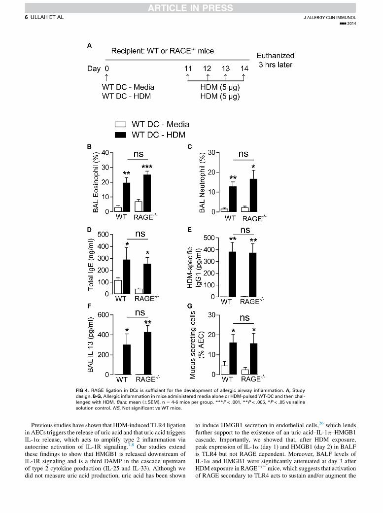
FIG 4. RAGE ligation in DCs is sufficient for the development of allergic airway inflammation. A, Study
design. B-G, Allergic inflammation in mice administered media alone or HDM-pulsed WT-DC and then chal-
lenged with HDM. Bars: mean (6SEM), n 5 4-6 mice per group. ***P < .001, **P < .005, *P < .05 vs saline
solution control. NS, Not significant vs WT mice.
J ALLERGY CLIN IMMUNOL
nnn 2014
6 ULLAH ET AL
Previous studies have shown that HDM-induced TLR4 ligationin AECs triggers the release of uric acid and that uric acid triggersIL-1a release, which acts to amplify type 2 inflammation viaautocrine activation of IL-1R signaling.7,8 Our studies extendthese findings to show that HMGB1 is released downstream ofIL-1R signaling and is a third DAMP in the cascade upstreamof type 2 cytokine production (IL-25 and IL-33). Although wedid not measure uric acid production, uric acid has been shown
to induce HMGB1 secretion in endothelial cells,26 which lendsfurther support to the existence of an uric acid–IL-1a–HMGB1cascade. Importantly, we showed that, after HDM exposure,peak expression of IL-1a (day 1) and HMGB1 (day 2) in BALFis TLR4 but not RAGE dependent. Moreover, BALF levels ofIL-1a and HMGB1 were significantly attenuated at day 3 afterHDM exposure in RAGE2/2mice, which suggests that activationof RAGE secondary to TLR4 acts to sustain and/or augment the
FIG 5. Expression and regulation of HMGB1 in AECs during allergen sensitization. A, Study design. HMGB1
expression during HDM sensitization (B) and day 3 after CR sensitization (C). D and E, Lung HMGB1
immunostaining (3400 magnification). F, HMGB1 cytoplasmic expression in AECs. Data: mean (6SEM),
n 5 3-9 mice per group. *P < .05 vs time 0. ###P < .001, ##P < .005, #P < .05 vs WT controls at respective
time points.
J ALLERGY CLIN IMMUNOL
VOLUME nnn, NUMBER nn
ULLAH ET AL 7
expression of these early response DAMPs. These data, togetherwith evidence that the first and second wave of HMGB1 cyto-plasmic expression in AECs appears to be regulated by thesequential activation of TLR4 and RAGE, respectively, stronglysupports the notion that TLR4 and RAGE are not redundant butrather act at critical but sequential points in the process of allergicsensitization, with interception of either point being sufficient toretard the progression of the allergic response.
Our finding that RAGE is engaged secondary to TLR4 duringallergen sensitization is significant for 2 reasons. First, it suggeststhat RAGE serves as a common mechanism downstream ofvarious allergen-activated PRR pathways. This point is bestillustrated by our demonstration that HMGB1 release occurredin response to 2 different extracts, HDM and CR, before theapparent onset of RAGE signaling. Because HDM and CR are
‘‘sensed’’ by distinct PRRs, primarily TLR4 and PAR-2,22,27
respectively, and HMGB1 is a well-characterized RAGEligand,14,16 it is conceivable that HMGB1 secretion downstreamof initial ‘‘sensing’’ PRRs converges on RAGE signaling to prop-agate the allergic inflammatory response to multiple allergens.Second, we observed that RAGE activation regulates HMGB1secretion and sustains its expression at the level of the airwayepithelium. Since we showed that HMGB1 mediates the releaseof the TH2-instructive cytokines IL-25 and IL-33, it appearsthat RAGE, by mediating the recognition and amplification ofDAMP signals required for the development of type 2inflammation, also is an important mechanism for theamplification of the allergic inflammatory response. Interestingly,although we found that RAGE mediated the expression of all 3type 2 cytokines studied (IL-25, IL-33, and TSLP), this was not
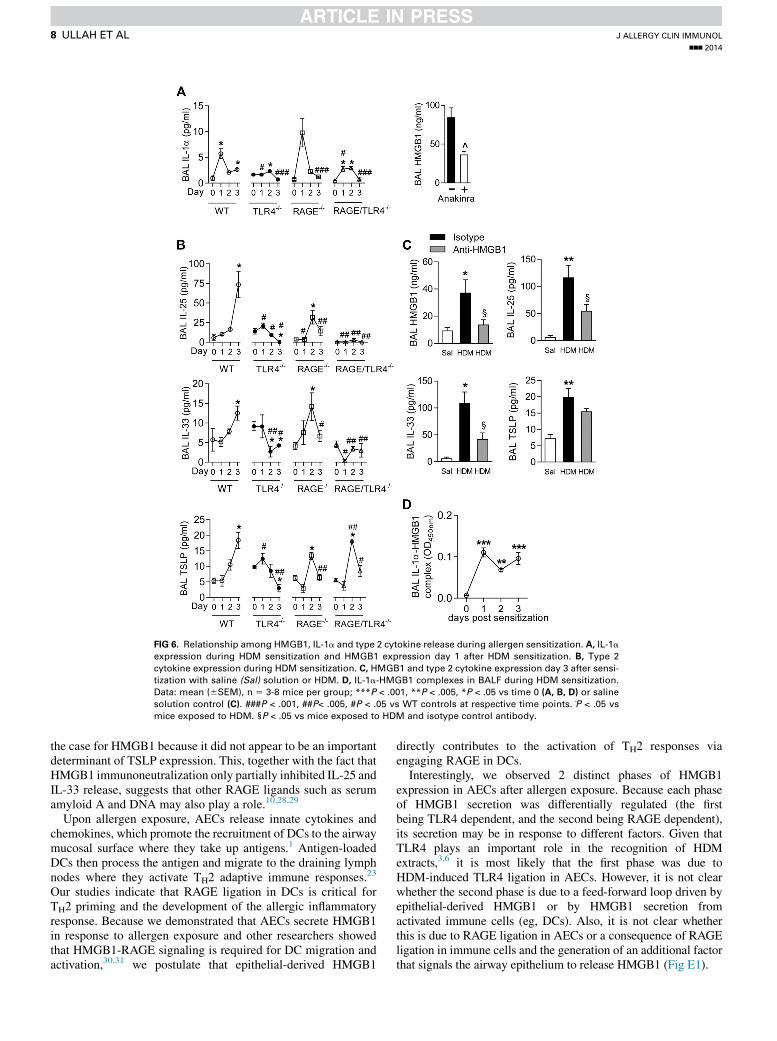
FIG 6. Relationship among HMGB1, IL-1a and type 2 cytokine release during allergen sensitization. A, IL-1a
expression during HDM sensitization and HMGB1 expression day 1 after HDM sensitization. B, Type 2
cytokine expression during HDM sensitization. C, HMGB1 and type 2 cytokine expression day 3 after sensi-
tization with saline (Sal) solution or HDM. D, IL-1a-HMGB1 complexes in BALF during HDM sensitization.
Data: mean (6SEM), n 5 3-8 mice per group; ***P < .001, **P < .005, *P < .05 vs time 0 (A, B, D) or saline
solution control (C). ###P < .001, ##P< .005, #P < .05 vs WT controls at respective time points. P̂ < .05 vs
mice exposed to HDM. §P < .05 vs mice exposed to HDM and isotype control antibody.
J ALLERGY CLIN IMMUNOL
nnn 2014
8 ULLAH ET AL
the case for HMGB1 because it did not appear to be an importantdeterminant of TSLP expression. This, together with the fact thatHMGB1 immunoneutralization only partially inhibited IL-25 andIL-33 release, suggests that other RAGE ligands such as serumamyloid A and DNA may also play a role.10,28,29
Upon allergen exposure, AECs release innate cytokines andchemokines, which promote the recruitment of DCs to the airwaymucosal surface where they take up antigens.1 Antigen-loadedDCs then process the antigen and migrate to the draining lymphnodes where they activate TH2 adaptive immune responses.23
Our studies indicate that RAGE ligation in DCs is critical forTH2 priming and the development of the allergic inflammatoryresponse. Because we demonstrated that AECs secrete HMGB1in response to allergen exposure and other researchers showedthat HMGB1-RAGE signaling is required for DC migration andactivation,30,31 we postulate that epithelial-derived HMGB1
directly contributes to the activation of TH2 responses viaengaging RAGE in DCs.
Interestingly, we observed 2 distinct phases of HMGB1expression in AECs after allergen exposure. Because each phaseof HMGB1 secretion was differentially regulated (the firstbeing TLR4 dependent, and the second being RAGE dependent),its secretion may be in response to different factors. Given thatTLR4 plays an important role in the recognition of HDMextracts,3,6 it is most likely that the first phase was due toHDM-induced TLR4 ligation in AECs. However, it is not clearwhether the second phase is due to a feed-forward loop driven byepithelial-derived HMGB1 or by HMGB1 secretion fromactivated immune cells (eg, DCs). Also, it is not clear whetherthis is due to RAGE ligation in AECs or a consequence of RAGEligation in immune cells and the generation of an additional factorthat signals the airway epithelium to release HMGB1 (Fig E1).
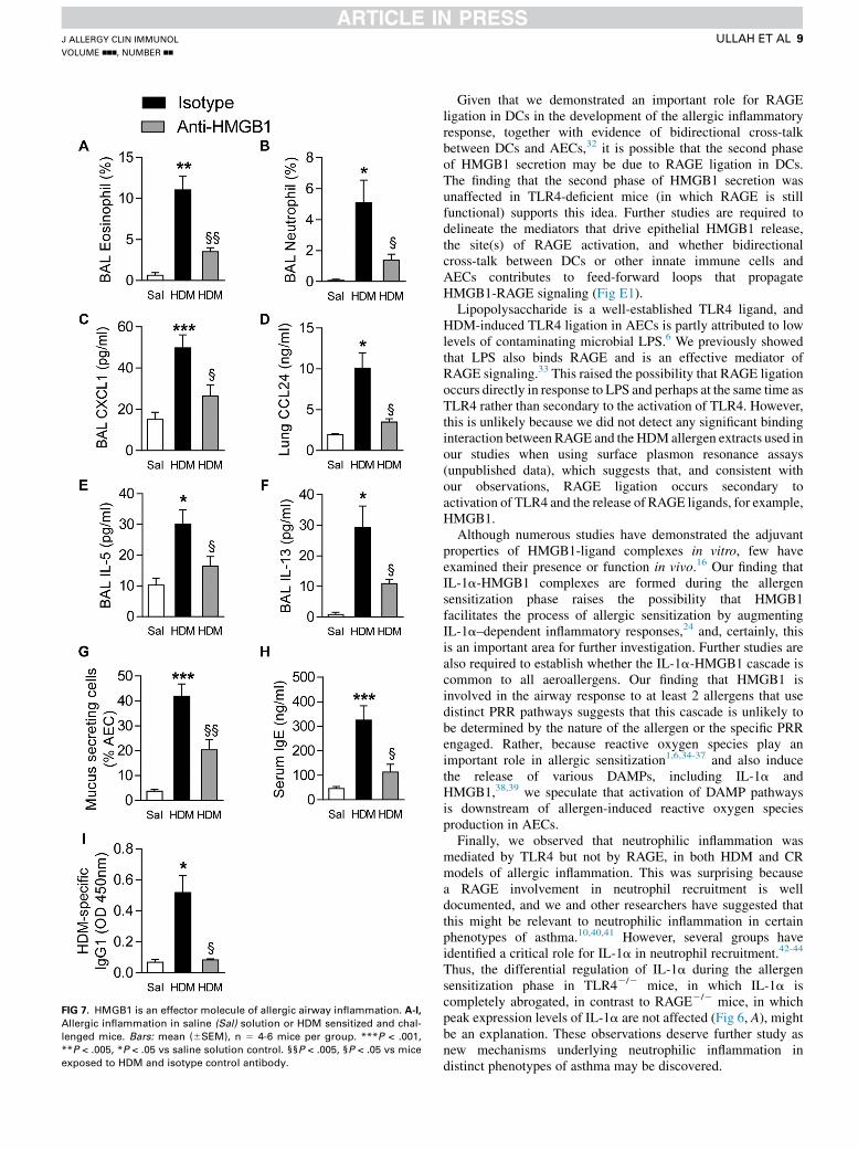
FIG 7. HMGB1 is an effector molecule of allergic airway inflammation. A-I,
Allergic inflammation in saline (Sal) solution or HDM sensitized and chal-
lenged mice. Bars: mean (6SEM), n 5 4-6 mice per group. ***P < .001,
**P < .005, *P < .05 vs saline solution control. §§P < .005, §P < .05 vs mice
exposed to HDM and isotype control antibody.
J ALLERGY CLIN IMMUNOL
VOLUME nnn, NUMBER nn
ULLAH ET AL 9
Given that we demonstrated an important role for RAGEligation in DCs in the development of the allergic inflammatoryresponse, together with evidence of bidirectional cross-talkbetween DCs and AECs,32 it is possible that the second phaseof HMGB1 secretion may be due to RAGE ligation in DCs.The finding that the second phase of HMGB1 secretion wasunaffected in TLR4-deficient mice (in which RAGE is stillfunctional) supports this idea. Further studies are required todelineate the mediators that drive epithelial HMGB1 release,the site(s) of RAGE activation, and whether bidirectionalcross-talk between DCs or other innate immune cells andAECs contributes to feed-forward loops that propagateHMGB1-RAGE signaling (Fig E1).
Lipopolysaccharide is a well-established TLR4 ligand, andHDM-induced TLR4 ligation in AECs is partly attributed to lowlevels of contaminating microbial LPS.6 We previously showedthat LPS also binds RAGE and is an effective mediator ofRAGE signaling.33 This raised the possibility that RAGE ligationoccurs directly in response to LPS and perhaps at the same time asTLR4 rather than secondary to the activation of TLR4. However,this is unlikely because we did not detect any significant bindinginteraction between RAGE and the HDM allergen extracts used inour studies when using surface plasmon resonance assays(unpublished data), which suggests that, and consistent withour observations, RAGE ligation occurs secondary toactivation of TLR4 and the release of RAGE ligands, for example,HMGB1.
Although numerous studies have demonstrated the adjuvantproperties of HMGB1-ligand complexes in vitro, few haveexamined their presence or function in vivo.16 Our finding thatIL-1a-HMGB1 complexes are formed during the allergensensitization phase raises the possibility that HMGB1facilitates the process of allergic sensitization by augmentingIL-1a–dependent inflammatory responses,24 and, certainly, thisis an important area for further investigation. Further studies arealso required to establish whether the IL-1a-HMGB1 cascade iscommon to all aeroallergens. Our finding that HMGB1 isinvolved in the airway response to at least 2 allergens that usedistinct PRR pathways suggests that this cascade is unlikely tobe determined by the nature of the allergen or the specific PRRengaged. Rather, because reactive oxygen species play animportant role in allergic sensitization1,6,34-37 and also inducethe release of various DAMPs, including IL-1a andHMGB1,38,39 we speculate that activation of DAMP pathwaysis downstream of allergen-induced reactive oxygen speciesproduction in AECs.
Finally, we observed that neutrophilic inflammation wasmediated by TLR4 but not by RAGE, in both HDM and CRmodels of allergic inflammation. This was surprising becausea RAGE involvement in neutrophil recruitment is welldocumented, and we and other researchers have suggested thatthis might be relevant to neutrophilic inflammation in certainphenotypes of asthma.10,40,41 However, several groups haveidentified a critical role for IL-1a in neutrophil recruitment.42-44
Thus, the differential regulation of IL-1a during the allergensensitization phase in TLR42/2 mice, in which IL-1a iscompletely abrogated, in contrast to RAGE2/2 mice, in whichpeak expression levels of IL-1a are not affected (Fig 6, A), mightbe an explanation. These observations deserve further study asnew mechanisms underlying neutrophilic inflammation indistinct phenotypes of asthma may be discovered.
J ALLERGY CLIN IMMUNOL
nnn 2014
10 ULLAH ET AL
In conclusion, we uncovered previously unknown roles forRAGE and its ligand HMGB1 in allergic airway sensitization andthe progression of the allergic airway inflammatory response.Combined with previous observations that inhibition of HMGB1during allergen challenge (but not during sensitization) signifi-cantly inhibits allergic airway inflammation in an ovalbuminmouse model of asthma,19,21 there is growing evidence that inhi-bition of the HMGB1-RAGE axis may have therapeutic benefit inasthma, both in primary prevention and in established disease.45
We thank Associate ProfessorMatthew J. Sweet, PhD, for providing TLR4-
deficient mice for use in these studies. We thank Professor Kevin J. Tracey,
MD, for his advice and support, and we also thank Professor Dirkje S. Postma,
MD, PhD, for her review of this manuscript.
Key messages
d Allergen-dependent activation of TLR4 induces secretionof the RAGE ligand HMGB1, a danger-associatedmolecular pattern critical to the development of allergicinflammation.
d HMGB1 release and RAGE activation is upstream ofIL-25 and IL-33, and contributes to allergic sensitization.
d Activation of the HMGB1-RAGE pathway occurs inresponse to more than 1 allergen, which suggests that itmay be a useful therapeutic target.
REFERENCES
1. Lambrecht BN, Hammad H. The airway epithelium in asthma. Nat Med 2012;18:
684-92.
2. Reijmerink NE, Bottema RWB, Kerkhof M, Gerritsen J, Stelma FF, Thijs C, et al.
TLR-related pathway analysis: novel gene–gene interactions in the development of
asthma and atopy. Allergy 2010;65:199-207.
3. Hammad H, Chieppa M, Perros F, Willart MA, Germain RN, Lambrecht BN.
House dust mite allergen induces asthma via Toll-like receptor 4 triggering of
airway structural cells. Nat Med 2009;15:410-6.
4. Trompette A, Divanovic S, Visintin A, Blanchard C, Hegde RS, Madan R, et al.
Allergenicity resulting from functional mimicry of a Toll-like receptor complex
protein. Nature 2009;457:585-8.
5. Phipps S, Lam CE, Kaiko GE, Foo SY, Collison A, Mattes J, et al. Toll/IL-1
signaling is critical for house dust mite-specific Th1 and Th17 responses.
Am J Respir Crit Care Med 2009;179:883-93.
6. Ryu J-H, Yoo J-Y, Kim M-J, Hwang S-G, Ahn KC, Ryu J-C, et al. Distinct
TLR-mediated pathways regulate house dust mite–induced allergic disease in
the upper and lower airways. J Allergy Clin Immunol 2013;131:549-61.
7. Willart MAM, Deswarte K, Pouliot P, Braun H, Beyaert R, Lambrecht BN, et al.
Interleukin-1a controls allergic sensitization to inhaled house dust mite via the
epithelial release of GM-CSF and IL-33. J Exp Med 2012;209:1505-17.
8. Kool M, Willart Monique AM, van Nimwegen M, Bergen I, Pouliot P, Virchow JC,
et al. An unexpected role for uric acid as an inducer of T helper 2 cell immunity to
inhaled antigens and inflammatory mediator of allergic asthma. Immunity 2011;34:
527-40.
9. Wills-Karp M. Allergen-specific pattern recognition receptor pathways. Curr Opin
Immunol 2010;22:777-82.
10. Sukkar M, Ullah M, Gan WJ, Wark PAB, Chung KF, Hughes JM, et al. RAGE:
a new frontier in chronic airways disease. Br J Pharmacol 2012;167:1161-76.
11. Sakaguchi M, Murata H, Yamamoto K-I, Ono T, Sakaguchi Y, Motoyama A, et al.
TIRAP, an adaptor protein for TLR2/4, transduces a signal from RAGE
phosphorylated upon ligand binding. PLoS One 2011;6:e23132.
12. Qin Y-H, Dai S-M, Tang G-S, Zhang J, Ren D, Wang Z-W, et al.
HMGB1 Enhances the proinflammatory activity of lipopolysaccharide by
promoting the phosphorylation of MAPK p38 through receptor for advanced
glycation end products. J Immunol 2009;183:6244-50.
13. Rojas A, Delgado-L�opez F, Gonz�alez I, P�erez-Castro R, Romero J, Rojas I. The
receptor for advanced glycation end-products: a complex signaling scenario for a
promiscuous receptor. Cell Signal 2013;25:609-14.
14. Rouhiainen A, Kuja-Panula J, Tumova S, Rauvala H. RAGE-mediated cell
signaling. In: Heizmann CW, editor. Calcium-binding proteins and RAGE: From
Structural Basics to Clinical Applications, Methods in Molecular Biology. New
York: Humana Press; 2013. Vol 963 p. 239-63.
15. Milutinovic PS, Alcorn JF, Englert JM, Crum LT, Oury TD. The receptor for
advanced glycation end products is a central mediator of asthma pathogenesis.
Am J Pathol 2012;181:1215-25.
16. Yanai H, Ban T, Taniguchi T. High-mobility group box family of proteins: ligand
and sensor for innate immunity. Trends Immunol 2012;33:633-40.
17. Watanabe T, Asai K, Fujimoto H, Tanaka H, Kanazawa H, Hirata K.
Increased levels of HMGB-1 and endogenous secretory RAGE in induced sputum
from asthmatic patients. Respir Med 2011;105:519-25.
18. Hou C, Zhao H, Liu L, Li W, Zhou X, Lv Y, et al. High mobility group protein B1
(HMGB1) in asthma: comparison of patients with chronic obstructive pulmonary
disease and healthy controls. Mol Med 2011;17:807-15.
19. Shim EJ, Chun E, Lee HS, Bang BR, Kim TW, Cho SH, et al. The role of
high-mobility group box-1 (HMGB1) in the pathogenesis of asthma. Clin Exp
Allergy 2012;42:958-65.
20. Zhou Y, Jiang Y-Q, Wang W-X, Zhou Z-X, Wang Y-G, Yang L, et al. HMGB1 and
RAGE levels in induced sputum correlate with asthma severity and neutrophil
percentage. Human Immunol 2012;73:1171-4.
21. Lee C-C, Lai Y-T, Chang H-T, Liao J-W, Shyu W-C, Li C-Y, et al. Inhibition of
high-mobility group box 1 in lung reduced airway inflammation and remodeling
in a mouse model of chronic asthma. Biochem Pharmacol 2013;86:940-9.
22. Arizmendi NG, Abel M, Mihara K, Davidson C, Polley D, Nadeem A, et al.
Mucosal allergic sensitization to cockroach allergens is dependent on proteinase
activity and proteinase-activated receptor-2 activation. J Immunol 2011;186:
3164-72.
23. Plantinga M, Guilliams M, Vanheerswynghels M, Deswarte K, Branco-Madeira F,
Toussaint W, et al. Conventional and monocyte-derived CD11b1 dendritic cells
initiate and maintain T helper 2 cell-mediated immunity to house dust mite
allergen. Immunity 2013;38:322-35.
24. Wahamaa H, Schierbeck H, Hreggvidsdottir H, Palmblad K, Aveberger A-C,
Andersson U, et al. High mobility group box protein 1 in complex with
lipopolysaccharide or IL-1 promotes an increased inflammatory phenotype in
synovial fibroblasts. Arthritis Res Ther 2011;13:R136.
25. Wilson RH, Maruoka S, Whitehead GS, Foley JF, Flake GP, Sever ML, et al. The
Toll-like receptor 5 ligand flagellin promotes asthma by priming allergic responses
to indoor allergens. Nat Med 2012;18:1705-10.
26. Rabadi MM, Kuo M-C, Ghaly T, Rabadi SM, Weber M, Goligorsky MS, et al.
Interaction between uric acid and HMGB1 translocation and release from
endothelial cells. Am J Physiol Renal Physiol 2012;302:F730-41.
27. Page K, Ledford J, Zhou P, Dienger K, Wills-Karp M. Mucosal sensitization
toGerman cockroach involves protease-activated receptor-2. Respir Res 2010;11:62.
28. Ather JL, Ckless K, Martin R, Foley KL, Suratt BT, Boyson JE, et al. Serum
amyloid A activates the NLRP3 inflammasome and promotes Th17 allergic asthma
in mice. J Immunol 2011;187:64-73.
29. Marichal T, Ohata K, Bedoret D, Mesnil C, Sabatel C, Kobiyama K, et al. DNA
released from dying host cells mediates aluminum adjuvant activity. Nat Med
2011;17:996-1002.
30. Yang D, Chen Q, Yang H, Tracey KJ, Bustin M, Oppenheim JJ. High mobility
group box-1 protein induces the migration and activation of human dendritic cells
and acts as an alarmin. J Leuk Biol 2007;81:59-66.
31. Dumitriu IE, Bianchi ME, Bacci M, Manfredi AA, Rovere-Querini P. The secretion
of HMGB1 is required for the migration of maturing dendritic cells. J Leukoc Biol
2007;81:84-91.
32. Strickland DH, Upham JW, Holt PG. Epithelial–dendritic cell interactions in
allergic disorders. Curr Opin Immunol 2010;22:789-94.
33. Yamamoto Y, Harashima A, Saito H, Tsuneyama K, Munesue S, Motoyoshi S,
et al. Septic shock is associated with receptor for advanced glycation end products
ligation of LPS. J Immunol 2011;186:3248-57.
34. Rangasamy T, Guo J, Mitzner WA, Roman J, Singh A, Fryer AD, et al. Disruption
of Nrf2 enhances susceptibility to severe airway inflammation and asthma in mice.
J Exp Med 2005;202:47-59.
35. Sevin CM, Newcomb DC, Toki S, Han W, Sherrill TP, Boswell MG, et al.
Deficiency of gp91phox inhibits allergic airway inflammation. Am J Respir Cell
Mol Biol 2013;49:396-402.
36. Boldogh I, Bacsi A, Choudhury BK, Dharajiya N, Alam R, Hazra TK, et al. ROS
generated by pollen NADPH oxidase provide a signal that augments
antigen-induced allergic airway inflammation. J Clin Invest 2005;115:2169-79.
37. Shalaby KH, Allard-Coutu A, O’Sullivan MJ, Nakada E, Qureshi ST, Day BJ, et al.
Inhaled birch pollen extract induces airway hyperresponsiveness via oxidative
stress but independently of pollen-intrinsic NADPH oxidase activity, or the
TLR4-TRIF pathway. J Immunol 2013;191:922-33.

J ALLERGY CLIN IMMUNOL
VOLUME nnn, NUMBER nn
ULLAH ET AL 11
38. McCarthy DA, Ranganathan A, Subbaram S, Flaherty NL, Patel N, Trebak M, et al.
Redox-control of the alarmin, interleukin-1a. Redox Biol 2013;1:218-25.
39. Tang D, Kang R, Zeh HJ, Lotze MT. HMGB1, Oxidative stress, and disease.
Antioxid Redox Signal 2011;14:1315-35.
40. Sukkar MB, Wood LG, Tooze M, Simpson JL, McDonald VM, Gibson PG, et al.
Soluble RAGE is deficient in neutrophilic asthma and chronic obstructive
pulmonary disease. Eur Respir J 2012;39:721-9.
41. Iwamoto H, Gao J, Koskela J, Kinnula V, Kobayashi H, Laitinen T, et al.
Differences in plasma and sputum biomarkers between COPD and
COPD-asthma overlap. Eur Respir J E-pub June 21, 2013. doi: 10.1183/
09031936.00024313.
42. Chen C-J, Kono H, Golenbock D, Reed G, Akira S, Rock KL. Identification of a
key pathway required for the sterile inflammatory response triggered by dying
cells. Nat Med 2007;13:851-6.
43. Lee PY, Kumagai Y, Xu Y, Li Y, Barker T, Liu C, et al. IL-1a modulates neutrophil
recruitment in chronic inflammation induced by hydrocarbon oil. J Immunol 2011;
186:1747-54.
44. Barry KC, Fontana MF, Portman JL, Dugan AS, Vance RE. IL-1a signaling
initiates the inflammatory response to virulent legionella pneumophila in vivo.
J Immunol 2013;190:6329-39.
45. Holt PG, Sly PD. Viral infections and atopy in asthma pathogenesis: new rationales
for asthma prevention and treatment. Nat Med 2012;18:726-35.
J ALLERGY CLIN IMMUNOL
nnn 2014
11.e1 ULLAH ET AL
METHODS
MiceAll mice strains were on C57Bl/6 background, and all studies were
performed in 8- to 12-week-old males. WT control mice were purchased from
the University of Queensland Biological Resources (Brisbane, Australia).
TLR42/2micewere provided byMatthew J. Sweet, PhD, (Institute forMolec-
ular Bioscience, University of Queensland, Australia), and RAGE2/2 mice
were provided by Ann Marie Schmidt, PhD (NYU Langone Medical Centre
and School of Medicine, NY). TLR42/2 mice were cross-bred with
RAGE2/2 mice to generate RAGE-TLR42/2 mice. All experimental proce-
dures and protocols were approved by the animal care and ethics committees
of the University of Queensland (Brisbane, Australia).
Allergen extractsHDM and CR extracts were purchased from Greer Laboratories, Lenoir,
NC. Extracts were prepared from HDMs and CRs that were grown under
controlled conditions. Extracts were prepared from the crushed bodies of
whole HDM and from CRs that were defatted, powdered, and dried. In each
case, the raw material was subjected to bilevel extraction in ammonium
bicarbonate solution and then dialyzed against distilled water. The endotoxin
level for HDM and CR extracts used were 3.5 EU and 395 EU per 100 mg
protein, respectively. Endotoxin concentration for HDM extracts was supplied
by the manufacturer, whereas endotoxin concentration for CR extracts was
determined by using Endosafe-PTS Limulus amebocyte lysate (Charles River
Laboratory, Charleston, SC) at the Protein Expression Facility (Australian
Institute of Bioengineering and Nanotechnology, The University of
Queensland).
Induction of allergic sensitization and airway
inflammationOn day 0, the mice were anesthetized with isoflurane and sensitized via
nasal instillation with either saline solution or 100 mg of HDM or CR extract.
At day 14, 15, 16, and 17, micewere challenged intranasally with either saline
solution or 5 mg of HDM or CR, and were euthanized 3 hours after the last
challenge (Fig 1, A). In experiments in which only the sensitization phase was
studied, themicewere euthanized at different time points from 1 to 5 days after
allergen sensitization (Figs 3, A and 5, A). In studies that examined the role of
HMGB1, 50mg of HMGB1 neutralizing antibody (clone 2G7, a kind gift from
Kevin J. Tracey, MD, Feinstein Institute of Medical Research, New York, NY)
was administered intranasally during the sensitization phase (30 minutes
before HDMadministration and on days 1 and 2) and/orHDMchallenge phase
(30 minutes before HDM challenge on days 14, 15, and 16). To study the role
of IL-1a signaling, 1 mg of an IL-1 receptor antagonist, Anakinra (Amgen,
Sydney, Australia, Pty Ltd; a kind gift from Mark R. Hutchinson, PhD, Uni-
versity of Adelaide, Australia) was administered intranasally 0.5, 6, and 12
hours after HDM sensitization.
DC generation and adoptive transfer studiesBone-marrow–derived DCs were generated from WT naive mice by
following the method of Lutz et alE1 with minor modification. Briefly, femurs
and tibias of WT mice were dissected, and bone marrow was flushed out with
RPMI-1640 growth medium (which contained 1 mM sodium pyruvate, 10%
FBS, 2 mM L-glutamine, 20 mMHEPES, 100 U/mL penicillin/streptomycin,
and 50 mM 2-mercaptoethanol). After red blood cell lysis, the cells were
cultured in growth media supplemented with 20 ng/mL of GM-CSF (R &
D Systems, Minneapolis, Minn) for 8 days (media was replenished on day 4
of culture). At the end of the culture period, nonadherent cells were harvested,
and CD11c positive selection was performed by using biotin-conjugated anti-
mouse CD11c, followed by streptavidin-conjugated magnetic beads. The
resulting CD11c1CD11b1 bone-marrow–derived DC population was 85%-
90% pure. The cells then were either left in media alone or pulsed with
HDM extract (100 mg/mL) for 24 hours, before they were harvested and
washed twice with PBS; 1 3 106 unstimulated or HDM treated bone-
marrow–derived DCs then were administered via the intranasal route to WT
or RAGE2/2 mice at day 0. At day 11, 12, 13, and 14, mice were challenged
with 5 mg of HDM and euthanized 3 hours after the last challenge (Fig 4, A).
This protocol did not induce significant lung inflammation in mice that
received unstimulated bone-marrow–derived DCs.
Analysis of allergic airway inflammationThe lungs were lavaged with 600mL of ice cold PBS, and the recovery was
consistently between 400 and 500 mL. To determine differential cell counts,
BAL fluids were spun down to separate cells. Red blood cells were then lysed,
and total blood cell counts were performed. Cytospins were prepared and
stained with May-Grunwald Giemsa solution (Sigma-Aldrich, St Louis, Mo).
A total of 400 cells were counted to enumerate differential cell counts. BALF
and/or cell lysates prepared from lung homogenates were assayed for cytokine
expression by ELISA. To prepare lung homogenates, lung lobes were
homogenized in radioimmunoprecipitation assay buffer (Sigma-Aldrich) by
using a Tissue Tearor homogenizer (Model 985-370; BioSpec Products
Bartlesville, Okla). Lung lobes were processed, and sections were stained to
enumerate mucus-producing goblet cells or mast cells. To examine TH2-cell
priming during the sensitization phase, draining lymph nodes were excised
5 days after HDM sensitization, and a single-cell suspension was prepared
and cultured with HDM 20 mg/mL for 4 days, as previously described.E2
Histologic analysis of mucus-secreting cells and
mast cellsParaffin-embedded lung biopsy specimens were stained with periodic
acid–Schiff reagents to enumerate mucus-secreting cells. The number of
mucus-secreting cells among the AECs were counted (4-5 airways per mouse)
and expressed as the percentage of total AECs in the respective airways. For
mast cell analysis, lung biopsy specimens were stained with toludine-blue
reagent, and the number of mast cells around the airways were counted and
described as the number of cells per 250 mm of airway epithelium basement
membrane.
Immunohistochemical detection of HMGB1Paraffin-embedded lung biopsy specimens were rehydrated and then
antigen retrieval was performed by incubating sections in citrate buffer
and heating in a pressure cooker microwave. After washing the sections,
goat serum was added to block nonspecific binding. Sections were
incubated at 48C with rabbit anti-mouse HMGB1 antibody (Abcam plc,
Cambridge, Mass) overnight, followed by anti-rabbit alkaline phosphatase
secondary antibody. Sections then were incubated in Fast Red solution,
counterstained with hematoxylin and mounted with glycerol. The number
of AECs positive for cytoplasmic HMGB1 was quantified and expressed as
the percentage of total AECs. A total of 4-5 airways per mouse were used
for quantification.
Enumeration of DCs by flow cytometryLung lobes and thoracic draining lymph nodes were excised 3 days after
HDM sensitization, and single-cell suspensions were prepared by gentle
pressurewith a syringe plunger over a cell strainer. Red blood cells were lysed
by using Gey lysis buffer. Cells then were incubated with FC receptor blocker
antibody and stained for DCs by using PerCP-cy5.5 conjugated anti-CD11b
(BD Biosciences, San Jose, Calif) and brilliant violet 785 conjugated CD11c
(Biolegend Inc, San Diego, Calif). BD LSRII was used for sample acquisition,
and BD FACS DIVA software was used for data analysis.
Measurement of serum IgE and IgG1Ninety-six–well plateswere coated overnight with either anti-IgE antibody,
HDM extract (50 mg/mL), or CRA extract (100 mg/mL). To block nonspecific
binding, wells were coated with 1% bovine serum albumin (BSA) in
PBS before adding serum or standards. Plates then were incubated with
biotinylated anti-IgE or anti-IgG1 followed by streptavidin-horseradish
peroxidase. Tetramethylbenzidine substrate was added, and the reaction was
stopped with 1 NH2SO4. Optical density was determined at 450 nm by using a
microplate reader (Bio-Rad, Hercules, Calif).
REFERENCES
E1. Lutz MB, Kukutsch N, Ogilvie ALJ, R€oßner S, Koch F, Romani N, et al.
An advanced culture method for generating large quantities of highly pure
dendritic cells from mouse bone marrow. J Immunol Methods 1999;223:
77-92.
E2. Phipps S, Lam CE, Kaiko GE, Foo SY, Collison A, Mattes J, et al. Toll/IL-1
signaling is critical for house dust mite-specific and type 2 and type 17 [cor-
rected] responses. Am J Respir Crit Care Med 2009;179:883-93.
E3. Venereau E, Casalgrandi M, Schiraldi M, Antoine DJ, Cattaneo A, De Marchis F,
et al. Mutually exclusive redox forms of HMGB1 promote cell recruitment or
proinflammatory cytokine release. J Exp Med 2012;209:1519-28.
J ALLERGY CLIN IMMUNOL
VOLUME nnn, NUMBER nn
ULLAH ET AL 11.e2
ELISAAll cytokine measurements were determined by ELISA according to the
manufacturer instructions. The detection limits of the assays were 15.6 pg/mL
for CCL24, 7.8 pg/mL for CXCL1, 7 pg/mL for IL-5, 1.5 pg/mL for IL-13,
3.5 pg/mL for IL-33 (R&DSystems); 1 pg/mL for IL-1a, 7.5 pg/mL for IL-25
(Biolegend Inc) and 1.6 ng/mL for HMGB1 (Chondrex Inc, Redmond,Wash).
To determine the presence of HMGB1–IL-1a complexes in BAL fluid, a
hybrid ELISAwas performed according to themethod of Venereau et alE3 with
minor modification. Briefly, 96-well plates were coated with an antimouse
HMGB1 antibody (2 mg/mL) (Abcam plc) overnight at room temperature.
Wells then were incubated with 1% BSA in PBS to block nonspecific
binding. BALF samples were then added to the wells and incubated for
2 hours at room temperature before addition of a biotinylated anti-IL-1a
antibody and streptavidin-horseradish peroxidase. Washing steps were
performed with 0.05% Tween-80/PBS solution. Tetramethylbenzidine
substrate was added, and the reaction was stopped with 1 N H2SO4. Optical
density was determined at 450 nm by using a microplate reader (Bio-Rad,
Hercules, Calif).
Statistical analysisAll the data are presented as mean (6SEM). Data sets were analyzed by
unpaired Student t test or 1-way ANOVA by using the software GraphPad
Prism version 5.0 (La Jolla, Calif). A P value <_.05 was considered to be
statistically significant.
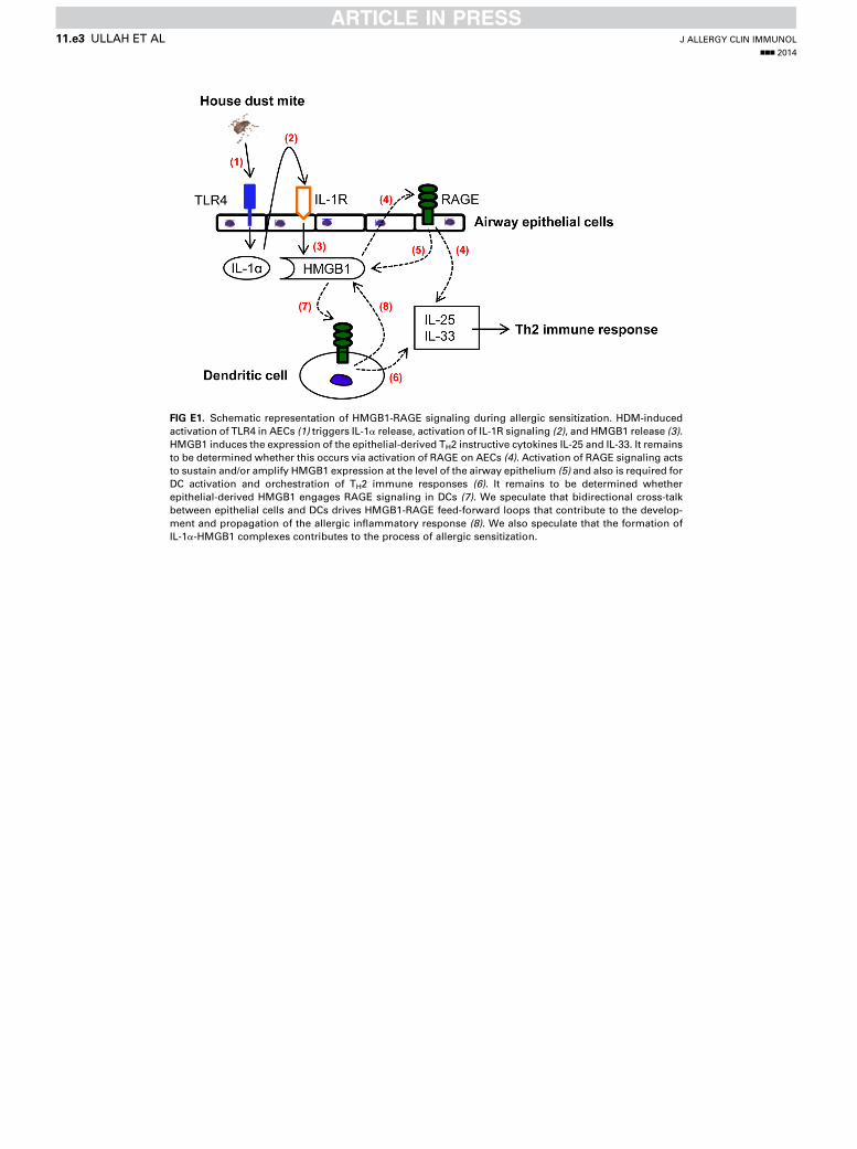
FIG E1. Schematic representation of HMGB1-RAGE signaling during allergic sensitization. HDM-induced
activation of TLR4 in AECs (1) triggers IL-1a release, activation of IL-1R signaling (2), and HMGB1 release (3).
HMGB1 induces the expression of the epithelial-derived TH2 instructive cytokines IL-25 and IL-33. It remains
to be determined whether this occurs via activation of RAGE on AECs (4). Activation of RAGE signaling acts
to sustain and/or amplify HMGB1 expression at the level of the airway epithelium (5) and also is required for
DC activation and orchestration of TH2 immune responses (6). It remains to be determined whether
epithelial-derived HMGB1 engages RAGE signaling in DCs (7). We speculate that bidirectional cross-talk
between epithelial cells and DCs drives HMGB1-RAGE feed-forward loops that contribute to the develop-
ment and propagation of the allergic inflammatory response (8). We also speculate that the formation of
IL-1a-HMGB1 complexes contributes to the process of allergic sensitization.
J ALLERGY CLIN IMMUNOL
nnn 2014
11.e3 ULLAH ET AL


![[Rapid airway access]](https://img.pdfslide.net/doc/110x75/6354cd1b765a645b3106d438/rapid-airway-access.jpg)


