Embed Size (px)
Citation preview
Vaccine 21 (2003) 3019–3029
Regional, but not systemic recruitment/expansion of dendriticcells by a pluronic-formulated Flt3-ligand plasmid with vaccine
adjuvant activity
Hongxun Sanga,b, Vladimir M. Pisarevb, Corey Mungerb, Simon Robinsonb, Jennifer Chavezb,Lori Hatcherb, Prahlad Parajulic, Yajun Guod, James E. Talmadgeb,∗
a International Joint Cancer Institute of Shanghai and Institute of Orthopaedics, Xijing Hospital, Xi’an 710032, PR Chinab Laboratory of Transplantation Immunology, Department of Pathology and Microbiology, University of Nebraska Medical Center,
Omaha, NE 68198-7660, USAc Department of Neurosurgery, Wayne State University& Karmanos Cancer Institute, Detroit, MI, USA
d Eppley Cancer Research Center, University of Nebraska Medical Center, Omaha, NE 68198-7696, USA
Received 9 October 2002; received in revised form 23 January 2003; accepted 28 January 2003
Abstract
Regional recruitment of dendritic cells (DCs) by the local administration of granulocyte macrophage-colony stimulating factor (GM-CSF)or Flt3-ligand (Flt3L) has vaccine adjuvant activity. However, Flt3L, with its DC growth factor activity, has not been extensively studiedas a vaccine adjuvant, particularly as a plasmid vector. We report that the intramuscular (IM) injection of a Flt3L plasmid (pNGVL-hFlex),when formulated in a pluronic carrier (SP1017, Supratek Pharma, Inc., Laval, Que., Canada), recruits DC to the injection site and regionallymph nodes (LNs) and augments immune responses to a p17 HIV plasmid vaccine to a greater extent than the injection of a naked DNAvaccine alone. Following IM administration of pNGVL-hFlex, Flt3L mRNA, Flt3L protein and infiltrating DC accumulate at the injectionsite. The number of DC in the draining LNs are also significantly increased with the greatest increase observed following injection of2.5�g of pNGVL-hFlex formulated in 0.01% SP1017. Flow cytometric studies demonstrate that the LN-infiltrating DC is mainly of theCD11c+CD11b− phenotype (IL-12 producing). Further, the co-injection of pNGVL3-hFlex and p17 HIV plasmids, formulated in SP1017,significantly increases the immune responses to the plasmid vaccine (pVAX-gag). The co-injection of pVAX-gag and pNGVL3-hFlex,formulated in SP1017, significantly increase delayed-type hypersensitivity responses and the numbers of antigen (Ag)-specific interferon-�secreting T cells in the spleen (Enzyme Linked Immune Spot (ELISpot) assay), compared to mice immunized with pVAX-gagformulatedin SP1017 alone. We conclude that the IM injection of pNGVL-hFlex with SP1017 can increase the number of DC in draining LN andat the site of injection, thereby providing adjuvant activity for a plasmid vaccine resulting in a significantly increased, Ag-specific T cellresponse.© 2003 Elsevier Science Ltd. All rights reserved.
Keywords:Dendritic cells; Flt3-ligand; Plasmid vaccine; Pluronic formulation; Gag
1. Introduction
Dendritic cells (DCs) are antigen (Ag)-presenting cells(APCs) with the unique ability to efficiently internalize,process and present Ag and stimulate both naı̈ve and mem-ory T cells [1–5]. The ex vivo expansion and maturationof DC from isolated progenitors (peripheral blood mono-cytes or bone marrow CD34+ cells) using granulocytemacrophage-colony stimulating factor (GM-CSF) and IL-4,followed by co-incubation with peptides or Ag vectors,
∗ Corresponding author. Tel.:+1-402-559-7844; fax:+1-402-559-4990.E-mail address:[email protected] (J.E. Talmadge).
provides the potential for vaccination with significant num-bers of Ag-presenting DC. However, this requires the invitro manipulation of DC, a complex and time-consumingprocess that adds the risk of microbial contamination. Onealternative approach to augment an immune response toa vaccine involves the manipulation of DC in vivo, eitherby in situ expansion and/or recruitment to the vaccinationsite.
The ligand for the fms-like tyrosine kinase-3 (Flt3L)[6,7]is a pluripotent hematopoietic growth factor that can expandand mobilize hematopoietic progenitor and stem cells[8]. Inaddition, Flt3L administration can increase the numbers ofDC in all lymphoid and parenchymal organs[9,10]and in the
0264-410X/03/$ – see front matter © 2003 Elsevier Science Ltd. All rights reserved.doi:10.1016/S0264-410X(03)00143-9

3020 H. Sang et al. / Vaccine 21 (2003) 3019–3029
blood, spleen and lymph nodes (LNs) of mice and humans[11–14]. The injection of Flt3L also augments non-specific,type 1 T cell responses, and if administered in associationwith a vaccine, can stimulate an Ag-specific, type 1 T cellresponse[15–23].
However, daily Flt3L injections for 8–10 days are neededto expand DC numbers, a requirement that limits the de-velopment of DC-based therapies. Recently, the in vivodelivery of the Flt3L transgene has been suggested as analternative to the injection of Flt3L protein[24]. In mousestudies, only hydrodynamic-based intravenous (IV) deliv-ery of naked DNA encoding a secreted form of humanFlt3L, a strategy with limited clinical utility, has beenshown to result in significantly increased DC numbers.However, Hung et al.[25] reported that intradermal deliv-ery (gene gun) of a recombinant chimera of Flt3L, linkedto a model Ag (papillomavirus-16 E7), increased the fre-quency of Ag-specific CD8+, but not CD4+, specific Tcells. Moore et al.[20] reported that co-administratingplasmids with transgenes for Flt3L and a cytokine that en-hances adoptive immune responses (IL-2, IL-12 or IL-15)also stimulated the responses to a plasmid DNA vaccine(Nef). The intramuscular (IM) injection of transgenes isan attractive route of vaccine/adjuvant delivery[20,26–28].However, only low levels of gene expression occur in themuscle following IM delivery of naked DNA,[29] possiblydue to poor transfection efficiency. Various cationic lipidcarrier systems have been developed that improve trans-fection efficiency; however, although they are effective invitro, they have not achieved great success in vivo[30]Another approach is the use of non-ionic carrier systems.It has been reported that SP1017, a recently developednon-ionic carrier, composed of two amphiphilic blockcopolymers, Pluronics L61 and F127,[31] has signifi-cantly higher transgene expression, following IM injectionof plasmid vectors compared to other non-ionic carriers[31].
To date, no studies on Flt3L expression or effects onDC number and recruitment by the IM injection of a plas-mid encoding Flt3L transgene have been reported. Becauseof the potential for low transgene expression, we exam-ined transgene expression following IM administration ofpNGVL3-hFlex formulated in SP1017 and its vaccine ad-juvant activity. Recently, we reported significant adjuvantactivity with the injection of recombinant Flt3L in associa-tion with an HGP-30 peptide derived from HIV-1 p17gag[15] and a genetic p53 vaccine[21]. Based on these stud-ies, we examined the adjuvant activity of the IM injectionof pNGVL-hFlex and pVAX-gag plasmids formulated inSP1017. We report herein that the concomitant administra-tion of pNGVL-hFlex and pVAX-gag plasmids formulatedin SP1017 significantly increased type 1, T cell responsesto genetic vaccines. The increase in adjuvant activity wasassociated with an increase in Flt3L mRNA and protein ex-pression at the injection site and an increased number of DCat the injection site and in the draining LN.
2. Materials and methods
2.1. Mice
Six- to eight-week-old female BALB/cAnNCrlBR micewere purchased from Charles River (Wilmington, MA) andallowed to acclimate for at least 2 weeks prior to use. Micewere housed in laminar airflow cages and provided foodand water ad libitum. Analyses of specimens from sentinelanimals revealed no evidence of exposure to viral, bacterialor parasitic pathogens during these studies.
2.2. Plasmids and plasmid injection
The pNGVL3-hFlex plasmid contains the gene for asecreted form of human Flt3L and was obtained fromthe National Gene Vector Laboratory at the Universityof Michigan (NGVL-UM). This plasmid also contains akanamycin-resistance gene for selection. Plasmid DNAwas purified using EndoFree Plasmid Giga kit (Qiagen,Valencia, CA) following the manufacturer’s protocol.
The pVAX-gagplasmid, which was developed in this lab-oratory, contains the p17/p24 region of HIV-1gag (aminoacids 78-732 ofgag) gene sequence derived from HXB2HIV-1 clade B (originally cloned by S. Dupont at the Uni-versity of Massachusetts Medical Center, Worcester, MA)into a pcDNA1/Neo plasmid. Thegag-coding sequence wascloned into the CMV promoter-driven pVAX1 plasmid vec-tor (InVitrogen, Carlsbad, CA) usingEcoRI and HindIIIcloning sites. The presence of the p17/p24-coding region inthe pVAX-gagplasmid was confirmed by DNA sequencingof restriction enzyme digests.
The pNGVL3-hFlex plasmid DNA formulated inDelbecco’s phosphate buffered saline (DPBS) or SP1017(generously provided by Supratek Pharma, Inc., Laval,Que., Canada) was injected bilaterally into the thigh muscleof Balb/c mice on days 1 and 6 and the mice sacrificedon day 12. Formulation of plasmid DNA with SP1017 wasperformed according to the protocol provided by the man-ufacturer. In the vaccination studies, pNGVL3-hFlex andpVax1-gag plasmids, combined or alone, were injected IMtwice (days 0 and 10), boosted with pVax1-gag alone onday 20 and then sacrificed on day 34. Negative controlsincluded the injection of plasmid DNA without SP1017,SP1017 alone or DPBS.
2.3. Peptides
The HIV-1 p17gagAg [(HGP-30 peptide or YSVHQRID-VKDTKEALDKIEEEQNKSKKKA and its non-apeptidederivatives DVKDTKEAL (P3), DTKEALDKI (P4) andDYKDTKEAL (P5)] were synthesized by Quality Con-trolled Biochemicals, Inc. (Hopkinton, MA) using f-mocchemistry and purified by high-pressure liquid chromatog-raphy. These peptides were generously provided by Dr. D.Zimmerman (Cel-Sci Corp., Vienna, VA).

H. Sang et al. / Vaccine 21 (2003) 3019–3029 3021
2.4. Flow cytometric (FCM) analysis
The inguinal LNs were harvested and a single cell sus-pension prepared by mechanical dissociation. One hun-dred microlitres aliquots of the cells at a concentrationof 106 cells/ml were labeled by incubating the cells with1�g/ml each of anti-CD11c fluorescein isothiocyanate(FITC) conjugated antibodies and anti-CD11b phycoery-thrin (PE)-conjugated antibodies (BD PharMingen, SanDiego, CA) for 30 min. Cells were washed, fixed with2% paraformaldehyde, and acquired with a FACScan Plus(Becton Dickinson, Boston, MA). Forward and side scat-ter were collected on a linear scale, while the PE andFITC signals were collected on a 4-decade log scale.Overlaps of emission spectra were electronically com-pensated and using the threshold on forward scatter toeliminate debris, 30,000–50,000 events were acquired andthe frequency distributions of DC subsets determined withAttractorsTM 3.0 software (Becton Dickinson, San Jose,CA).
2.5. RT-PCR and southern blot analysis
Injected muscles were harvested and samples dividedfor analysis by RT-PCR and immunohistochemistry(IHC). Total mRNA was extracted from the muscle us-ing Trizol® (Gibco BRL, Carlsbad, CA) according to themanufacturer’s instructions. RNA was reverse transcribedto cDNA and used for PCR amplification using the Am-pliTaq Gold PCR Kit (Roche, Inc., Foster City, CA). ThePCR assay used 5′-ACAACCTATCTCCTCCTGCTG-3′and 5′-GGCACATTTGGTGACAAAGTG-3′ as the primerpairs. This primer pair is specific for hFlt3L and amplifiesa 307 bp sequence after 35 cycles (1 min at 95◦C, 1 min at61◦C, and 1 min at 72◦C). Expression levels of GAPDHwere used as the housekeeping gene as previously reported[32]. PCR products were analyzed by electrophoresis in2% agarose gels containing ethidium bromide (0.5�g/ml).Products were visualized, photographed and transferredto nitrocellulose membranes for hybridization with a32P-labeled cytokine-specific oligonucleotide probe for hu-man Flt3L (5′-ACTTCGCTGACAAAATCCGT-3′). Theresulting Southern blots were subjected to autoradiographyusing a PhosphorImager (Molecular Dynamics, Sunnyvale,CA) and digital image analysis performed using the ImageQuant® analysis system (Molecular Dynamics, Sunnyvale,CA). Gene expression was calculated as an expression index(EI) versus the housekeeping gene GAPDH.
2.6. Immunohistochemical (IHC) staining
The harvested muscles were embedded in Tissue-Tek®
OCT (Bakura Finetec, Inc., Torrance, CA) and rapidlycooled to−80◦C. Tissue sections (5–10�m thick) were at-tached to Superfrost® microscope slides (Fisher Scientific)
and fixed with cold acetone. Endogenous peroxidase activ-ity was blocked with hydrogen peroxide, the slides rinsedand non-specific antibody binding reduced by incubationin 10% normal rabbit serum. Primary antibodies wereadded individually and included goat anti-human Flt3L(RDI-Flt3LnabG from RDI, Flanders, NJ); rat anti-mousefollicular DC (FDC-M1, BD PharMingen, San Diego,CA); and rat anti-mouse interdigitating DC (DEC-205,Serotec, Inc., Raleigh, NC). The slides were incubatedovernight at 4◦C, rinsed with phosphate-buffered saline(PBS), and incubated with biotinylated secondary an-tibodies (biotinylated rabbit anti-goat or rabbit anti-ratIgG, H + L, Vector, Inc., Burlingame, CA). Slides wererinsed and incubated with Vectastain® ABC (Vector, Inc.,Burlingame, CA) for 30 min before staining with DABsubstrate (Vector, Inc., Burlingame, CA). The sectionswere counterstained with hematoxylin and viewed by lightmicroscopy.
2.7. Delayed-type hypersensitivity (DTH) assay
Seven days after the last immunization, the mice werechallenged in one pinna with 2�g of HGP-30 (test ear) ina volume of 50�l or an equivalent volume of sterile DPBS(control ear). Twenty-four hours after challenge, the edemaof the test and control ears was measured using an electroniccaliper (Digimatic Indicator IDC Series 543 from MitutoyoCorp., Tokyo, Japan) with an accuracy of 3.8 × 10⁻³ mm.To determine HGP-30-specific edema, the thickness of thecontrol ear was subtracted from the thickness of the testear.
2.8. Enzyme-linked immune spot (ELISpot) assay
A spleen cell suspension was prepared from immunizedmice as “responder” cells and 1× 10⁷ spleen cells fromeach mouse were cultured for 48 h in separate wells of24-well plates containing 1×10⁶ irradiated (50 cGy) spleencells per well as APC from non-immunized syngeneic mice.The cells to be used as APC were incubated for 2 h withan HGP-30 peptide (final concentration 30�g/ml) priorto use. After co-incubation, the cells were harvested andan ELISpot assay performed as described previously[33].Briefly, MultiScreen-HA 96-well assay plates (Millipore,Bedford, MA) were coated with 100�l of cytokine captureantibodies R4-6A2 (anti-IFN-�) or 11B11 (anti-IL-4), bothfrom Pharmingen, San Diego, CA, diluted to 4�g/ml inPBS. Following overnight incubation at 4◦C, wells werewashed, blocked with 10% fetal bovine serum (FBS),and responder cells transferred to capture antibody-coatedplates using two dilutions (2× 10⁴ and 2× 10⁵ viable cellsper well) in duplicate wells and incubated for 24 h. Thecytokine-secreting cells were visualized using 1�g/ml bi-otinylated secondary Ab to IFN-� or IL-4 (Jackson ImmunoResearch Laboratories, Inc., West Grove, PA) followed by

3022 H. Sang et al. / Vaccine 21 (2003) 3019–3029
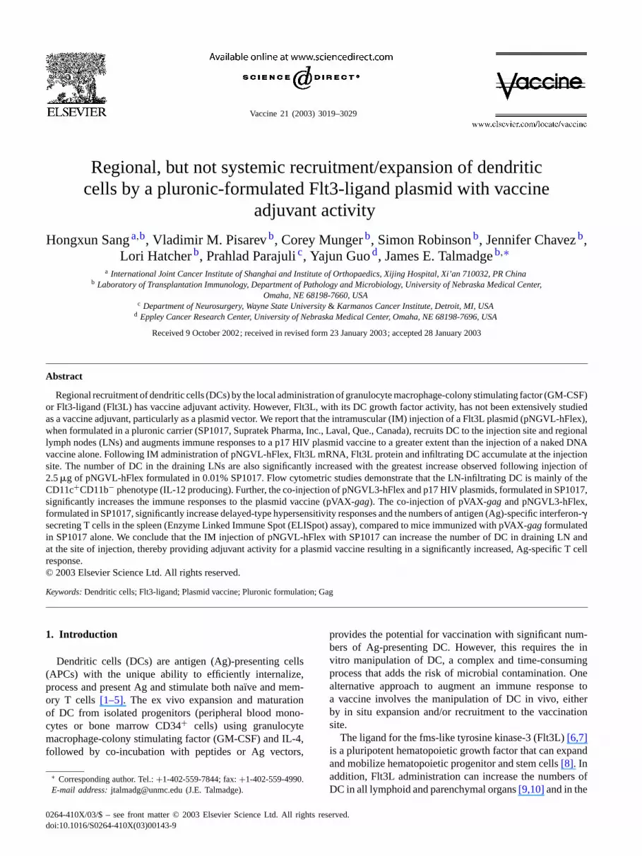
Fig. 1. RT-PCR and southern blot analysis of hFlt3L mRNA expression in mouse muscle after pNGVL3-hFlex IM injection. RT-PCR analysis forFlt3L gene expression was undertaken with muscle from the injection site in mice injected with SP1017, NGVL3-hFlex or pNGVL3-hFlex formulatedin SP1017. The upper two gels show Flt3L and GAPDH (housekeeping gene) expression. The lower gel shows a Southern blot performed using anFlt3L-specific probe. The sample loaded into each lane is shown above each gel.
co-incubation with alkaline phosphatase-conjugated avidin(Extravidin, Sigma) and Western Blue Substrate (Promega,Madison, WI). The number of spot-forming cells wasenumerated by an automatic computer ELISpot readingsystem, the Axioplan 2 Imaging System with KS ELISpotsoftware (Carl Zeiss Vision, Munchen-Hallbergmous, Ger-many). The total number of spots was determined perwell and the number of spots observed in wells containingonly irradiated spleen cells from non-immunized controlmice (background) subtracted. The average frequency ofcytokine-producing (spot-forming) cells was calculated.
2.9. Statistical analysis
Statistical analyses of data were performed using SPSS10.0 for Windows (SPSS, Inc., Chicago, IL). Means werecompared using the Student’s two-samplet-test; with sig-nificance assumed atP = 0.05.
3. Results
3.1. Flt3L gene and protein expression after IM injectionof pNGVL3-hFlex with or without SP1017
Flt3L gene expression (RT-PCR and Southern blotting)and protein production (IHC) after IM administration ofpNGVL3-hFlex formulated in SP1017 or DPBS, were de-termined using muscle samples from the injection sites.As shown inFig. 1, significantly increased levels of Flt3L
mRNA were observed in all samples obtained from miceinjected with pNGVL3-hFlex, as compared to mice injectedwith SP1017 alone, regardless of DNA dose level andformulation in SP1017 (Table 1). However, mice injectedwith 2.5�g of pNGVL3-hFlex plasmid DNA formulatedin 0.01% SP1017 had significantly greater levels of Flt3LmRNA by RT-PCR and Southern blotting analysis comparedto mice injected with 10�g of pNGVL3-hFlex alone. In con-trast, higher and lower doses of DNA formulated in 0.01%SP1017 were not significantly more active than the injectionof 10�g of pNGVL-hFlex injected alone. Other studies todetermine the optimal dose of SP1017 for the expression ofthe Flt3L transgene expression revealed that the injection of
Table 1Expression index of hFlt3L mRNA in the muscle of mice receivingdifferent doses of pNGVL3-hFlex DNA formulated in 0.01% (v/v) SP1017or DPBS
Group Mean± S.E.M.
DPBS 4.94± 3.270.01% SP1017 2.17± 0.74DNA 10�g 90.27 ab± 19.34DNA 10�g + SP1017 71.82 ab± 27.50DNA 5 �g + SP1017 113.61 ab± 25.27DNA 2.5�g + SP1017 129.80 abc± 8.45DNA 1.25�g + SP1017 84.39 ab± 42.10
Data shown is the expression index (EI) relative to GAPDH. Data pre-sented as the mean EI± standard error of the mean (S.E.M.). Student’st-test results for comparisons as follows:P < 0.05, a = vs. DPBS;b = vs. SP1017 alone; and c= vs. pNGVL3-hFlex plasmid alone. Thesestudies were done twice with anN of 4 in each study, data are pooledfor analysis.

H. Sang et al. / Vaccine 21 (2003) 3019–3029 3023
Table 2Expression of hFlt3L mRNA in the muscle of mice after administrationof pNGVL3-hFlex DNA
Group Mean± S.E.M.
DPBS 4.10± 1.96SP1017 alone 0.01% 6.56± 3.00pNGVL3-hFlex 2.5�g 188.97 ab± 24.93pNGVL3-hFlex 2.5�g + SP 0.005% 270.00 abcd± 7.44pNGVL3-hFlex 2.5�g + SP 0.01% 248.60 abcd± 7.33pNGVL3-hFlex 2.5�g + SP 0.02% 179.08 ab± 1.64
Mice received 2.5�g of pNGVL3-hFlex DNA formulated in varyingconcentrations of SP1017 (0.02–0.005%) or DPBS. Data shown are theexpression index (EI) relative to GAPDH and expressed as the meanEI ± standard error of the mean (S.E.M.). Student’st-test results forcomparisons as follows:P < 0.05, a= vs. DPBS; b= vs. SP1017 alone;c = vs. pNGVL3-hFlex alone; and d= pNGVL3-hFlex and SP10170.02%. These studies were performed twice with anN of 4 in each study,data are pooled for analysis.
2.5�g of the pNGVL3-hFlex plasmid DNA formulated inlower concentrations of SP1017 (0.005 and 0.01%) also sig-nificantly increased transcription compared to mice injectedwith 2.5�g per mouse pNGVL3-hFlex alone (Table 2).Further, the level of Flt3L mRNA was significantly greaterafter the injection of 2.5�g of pNGVL3-hFlex formulatedwith 0.005 or 0.01% SP1017 compared to the injection ofthe same dose of pNGVL3-hFlex formulated in a higher(0.02%) concentration of SP1017.
3.2. DC recruitment to draining LN after IM injection ofpNGVL3-hFlex and the effect of formulation in SP1017
The effect of pNGVL3-hFlex injection IM on DC num-bers in draining LN was also examined to identify the opti-mal dose(s) of pNGVL3-hFlex plasmid DNA and SP1017carrier. Following IM injection, FCM was used to assess thenumbers of CD11c+CD11b− (DC1) and CD11c+CD11b+
Fig. 2. DC cell number in inguinal LN after the administration of pNGVL3-hFlex formulated in SP1017 or in DPBS. In these studies, quadriceps muscleswere injected with different doses of the pNGVL3-hFlex plasmid (1.25–10�g) formulated in SP1017 (0.01%) or in DPBS. The frequency of DC subsets(CD11c+CD11b− and CD11c+CD11b+) in the draining inguinal LN was determined by flow cytometry. Data are presented as mean± standard errorof the mean (S.E.M.) (five mice per treatment group and each study repeated at least twice). (#) Significantly different from DPBS, (@) significantlydifferent from SP1017 alone and (&) significantly different from pNGVL3-hFlex alone.
(DC2) cells in the LN (Fig. 2). When pNGVL3-hFlexwas formulated in 0.01% SP1017, the absolute numberof DC1 in the inguinal LN was significantly increased,compared to mice receiving pNGVL3-hFlex formulated inDPBS, SP1017 alone or DPBS alone and was independentof the plasmid dose (Fig. 2). The number of DC2 wassignificantly increased only in the group receiving 2.5�gpNGVL3-hFlex formulated with 0.01% SP1017 (Fig. 2).In another study (Fig. 3), where the same pNGVL3-hFlexplasmid DNA dose (2.5�g) was formulated with differentSP1017 concentrations, the number of DC1 in the inguinalLN was significantly increased in the group injected withpNGVL3-hFlex formulated in 0.01% SP1017. In contrast,the only significant increase in DC2 cell number, relativeto the pNGVL-hFlex plasmid alone was observed in miceinjected with pNGVL3-hFlex formulated in 0.02% SP1017(Fig. 3). In both of these studies, changes in DC numberfrom the draining popliteal LNs were similar to those ob-served with the inguinal LN, but were of lower magnitude.As observed with the inguinal LN, the increase in DC num-ber was also due primarily to increased numbers of DC1(results not shown).
3.3. IHC staining of Flt3L expression and DC infiltrationat the injection site
To investigate the relationship between pNGVL3-hFlexinjection and Flt3L protein levels, IHC staining of the in-jection site was undertaken. Staining for Flt3L was negativein mice injected with SP1017 alone (Fig. 4, upper panel),and positive in the cytoplasm of myocytes from mice in-jected with pNGVL3-hFlex (Fig. 4, middle panel) with anincreased intensity in mice injected with pNGVL3-hFlexformulated in SP1017 (Fig. 4, lower panel). Consistent withthe low levels of Flt3L, IHC-staining of the injection siterevealed no increase in DC infiltration in mice injected with

3024 H. Sang et al. / Vaccine 21 (2003) 3019–3029
Fig. 3. DC number in inguinal LN after injection of pNGVL3-hFlex formulated in different SP1017 concentrations. In these studies, quadriceps muscleswere injected with 2.5�g of pNGVL3-hFlex plasmid formulated in different concentrations (0.005–0.02%) of SP1017 or in DPBS. The frequency of DCsubsets (CD11c+CD11b− and CD11c+CD11b+) in the inguinal LN was determined by flow cytometry. Data are presented as mean± standard error ofthe mean (S.E.M.) (five mice per treatment group, repeated at least twice and the data pooled). (#) Significantly different from DPBS, (@) significantlydifferent from pNGVL3-hFlex formulated in DPBS or 0.005% SP1017.
SP1017 alone (Fig. 5, upper panel). However, mice injectedwith pNGVL3-hFlex (Fig. 5, middle and lower panels), re-vealed increased numbers of DC with the greatest increasein mice injected with Flt3L formulated in SP1017. Stain-ing with antibodies to follicular (FDC-M1) and interdigitat-ing DC (DEC205) also revealed the presence of DC at theinjection site, although the FDC-M1 positive-staining cellspredominated (results not shown). As shown inFig. 5, DCinfiltration in the muscle followed the needle path and thetissue immediately surrounding the injection site.
3.4. pNGVL3-hFlex augmentation of the DTH response toa pVAX-gag vaccine
To investigate the ability of pNGVL3-hFlex to augmentT cell immune responses to an HIV-gagvaccine, we exam-ined the DTH responses to HGP-30, an Ag recognition sitefor HIV gag p17, following vaccination. In these studies,mice were immunized with: (a) the pVAX-gagplasmid for-mulated in SP1017, (b) the pNGVL3-hFlex plasmid formu-lated in SP1017 or (c) the combination of pVAX-gag vac-cine and pNGVL3-hFlex plasmids formulated in SP1017.Control groups included administration of DPBS or SP1017alone. These studies revealed a significantly increased DTHresponse measured as pinna edema only in the group re-ceiving the combination of pVAX-gag and pNGVL3-hFlexplasmids formulated in SP1017 (Fig. 6).
3.5. Frequency of Ag-specific IFN-γ secreting cells(ELISpot assay) after immunization
To determine whether the Ag-specific T cell immune re-sponses were type 1 or 2, we performed an ELISpot as-say to quantify IFN-�-and IL-4-secreting cell frequencies
in the spleen. As shown inFig. 7, there was a signifi-cantly increased frequency of IFN-�-secreting cells in re-sponse to stimulation with HGP30 peptides in the spleensof mice immunized with the combination of pVAX-gagandpNGVL3-hFlex plasmids formulated in SP1017 comparedto all other groups. In contrast, there was no significant dif-ference in the frequency of IL-4-secreting cells between thegroups (data not shown). These results demonstrate that vac-cination with the combination of pVAX-gag vaccine andpNGVL3-hFlex plasmids formulated in SP1017 can inducea type 1, Ag-specific T cell response.
4. Discussion
We report that IM injection of the pNGVL3-hFlex plas-mid, formulated with SP1017, can induce DC recruitmentto the injection site and to regional LN. Formulations ofpNGVL3-hFlex plasmid DNA in a narrow concentrationrange (0.01–0.002%) of the pluronic carrier SP1017 sig-nificantly enhanced Flt3L mRNA expression and proteinproduction following IM injection. This resulted in the in-filtration of DC to the injection site and augmentation of theimmune response to a p17gag plasmid vaccine as shownby both DTH and ELISpot immune assays.
DCs are APC, with a potent ability to evoke an Ag-specificimmune response[1–5]. However, DCs occur at a low fre-quency in vivo, restricting their use as APC in a vaccinestrategy. This difficulty can be ameliorated in part with theuse of Flt3L, which can stimulate DC expansion[11–14].Further, the injection of Flt3L protein has been shown topreferentially expand the DC1 subset in mice, thereby aug-menting Ag-specific T and B cell responses[23,34–36]. In-deed, the daily injection of Flt3L for 14 days in patients

H. Sang et al. / Vaccine 21 (2003) 3019–3029 3025
Fig. 4. Immunohistochemical staining of Flt3L protein in pNGVL3-hFlexinjected muscle. Mice were injected twice (5 days between injections) withSP1017 alone, pNGVL3-hFlex formulated in DPBS or pNGVL3-hFlexformulated in 0.01% SP1017. The injection site in the quadriceps mus-cle was dissected from the surrounding tissue 5 days after the last in-jection and IHC for Flt3L undertaken. The upper panel shows muscleinjected with SP1017 alone, the middle panel shows muscle injected withpNGVL3-hFlex formulated in DPBS and the lower panel shows mus-cle injected with pNGVL3-hFlex formulated in SP1017 (magnification:100×).
with metastatic colon cancer has been shown to increasethe frequency of circulating DC and to increase peritumoralDC infiltration, [37] although, high doses and multiple in-jections of recombinant Flt3L are required to induce theseeffects. Therefore, genetic delivery of Flt3L provides a po-tential approach to recruit DC to a tumor or vaccination siteand thereby increase Ag-specific immune responses as hasbeen shown clinically with Flt3L protein[19].
A previous study demonstrated that hydrodynamic IVinjection of pNGVL3-hFlex in mice could increase thenumber of DC in the spleen and LN[24]. However, thisapproach is effective only when the plasmid is delivered tomice in a volume of 1.5 ml via a rapid IV push, which can-not be considered clinically. A more practical approach toimprove Flt3L delivery is the IM injection of a vector withthe Flt3L transgene in a formulation to improve transfection
Fig. 5. Immunohistochemical staining of DCs in pNGVL3-hFlex injectedmuscle. Mice were injected twice (5 days between injections) with SP1017alone, pNGVL3-hFlex formulated in DPBS or pNGVL3-hFlex formulatedin 0.01% SP1017. The injection site in the quadriceps muscle was dis-sected from the surrounding tissue 5 days after the last injection and pro-cessed for IHC and staining for DC (blue staining cells) using an antibodyspecific for DEC205, counterstained with hematoxylin and viewed by lightmicroscopy. The upper panel shows muscle injected with SP1017 alone,the middle panel shows muscle injected with pNGVL3-hFlex formulatedin DPBS and the lower panel shows muscle injected with pNGVL3-hFlexformulated in SP1017 (magnification: 100×).
efficacy. The IM injection of naked plasmid DNA results intransgene expression by transfected skeletal myocytes[38].Further, IM injection of a non-viral vector results in chronicexpression of an antigenic transgene and the induction ofhumoral and cell-mediated immune responses with the po-tential for prophylactic activity against infections or cancer[39,40]. In one study, it was found that the concomitantIM injection of a non-viral vector encoding GM-CSF andan Ag-encoding plasmid enhanced an Ag-specific immuneresponse that was associated with DC infiltration of the IMinjection site[41]. However, this vaccine approach may belimited by the low levels of transgene expression associ-ated with the injection of non-viral vectors. Carrier systemshave been developed to enhance transfection and gene ex-pression, and although they do not achieve high systemicprotein levels, they may increase local transgene expression.

3026 H. Sang et al. / Vaccine 21 (2003) 3019–3029
Fig. 6. DTH response togagpeptide following immunization with pVAX-gagand pNGVL3-hFlex Mice were immunized twice (5 days between injections)with the pVAX-gag plasmid formulated in 0.01% SP1017, a combination of pVAX-gag and the pNGVL3-hFlex plasmids formulated in 0.01% SP1017,0.01% SP1017 in DPBS or DPBS. One week after the final immunization, mice were challenged by the intradermal administration of 4�g HGP-30 inone pinna (test) and DPBS in the other pinna (control). After 24 h, the edema (thickness) of test and control ears was measured. Results are shown asthe difference in ear thickness between the test and control ears (Ag-specific edema). Data are presented as mean± standard error of the mean (S.E.M.)(five mice per treatment group): () significantly different from DPBS, 0.01% SP1017 alone and pVAX-gag formulated in 0.01% SP1017.
SP1017 is a pluronic carrier composed of two amphiphilicblock copolymers (pluronics L61 and F127) that has beenreported to augment transgene expression following IM in-jection [31]. In this study, reporter genes were injected IM
Fig. 7. Frequency of IFN-�-secreting cells in the spleen following immunization with pVAX-1-gag and pNGVL3-hFlex formulated in SP1017. Mice wereimmunized with the concomitant injection of pVAX-gag and pNGVL3-hFlex plasmids formulated in 0.01% SP1017. The relevant controls were also usedas in the immunization protocol. Two weeks after the final immunization, mice were sacrificed and splenocytes isolated. Splenocytes were stimulatedin the presence or absence of HGP-30 peptides and after 48 h, the frequency of IFN-� secreting cells was determined by ELISpot assay. Cells wereplated in duplicate using two different concentrations and the results normalized and averaged. Data represents the Ag-specific responses calculated bythe subtraction of the number of spots developed in non-HGP-30-stimulated cultures (background) from the number of spots developed in the presenceof HGP-30. Data are presented as mean± standard error of the mean (S.E.M.): () significantly different from DPBS, 0.01% SP1017 and pVAX-gagformulated in 0.01% SP1017.
with and without SP1017 into rodents. SP1017 increasedgene expression by about 10-fold and maintained highergene expression compared with naked DNA. In a similarstudy, the IM injection of a plasmid with a transgene for

H. Sang et al. / Vaccine 21 (2003) 3019–3029 3027
luciferase, which was formulated in poloxamer 188(Pluronic F68) demonstrated a three-fold increase in lu-ciferase expression compared to injection with out theformulation [42]. However, the expression levels ob-served were lower than that obtained with electropora-tion.
We report, herein, that the IM injection of the pNGVL3-hFlex plasmid when formulated in SP1017 resulted in therecruitment of DC to the injection site. This was associatedwith an increase in local, but not systemic, Flt3L gene ex-pression and protein production. Further, the IM injectionof the pNGVL3-hFlex plasmid formulated in SP1017 hadadjuvant activity for a non-viral vaccine (pVAX-gag). AnAg-specific, type 1 T cell response was observed whenthe pNGVL3-hFlex plasmid formulated in SP1017 wasco-administered with pVAX-gag as shown by DTH andELISpot assays. It is noted that despite the regional effecton DC numbers, no effect on the numbers of DC in theblood or spleen were detected and serum Flt3L levels wasnot increased (results not shown). These results suggestthat low, regional levels of Flt3L production stimulating thelocal expansion and/or recruitment of DC can significantlyincrease systemic T cell responses. No studies to date havereported DC expansion following IM injection of an Flt3Lplasmid or adjuvant activity for T cell responses. Indeed,one recent study suggested that co-injection of a Flt3Lplasmid decreased the antibody response to the HepatitisB virus core antigen[16]. However, one can hypothesizethat this was due to the induction of a type 1 responsethat inhibited a type 2-antibody response. One recent studydemonstrated adjuvant activity by the co-administrationof Flt3L and T cell augmenting cytokine (IL-2, IL-12 orIL-15) plasmids together with a Nef DNA vaccine[43]. Incontrast, the IM injection of a pluronic-based formulationof the Flt3L plasmid, in our studies, was sufficient to in-duce a T cell response to a DNA vaccine. This could bea consequence of increased levels of Flt3L at the injectionsite, as compared to mice injected with the pNGVL3-hFlexplasmid formulated in DPBS. Similarly, formulation ofthe pNGVL3-hFlex plasmid in SP1017 significantly in-creased the number of DC at the injection site and in thedraining LN. We suggest that these infiltrating DC takeup, process and present Ags at the injection site or inthe draining LN, resulting in an Ag-specific immune re-sponse with potential activity against infections or solidtumors.
In an earlier vaccine adjuvant study,[25] it was found thatan immune response could be enhanced by linking the genesfor an Ag and Flt3L and injecting them IM, resulting in theinduction of an Ag-specific CD8+ T cell-mediated immuneresponse and antitumor prophylaxis in vivo. In our study,separate plasmids containing transgenes for the HIV p17gagand Flt3L were co-injected and we found that a significant Tcell response was induced when the injected plasmids wereformulated in SP1017. This may explain differences betweenour studies and those of Nabel and colleagues[43] who used
a naked plasmid DNA and showed an increased T cell IFN-�response when the plasmid encoding vaccine antigen wasadministered at a dose of 20�g per animal. In our study, for-mulation with SP1017 resulted in the expression of plasmidDNA at the injection site (as revealed by RT-PCR and IHC),local DC recruitment and the induction of a systemic T cellresponse, using an immunization protocol with lower dosesof the Flt3L plasmid and antigen (<10�g per animal andless).
It has previously been shown that Flt3L protein in-jected intralesionally into tumor-bearing mice increasesthe immune response and in some instances, inducestherapeutic activity[23,34,44,45]. Moreover, injection ofFlt3L-transduced tumor cells inhibits tumor growth andprotects against subsequent tumor challenge[46]. Themechanism of T cell augmentation by Flt3L remains un-clear, although Flt3L administration increases the numberof DCs with a CD11c+CD11b− phenotype, and can aug-ment an Ag-specific, type 1 T cell response[47]. Theinjection of pNGVL3-hFlex induced primarily the expan-sion of DC1, as revealed by flow cytometry of cells in thedraining LNs. Further, the DTH response to Ag challengewas significantly better in mice immunized with the com-bination of pVAX-gag and pNGVL3-hFlex formulated inSP1017 as compared to formulation in DPBS. In addition,in mice immunized with the combination of pVAX-gagand pNGVL3-hFlex formulated with SP1017, a higher fre-quency of Ag-specific IFN-�-producing (type 1) T cellswas observed, while the frequency of type 2 (IL-4) cytokinesecreting cells remained at a low level (results not shown).Together, these results demonstrate that immunization withthe combination of pVAX-gag and pNGVL3-hFlex formu-lated with SP1017 stimulates an Ag-specific type 1 T cellresponse in association with the regional expansion and/orrecruitment of DC1 to the site of immunization and to localLN.
Acknowledgements
The authors wish to thank Dr. Rakesh K. Singh, MichelleVarney, Dr. Aihua Li and Matthew Backora for their discus-sion on these studies. The authors also wish to thank RichardMurcek, Lisa Chudomelka and Tina Winekauf for assist-ing with the preparation of the manuscript. This researchwas supported by grants from amfAR #02705-28-RGV, theNebraska Research Initiative Grants on Gene Therapy andMolecular Therapeutics and an international exchange awardfrom National Science Foundation and the Department ofHealth, PR China.
References
[1] Nestle FO, Banchereau J, Hart D. Dendritic cells: on the move frombench to bedside. Nat Med 2001;7(7):761–5.

3028 H. Sang et al. / Vaccine 21 (2003) 3019–3029
[2] Nouri-Shirazi M, Banchereau J, Fay J, Palucka K. Dendritic cellbased tumor vaccines. Immunol Lett 2000;74(1):5–10.
[3] Liu YJ, Kanzler H, Soumelis V, Gilliet M. Dendritic cell lineage,plasticity and cross-regulation. Nat Immunol 2001;2(7):585–9.
[4] Pulendran B, Banchereau J, Maraskovsky E, Maliszewski C.Modulating the immune response with dendritic cells and their growthfactors. Trends Immunol 2001;22(1):41–7.
[5] Lutzker SG, Lattime EC. Use of dendritic cells to immunize againstcancers overexpressing p53. Clin Cancer Res 2001;7(1):2–4.
[6] Lyman SD, James L, Vanden Bos T, et al. Molecular cloning of aligand for the flt3/flk-2 tyrosine kinase receptor: a proliferative factorfor primitive hematopoietic cells. Cell 1993;75(6):1157–67.
[7] Hannum C, Culpepper J, Campbell D, et al. Ligand for FLT3/FLK2receptor tyrosine kinase regulates growth of haematopoietic stemcells and is encoded by variant RNAs. Nature 1994;368(6472):643–8.
[8] Lyman SD, James L, Johnson L, et al. Cloning of the humanhomologue of the murine Flt3-ligand: a growth factor for earlyhematopoietic progenitor cells. Blood 1994;83(10):2795–801.
[9] McKenna HJ. Role of hematopoietic growth factors/Flt3-ligand inexpansion and regulation of dendritic cells. Curr Opin Hematol2001;8(3):149–54.
[10] Antonysamy MA, Thomson AW. Flt3-ligand (FL) and its influenceon immune reactivity. Cytokine 2000;12(2):87–100.
[11] Brasel K, McKenna HJ, Morrissey PJ. Hematologic effects ofFlt3-ligand in vivo in mice. Blood 1996;88(6):2004–12.
[12] Xu ZX, Zhu JK, Zhang ZH, et al. Recombinant human Flt3-ligandexerts both direct and indirect effects on hematopoiesis. Indian JPharmacol 2001;32:1–6.
[13] Pulendran B, Banchereau J, Burkeholder S, et al. Flt3-ligandand granulocyte colony-stimulating factor mobilize distinct humandendritic cell subsets in vivo. J Immunol 2000;165(1):566–72.
[14] Mosley RL, Parajuli P, Pisarev V, et al. Flt3-ligand augmentation ofT cell mitogenesis and expansion of type 1 effector/memory T cells.Int Immunopharmacol 2002;2(7):925–40.
[15] Pisarev VM, Parajuli P, Mosley RL, et al. Flt3-ligand enhancesthe immunogenicity of a gag-based HIV-1 vaccine. Int JImmunopharmacol 2000;22(11):865–76.
[16] Kwon TK, Park JW. Intramuscular co-injection of naked DNAencoding HBV core antigen and Flt3-ligand suppresses anti-HBcantibody response. Immunol Lett 2002;81(3):229–34.
[17] Pisarev VM, Parajuli P, Mosley RL, et al. Flt3-ligand and conjugationto IL-1B peptide as adjuvants for a type 1. T cell response to anHIV gag p17 vaccine. Vaccine 2002;17–18:2358–68.
[18] O’Keeffe M, Hochrein H, Vremec D, et al. Effects of administrationof progenipoietin 1, Flt-3 ligand, granulocyte colony-stimulatingfactor, and pegylated granulocyte macrophage-colony stimulatingfactor on dendritic cell subsets in mice. Blood 2002;99(6):2122–30.
[19] Disis ML, Rinn K, Knutson KL, et al. Flt3-ligand as avaccine adjuvant in association with HER-2/neu peptide-basedvaccines in patients with HER-2/neu-overexpressing cancers. Blood2002;99(8):2845–50.
[20] Moore AC, Kong WP, Chakrabarti BK, Nabel GJ. Effectsof antigen and genetic adjuvants on immune responses tohuman immunodeficiency virus DNA vaccines in mice. J Virol2002;76(1):243–50.
[21] Parajuli P, Pisarev V, Sublet J, et al. Immunization with wild-typep53 gene sequences coadministered with Flt3-ligand induces anantigen-specific type 1 T-cell response. Cancer Res 2001;61:8227–34.
[22] Gregory SH, Sagnimeni AJ, Zurowski NB, Thomson AW.Flt3-ligand pretreatment promotes protective immunity to Listeriamonocytogenes. Cytokine 2001;13(4):202–8.
[23] Lynch DH, Andreasen A, Maraskovsky E, Whitmore J, Miller RE,Schuh JCL. Flt3-ligand induces tumor regression and antitumorimmune responses in vivo. Nat Med 1997;3(6):625–31.
[24] He Y, Pimenov AA, Nayak JV, Plowey J, Falo Jr LD, Huang L.Intravenous injection of naked DNA encoding secreted Flt3-ligand
dramatically increases the number of dendritic cells and natural killercells in vivo. Hum Gene Ther 2000;11(4):547–54.
[25] Hung CF, Hsu KF, Cheng WF, et al. Enhancement of DNAvaccine potency by linkage of antigen gene to a gene encoding theextracellular domain of Fms-like tyrosine kinase 3-ligand. CancerRes 2001;61(3):1080–8.
[26] Bonadio J. Tissue engineering via local gene delivery: update andfuture prospects for enhancing the technology. Adv Drug Deliv Rev2000;44(2–3):185–94.
[27] Shedlock DJ, Weiner DB. DNA vaccination: antigen presentationand the induction of immunity. J Leukoc Biol 2000;68(6):793–806.
[28] Schatzlein AG. Non-viral vectors in cancer gene therapy: principlesand progress. Anticancer Drugs 2001;12(4):275–304.
[29] Liu F, Song Y, Liu D. Hydrodynamics-based transfection inanimals by systemic administration of plasmid DNA. Gene Ther1999;6(7):1258–66.
[30] Yew NS, Wang KX, Przybylska M, et al. Contribution of plasmidDNA to inflammation in the lung after administration of cationiclipid: pDNA complexes. Hum Gene Ther 1999;10(2):223–34.
[31] Lemieux P, Guerin N, Paradis G, et al. A combination of poloxamersincreases gene expression of plasmid DNA in skeletal muscle. GeneTher 2000;7(11):986–91.
[32] Krzesicki RF, Winterrowd GE, Brashler JR, et al. Identificationof cytokine and adhesion molecule mRNA in murine lung tissueand isolated T cells and eosinophils by semi-quantitative reversetranscriptase-polymerase chain reaction. Am J Respir Cell Mol Biol1997;16(6):693–701.
[33] Fushimi T, Inoue A, Koh CS, Yamazaki M, Ishihara Y, KimBS. The effect of pentoxyfilline (PTX) on Theiler’s murineencephalomyelitis virus (TMEV)-induced demyelinating disease. CellImmunol 1998;186(2):140–6.
[34] Braun SE, Chen K, Blazar BR, et al. Flt3-ligand antitumor activityin a murine breast cancer model: a comparison with granulocytemacrophage-colony stimulating factor and a potential mechanism ofaction. Hum Gene Ther 1999;10(13):2141–51.
[35] Brasel K, De Smedt T, Smith JL, Maliszewski CR. Generation ofmurine dendritic cells from flt3-ligand-supplemented bone marrowcultures. Blood 2000;96(9):3029–39.
[36] Fong L, Hou Y, Rivas A, et al. Altered peptide ligand vaccinationwith Flt3-ligand expanded dendritic cells for tumor immunotherapy.Proc Natl Acad Sci USA 2001;98(15):8809–14.
[37] Morse MA, Nair S, Fernandez-Casal M, et al. Preoperativemobilization of circulating dendritic cells by Flt3-ligandadministration to patients with metastatic colon cancer. J Clin Oncol2000;18(23):3883–93.
[38] Wolff JA, Malone RW, Williams P, et al. Direct gene transfer intomouse muscle in vivo. Science 1990;247(4949 Pt 1):1465–8.
[39] Dupuis M, Denis-Mize K, Woo C, et al. Distribution of DNA vaccinesdetermines their immunogenicity after intramuscular injection inmice. J Immunol 2000;165(5):2850–8.
[40] Horton HM, Anderson D, Hernandez P, Barnhart KM, Norman JA,Parker SE. A gene therapy for cancer using intramuscular injectionof plasmid DNA encoding interferon alpha. Proc Natl Acad Sci USA1999;96(4):1553–8.
[41] Haddad D, Ramprakash J, Sedegah M, et al. Plasmid vaccineexpressing granulocyte macrophage-colony stimulating factor attractsinfiltrates including immature dendritic cells into injected muscles.J Immunol 2000;165(7):3772–81.
[42] Hartikka J, Sukhu L, Buchner C, et al. Electroporation-facilitateddelivery of plasmid DNA in skeletal muscle: plasmid dependenceof muscle damage and effect of poloxamer 188. Mol Ther2001;4(5):407–15.
[43] Moore AC, Kong WP, Chakrabarti BK, Nabel GJ. Effectsof antigen and genetic adjuvants on immune responses tohuman immunodeficiency virus DNA vaccines in mice. J Virol2002;76(1):243–50.

H. Sang et al. / Vaccine 21 (2003) 3019–3029 3029
[44] Esche C, Subbotin VM, Maliszewski C, Lotze MT, Shurin MR.Flt3-ligand administration inhibits tumor growth in murine melanomaand lymphoma. Cancer Res 1998;58(3):380–3.
[45] Chen K, Braun S, Lyman S, et al. Antitumor activity andimmunotherapeutic properties of Flt3-ligand in a murine breast cancermodel. Cancer Res 1997;57(16):3511–6.
[46] Lowrie DB, Tascon RE, Bonato VL, et al. Therapy of tuber-culosis in mice by DNA vaccination. Nature 1999;400(6741):269–71.
[47] Pulendran B, Smith JL, Caspary G, et al. Distinct dendritic cellsubsets differentially regulate the class of immune response in vivo.Proc Natl Acad Sci USA 1999;96(3):1036–41.


