Embed Size (px)
Citation preview

Residual levels of tripeptidyl-peptidase I activity dramaticallyameliorate disease in late infantile neuronal ceroid lipofuscinosis
David E. Sleat1,2, Mukarram El-Banna1, Istvan Sohar1, Kwi-Hye Kim1, Kostantin Dobrenis3,Steven U. Walkley3, and Peter Lobel1,2
1Center for Advanced Biotechnology and Medicine, University of Medicine and Dentistry of New Jersey,Piscataway, NJ 08854
2Department of Pharmacology, University of Medicine and Dentistry of New Jersey, Piscataway, NJ 08854
3Sidney Weisner Laboratory of Genetic Neurological Disease, Department of Neuroscience, Rose F. KennedyCenter for Research in Mental Retardation and Human Development, Albert Einstein College of Medicine,Bronx, NY 10461.
AbstractClassical late-infantile neuronal ceroid lipofuscinosis (LINCL) is a hereditary neurodegenerativedisease of childhood that is caused by mutations in the gene (CLN2) encoding the lysosomal proteasetripeptidyl-peptidase I (TPPI). LINCL is fatal and there is no treatment of demonstrated efficacy inaffected children but preclinical studies with AAV-mediated gene therapy have demonstratedpromise in a mouse model. Here, we have generated mouse CLN2 mutants that express differentamounts of TPPI activity to benchmark levels required for therapeutic benefits. Approximately 3%of normal TPPI activity in brain delayed disease onset and doubled lifespan to a median of ~9 monthscompared to mice expressing ~0.2% of normal levels. Expression of 6% of normal TPPI activitydramatically attenuated disease, with a median lifespan of ~20 months which approaches that ofunaffected mice. While the life-span of this hypomorph is shortened, disease is late-onset, less severeand progresses slowly compared to mice expressing lower TPPI levels. For gene therapy and otherapproaches that restore enzyme activity, these results suggest that 6% of normal TPPI activitythroughout the CNS of affected individuals will provide a significant therapeutic benefit but higherlevels will be required to cure this disease.
Keywordsneuronal ceroid lipofuscinosis; mouse model; hypomorph; lysosomal storage disease
INTRODUCTIONThe neuronal ceroid lipofuscinoses (NCLs) are a group of genetically distinct but clinicallyrelated diseases of which the classical late-infantile form (LINCL) is one of the most frequentlyencountered [1]. LINCL is a fatal neurodegenerative lysosomal storage disease that resultsfrom mutations in the gene CLN2 [2] that encodes the lysosomal protease tripeptidyl-peptidase
Correspondence should be addressed to P.L. ([email protected]) and D.E.S. ([email protected]). Tel.: +732 235 5032; Fax:+ 732 235 4466.Publisher's Disclaimer: This is a PDF file of an unedited manuscript that has been accepted for publication. As a service to our customerswe are providing this early version of the manuscript. The manuscript will undergo copyediting, typesetting, and review of the resultingproof before it is published in its final citable form. Please note that during the production process errors may be discovered which couldaffect the content, and all legal disclaimers that apply to the journal pertain.
NIH Public AccessAuthor ManuscriptMol Genet Metab. Author manuscript; available in PMC 2009 June 1.
Published in final edited form as:Mol Genet Metab. 2008 June ; 94(2): 222–233. doi:10.1016/j.ymgme.2008.01.014.
NIH
-PA Author Manuscript
NIH
-PA Author Manuscript
NIH
-PA Author Manuscript

I (TPPI) [3]. This disease is characterized by seizures, progressive mental decline, loss of visionand locomotor function, and shortened lifespan. Mutations in most LINCL patients are nullalleles [4] and typically result in diagnosis at around 4 years and survival to between 7 and 15years of age. However, like many other lysosomal storage diseases [5], some LINCL casesexhibit a less severe clinical phenotype. Here, compound heterozygosity for a null allele anda presumed hypomorphic Arg447His missense mutation [4] results in a later onset (~8 years)and protracted disease with survival into the third or fourth decade of life.
There is currently no treatment of demonstrated efficacy for LINCL patients but there ispromising progress, particularly with respect to adeno-associated virus (AAV)-mediated genetherapy (reviewed in [6]). Initial studies examining the expression of TPPI by recombinantAAV and other viral vectors demonstrated high levels and widespread expression of the proteinthroughout the central nervous system (CNS) of healthy rodents [7,8]. More recently, AAV-mediated gene therapy has been evaluated in a mouse model for LINCL. This gene-targetedmouse was created by the disruption of CLN2 with an Arg446His missense mutation (whichis equivalent to the late-onset human allele) and insertion of a neomycin selection cassettewithin an adjacent intron, resulting in a disruption of normal splicing [9]. The synergistic effectof the missense mutation and the splicing defect results in levels of TPPI that are below thethreshold of detection (~1% of normal levels) and this mouse recapitulates many of the clinicalfeatures of the human disease [9]. Mice appear to be healthy at birth but develop tremors byabout 7 weeks. In symptomatic mice, there is pervasive neuronal pathology with a cytoplasmicaccumulation of autofluorescent storage material, selective loss of some Purkinje cells andwidespread axonal degeneration. Lifespan is greatly shortened with a median survival of ~ 20weeks.
Treatment of the CLN2-targeted mice with an AAV vector expressing TPPI initially showedexpression of the recombinant protein and a slowing of the cellular pathology that is associatedwith disease [10]. More recently, it has been demonstrated that AAV-mediated gene therapy,in addition to attenuating cellular pathology, can also slow or halt the decline in locomotorfunction and significantly increase survival of the mutant mice [11,12]. An importantobservation to emerge from these studies is that while AAV treatment of symptomatic micehas some positive effects, treatment must be conducted before onset of disease for the greatesttherapeutic benefits [11].
Levels of TPPI activity achievable by AAV-mediated gene therapy vary from study to study,reflecting, in addition to other variables, viral serotype and titer as well as the number andlocation of injection sites. Passini et al [10] achieved levels that approached or exceeded wild-type activities throughout most of the brain. More recently, CNS levels of TPPI have beenachieved in AAV-treated mutant mice that were 10 to 100-fold [11] or up to 27-fold [12] higherthan found in wild-type mice.
While the induction of such levels is clearly beneficial in the mouse model, it seems unlikelythat similar levels of TPPI activity will be attainable throughout the brain of human LINCLpatients by gene therapy or enzyme replacement. However, the minimum level of TPPI activitythat will be required to successfully treat LINCL remains unknown. In this study, we havetaken a direct approach to this fundamental question by investigating the disease phenotype ofa series of CLN2-targeted mouse mutants that express different amounts of residual TPPIactivity.
Sleat et al. Page 2
Mol Genet Metab. Author manuscript; available in PMC 2009 June 1.
NIH
-PA Author Manuscript
NIH
-PA Author Manuscript
NIH
-PA Author Manuscript

MATERIALS AND METHODSGeneration of CLN2 mouse hypomorphs
All experiments and procedures involving live animals were conducted in compliance withapproved Institutional Animal Care and Use Committee protocols. The CLN2-targeted mousemodel for LINCL (CLN2−/−) has been described previously [9] and was created by thesynergistic effect of an Arg446His missense mutation and a splicing defect resulting from theinsertion of a floxed neo selection marker (Fig. 1, Panel A).
Removal of the neo selection marker was achieved by crossing male CLN2 +/− mice withfemale Zp3-cre mice (The Jackson Laboratory, Bar Harbor, Maine) which is a transgenic linein a C57BL/6 background that expresses cre within the oocyte from the zona pellucida 3 genepromoter [13]. Progeny of this mating were screened for cre-mediated excision of neo by usingprimers (ATCTGATGGCTACTGGGTGG, CCCGGTAGAATTCCGATCAT andCCCCCAAACACTGGAGTAGA) that generate 402 nt and 328 nt products from the wild-type and neo-containing targeted alleles, respectively and 529 nt from the mutant allele afterexcision of neo (Fig. 1, Panel B). Founders that were heterozygous for the neo excision (+/f)were backcrossed against C57BL/6 mice and were selected for loss of the cre transgene usinga primer set (GCGGTCTGGCAGTAAAAACTATC, GTGAAACAGCATTGCTGTCACTT)that generates a 100 nt product from this sequence. Complete annotated CLN2 sequences forall three alleles (wild-type, targeted and floxed-targeted) are provided as Online SupplementaryMaterial. Data presented was obtained from −/− and f/− mice after 10 back crosses to C57BL/6 and +/−, f/+, +/+, and f/f mice after at least 5 backcrosses to C57BL/6.
Detection of CLN2 transcriptsNorthern blotting for CLN2 transcripts were preformed on total RNA purified using an RNEasykit (Qiagen, Valencia, CA) as described previously [9].
Subcellular fractionationBrain and liver homogenates (H fraction) were prepared using a Potter homogenizer asdescribed [14] in 0.25M sucrose. Nuclei and heavy mitochondrial components were pelletedby centrifugation for 3 minutes at 12,500 rpm in a 50 Ti rotor (Beckman Coulter, Fullerton,CA). The supernatant was then centrifuged for 6.42 minutes at 25,000 rpm in a 50 Ti rotor topellet the light mitochondrial differential fraction (L fraction) which was washed andresuspended in 0.25 M sucrose.
Measurement of TPPI activityTPPI was measured in the H and L fractions after dilution with cold 0.15M NaCl-0.1% TritonX-100 using 200 µM Arg-Ala-Phe-ACC (generously provided by Dr. John W. Taylor, RutgersUniversity) as substrate using the kinetic assay described previously [15]. For determinationof KM, TPPI was measured using Arg-Ala-Phe-ACC and Ala-Ala-Phe-AMC (Sigma, St. Louis,MO). β-Galactosidase activity was measured using 4-methylumbelliferyl-β-D-galactopyranoside substrate [16]. For both enzymes, fluorescence was measured using aCytoFluor 4000 multiwell plate reader (PerSeptive Biosystems, Framingham, MA). Sevendifferent dilutions of each sample, corresponding to ~0.15 g tissue equivalents/mL and sixsuccessive 2-fold dilutions, were used for enzyme activity measurements. TPPI activity valueswere calculated from 2–3 dilutions in the linear range using the lowest (most concentrated)dilutions for the f/f and −/− specimens and the highest dilutions for +/+ specimens. Activitieswere standardized to protein concentration measured with Advanced Protein Assay reagent(Cytoskeleton Inc., Denver, CO).
Sleat et al. Page 3
Mol Genet Metab. Author manuscript; available in PMC 2009 June 1.
NIH
-PA Author Manuscript
NIH
-PA Author Manuscript
NIH
-PA Author Manuscript

Behavioral testingMutant mice were analyzed for gait abnormalities and ataxia as reviewed [17]. Footprintpatterns were visualized by painting the feet of mutant mice with non-toxic washable paint(forefeet, purple and hindfeet, orange) and placing the mice at the entrance of a dark tunnel(35cm long × 9cm wide × 6cm high) placed over white paper.
PathologyFor immunohistochemical detection of TPPI, mice were euthanized at 52–57 days of age andwere transcardially infused with Bouin’s fixative. Brains were removed, bisected and drop-fixed at 4C for 16 hours. Samples were then washed three times with PBS and saturated withPBS containing 15% then 30% sucrose in consecutive 48hr incubations. Tissue samples wereembedded in Shandon M-1 Embedding Matrix (Thermo Scientific) and 10 µm cryosectionswere prepared. Immunohistochemistry using a 1:100 dilution of a primary goat anti-rabbitpolyclonal antibody (R72) was as described previously [18] with fluorescent staining using anAlexa Fluor 488 dye-labeled secondary antibody (Invitrogen) at a dilution of 1:400. Forimmunohistochemical detection of the subunit C of mitochondrial ATP synthase, tissues werefixed in a manner similar to the above but using 4.0% paraformaldehyde in 0.1 M phosphatebuffer. Blocks of tissue were removed and sectioned at 35 microns on a Leica 9000S Vibratome.This was followed by staining overnight with a 1:250 dilution of a rabbit polyclonal antibody(generously provided by Dr. E. F. Neufeld) raised against a peptide corresponding to the aminoterminus of the mature protein c subunit ([19]). After washing, sections were incubated in abiotinylated goat-anti-rabbit IgG (1:200) for one hour, followed by incubation in VectastainABC Elite complex (Vector Laboratories, Burlingame, CA). Antibody binding was visualizedusing a diaminaobenzidine substrate kit (Vector Laboratories). Sections were placed ongelatinized slides and dried overnight, followed by dehydration and cover-slipping, andexamination on an Olympus research microscope. Autofluorescent storage was examined in35 µm coronal vibratome sections that were cut from fixed brain tissue, washed with PBS andmounted on slides with Prolong Gold antifading reagent (Invitrogen: Molecular Probes).Sections were examined using the Zeiss Meta Duo V2 confocal system by simultaneousexcitation with 458 and 488 nm lines of a multi-line Argon laser and collective emission captureusing the Meta spectral scanner set for a band of 501–694 nm. This bandwidth was selected inpart based on initial full spectrum scans to profile peaks of emission. Multiple sections fromeach of duplicate mice for each genotype and age were surveyed.
Survival analysisMice were handled gently as environmental disturbances can induce fatal seizures in the −/−animals [9]. Moribund animals (typically, severe ataxia and tremors restricting ability to feed)were euthanized under IACUC policy and were scored as dying from disease on the day ofeuthanasia. Kaplan-Meier analysis was conducted using Prism v 4.03 (GraphPad Software,San Diego).
Western blottingTissue homogenates were prepared in 250 mM sucrose and 10 or 20 ug protein equivalentsfractionated by electrophoresis on 4–12 % Bis-Tris gradient gels (Invitrogen) with MESrunning buffer for SCMAS or 10% Bis-Tris gels with MOPS running buffer for TPPI. Proteinswere transferred to nitrocellulose. SCMAS was detected using the rabbit polyclonal antibodydescribed above which was diluted 1:3000-fold in PBS containing 0.2% Tween -20 and 5%BSA. TPPI was detected using an affinity-purified rabbit polyclonal antibody raised againstrecombinant TPPI which was diluted in PBS containing 10% fat free milk and 0.2% Tween-20.In both cases, the secondary antibody was a I125-labeled goat anti-rabbit polyclonal and signal
Sleat et al. Page 4
Mol Genet Metab. Author manuscript; available in PMC 2009 June 1.
NIH
-PA Author Manuscript
NIH
-PA Author Manuscript
NIH
-PA Author Manuscript

was visualized using Typhoon scanner (GE Healthcare) and quantified using Imagequant 5.2software.
RESULTSSurvival of CLN2-mutant mice is strain dependent
In our previous characterization of the CLN2-targeted mouse, we measured survival in eitheran isogenic 129Sv or a mixed C57BL/6, 129Sv strain background. We found that survival ofthe isogenic 129Sv mutant was longer (median, 164 days) than the mixed strain (median, 138days). We interpreted these results to possibly indicate the presence of a strain-specific modifier(s) of the disease phenotype which could, for example, influence triggering of or susceptibilityto fatal seizures. In this study, we have analyzed the effects of CLN2 mutations on phenotypeby comparing congenic or, because of time constraints reflecting the life-span of the mutants,near-congenic (≥ 6 backcrosses) mice in a C57BL/6 genetic background. To determine theeffect of this genetic background on LINCL phenotype, we compared the survival of −/− mutantmice that were in either a C57BL/6 or 129Sv background (Fig. 2). We find that survival of theCLN2 mutant in a 129Sv background (median 155 days) is extended (p=0.0008) compared tothe mice in a congenic C57BL/6 background (median 132 days). Survival of the CLN2 mutantin a first generation cross between C57BL/6 and 129Sv was essentially the same as in the129Sv background.
Generation of mouse models expressing different levels of TPPI activityThe aim of this study was to generate mouse models of LINCL with hypomorphic mutationsthat express low levels of TPPI to determine thresholds of activity that provide therapeuticbenefits. Our approach to achieving this has been to genetically modify our existing CLN2mutant (technically designated neoinsArg446HisCLN2 allele (The Mouse Genome Initiative,http://www.informatics.jax.org) and designated here as −/− for brevity) to allow limitedexpression of TPPI. An explanation of this strategy requires a description of the moleculardetails by which our CLN2-targeted mouse model was generated and this is provided elsewhere[9]. In brief, CLN2 was disrupted by the synergistic effects of an Arg446His missense mutationin exon 11 and a splicing defect caused by the presence of cryptic splice sites within theneomycin selection marker inserted into intron 11 (Fig. 1, Panel A). Most transcripts from thetargeted allele contained an antisense neo insertion within the CLN2 transcript but a smallamount of mRNA (corresponding to ~4% of wild-type levels) was correctly spliced. Together,these mutations resulted in <1% (the lower limit of detection of our previous assay) of thenormal levels of TPPI activity in homozygous targeted mice. In order to generate a TPPIhypomorph mutant with residual TPPI activity, we used cre-mediated recombination to removethe disruptive neo insertion to allow for normal splicing of a transcript containing theArg446His missense mutation (Fig. 1, Panel B). Mice that are homozygous for the mutantallele in which the neo selection marker is removed are designated neodelArg446HisCLN2 andare referred to here as f/f. The compound heterozygote of these mutant CLN2 alleles is referredto as f/−.
We investigated the effect of the removal of neo on CLN2 transcription and splicing by northernblotting. Wild-type mouse RNA have two transcripts for CLN2 of ~3.5 and 4.5 kb (Fig. 3,Panel A) that result from transcription termination at two alternate polyadenylation sites asobserved previously [9]. Levels of TPPI mRNA in the CLN2 −/− mutant were greatly reducedand transcripts were larger than observed in the wild-type mice, reflecting the insertion of ~500 nts corresponding to an antisense neo transcript. As predicted, size and abundance of thetranscript from the mutant allele after excision of neo was indistinguishable with the wild-typemRNA (Fig. 3, Panel A). These results indicate that the CLN2 mRNA is stable when encodingthe Arg446His missense mutation and that splicing of the primary transcription product is
Sleat et al. Page 5
Mol Genet Metab. Author manuscript; available in PMC 2009 June 1.
NIH
-PA Author Manuscript
NIH
-PA Author Manuscript
NIH
-PA Author Manuscript

unaffected by the 175-nt insertion within intron 11 that results from targeting vector sequenceand the single loxP site that remains after recombination (Fig. 1, Panel B).
TPPI is expressed in the CLN2-targeted hypomorphsGiven that a precise determination of TPPI levels in the CLN2 mutants is key to theinterpretation of this study, TPPI expression in the CLN2-targeted mice was measured both byfunctional enzymatic assay and by immunological approaches. Enzyme activity measurementswere conducted using a new substrate that is highly specific for TPPI on total homogenatesand differential centrifugation light mitochondrial (L) fractions from both liver and brain (Table1). The average TPPI specific activities in brain homogenates of f/f and −/− mutants are 5.9%and 0.3%, respectively, of wild type, with the values for the −/− samples approachingbackground levels of the assay. The L fractions are enriched in lysosomes and TPPI specificactivities of f/f and −/− mutants are 4.7% and 0.2% of wild type. While the differentialcentrifugation step increases the specific activity of lysosomal enzymes and thus the signal tonoise of the TPPI assay, the relative enrichments for the different samples differ: whenconsidering the ratio of β-galactosidase specific activity in a given L fraction and homogenate(“enrichment factor”), the average enrichment factors are 2.28, 1.86 and 1.09 for the +/+, f/fand −/− brain samples, respectively. When correcting for this and using data from analysis ofL fractions, we estimate that the relative TPPI activities in f/f and −/− mutant brain are 5.7%and 0.46%, respectively, of wild type.
The above analysis yields a consistent estimate of 6% for the relative activity of TPPI in f/fbrain. The values for the −/− mutant are less certain given the residual activity is near thedetection limit of our assay. We have previously used rtPCR to determine that the levels of thecorrectly spliced Arg447His mutant transcript in −/− brain are 4% of the corresponding normaltranscript in wild type brain [9]. Given that the f/f and+/+ mice have indistinguishable levelsof correctly spliced transcript (Fig. 3, Panel A) and that “normal” levels of the Arg447Histranscript yield 6% of TPPI enzymatic activity, then a realistic estimate of the activity in −/−brain is ~0.2 % (6% × 4%) of wild type. In an independent experiment, levels of TPPI activityin crude homogenates from the f/− compound heterozygote were found to be 2.9% of wild-type levels (data not shown) which, as predicted, is intermediate between the f/f and −/−mutants.
Analysis of liver homogenates and differential centrifugation L fractions indicates that theTPPI activity of the f/f mutant is ~1% of wild type, while the value for the −/− mutant is belowthe limit of detection of our assay (Table 1). The reason for the lower relative levels of TPPIactivity in f/f and −/− liver compared to brain is not clear but it could potentially reflectdifferences in the stability of the mutant protein in these different tissues. Interestingly, thespecific activity of beta-galactosidase in the mutant liver samples are decreased ~50%compared to wild-type while the beta-galacosidase levels of the brain samples are essentiallyindistinguishable (Table 1).
Given that levels of the correctly spliced CLN2 transcript in the f/f mutant are similar to wild-type, there are two possible explanations for the reduced TPPI activity in this mutant. First,TPPI protein may be present at normal levels but the mutation could result in reduced activityof the enzyme. Second, the mutant protein may have normal activity but could be unstable andpresent at reduced levels. To distinguish between these possibilities, we compared the activityof TPPI in wild-type and f/f samples using two different synthetic substrates (Fig. 4). It is clearthat the KM for both control and mutant TPPI is essentially identical when measured with eithersubstrate, strongly suggesting that while transcription is normal, levels of TPPI protein arereduced in correspondence with activity.
Sleat et al. Page 6
Mol Genet Metab. Author manuscript; available in PMC 2009 June 1.
NIH
-PA Author Manuscript
NIH
-PA Author Manuscript
NIH
-PA Author Manuscript
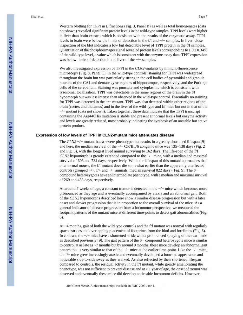
Western blotting for TPPI in L fractions (Fig. 3, Panel B) as well as total homogenates (datanot shown) revealed significant protein levels in the wild-type samples. TPPI levels were higherin liver than brain extracts which is consistent with the results of the enzymatic assay. TPPIlevels in brain were below the limits of detection in the f/f and −/− samples. In liver, closeinspection of the blot indicates a low but detectable level of TPPI protein in the f/f samples.Quantitation of the phosphorimager signal revealed protein levels corresponding to 1.0 ± 0.34%of the wild-type level, a value which is consistent with the enzyme assay data. TPPI expressionwas below limits of detection in the liver of the −/− samples.
We also investigated expression of TPPI in the CLN2-mutants by immunofluorescencemicroscopy (Fig. 3, Panel C). In the wild-type controls, staining for TPPI was widespreadthroughout the brain but was particularly strong in the cell bodies of pyramidal and granuleneurons of the CA1 and dentate gyrus regions of hippocampus, respectively, and the Purkinjecells of the cerebellum. Staining was punctate and cytoplasmic which is consistent withlysosomal localization. TPPI was detectable in the same regions of the brain in the f/fhypomorph but was less intense than observed in the wild-type control. Essentially no stainingfor TPPI was detected in the −/− mutant. TPPI was also detected within other regions of thebrain (cortex and thalamus) and in the liver of the wild-type and f/f mice but not in that of the−/− mutant (data not shown). Taken together, these data indicate that the TPPI transcriptcontaining the Arg446His mutation is stable and present at normal levels but enzyme activityand levels are greatly reduced, most probably indicating the synthesis of an unstable but activeprotein product.
Expression of low levels of TPPI in CLN2-mutant mice attenuates diseaseThe CLN2 −/− mutant has a severe phenotype that results in a greatly shortened lifespan [9]and here, the median survival of the −/− C57BL/6 congenic mice was 135–138 days (Fig. 2and Fig. 5), with the longest lived animal surviving to 162 days. The life-span of the f/fCLN2 hypomorph is greatly extended compared to the −/− mice, with a median and maximalsurvival of 603 and 734 days, respectively. While the lifespan of this mutant approaches thatof a normal mouse, the f/f mutant does die somewhat earlier than the apparently unaffectedcontrols (grouped +/+, f/+ and −/+ animals, median survival 822 days) (Fig. 5). The f/−compound heterozygotes have an intermediate phenotype, with a median and maximal survivalof 269 and 438 days, respectively.
At around 7 weeks of age, a constant tremor is detected in the −/− mice which becomes morepronounced as they age and is eventually accompanied by ataxia and an abnormal gait. Bothof the CLN2 hypomorphs described here show a similar disease progression but with a lateronset and slower progression that is in proportion to the overall survival of the mice. As ageneral indicator of disease progression from a locomotor perspective, we measured thefootprint patterns of the mutant mice at different time-points to detect gait abnormalities (Fig.6).
At ~4 months, gait of both the wild type controls and the f/f mutant was normal with regularlyspaced strides and overlapping placement of footprints from the hind and forelimbs (Fig. 6).In contrast, the −/− mice have a shortened stride with a pronounced splaying of the rear limbsas described previously [9]. The gait pattern of the f/− compound heterozygote mice is similarto control at as late as ~7 months but by around 9 months, these mice develop an abnormal gaitpattern that is very similar to that of the −/− mice at the earlier time-point. Like the −/− mice,the f/− mice grew increasingly ataxic and eventually developed a hunched appearance andnoticeable side-to-side sway as they walked. As also reflected by their shortened lifespancompared to controls, the residual activity in the f/f mutant, while greatly ameliorating thephenotype, was not sufficient to prevent disease and at > 1 year of age, the onset of tremor wasobserved and eventually these mice did develop noticeable locomotor deficits. However,
Sleat et al. Page 7
Mol Genet Metab. Author manuscript; available in PMC 2009 June 1.
NIH
-PA Author Manuscript
NIH
-PA Author Manuscript
NIH
-PA Author Manuscript

disease progression was slow and these mice appeared relatively healthy for most of their life-span when compared to the −/− mutants. Disease phenotype at the end of the lifespan of the f/f mutant was not as severe as observed in the end-stage −/− or f/− mutants. This is illustratedby the gait pattern of these mice at ~ 2 years of age, which is not dissimilar to that of wild-typecontrols of similar age.
Lysosomal storage is reduced by low levels of TPPI activitySeveral of the genetically-distinct forms of neuronal ceroid lipofuscinosis, including the late-infantile form, accumulate a small proteolipid, subunit c of mitochondrial ATP synthase(SCMAS), as a major component of the storage material [20]. Accumulation of SCMAS incontrol and mutant mouse brain was measured by western blotting and immunodetection (Fig.7, Panel A). At 60 days of age, significant accumulation of SCMAS was already detectable inthe −/− mutant by western blotting even though the neurodegenerative phenotype was stillmild. As the −/− mice continued to age (143 days), levels of SCMAS continued to increase.In contrast, there was no significant accumulation of SCMAS in the f/f hypomorph even whenthe mice were ~ 1 year of age. Immunohistochemical analysis revealed that levels of SCMASwere significantly elevated in the Purkinje cell layer of the cerebellar cortex in the −/− mouseat ~ 4 months of age (Fig. 7, Panel B). At the same age, levels of SCMAS were slightly elevatedin the f/− mutant but staining in the f/f mutant appeared to be the same as observed in the wild-type controls. At ~ 22 months, staining for SCMAS was slightly increased in the f/f mutantcompared to control. In cerebral cortex (Fig. 7, Panel C), accumulation of SCMAS followedthe same pattern with storage being proportional to the severity of disease in the differentmodels.
One of the clinical hallmarks of LINCL and other NCLs is the presence of autofluorescentlysosomal storage material within the cells of affected individuals and this phenotype isrecapitulated in the −/− mouse model [9]. At 128 days, there is a significant accumulation ofpunctuate cytoplasmic autofluorescent material in the cerebral cortex of the −/− mousecompared to the wild-type control (Fig. 8). In contrast, autofluorescence in the f/f and f/− micewas markedly less and similar to the wild-type mice at this age. There is an increase inautofluorescence in the f/f mouse at ~660 days compared to younger ages but it is essentiallyindistinguishable from that in the wild-type mice and presumably reflects the accumulation ofnormal aging lipofuscin rather than disease-related storage material.
DISCUSSIONPromising treatment approaches for lysosomal storage diseases (reviewed in [21] aim to eitherreplace the defective protein with a functional recombinant version (e.g. gene, stem-cell andenzyme replacement therapies) or stimulate the production of a functional protein from anendogenous mutant gene (e.g. nonsense suppression and chemical chaperone therapy). Forthese, understanding the therapeutic benefits that are likely to be associated with any givenlevel of a restored activity in preclinical models is critical in justifying whether such approachesshould be extended to patients. However, it is generally accepted that the restoration ofrelatively low levels of the deficient activity will be useful and for most LSDs, <10% of normalactivity is generally thought to have therapeutic benefits [6]. This is threshold is has arisenfrom measurements of residual activity that is present in patients with LSDs with attenuated,late-onset and/or slowly progressing disease and is supported by in vitro experimentsdemonstrating that the turnover of lysosomal enzyme substrates is normal in that cells containas low as 15% of the normal activity of the respective enzymes [22].
Clinical correlates can certainly provide useful clues to the levels of lysosomal activities thatare likely to be necessary for successful treatment but there are significant limitations to thisapproach. Although progression of LINCL tends to be fairly uniform, progression of other
Sleat et al. Page 8
Mol Genet Metab. Author manuscript; available in PMC 2009 June 1.
NIH
-PA Author Manuscript
NIH
-PA Author Manuscript
NIH
-PA Author Manuscript

lysosomal storage diseases is typically very variable even when considering null alleles,reflecting environmental influences as well as possible epigenetic factors and modifying genes.The range of enzyme activities measured in different classes of severity of a given lysosomalstorage disease are often overlapping and as a result, clinical genotype-phenotype correlationsare frequently difficult or uninformative. In addition, clinical correlation can, by definition,only provide an indication of the residual levels of a lysosomal enzyme activity that areassociated with the least severe variant of a given disorder, which is not equivalent to theminimum activity that is associated with an absence of disease.
A few studies have directly investigated the effect of residual lysosomal activities on respectivedisease phenotype. Several missense mutations were engineered into the β-glucuronidase genein mouse models of mucopolysaccharidosis VII [23], resulting in low but measurable levelsof β-glucuronidase activities (0.1–0.7% of normal levels, depending on tissue source). Theselevels were associated with a milder disease phenotype and decreased storage. Missensemutations of β-glucosidase have also been generated in mouse models of Gaucher disease[24] where null mutations are neonatally lethal. Most of the missense mutants were viable andthese, regardless of whether they were homozygotes or compound heterozygotes with a nullallele, retained ~ 25% normal activity within the brain. This was associated with greatlyameliorated disease. Another mouse model of Gaucher disease that retained 15–20% of normalβ-glucosidase activity also presented a greatly attenuated phenotype [25]. While these studiesdo not identify minimum activities required to prevent the respective diseases, taken together,they do indicate that a small amount of residual lysosomal activity can attenuate disease andthat ~25% of normal levels can result in a substantially corrected phenotype.
In this study, we have determined the effect of different residual levels of TPPI on the phenotypeof gene-targeted mouse models of LINCL. Understanding the levels of enzymatic activity foruseful therapeutic benefits is of particular relevance in LINCL because this is one of the firstlysosomal storage diseases for which viral-mediated gene therapy trials has been undertakenwith affected individuals (Clinicaltrials.gov NCT00151216, [26]). In addition, clinical trialsare also underway for the use of human central nervous system stem cell transplantation inLINCL (identifier NCT00337636, [27]). We find that a hypomorphic mutant with 3% of wild-type activity in brain survived for twice as long as a mutant lacking detectable TPPI (mediansurvival, ~ 9 months compared to 4.5 months). Albeit delayed, the disease phenotype of thishypomorph was severe and resembled that of the mouse that lacked detectable TPPI. However,we also find that 6% residual activity of TPPI in the brain of another hypomorph, while notsufficient to completely cure disease, elicit an improvement in phenotype that is dramatic whencompared to the rapid progression of disease and death of the mouse lacking detectable TPPI.This hypomorph survives with a median life-span of 20 months and while this is somewhatshortened compared to wild-type (median survival ~ 25 months), the phenotype in terms ofpathology and locomotor dysfunction was relatively mild, even at the later stages of disease.As the KM for TPPI in the wild-type and f/f specimens are indistinguishable (Fig. 4) and steadystate TPPI protein levels are dramatically decreased in the f/f specimens (Fig. 3), it is highlylikely that 6% of wild-type activity can be equated to 6% of the normal levels of protein.
If our results can be extrapolated from the mouse model to the clinic (and there is no reason tosuspect otherwise given the accuracy with which the LINCL mouse recapitulates the humandisease), then ≤6% of normal TPPI activity, even if it were possible to achieve this level inevery cell of the CNS, will not be sufficient to cure or halt disease. It should be noted than anaverage CNS activity of 6% of wild-type may not be therapeutically equivalent to 6% in everycell. This is an important consideration for stem-cell or gene therapies given the chimeric natureof these approaches where individual corrected cells cross-protect other mutant cells. Thus, asan example, 18% of wild-type activity in one out of every three cells is likely not to beequivalent to 6% activity in every cell. Higher levels of TPPI (10% of normal ?) in each cell
Sleat et al. Page 9
Mol Genet Metab. Author manuscript; available in PMC 2009 June 1.
NIH
-PA Author Manuscript
NIH
-PA Author Manuscript
NIH
-PA Author Manuscript

will likely be needed to completely prevent the onset of LINCL and whether this target can beachieved in patients using current recombinant approaches remains to be determined.
Finally, the observation that residual levels of TPPI can result in a late-onset, slowlyprogressing neurodegenerative phenotype in mice suggests that CLN2 defects may be worthconsidering in some human neurological disorders of unknown molecular etiology. Oneobvious candidate is the adult form of NCL, which can share many of the clinical features ofLINCL including storage of SCMAS and curvilinear storage bodies [1].
Supplementary MaterialRefer to Web version on PubMed Central for supplementary material.
ACKNOWLEDGEMENTSThis work was supported by National Institutes of Health Grants NS37918 (P.L.) and HD045561 (S.U.W.). We wouldlike to thank Drs. Elizabeth Neufeld (UCLA) and John Taylor (Rutgers University) for generously providing theSCMAS antibody and substrate for TPPI assay, respectively.
REFERENCES1. Goebel, HH.; Mole, SE.; Lake, BD. Neuronal Ceroid Lipofuscinoses (Batten Disease). Ios Pr Inc;
1999.2. Sleat DE, Donnelly RJ, Lackland H, Liu CG, Sohar I, Pullarkat RK, Lobel P. Association of mutations
in a lysosomal protein with classical late-infantile neuronal ceroid lipofuscinosis. Science1997;277:1802–1805. [PubMed: 9295267]
3. Rawlings ND, Barrett AJ. Tripeptidyl-peptidase I is apparently the CLN2 protein absent in classicallate-infantile neuronal ceroid lipofuscinosis. Biochim Biophys Acta 1999;1429:496–500. [PubMed:9989235]
4. Sleat DE, Gin RM, Sohar I, Wisniewski K, Sklower-Brooks S, Pullarkat RK, Palmer DN, Lerner TJ,Boustany RM, Uldall P, Siakotos AN, Donnelly RJ, Lobel P. Mutational analysis of the defectiveprotease in classic late-infantile neuronal ceroid lipofuscinosis, a neurodegenerative lysosomal storagedisorder. Am J Hum Genet 1999;64:1511–1523. [PubMed: 10330339]
5. Maire I. Is genotype determination useful in predicting the clinical phenotype in lysosomal storagediseases? J Inherit Metab Dis 2001;24:57–61. [PubMed: 11758680]discussion 45–56
6. Sands MS, Davidson BL. Gene therapy for lysosomal storage diseases. Mol Ther 2006;13:839–849.[PubMed: 16545619]
7. Hackett NR, Redmond DE, Sondhi D, Giannaris EL, Vassallo E, Stratton J, Qiu J, Kaminsky SM,Lesser ML, Fisch GS, Rouselle SD, Crystal RG. Safety of direct administration of AAV2(CU)hCLN2,a candidate treatment for the central nervous system manifestations of late infantile neuronal ceroidlipofuscinosis, to the brain of rats and nonhuman primates. Hum Gene Ther 2005;16:1484–1503.[PubMed: 16390279]
8. Haskell RE, Hughes SM, Chiorini JA, Alisky JM, Davidson BL. Viral-mediated delivery of the late-infantile neuronal ceroid lipofuscinosis gene, TPP-I to the mouse central nervous system. Gene Ther2003;10:34–42. [PubMed: 12525835]
9. Sleat DE, Wiseman JA, El-Banna M, Kim KH, Mao Q, Price S, Macauley SL, Sidman RL, Shen MM,Zhao Q, Passini MA, Davidson BL, Stewart GR, Lobel P. A mouse model of classical late-infantileneuronal ceroid lipofuscinosis based on targeted disruption of the CLN2 gene results in a loss oftripeptidyl-peptidase I activity and progressive neurodegeneration. J Neurosci 2004;24:9117–9126.[PubMed: 15483130]
10. Passini MA, Dodge JC, Bu J, Yang W, Zhao Q, Sondhi D, Hackett NR, Kaminsky SM, Mao Q,Shihabuddin LS, Cheng SH, Sleat DE, Stewart GR, Davidson BL, Lobel P, Crystal RG. Intracranialdelivery of CLN2 reduces brain pathology in a mouse model of classical late infantile neuronal ceroidlipofuscinosis. J Neurosci 2006;26:1334–1342. [PubMed: 16452657]
Sleat et al. Page 10
Mol Genet Metab. Author manuscript; available in PMC 2009 June 1.
NIH
-PA Author Manuscript
NIH
-PA Author Manuscript
NIH
-PA Author Manuscript

11. Cabrera-Salazar MA, Roskelley EM, Bu J, Hodges BL, Yew N, Dodge JC, Shihabuddin LS, SoharI, Sleat DE, Scheule RK, Davidson BL, Cheng SH, Lobel P, Passini MA. Timing of TherapeuticIntervention Determines Functional and Survival Outcomes in a Mouse Model of Late InfantileBatten Disease. Mol Ther. 2007
12. Sondhi D, Hackett NR, Peterson DA, Stratton J, Baad M, Travis KM, Wilson JM, Crystal RG.Enhanced Survival of the LINCL Mouse Following CLN2 Gene Transfer Using the rh.10 RhesusMacaque-derived Adeno-associated Virus Vector. Mol Ther 2007;15:481–491. [PubMed:17180118]
13. de Vries WN, Binns LT, Fancher KS, Dean J, Moore R, Kemler R, Knowles BB. Expression of Crerecombinase in mouse oocytes: a means to study maternal effect genes. Genesis 2000;26:110–112.[PubMed: 10686600]
14. Della Valle MC, Sleat DE, Sohar I, Wen T, Pintar JE, Jadot M, Lobel P. Demonstration of lysosomallocalization for the mammalian ependymin-related protein using classical approaches combined witha novel density shift method. J Biol Chem 2006;281:35436–35445. [PubMed: 16954209]
15. Tian Y, Sohar I, Taylor JW, Lobel P. Determination of the substrate specificity of tripeptidyl-peptidaseI using combinatorial peptide libraries and development of improved fluorogenic substrates. J BiolChem 2006;281:6559–6572. [PubMed: 16339154]
16. Sleat DE, Sohar I, Lackland H, Majercak J, Lobel P. Rat brain contains high levels of mannose-6-phosphorylated glycoproteins including lysosomal enzymes and palmitoyl-protein thioesterase, anenzyme implicated in infantile neuronal lipofuscinosis. J Biol Chem 1996;271:19191–19198.[PubMed: 8702598]
17. Crawley, JN. What's wrong with my mouse? : behavioral phenotyping of transgenic and knockoutmice. New York: Wiley-Liss; 2000.
18. Lin L, Lobel P. Production and characterization of recombinant human CLN2 protein for enzyme-replacement therapy in late infantile neuronal ceroid lipofuscinosis. Biochem J 2001;357:49–55.[PubMed: 11415435]
19. Ryazantsev S, Yu WH, Zhao HZ, Neufeld EF, Ohmi K. Lysosomal accumulation of SCMAS (subunitc of mitochondrial ATP synthase) in neurons of the mouse model of mucopolysaccharidosis III B.Mol Genet Metab 2007;90:393–401. [PubMed: 17185018]
20. Palmer DN, Fearnley IM, Medd SM, Walker JE, Martinus RD, Bayliss SL, Hall NA, Lake BD, WolfeLS, Jolly RD. Lysosomal storage of the DCCD reactive proteolipid subunit of mitochondrial ATPsynthase in human and ovine ceroid lipofuscinoses. Adv Exp Med Biol 1989;266:211–222. [PubMed:2535017]discussion 223
21. Beck M. New therapeutic options for lysosomal storage disorders: enzyme replacement, smallmolecules and gene therapy. Hum Genet 2007;121:1–22. [PubMed: 17089160]
22. Leinekugel P, Michel S, Conzelmann E, Sandhoff K. Quantitative correlation between the residualactivity of beta-hexosaminidase A and arylsulfatase A and the severity of the resulting lysosomalstorage disease. Hum Genet 1992;88:513–523. [PubMed: 1348043]
23. Tomatsu S, Orii KO, Vogler C, Grubb JH, Snella EM, Gutierrez MA, Dieter T, Sukegawa K, Orii T,Kondo N, Sly WS. Missense models [Gustm(E536A)Sly, Gustm(E536Q)Sly, and Gustm(L175F)Sly] of murine mucopolysaccharidosis type VII produced by targeted mutagenesis. Proc Natl AcadSci U S A 2002;99:14982–14987. [PubMed: 12403825]
24. Xu YH, Quinn B, Witte D, Grabowski GA. Viable mouse models of acid beta-glucosidase deficiency:the defect in Gaucher disease. Am J Pathol 2003;163:2093–2101. [PubMed: 14578207]
25. Mizukami H, Mi Y, Wada R, Kono M, Yamashita T, Liu Y, Werth N, Sandhoff R, Sandhoff K, ProiaRL. Systemic inflammation in glucocerebrosidase-deficient mice with minimal glucosylceramidestorage. J Clin Invest 2002;109:1215–1221. [PubMed: 11994410]
26. Arkin LM, Sondhi D, Worgall S, Suh LH, Hackett NR, Kaminsky SM, Hosain SA, Souweidane MM,Kaplitt MG, Dyke JP, Heier LA, Ballon DJ, Shungu DC, Wisniewski KE, Greenwald BM, HollmannC, Crystal RG. Confronting the issues of therapeutic misconception, enrollment decisions, andpersonal motives in genetic medicine-based clinical research studies for fatal disorders. Hum GeneTher 2005;16:1028–1036. [PubMed: 16149901]
27. Taupin P. HuCNS-SC (StemCells). Curr Opin Mol Ther 2006;8:156–163. [PubMed: 16610769]
Sleat et al. Page 11
Mol Genet Metab. Author manuscript; available in PMC 2009 June 1.
NIH
-PA Author Manuscript
NIH
-PA Author Manuscript
NIH
-PA Author Manuscript

Figure 1. Schematic of the generation of the CLN2 hypomorphPanel A, The CLN2 targeted allele was originally generated by the synergistic combination ofan Arg446His allele with the insertion of a neomycin selection cassette into intron 10 of themurine CLN2 gene which results in an unstable mRNA containing a ~ 500 nt reverse orientationneo insertion [9]. The targeted CLN2 alleles described in this study were generated by the cre-mediated excision of the neo marker allowing for transcription of a normally spliced and stableCLN2 mRNA that differs from the wild-type transcript only in the presence of the missensemutation. Panel B, Details of the targeted CLN2 allele and genotyping strategy.
Sleat et al. Page 12
Mol Genet Metab. Author manuscript; available in PMC 2009 June 1.
NIH
-PA Author Manuscript
NIH
-PA Author Manuscript
NIH
-PA Author Manuscript

Figure 2. Strain-dependence of survival of the CLN2-mutant miceMice were isogenic for either C57BL/6 or 129SvEv genetic background or were a firstgeneration cross between these strains. 129Sv, n=21; C57BL/6, n=33, 129Sv × C57BL/6 F1,n=25.
Sleat et al. Page 13
Mol Genet Metab. Author manuscript; available in PMC 2009 June 1.
NIH
-PA Author Manuscript
NIH
-PA Author Manuscript
NIH
-PA Author Manuscript

Figure 3. Expression of TPPI in the CLN2-mutant micePanel A, CLN2 mRNA was detected by northern blotting in 10ug total RNA purified frommouse brain. Note the two sizes of transcript resulting from the use of alternate polyadenylationsites, designated u (upper) and l (lower) for transcripts from the wild-type and floxed mutantalleles, or u* and l* for the misspliced transcript from mutant allele containing the neo insertion(Fig. 1). Quantitation of correctly spliced CLN2 transcripts (u and l) as a percentage of theaverage levels in the wild-type control. Error bars represent the range of duplicatedeterminations. Panel B, Detection of TPPI by western blotting in L fractions prepared frombrain or liver. Protein extracts (20µg equivalents) were fractionated by SDS-PAGE, transferredto nitrocellulose and TPPI visualized using a rabbit polyclonal antibody and radiolabeledsecondary antibody. Bands corresponding to TPPI are indicated with arrows and non-specific
Sleat et al. Page 14
Mol Genet Metab. Author manuscript; available in PMC 2009 June 1.
NIH
-PA Author Manuscript
NIH
-PA Author Manuscript
NIH
-PA Author Manuscript

bands found in all samples are indicated by asterices. Mice were 56–68 days old. Panel C,Immunohistochemical detection of TPP 1 in sections from cerebellum and hippocampus (CA1region and dentate gyrus) using a rabbit polyclonal anti-TPPI antibody and visualized using afluorescent-labeled secondary antibody. Mice were between 52 and 57 days of age and multiplesections were examined from independent duplicates for each genotype before selection ofrepresentative images. Images were acquired using a Zeiss LSM-410 confocal laser scanningmicroscope using a 63x oil objective. All mice were congenic for the C57BL/6 strainbackground.
Sleat et al. Page 15
Mol Genet Metab. Author manuscript; available in PMC 2009 June 1.
NIH
-PA Author Manuscript
NIH
-PA Author Manuscript
NIH
-PA Author Manuscript

Figure 4. Substrate saturation curves for normal and mutant TPPITPPI activity was measured using two synthetic substrates Arg-Ala-Phe-ACC (RAF-ACC) orAla-Ala-Phe-AMC (AAF-AMC) in mouse liver L fractions. L fraction dilutions were adjustedsuch that the Vmax measured for both wild-type and mutant TPPI was identical. Open and filledcircles represent wild-type and f/f mutant samples, respectively
Sleat et al. Page 16
Mol Genet Metab. Author manuscript; available in PMC 2009 June 1.
NIH
-PA Author Manuscript
NIH
-PA Author Manuscript
NIH
-PA Author Manuscript

Figure 5. Survival analysis of CLN2 mutantsMice were in either congenic or near congenic (≥ 6 backcrosses) C57BL/6. Data represent thefollowing numbers of mice: −/−, n=47; f/−, n=35; f/f, n=36; control +, n=33 (+/−, n=15; +/+,n=5; +/f, n=13).
Sleat et al. Page 17
Mol Genet Metab. Author manuscript; available in PMC 2009 June 1.
NIH
-PA Author Manuscript
NIH
-PA Author Manuscript
NIH
-PA Author Manuscript

Figure 6. Gait analysis of CLN2 mutant miceGait analysis was conducted on mutant and control mice (n=2 for each genotype at eachtimepoint) and representative patterns shown. Mice were either congenic or near congenic (≥6 backcrosses) C57BL/6.
Sleat et al. Page 18
Mol Genet Metab. Author manuscript; available in PMC 2009 June 1.
NIH
-PA Author Manuscript
NIH
-PA Author Manuscript
NIH
-PA Author Manuscript

Figure 7. Accumulation of SCMAS in the brain of CLN2-mutant micePanel A, Homogenates of mouse brain (10 ug protein equivalents) were fractionated by SDS-PAGE, transferred to nitrocellulose and SCMAS detected with a rabbit polyclonal antibodyand visualized with an I125-labeled secondary antibody. Ages of congenic or near-congenicC57BL/6 mice were: +/+, 60, 63, 133 and 344 days; f/f, 59, 193, 319 and 392 days; −/−, 56and 143 days. SCMAS levels in the mutant mice were determined from duplicate animals anderror bars represent the range between these independent measurements. SCMAS was alsodetected by immunohistochemistry in the Purkinje cell layer of the cerebellum (Panel B) orcerebral cortex (Panel C). Arrows depicted individual neuronal cell bodies that stain forSCMAS. Age and genotype of animals are indicated for each panel.
Sleat et al. Page 19
Mol Genet Metab. Author manuscript; available in PMC 2009 June 1.
NIH
-PA Author Manuscript
NIH
-PA Author Manuscript
NIH
-PA Author Manuscript

Figure 8. Autofluorescent storage material within the cerebral cortex of CLN2 mutant miceAge and genotype of animals are indicated for each panel. Note that the autofluorescent storagematerial in the wild-type mice is significantly increased in the older compared to younger mice,presumably reflecting the contribution of normal lipofuscin aging pigment. Images orientedwith the pial surface just beyond the top of each panel. Mice were either congenic or nearcongenic (≥ 6 backcrosses) C57BL/6. Scale bar = 40µm.
Sleat et al. Page 20
Mol Genet Metab. Author manuscript; available in PMC 2009 June 1.
NIH
-PA Author Manuscript
NIH
-PA Author Manuscript
NIH
-PA Author Manuscript

NIH
-PA Author Manuscript
NIH
-PA Author Manuscript
NIH
-PA Author Manuscript
Sleat et al. Page 21Ta
ble
1T
PPI a
ctiv
ity in
the
CLN
2-m
utan
t mic
eTP
PI a
nd β
-gal
acto
sida
se w
ere
mea
sure
d in
cru
de h
omog
enat
es o
r lys
osom
e-en
riche
d su
bcel
lula
r fra
ctio
ns (L
frac
tion)
pre
pare
d fr
omei
ther
live
r or b
rain
. Dat
a ar
e ex
pres
sed
as a
vera
ge a
rbitr
ary
units
of f
luor
esce
nt p
rodu
ct p
rodu
ced
per h
our p
er m
icro
gram
of p
rote
in.
Erro
rs re
pres
ent t
he st
anda
rd d
evia
tion
of m
easu
rem
ents
from
thre
e an
imal
s per
gen
otyp
e. A
ll an
imal
s wer
e 56
–68
days
of a
ge a
nd w
ere
in a
con
geni
c C
57B
L/6
stra
in b
ackg
roun
d.G
EN
OT
YPE
TIS
SUE
FRA
CT
ION
EN
ZY
ME
wild
type
f/f−/−
Liv
erH
omog
enat
eβ-
gal
352
± 34
194
± 17
205
± 19
TPP
I11
40 ±
108
12 ±
11
± 0
L F
ract
ion
β-ga
l21
20 ±
529
1460
± 2
7389
8 ±
277
TPP
I93
40 ±
237
010
2 ±
195
± 4
Bra
inH
omog
enat
eβ-
gal
209
± 23
178
± 4
202
± 9
TPP
I49
0 ±
101
27 ±
11
± 1
L F
ract
ion
β-ga
l47
6 ±
7033
1 ±
2122
1 ±
29
TPP
I11
10 ±
239
52 ±
10
2 ±
1
Mol Genet Metab. Author manuscript; available in PMC 2009 June 1.


