Embed Size (px)
Citation preview

RESPIRATORY SYSTEMWEEK 2
Assoc. Prof. Dr. Yasemin SALGIRLI DEMİRBAŞ
TYPES OF BREATHING
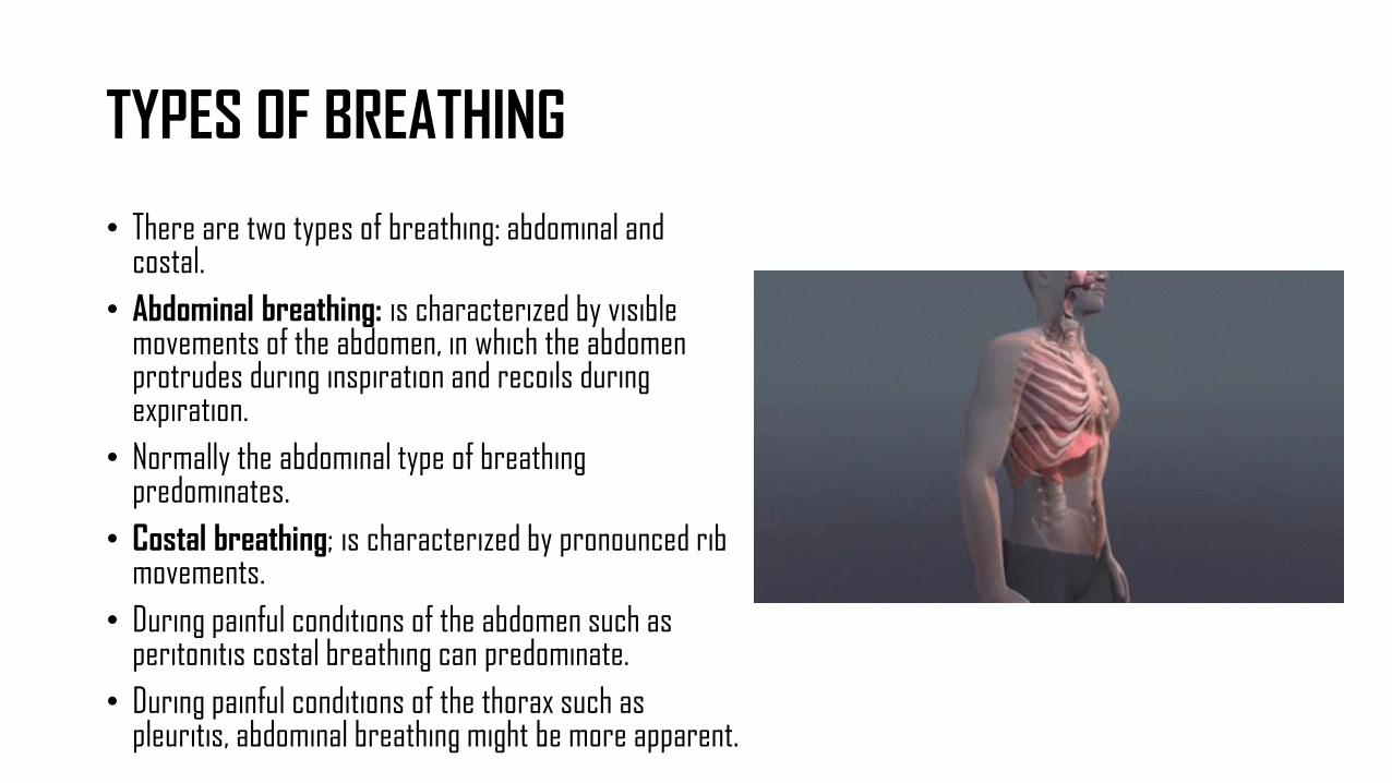
• There are two types of breathing: abdominal and costal.
• Abdominal breathing: is characterized by visible movements of the abdomen, in which the abdomen protrudes during inspiration and recoils during expiration.
• Normally the abdominal type of breathing predominates.
• Costal breathing; is characterized by pronounced rib movements.
• During painful conditions of the abdomen such as peritonitis costal breathing can predominate.
• During painful conditions of the thorax such as pleuritis, abdominal breathing might be more apparent.
STATES OF BREATHING
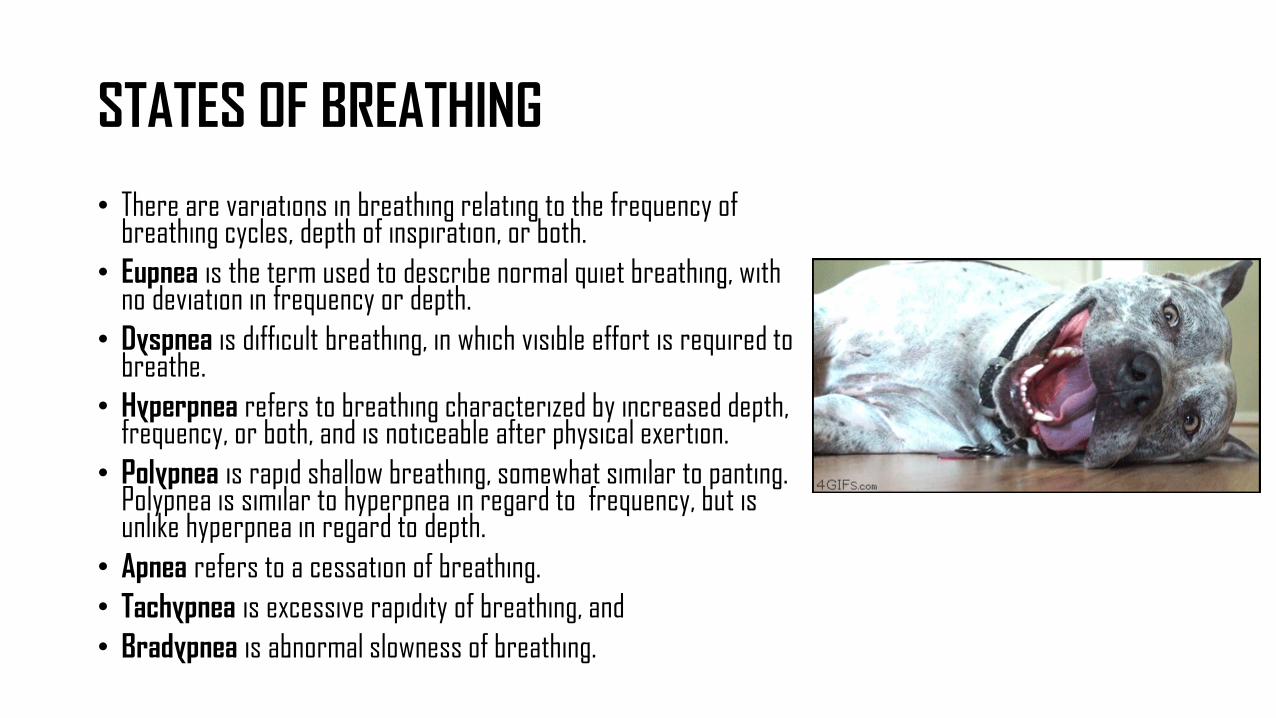
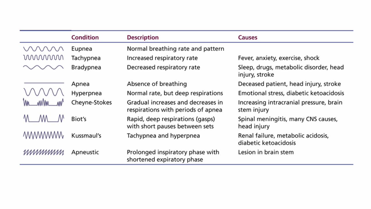
• There are variations in breathing relating to the frequency of breathing cycles, depth of inspiration, or both.
• Eupnea is the term used to describe normal quiet breathing, with no deviation in frequency or depth.
• Dyspnea is difficult breathing, in which visible effort is required to breathe.
• Hyperpnea refers to breathing characterized by increased depth, frequency, or both, and is noticeable after physical exertion.
• Polypnea is rapid shallow breathing, somewhat similar to panting. Polypnea is similar to hyperpnea in regard to frequency, but is unlike hyperpnea in regard to depth.
• Apnea refers to a cessation of breathing. • Tachypnea is excessive rapidity of breathing, and • Bradypnea is abnormal slowness of breathing.
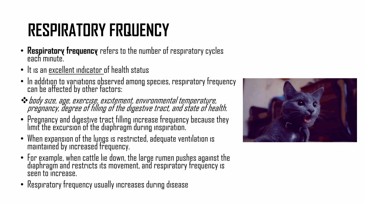
RESPIRATORY FRQUENCY• Respiratory frequency refers to the number of respiratory cycles
each minute. • It is an excellent indicator of health status• In addition to variations observed among species, respiratory frequency
can be affected by other factors:body size, age, exercise, excitement, environmental temperature,
pregnancy, degree of filling of the digestive tract, and state of health. • Pregnancy and digestive tract filling increase frequency because they
limit the excursion of the diaphragm during inspiration. • When expansion of the lungs is restricted, adequate ventilation is
maintained by increased frequency. • For example, when cattle lie down, the large rumen pushes against the
diaphragm and restricts its movement, and respiratory frequency is seen to increase.
• Respiratory frequency usually increases during disease
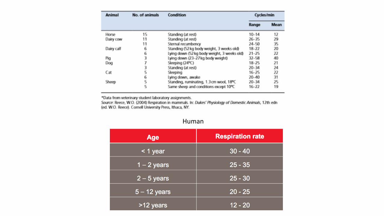
Human
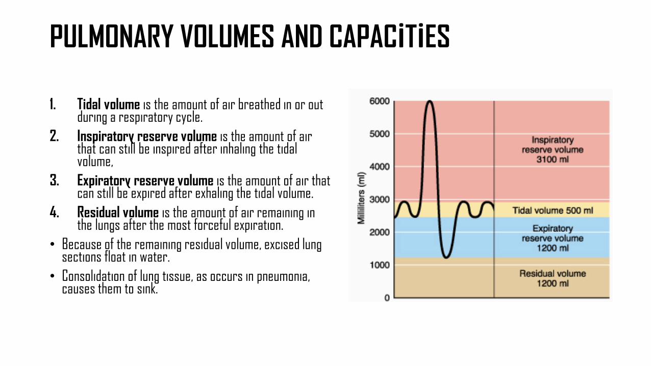
PULMONARY VOLUMES AND CAPACİTİES
1. Tidal volume is the amount of air breathed in or out during a respiratory cycle.
2. Inspiratory reserve volume is the amount of air that can still be inspired after inhaling the tidal volume,
3. Expiratory reserve volume is the amount of air that can still be expired after exhaling the tidal volume.
4. Residual volume is the amount of air remaining in the lungs after the most forceful expiration.
• Because of the remaining residual volume, excised lung sections float in water.
• Consolidation of lung tissue, as occurs in pneumonia, causes them to sink.
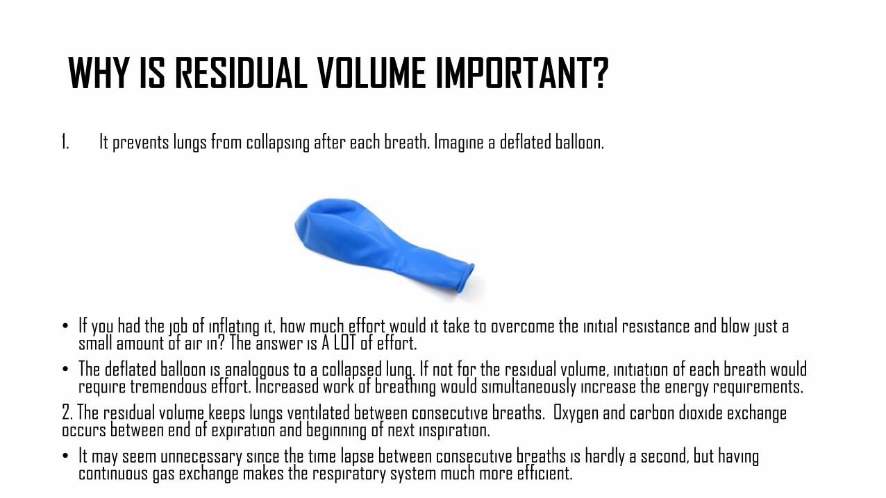
WHY IS RESIDUAL VOLUME IMPORTANT?1. It prevents lungs from collapsing after each breath. Imagine a deflated balloon.
• If you had the job of inflating it, how much effort would it take to overcome the initial resistance and blow just a small amount of air in? The answer is A LOT of effort.
• The deflated balloon is analogous to a collapsed lung. If not for the residual volume, initiation of each breath would require tremendous effort. Increased work of breathing would simultaneously increase the energy requirements.
2. The residual volume keeps lungs ventilated between consecutive breaths. Oxygen and carbon dioxide exchange occurs between end of expiration and beginning of next inspiration.• It may seem unnecessary since the time lapse between consecutive breaths is hardly a second, but having
continuous gas exchange makes the respiratory system much more efficient.
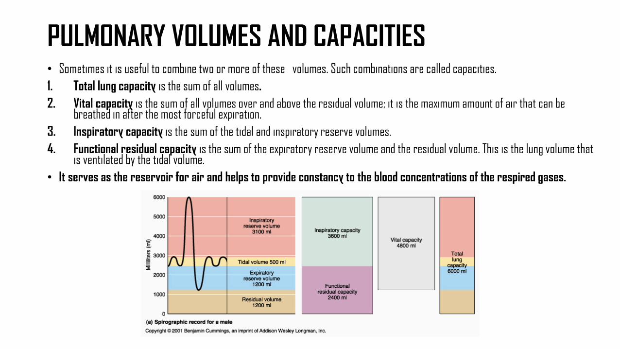
PULMONARY VOLUMES AND CAPACITIES• Sometimes it is useful to combine two or more of these volumes. Such combinations are called capacities. 1. Total lung capacity is the sum of all volumes. 2. Vital capacity is the sum of all volumes over and above the residual volume; it is the maximum amount of air that can be
breathed in after the most forceful expiration. 3. Inspiratory capacity is the sum of the tidal and inspiratory reserve volumes. 4. Functional residual capacity is the sum of the expiratory reserve volume and the residual volume. This is the lung volume that
is ventilated by the tidal volume.• It serves as the reservoir for air and helps to provide constancy to the blood concentrations of the respired gases.
What have we learned today?
• Differentiate between abdominal and costal breathing. • What are some commonly referred to states of breathing? • What is the difference between a lung volume subdivision and a lung capacity subdivision? • When expansion of the lungs is restricted, how is adequate ventilation maintained? • What are some factors that affect respiratory frequency?• What is the difference between pulmonary volumes and pulmonary capacities?• What is the definition of vital capacity? • What is the functional residual capacity?
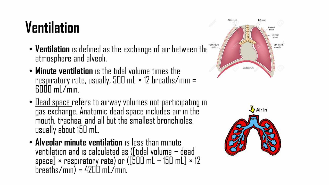
Ventilation• Ventilation is defined as the exchange of air between the
atmosphere and alveoli.• Minute ventilation is the tidal volume times the
respiratory rate, usually, 500 mL × 12 breaths/min = 6000 mL/min.
• Dead space refers to airway volumes not participating in gas exchange. Anatomic dead space includes air in the mouth, trachea, and all but the smallest bronchioles, usually about 150 mL.
• Alveolar minute ventilation is less than minute ventilation and is calculated as ([tidal volume − dead space] × respiratory rate) or ([500 mL − 150 mL] × 12 breaths/min) = 4200 mL/min.
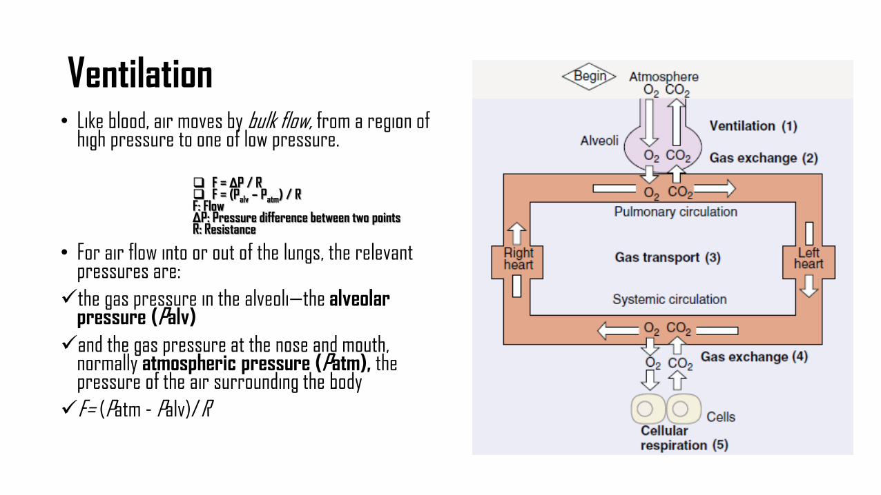
Ventilation• Like blood, air moves by bulk flow, from a region of
high pressure to one of low pressure.
F = ΔP / R F = (Palv – Patm) / RF: FlowΔP: Pressure difference between two pointsR: Resistance
• For air flow into or out of the lungs, the relevant pressures are:the gas pressure in the alveoli—the alveolar
pressure (Palv)and the gas pressure at the nose and mouth,
normally atmospheric pressure (Patm), the pressure of the air surrounding the bodyF= (Patm - Palv)/R
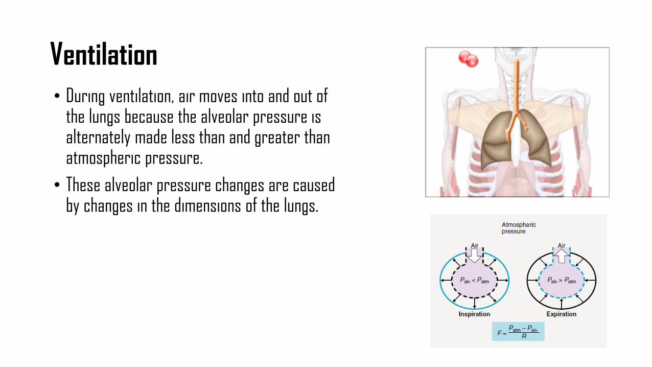
Ventilation• During ventilation, air moves into and out of
the lungs because the alveolar pressure is alternately made less than and greater than atmospheric pressure.
• These alveolar pressure changes are causedby changes in the dimensions of the lungs.
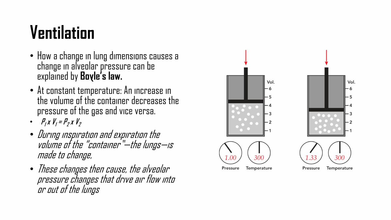
Ventilation• How a change in lung dimensions causes a
change in alveolar pressure can be explained by Boyle’s law.
• At constant temperature: An increase in the volume of the container decreases thepressure of the gas and vice versa.
• P1 x V1 = P2 x V2
• During inspiration and expiration the volume of the “container”—the lungs—is made to change,
• These changes then cause, the alveolar pressure changes that drive air flow into or out of the lungs
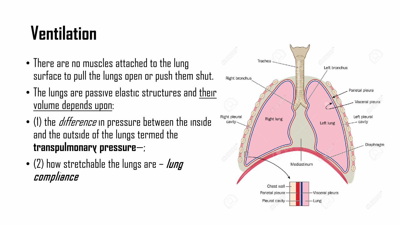
Ventilation• There are no muscles attached to the lung
surface to pull the lungs open or push them shut. • The lungs are passive elastic structures and their
volume depends upon:• (1) the difference in pressure between the inside
and the outside of the lungs termed the transpulmonary pressure—;
• (2) how stretchable the lungs are – lungcompliance
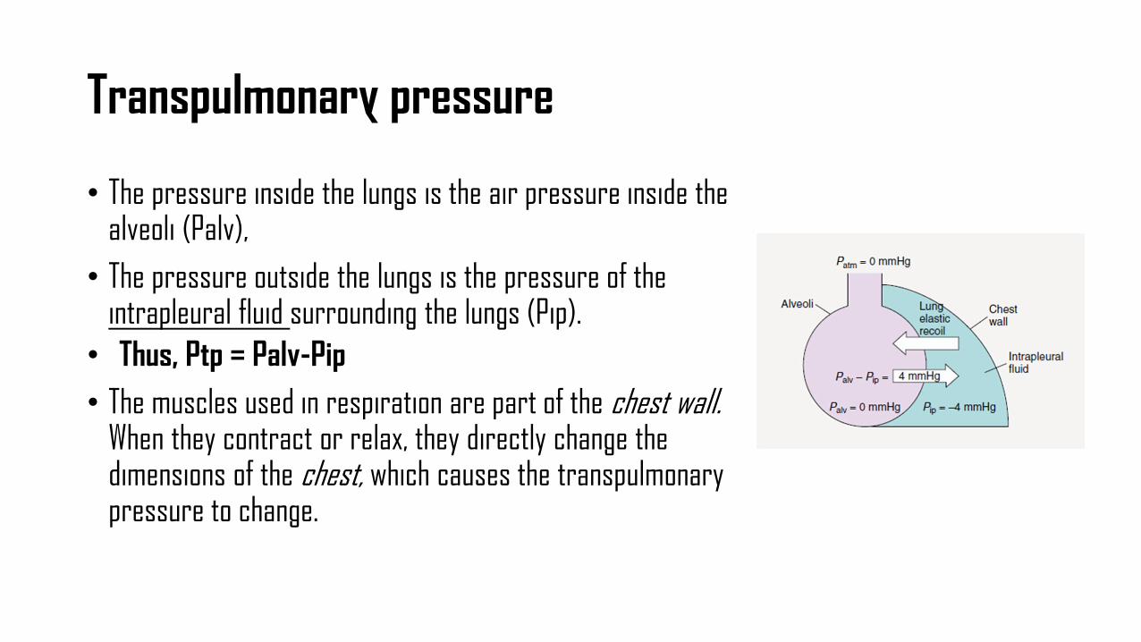
Transpulmonary pressure
• The pressure inside the lungs is the air pressure inside the alveoli (Palv),
• The pressure outside the lungs is the pressure of the intrapleural fluid surrounding the lungs (Pip).
• Thus, Ptp = Palv-Pip• The muscles used in respiration are part of the chest wall.
When they contract or relax, they directly change the dimensions of the chest, which causes the transpulmonarypressure to change.
Transpulmonary pressure
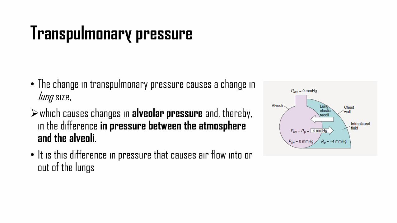
• The change in transpulmonary pressure causes a change in lung size, which causes changes in alveolar pressure and, thereby,
in the difference in pressure between the atmosphere and the alveoli.
• It is this difference in pressure that causes air flow into or out of the lungs
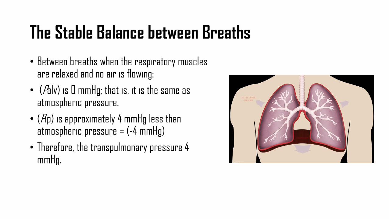
The Stable Balance between Breaths• Between breaths when the respiratory muscles
are relaxed and no air is flowing:• (Palv) is 0 mmHg; that is, it is the same as
atmospheric pressure. • (Pip) is approximately 4 mmHg less than
atmospheric pressure = (-4 mmHg)• Therefore, the transpulmonary pressure 4
mmHg.
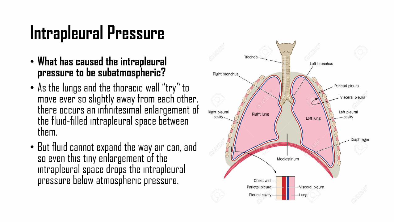
Intrapleural Pressure• What has caused the intrapleural
pressure to be subatmospheric?• As the lungs and the thoracic wall “try” to
move ever so slightly away from each other, there occurs an infinitesimal enlargement of the fluid-filled intrapleural space between them.
• But fluid cannot expand the way air can, and so even this tiny enlargement of the intrapleural space drops the intrapleuralpressure below atmospheric pressure.
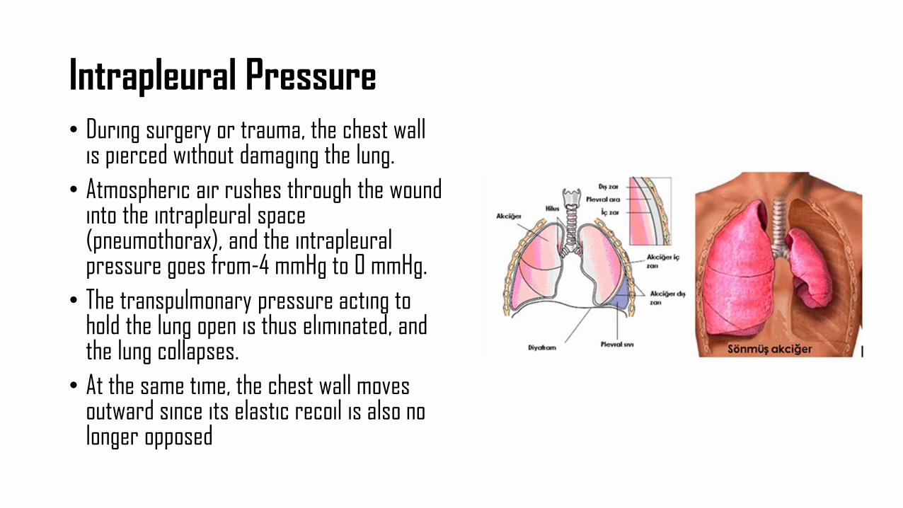
Intrapleural Pressure• During surgery or trauma, the chest wall
is pierced without damaging the lung.• Atmospheric air rushes through the wound
into the intrapleural space (pneumothorax), and the intrapleuralpressure goes from-4 mmHg to 0 mmHg.
• The transpulmonary pressure acting to hold the lung open is thus eliminated, and the lung collapses.
• At the same time, the chest wall moves outward since its elastic recoil is also no longer opposed
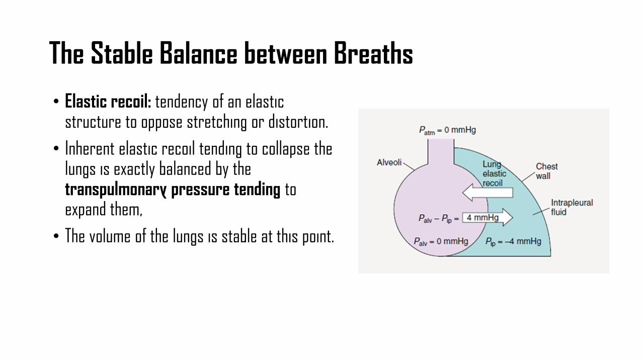
The Stable Balance between Breaths• Elastic recoil: tendency of an elastic
structure to oppose stretching or distortion. • Inherent elastic recoil tending to collapse the
lungs is exactly balanced by the transpulmonary pressure tending toexpand them,
• The volume of the lungs is stable at this point.
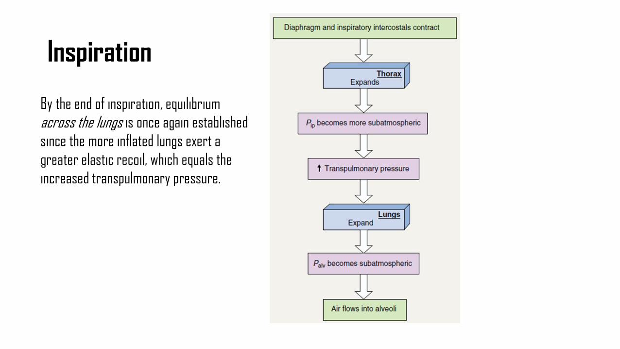
Inspiration
By the end of inspiration, equilibrium across the lungs is once again establishedsince the more inflated lungs exert a greater elastic recoil, which equals the increased transpulmonary pressure.
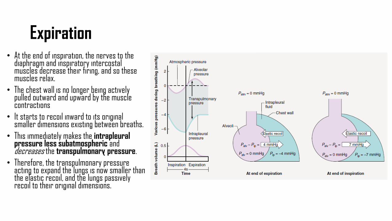
Expiration• At the end of inspiration, the nerves to the
diaphragm and inspiratory intercostalmuscles decrease their firing, and so these muscles relax.
• The chest wall is no longer being actively pulled outward and upward by the muscle contractions
• It starts to recoil inward to its original smaller dimensions existing between breaths.
• This immediately makes the intrapleuralpressure less subatmospheric and decreases the transpulmonary pressure.
• Therefore, the transpulmonary pressure acting to expand the lungs is now smaller than the elastic recoil, and the lungs passively recoil to their original dimensions.

ANY QUESTIONS?