Embed Size (px)
Citation preview
E
R(iE
CRF
a
Mb
c
Md
Me
RA
h0
pilepsy Research (2015) 113, 98—103
j ourna l h om epa ge: www.elsev ier .com/ locate /ep i lepsyres
esponders to vagus nerve stimulationVNS) in refractory epilepsy have reducednterictal cortical synchronicity on scalpEG
lémentine Bodina, Sandrine Auberta, Géraldine Daquinb,omain Carronc, Didier Scavardad, Aileen McGonigal a,abrice Bartolomeia,e,∗
CHU Timone, Service de Neurophysiologie Clinique, Assistance Publique des Hôpitaux de Marseille,arseille F-13005, FranceHôpital Henri Gastaut, 13009 Marseille, FranceCHU Timone, Stereotactic and Functional Neurosurgery Department, Assistance Publique des Hôpitaux dearseille, Marseille F-13005, FranceCHU Timone, Pediatric Neurosurgery Department, Assistance Publique des Hôpitaux de Marseille,arseille F-13005, FranceInstitut de Neurosciences des Systèmes, INSERM UMR 1106, Marseille, France
eceived 15 February 2015; received in revised form 18 March 2015; accepted 28 March 2015vailable online 9 April 2015
KEYWORDSVNS;EEG;Functional
Summary EEG desynchronization has been proposed to be an important mechanism forantiepileptic effect of vagus nerve stimulation (VNS) but has never been clearly documentedin human. The aim of this study was to evaluate impact of VNS on the synchronicity of inter-ictal EEG rhythms. We estimated synchronization between scalp EEG signals using phase lag
connectivity;Synchrony;Epilepsy
index (PLI) in 19 patients with chronic VNS therapy. We estimated changes in synchronizationbetween ON and OFF phases and between responder (R) and non-responder (NR) patients. Wefound that R have a lower global level of synchronization (EEG broadband) than NR (p < 0.0001)In addition, ON periods were characterized by lower values in comparison with OFF periods(p < 0.001). R had significantly lower global synchronization levels in delta and alpha frequency
bands (p < 0.0001).∗ Corresponding author at: CHU Timone, Service de Neurophysiologie Clinique, CHU Timone-264 Rue St Pierre, F-13005 Marseille, France.E-mail address: [email protected] (F. Bartolomei).
ttp://dx.doi.org/10.1016/j.eplepsyres.2015.03.018920-1211/© 2015 Elsevier B.V. All rights reserved.
VNS and cortical synchrony 99
Patients responding to VNS have thus a lower level of broadband EEG synchronization than non-responders. Estimating changes of synchronization level is thus a promising tool for predictingresponse to VNS.© 2015 Elsevier B.V. All rights reserved.
‘awd(A(
E
Sufwasbjsabetst
P
Tpal
eadcesibeslthe PLI only quantifies the relative phase difference dis-tribution asymmetry. PLI values range between 0 and 1.A PLI value of zero indicates either no coupling or cou-pling with a phase difference around 0 while a PLI value
Introduction
In a large proportion (∼30%) of patients with epilepsy,seizures persist despite appropriate antiepileptic treat-ments (Wilcox et al., 2013). In this context, vagus nervestimulation (VNS) represents a non-pharmacological low-risksurgical option. A recent meta-analysis has confirmed VNS tobe an effective and reasonably safe adjunctive in medicallyrefractory epilepsy, with an overall responder rate of 57%(Englot et al., 2011). However the mechanisms of actionof VNS remain poorly understood. Initially based on animalstudies, desynchronization of the EEG has been proposed tobe an important mechanism for antiepileptic effect (Jaseja,2010) but this effect has never been demonstrated in humanepilepsies. The effect of VNS on awake EEG is uncertain andcontroversial (Marrosu et al., 2005; Salinsky and Burchiel,1993). More recently a study found desynchronization in thegamma band frequency for responder patients (Fraschiniet al., 2013). However this result is disputable since it hasbeen shown that gamma activity is unlikely to be recordedfrom scalp EEG (Whitham et al., 2007).
The aim of this study was to evaluate impact of VNSon the synchronicity of interictal EEG rhythms. We particu-larly wished to evaluate changes in synchronization betweenON and OFF phases and to compare responders with non-responder patients.
Materials and methods
Subjects
Nineteen patients (7 females, 12 males, 14—54 y) withdrug-resistant epilepsy, non-eligible for epilepsy surgery,were retrospectively selected for this study. All patientswere implanted with a vagus nerve stimulator device (VNS,Cyberonics, Houston, TX) according to standard procedure(Landre, 2004). They were selected on the following crite-ria:
- at least one scalp EEG recording was done after VNSimplantation,
- EEG were recorded using an additional bipolar electrodecapturing the VNS artifact of stimulation,
- EEG segments without artifacts were available for analy-sis.
The subjects were divided into two groups based on theirresponse to the VNS therapy at the time of the studiedEEG. Those whose seizures were reduced by at least 50%in frequency (over the six months preceding the EEG) were
qualified as ‘‘responders’’. Others were qualified as ‘‘non-responders’’.oT
In all the included patients the ‘‘ON’’ period was 30 s, the‘OFF’’ periods were 3 or 5 min; 30 Hz frequency was system-tically used. Intensities ranged from 0.5 to 2.5 mA and pulseidth was 250 or 500 �s. Patients (range age: 17—41) wereiagnosed with drug-resistant generalized (n = 9) or partialmultifocal, bilateral, or in eloquent areas, n = 10) epilepsy.mong them, 10 were responders (R) and 9 non-respondersNR) at the time of the EEG (Table 1).
EG analysis
calp electroencephalographic (EEG) signals were recordedsing a 21 channel EEG system (Natus, France) and was per-ormed at least 3 months after the VNS surgery. Signalsere acquired during a 20 min eyes-closed resting state,t a distance (at least 3 h) from seizures or post-ictaltate, digitized with sample frequency set to 256 Hz andand pass filtered between 0.5 and 70 Hz. For each sub-ect ten artifact-free epochs using average reference wereelected for analysis. To precisely differentiate the ‘‘ON’’nd ‘‘OFF’’ stimulation periods we made use of additionalipolar recording to capture the stimulation artifact. Twolectrodes were placed, one on the neck close to the elec-rode scar and the other on the thorax. During the ON cycle,timulation was associated with 30 Hz activity detected onhese electrodes.
hase lag index
he PLI is a measure of the asymmetry of the distribution ofhase differences between two signals (Stam et al., 2007)nd it seems to be a convenient indicator of synchronizationevels in EEG.
Functional connectivity between all possible pairs of 21lectrodes was calculated using the phase lag index (PLI),
measure of the asymmetry of the distribution of phaseifferences between EEG signals. A detailed method foralculating the PLI is described in previous work (Stamt al., 2007). The PLI performs at least as well as theynchronization likelihood or phase coherence in detect-ng true changes in synchronization but is less affectedy the influence of common sources and/or active ref-rence electrodes, an important potential problem whencalp EEG is used. This is due to the fact that the zero-ag synchronization is removed from the analyses, and that
f 1 indicates perfect phase locking plus or minor pi.he more consistent this non-zero phase locking is, the
100 C. Bodin et al.
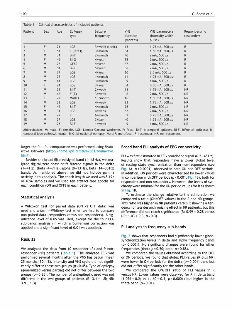
Table 1 Clinical characteristics of included patients.
Patient Sex Age Epilepsytype
Seizurefrequency
VNSduration(months)
VNS parameters(intensity widthpulse)
Responders/noresponders
1 F 21 LGS 3/week (tonic) 13 1.75 mA, 500 �s R2 F 54 F (left I) 3/month 34 1.50 mA, 500 �s R3 M 31 Bi-T 2/month 40 2 mA, 500 �s R4 F 45 Bi-O 4/year 32 2 mA, 500 �s R5 M 28 GEFS+ 4/year 32 2 mA, 500 �s R6 M 54 Bi-F 5/year 38 2 mA, 500 �s R7 M 37 LGS 4/year 60 2.5 mA, 500 �s R8 M 25 LGS 1/month 14 1.25 mA, 500 �s R9 M 14 LGS 3/month 9 1 mA, 500 �s R10 F 21 LGS 3/year 4 0.50 mA, 500 �s R11 M 21 Bi-T 2/week 11 1.75 mA, 500 �s NR12 M 12 F (T) 3/week 6 2 mA, 500 �s NR13 F 27 Multi F 10/month 16 1.50 mA, 500 �s NR14 M 32 LGS 4/week 23 1.75 mA, 500 �s NR15 F 42 Bi-T 4/month 26 2 mA, 500 �s NR16 M 31 LGS 4/week 67 2 mA, 500 �s NR17 M 27 F 6/month 7 0.75 mA, 500 �s NR18 M 27 LGS 3/day 40 1.25 mA, 500 �s NR19 M 25 Bi-F 1/week 9 1 mA, 500 �s NR
me,
F: mu
lwh
l(baoe
S
Aunnsa
R
Wrp2c(gd3
B
PRoFIirci
cTddN
P
Fs(f
owd
Abbreviations: M: male, F: female, LGS: Lennox Gastaut syndrotemporal lobe epilepsyI: insula; Bi-O: bi-occipital epilepsy; Multi
arger the PLI. PLI computation was performed using Brain-ave software (http://home.kpn.nl/stam7883/brainwave.tml).
Besides the broad filtered signal band (1—48 Hz), we ana-yzed digital zero-phase shift filtered signals in the delta1—4 Hz), theta (4—7 Hz), alpha (8—13 Hz), beta (14—30 Hz)ands. As mentioned above, we did not include gammactivity in this analysis. The epoch length we used was 8.19 sr 4096 samples and we used ten artifact-free epochs forach condition (ON and OFF) in each patient.
tatistical analysis
Wilcoxon test for paired data (ON vs OFF data) wassed and a Mann—Whitney test when we had to compareon-paired data (responders versus non-responders). A sig-ificance level of 0.05 was used, except for the four EEGub-bands analysis (in which a Bonferroni correction waspplied and a significant level of 0.01 was applied).
esults
e analyzed the data from 10 responder (R) and 9 non-esponder (NR) patients (Table 1). The analyzed EEG waserformed several months after the VNS has begun (mean5 months, SD: 18). Intensity and VNS cycle did not signifi-antly differ in these two groups (p = 0.45). Type of epilepsy
generalized versus partial) did not differ between the tworoups (p = 0.25). The number of antiepileptic used was notifferent in the two groups of patients (R: 3.1 ± 1.5; NR:.9 ± 1.3).v(t
F: focal, Bi-T: bitemporal epilepsy, Bi-F: bifrontal epilepsy; T:ltifocal; R: responder; NR: non responder.
road band PLI analysis of EEG connectivity
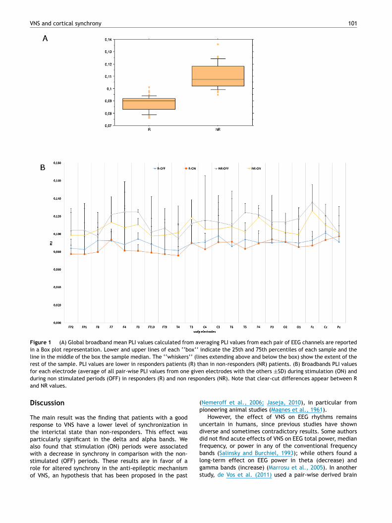
LI was first estimated in EEG broadband signal (0.5—48 Hz).esults show that responders have a lower global levelf resting state synchronization than non-responders (seeig. 1A, p < 0.0001), observed in both ON and OFF periods.n addition, ON periods were characterized by lower valuesn comparison with OFF periods (p < 0.001; Fig. 1B), both foresponders and non responders. However, the levels of syn-hrony were minimal for the ON period values for R as shownn Fig. 1B.
To estimate the change relative to the stimulation weompared a ratio (ON/OFF values) in the R and NR groups.his ratio was higher in NR patients versus R showing a ten-ency for less desynchronizing effect in NR patients; but thisifference did not reach significance (R: 0.99 ± 0.28 versusR: 1.03 ± 0.3, p = 0.3).
LI analysis in frequency sub-bands
ig. 2 shows that responders had significantly lower globalynchronization levels in delta and alpha frequency bandsp < 0.0001). No significant changes were found for otherrequencies (theta p = 0.50; beta, p = 0.88).
We compared the values obtained according to the OFFr ON periods. We found that global PLI values (R plus NR)ere lower in ON periods for the delta (p = 0.004) band butid not differ significantly for the other bands.
We compared the ON/OFF ratio of PLI values in Rersus NR. Lower values were observed for R in delta band1.026 ± 0.2; vs 1.160 ± 0.3, p = 0.0001) but higher in theheta band (p = 0.01).
VNS and cortical synchrony 101
Figure 1 (A) Global broadband mean PLI values calculated from averaging PLI values from each pair of EEG channels are reportedin a Box plot representation. Lower and upper lines of each ‘‘box’’ indicate the 25th and 75th percentiles of each sample and theline in the middle of the box the sample median. The ‘‘whiskers’’ (lines extending above and below the box) show the extent of therest of the sample. PLI values are lower in responders patients (R) than in non-responders (NR) patients. (B) Broadbands PLI valuesfor each electrode (average of all pair-wise PLI values from one given electrodes with the others ±SD) during stimulation (ON) and
respo
(p
uddfb
during non stimulated periods (OFF) in responders (R) and non
and NR values.
Discussion
The main result was the finding that patients with a goodresponse to VNS have a lower level of synchronization inthe interictal state than non-responders. This effect wasparticularly significant in the delta and alpha bands. Wealso found that stimulation (ON) periods were associated
with a decrease in synchrony in comparison with the non-stimulated (OFF) periods. These results are in favor of arole for altered synchrony in the anti-epileptic mechanismof VNS, an hypothesis that has been proposed in the pastlgs
nders (NR). Note that clear-cut differences appear between R
Nemeroff et al., 2006; Jaseja, 2010), in particular fromioneering animal studies (Magnes et al., 1961).
However, the effect of VNS on EEG rhythms remainsncertain in humans, since previous studies have showniverse and sometimes contradictory results. Some authorsid not find acute effects of VNS on EEG total power, medianrequency, or power in any of the conventional frequencyands (Salinsky and Burchiel, 1993); while others found a
ong-term effect on EEG power in theta (decrease) andamma bands (increase) (Marrosu et al., 2005). In anothertudy, de Vos et al. (2011) used a pair-wise derived brain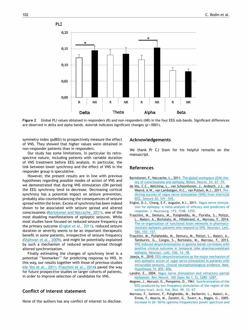
102 C. Bodin et al.
Figure 2 Global PLI values obtained in responders (R) and non-responders (NR) in the four EEG sub-bands. Significant differencesa cant
son
solr
hwtspsscmmtdb(ba
pt(fi
C
N
A
Wm
R
B
d
E
F
F
J
L
M
re observed in delta and alpha bands. Asterisk indicates signifi
ymmetry index (pdBSI) to prospectively measure the effectf VNS. They showed that higher values were obtained inon-responder patients than in responders.
Our study has some limitations, in particular its retro-pective nature, including patients with variable durationf VNS treatment before EEG analysis. In particular, theink between lower synchrony and the effect of VNS in theesponder group is speculative.
However, the present results are in line with previousypotheses regarding possible modes of action of VNS ande demonstrated that during VNS stimulation (ON period)
he EEG synchrony tend to decrease. Decreasing corticalynchrony has a potential impact on seizure prevention,robably also counterbalancing the consequences of seizurepread within the brain. Excess of synchrony has been indeedhown to be associated with seizure spread and alteredonsciousness (Bartolomei and Naccache, 2011), one of theost disabling manifestations of epileptic seizures. Whileost studies have focused on reduced seizure frequency as
he primary outcome (Englot et al., 2011), reduced seizureuration or severity seems to be an important therapeuticenefit in some patients, irrespective of seizure frequencyShahwan et al., 2009), and might be potentially explainedy such a mechanism of reduced seizure spread throughltered synchronization.
Finally estimating the changes of synchrony level is aotential ‘‘biomarker’’ for predicting response to VNS. Inhis way, our results together with those of previous studiesde Vos et al., 2011; Fraschini et al., 2014) paved the wayor future prospective studies on larger cohorts of patients,n order to improve selection of candidates for VNS.
onflict of interest statement
one of the authors has any conflict of interest to disclose.
M
changes (p < 0001).
cknowledgements
e thank Pr CJ Stam for his helpful remarks on theanuscript.
eferences
artolomei, F., Naccache, L., 2011. The global workspace (GW) the-ory of consciousness and epilepsy. Behav. Neurol. 24, 67—74.
e Vos, C.C., Melching, L., van Schoonhoven, J., Ardesch, J.J., deWeerd, A.W., van Lambalgen, H.C., van Putten, M.J., 2011. Pre-dicting success of vagus nerve stimulation (VNS) from interictalEEG. Seizure 20, 541—545.
nglot, D.J., Chang, E.F., Auguste, K.I., 2011. Vagus nerve stimula-tion for epilepsy: a meta-analysis of efficacy and predictors ofresponse. J. Neurosurg. 115, 1248—1255.
raschini, M., Demuru, M., Puligheddu, M., Floridia, S., Polizzi,L., Maleci, A., Bortolato, M., Hillebrand, A., Marrosu, F., 2014.The re-organization of functional brain networks in pharmaco-resistant epileptic patients who respond to VNS. Neurosci. Lett.580, 153—157.
raschini, M., Puligheddu, M., Demuru, M., Polizzi, L., Maleci, A.,Tamburini, G., Congia, S., Bortolato, M., Marrosu, F., 2013.VNS induced desynchronization in gamma bands correlates withpositive clinical outcome in temporal lobe pharmacoresistantepilepsy. Neurosci. Lett. 536, 14—18.
aseja, H., 2010. EEG-desynchronization as the major mechanism ofanti-epileptic action of vagal nerve stimulation in patients withintractable seizures: clinical neurophysiological evidence. Med.Hypotheses 74, 855—856.
andre, E., 2004. Vagus nerve stimulation and refractory partialepilepsies. Rev. Neurol. 160 (Spec No 1, 5), S280—S287.
agnes, J., Moruzzi, G., Pompeino, O., 1961. Synchronization of theEEG produced by low frequency stimulation of the region of the
solitary tract. Arch. Ital. Biol. 99, 33—67.arrosu, F., Santoni, F., Puligheddu, M., Barberini, L., Maleci, A.,Ennas, F., Mascia, M., Zanetti, G., Tuveri, A., Biggio, G., 2005.Increase in 20—50 Hz (gamma frequencies) power spectrum and
S
W
W
VNS and cortical synchrony
synchronization after chronic vagal nerve stimulation. Clin. Neu-rophysiol. 116, 2026—2036 (official journal of the InternationalFederation of Clinical Neurophysiology).
Nemeroff, C.B., Mayberg, H.S., Krahl, S.E., McNamara, J., Frazer,A., Henry, T.R., George, M.S., Charney, D.S., Brannan, S.K.,2006. VNS therapy in treatment-resistant depression: clinicalevidence and putative neurobiological mechanisms. Neuropsy-chopharmacology 31, 1345—1355 (official publication of theAmerican College of Neuropsychopharmacology).
Salinsky, M.C., Burchiel, K.J., 1993. Vagus nerve stimulation hasno effect on awake EEG rhythms in humans. Epilepsia 34,299—304.
Shahwan, A., Bailey, C., Maxiner, W., Harvey, A.S., 2009.Vagus nerve stimulation for refractory epilepsy in children:more to VNS than seizure frequency reduction. Epilepsia 50,1220—1228.
103
tam, C.J., Nolte, G., Daffertshofer, A., 2007. Phase lag index:assessment of functional connectivity from multi channel EEGand MEG with diminished bias from common sources. Hum. BrainMapp. 28, 1178—1193.
hitham, E.M., Pope, K.J., Fitzgibbon, S.P., Lewis, T., Clark, C.R.,Loveless, S., Broberg, M., Wallace, A., DeLosAngeles, D., Lil-lie, P., Hardy, A., Fronsko, R., Pulbrook, A., Willoughby, J.O.,2007. Scalp electrical recording during paralysis: quantitativeevidence that EEG frequencies above 20 Hz are contaminatedby EMG. Clin. Neurophysiol. 118, 1877—1888 (official journal ofthe International Federation of Clinical Neurophysiology).
ilcox, K.S., Dixon-Salazar, T., Sills, G.J., Ben-Menachem, E.,
White, H.S., Porter, R.J., Dichter, M.A., Moshe, S.L., Noebels,J.L., Privitera, M.D., Rogawski, M.A., 2013. Issues related todevelopment of new antiseizure treatments. Epilepsia 54 (Suppl.4), 24—34.


