Embed Size (px)
Citation preview
Role of SV40 Integration Site at Chromosomal Interval 1q21.1 inImmortalized CRL2504 Cells
Jinglan Liu1, Gurpreet Kaur1, Vikramjit K. Zhawar1, Drazen B. Zimonjic2, Nicholas C.Popescu2, Raj P. Kandpal3, and Raghbir S. Athwal1,*1 Fels Institute for Cancer Research and Molecular Biology, Temple University School ofMedicine, Philadelphia, PA 191402 Laboratory of Experimental Carcinogenesis, National Cancer Institute, Bethesda MD20892-42623 Department of Basic Medical Sciences, Western University of Health Sciences, Pomona, CA91766
AbstractWe have applied a functional gene transfer strategy to demonstrate the importance of viralintegration site in cellular immortalization. The large tumor antigen of SV40 is capable ofextending the cellular life span by sequestering tumor suppressor proteins pRB and p53 in virus-transformed human cells. Although SV40-LT is essential, it is not sufficient for cellularimmortalization, suggesting that additional alterations in cellular genes are required to attaininfinite proliferation. We demonstrate here that the disruption of human chromosomal interval at1q21.1, by SV40 integration, can be an essential step for cellular immortalization. The transfer ofa 150Kb bacterial artificial chromosome (BAC) clone, RP364B14, corresponding to viralintegration site in CRL2504 cells, reverted their immortal phenotype. Interestingly, the BACtransfer clones of CRL-2504 cells displayed characteristics of either senescence as shown by β-galactosidase activity or apoptosis as revealed by positive staining with M30 cytoDeath antibody.The SV40 integration at 1q21.1, in the vicinity of epidermal differentiation complex genes,resulted in the down-regulation of the filaggrin (FLG) gene that is part of the epidermaldifferentiation complex. FLG gene expression was restored to its normal levels in BAC transfersenescent and apoptotic clones. Our results suggest that the disruption of native genomic sequenceby SV40 may alter expression of genes involved in senescence and apoptosis by modulatingchromatin structure. These studies imply that identification of genes located in the vicinity of viralintegration sites in human cancers may be helpful in developing new diagnostic and therapeuticstrategies.
KeywordsSV40 Integration Site; Senescence; Apoptosis; Filaggrin; Chromatin Structure
INTRODUCTIONThe mechanisms of neoplastic transformation have been addressed by using human cellsimmortalized with DNA tumor viruses or viral sequences, such as SV40 large T antigen
*Please address correspondence to: Dr. Raghbir S. Athwal, Fels Institute for Cancer Research and Molecular Biology, TempleUniversity School of Medicine, Philadelphia, PA [email protected].
NIH Public AccessAuthor ManuscriptCancer Res. Author manuscript; available in PMC 2010 October 1.
Published in final edited form as:Cancer Res. 2009 October 1; 69(19): 7819–7825. doi:10.1158/0008-5472.CAN-09-1003.
NIH
-PA Author Manuscript
NIH
-PA Author Manuscript
NIH
-PA Author Manuscript

(SV40-LT), HPV16-E6 and E7 and adenovirus E1A and E1B (1). A two stage modelconsisting of M1 (mortality stage 1) and M2 (mortality stage 2) has been proposed forSV40-LT induced immortalization of human cells (2). The cells challenged with SV40 largeT antigen bypass M1 stage and continue to multiply until they reach M2 stage (2).Subsequently, the entire cell population enters a state of crisis that is characterized bymassive cell death through apoptosis or necrosis (3). However, a rare variant cell (1 in107–8) survives the crisis state and continues to multiply indefinitely (2). These studiessuggest that M1 and M2 are regulated through separate genetic controls, and immortalizedcells have either inactivated or bypassed regulatory pathways involved in both M1 and M2stages.
The presence of SV40 has been reported in some human tumors such as mesothelioma,osteosarcoma, ependymomas (4–7), bronchopulmonary carcinoma and non-malignantpulmonary disease (8). Despite these observations, the role of SV40 in human cancersremains controversial. On the other hand, HPV has been detected in greater than 99% ofcervical cancer cases (9). Although the above observations suggest viral etiology of thesecancers, the contribution of disrupted host genes due to integration of viral genome has notbeen addressed.
It has been demonstrated that SV40-LT releases the host cell from the G1 checkpoint (M1stage) and extends its life span by sequestering pRB and p53 family of tumor suppressorproteins (10–12). In order for cell to attain indefinite growth potential, however, additionalmutations in cellular genes are essential to bypass M2 stage (2). Although SV40 is known tointegrate randomly at multiple sites in the host genome, the immortalized cells often showits integration at a unique site. We propose that the disruption of a specific genomic site dueto the viral integration may contribute to malignant transformation of human cells.Furthermore, the integration of SV40 into a specific genomic site may promote genomicinstability and likely confer growth advantage.
We have applied a functional approach to restore senescence in SV40 immortalized cellsand investigated the role of the integration site in cellular immortalization. We havecharacterized the genomic region in the vicinity of the site interrupted by SV40 integration.The introduction of a genomic clone, corresponding to the genomic site disrupted by SV40at 1q21.1, restores senescence or apoptosis in CRL2504, an immortalized cell line.
MATERIALS AND METHODSCell lines and growth conditions
A single cell subclone of a human bronchial epithelial cell line, immortalized with a clonedOri− SV40 (13), was used in these studies. CRL-2504 cells contain a single integrated copyof SV40 genome and do not contain any free virus (J. Liu and R. S. Athwal, unpublishedobservations). The cell lines used for comparison included normal human diploid fibroblasts(HDFs), GMO3468A (Genetic Mutant Cell repository, Camden, NJ) and FS-2 (generated inthe lab). The cell lines were routinely cultured at 37°C in a 7.5% CO2 atmosphere in DF/12media supplemented with 10% fetal bovine serum (FBS) containing 1% penicillin andstreptomycin. The gene transfer clones carrying neo marker were isolated and maintained inthe medium containing G418 (400 μg/ml)
Isolation of human DNA flanking SV40 insertion site by inverse PCRThe EcoRI digested CRL-2504 DNA (400 ng) was circularized by overnight incubation at16°C in an appropriate reaction buffer containing 2 μl of T4 DNA ligase (New EnglandBiolabs, Beverly, MA). The circularized DNA was amplified with the primer pair SV40F1726/SV40R796 specific to the SV40 sequences flanking the human genomic DNA. The
Liu et al. Page 2
Cancer Res. Author manuscript; available in PMC 2010 October 1.
NIH
-PA Author Manuscript
NIH
-PA Author Manuscript
NIH
-PA Author Manuscript

amplification was performed in a 25 μl reaction mixture containing 5 μl of ligation products,2 mM MgCl2, 200 μM each of dNTPs, 1 μM of each primer and 1 unit of Taq polymerase(Promega, Madison, WI). The reaction conditions included initial denaturation at 96°C for 5minutes, followed by 35 cycles of 1 min at 94° C, 1 min at 58° C and 2 min at 72° C. Theamplified products were fractionated by electrophoresis in a 2% agarose gel, visualized byethidium bromide staining and purified from the gel for cloning.
Transfer of BAC clone into CRL-2504 cellsThe BAC clones BAC RP11-364B14 and 152L6 were retrofitted with MJ0X166 toincorporate neo marker, using a vector exchange procedure (14). Briefly, human DNAinserts from the BAC clones were released by digestion with NotI restriction enzyme (NewEngland Biolab, Beverly, MA) and separated by pulsed- field gel electrophoresis (PFGE).The purified inserts were mixed separately with NotI digested and dephosphorylatedretrofitting vector pJMOX166 at a molar ratio of 2:1 or 4:1 and ligated using T4 DNA ligaseat 16° C for 16 hours. An aliquot (1 μl) of the ligated products was transformed intocompetent DH-5α cells and cells were plated on LB agar plates containing chloramphenicol(12.5μg/ml) and kanamycin (30μg/ml). The DNA isolated from the retrofitted clones wasdigested with NotI, fractionated by PFGE and blotted onto a membrane. The blot washybridized with DNA probes for the co-localization of human insert and pJMOX166. Thepresence of neo marker in the retrofitted BAC clones was confirmed by PCR.
The DNA from retrofitted BAC clones 364B14 and 152L6 were transferred into mammaliancells by eletroporation using published procedure (15). Briefly, exponentially growing cellswere harvested by trypisnization, washed twice in media without serum and suspended in0.4 ml of serum free media at a density of 2.5 × 10⁷ cells per ml. The BAC DNA (5 μg) wasadded to the cell suspension, mixed gently and transferred into a cuvette (BioRad) forelectroporation using 350V and 500 μFb that produced a time constant in the range of 10–20seconds (BioRad’s Gene Pulser Apparatus with Capacitance Extender). The cells wereplaced on ice for 5 minutes immediately after electroporation and then plated on five tissueculture dishes (100 mm) in complete media. The medium was replaced after 24 hours withselection medium containing G418 (400ug/ml) and refreshed every two days on a regularbasis. A control experiment was also performed by transferring empty vector pJMOX166into cells and the transfected cells were selected as above.
Analysis of BAC transfer clonesThe BAC transfer colonies that grew in selection medium were either isolated individuallyor followed in plates. The growth of individual colonies was monitored and theirmorphology visualized using a phase contrast microscope and photographed at regularintervals.
i. Staining for senescence associated β-galactosidase (SA β-gal) activity—Thecells in culture dishes were washed with PBS and fixed in 2% formaldehyde/0.2%glutaraldehyde at room temperature. The staining solution (1 mg of 5-bromo-4-chloro-3-indolyl β-galactosidase per ml, 40 mM citric acid/sodium phosphate, pH 6.0, 5 mMpotassium ferrocyanide, 5 mM potassium ferricyanide, 150 mM NaCl, 2 mM MgCl2) wasoverlaid on fixed cells and incubated at 37°C for 16h (16). The stained cells were visualizedusing an inverted phase contrast microscope and photographed.
ii. BrdU incorporation assay for DNA replication—The cells (2000 cells/chamber) inan 8-chamber slide (Nalgene Nunc International., Naperville, IL) were incubated for 24h at37°C and then BrdU (10 μM) was added to the medium. The incubation with BrdU wascarried out for 24 hours and the cells were subsequently fixed for 45 min in a fixative
Liu et al. Page 3
Cancer Res. Author manuscript; available in PMC 2010 October 1.
NIH
-PA Author Manuscript
NIH
-PA Author Manuscript
NIH
-PA Author Manuscript

solution (7 volumes ethanol and 3 volumes 50 mM glycine). The fixed cells were hybridizedwith a 1:600 dilution of mouse anti-BrdU monoclonal antibodies (Molecular probes,Eugene, OR) and signal was detected by staining with a 1:200 dilution of the Alexa Fluor488 conjugated donkey anti-mouse IgG. The nuclei were stained with DAPI and cellsvisualized under a fluorescence microscope.
Analysis for ApoptosisCytoDEATH antidody M30 (Roche Diagnostic Corp., Indianapolis, IN), which recognizes aspecific caspase cleavage site within CK18, were used to detect apoptosis (17). The cellswere seeded on 8-chamber slides (Nalgene Nunc International, Naperville, IL) and fixed inice-cold methanol at −20°C for 30 minutes. The fixed cells were treated with 0.25% TritonX-100 (Sigma, Saint Louis, Mo) and then incubated for 2 h with donkey serum (MolecularProbes, Eugene, OR). The slides were overlaid with a solution (100μl) containing M30antibody and incubated at 4°C overnight. The signal was detected by staining with a 1:200dilution of donkey anti-mouse IgG linked with Alexa Fluor 488 (Molecular probes, Eugene,OR) and cells photographed using Olympus AHVT3 fluorescence research photomicroscopesystem.
Sequences of primersSequences of various PCR primers used are as the follows
SV40-1726F: 5′-ACA GTT TAC AGA TGA CTC TCC-3′
SV40-796R: 5′-TTG CAG TAA AGC TGC AAA TCC-3′
SV40-1745F: 5′-CCA GAC AAA GAA CAA CTG CC-3′
SV40-477 R: 5′-CCG TCA ACA GTA TCT TCC CC-3′
H-F87: 5′-GAT CCC AAC TAA AAC ATC ACC-3′
H-R484: 5′-AGC CCT TAT TGT TTA AAA GAC C-3′
SV2504-IS-F165: 5′-CTT CTG TTA TAT CAT TTG ACC C-3′
SV2504-IS-R453: 5′-TAC TGT TTC ATT CCT TGA GCC-3′
Neo-F1289: 5′-TCA CGA CGA GAT CCT CGC C- 3′
Neo-R1705: 5′-TTG TCA AGA CCG ACC TGT CC-3′
GAPDH-F420: 5′-AGA AGG CTG GGG CTC ATT TG – 3′
GAPDH-R1080: 5′-TCC ACC ACC CTG TTG CTG TA – 3′
Fluorecent In Situ Hybridization (FISH)Normal human mononuclear cells from peripheral blood were cultured in the presence of0.05 μg/ml phytohaemagglutinin (Gibco, Grand Island, NY), treated with 0.05–0.1 μg/mlcolcemide (Gibco) for one hour, exposed to standard hypotonic solution (75 mM KCl) andfixed in 3:1 absolute methanol/glacial acetic acid. Fixed cells were transferred to glass slidesto prepare metaphase chromosome spreads. The metaphase chromosome spreads werehybridized with a mixture of 1 μg of biotin labeled BAC DNA and 100 μg of human Cot-1DNA (Clontech Laboratories). The detection of the hybridized BAC DNA, digital imageacquisition, processing and direct visualization on G-banded chromosomes were all carriedout as described earlier (18).
Liu et al. Page 4
Cancer Res. Author manuscript; available in PMC 2010 October 1.
NIH
-PA Author Manuscript
NIH
-PA Author Manuscript
NIH
-PA Author Manuscript

RESULTSCloning of human DNA sequence flanking the SV40 DNA
A restriction map for the integrated p129-SV40 in CRL-2504 was first deduced by Southernblot analysis of the DNA digested with BamHI, BstXI, EcoRI, KpnI, PstI, and PvuII eitherindividually or with a combination of two enzymes. The digestion of CRL2504 DNA withEcoRI generated 2.2kb and 5.1kb fragments that were linked to SV40 DNA. The inversePCR with primer pair SV40-1726F/796R directed to 2.2 Kb fragment of EcoRI digestedCRL-2504 DNA generated a 1200bp amplicon. The origin of this fragment from the SV40-human DNA junction was confirmed by PCR using primer pair SV40-1745F/477R that wereinternal to the first set of primers. The 1200bp PCR product, designated as SV2504-IS, wascloned into pGEM-T easy vector (Promega, Madison, WI) and sequenced. The databasesearch and BLAST comparison revealed this fragment to contain 552bp of human DNAlinked to 648bp of SV40 sequence (Figure 1A). The identity of the rescued DNA wasfurther confirmed by PCR amplification of human DNA with primer pair H-F87/H-R484designed from the recovered human DNA sequence. These primes amplified an expected297bp product from the human DNA template (data not shown).
Identification of a BAC clone corresponding to SV40 integration site in CRL-2504 cellsA human PCR ready BAC library (Research Genetics, Huntsville, AL) was screened withprimer pair H-F87/H-R484 that led to the identification of a 115kb clone RP11-364B14.This clone, obtained from Research Genetics (Huntsville, AL), was found to represent thehuman–SV40 junction sequence. The position of the BAC was mapped to chromosome 1 byaligning the 552 bp cloned human DNA sequence against genome database. The location ofthe BAC at 1q21.1 was further confirmed by FISH to the chromosomes prepared fromperipheral blood lymphocytes (Figure 1B).
Functional testing of BAC RP11-364B14 by introduction into CRL 2504 cellsThe neo marker containing retrofitted BAC clones RP11-364B14 and 152L6 (14) and emptyvector pJMOX166 were transfected into CRL-2504 cells by electroporation. The emptyvector pJMOX166 and BAC 152L6, known not to affect the growth of immortal cell lines(RSA Unpublished Results), served as control transfections. The BAC transfer colonies,which grew in G418 medium were isolated individually into 60 mm dishes or followed inthe plate. A total of 89 independent colonies were isolated following the transfer ofRP11-364B14, and 50 colonies were isolated for each of the control transfections. Thepresence of the transferred DNA in these clones was ascertained by PCR using neo-specificprimer pair F1289/R1705 (data not shown).
BAC transfer colonies and control colonies were classified as senescent, apoptotic orimmortal based on their growth characteristics and morphology. Eleven out of 89CRL-2504/364B14neo colonies (12.4%) displayed characteristic senescent phenotype(Figure 2A), 9 colonies (10.1%) became apoptotic (Figure 2B), and 69 colonies (77.5%)retained the immortal parental cell phenotypes. Our data may be biased in favor of immortalcolonies for the reason that some of the senescent colonies may have been eliminated at anearly stage prior to being recognized and/or camouflaged by the fast growing immortalcolonies.
Analysis of BAC transfer coloniesi) Senescent colonies—Senescent colonies grew at a slow rate and the cells appearedenlarged, flattened and highly vacuolated (Figure 2A). The doubling time of cells wasassessed by counting cells at regular intervals, either under a phase contrast microscope or in
Liu et al. Page 5
Cancer Res. Author manuscript; available in PMC 2010 October 1.
NIH
-PA Author Manuscript
NIH
-PA Author Manuscript
NIH
-PA Author Manuscript

photomicrographs. These cells displayed an initial doubling time of 30–36h thatprogressively increased to 4–7 days and terminated in complete growth arrest after a periodof 6–8 weeks. These cells were attached to the plates and their senescent phenotype waschemically detectable by the SA-β gal activity (Figure 3B).
The lack of DNA replication, a hallmark of senescent cells, was assessed by BrdUincorporation in specific BAC transfer colonies and cells from control transfer experiments.The cells were subsequently stained with 4′,6-Diamidino-2-phenylindole (DAPI) tovisualize nuclei and determine the relative abundance of BrdU positive cells. A smallproportion (1–7%) of cells in the senescent colonies were positive for DNA replication,while more than 90% of cells in control cultures were undergoing DNA replication (Figure3A).
ii) Apoptotic colonies—The proliferation rate for nine BAC transfer colonies was similarto the parental CRL-2504 cells during the initial period lasting for about 21–30 days (datanot shown). However, after about 4 weeks, these cells became rounded, detached from theplate surface and entered a phase of apoptosis (Figure 2B). The apoptotic cells werecollected from each colony and examined after staining with M30 CytoDEATH antidody(Roche Diagnostic Corporation, Indianapolis, IN). All nine colonies stained positive forM-30 CytoDEATH antibody (Figure 4), while colonies recovered from control experimentsdid not show any staining. These data demonstrate that BAC 364B11 induces apoptosis inCRL-2504 cells.
iii) Immortal colonies—The immortal neo positive colonies recovered from 364B14neotransfer and control transfer experiments consisted of compact cells that were identical tountransfected CRL-2504. The immortality of these colonies was confirmed by their abilityto proliferate in culture beyond 15 passages. These cells did not show SA-β gal activity andwere positive for BrdU staining (data not shown). Although the structure of integrated BACin these cells was not characterized, we speculate that the BAC became rearranged or hadlost DNA sequence during transfer or after integration into the host cell genome.
Expression of profilaggrin (FLG) transcript is altered in CRL2504 cellsThe chromosome interval at 1q21.1 encodes the epidermal differentiation complex (EDC)that consists of S100A11, thrichohyalin (THH), and profilaggrin (FLG) genes. All threegenes are expressed in terminally differentiated epithelial cells (19). A semi-quantitativeRT-PCR revealed that FLG transcript was expressed at very low levels in CRL2504 cells,while its expression was restored to normal levels in the BAC transfer senescent cells(Figure 5). These results suggest that FLG down-regulation may be associated with cellularimmortalization.
Our data clearly demonstrate that the introduction of genomic DNA, corresponding to theviral integration site in SV40 immortalized cells, induces senescence and/or apoptosis in theparent cells. The results are consistent with the notion that the site of viral insertion in thehost genome is important for immortalization of human cells. Our observations may helpexplain the virus-induced carcinogenesis in cancers such as cervical cancer.
DISCUSSIONThe transfer of a native 150 kb human BAC clone that spans the SV40 integration site at1q21.1 in CRL-2504 cells, restores cellular senescence and/or apoptosis. The newlyacquired senescent phenotype in the transfer clones is likely due to the complementation ofthe loci disrupted by SV40 integration in CRL-2504 cells. These findings support thehypothesis that integration of SV40 in the host genome may be a prerequisite for the
Liu et al. Page 6
Cancer Res. Author manuscript; available in PMC 2010 October 1.
NIH
-PA Author Manuscript
NIH
-PA Author Manuscript
NIH
-PA Author Manuscript

acquisition and maintenance of the immortal phenotype in human cells. Furthermore,immortalization may likely involve altered expression of the senescence and/or apoptosisgenes mapping proximal to the SV40 integration site.
Although SV40 T antigen-mediated inactivation of pRb and p53 pathways extends the lifespan of cells, these changes by themselves are not sufficient to promote infinite cellproliferation. To acquire infinite growth potential, the cells require additional mutations oraltered expression of host genes. The disruption of the host genome structure due to theintegration of SV40 sequences may contribute toward immortalization of host cells.However, the implication of SV40 integration site for immortalization is complicated by thepresence of episomal virus that can self-replicate and cause multiple random integrations inthe host genome (RSA unpublished results, 20, 21, 23). The CRL2504 cells used in ourstudies, carry a single integrated copy of the viral genome and contain no episomal viralparticles (data not shown), thus making it feasible to address the role of a specificintegration site in immortalization. We have clearly demonstrated that functionalreconstitution of the disrupted site is sufficient to restore normal growth pattern, senescenceand/or apoptosis in CRL2504 cells.
FRA1F, a common aphidicolin-inducible fragile site, maps near the SV40 integration site inCRL2504 cells. The disruption of fragile sites is known to mediate aberrant gene expressionas evidenced by the amplification of MET oncogene and deletions of FHIT and WWOXtumor suppressor genes (24). Similarly, recombinant adeno-associated virus integrates atfragile sites harboring oncogene or tumor suppressor genes (25), and recombinant murineleukemia virus has been found within FRA11E fragile site near the LMO2 oncogene (26).The altered expression of specific genes is illustrated by integration of HBV into cyclin Agene (27), MLV into transcription start sites (28), HIV-1 into transcriptionaly active genes(29), TBLV near c-myc locus (30), and ecotropic murine leukemia virus into STAT5A gene(31).
The SV40 integration site on chromosomal interval 1q21.1 in CRL-2504 cells maps withinthe epidermal differentiation complex (19) that comprises S100 proteins (32), cell envelopprecursor proteins, and the fused-type proteins such as profilaggrin (FLG) and trichohyalin-like 1 (THHL1) (33). These genes are clustered within a 2.0 Mb of genomic DNA interval at1q21.1. In particular, S100A11 and S100A10 genes, members of a large family of EF-handcalcium-binding proteins (34), are located in the close vicinity of BAC364B14. The alteredexpression of specific members of S100 family has been reported in colorectal cancers (35),breast cancer-derived metastatic axillary lymph nodes (36) and skin cancers (32).Interestingly, S100A11 is one of the 376 genes specifically up-regulated during senescencein human fibroblasts (37). FLG, a major protein component of the keratohyalin granules ofmammalian epidermis, has been implicated in several keratinizing disorders (38–40). Wehave shown reduced expression of FLG transcript in CRL-2504 cells and restoration of thetranscript to its normal levels in senescent BAC transfer clones. Our results conform toaltered expression of EDC genes observed in cancers and other disorders and point topossible involvement of FLG protein in epithelial cell senescence. It is noteworthy that wehave been unable to demonstrate the disruption of any specific gene through SV40integration. The reduced expression of FLG transcript may be attributed to inactivation ofone allele by alterations in a regulatory region upstream of the gene. Alternatively,disruption of the higher order chromatin structure and epigenetic programming (41),mediated by SV40 insertion may also contribute to the decreased gene expression. Suchdecrease in transcript levels may lead to haploinsufficiency of the gene product as shown fortumor suppressor genes Pten and Smad4/Dpc4 (42,43).
Liu et al. Page 7
Cancer Res. Author manuscript; available in PMC 2010 October 1.
NIH
-PA Author Manuscript
NIH
-PA Author Manuscript
NIH
-PA Author Manuscript

The senescent and apoptotic phenotypes observed in BAC transfer clones of CRL-2504 cellswarrant a comparison of their biological relevance. The evolution of cellular senescence isconsidered to be a fail-safe mechanism against the risk for neoplastic transformation innormal cells. For this reason, the underlying significance of cellular senescence is quitesimilar to apoptosis. While apoptosis eliminates potential cancer cells, cellular senescenceirreversibly arrests their growth (44). The other similarities in underlying mechanisms maybe drawn from the fact that the induction of both these phenotypes can be mediated by p53and pRB pathways and a common set of stimuli, such as dysfunctional telomeres, DNAdamage, disrupted chromatin structures, altered expression of certain oncogenes andsupraphysiologic mitogenic signals (44).
In conclusion, we have demonstrated the importance of SV40 integration site in cellimmortalization and characterized the ability of the native genomic sequence containingBAC clone to restore the senescent or apoptotic phenotype. Our results are consistent withthe proposed ability of the integrated viral genome to modulate the chromatin structure andexpression of senescence and apoptosis genes mapping to the chromosomal interval 1q21.1.These observations set the foundation for investigating the integration sites for otheroncogenic viruses such as HPV and the role of disrupted genes in the development of humancancers.
AcknowledgmentsThis work was supported in part by the grants from National Institute of Health (CA74983), US Army BreastCancer Research Program (DAMD17-99-1-9393 and DAMD17-02-1-0574) and Susan G. Komen Breast CancerFoundation (BCTR9830 and BCTR1092).
References1. Ozer HL. SV40-mediated immortalization. Prog Mol Subcell Biol 2000;24:121–53. [PubMed:
10547861]2. Wright WE, Pereira-Smith OM, Shay JW. Reversible cellular senescence: implications for
immortalization of normal human diploid fibroblasts. Mol Cell Biol 1989;9:3088–92. [PubMed:2779554]
3. Shay JW, Pereira-Smith OM, Wright WE. A role for both RB and p53 in the regulation of humancellular senescence. Exp Cell Res 1991;196:33–9. [PubMed: 1652450]
4. Carbone M, Rizzo P, Procopio A, et al. SV40-like sequences in human bone tumors. Oncogene1996;13:527–35. [PubMed: 8760294]
5. Carbone M, Rizzo P, Grimley PM, et al. Simian virus-40 large-T antigen binds p53 in humanmesotheliomas. Nat Med 1997;3:908–12. [PubMed: 9256284]
6. Carbone M, Rizzo P, Pass HI. Simian virus 40, poliovaccines and human tumors: a review of recentdevelopments. Oncogene 1997;15:1877–88. [PubMed: 9365233]
7. Testa JR, Carbone M, Hirvonen A, et al. A multi-institutional study confirms the presence andexpression of simian virus 40 in human malignant mesotheliomas. Cancer Res 1998;58:4505–9.[PubMed: 9788590]
8. Galateau-Salle F, Bidet P, Iwatsubo Y, Gennetay E, et al. SV40-like DNA sequences in pleuralmesothelioma, bronchopulmonary carcinoma, and non-malignant pulmonary diseases. J Path1998;184:252–7. [PubMed: 9614376]
9. Dunne EF, Datta SD, Markowitz L. A review of prophylactic human papillomavirus vaccines:Recommendations and monitoring in the US. Cancer 2008;113:2995–3003. [PubMed: 18980283]
10. Hunter T. Signal transduction. Cytokine connections. Nature 1993;366:114–6. [PubMed: 8232550]11. Hara E, Smith R, Parry D, Tahara H, Stone S, Peters G. Regulation of p16CDKN2 expression and
its implications for cell immortalization and senescence. Mol Cell Biol 1996;16:859–67. [PubMed:8622687]
Liu et al. Page 8
Cancer Res. Author manuscript; available in PMC 2010 October 1.
NIH
-PA Author Manuscript
NIH
-PA Author Manuscript
NIH
-PA Author Manuscript

12. Zindy F, Quelle DE, Roussel MF, Sherr CJ. Expression of the p16INK4a tumor suppressor versusother INK4 family members during mouse development and aging. Oncogene 1997;15:203–11.[PubMed: 9244355]
13. Schiller JH, Bittner G, Oberley TD, Kao C, Harris C, Meisner LF. Establishment andcharacterization of a SV40 T-antigen immortalized human bronchial epithelial cell line. In VitroCell Dev Biol 1992;28A:461–4. [PubMed: 1522038]
14. Mejia JE, Monaco AP. Retrofitting vectors for Escherichia coli-based artificial chromosomes(PACs and BACs) with markers for transfection studies. Genome Res 1997;7:179–86. [PubMed:9049635]
15. Hejna JA, Johnstone PL, Kohler SL, Bruun DA, Reifsteck CA, Olson SB, et al. Functionalcomplementation by electroporation of human BACs into mammalian fibroblast cells. NucleicAcids Res 1998;26:1124–5. [PubMed: 9461477]
16. Dimri GP, Lee X, Basile G, Acosta M, Scott G, Roskelley C, et al. A biomarker that identifiessenescent human cells in culture and in aging skin in vivo. Proc Natl Acad Sci USA1995;92:9363–7. [PubMed: 7568133]
17. Leers MP, Kolgen W, Bjorklund V, Bergman T, Tribbick G, Persson B, et al.Immunocytochemical detection and mapping of a cytokeratin 18 neo-epitope exposed during earlyapoptosis. J Pathol 1999;187:567–72. [PubMed: 10398123]
18. Zimonjic DB, Rezanka L, Popescu NC. Refined localization of the erbB-3 proto-oncogene bydirect visualization of FISH Signals on LUT-inverted and contrast-enhanced digital images ofDAPI-banded chromosomes. Cancer Genet Cytogenet 1995;80:100–2. [PubMed: 7736422]
19. Mischke D, Korge BP, Marenholz I, Volz A, Ziegler A. Genes encoding structural proteins ofepidermal cornification and S100 calcium-binding proteins form a gene complex (“epidermaldifferentiation complex”) on human chromosome 1q21. J Invest Dermatol 1996;106:989–92.[PubMed: 8618063]
20. Huang KC, Yamasaki EF, Snapka RM. Maintenance of episomal SV40 genomes in GM637 humanfibroblasts. Virology 1999;262:457–69. [PubMed: 10502524]
21. Romani M, Baldini A, Volpi EV, Casciano I, Nobile C, Muresu R, et al. Concurrent mapping of anadenovirus 5/SV40 integration site and the U1 snRNA cluster (RNU1) within 400 kb of thechromosome region 1p36.1. Cytogenet Cell Genet 1994;67:37–40. [PubMed: 8187549]
22. Lebeau J, Gerbault-Seureau M, Lemieux N, Apiou F, Calvo F, Berthon P, et al. Loss ofchromosome 3p arm differentiating tumorigenic from non-tumorigenic cells derived from thesame SV40-transformed human mammary epithelial cells. Int J Cancer 1995;60:244–8. [PubMed:7829223]
23. Lu YJ, Dong XY, Guo SP, Ke Y, Cheng SJ. Integration of SV40 at 12q23 in SV40-immortalizedhuman bronchial epithelial cells. Carcinogenesis 1996;17:2089–91. [PubMed: 8824541]
24. Popescu NC. Genetic alterations in cancer as a result of breakage at fragile sites. Cancer Lett2003;192:1–17. [PubMed: 12637148]
25. Rivadeneira ED, Popescu NC, Zimonjic DB, Cheng GS, et al. Sites of recombinant adeno-associated virus integration. Int J Oncol 1998;12:805–10. [PubMed: 9499439]
26. Bester AC, Schwartz M, Schmidt M, et al. Fragile sites are preferential targets for integrations ofMLV vectors in gene therapy. Gene Ther 2006;13:1057–9. [PubMed: 16511518]
27. Wang J, Chenivesse X, Henglein B, Brechot C. Hepatitis B virus integration in a cyclin A gene in ahepatocellular carcinoma. Nature 1990;343:555–7. [PubMed: 1967822]
28. Wu X, Li Y, Crise B, Burgess SM. Transcription start regions in the human genome are favoredtargets for MLV integration. Science 2003;300:1749–51. [PubMed: 12805549]
29. Schroder AR, Shinn P, Chen H, Berry C, Ecker JR, Bushman F. HIV-1 integration in the humangenome favors active genes and local hotspots. Cell 2002;110:521–9. [PubMed: 12202041]
30. Broussard DR, Mertz JA, Lozano M, Dudley JP. Selection for c-myc integration sites in polyclonalT-cell lymphomas. J Virol 2002;76:2087–99. [PubMed: 11836386]
31. Tsuruyama T, Nakamura T, Jin G, Ozeki M, Yamada Y, Hiai H. Constitutive activation of Stat5aby retrovirus integration in early pre-B lymphomas of SL/Kh strain mice. Proc Natl Acad Sci USA2002;99:8253–8. [PubMed: 12048235]
Liu et al. Page 9
Cancer Res. Author manuscript; available in PMC 2010 October 1.
NIH
-PA Author Manuscript
NIH
-PA Author Manuscript
NIH
-PA Author Manuscript

32. Eckert RL, Broome AM, Ruse M, Robinson N, Ryan D, Lee K. S100 proteins in the epidermis. JInvest Dermatol 2004;123:23–33. [PubMed: 15191538]
33. Huber M, Siegenthaler G, Mirancea N, Marenholz I, Nizetic D, Breitkreutz D, et al. Isolation andcharacterization of human repetin, a member of the fused gene family of the epidermaldifferentiation complex. J Invest Dermatol 2005;124:998–1007. [PubMed: 15854042]
34. Wicki R, Marenholz I, Mischke D, Schafer BW, Heizmann CW. Characterization of the humanS100A12 (calgranulin C, p6, CAAF1, CGRP) gene, a new member of the S100 gene cluster onchromosome 1q21. Cell Calcium 1996;20:459–64. [PubMed: 8985590]
35. Tanaka M, Adzuma K, Iwami M, Yoshimoto K, Monden Y, Itakura M. Human calgizzarin; onecolorectal cancer-related gene selected by a large scale random cDNA sequencing and northernblot analysis. Cancer Lett 1995;89: 195–200. [PubMed: 7889529]
36. Tomasetto C, Regnier C, Moog-Lutz C, Mattei MG, Chenard MP, Lidereau R, et al. Identificationof four novel human genes amplified and overexpressed in breast carcinoma and localized to theq11-q21.3 region of chromosome 17. Genomics 1995;28:367–76. [PubMed: 7490069]
37. Zhang Y, Song S, Fong CC, Tsang CH, Yang Z, Yang M. cDNA microarray analysis of geneexpression profiles in human fibroblast cells irradiated with red light. J Invest Dermatol2003;120:849–57. [PubMed: 12713592]
38. Baden HP, Roth SI, Goldsmith LA, et al. Keratohyalin protein in disorders of keratinization. JInvest Dermatol 1974;62:411–4. [PubMed: 4132224]
39. Holbrook KA, Dale BA, Brown KS. Abnormal epidermal keratinization in the repeated epilationmutant mouse. J Cell Biol 1982;92:387–97. [PubMed: 6174530]
40. Sybert VP, Dale BA, Holbrook KA. Ichthyosis vulgaris: identification of a defect in synthesis offilaggrin correlated with an absence of keratohyaline granules. J Invest Dermatol 1985;84:191–4.[PubMed: 2579164]
41. Spofford JB. Single-locus modification of position-effect variegation in Drosophila melanogaster.I. White variegation. Genetics 1967;57:751–66. [PubMed: 6082620]
42. Kwabi-Addo B, Giri D, Schmidt K, Podsypanina K, Parsons R, Greenberg N, Ittmann M.Haploinsufficiency of the Pten tumor suppressor gene promotes prostate cancer progression. ProcNatl Acad Sci U S A 2001;98:11563–8. [PubMed: 11553783]
43. Izeradjene K, Combs C, Best M, Gopinathan A, Wagner A, Grady WM, Deng CX, Hruban RH,Adsay NV, Tuveson DA, Hingorani SR. Kras(G12D) and Smad4/Dpc4 haploinsufficiencycooperate to induce mucinous cystic neoplasms and invasive adenocarcinoma of the pancreas.Cancer Cell 2007;11:229–43. [PubMed: 17349581]
44. Campisi J. Cellular senescence and apoptosis: how cellular responses might influence agingphenotypes. Exp Gerontol 2003;38:5–11. [PubMed: 12543256]
Liu et al. Page 10
Cancer Res. Author manuscript; available in PMC 2010 October 1.
NIH
-PA Author Manuscript
NIH
-PA Author Manuscript
NIH
-PA Author Manuscript
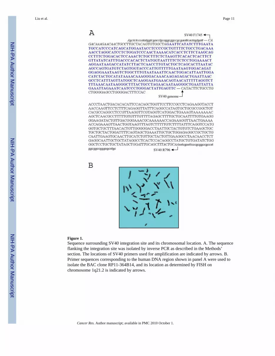
Figure 1.Sequence surrounding SV40 integration site and its chromosomal location. A. The sequenceflanking the integration site was isolated by inverse PCR as described in the Methods’section. The locations of SV40 primers used for amplification are indicated by arrows. B.Primer sequences corresponding to the human DNA region shown in panel A were used toisolate the BAC clone RP11-364B14, and its location as determined by FISH onchromosome 1q21.2 is indicated by arrows.
Liu et al. Page 11
Cancer Res. Author manuscript; available in PMC 2010 October 1.
NIH
-PA Author Manuscript
NIH
-PA Author Manuscript
NIH
-PA Author Manuscript

Figure 2.Morphology of senescent and apoptotic transfer clones. A. Senescent transfer clones ofCRL2504/BAC364B14 were grown and photographed after 21 days (panel 1), 28 days(panel 2), 35 days (panel 3) and 42 days (panel 4). B. CRL2504 cells 8 weeks aftertransfection with the empty vector (panel 1), 8 weeks after transfer of an unrelated BAC152L6 (panel 2), 23 days after transfer of BAC364B14 (panel 3) and 27 days after transferof BAC364B14 (panel 4) were photographed as described in Methods’ section.
Liu et al. Page 12
Cancer Res. Author manuscript; available in PMC 2010 October 1.
NIH
-PA Author Manuscript
NIH
-PA Author Manuscript
NIH
-PA Author Manuscript

Figure 3.Visualization of cells after BrdU incorporation and senescence associated β-galactosidase(SA-βgal) assay. A. Native CRL2504 cells (panel 1) and BAC364B14 transfer clone #10(panel 3) were visualized with anti-BrdU antibody after growing the cells in the presence ofBrdU. The nuclei of native cells (panel 2) and the cells from BAC364B14 transfer clone #10(panel 4) were subsequently stained with DAPI. The cells were photographed as describedin Methods’ section. B. The cells from a confluent culture of native CRL2504 (panel 1),GMO3468 cells after 62 passages (panel 2), BAC364B14 transfer clone #10 after 7 weeks(panel 3) and BAC364B14 transfer clone # sen II after 6 weeks (panel 4) were assayed forthe presence of SA-βgal activity. The cells were photographed as above.
Liu et al. Page 13
Cancer Res. Author manuscript; available in PMC 2010 October 1.
NIH
-PA Author Manuscript
NIH
-PA Author Manuscript
NIH
-PA Author Manuscript

Figure 4.Staining of apoptotic cells with cytoDEATH M30 antibody. Native CRL2504 cells (A),vector transfer clone (panel B), BAC364B14 transfer clone (panel C) and floating cells fromthe dish containing BAC364B14 transfer clone (panel D) were stained with cytoDEATHM30 antibody and photographed as described in Methods’ section.
Liu et al. Page 14
Cancer Res. Author manuscript; available in PMC 2010 October 1.
NIH
-PA Author Manuscript
NIH
-PA Author Manuscript
NIH
-PA Author Manuscript

Figure 5.Semi-quantitaive evaluation of filaggrin (FLG) transcript by RT-PCR. A. RT-PCR wasperformed with FLG specific primers and GAPDH primers on total RNA isolated fromCRL2504 parent (lane 1), CRL2504/pJMOX166 vector control (lane 2), CRL-2504/152L16(lane 3), immortal CRL2504/364B14 Clone 5.1 (lane 4), immortal CRL2504/364B14 Clone5.3 (lane 5), immortal CRL2504/364B14 Clone 5.7(lane 6), senescent CRL-2504/364B14clone 5.5 (lane 7), senescent CRL 2504/364B14 clone 5.9 (lane 8) and senescent CRL2504/364B14 clone 5.17 (lane 9), and the amplified products were electrophoresed on anagarose gel as described. The arrows indicate the amplified products. B. The FLG transcriptwas normalized by calculating the ratio of FLG and GAPDH products. The ratios areexpressed as percentages.
Liu et al. Page 15
Cancer Res. Author manuscript; available in PMC 2010 October 1.
NIH
-PA Author Manuscript
NIH
-PA Author Manuscript
NIH
-PA Author Manuscript


