Embed Size (px)
Citation preview

TH
EJ
OU
RN
AL
OF
CE
LL
BIO
LO
GY
JCB: ARTICLE
The Journal of Cell Biology, Vol. 167, No. 6, December 20, 2004 1195–1204http://www.jcb.org/cgi/doi/10.1083/jcb.200406025
JCB 1195
Roles of uroplakins in plaque formation, umbrella cell enlargement, and urinary tract diseases
Xiang-Tian Kong,
1
Fang-Ming Deng,
1,2
Ping Hu,
1
Feng-Xia Liang,
1
Ge Zhou,
1
Anna B. Auerbach,
6
Nancy Genieser,
3
Peter K. Nelson,
3
Edith S. Robbins,
4
Ellen Shapiro,
2
Bechara Kachar,
8
and Tung-Tien Sun
1,2,5,7
1
Epithelial Biology Unit, The Ronald O. Perelman Department of Dermatology,
2
Department of Urology,
3
Department of Radiology,
4
Department of Cell Biology,
5
Department of Pharmacology,
6
Skirball Institute of Biomolecular Medicine, and
7
New York University Cancer Institute, New York University School of Medicine, New York, NY 10016
8
Section on Structural Cell Biology, National Institute on Deafness and Other Communication Disorders, National Institutes of Health, Bethesda, MD 20892
he apical surface of mouse urothelium is covered bytwo-dimensional crystals (plaques) of uroplakin(UP) particles. To study uroplakin function, we ab-
lated the mouse UPII gene. A comparison of the pheno-types of UPII- and UPIII-deficient mice yielded new insightsinto the mechanism of plaque formation and some funda-mental features of urothelial differentiation. AlthoughUPIII knockout yielded small plaques, UPII knockoutabolished plaque formation, indicating that bothuroplakin heterodimers (UPIa/II and UPIb/III or IIIb) arerequired for plaque assembly. Both knockouts had elevated
T
UPIb gene expression, suggesting that this is a generalresponse to defective plaque assembly. Both knockoutsalso had small superficial cells, suggesting that continuedfusion of uroplakin-delivering vesicles with the apicalsurface may contribute to umbrella cell enlargement. Bothknockouts experienced vesicoureteral reflux, hydro-nephrosis, renal dysfunction, and, in the offspring ofsome breeding pairs, renal failure and neonatal death.These results highlight the functional importance ofuroplakins and establish uroplakin defects as a possiblecause of major urinary tract anomalies and death.
Introduction
The apical surface of mammalian urothelium that is in contactwith the urine is highly specialized, featuring two-dimensional(2D) crystals (urothelial plaques) of hexagonally packed 16-nmprotein particles (Hicks and Ketterer, 1969; Vergara et al.,1969; Staehelin et al., 1972; Brisson and Wade, 1983; Walz etal., 1995; Kachar et al., 1999; Oostergetel et al., 2001; Min etal., 2003). These plaques contain four major uroplakins (UPs):Ia (27 kD), Ib (28 kD), II (15 kD), and III (47 kD) (Wu et al.,1994; Sun et al., 1999). Uroplakins Ia and Ib have four trans-membrane domains, are
�
40% identical in sequence, and belongto the “tetraspanin” family (Yu et al., 1994). The tetraspaninfamily contains many cell surface molecules, including CD9,CD63, CD81, CD82, and CD151, that play key roles in cellularevents involving membrane organization (Maecker et al., 1997;Levy et al., 1998; Berditchevski, 2001; Boucheix and Rubinstein,2001; Hemler, 2001, 2003; Tarrant et al., 2003; Yunta andLazo, 2003). Uroplakins II and III both have a single trans-
membrane domain, and they share a common stretch of
�
12amino acid residues on the exoplasmic side of the transmem-brane domain (Wu and Sun, 1993; Lin et al., 1994; Deng et al.,2002). Within the plaques, these four major uroplakins areorganized into two heterodimers consisting of Ia/II and Ib/III,as demonstrated by chemical cross-linking (Wu et al., 1995)and protein isolation (Liang et al., 2001). In addition, transfectionstudies show that when individual uroplakins are expressed in293T cells, they are retained in the ER; however, coexpressionof uroplakins Ia plus II or Ib plus III permits the heterodimersto exit from the ER; these events suggest that the formation ofspecific heterodimers is a prerequisite for uroplakins to reachthe cell surface (Tu et al., 2002).
To study the functional and disease implications ofuroplakins, we previously generated uroplakin III–deficientmice using the gene-targeting approach (Hu et al., 2000). Suchmice lack a typical urothelial umbrella cell layer. They have areduced urothelial plaque size, compromised urothelial perme-ability barrier function, and retrograde flow of urine fromthe bladder into the ureters (vesicoureteral reflux [VUR]). In
�
70% of the mice, accumulation of urine in the renal pelvis(hydronephrosis) occurs (Hu et al., 2000, 2002). These resultsestablish that uroplakin III is an integral subunit of urothelialplaques, which contribute to the permeability barrier function
X.-T. Kong, F.-M. Deng, and P. Hu contributed equally to this paper.Correspondence to Tung-Tien Sun: [email protected]. Hu’s present address is Procter & Gamble Corporate Research Biotechnology,Cincinnati, OH 45252.Abbreviations used in this paper: 2D, two-dimensional; BUN, blood ureanitrogen; ES, embryonic stem; IVP, i.v. pyelogram; UP, uroplakin; VUR, vesi-coureteral reflux.
on June 1, 2016jcb.rupress.org
Dow
nloaded from
Published December 20, 2004

JCB • VOLUME 167 • NUMBER 6 • 20041196
of the urothelial apical surface, and suggest that urothelial de-fects may play a role in VUR (Hu et al., 2000, 2002).
Some of the phenotypes of these UPIII-deficient mice re-main hardly understood. For example, the observation thatUPIII deficiency leads to the formation of small plaques seemsincompatible with our current assumption that all four majoruroplakins are required for the formation of the 16-nm particle(Sun et al., 1999). One possible explanation is that UPIIIb, anewly discovered, minor UPIII isoform (Deng et al., 2002), isresponsible for the formation of the residual plaques observedin the UPIII-deficient urothelium. If it is, one would predictthat knockout of uroplakin II, which does not have a knownisoform, should completely abolish plaque formation. Anotherquestion has to do with the possible role of uroplakin mutationin VUR. Although knockout of the UPIII gene leads to VUR inmice, screening of human VUR patients revealed that polymor-phism of uroplakin genes is only marginally associated withVUR (Giltay et al., 2004; Jiang et al., 2004). Moreover, al-though polymorphism was found in these VUR patients, no de-letion, truncation, or frameshift mutations have been detectedso far (Giltay et al., 2004; Jiang et al., 2004). The role ofuroplakin defects in VUR and other lower urinary tract anoma-lies thus remains unclear.
To address these issues, we have genetically ablated themouse UPII gene to perturb the other uroplakin pair consistingof UPIa/II. This led to the entrapment of UPIa in the ER, but al-lowed the remaining UPIb/III pair to reach the apical urothelialsurface. No 16-nm particle or apical plaques were formed,however, which indicates that both uroplakin heterodimerswere required for plaque formation. The small size of the su-perficial cells in both UPII- and UPIII-deficient urothelia sug-gested that the incorporation of fully assembled uroplakinplaques into the apical surface through fusion of the fusiformvesicles with the apical membrane played a role in the enlarge-ment of umbrella cells. Because entire litters of UPII-deficientmice from some breeding pairs (originating from the inbred129/SvEv and outbred Swiss Webster strains) reproduciblydied 8–10 d postnatally, as a result of renal failure, it seems thatmajor uroplakin defects such as gene deletion, frameshift, ortruncation might cause urinary tract anomalies that, in severecases, could lead to death. These results highlight the criticalfunctional importance of uroplakins in the formation of a spe-cialized urothelial apical surface, and shed light on the mecha-nisms of plaque formation, umbrella cell enlargement, and cer-tain urinary tract abnormalities.
Results
Inactivation of the mouse uroplakin II gene
After transfecting embryonic stem (ES) cells (from 129/SvEvmice) with a vector designed to delete the first four exons and apart of the fifth exon of the mouse uroplakin II gene (Fig. 1 A),we screened 320 ES cell colonies and found 4 that were harbor-ing the correct homologous recombination events as determinedby long template PCR (Fig. 1 B) and Southern blot (not de-picted). 18 chimeric mice from three ES cell lines were germ-
line transmitting and were bred with black Swiss Webster miceto yield homozygous UPII knockout mice. Northern (Fig. 1, Cand D) and Western (Fig. 1 E) blotting confirmed the absenceof UPII in homozygous uroplakin II–deficient (
�
/
�
) mice. The
Figure 1. Inactivation of the mouse UPII gene (upk2) perturbed theexpression of other uroplakins. (A) Alignment of the mouse UPII locus, thetargeting vector (V), and the mutated locus (KO). Black boxes, exons; thicklines, mouse genomic sequences included in the vector; crosses, potentialcrossover sites; PCR1 and 2, PCR products characteristic of the wild-type andKO alleles, respectively; Neo, neomycin-resistant gene; TK, thymidine kinaseof the herpes simplex virus; B, BamHI; C, ClaI; E, EcoRI; S, SacI; X, Xhol. (B)PCR analysis of the genomic DNA using a mixture of two pairs of primers thatgenerated 4.0-kb and 3.8-kb PCR products characteristic of the wild-type (�)and KO (�) alleles, respectively. M, �C, and �C denote size markers, posi-tive controls (�/� genomic DNA or vector used as templates), and a nega-tive control (no template), respectively. (C) Northern blot analysis. 5 �g of totalRNA samples was resolved electrophoretically and probed with cDNA ofmouse uroplakins Ia, Ib, II, and III, and glyceraldehyde phosphate dehydro-genase (GAPDH) as a loading control. Note, in the �/� animals, theabsence of UPII message and the �2� and 10–20� increases in UPIa andUPIb message, respectively. (D) Ethidium bromide–stained rRNA of the sam-ples in C serving as a loading control. (E) Immunoblot analysis of mouseuroplakin proteins Ia (27 kD), Ib (28 kD), II (15 kD), and III (47 kD). FGdenotes the fast green–stained total mouse urothelial proteins used for immuno-blotting. MAPK and tubulin of the same samples were immunoblotted forcomparison. Note in the �/� mice the absence of UPII protein, the significantdecrease in the levels of the other three uroplakin proteins, and the absenceof the highest mol wt (�30 kD) UPIb species due to defective glycosylation.
on June 1, 2016jcb.rupress.org
Dow
nloaded from
Published December 20, 2004

UROPLAKIN FUNCTIONS • KONG ET AL.
1197
protein levels of uroplakin Ia, UPII’s partner, and of uroplakinsIb and III (of the uroplakin Ib/III pair) were reduced 10–20-fold(on a per total cellular protein basis) when compared with thoseof the normal control mice (
�
/
�
; Fig. 1 E). The mRNA levelsof UPIa and UPIII were slightly up-regulated (about twofold),whereas that of UPIb was drastically up-regulated by 10–20-fold (Fig. 1 C; see Discussion).
Uroplakin expression and plaque formation
Consistent with the UPIII knockout results, the UPII-deficienturothelium lacked a typical superficial umbrella cell layer (Fig.2 b). This urothelium was hyperplastic, as indicated by an ele-vated level of BrdU incorporation (the labeling index increasedalmost 100-fold from 0.12%, which is normal, to 10.7%; Fig.2, c and d), and was three- to sixfold thicker than normal (Fig.2, a and b). Immunohistochemical staining with monospecificrabbit and mouse antibodies to individual uroplakins confirmedthat all four major uroplakins were preferentially expressed insuperficial umbrella cells in normal mouse urothelium (Fig. 3,a, c, e, g, and i; Hu et al., 2000). The staining of the UPII-defi-cient urothelium showed that UPIa (the partner of UPII) wasdiffusely distributed in all upper cells, which is consistent withits entrapment in the ER (Fig. 3 d). Uroplakins Ib and III (of theother uroplakin pair), although expressed at a lower level thannormal, were clearly associated with the apical, as well as somebasal/lateral, cell surface(s) (Fig. 3, f and j).
Unlike normal urothelium, which was covered by largesquamous superficial umbrella cells that could be as large as100
�
m in diameter (Fig. 4 a) and were covered by rigid-look-
Figure 2. UPII-deficient urothelium lacked a typical umbrella cell layerand became hyperplastic. Paraffin sections of the normal (a) and UPII-deficient(b) bladders of 1-mo-old male mice were stained with hematoxylin andeosin. Those of normal (c) and UPII-deficient (d) bladders of 19-day-oldmale mice were isolated 2 hr after the final i.p. injection of BrdU and werestained immunohistochemically for BrdU. The inset in c shows an areawith some BrdU-incorporating superficial cells. Note the increased thick-ness and the elevated BrdU incorporation (10.7 � 1.1% vs. 0.12 �0.07%) of the UPII-deficient urothelium. Also note that although most of thelabeling occurred in basal cells (arrows), there was occasional labeling ofsuperficial cells (arrowheads). A total of eight UPII-deficient mice and eightnormal mice, half of them male, were studied; 1,000–2,000 urothelialcells were examined per section from each animal; P � 0.001; with nosignificant gender differences. Bar, 50 �m.
Figure 3. Altered uroplakin expression pattern in the UPII-deficienturothelium. Paraffin sections of wild-type (WT; a, c, e, g, and i) or UPII-deficient (UPII KO; b, d, f, h, and j) mouse urothelium were immunohis-tochemically stained for keratins (a and b) using a mixture of AE1 andAE3 mouse monoclonal antibodies (Tseng et al., 1982; for review seeCooper et al., 1985), uroplakin Ia (c and d), UPIb (e and f), UPII (g andh), and UPIII (i and j). k shows the silver nitrate–stained (SN) SDS-PAGEpatterns of the detergent-insoluble membrane proteins from (lanes 1)bovine urothelium, (2) normal mouse urothelium, (3) UPIII-deficientmouse urothelium, and (4) UPII-deficient mouse urothelium, and the immuno-blotting of these proteins using antibodies to UPs Ia, Ib, II, IIIa, and IIIb.Note, in the thickened UPII KO urothelium, the absence of UPII, the dif-fused cytoplasmic distribution and lack of apical surface association ofUPIa, the greatly reduced but still noticeably apical surface association(arrowheads) of UPIb and UPIII, and the reduced amounts of detergent-insoluble uroplakins in the UPIII-deficient urothelium (lanes 3) and theirfurther reduction in the UPII-deficient urothelium (lanes 4). Bar, 50 �m.
ing plaques (Figs. 4 c and 5 a), superficial cells of the UPII-deficient urothelium were uniformly small (20–30
�
m; Fig.4 b) and were completely devoid of the rigid-looking, apical
on June 1, 2016jcb.rupress.org
Dow
nloaded from
Published December 20, 2004

JCB • VOLUME 167 • NUMBER 6 • 20041198
plaques (Fig. 4 d). In addition, the uroplakin-delivering fusi-form vesicles (0.7–1
�
m in diameter; Fig. 5 a) were totally re-placed by numerous small, spherical vesicles (Fig. 5 b). Thesesmall vesicles (150–200 nm in diameter) in the UPIII- andUPII-deficient urothelia were moderately and weakly labeled,respectively, by antibodies to uroplakins (Fig. 5, e–h), whichindicates that they were involved in delivering the remaininguroplakins to the cell surface. Consistent with these findings,the detergent-insoluble membrane fraction of the UPIII-defi-cient mouse urothelium showed a significantly reduced numberof uroplakins (Fig. 3 k, lanes 3), whereas that of the UPII-defi-cient urothelium contained almost no detectable uroplakins(Fig. 3 k, lanes 4). Together, these results indicated that UPIIablation completely abolished plaque formation.
VUR, ureteral obstruction, and hydronephrosis
By administering an India ink solution into the bladders of live,anesthetized mice (Fig. 6, a and b), we determined the hydro-static pressures at which micturition (P
M
) and VUR (P
R
) oc-curred. Normal mice had a P
M
of 28 cm of H
2
O pressure (Fig. 6c), and most of them did not reflux—unless the urethra was li-gated, causing outlet obstruction, and the hydrostatic pressurewas increased to 40–80 cm of H
2
O (Fig. 6 d, WT). Althoughthe UPII knockout mice had a normal micturition pressure of
�
24 cm of H
2
O (Fig. 6 c),
�
50% (11 out of 20) of the mice re-fluxed at a pressure lower than this (Fig. 6, d and e). Manyof the (
�
/
�
) mice developed severe hydronephrosis with agreatly expanded renal pelvis (Fig. 7, b and d) and associ-ated renal morphological changes (Fig. 7, a–f). To determinewhether reflux caused hydronephrosis, we calculated the dif-ference between the P
M
and the P
R
of each ureter (P
K
[hydro-
static pressure to the kidney]
�
P
M
�
P
R
). For a normal mousethat had a P
M
of 25 cm of H
2
O and a P
R
of 50 cm, an intravesic-ular pressure of
�
25 cm of H
2
O would result in micturition anddissipation of pressure. However, for a UPII knockout mousethat had a P
M
of 25 cm of H
2
O but a lower P
R
of 18 cm of H
2
O,intravesicular pressures of 18–25 cm of H
2
O would lead to re-flux, thus potentially transmitting up to 7 cm of hydrostaticpressure to the kidney (P
K
). We wanted to see whether this re-nal pressure correlated with the grade (G) of hydronephrosis—which we defined as G
�
D/T, where D was the internal di-ameter of the renal pelvis and T was the thickness of theremaining renal parenchyma (Fig. 7, c and d). If reflux were amain cause of hydronephrosis, there should have been a posi-tive correlation between P
K
and G. However, we found thatthese two parameters were independent (P
�
0.2), which sug-
Figure 4. The UPII-deficient urothelium had small superficial cells and aparticle-free apical surface. The urothelia from age-matched 3-mo-oldwild-type (a and c) and UPII-deficient (b and d) mice were examined byscanning EM (a and b) and quick-freeze deep etch transmission EM (c and d).Note the replacement of large surface umbrella cells (U) by small super-ficial cells (S) (a and b), and the complete absence of the 16-nm particlesand urothelial plaques on the apical surface of the UPII-deficient urothelium(d). cyt, cytoplasm; m, membrane; ups, uroplakin particles. Bars: (a and b)20 �m; (c and d) 0.2 �m.
Figure 5. The replacement of the fusiform vesicles by small sphericalvesicles that delivered the remaining uroplakin pair in uroplakin-deficienturothelium. The urothelia from age-matched 3-mo-old wild-type (a, c, and d),UPII-deficient (b, g, and h), and UPIII-deficient (e and f) mice were exam-ined by thin section (a and b) and immunolabeling (c–h) transmission EM.Normal uroplakin-delivering fusiform vesicles (a, c, and d) were completelyreplaced by numerous small, spherical, immature-looking spherical vesiclesin UPIII-deficient (e and f) and UPII-deficient (b, g, and h) urothelia. Notethe strong labeling of normal fusiform vesicles (*) by antibodies touroplakins Ia (c) and Ib (d), the moderate staining of the spherical vesiclesin the UPIII-deficient urothelium (which still expressed the UPIIIb isoform;e and f), and the weak staining of the small vesicles (arrows) in the UPII-deficient urothelium by anti-UPIII (g) and by a rabbit antiserum to totaluroplakins (h). Arrowheads (b) indicate the smooth apical surface of theUPII-deficient urothelium. Fv, fusiform vesicle; P, plaque; Sv, small vesicle.Bars: (a and b) 0.5 �m; (c–h) 1 �m.
on June 1, 2016jcb.rupress.org
Dow
nloaded from
Published December 20, 2004

UROPLAKIN FUNCTIONS • KONG ET AL.
1199
gests that reflux could not be the major etiology of hydrone-phrosis in this system (Fig. 7 g).
Another possible cause of hydronephrosis is ureteral ob-struction, which impedes the flow of urine from the kidney. Toinvestigate this possibility, we performed i.v. pyelogram (IVP)by injecting 3
�
l/g (of body weight) of Omnipaque into the or-bital sinus, followed by serial radiography (Fig. 8, a and b). Theresults indicated that UPII knockout mice had a significantlydelayed excretion of Omnipaque, indicating obstruction. Serialsectioning of the urinary tracts of UPII knockout mice revealedareas of the ureter with epithelial polyps or complete epithelialocclusion, which caused structural obstruction (Fig. 8, c–h; seeDiscussion). These results suggest that both VUR and structuraland/or functional obstruction of the ureters may be responsiblefor the observed hydronephrosis in UPII knockout mice.
Renal dysfunction and death
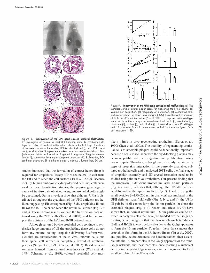
Using a filter paper assay, we found that UPII knockout micehad higher volume per micturition (Fig. 9 b), micturition fre-quency (Fig. 9 c), and total urine output (Fig. 9 d) than normalmice; no statistically significant gender differences were noted(Fig. 9, b–d). The levels of many urinary components, includinguric acid, creatinine, potassium, sodium, and chloride ions, wereslightly reduced (Fig. 9, f–j), possibly because of defects inmechanisms of urine concentration (Fig. 9 d). The concentrationof blood urea nitrogen (BUN) almost doubled (Fig. 9 e), suggest-ing a compromised renal function. Interestingly, although mostof the breeding pairs yielded litters that survived into adulthood,the litters of some breeding pairs (129/SvEv
�
Swiss Webster)
reproducibly died around days 8–10 postnatally (Fig. 10, a andb). These litters were characterized by retarded growth (Fig. 10c) and a sharp surge in the BUN level (Fig. 10 d), which suggestsrenal failure as a cause of death, possibly resulting from ureteralobstruction (Fig. 8, c– h) and a defective renal pelvis urotheliumthat normally expressed uroplakins (Fig. 10 e).
Discussion
Mammalian urothelium has three unique biological features: itsapical membrane is highly specialized, harboring 2D crystals ofhexagonally packed 16-nm particles (Hicks and Ketterer, 1969;Vergara et al., 1969; Staehelin et al., 1972; Brisson and Wade,1983); its superficial umbrella cells are greatly flattened and ex-panded, with a diameter reaching over 100
�
m (Porter and Bon-neville, 1963; Hicks, 1965; Koss, 1969; Lewis, 2000; Veranic etal., 2004; for review see Hicks, 1975); and it is one of the slow-est-cycling stratified epithelia, with a BrdU-labeling index of
�
0.1% (Martin, 1972; Farsund, 1975). The ablation of a singlegene that encodes uroplakin II perturbed all of these urothelialproperties and led to major urinary tract abnormalities.
Mechanism of uroplakin plaque formation
As mentioned earlier, the four major uroplakins can form twoheterodimer pairs consisting of uroplakins Ia/II and Ib/III (Wuet al., 1995; Liang et al., 2001; Tu et al., 2002). Transfection
Figure 6. Inactivation of the UPII gene led to vesicoureteral reflux. An Indiaink suspension (0.1% in PBS) was instilled into the bladders of 11 normal3-mo--old mice and 10 UPII knockout mice, and the hydrostatic pressure atwhich micturition (micturition pressure [PM]) and reflux (reflux pressure [PR])occurred for individual ureters were recorded. Note in a the presence ofIndia ink in the bladder (B) and ureters (U) and, in b, in the left kidney(K)—but not in the right kidney, suggesting obstruction of the right ureter.Also note that, although the UPII-deficient (KO) mice had a normal micturitionpressure (c), 11 out of 20 ureters (55%) had retrograde flow of urine intothe ureters (occurring at a pressure lower than the micturition pressure; d,closed circles), compared with 3 out of 22 ureters (13.6%) in wild-type(WT) mice. Open circles denote cases where reflux occurred at a pressurehigher than the micturition pressure (and thus would not occur naturally).e shows the mean values of PR based on the data shown in d. Error barsrepresent 1 SD.
Figure 7. Inactivation of the UPII gene caused hydronephrosis and renalabnormalities. The kidneys of normal (a, c, and e) and UPII-deficient (b, d,and f) mice were compared in terms of their gross appearance (a and b),whole kidney section (c and d), and histological appearance (e and f).The mice used were 6 mo old for a and b, and 3 mo old for c–g. g showsa plot of renal pressure (PM � PR) versus the grade of hydronephrosis (D/T,where D and T were the diameter of the renal pelvis cavity and the thicknessof the renal parenchyma, respectively). More than 100 knockout micewere studied and �80% of them showed various degrees of hydronephrosis.Note a lack of positive correlation between the grade of hydronephrosisand renal pressure (Pk) (t test, t � 0.177, P � 0.2), which suggests thatreflux could not be a major cause of hydronephrosis. (c, e, and f) C, cortex;G, glomeruli; M, medulla; T, tubules; U, urothelium. Bar, 50 �m.
on June 1, 2016jcb.rupress.org
Dow
nloaded from
Published December 20, 2004
JCB • VOLUME 167 • NUMBER 6 • 20041200
studies indicated that the formation of correct heterodimer isrequired for uroplakins (except UPIb; see below) to exit fromthe ER and to reach the cell surface (Tu et al., 2002). Because293T (a human embryonic kidney–derived cell line) cells wereused in these transfection studies, the physiological signifi-cance of in vitro data obtained using nonurothelial cells mightbe questioned. Our in vivo data show that although UPIa is dis-tributed throughout the cytoplasm of the UPII-deficient urothe-lium, suggesting ER entrapment (Fig. 3 d), uroplakins Ib andIII (of the Ib/III pair) can reach the urothelial surface (Fig. 3, fand j). These in vivo results validate the transfection data ob-tained using the 293T cells (Tu et al., 2002), and further sup-port the existence of the Ia/II and Ib/III heterodimers.
Although cultured bovine urothelial cells continue to syn-thesize large amounts of all the uroplakins, these cells do notform any mature-looking, uroplakin-delivering fusiform vesi-cles that are characteristic of the in vivo umbrella cells, andtheir apical cell surface is completely devoid of urothelialplaques (Surya et al., 1990; Chen et al., 2003). Based on whatwe have learned from cultured keratinocytes (Weiss et al.,1984; Schermer et al., 1989), cultured urothelial cells most
likely mimic in vivo regenerating urothelium (Surya et al.,1990; Chen et al., 2003). The inability of regenerating urothe-lial cells to assemble plaques could be functionally important,because a cell surface laden with the rigid-looking plaques maybe incompatible with cell migration and proliferation duringwound repair. Therefore, although we can study certain earlysteps of uroplakin interaction in the currently available, cul-tured urothelial cells and transfected 293T cells, the final stagesof uroplakin assembly and 2D crystal formation need to bestudied using the in vivo urothelium. Our present finding thatthe uroplakin II–deficient urothelium lacks 16-nm particles(Fig. 4, c and d) indicates that, although the UPIb/III pair canbe delivered to the apical surface (Fig. 3, f and j) using thesmall vesicles (�150–200 nm in diameter) accumulated in theUPII-deficient superficial cells (Fig. 5, b, g, and h), the UPIb/III pair by itself cannot form the 16-nm particle, let alone theurothelial plaques (Fig. 4 d). Severs and Hicks (1979) haveshown that, in normal urothelium, 16-nm particles can be de-tected in early vesicles that have just budded off the Golgi ap-paratus, which suggests that the two uroplakin heterodimers(Ia/II and Ib/III) interact before they leave the Golgi apparatusto form the 16-nm particle. Together, these data suggest thaturoplakins first form, in the ER, heterodimers (Tu et al., 2002),and possibly heterotetramers; the heterotetramers then assem-ble into the 16-nm particles in the Golgi apparatus or the trans-Golgi network, and these particles, once reaching a sufficientdensity in the post-Golgi vesicles, can then aggregate to formsmall and, later, large 2D crystals.
Figure 8. Inactivation of the UPII gene caused ureteral obstruction.I.v. pyelogram of normal (a) and UPII knockout mice (b) established de-layed excretion of contrast in the latter. c–h show the histological sectionsof the ureters of normal (c and e), UPII knockout (d and f), and UPIII knock-out (g and h) mice. Samples were taken from proximal (c and d) or distal(e–h) ureter. Note the formation of epithelial outgrowth filling the ureterallumen (f), sometimes forming a complete occlusion (h). B, bladder; EO,epithelial occlusion; EP, epithelial plug; K, kidney; L, lumen. Bar, 50 �m.
Figure 9. Inactivation of the UPII gene caused renal malfunction. (a) Thestandard curve of a filter paper assay for measuring the urine volume. (b)Volume per micturition. (c) Frequency of micturition. (d) Cumulative totalmicturition volume. (e) Blood urea nitrogen (BUN). Note the twofold increaseof BUN in UPII-deficient mice (P � 0.00001) compared with wild-typemice. f–j show the urinary concentrations of uric acid (f), creatinine (g),potassium (h), sodium (i), and chloride (j). Urine and sera from 12 wild-typeand 12 knockout 3-mo-old mice were pooled for these analyses. Errorbars represent 1 SD.
on June 1, 2016jcb.rupress.org
Dow
nloaded from
Published December 20, 2004

UROPLAKIN FUNCTIONS • KONG ET AL. 1201
Given our current assumption that both uroplakin pairsare required for the formation of the 16-nm particle, it was baf-fling that the UPIII-deficient urothelium still made 2D crystals,albeit small ones, from the 16-nm uroplakin particles (Hu et al.,2000). However, we now know that highly purified urothelialplaques contain a minor isoform of UPIII, which we recentlycharacterized (Deng et al., 2002). This 35-kD protein wasnamed uroplakin IIIb because it is urothelium specific, it sharesa similar transmembrane topology and significant sequence ho-mologies with UPIII (also known as UPIIIa), and it forms aheterodimer with UPIb, UPIIIa’s partner (Deng et al., 2002).This newly found uroplakin IIIb, which is up-regulated in UPI-IIa-deficient urothelium (Deng et al., 2002), should allow theformation of small amounts of heterotetramers containing het-erodimers UPIb/UPIIIb and UPIa/UPII. The heterodimers canthen be delivered by the 150–200-nm vesicles (Fig. 5, e and f)and account for the formation of small surface plaques in theUPIIIa-deficient urothelium. This interpretation is consistentwith our finding that the ablation of UPII, which does not havea known isoform, greatly diminishes the amounts of small ves-icle-associated uroplakins and completely abolishes the forma-tion of the 16-nm particles (Fig. 4, c and d).
We showed previously that the ablation of UPIII led to ab-normal synthesis and processing of UPIb, i.e., the level of UPIbmRNA was greatly increased, whereas the amount of UPIb pro-tein was reduced, became hypoglycosylated, and was mistar-geted to the basal/lateral cell surface (Hu et al., 2000). BecauseUPIII and UPIb were known to interact, we speculated that theseUPIb changes were caused by the removal of its partner, UPIII(Hu et al., 2000). Our finding that UPII ablation led to a similarup-regulation of the UPIb mRNA level (Fig. 1 C) and hypogly-cosylation of the UPIb protein (Fig. 1 E) indicates, however, that
these UPIb changes represent a general response to a perturbeduroplakin assembly. This may explain why, in cultured humanurothelial cells, the level of mRNA for UPIb is greatly elevatedcompared with those for other uroplakins (Varley et al., 2004).Uroplakin Ib is also unique in that it is the only uroplakin that,when expressed alone in 293T cells, can exit from the ER toreach the cell surface (although it is required for its partner,UPIII, to exit from the ER [Tu et al., 2002]); and in that it is theonly uroplakin that is expressed in nonurothelial tissues, includ-ing corneal and conjunctival epithelia (Adachi et al., 2000) and,possibly, lung epithelium (Kallin et al., 1991; Olsburgh et al.,2003). These data indicate that uroplakin Ib is unique among theuroplakins in its regulation and function.
Uroplakin assembly and umbrella cell enlargementNormal mouse urothelium is covered by large umbrella cellsthat have an average diameter of �100 �m (Hicks, 1965; Koss,1969). The greatly enlarged superficial cells can minimize theintercellular space, thus contributing to the permeability barrierfunction of the apical urothelial surface (Negrete et al., 1996;Zeidel, 1996; Lewis, 2000; Apodaca, 2004). The mechanismby which these large umbrella cells, which are frequently tetra-ploid or octoploid (Farsund, 1975), are formed is unclear, al-though both arrested cytokinesis (Farsund, 1976; Farsund andDahl, 1978) and cell fusion (Martin and Wong, 1981) havebeen suggested. Our finding that the superficial cells of bothUPII- and UPIII-deficient urothelia failed to enlarge (Fig. 4, aand b; Hu et al., 2000) suggests that urothelial plaque forma-tion may play a role in umbrella cell enlargement. It is possiblethat the continued insertion of fusiform vesicles into the apicalsurface not only expands (Porter and Bonneville, 1963; Hicks,
Figure 10. Inactivation of the UPII gene cancause neonatal death of the entire litters fromsome mouse breeding pairs. (a) The pedigreeof one pair of founder mice. Note that theentire litter of pups from some breeding pairs,but not from other pairs, died around day 10postnatally. (b) The fractional survival curvesof two litters from different breeding pairs.One underwent neonatal death (2KO-death),whereas the other (2KO-health), like the nor-mal wild-type control (WT), survived into adult-hood. (c) The growth (increase in bodyweight) of the 2KO-death mice was severelyretarded. (d) The blood urea nitrogen (BUN)of the 2KO-death mice increased drastically(reaching �10-fold the normal value) beforethe animals’ death. (e) Immunohistochemicalstaining of normal mouse kidney (embryonicday 16.5), showing that the renal pelvis (RP)urothelium (U) and the simple epithelium cov-ering the papilla (arrowheads) are uroplakinpositive. Arrowheads indicate the single-layeredpapillary epithelium. CD, collecting ducts.Bar, 50 �m.
on June 1, 2016jcb.rupress.org
Dow
nloaded from
Published December 20, 2004

JCB • VOLUME 167 • NUMBER 6 • 20041202
1965; Koss, 1969; Staehelin et al., 1972; Lewis and de Moura,1982; Truschel et al., 2002; Apodaca, 2004), but also alters/sta-bilizes, the luminal surface, thus hindering cytokinesis; the factthat some umbrella cells can incorporate BrdU (Fig. 2, c and d;Erman et al., 2004) makes it plausible that arrested cytokinesiscould contribute to polyploidy and cell enlargement. A combi-nation of cell surface expansion and arrested cytokinesis maythus account for the formation of large polyploid umbrellacells, without resorting to cell fusion. This “surface alterationhypothesis” is supported by cell kinetic data suggesting ar-rested cytokinesis (Farsund, 1976; Farsund and Dahl, 1978);however, additional studies are needed to test this hypothesis.
Uroplakins and urothelial growthA striking phenotype of both uroplakin II– and uroplakin III–deficient urothelia is that they become hyperplastic, with aBrdU-labeling index of �10%, versus a normal value of 0.1%(Fig. 2, c and d) (Hu et al., 2000). It is possible that the per-meability barrier function of the uroplakin-deficient urotheliais compromised (Hu et al., 2002) so that the EGF-related mi-togens that are known to be present in high concentrations inthe mouse urine (Lakshmanan et al., 1990; Parries et al.,1995) can reach urothelial basal cells to stimulate cell prolif-eration (Rebel et al., 1994). Alternatively, fully assembledurothelial plaques can trigger certain growth-inhibiting sig-nals, which are now absent in the uroplakin-deficient urothe-lia, leading to hyperplasia. Additional studies are needed todistinguish these possibilities.
Roles of uroplakin defects in urinary tract diseasesOur earlier observation that inactivation of the mouse uroplakinIII gene resulted in VUR and hydronephrosis raised the possi-bility that uroplakin defects may cause VUR in humans (Hu etal., 2000; Mak and Kuo, 2003). By surveying 76 VUR patients,we identified 18 single-nucleotide polymorphisms; 7 of themare missense, with no truncation or frameshift mutations (Jianget al., 2004). Only two of the missense polymorphisms (anAla7Val change in the UPIa gene and a Pro154Ala change inthe UPIII gene) were found to be marginally associated withVUR (both P � 0.08). These results, as well as those by Giltayet al. (2004), suggest that the uroplakin point mutations thathave been detected so far do not play a major role in causingVUR (Jiang et al., 2004). There are two possible explanationsfor this apparent inconsistency between the human and mousedata. First, the results of mouse studies cannot be directly ex-trapolated to humans because of minor, but still significant, dif-ferences in the development of the lower urinary tracts. Perhapsfor this reason, inactivation of angiotensin II receptor (AT2)gene can cause VUR in mice (Nishimura et al., 1999), but poly-morphisms in this gene do not seem to be involved in causingprimary familial VUR in humans (Hohenfellner et al., 1999;Yoneda et al., 2002). Second, and perhaps more importantly, notruncation or frameshift mutations have been detected in any ofthe VUR patients that have been studied so far, and some breed-ing pairs of the UPII and UPIII knockout mice reproducibly diepostnatally around days 8–10, as a result of renal failure (Fig.
10; Hu et al., 2000), suggesting that certain uroplakin defects(such as deletion, truncation, or mutations in some structurallycrucial amino acid residues), in combination with certain ge-netic backgrounds (Sanford et al., 2001; Wolfer et al., 2002),may be embryologically or postnatally lethal and are thus unde-tected in VUR patients. It is interesting to note that a recent sur-vey showed that urinary tract abnormalities account for 20% ofall congenital anomalies detected during pregnancies and up toone year after birth (Scott, 2002). Of the 560 cases of prenatal orneonatal deaths resulting from congenital abnormalities, 58%were because of renal anomalies, including a significant numberwith obstruction (Scott, 2002).
An important finding of this study is that urothelial ab-normalities caused by uroplakin defects can lead to renal fail-ure and death (Fig. 10). There are several possible mechanismsby which this could occur. First, defects in the uroplakin-posi-tive renal pelvis urothelium, as well as in the simple epitheliumthat covers the renal papilla expressing uroplakin prenatally(Fig. 10 e; Jiang et al., 2004), may lead to renal dysfunctionand failure. Second, urothelial hyperplasia can cause obstruc-tion of the ureteral lumen (Fig. 8, f and h). Although it hasbeen well-established that mechanical obstruction can induceurothelial hyperplasia (Monson et al., 1992; Curhan et al.,2000), it is unclear whether urothelial hyperplasia, per se, cancause obstruction. In our system, urothelial hyperplasia seemsto be the cause, rather than the effect, of obstruction because(a) urothelial hyperplasia precedes obstruction and hydrone-phrosis; and (b) although only 70–80% of the mice develop hy-dronephrosis, 100% of the animals show urothelial hyperplasiathroughout the urinary tract, thus ruling out obstruction as acause of urothelial hyperplasia (but see Gamp et al., 2003).Third, defects in the ureteral urothelium may interfere with thedevelopment of its underlying smooth muscle (Master et al.,2003), leading to abnormal peristalsis or functional obstruction(Santicioli and Maggi, 1998)—a possibility supported by ourrecent finding that uroplakin defects lead to abnormal bladdermuscle contractility and/or function (Christ, G., personal com-munication). These possible mechanisms, or a combinationthereof, may cause a functional/structural obstruction of theurinary tract, leading to abnormal renal development (Chungand Chevalier, 1996), obstructive nephropathy, hydronephro-sis, and, in severe cases, renal failure and death.
Materials and methodsProduction of the UPII knockout miceGenomic clones of the mouse UPII gene were isolated from a 129/Olamouse P1 genomic library (Genome Systems). The targeting vector wasdesigned to delete the first four exons, and a part of the fifth, of the UPIIgene (Fig. 1 A). Three primers were used for genotyping; one forward (5-gagggagttaagactcaagaatcaatcaagga-3) and two reverse (5-cttctatcgcct-tcttgacgagttcttctgagg-3 for detecting a 3.8-kb product of the neomycin se-quence, and 5-cagatttctagcagtccactcttgtagaacgg-3 for a 4.0-kb productof the native UPII gene; Fig. 1, A and B).
Morphological studiesMouse urothelium was examined by scanning EM (model JSM-840; JEOL)and transmission EM (model 200CX; JEOL) (Hu et al., 2000). Quick-freezedeep etch was performed as described previously (Kachar et al., 1999).Cell proliferation was assessed based on the nuclear incorporation of BrdU(Sigma-Aldrich). 19-d-old mice received five intraperitoneal injections of
on June 1, 2016jcb.rupress.org
Dow
nloaded from
Published December 20, 2004

UROPLAKIN FUNCTIONS • KONG ET AL. 1203
BrdU (100 mg/kg of body weight, in PBS), 1.5 h apart, and were killed2 h after the final injection. Various tissues, including esophagus and urinarybladder tissues, were removed, fixed in 10% formalin overnight, and pro-cessed for immunostaining with a horseradish peroxidase–conjugated anti-BrdU monoclonal antibody (CHEMICON International). Samples were visu-alized with a microscope (Axiophot; Carl Zeiss MicroImaging, Inc.) with10�/0.32 and 20�/0.60 (Plan-Apochromat) or 40�/0.75 (Plan-Neo-fluar) objective lenses. Images were captured with a digital camera (modelDKC-5000; Sony) at room temperature. The images were processed in sizeand contrast/brightness with Adobe Photoshop 6.0.
Isolation of detergent-insoluble proteins from mouse urotheliumMouse urothelial cells were homogenized in buffer A (10 mM Hepes, pH7.5, 1 mM EDTA, 1 mM EGTA, and 1 mM PMSF), loaded onto a 1.6-M su-crose cushion (in buffer A), and centrifuged at 16,000 rpm for 25 min at4C (SW41; Beckman Coulter). The crude membranes concentrated at theinterface were isolated, washed with buffer A, treated with 2% Sarkosyl inbuffer A for 10 min at 25C, and pelleted. The detergent-insoluble mem-branes, in which the urothelial plaques were highly enriched, were washedwith buffer A before they were solubilized in SDS-PAGE sample buffer andanalyzed by SDS-PAGE, followed by silver nitrate staining, or by immuno-blotting (Wu et al., 1994; Liang et al., 1999; Zhou et al., 2001).
Determination of reflux pressure and hydronephrosisThe reflux pressure was recorded as the hydrostatic pressure (i.e., cm ofH2O) at which an India ink suspension in PBS backflowed from the blad-ders into the ureters of anesthetized mice (Hu et al., 2000). At the end ofthe experiment, the kidneys were dissected longitudinally to confirm thepresence of the ink in the renal pelvis (Fig. 6 b).
Determination of the micturition pattern and urine/blood chemistryThe micturition pattern of the mice was recorded using special cages, asdescribed previously (Hu et al., 2000). A total of �500 �l of mouse urinewas collected from each mouse and used for assaying the concentrationsof uric acid, creatinine, potassium, sodium, and chloride ions (Tufts Veteri-nary Diagnostic Laboratory). Mouse sera were used for assaying the BUNlevel (Anilytics).
Determination of obstruction by IVP and serial sectioning of the mouse ureterIVP was performed in anesthetized 4-mo-old wild-type (n � 2) anduroplakin II–deficient (n � 4) mice. The contrast Omnipaque (Iohexol; 3�l/g of body weight) was injected into the orbital sinus, and serial radio-graphs were made at 0, 5, 15, 30, 45, 60, and 90 min. After lethal anes-thesia, the urinary tracts were dissected and fixed in 10% formalin over-night. The ureters were embedded in paraffin and serially sectioned.Sections were numbered from superior to inferior orientation and stainedwith hematoxylin and eosin.
We thank Alexandra Joyner for her help in generating the knockout mice;Songshan Jiang and Jordan Gitlin for experimental assistance; Gloria Galloand Adrian S. Woolf for reviewing renal histology; the Interstitial Cystitis Asso-ciation and the NYU Urology and Dermatology Research Programs for sup-port; David Sabatini, Xiang-Peng Kong, Gert Kreibich, Angel Pellicer, and Xue-Ru Wu for helpful discussions; and Herbert Lepor and Irwin M. Freedberg fortheir encouragement and support.
This work was funded by National Institutes of Health grants DK39753,DK52206, and DK66491.
Submitted: 7 June 2004Accepted: 9 November 2004
ReferencesAdachi, W., K. Okubo, and S. Kinoshita. 2000. Human uroplakin Ib in ocular
surface epithelium. Invest. Ophthalmol. Vis. Sci. 41:2900–2905.
Apodaca, G. 2004. The uroepithelium: not just a passive barrier. Traffic. 5:117–128.
Berditchevski, F. 2001. Complexes of tetraspanins with integrins: more thanmeets the eye. J. Cell Sci. 114:4143–4151.
Boucheix, C., and E. Rubinstein. 2001. Tetraspanins. Cell. Mol. Life Sci. 58:1189–1205.
Brisson, A., and R.H. Wade. 1983. Three-dimensional structure of luminalplasma membrane protein from urinary bladder. J. Mol. Biol. 166:21–36.
Chen, Y., X. Guo, F.M. Deng, F.X. Liang, W. Sun, M. Ren, T. Izumi, D.D. Sa-batini, T.T. Sun, and G. Kreibich. 2003. Rab27b is associated with fusi-
form vesicles and may be involved in targeting uroplakins to urothelialapical membranes. Proc. Natl. Acad. Sci. USA. 100:14012–14017.
Chung, K.H., and R.L. Chevalier. 1996. Arrested development of the neonatalkidney following chronic ureteral obstruction. J. Urol. 155:1139–1144.
Cooper, D., A. Schermer, and T.-T. Sun. 1985. Classification of human epitheliaand their neoplasms using monoclonal antibodies to keratins: strategies,applications, and limitations. Lab. Invest. 52:243–256.
Curhan, G.C., W.S. McDougal, and M.L. Zeidel. 2000. Urinary tract obstruc-tion. In The Kidney. G.M. Brenner, editor. W.B. Saunders, Philadelphia.1820–1843.
Deng, F.M., F.X. Liang, L. Tu, K.A. Resing, P. Hu, M. Supino, C.C. Hu, G.Zhou, M. Ding, G. Kreibich, and T.T. Sun. 2002. Uroplakin IIIb, aurothelial differentiation marker, dimerizes with uroplakin Ib as an earlystep of urothelial plaque assembly. J. Cell Biol. 159:685–694.
Erman, A., G. Vidmar, and K. Jezernik. 2004. Temporal and spatial dimensionsof postnatal growth of the mouse urinary bladder urothelium. Histochem.Cell Biol. 121:63–71.
Farsund, T. 1975. Cell kinetics of mouse urinary bladder epithelium. I. Circa-dian and age variations in cell proliferation and nuclear DNA content.Virchows Arch. B Cell Pathol. 18:35–49.
Farsund, T. 1976. Cell kinetics of mouse urinary bladder epithelium. II.Changes in proliferation and nuclear DNA content during necrosis re-generation, and hyperplasia caused by a single dose of cyclophospha-mide. Virchows Arch. B Cell Pathol. 21:279–298.
Farsund, T., and E. Dahl. 1978. Cell kinetics of mouse urinary bladder epithe-lium. III. A histologic and ultrastructural study of bladder epitheliumduring regeneration after a single dose of cyclophosphamide, with spe-cial reference to the mechanism by which polyploid cells are formed.Virchows Arch. B Cell Pathol. 26:215–223.
Gamp, A.C., Y. Tanaka, R. Lullmann-Rauch, D. Wittke, R. D’Hooge, P.P. DeDeyn, T. Moser, H. Maier, D. Hartmann, K. Reiss, et al. 2003. LIMP-2/LGP85 deficiency causes ureteric pelvic junction obstruction, deafnessand peripheral neuropathy in mice. Hum. Mol. Genet. 12:631–646.
Giltay, J.C., J. van de Meerakker, H.K. van Amstel, and T.P. de Jong. 2004. Nopathogenic mutations in the uroplakin III gene of 25 patients with pri-mary vesicoureteral reflux. J. Urol. 171:931–932.
Hemler, M.E. 2001. Specific tetraspanin functions. J. Cell Biol. 155:1103–1107.
Hemler, M.E. 2003. Tetraspanin proteins mediate cellular penetration, invasion,and fusion events and define a novel type of membrane microdomain.Annu. Rev. Cell Dev. Biol. 19:397–422.
Hicks, R.M. 1965. The fine structure of the transitional epithelium of rat ureter.J. Cell Biol. 26:25–48.
Hicks, R.M. 1975. The mammalian urinary bladder: an accommodating organ.Biol. Rev. Camb. Philos. Soc. 50:215–246.
Hicks, R.M., and B. Ketterer. 1969. Hexagonal lattice of subunits in the thick lu-minal membrane of the rat urinary bladder. Nature. 224:1304–1305.
Hohenfellner, K., T.E. Hunley, E. Yerkes, P. Habermehl, R. Hohenfellner, andV. Kon. 1999. Angiotensin II, type 2 receptor in the development ofvesico-ureteric reflux. BJU Int. 83:318–322.
Hu, P., F.M. Deng, F.X. Liang, C.M. Hu, A.B. Auerbach, E. Shapiro, X.R. Wu,B. Kachar, and T.T. Sun. 2000. Ablation of uroplakin III gene results insmall urothelial plaques, urothelial leakage, and vesicoureteral reflux. J.Cell Biol. 151:961–972.
Hu, P., S. Meyers, F.X. Liang, F.M. Deng, B. Kachar, M.L. Zeidel, and T.T.Sun. 2002. Role of membrane proteins in permeability barrier function:uroplakin ablation elevates urothelial permeability. Am. J. Physiol. RenalPhysiol. 283:F1200–F1207.
Jiang, S., J. Gitlin, F.M. Deng, F.X. Liang, A. Lee, A. Atala, S.B. Bauer, G.D.Ehrlich, S.A. Feather, J.D. Goldberg, et al. 2004. Lack of major involve-ment of human uroplakin genes in vesicoureteral reflux: implications fordisease heterogeneity. Kidney Int. 66:10–19.
Kachar, B., F. Liang, U. Lins, M. Ding, X.R. Wu, D. Stoffler, U. Aebi, and T.-T.Sun. 1999. Three-dimensional analysis of the 16 nm urothelial plaqueparticle: luminal surface exposure, preferential head-to-head interaction,and hinge formation. J. Mol. Biol. 285:595–608.
Kallin, B., R. de Martin, T. Etzold, V. Sorrentino, and L. Philipson. 1991. Cloningof a growth arrest-specific and transforming growth factor beta-regulatedgene, TI 1, from an epithelial cell line. Mol. Cell. Biol. 11:5338–5345.
Koss, L.G. 1969. The asymmetric unit membranes of the epithelium of the uri-nary bladder of the rat. An electron microscopic study of a mechanism ofepithelial maturation and function. Lab. Invest. 21:154–168.
Lakshmanan, J., E.C. Salido, R. Lam, L. Barajas, and D.A. Fisher. 1990. Identi-fication of pro-epidermal growth factor and high molecular weight epi-dermal growth factors in adult mouse urine. Biochem. Biophys. Res.Commun. 173:902–911.
Levy, S., S.C. Todd, and H.T. Maecker. 1998. CD81 (TAPA-1): a molecule in-volved in signal transduction and cell adhesion in the immune system.
on June 1, 2016jcb.rupress.org
Dow
nloaded from
Published December 20, 2004

JCB • VOLUME 167 • NUMBER 6 • 20041204
Annu. Rev. Immunol. 16:89–109.
Lewis, S.A. 2000. Everything you wanted to know about the bladder epitheliumbut were afraid to ask. Am. J. Physiol. Renal Physiol. 278:F867–F874.
Lewis, S.A., and J.L. de Moura. 1982. Incorporation of cytoplasmic vesiclesinto apical membrane of mammalian urinary bladder epithelium. Nature.297:685–688.
Liang, F., B. Kachar, M. Ding, Z. Zhai, X.R. Wu, and T.-T. Sun. 1999. Urothe-lial hinge as a highly specialized membrane: detergent- insolubility, uro-hingin association, and in vitro formation. Differentiation. 65:59–69.
Liang, F.X., I. Riedel, F.M. Deng, G. Zhou, C. Xu, X.R. Wu, X.P. Kong, R.Moll, and T.T. Sun. 2001. Organization of uroplakin subunits: trans-membrane topology, pair formation and plaque composition. Biochem. J.355:13–18.
Lin, J.H., X.R. Wu, G. Kreibich, and T.-T. Sun. 1994. Precursor sequence, pro-cessing, and urothelium-specific expression of a major 15-kDa proteinsubunit of asymmetric unit membrane. J. Biol. Chem. 269:1775–1784.
Maecker, H.T., S.C. Todd, and S. Levy. 1997. The tetraspanin superfamily: mo-lecular facilitators. FASEB J. 11:428–442.
Mak, R.H., and H.J. Kuo. 2003. Primary ureteral reflux: emerging insights frommolecular and genetic studies. Curr. Opin. Pediatr. 15:181–185.
Martin, B.F. 1972. Cell replacement and differentiation in transitional epithe-lium: a histological and autoradiographic study of the guinea-pig bladderand ureter. J. Anat. 112:433–455.
Martin, B.F., and Y.C. Wong. 1981. Development and maturation of the bladderepithelium of the guinea pig. Acta Anat. (Basel). 110:359–375.
Master, V.A., G. Wei, W. Liu, and L.S. Baskin. 2003. Urothlelium facilitatesthe recruitment and trans-differentiation of fibroblasts into smooth mus-cle in acellular matrix. J. Urol. 170:1628–1632.
Min, G., G. Zhou, M. Schapira, T.T. Sun, and X.P. Kong. 2003. Structural basisof urothelial permeability barrier function as revealed by cryo-EM stud-ies of the 16 nm uroplakin particle. J. Cell Sci. 116:4087–4094.
Monson, F.C., B.A. McKenna, A.J. Wein, and R.M. Levin. 1992. Effect of out-let obstruction on 3H-thymidine uptake: a biochemical and radioauto-graphic study. J. Urol. 148:158–162.
Negrete, H.O., J.P. Lavelle, J. Berg, S.A. Lewis, and M.L. Zeidel. 1996. Perme-ability properties of the intact mammalian bladder epithelium. Am. J.Physiol. 271:F886–F894.
Nishimura, H., E. Yerkes, K. Hohenfellner, Y. Miyazaki, J. Ma, T.E. Hunley, H.Yoshida, T. Ichiki, D. Threadgill, J.A. Phillips III, et al. 1999. Role ofthe angiotensin type 2 receptor gene in congenital anomalies of the kid-ney and urinary tract, CAKUT, of mice and men. Mol. Cell. 3:1–10.
Olsburgh, J., P. Harnden, R. Weeks, B. Smith, A. Joyce, G. Hall, R. Poulsom, P.Selby, and J. Southgate. 2003. Uroplakin gene expression in normal hu-man tissues and locally advanced bladder cancer. J. Pathol. 199:41–49.
Oostergetel, G.T., W. Keegstra, and A. Brisson. 2001. Structure of the majormembrane protein complex from urinary bladder epithelial cells by cryo-electron crystallography. J. Mol. Biol. 314:245–252.
Parries, G., K. Chen, K.S. Misono, and S. Cohen. 1995. The human urinary epi-dermal growth factor (EGF) precursor. Isolation of a biologically active160-kilodalton heparin-binding pro-EGF with a truncated carboxyl ter-minus. J. Biol. Chem. 270:27954–27960.
Porter, K.R., and M.A. Bonneville. 1963. An Introduction to the Fine Structureof Cells and Tissues. Lea & Febiger, Philadelphia. 196 pp.
Rebel, J.M., W.I. De Boer, C.D. Thijssen, M. Vermey, E.C. Zwarthoff, andT.H. Van der Kwast. 1994. An in vitro model of urothelial regeneration:effects of growth factors and extracellular matrix proteins. J. Pathol.173:283–291.
Sanford, L.P., S. Kallapur, I. Ormsby, and T. Doetschman. 2001. Influence ofgenetic background on knockout mouse phenotypes. Methods Mol. Biol.158:217–225.
Santicioli, P., and C.A. Maggi. 1998. Myogenic and neurogenic factors in thecontrol of pyeloureteral motility and ureteral peristalsis. Pharmacol. Rev.50:683–722.
Schermer, A., J.V. Jester, C. Hardy, D. Milano, and T.-T. Sun. 1989. Transientsynthesis of K6 and K16 keratins in regenerating rabbit corneal epithe-lium: keratin markers for an alternative pathway of keratinocyte differen-tiation. Differentiation. 42:103–110.
Scott, J.E. 2002. Fetal, perinatal, and infant death with congenital renal anom-aly. Arch. Dis. Child. 87:114–117.
Severs, N.J., and R.M. Hicks. 1979. Analysis of membrane structure in the tran-sitional epithelium of rat urinary bladder. 2. The discoidal vesicles andGolgi apparatus: their role in luminal membrane biogenesis. J. Ultra-struct. Res. 69:279–296.
Staehelin, L.A., F.J. Chlapowski, and M.A. Bonneville. 1972. Lumenal plasmamembrane of the urinary bladder. I. Three-dimensional reconstructionfrom freeze-etch images. J. Cell Biol. 53:73–91.
Sun, T.-T., F.X. Liang, and X.R. Wu. 1999. Uroplakins as markers of urothelialdifferentiation. Adv. Exp. Med. Biol. 462:7–18.
Surya, B., J. Yu, M. Manabe, and T.-T. Sun. 1990. Assessing the differentiationstate of cultured bovine urothelial cells: elevated synthesis of stratifica-tion-related K5 and K6 keratins and persistent expression of uroplakin I.J. Cell Sci. 97:419–432.
Tarrant, J.M., L. Robb, A.B. van Spriel, and M.D. Wright. 2003. Tetraspanins: mo-lecular organisers of the leukocyte surface. Trends Immunol. 24:610–617.
Truschel, S.T., E. Wang, W.G. Ruiz, S.M. Leung, R. Rojas, J. Lavelle, M.Zeidel, D. Stoffer, and G. Apodaca. 2002. Stretch-regulated exocytosis/endocytosis in bladder umbrella cells. Mol. Biol. Cell. 13:830–846.
Tseng, S.C., M.J. Jarvinen, W.G. Nelson, J.W. Huang, M.J. Woodcock, andT.-T. Sun. 1982. Correlation of specific keratins with different typesof epithelial differentiation: monoclonal antibody studies. Cell. 30:361–372.
Tu, L., T.T. Sun, and G. Kreibich. 2002. Specific heterodimer formation is aprerequisite for uroplakins to exit from the endoplasmic reticulum. Mol.Biol. Cell. 13:4221–4230.
Varley, C.L., J. Stahlschmidt, W.C. Lee, J. Holder, C. Diggle, P.J. Selby, L.K.Trejdosiewicz, and J. Southgate. 2004. Role of PPAR� and EGFR sig-nalling in the urothelial terminal differentiation programme. J. Cell Sci.117:2029–2036.
Veranic, P., R. Romih, and K. Jezernik. 2004. What determines differentiationof urothelial umbrella cells? Eur. J. Cell Biol. 83:27–34.
Vergara, J.A., W. Longley, and J.D. Robertson. 1969. A hexagonal arrange-ment of subunits in membrane of mouse urinary bladder. J. Mol. Biol.46:593–596.
Walz, T., M. Haner, X.R. Wu, C. Henn, A. Engel, T.-T. Sun, and U. Aebi. 1995.Towards the molecular architecture of the asymmetric unit membrane ofthe mammalian urinary bladder epithelium: a closed “twisted ribbon”structure. J. Mol. Biol. 248:887–900.
Weiss, R.A., R. Eichner, and T.-T. Sun. 1984. Monoclonal antibody analysis ofkeratin expression in epidermal diseases: a 48- and 56-kdalton keratinas molecular markers for hyperproliferative keratinocytes. J. Cell Biol.98:1397–1406.
Wolfer, D.P., W.E. Crusio, and H.P. Lipp. 2002. Knockout mice: simple solu-tions to the problems of genetic background and flanking genes. TrendsNeurosci. 25:336–340.
Wu, X.R., and T.-T. Sun. 1993. Molecular cloning of a 47 kDa tissue-specificand differentiation-dependent urothelial cell surface glycoprotein. J. CellSci. 106:31–43.
Wu, X.R., J.H. Lin, T. Walz, M. Haner, J. Yu, U. Aebi, and T.-T. Sun. 1994.Mammalian uroplakins. A group of highly conserved urothelial differen-tiation-related membrane proteins. J. Biol. Chem. 269:13716–13724.
Wu, X.R., J.J. Medina, and T.-T. Sun. 1995. Selective interactions of UPIa andUPIb, two members of the transmembrane 4 superfamily, with distinctsingle transmembrane-domained proteins in differentiated urothelialcells. J. Biol. Chem. 270:29752–29759.
Yoneda, A., S. Cascio, A. Green, D. Barton, and P. Puri. 2002. Angiotensin IItype 2 receptor gene is not responsible for familial vesicoureteral reflux.J. Urol. 168:1138–1141.
Yu, J., J.H. Lin, X.R. Wu, and T.-T. Sun. 1994. Uroplakins Ia and Ib, two majordifferentiation products of bladder epithelium, belong to a family of fourtransmembrane domain (4TM) proteins. J. Cell Biol. 125:171–182.
Yunta, M., and P.A. Lazo. 2003. Tetraspanin proteins as organisers of membranemicrodomains and signalling complexes. Cell. Signal. 15:559–564.
Zeidel, M.L. 1996. Low permeabilities of apical membranes of barrier epithe-lia: what makes watertight membranes watertight? Am. J. Physiol.271:F243–F245.
Zhou, G., W.J. Mo, P. Sebbel, G. Min, T.A. Neubert, R. Glockshuber, X.R. Wu,T.T. Sun, and X.P. Kong. 2001. Uroplakin Ia is the urothelial receptorfor uropathogenic Escherichia coli: evidence from in vitro FimH bind-ing. J. Cell Sci. 114:4095–4103.
on June 1, 2016jcb.rupress.org
Dow
nloaded from
Published December 20, 2004


