Embed Size (px)
Citation preview
RESEARCH Open Access
Runx2 transcriptome of prostate cancer cells:insights into invasiveness and bone metastasisSanjeev K Baniwal1,4,6*, Omar Khalid1,4,6, Yankel Gabet1,4,6, Ruchir R Shah7,8, Daniel J Purcell1,6, Deepak Mav7,Alice E Kohn-Gabet4,6, Yunfan Shi4,6, Gerhard A Coetzee3,5,6, Baruch Frenkel1,2,4,6*
Abstract
Background: Prostate cancer (PCa) cells preferentially metastasize to bone at least in part by acquiringosteomimetic properties. Runx2, an osteoblast master transcription factor, is aberrantly expressed in PCa cells, andpromotes their metastatic phenotype. The transcriptional programs regulated by Runx2 have been extensivelystudied during osteoblastogenesis, where it activates or represses target genes in a context-dependent manner.However, little is known about the gene regulatory networks influenced by Runx2 in PCa cells. We thereforeinvestigated genome wide mRNA expression changes in PCa cells in response to Runx2.
Results: We engineered a C4-2B PCa sub-line called C4-2B/Rx2dox, in which Doxycycline (Dox) treatment stimulatesRunx2 expression from very low to levels observed in other PCa cells. Transcriptome profiling using whole genomeexpression array followed by in silico analysis indicated that Runx2 upregulated a multitude of genes withprominent cancer associated functions. They included secreted factors (CSF2, SDF-1), proteolytic enzymes (MMP9,CST7), cytoskeleton modulators (SDC2, Twinfilin, SH3PXD2A), intracellular signaling molecules (DUSP1, SPHK1,RASD1) and transcription factors (Sox9, SNAI2, SMAD3) functioning in epithelium to mesenchyme transition (EMT),tissue invasion, as well as homing and attachment to bone. Consistent with the gene expression data, induction ofRunx2 in C4-2B cells enhanced their invasiveness. It also promoted cellular quiescence by blocking the G1/S phasetransition during cell cycle progression. Furthermore, the cell cycle block was reversed as Runx2 levels declinedafter Dox withdrawal.
Conclusions: The effects of Runx2 in C4-2B/Rx2dox cells, as well as similar observations made by employing LNCaP,22RV1 and PC3 cells, highlight multiple mechanisms by which Runx2 promotes the metastatic phenotype of PCacells, including tissue invasion, homing to bone and induction of high bone turnover. Runx2 is therefore anattractive target for the development of novel diagnostic, prognostic and therapeutic approaches to PCamanagement. Targeting Runx2 may prove more effective than focusing on its individual downstream genes andpathways.
IntroductionRunx2 together with Runx1 and Runx3 comprise theRunx class of transcription factors, defined by theirhighly homologous Runt-related DNA-binding domain.As heterodimers with Cbfß, Runx proteins bind to cog-nate DNA elements with the consensus nucleotidesequence 5’-ACCACA in the promoters/enhancers oftheir target genes [1]. The three Runx proteins coordi-nate proliferation and differentiation of various cell
types [2]. Runx1 is important for hematopoiesis [3,4];Runx2 is pivotal in osteogenesis [1,5,6]; and Runx3 iscritical for neurogenesis [7], thymopoiesis [4], and main-tenance of the gastric epithelium [4,8]. While promotingspecific cellular phenotypes, Runx proteins have evolvedto inhibit cell proliferation. Runx3 is a bona fide tumorsuppressor [9] as down-regulation of its promoter byhypermethylation contributes to the development of gas-tric cancer [10,11]. Ablation of Runx1 activity leads toleukemia [12] and disruption of Runx2 results inderegulated cell proliferation and immortalization[13-17]. Paradoxically, Runx2 is also implicated in carci-nogenesis. In a mouse screen for c-Myc-collaborating
* Correspondence: [email protected]; [email protected] of Biochemistry & Molecular Biology, University of SouthernCalifornia, Los Angeles, USAFull list of author information is available at the end of the article
Baniwal et al. Molecular Cancer 2010, 9:258http://www.molecular-cancer.com/content/9/1/258
© 2010 Baniwal et al; licensee BioMed Central Ltd. This is an Open Access article distributed under the terms of the Creative CommonsAttribution License (http://creativecommons.org/licenses/by/2.0), which permits unrestricted use, distribution, and reproduction inany medium, provided the original work is properly cited.
oncogenes, MLV-induced leukemia occurred most fre-quently when the provirus integrated into the Runx2locus resulting in its ectopic expression [18]. It was sug-gested that Runx2 initially provides the cells with a sur-vival advantage, and its anti-mitogenic activity iscounteracted by the CD2-Myc transgene present in themouse model used for this screen [2,19]. Therefore,Runx2-mediated tumorigenesis likely requires additionalloss of check-point genes such as Trp53 or improperregulation of an oncogene such as c-Myc [19].Runx2 has been extensively studied in the context of
osteoblastogenesis from mesenchymal progenitors,where as a master regulator it stimulates the expressionof various bone matrix components such as osteocalcinand bone sialoprotein (BSP) [20]. Runx2-/- mice diesoon after birth due to the lack of differentiated osteo-blasts and thus a mineralized skeleton [1,5,6]. Runx2haploinsufficiency in humans causes the rare skeletaldisorder Cleidocranial Dysplasia [21]. In search forhints to explain the high predilection of prostate andbreast cancer to metastasize to bone, investigators havenoticed ectopic expression of Runx2 and some of its tar-get genes in biopsies from advanced tumors and theirderivative cell lines [22-26]. In a mouse model of PCa,conditional deletion of Pten in prostate epithelial cellsresulted in the development of tumors with progressiveincrease in Runx2 expression [27]. Among the osteomi-metic properties of prostate and breast cancer cells areexpression of the Runx2 target genes MMP9 [28], BSP[29] and VEGFA [30], as well as induction of minerali-zation [25].In addition to promoting osteoblast differentiation,
Runx2 drives the expression of osteoclastogenic signals,both in osteoblasts [31,32] and in the PC3 bone metas-tasis-derived PCa cell line [22]. PC3 cells robustlyexpress Runx2 [33], and its silencing decreased theirosteoclastogenic property in vitro and their growthwithin the bone microenvironment in vivo [22]. Runx2also promotes metastatic aspects not necessarily relatedto bone. Invasion of PC3 cells through Matrigel™, abasement membrane-like preparation, decreased afterRunx2 silencing [22], and its ectopic expression inmammary epithelial cells increased their proliferationand disrupted their normal acinar organization [34]. Anoncogenic role for Runx2 has also been suggested intumors that do not exhibit high predilection to bone,including pancreatic ductal adenocarcinoma [35] andthyroid papillary carcinoma [36].Whereas Runx2 is being increasingly recognized as a
pro-metastatic factor, little is known about the underly-ing transcriptional programs. To establish gene regula-tory networks downstream of Runx2 in aggressive PCa,we analyzed gene expression in response to Runx2 inthe C4-2B PCa cell line. These cells are castration-
resistant derivatives of the androgen-dependent LNCaPcells, and serve as a model for the aggressive stage ofbone metastatic PCa [37,38]. Although C4-2B cellsexpress Runx2 at levels higher than LNCaP cells [25],these levels are far lower than those observed in PC3cells or osteoblasts [22]. We therefore engineered a C4-2B sub-line that allowed us to profile gene expressionafter induction of Runx2 with Doxycycline to levels seenin PC3 cells. Remarkably, the most significant changeswere the up-regulation of genes implicated in cancerprogression and cellular movement, and the down-regu-lation of genes involved in cell cycle progression. Con-sistent with these changes in gene expression, Runx2enhanced PCa cell invasiveness and inhibited theirproliferation.
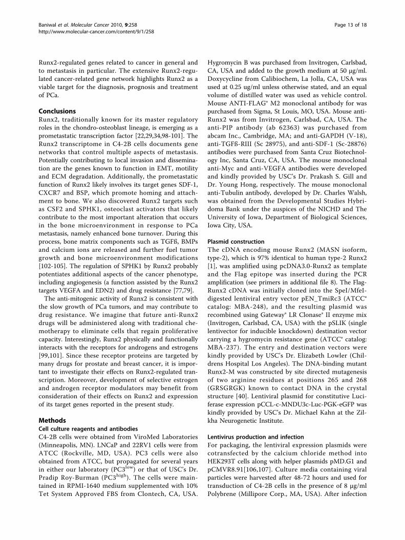
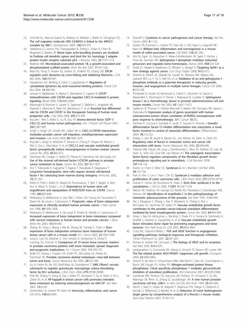
Results and DiscussionEstablishment of C4-2B PCa cells with conditional Runx2expressionTo establish a C4-2B cell line that conditionallyexpresses Runx2, we employed the recently describedlentivirus-based pSLIK vector system, which allows tightDoxycycline (Dox)-inducible, RNA PolII-mediated tran-scription of a gene of interest [39]. C4-2B cells weretransduced with Flag-tagged Runx2-encoding lenti-viruses (Figure 1A), resulting in the C4-2B/Rx2dox sub-line. As control, we established the C4-2B/Rx2-Mdox
subline, where Dox treatment induced expression of thetranscriptionally inactive Flag-Runx2-M (Figure 1A)[40]. Western blot analysis with anti-Flag antibodiesconfirmed roughly equal expression levels of the wildtype and mutant Runx2 proteins, which were strictlyand dose-dependently regulated by Dox (Figure 1B).RT-qPCR analysis revealed that the Dox treatmentincreased Runx2 mRNA by ~20-fold compared to itsendogenous levels, and that the induced level was com-parable to that observed in the PC3high sub-line (Figure1C). Western analysis using anti-Runx2 antibodies indi-cated that the level of endogenous Runx2 protein wasnegligible in untreated C4-2B cells, and that Doxinduced expression of the exogenous Runx2 to the levelsnormally found in osteoblasts (Figure 1D) [41].The transcriptional activity of Dox-induced Runx2 was
initially assessed using luciferase reporter assay (Figure1E). In the reporter plasmid 6XOSE2-Luc, luciferaseexpression was controlled by six copies of the osteo-blast-specific element 2 (OSE2) from the Runx2-regu-lated osteocalcin (OC) gene promoter [42]. In theabsence of Dox, 6XOSE2-luc activity was indistinguish-able from the background luciferase activity observedwithout any cell extract, suggesting lack of endogenousRunx2 activity (Figure 1E). The luciferase reporter wasstrongly stimulated by WT but not by the mutant formof Runx2 (Figure 1E). As shown in Figure 1F, Runx2
Baniwal et al. Molecular Cancer 2010, 9:258http://www.molecular-cancer.com/content/9/1/258
Page 2 of 18
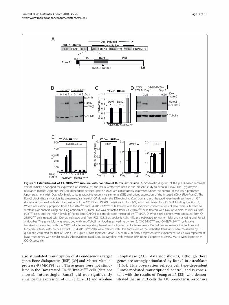
also stimulated transcription of its endogenous targetgenes Bone Sialoprotein (BSP) [29] and Matrix Metallo-protease-9 (MMP9) [28]. These genes were not stimu-lated in the Dox-treated C4-2B/Rx2-Mdox cells (data notshown). Interestingly, Runx2 did not significantlyenhance the expression of OC (Figure 1F) and Alkaline
Phophatase (ALP; data not shown), although thesegenes are strongly stimulated by Runx2 in osteoblasts[1,43]. This observation reflects cell type-dependentRunx2-mediated transcriptional control, and is consis-tent with the results of Yeung et al. [33], who demon-strated that in PC3 cells the OC promoter is responsive
B
GAPDH
Runx2
Runx2-Mdox
0.1 0.5Runx2dox
(α-Flag)
0.1 0.5--
A
C
0100
200
300
400
500
Dox: ( μg/ml)
6xOSE2-Luc
RLU
E
Runx2-MdoxRunx2dox0.1 0.5- 0.1 0.5-
:Dox (μg/ml)
D
F
Rel
. mR
NA
/GA
PD
H
0
0.1
0.2
0.3 OC
Day: 1 2Day: 1 20
0.1
0.2
0.3MMP9
0
0.1
0.2
0.3
0.4BSP
(α-c-term)
Tubulin
-0.25
ROS17/2.8
C4-2B/Rx2dox
Runx2
- :Dox (μg/ml)
0.25-Day 1 Day 2
Day: 1 2
QA
8251
PSTRuntQA
8251
PSTRunt
5’LTR FLAP TRE Ubi -c rtTA3 IRES Hyg WRE 3’SIN LTR5’LTR FLAP TRE Ubi -c rtTA3 IRES Hyg WRE 3’SIN LTRconstitutiveconstitutive
Dox inducedDox induced
FlagRunx2:
R265D, R268D
pSLIK -Runx2
Rel
. mR
NA
/GA
PD
H
00.2
0.4
0.6
0.8
1.0
1.2Veh Dox
Day1 Day2
PC3highC4-2B/Rx2dox
Veh DoxVeh Dox
Figure 1 Establishment of C4-2B/Rx2dox sub-line with conditional Runx2 expression. A, Schematic diagram of the pSLIK-based lentiviralvector. Initially developed for expression of shRNAs [39] the pSLIK vector was used in the present study to express Runx2. The Hygromycinresistance marker (Hyg) and the Dox-dependent activator protein rtTA3 are constitutively expressed under the control of the Ubi-c promoter.Upon treatment with Dox, rtTA binds to its tetracycline responsive elements (TRE) and drives expression of the inserted cDNA (Flag-Runx2). TheRunx2 block diagram depicts its glutamine/alanine-rich QA domain, the DNA-binding Runt domain, and the proline/serine/threonine-rich PSTdomain. Arrowhead indicates the position of the R265D and R268D mutations in Runx2-M, which eliminate Runx2’s DNA binding function. B,Whole cell extracts, prepared from C4-2B/Rx2dox and C4-2B/Rx2-Mdox cells treated with the indicated concentrations of Dox, were subjected towestern blot analysis using anti-Flag antibodies. C, Total RNA was extracted from C4-2B/Rx2dox cells treated with Dox or vehicle, as well as fromPC3high cells, and the mRNA levels of Runx2 (and GAPDH as control) were measured by RT-qPCR. D, Whole cell extracts were prepared from C4-2B/Rx2dox cells treated with Dox as indicated and from ROS 17.8/2 osteoblastic cells [41], and subjected to western blot analysis using anti-Runx2antibodies. The same blot was re-probed with anti-Tubulin antibodies as loading control. E, C4-2B/Rx2dox and C4-2B/Rx2-Mdox cells weretransiently transfected with the 6XOSE2-luciferase reporter plasmid and subjected to luciferase assay. Dotted line represents the backgroundluciferase activity with no cell extract. F, C4-2B/Rx2dox cells were treated with Dox and levels of the indicated transcripts were measured by RT-qPCR and corrected for that of GAPDH. In Figure 1, bars represent Mean ± SEM (n = 3) from a representative experiment, which was repeated atleast three times with similar results. Abbreviations used: Dox, Doxycycline; Veh, vehicle; BSP, Bone Sialoprotein; MMP9, Matrix Metalloprotein-9;OC, Osteocalcin.
Baniwal et al. Molecular Cancer 2010, 9:258http://www.molecular-cancer.com/content/9/1/258
Page 3 of 18
to the transcription factors AP-1 and SP1, but notRunx2. To identify Runx2-regulated genes and pathwaysin advanced PCa cells in an unbiased manner, we sub-jected C4-2B/Rx2dox cells to global gene expressionprofiling.
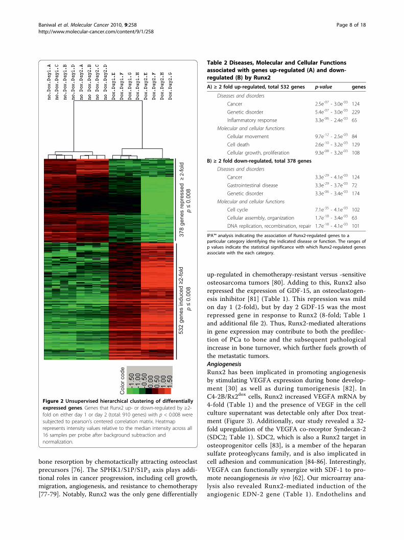
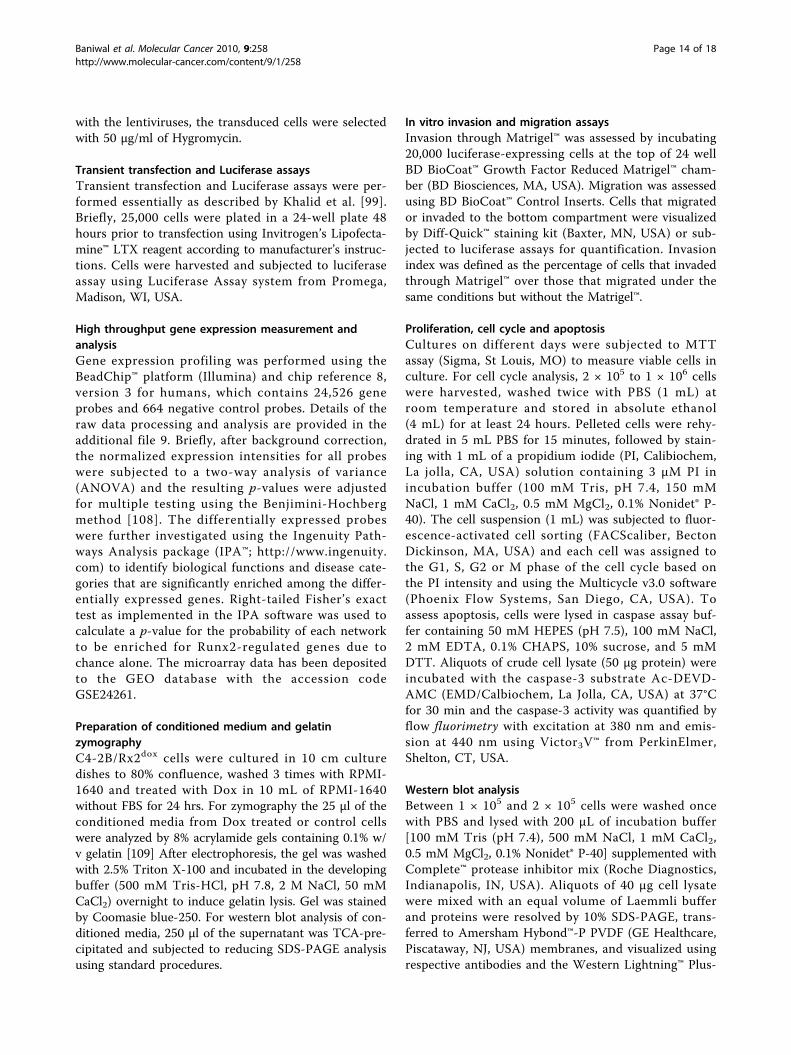
Runx2-regulated global gene expression and in silicoassessment of associated pathwaysC4-2B/Rx2dox cells were subjected to microarray geneexpression analysis after one- and two- days of treat-ment with either Dox or vehicle in biological quadrupli-cates (a total of 16 samples). Of 24,526 probesrepresented in the microarray, 532 genes showed ≥2-fold increased expression and 378 genes showed ≥2-folddecreased expression with high statistical significance(p < 0.008) on either day of treatment (see additionalfile 1). RT-qPCR analysis of 50 representative genes con-formed to the microarray data (Table 1 and additionalfile 2).An unsupervised hierarchical analysis of these 910 up-and down-regulated genes resulted in a clear separationbetween the Dox-treated and control samples (Figure 2).The variation among the biological quadruplicates wassmall, indicating the overall robustness of the methodol-ogy utilized. Gene clusters showing changes in expres-sion pattern with respect to time and Dox treatmentwere clearly discernable. In general, changes observedon day 1 of treatment were maintained or intensified byday 2.We next employed the Ingenuity Pathway Analysis
(IPA™) platform to indentify disease pathways, as well asmolecular and cellular functions associated with Runx2-regulated genes. The analysis suggested ‘cancer’ as thedisease most significantly associated with both the up-and the down-regulated gene groups (Table 2). A totalof 248 genes, half from each group, had highly signifi-cant cancer related function (see additional file 3). Addi-tionally, the up-regulated genes were strongly associatedwith genetic disorders, inflammatory responses, and gas-trointestinal diseases (Table 2A). Among the most sig-nificant molecular and cellular functions, cellularmovement, cell death, cellular growth, and proliferationwere associated with the up-regulated genes, whereascell cycle, cell death, cellular assembly and DNA replica-tion functions were associated with the down-regulatedgenes (Table 2B).
Runx2-modulated genes are involved in tumor metastasisPromotion of tissue invasion, metastasis and cytoskeletondynamicsThe major functions reported for the up-regulated genesbelonged to cancer progression (Table 2). Importantly,these genes encode transcriptional regulators, cytoskele-tal components, signaling molecules and peptidases,
which have been implicated in tumor metastasis (Table1). The transcription factors Sox9 and SNAI2, and theextracellular-matrix (ECM) protein LCN2, all major reg-ulators of epithelial-to-mesenchymal transition (EMT)[44,45], were up-regulated by ~4-fold after one day andby > 6-fold after two days of Runx2 induction, and theirupregulation was confirmed by RT-qPCR (Table 1).However, the functional significance of these EMT mar-kers requires further investigation in light of the unex-pected increase in E-cadherin mRNA (see additional file4). Runx2 also enhanced the expression levels of multi-ple transcripts encoding matrix modifying peptidases(Table 1). These included MMP9, a known Runx2 targetin BCa cells [28] and aspartyl proteases with fibronectindegrading activities such as Prolactin Induced Protein(PIP) and Pepsinogen (PGC) [46,47]. The latter twoshowed a rapid ~10-fold increase within 24 hours(Table 1) and PIP exhibited the highest change (79-fold)in response to Runx2 on day 2 (Table 1 and additionalfile 2). PIP protein in the C4-2B/Rx2dox culture superna-tant was below detectable levels under control condi-tions, but was readily detected after induction of Runx2(Figure 3). We also found increased transcript levels forCystatin 7 (CST7), S100A4 and SMAD3, with a mild~3-fold increase on day 1, but a robust > 20-foldincrease on day 2 (Table 1). These genes function asmetastasis promoters [48,49]. Interestingly, S100A4 andSMAD3 physically interact to potentiate cancer cellinvasiveness [50].Runx2 also up-regulated genes involved in cellular
movement and cytoskeleton remodeling (Table 1).SH3PXD2A, which was up-regulated by 7-fold on day 2,is a scaffold protein involved in the formation of invado-pedia [51,52], which are matrix digesting, actin rich,short lived protrusions observed in osteoclasts and can-cer cells [53]. Runx2 up-regulated by > 9-fold the tran-scripts for Nav2, a scaffold protein crucial for actincytoskeleton remodeling [53]. Other genes that were up-regulated by > 3-fold, with known roles in actin cytoske-leton dynamics included ESPN, which interacts with theSrc-homology 3 (SH3) adaptor proteins to regulatecytoskeletal actin functions [54]; MAP1B, known tomaintain cytoskeletal integrity [55]; LIMA1, whichcross-links actin monomers [56]; and PTK9L (a.k.a.twinfiln), which sequesters ADP-actin monomers in thecytoplasm and delivers them to sites of rapid actin-fila-ment assembly [57].Metastasis to bone and modification of the bonemicroenvironmentThe expression of SDF-1 and its receptor CXCR7 wasenhanced by > 5-fold based on the microarray analysisand by > 20-fold based on the RT-qPCR results (Table1). Runx2-induced SDF-1 protein was also detectable inthe culture supernatant (Figure 3). SDF-1 signaling is
Baniwal et al. Molecular Cancer 2010, 9:258http://www.molecular-cancer.com/content/9/1/258
Page 4 of 18
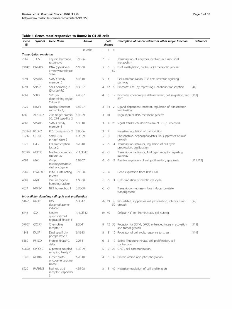
Table 1 Genes most responsive to Runx2 in C4-2B cells
GeneID
Symbol Gene Name Anova Foldchange
Description of cancer related or other major function Reference
p value I II q
Transcription regulators
7069 THRSP Thyroid hormoneresponsive
3.5E-06 7 5 Transcription of enzymes involved in tumor lipidmetabolism
29947 DNMT3L DNA (cytosine-5-)-methyltransferase3-like
5.5E-08 5 6 >50
DNA methylation, nucleic acid metabolic process
4091 SMAD6 SMAD familymember 6
8.1E-10 5 4 Cell communication, TGF-beta receptor signalingpathway
6591 SNAI2 Snail homolog 2(Drosophila)
8.8E-07 4 12 6 Promotes EMT by repressing E-cadherin transcription [44]
6662 SOX9 SRY (sexdetermining regionY)-box 9
4.4E-07 4 6 17 Promotes chondrocyte differentiation, cell migration, andEMT
[110]
7025 NR2F1 Nuclear receptorsubfamily 2,
3.5E-07 3 14 2 Ligand-dependent receptor, regulation of transcriptiontermination
678 ZFP36L2 Zinc finger protein36, C3H type-like 2
4.1E-09 3 10 Regulation of RNA metabolic process
4088 SMAD3 SMAD familymember 3
6.3E-10 3 7 25 Signal transducer downstream of TGF-b receptors
283248 RCOR2 REST corepressor 2 2.9E-06 3 7 Negative regulation of transcription
10217 CTDSPL Small CTDphosphatase 3
1.9E-09 -2 -3 Phosphatase, dephosphorylates Rb, suppresses cellulargrowth
1870 E2F2 E2F transcriptionfactor 2
8.2E-10 -2 -5 -4 Transcription activator, regulation of cell cycleprogression, proliferation
90390 MED30 Mediator complexsubunit 30
< 1.0E-12 -2 -3 Transcription activator, Androgen receptor signalingpathway
4609 MYC V-mycmyelocytomatosisviral oncogene
2.9E-07 -2 -3 -2 Positive regulation of cell proliferation, apoptosis [111,112]
29893 PSMC3IP PSMC3 interactingprotein
3.5E-08 -2 -4 Gene expression from RNA PolII
4602 MYB Viral oncogenehomolog (avian)
1.6E-08 -3 -5 -3 G1/S transition of mitotic cell cycle
4824 NKX3-1 NK3 homeobox 1 3.7E-08 -3 -3 Transcription repressor, loss induces prostatetumorigenesis
Intracellular signaling, cell cycle and proliferation
51655 RASD1 RAS,dexamethasone-induced 1
6.8E-12 26 19 >50
Ras related, suppresses cell proliferation, inhibits tumorgrowth
[92]
6446 SGK Serum/glucocorticoidregulated kinase 1
< 1.0E-12 19 45 Cellular Na+ ion homeostasis, cell survival
57007 CXCR7 Chemokinereceptor 7
9.2E-11 8 12 30 Receptor for SDF-1, GPCR, enhanced integrin activationand tumor growth
[113]
1843 DUSP1 Dual specificityphosphatase 1
9.1E-13 8 8 10 Regulator of cell cycle, response to stress [114]
5580 PRKCD Protein kinase C,delta
2.0E-11 6 5 12 Serine-Threonine Kinase, cell proliferation, cellcontraction
55890 GPRC5C G protein-coupledreceptor, family C
1.3E-09 5 5 25 GPCR, cell communication
10461 MERTK C-mer proto-oncogene tyrosinekinase
6.2E-10 4 6 39 Protein amino acid phosphorylation
5920 RARRES3 Retinoic acidreceptor responder3
4.3E-08 3 8 40 Negative regulation of cell proliferation
Baniwal et al. Molecular Cancer 2010, 9:258http://www.molecular-cancer.com/content/9/1/258
Page 5 of 18
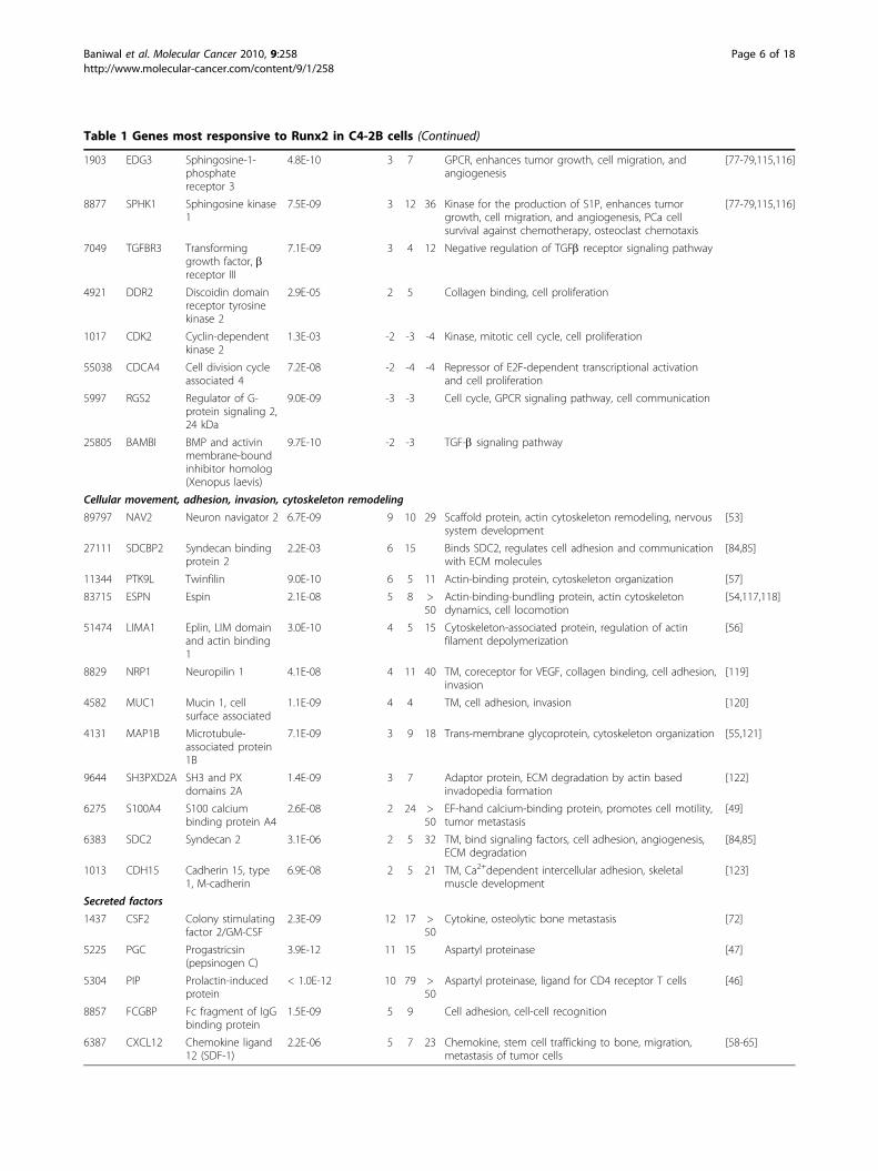
Table 1 Genes most responsive to Runx2 in C4-2B cells (Continued)
1903 EDG3 Sphingosine-1-phosphatereceptor 3
4.8E-10 3 7 GPCR, enhances tumor growth, cell migration, andangiogenesis
[77-79,115,116]
8877 SPHK1 Sphingosine kinase1
7.5E-09 3 12 36 Kinase for the production of S1P, enhances tumorgrowth, cell migration, and angiogenesis, PCa cellsurvival against chemotherapy, osteoclast chemotaxis
[77-79,115,116]
7049 TGFBR3 Transforminggrowth factor, breceptor III
7.1E-09 3 4 12 Negative regulation of TGFb receptor signaling pathway
4921 DDR2 Discoidin domainreceptor tyrosinekinase 2
2.9E-05 2 5 Collagen binding, cell proliferation
1017 CDK2 Cyclin-dependentkinase 2
1.3E-03 -2 -3 -4 Kinase, mitotic cell cycle, cell proliferation
55038 CDCA4 Cell division cycleassociated 4
7.2E-08 -2 -4 -4 Repressor of E2F-dependent transcriptional activationand cell proliferation
5997 RGS2 Regulator of G-protein signaling 2,24 kDa
9.0E-09 -3 -3 Cell cycle, GPCR signaling pathway, cell communication
25805 BAMBI BMP and activinmembrane-boundinhibitor homolog(Xenopus laevis)
9.7E-10 -2 -3 TGF-b signaling pathway
Cellular movement, adhesion, invasion, cytoskeleton remodeling
89797 NAV2 Neuron navigator 2 6.7E-09 9 10 29 Scaffold protein, actin cytoskeleton remodeling, nervoussystem development
[53]
27111 SDCBP2 Syndecan bindingprotein 2
2.2E-03 6 15 Binds SDC2, regulates cell adhesion and communicationwith ECM molecules
[84,85]
11344 PTK9L Twinfilin 9.0E-10 6 5 11 Actin-binding protein, cytoskeleton organization [57]
83715 ESPN Espin 2.1E-08 5 8 >50
Actin-binding-bundling protein, actin cytoskeletondynamics, cell locomotion
[54,117,118]
51474 LIMA1 Eplin, LIM domainand actin binding1
3.0E-10 4 5 15 Cytoskeleton-associated protein, regulation of actinfilament depolymerization
[56]
8829 NRP1 Neuropilin 1 4.1E-08 4 11 40 TM, coreceptor for VEGF, collagen binding, cell adhesion,invasion
[119]
4582 MUC1 Mucin 1, cellsurface associated
1.1E-09 4 4 TM, cell adhesion, invasion [120]
4131 MAP1B Microtubule-associated protein1B
7.1E-09 3 9 18 Trans-membrane glycoprotein, cytoskeleton organization [55,121]
9644 SH3PXD2A SH3 and PXdomains 2A
1.4E-09 3 7 Adaptor protein, ECM degradation by actin basedinvadopedia formation
[122]
6275 S100A4 S100 calciumbinding protein A4
2.6E-08 2 24 >50
EF-hand calcium-binding protein, promotes cell motility,tumor metastasis
[49]
6383 SDC2 Syndecan 2 3.1E-06 2 5 32 TM, bind signaling factors, cell adhesion, angiogenesis,ECM degradation
[84,85]
1013 CDH15 Cadherin 15, type1, M-cadherin
6.9E-08 2 5 21 TM, Ca2+dependent intercellular adhesion, skeletalmuscle development
[123]
Secreted factors
1437 CSF2 Colony stimulatingfactor 2/GM-CSF
2.3E-09 12 17 >50
Cytokine, osteolytic bone metastasis [72]
5225 PGC Progastricsin(pepsinogen C)
3.9E-12 11 15 Aspartyl proteinase [47]
5304 PIP Prolactin-inducedprotein
< 1.0E-12 10 79 >50
Aspartyl proteinase, ligand for CD4 receptor T cells [46]
8857 FCGBP Fc fragment of IgGbinding protein
1.5E-09 5 9 Cell adhesion, cell-cell recognition
6387 CXCL12 Chemokine ligand12 (SDF-1)
2.2E-06 5 7 23 Chemokine, stem cell trafficking to bone, migration,metastasis of tumor cells
[58-65]
Baniwal et al. Molecular Cancer 2010, 9:258http://www.molecular-cancer.com/content/9/1/258
Page 6 of 18
critical for homing of hematopoietic cells to the bonemarrow space and their survival in this environment[58-65]. Within one day, Runx2 also increased by 10-fold the mRNA for BSP (Figure 1F), whose abundantexpression by bone metastatic tumor cells facilitatestheir attachment to the bone matrix [66-69]. Oncesettled in the bone microenvironment, the metastaticcells secrete regulatory molecules that stimulate boneturnover [70]. Remarkably, Runx2 enhanced the expres-sion of the osteoclastogenic cytokine CSF2 by > 50-foldwithin 48 hours (Table 1). This presumably occurred bydirect binding of Runx2 to the CSF2 promoter [71].
Runx2-mediated induction of CSF2 in PCa cells likelycontributes to the increased bone turnover in bonemetastatic sites, similar to the role of this cytokine inbreast cancer bone metastasis [72]. CSF2 production bytumor cells may also contribute to accumulation ofmacrophages, inflammatory T cells, and cytokines[73,74] that exacerbate morbidity and mortality [75].Two additional Runx2-up-regulated genes associatedwith osteoclast function are SPHK1, a kinase responsiblefor the production of sphingosine 1 phosphate (S1P),and S1P receptor 3 (S1P3) a.k.a. EDG3 (Table 1). Pro-duction of S1P in the bone microenvironment promotes
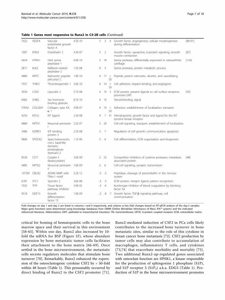
Table 1 Genes most responsive to Runx2 in C4-2B cells (Continued)
7422 VEGFA Vascularendothelial growthfactor A
4.5E-10 5 3 4 Growth factor, angiogenesis, cellular morphogenesisduring differentiation
[88-91]
1907 EDN2 Endothelin 2 4.3E-07 5 2 Growth factor, vasoactive, G-protein signaling, smoothmuscle contraction
[87]
5654 HTRA1 HtrA serinepeptidase 1
6.9E-10 3 18 Serine protease, differentially expressed in osteoarthriticcartillage
[124]
3817 KLK2 Kallikrein-relatedpeptidase 2
1.5E-08 4 3 Serine protease, protein metabolic process
4880 NPPC Natriuretic peptideprecursor C
1.8E-10 4 17 >50
Peptide, potent natriuretic, diuretic, and vasodilating
7057 THBS1 Thrombospondin 1 3.0E-10 4 14 >50
Cell adhesion, heparin-binding, anti-angiogenic
3934 LCN2 Lipocalin 2 5.1E-08 4 10 5 ECM protein, present ligands to cell surface receptors,promotes EMT
[45]
6462 SHBG Sex hormone-binding globulin
8.7E-10 4 10 Steroid-binding, signal
57642 COL20A1 Collagen, type XX,a 1
4.9E-07 4 10 >50
Adhesion, establishment of localization, transport
4254 KITLG KIT ligand 2.3E-08 4 7 41 Hematopoietic growth factor and ligand for the KITtyrosine kinase receptor
4884 NPTX1 Neuronal pentraxinI
2.5E-07 3 20 Cell-cell signaling, transport, establishment of localization
3486 IGFBP3 IGF bindingprotein 3
2.2E-08 3 7 Regulation of cell growth, communication, apoptosis
9806 SPOCK2 Sparc/osteonectin,cwcv, kazal-likedomainsproteoglycan(testican) 2
1.1E-06 3 4 Cell differentiation, ECM organization and biogenesis
8530 CST7 Cystatin F(leukocystatin)
3.0E-09 2 25 Competitive inhibitors of cysteine proteases, metastasisassociated protein
[48]
4885 NPTX2 Neuronal pentraxinII
7.6E-09 2 6 Cell-cell signaling, synaptic transmission
147381 CBLN2 ADAM MMP withThbs-1 motif
5.2E-12 -3 -3 Peptidase, cleavage of precerebellin in the nervoussystem
6781 STC1 Spondin 2 3.6E-08 -3 -3 ECM protein, integrin ligand, pattern recognition
7035 TFPI Tissue factorpathway inhibitor
3.9E-05 -3 -4 Kunitz-type inhibitor of blood coagulation by blockingfactor Xa
9518 GDF15 Growthdifferentiationfactor 15
1.6E-09 -2 -8 -7 Growth factor, TGF-b signaling pathway, cellcommunication
Fold changes on day 1 and day 2 are listed in columns I and II respectively, and column q lists fold changes based on RT-qPCR analysis of the day-2 samples.Major gene functions were determined using knowledge databases from OMIM (Online Mendelian Inheritance of Man), IPA™ systems and the indicatedreferenced literature. Abbreviations: EMT, epithelial to mesenchymal transition; TM, transmembrane; GPCR, G-protein coupled receptor; ECM, extracellular matrix.
Baniwal et al. Molecular Cancer 2010, 9:258http://www.molecular-cancer.com/content/9/1/258
Page 7 of 18
bone resorption by chemotactically attracting osteoclastprecursors [76]. The SPHK1/S1P/S1P3 axis plays addi-tional roles in cancer progression, including cell growth,migration, angiogenesis, and resistance to chemotherapy[77-79]. Notably, Runx2 was the only gene differentially
up-regulated in chemotherapy-resistant versus -sensitiveosteosarcoma tumors [80]. Adding to this, Runx2 alsorepressed the expression of GDF-15, an osteoclastogen-esis inhibitor [81] (Table 1). This repression was mildon day 1 (2-fold), but by day 2 GDF-15 was the mostrepressed gene in response to Runx2 (8-fold; Table 1and additional file 2). Thus, Runx2-mediated alterationsin gene expression may contribute to both the predilec-tion of PCa to bone and the subsequent pathologicalincrease in bone turnover, which further fuels growth ofthe metastatic tumors.AngiogenesisRunx2 has been implicated in promoting angiogenesisby stimulating VEGFA expression during bone develop-ment [30] as well as during tumorigenesis [82]. InC4-2B/Rx2dox cells, Runx2 increased VEGFA mRNA by4-fold (Table 1) and the presence of VEGF in the cellculture supernatant was detectable only after Dox treat-ment (Figure 3). Additionally, our study revealed a 32-fold upregulation of the VEGFA co-receptor Syndecan-2(SDC2; Table 1). SDC2, which is also a Runx2 target inosteoprogenitor cells [83], is a member of the heparansulfate proteoglycans family, and is also implicated incell adhesion and communication [84-86]. Interestingly,VEGFA can functionally synergize with SDF-1 to pro-mote neoangiogenesis in vivo [62]. Our microarray ana-lysis also revealed Runx2-mediated induction of theangiogenic EDN-2 gene (Table 1). Endothelins and
532
gene
s in
duce
d ≥ 2
-fold
37
8 ge
nes
repr
esse
d ≥
2-fo
ld
Col
or c
ode
p≤
0.00
8p
≤0.
008
Figure 2 Unsupervised hierarchical clustering of differentiallyexpressed genes. Genes that Runx2 up- or down-regulated by ≥2-fold on either day 1 or day 2 (total: 910 genes) with p < 0.008 weresubjected to pearson’s centered correlation matrix. Heatmaprepresents intensity values relative to the median intensity across all16 samples per probe after background subtraction andnormalization.
Table 2 Diseases, Molecular and Cellular Functionsassociated with genes up-regulated (A) and down-regulated (B) by Runx2
A) ≥ 2 fold up-regulated, total 532 genes p-value genes
Diseases and disorders
Cancer 2.5e-07 - 3.0e-03 124
Genetic disorder 5.4e-07 - 3.0e-03 229
Inflammatory response 3.3e-06 - 2.4e-03 65
Molecular and cellular functions
Cellular movement 9.7e-12 - 2.5e-03 84
Cell death 2.6e-10 - 3.2e-03 129
Cellular growth, proliferation 9.3e-08 - 3.2e-03 108
B) ≥ 2 fold down-regulated, total 378 genes
Diseases and disorders
Cancer 3.3e-29 - 4.1e-03 124
Gastrointestinal disease 3.3e-29 - 3.7e-03 72
Genetic disorder 3.3e-06 - 3.4e-03 174
Molecular and cellular functions
Cell cycle 7.1e-35 - 4.1e-03 102
Cellular assembly, organization 1.7e-18 - 3.4e-03 63
DNA replication, recombination, repair 1.7e-18 - 4.1e-03 101
IPA™ analysis indicating the association of Runx2-regulated genes to aparticular category identifying the indicated disease or function. The ranges ofp values indicate the statistical significance with which Runx2-regulated genesassociate with the each category.
Baniwal et al. Molecular Cancer 2010, 9:258http://www.molecular-cancer.com/content/9/1/258
Page 8 of 18
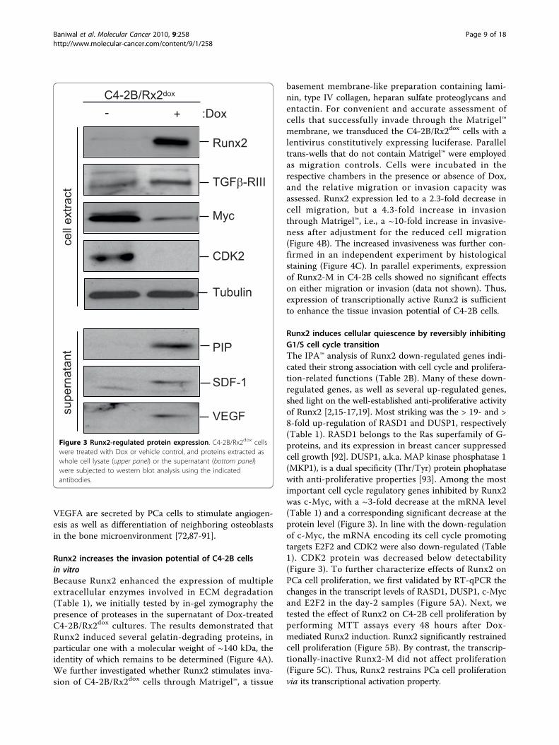
VEGFA are secreted by PCa cells to stimulate angiogen-esis as well as differentiation of neighboring osteoblastsin the bone microenvironment [72,87-91].
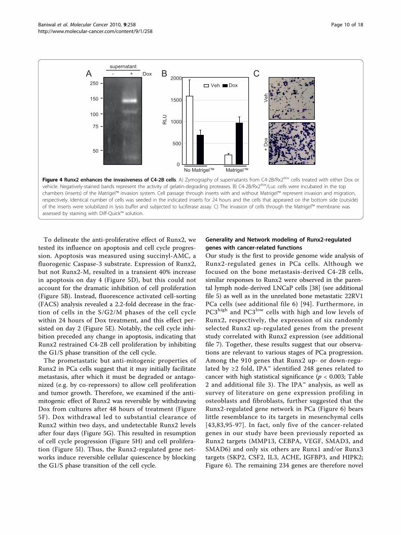
Runx2 increases the invasion potential of C4-2B cellsin vitroBecause Runx2 enhanced the expression of multipleextracellular enzymes involved in ECM degradation(Table 1), we initially tested by in-gel zymography thepresence of proteases in the supernatant of Dox-treatedC4-2B/Rx2dox cultures. The results demonstrated thatRunx2 induced several gelatin-degrading proteins, inparticular one with a molecular weight of ~140 kDa, theidentity of which remains to be determined (Figure 4A).We further investigated whether Runx2 stimulates inva-sion of C4-2B/Rx2dox cells through Matrigel™, a tissue
basement membrane-like preparation containing lami-nin, type IV collagen, heparan sulfate proteoglycans andentactin. For convenient and accurate assessment ofcells that successfully invade through the Matrigel™membrane, we transduced the C4-2B/Rx2dox cells with alentivirus constitutively expressing luciferase. Paralleltrans-wells that do not contain Matrigel™ were employedas migration controls. Cells were incubated in therespective chambers in the presence or absence of Dox,and the relative migration or invasion capacity wasassessed. Runx2 expression led to a 2.3-fold decrease incell migration, but a 4.3-fold increase in invasionthrough Matrigel™, i.e., a ~10-fold increase in invasive-ness after adjustment for the reduced cell migration(Figure 4B). The increased invasiveness was further con-firmed in an independent experiment by histologicalstaining (Figure 4C). In parallel experiments, expressionof Runx2-M in C4-2B cells showed no significant effectson either migration or invasion (data not shown). Thus,expression of transcriptionally active Runx2 is sufficientto enhance the tissue invasion potential of C4-2B cells.
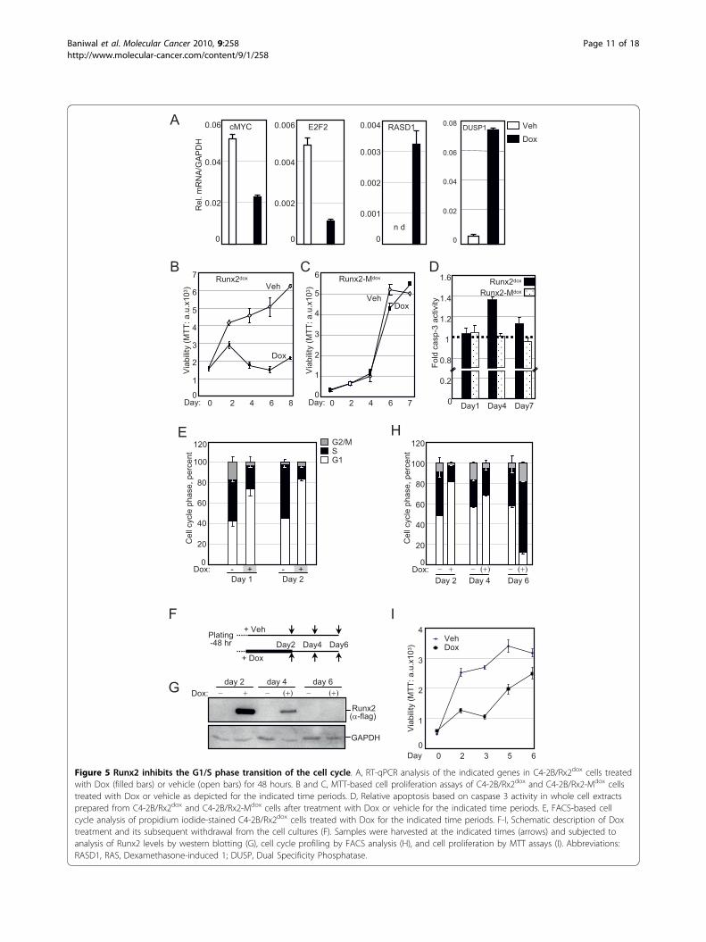
Runx2 induces cellular quiescence by reversibly inhibitingG1/S cell cycle transitionThe IPA™ analysis of Runx2 down-regulated genes indi-cated their strong association with cell cycle and prolifera-tion-related functions (Table 2B). Many of these down-regulated genes, as well as several up-regulated genes,shed light on the well-established anti-proliferative activityof Runx2 [2,15-17,19]. Most striking was the > 19- and >8-fold up-regulation of RASD1 and DUSP1, respectively(Table 1). RASD1 belongs to the Ras superfamily of G-proteins, and its expression in breast cancer suppressedcell growth [92]. DUSP1, a.k.a. MAP kinase phosphatase 1(MKP1), is a dual specificity (Thr/Tyr) protein phophatasewith anti-proliferative properties [93]. Among the mostimportant cell cycle regulatory genes inhibited by Runx2was c-Myc, with a ~3-fold decrease at the mRNA level(Table 1) and a corresponding significant decrease at theprotein level (Figure 3). In line with the down-regulationof c-Myc, the mRNA encoding its cell cycle promotingtargets E2F2 and CDK2 were also down-regulated (Table1). CDK2 protein was decreased below detectability(Figure 3). To further characterize effects of Runx2 onPCa cell proliferation, we first validated by RT-qPCR thechanges in the transcript levels of RASD1, DUSP1, c-Mycand E2F2 in the day-2 samples (Figure 5A). Next, wetested the effect of Runx2 on C4-2B cell proliferation byperforming MTT assays every 48 hours after Dox-mediated Runx2 induction. Runx2 significantly restrainedcell proliferation (Figure 5B). By contrast, the transcrip-tionally-inactive Runx2-M did not affect proliferation(Figure 5C). Thus, Runx2 restrains PCa cell proliferationvia its transcriptional activation property.
Runx2
TGFβ-RIII
Myc
PIP
SDF-1
VEGF
- + :Dox
cell
extra
ctsu
pern
atan
t
CDK2
Tubulin
C4-2B/Rx2dox
Figure 3 Runx2-regulated protein expression. C4-2B/Rx2dox cellswere treated with Dox or vehicle control, and proteins extracted aswhole cell lysate (upper panel) or the supernatant (bottom panel)were subjected to western blot analysis using the indicatedantibodies.
Baniwal et al. Molecular Cancer 2010, 9:258http://www.molecular-cancer.com/content/9/1/258
Page 9 of 18
To delineate the anti-proliferative effect of Runx2, wetested its influence on apoptosis and cell cycle progres-sion. Apoptosis was measured using succinyl-AMC, afluorogenic Caspase-3 substrate. Expression of Runx2,but not Runx2-M, resulted in a transient 40% increasein apoptosis on day 4 (Figure 5D), but this could notaccount for the dramatic inhibition of cell proliferation(Figure 5B). Instead, fluorescence activated cell-sorting(FACS) analysis revealed a 2.2-fold decrease in the frac-tion of cells in the S/G2/M phases of the cell cyclewithin 24 hours of Dox treatment, and this effect per-sisted on day 2 (Figure 5E). Notably, the cell cycle inhi-bition preceded any change in apoptosis, indicating thatRunx2 restrained C4-2B cell proliferation by inhibitingthe G1/S phase transition of the cell cycle.The prometastatic but anti-mitogenic properties of
Runx2 in PCa cells suggest that it may initially facilitatemetastasis, after which it must be degraded or antago-nized (e.g. by co-repressors) to allow cell proliferationand tumor growth. Therefore, we examined if the anti-mitogenic effect of Runx2 was reversible by withdrawingDox from cultures after 48 hours of treatment (Figure5F). Dox withdrawal led to substantial clearance ofRunx2 within two days, and undetectable Runx2 levelsafter four days (Figure 5G). This resulted in resumptionof cell cycle progression (Figure 5H) and cell prolifera-tion (Figure 5I). Thus, the Runx2-regulated gene net-works induce reversible cellular quiescence by blockingthe G1/S phase transition of the cell cycle.
Generality and Network modeling of Runx2-regulatedgenes with cancer-related functionsOur study is the first to provide genome wide analysis ofRunx2-regulated genes in PCa cells. Although wefocused on the bone metastasis-derived C4-2B cells,similar responses to Runx2 were observed in the paren-tal lymph node-derived LNCaP cells [38] (see additionalfile 5) as well as in the unrelated bone metastatic 22RV1PCa cells (see additional file 6) [94]. Furthermore, inPC3high and PC3low cells with high and low levels ofRunx2, respectively, the expression of six randomlyselected Runx2 up-regulated genes from the presentstudy correlated with Runx2 expression (see additionalfile 7). Together, these results suggest that our observa-tions are relevant to various stages of PCa progression.Among the 910 genes that Runx2 up- or down-regu-lated by ≥2 fold, IPA™ identified 248 genes related tocancer with high statistical significance (p < 0.003; Table2 and additional file 3). The IPA™ analysis, as well assurvey of literature on gene expression profiling inosteoblasts and fibroblasts, further suggested that theRunx2-regulated gene network in PCa (Figure 6) bearslittle resemblance to its targets in mesenchymal cells[43,83,95-97]. In fact, only five of the cancer-relatedgenes in our study have been previously reported asRunx2 targets (MMP13, CEBPA, VEGF, SMAD3, andSMAD6) and only six others are Runx1 and/or Runx3targets (SKP2, CSF2, IL3, ACHE, IGFBP3, and HIPK2;Figure 6). The remaining 234 genes are therefore novel
250
150
100
75
50
- + :Dox supernatant
C
+ D
oxV
eh
B
0
500
1000
1500
2000
No Matrigel™ Matrigel™R
LU
AVeh Dox
Figure 4 Runx2 enhances the invasiveness of C4-2B cells. A) Zymography of supernatants from C4-2B/Rx2dox cells treated with either Dox orvehicle. Negatively-stained bands represent the activity of gelatin-degrading proteases. B) C4-2B/Rx2dox/Luc cells were incubated in the topchambers (inserts) of the Matrigel™ invasion system. Cell passage through inserts with and without Matrigel™ represent invasion and migration,respectively. Identical number of cells was seeded in the indicated inserts for 24 hours and the cells that appeared on the bottom side (outside)of the inserts were solubilized in lysis buffer and subjected to luciferase assay. C) The invasion of cells through the Matrigel™ membrane wasassessed by staining with Diff-Quick™ solution.
Baniwal et al. Molecular Cancer 2010, 9:258http://www.molecular-cancer.com/content/9/1/258
Page 10 of 18
Day 1- +
Day 2- +
E
Dox:
Cel
l cyc
le p
hase
, per
cent
G2/MSG1
0
20
40
60
80
100
120
B
Via
bilit
y (M
TT: a
.u.x
103 )
0
1
2
3
4
5
6
7
0 2 4 6 8
Runx2dox
Veh
Dox
Day:
C
0
1
2
3
4
5
6
0 2 4 6 7
Runx2-Mdox
VehDox
Day:
Via
bilit
y (M
TT: a
.u.x
103 )
I
0
1
2
3
4
0 2 3 5 6
VehDox
Day
F
H
Day4 Day7Day10
0.2
0.8
1
1.2
1.4
Fold
cas
p-3
activ
ity
Runx2dox
Runx2-Mdox
1.6D
Plating-48 hr Day2 Day4 Day6
+ Dox
+ Veh
Cel
l cyc
le p
hase
, per
cent
0
20
40
60
80
100
120
Dox: − +Day 2
(+)−Day 4
(+)−Day 6
Via
bilit
y (M
TT: a
.u.x
103 )
GRunx2
(α-flag)
GAPDH
Dox: − +day 2
(+)−day 4
(+)−day 6
0
0.001
0.002
0.003
0.004
n d
RASD1
0
0.02
0.04
0.06
0.08 DUSP1
0
0.02
0.04
0.06 cMYC
0
0.002
0.004
0.006 E2F2
Rel
. mR
NA
/GA
PD
H
A VehDox
Figure 5 Runx2 inhibits the G1/S phase transition of the cell cycle. A, RT-qPCR analysis of the indicated genes in C4-2B/Rx2dox cells treatedwith Dox (filled bars) or vehicle (open bars) for 48 hours. B and C, MTT-based cell proliferation assays of C4-2B/Rx2dox and C4-2B/Rx2-Mdox cellstreated with Dox or vehicle as depicted for the indicated time periods. D, Relative apoptosis based on caspase 3 activity in whole cell extractsprepared from C4-2B/Rx2dox and C4-2B/Rx2-Mdox cells after treatment with Dox or vehicle for the indicated time periods. E, FACS-based cellcycle analysis of propidium iodide-stained C4-2B/Rx2dox cells treated with Dox for the indicated time periods. F-I, Schematic description of Doxtreatment and its subsequent withdrawal from the cell cultures (F). Samples were harvested at the indicated times (arrows) and subjected toanalysis of Runx2 levels by western blotting (G), cell cycle profiling by FACS analysis (H), and cell proliferation by MTT assays (I). Abbreviations:RASD1, RAS, Dexamethasone-induced 1; DUSP, Dual Specificity Phosphatase.
Baniwal et al. Molecular Cancer 2010, 9:258http://www.molecular-cancer.com/content/9/1/258
Page 11 of 18
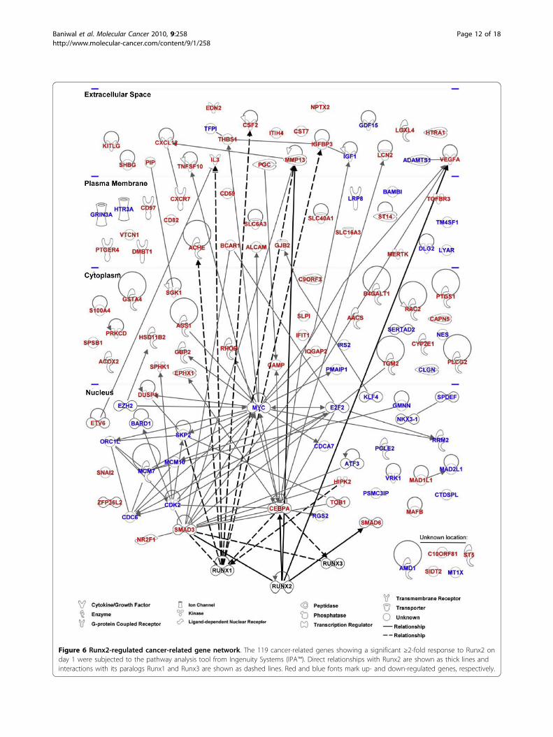
Figure 6 Runx2-regulated cancer-related gene network. The 119 cancer-related genes showing a significant ≥2-fold response to Runx2 onday 1 were subjected to the pathway analysis tool from Ingenuity Systems (IPA™). Direct relationships with Runx2 are shown as thick lines andinteractions with its paralogs Runx1 and Runx3 are shown as dashed lines. Red and blue fonts mark up- and down-regulated genes, respectively.
Baniwal et al. Molecular Cancer 2010, 9:258http://www.molecular-cancer.com/content/9/1/258
Page 12 of 18
Runx2-regulated genes related to cancer in general andto metastasis in particular. The extensive Runx2-regu-lated cancer-related gene network highlights Runx2 as aviable target for the diagnosis, prognosis and treatmentof PCa.
ConclusionsRunx2, traditionally known for its master regulatoryroles in the chondro-osteoblast lineage, is emerging as aprometastatic transcription factor [22,29,34,98-101]. TheRunx2 transcriptome in C4-2B cells documents genenetworks that control multiple aspects of metastasis.Potentially contributing to local invasion and dissemina-tion are the genes known to function in EMT, motilityand ECM degradation. Additionally, the prometastaticfunction of Runx2 likely involves its target genes SDF-1,CXCR7 and BSP, which promote homing and attach-ment to bone. We also discovered Runx2 targets suchas CSF2 and SPHK1, osteoclast activators that likelycontribute to the most important alteration that occursin the bone microenvironment in response to PCametastasis, namely enhanced bone turnover. During thisprocess, bone matrix components such as TGFß, BMPsand calcium ions are released and further fuel tumorgrowth and bone microenvironment modifications[102-105]. The regulation of SPHK1 by Runx2 probablypotentiates additional aspects of the cancer phenotype,including angiogenesis (a function assisted by the Runx2targets VEGFA and EDN2) and drug resistance [77,79].The anti-mitogenic activity of Runx2 is consistent with
the slow growth of PCa tumors, and may contribute todrug resistance. We imagine that future anti-Runx2drugs will be administered along with traditional che-motherapy to eliminate cells that regain proliferativecapacity. Interestingly, Runx2 physically and functionallyinteracts with the receptors for androgens and estrogens[99,101]. Since these receptor proteins are targeted bymany drugs for prostate and breast cancer, it is impor-tant to investigate their effects on Runx2-regulated tran-scription. Moreover, development of selective estrogenand androgen receptor modulators may benefit fromconsideration of their effects on Runx2 and expressionof its target genes reported in the present study.
MethodsCell culture reagents and antibodiesC4-2B cells were obtained from ViroMed Laboratories(Minneapolis, MN). LNCaP and 22RV1 cells were fromATCC (Rockville, MD, USA). PC3 cells were alsoobtained from ATCC, but propagated for several yearsin either our laboratory (PC3low) or that of USC’s Dr.Pradip Roy-Burman (PC3high). The cells were main-tained in RPMI-1640 medium supplemented with 10%Tet System Approved FBS from Clontech, CA, USA.
Hygromycin B was purchased from Invitrogen, Carlsbad,CA, USA and added to the growth medium at 50 μg/ml.Doxycycline from Calibiochem, La Jolla, CA, USA wasused at 0.25 ug/ml unless otherwise stated, and an equalvolume of distilled water was used as vehicle control.Mouse ANTI-FLAG® M2 monoclonal antibody for waspurchased from Sigma, St Louis, MO, USA. Mouse anti-Runx2 was from Invitrogen, Carlsbad, CA, USA. Theanti-PIP antibody (ab 62363) was purchased fromabcam Inc., Cambridge, MA; and anti-GAPDH (V-18),anti-TGFß-RIII (Sc 28975), and anti-SDF-1 (Sc-28876)antibodies were purchased from Santa Cruz Biotechnol-ogy Inc, Santa Cruz, CA, USA. The mouse monoclonalanti-Myc and anti-VEGFA antibodies were developedand kindly provided by USC’s Dr. Prakash S. Gill andDr. Young Hong, respectively. The mouse monoclonalanti-Tubulin antibody, developed by Dr. Charles Walsh,was obtained from the Developmental Studies Hybri-doma Bank under the auspices of the NICHD and TheUniversity of Iowa, Department of Biological Sciences,Iowa City, USA.
Plasmid constructionThe cDNA encoding mouse Runx2 (MASN isoform,type-2), which is 97% identical to human type-2 Runx2[1], was amplified using pcDNA3.0-Runx2 as templateand the Flag epitope was inserted during the PCRamplification (see primers in additional file 8). The Flag-Runx2 cDNA was initially cloned into the SpeI/MfeI-digested lentiviral entry vector pEN_TmiRc3 (ATCC®catalog: MBA-248), and the resulting plasmid wasrecombined using Gateway® LR Clonase® II enzyme mix(Invitrogen, Carlsbad, CA, USA) with the pSLIK (singlelentivector for inducible knockdown) destination vectorcarrying a hygromycin resistance gene (ATCC® catalog:MBA-237). The entry and destination vectors werekindly provided by USC’s Dr. Elizabeth Lowler (Chil-drens Hospital Los Angeles). The DNA-binding mutantRunx2-M was constructed by site directed mutagenesisof two arginine residues at positions 265 and 268(GRSGRGK) known to contact DNA in the crystalstructure [40]. Lentiviral plasmid for constitutive Luci-ferase expression pCCL-c-MNDU3c-Luc-PGK-eGFP waskindly provided by USC’s Dr. Michael Kahn at the Zil-kha Neurogenetic Institute.
Lentivirus production and infectionFor packaging, the lentiviral expression plasmids werecotransfected by the calcium chloride method intoHEK293T cells along with helper plasmids pMD.G1 andpCMVR8.91[106,107]. Culture media containing viralparticles were harvested after 48-72 hours and used fortransduction of C4-2B cells in the presence of 8 μg/mlPolybrene (Millipore Corp., MA, USA). After infection
Baniwal et al. Molecular Cancer 2010, 9:258http://www.molecular-cancer.com/content/9/1/258
Page 13 of 18
with the lentiviruses, the transduced cells were selectedwith 50 μg/ml of Hygromycin.
Transient transfection and Luciferase assaysTransient transfection and Luciferase assays were per-formed essentially as described by Khalid et al. [99].Briefly, 25,000 cells were plated in a 24-well plate 48hours prior to transfection using Invitrogen’s Lipofecta-mine™ LTX reagent according to manufacturer’s instruc-tions. Cells were harvested and subjected to luciferaseassay using Luciferase Assay system from Promega,Madison, WI, USA.
High throughput gene expression measurement andanalysisGene expression profiling was performed using theBeadChip™ platform (Illumina) and chip reference 8,version 3 for humans, which contains 24,526 geneprobes and 664 negative control probes. Details of theraw data processing and analysis are provided in theadditional file 9. Briefly, after background correction,the normalized expression intensities for all probeswere subjected to a two-way analysis of variance(ANOVA) and the resulting p-values were adjustedfor multiple testing using the Benjimini-Hochbergmethod [108]. The differentially expressed probeswere further investigated using the Ingenuity Path-ways Analysis package (IPA™; http://www.ingenuity.com) to identify biological functions and disease cate-gories that are significantly enriched among the differ-entially expressed genes. Right-tailed Fisher’s exacttest as implemented in the IPA software was used tocalculate a p-value for the probability of each networkto be enriched for Runx2-regulated genes due tochance alone. The microarray data has been depositedto the GEO database with the accession codeGSE24261.
Preparation of conditioned medium and gelatinzymographyC4-2B/Rx2dox cells were cultured in 10 cm culturedishes to 80% confluence, washed 3 times with RPMI-1640 and treated with Dox in 10 mL of RPMI-1640without FBS for 24 hrs. For zymography the 25 μl of theconditioned media from Dox treated or control cellswere analyzed by 8% acrylamide gels containing 0.1% w/v gelatin [109] After electrophoresis, the gel was washedwith 2.5% Triton X-100 and incubated in the developingbuffer (500 mM Tris-HCl, pH 7.8, 2 M NaCl, 50 mMCaCl2) overnight to induce gelatin lysis. Gel was stainedby Coomasie blue-250. For western blot analysis of con-ditioned media, 250 μl of the supernatant was TCA-pre-cipitated and subjected to reducing SDS-PAGE analysisusing standard procedures.
In vitro invasion and migration assaysInvasion through Matrigel™ was assessed by incubating20,000 luciferase-expressing cells at the top of 24 wellBD BioCoat™ Growth Factor Reduced Matrigel™ cham-ber (BD Biosciences, MA, USA). Migration was assessedusing BD BioCoat™ Control Inserts. Cells that migratedor invaded to the bottom compartment were visualizedby Diff-Quick™ staining kit (Baxter, MN, USA) or sub-jected to luciferase assays for quantification. Invasionindex was defined as the percentage of cells that invadedthrough Matrigel™ over those that migrated under thesame conditions but without the Matrigel™.
Proliferation, cell cycle and apoptosisCultures on different days were subjected to MTTassay (Sigma, St Louis, MO) to measure viable cells inculture. For cell cycle analysis, 2 × 10⁵ to 1 × 10⁶ cellswere harvested, washed twice with PBS (1 mL) atroom temperature and stored in absolute ethanol(4 mL) for at least 24 hours. Pelleted cells were rehy-drated in 5 mL PBS for 15 minutes, followed by stain-ing with 1 mL of a propidium iodide (PI, Calibiochem,La jolla, CA, USA) solution containing 3 μM PI inincubation buffer (100 mM Tris, pH 7.4, 150 mMNaCl, 1 mM CaCl2, 0.5 mM MgCl2, 0.1% Nonidet® P-40). The cell suspension (1 mL) was subjected to fluor-escence-activated cell sorting (FACScaliber, BectonDickinson, MA, USA) and each cell was assigned tothe G1, S, G2 or M phase of the cell cycle based onthe PI intensity and using the Multicycle v3.0 software(Phoenix Flow Systems, San Diego, CA, USA). Toassess apoptosis, cells were lysed in caspase assay buf-fer containing 50 mM HEPES (pH 7.5), 100 mM NaCl,2 mM EDTA, 0.1% CHAPS, 10% sucrose, and 5 mMDTT. Aliquots of crude cell lysate (50 μg protein) wereincubated with the caspase-3 substrate Ac-DEVD-AMC (EMD/Calbiochem, La Jolla, CA, USA) at 37°Cfor 30 min and the caspase-3 activity was quantified byflow fluorimetry with excitation at 380 nm and emis-sion at 440 nm using Victor3V™ from PerkinElmer,Shelton, CT, USA.
Western blot analysisBetween 1 × 10⁵ and 2 × 10⁵ cells were washed oncewith PBS and lysed with 200 μL of incubation buffer[100 mM Tris (pH 7.4), 500 mM NaCl, 1 mM CaCl2,0.5 mM MgCl2, 0.1% Nonidet® P-40] supplemented withComplete™ protease inhibitor mix (Roche Diagnostics,Indianapolis, IN, USA). Aliquots of 40 μg cell lysatewere mixed with an equal volume of Laemmli bufferand proteins were resolved by 10% SDS-PAGE, trans-ferred to Amersham Hybond™-P PVDF (GE Healthcare,Piscataway, NJ, USA) membranes, and visualized usingrespective antibodies and the Western Lightning™ Plus-
Baniwal et al. Molecular Cancer 2010, 9:258http://www.molecular-cancer.com/content/9/1/258
Page 14 of 18
ECL kit (PerkinElmer Inc, Waltham, MA, USA) followedby exposure to X-ray film (ISCBioExpress®, Kaysville,UT, USA).
RT-qPCRTotal RNA was isolated using Aurum Total RNA kit(Bio-Rad Laboratories Inc., Hercules, CA, USA) follow-ing the manufacturer’s recommendations and 1 μg wasreverse transcribed using the Superscript III kit (Invitro-gen, CA, USA). The cDNA was subjected to real-timePCR amplification using iQ SYBR Green Supermix anda Opticon™2 real time PCR machine from Bio-Rad, Her-cules, CA. The sequences of primers for amplification ofthe cDNA of interest and the control GAPDH are listedin the additional file 8.GEO accession code for the microarray data:
GSE24261
Additional material
Additional file 1: ANOVA analysis of the microarray data. List of allprobes with the microarray gene expression data in log scale (base 2)along with the Fold change, P-values and gene annotations.
Additional file 2: Runx2-regulated genes involved in cellularmetabolism. List of genes with functions in cellular metabolism, theirfold changes, and known functions.
Additional file 3: Runx2 -regulated genes involved in cancer. List of256 genes with established roles in cancer.
Additional file 4: E-Cadherin expression in C4-2B/Rx2dox cells uponRunx2 expression. Western blot and RT-qPCR analysis of C4-2B/Rx2dox
cells in response to Runx2 expression.
Additional file 5: Generation and characterization of LNCaP/Rx2dox
cells. RT-PCR to detect Runx2 transcript in PC3, C4-2B, and LNCaP cells.Proliferation of the LNCaP/Rx2dox cells by using MTT, and RT-qPCRanalysis of Runx2-regulated genes.
Additional file 6: Generation and characterization of 22RV1/Rx2dox
cells. Western blot analysis of Dox-induced Runx2, and MTT basedproliferation analysis of 22RV1/Rx2dox cells in response to Runx2expression.
Additional file 7: Expression of PGC, CST7, S100A4, SDF-1, CSF2,and DUSP1 in two PC3 sub-lines with different Runx2 levels. RT-qPCR analysis using PC3high and PC3low cells to examine the expressionof Runx2-regulated genes.
Additional file 8: List of primers used in the present study to PCR-amplify the indicated gene sequences. Nucleotide sequence ofprimers.
Additional file 9: Bioinformatics analysis of the microarray data.Detailed description of microarray quality control and data processing(such as quality control, normalization and box/density plots); andmethodology employed for the identification of differentially expressedprobes.
AcknowledgementsWe thank Dr. Joseph Hacia and Dr. Nyam-Osor Chimge at the University ofSouthern California for their critical comments during the preparation of themanuscript. We also thank Dr. Gerard Karsenty (Columbia University, NewYork, NY) for the 6XOSE2-luciferase reporter and pCDNA3.1-Runx2 (mouse)plasmids. The microarray analysis was performed at the Southern CaliforniaGenotyping Consortium at UCLA http://scgc.genetics.ucla.edu/ under the
direction of Mr. Joseph DeYoung. Further analysis of the gene expressionprofiles was performed with help from Dr. Yibu Chen at the USC NorrisMedical Library. We thank Dr. Shinwu Jeong and Dr. Gabriel M Gordon fortheir help with the zymography assay. We also thank Dr. Pradip Roy-Burmanand Helty Adisetiyo for kindly providing the PC3high cell line. The anti-TGFß-RIII antibody was kindly provided by Dr. Yang Chai at Center for CraniofacialMolecular Biology; and anti-SDF-1 antibody was kindly provided by Dr.Henry Sucov at the University of Southern California. This work was fundedby NIH grants R01 CA 109147 and R01 CA 136924. SKB was supported by apostdoctoral Innovative Chapter Research Award; and YG by a Meyer YoungInvestigator Fellowship, both from the Arthritis Foundation SouthernCalifornia Chapter. BF holds the J. Harold and Edna L. LaBriola Chair inGenetic Orthopedic Research at USC.
Author details1Department of Biochemistry & Molecular Biology, University of SouthernCalifornia, Los Angeles, USA. 2Department of Orthopaedic Surgery, Universityof Southern California, Los Angeles, USA. 3Department of Urology, Universityof Southern California, Los Angeles, USA. 4Institute for Genetic Medicine,Keck School of Medicine at the University of Southern California, LosAngeles, USA. 5Norris Cancer Center, University of Southern California, LosAngeles, USA. 6Keck School of Medicine at the University of SouthernCalifornia, Los Angeles, USA. 7SRA International, 2605 Meridian Parkway,Durham, USA. 8Sciome LLC, 2 Davis Drive, Research Triangle Park, USA.
Authors’ contributionsConceived and designed the experiments: SKB and BF. Performed theexperiments: SKB, OK, YG, AEG, SS and DJP. Bioinformatics analysis: RRS andDM. Analyzed data and wrote the paper: SKB, GAC and BF. All authors haveread and approved the final manuscript.
Competing interestsThe authors declare that they have no competing interests.
Received: 8 April 2010 Accepted: 23 September 2010Published: 23 September 2010
References1. Ducy P, Zhang R, Geoffroy V, Ridall AL, Karsenty G: Osf2/Cbfa1: a
transcriptional activator of osteoblast differentiation. Cell 1997,89:747-754.
2. Cameron ER, Neil JC: The Runx genes: lineage-specific oncogenes andtumor suppressors. Oncogene 2004, 23:4308-4314.
3. Lo Coco F, Pisegna S, Diverio D: The AML1 gene: a transcription factorinvolved in the pathogenesis of myeloid and lymphoid leukemias.Haematologica 1997, 82:364-370.
4. Woolf E, Xiao C, Fainaru O, Lotem J, Rosen D, Negreanu V, Bernstein Y,Goldenberg D, Brenner O, Berke G, et al: Runx3 and Runx1 are requiredfor CD8 T cell development during thymopoiesis. Proc Natl Acad Sci USA2003, 100:7731-7736.
5. Komori T, Yagi H, Nomura S, Yamaguchi A, Sasaki K, Deguchi K, Shimizu Y,Bronson RT, Gao YH, Inada M, et al: Targeted disruption of Cbfa1 resultsin a complete lack of bone formation owing to maturational arrest ofosteoblasts. Cell 1997, 89:755-764.
6. Otto F, Thornell AP, Crompton T, Denzel A, Gilmour KC, Rosewell IR,Stamp GW, Beddington RS, Mundlos S, Olsen BR, et al: Cbfa1, a candidategene for cleidocranial dysplasia syndrome, is essential for osteoblastdifferentiation and bone development. Cell 1997, 89:765-771.
7. Levanon D, Bettoun D, Harris-Cerruti C, Woolf E, Negreanu V, Eilam R,Bernstein Y, Goldenberg D, Xiao C, Fliegauf M, et al: The Runx3transcription factor regulates development and survival of TrkC dorsalroot ganglia neurons. Embo J 2002, 21:3454-3463.
8. Ito Y: Oncogenic potential of the RUNX gene family: ‘overview’.Oncogene 2004, 23:4198-4208.
9. Bae SC, Choi JK: Tumor suppressor activity of RUNX3. Oncogene 2004,23:4336-4340.
10. Levanon D, Groner Y: Structure and regulated expression of mammalianRUNX genes. Oncogene 2004, 23:4211-4219.
11. Oshimo Y, Oue N, Mitani Y, Nakayama H, Kitadai Y, Yoshida K, Ito Y,Chayama K, Yasui W: Frequent loss of RUNX3 expression by promoterhypermethylation in gastric carcinoma. Pathobiology 2004, 71:137-143.
Baniwal et al. Molecular Cancer 2010, 9:258http://www.molecular-cancer.com/content/9/1/258
Page 15 of 18
12. Rossetti S, Van Unen L, Touw IP, Hoogeveen AT, Sacchi N: Myeloidmaturation block by AML1-MTG16 is associated with Csf1r epigeneticdownregulation. Oncogene 2005, 24:5325-5332.
13. Kilbey A, Blyth K, Wotton S, Terry A, Jenkins A, Bell M, Hanlon L,Cameron ER, Neil JC: Runx2 disruption promotes immortalization andconfers resistance to oncogene-induced senescence in primary murinefibroblasts. Cancer Res 2007, 67:11263-11271.
14. Zaidi SK, Pande S, Pratap J, Gaur T, Grigoriu S, Ali SA, Stein JL, Lian JB, vanWijnen AJ, Stein GS: Runx2 deficiency and defective subnuclear targetingbypass senescence to promote immortalization and tumorigenicpotential. Proc Natl Acad Sci USA 2007, 104:19861-19866.
15. Galindo M, Pratap J, Young DW, Hovhannisyan H, Im HJ, Choi JY, Lian JB,Stein JL, Stein GS, van Wijnen AJ: The bone-specific expression of Runx2oscillates during the cell cycle to support a G1-related antiproliferativefunction in osteoblasts. J Biol Chem 2005, 280:20274-20285.
16. Pratap J, Galindo M, Zaidi SK, Vradii D, Bhat BM, Robinson JA, Choi JY,Komori T, Stein JL, Lian JB, et al: Cell growth regulatory role of Runx2during proliferative expansion of preosteoblasts. Cancer Res 2003,63:5357-5362.
17. Thomas DM, Johnson SA, Sims NA, Trivett MK, Slavin JL, Rubin BP,Waring P, McArthur GA, Walkley CR, Holloway AJ, et al: Terminal osteoblastdifferentiation, mediated by runx2 and p27KIP1, is disrupted inosteosarcoma. J Cell Biol 2004, 167:925-934.
18. Blyth K, Terry A, Mackay N, Vaillant F, Bell M, Cameron ER, Neil JC,Stewart M: Runx2: a novel oncogenic effector revealed by in vivocomplementation and retroviral tagging. Oncogene 2001, 20:295-302.
19. Blyth K, Cameron ER, Neil JC: The RUNX genes: gain or loss of function incancer. Nat Rev Cancer 2005, 5:376-387.
20. Komori T: Regulation of osteoblast differentiation by transcriptionfactors. J Cell Biochem 2006, 99:1233-1239.
21. Otto F, Kanegane H, Mundlos S: Mutations in the RUNX2 gene in patientswith cleidocranial dysplasia. Hum Mutat 2002, 19:209-216.
22. Akech J, Wixted JJ, Bedard K, van der Deen M, Hussain S, Guise TA, vanWijnen AJ, Stein JL, Languino LR, Altieri DC, et al: Runx2 association withprogression of prostate cancer in patients: mechanisms mediating boneosteolysis and osteoblastic metastatic lesions. Oncogene 2009, 811-821.
23. Koeneman KS, Yeung F, Chung LW: Osteomimetic properties of prostatecancer cells: a hypothesis supporting the predilection of prostate cancermetastasis and growth in the bone environment. Prostate 1999,39:246-261.
24. Chua CW, Chiu YT, Yuen HF, Chan KW, Man K, Wang X, Ling MT, Wong YC:Suppression of androgen-independent prostate cancer cellaggressiveness by FTY720: validating Runx2 as a potential antimetastaticdrug screening platform. Clin Cancer Res 2009, 15:4322-4335.
25. Lin DL, Tarnowski CP, Zhang J, Dai J, Rohn E, Patel AH, Morris MD, Keller ET:Bone metastatic LNCaP-derivative C4-2B prostate cancer cell linemineralizes in vitro. Prostate 2001, 47:212-221.
26. Selvamurugan N, Kwok S, Partridge NC: Smad3 interacts with JunB andCbfa1/Runx2 for transforming growth factor-beta1-stimulatedcollagenase-3 expression in human breast cancer cells. J Biol Chem 2004,279:27764-27773.
27. Lim M, Zhong C, Yang S, Bell AM, Cohen MB, Roy-Burman P: Runx2regulates survivin expression in prostate cancer cells. Lab Invest90:222-233.
28. Pratap J, Javed A, Languino LR, van Wijnen AJ, Stein JL, Stein GS, Lian JB:The Runx2 osteogenic transcription factor regulates matrixmetalloproteinase 9 in bone metastatic cancer cells and controls cellinvasion. Mol Cell Biol 2005, 25:8581-8591.
29. Barnes GL, Javed A, Waller SM, Kamal MH, Hebert KE, Hassan MQ,Bellahcene A, Van Wijnen AJ, Young MF, Lian JB, et al: Osteoblast-relatedtranscription factors Runx2 (Cbfa1/AML3) and MSX2 mediate theexpression of bone sialoprotein in human metastatic breast cancer cells.Cancer Res 2003, 63:2631-2637.
30. Zelzer E, Glotzer DJ, Hartmann C, Thomas D, Fukai N, Soker S, Olsen BR:Tissue specific regulation of VEGF expression during bone developmentrequires Cbfa1/Runx2. Mech Dev 2001, 106:97-106.
31. Geoffroy V, Kneissel M, Fournier B, Boyde A, Matthias P: High boneresorption in adult aging transgenic mice overexpressing cbfa1/runx2 incells of the osteoblastic lineage. Mol Cell Biol 2002, 22:6222-6233.
32. Maruyama Z, Yoshida CA, Furuichi T, Amizuka N, Ito M, Fukuyama R,Miyazaki T, Kitaura H, Nakamura K, Fujita T, et al: Runx2 determines bone
maturity and turnover rate in postnatal bone development and isinvolved in bone loss in estrogen deficiency. Dev Dyn 2007,236:1876-1890.
33. Yeung F, Law WK, Yeh CH, Westendorf JJ, Zhang Y, Wang R, Kao C,Chung LW: Regulation of human osteocalcin promoter in hormone-independent human prostate cancer cells. J Biol Chem 2002,277:2468-2476.
34. Pratap J, Imbalzano KM, Underwood JM, Cohet N, Gokul K, Akech J, vanWijnen AJ, Stein JL, Imbalzano AN, Nickerson JA, et al: Ectopic runx2expression in mammary epithelial cells disrupts formation of normalacini structure: implications for breast cancer progression. Cancer Res2009, 69:6807-6814.
35. Kayed H, Jiang X, Keleg S, Jesnowski R, Giese T, Berger MR, Esposito I,Lohr M, Friess H, Kleeff J: Regulation and functional role of the Runt-related transcription factor-2 in pancreatic cancer. Br J Cancer 2007,97:1106-1115.
36. Endo T, Ohta K, Kobayashi T: Expression and function of Cbfa-1/Runx2 inthyroid papillary carcinoma cells. J Clin Endocrinol Metab 2008,93:2409-2412.
37. Horoszewicz JS, Leong SS, Kawinski E, Karr JP, Rosenthal H, Chu TM,Mirand EA, Murphy GP: LNCaP model of human prostatic carcinoma.Cancer Res 1983, 43:1809-1818.
38. Wu HC, Hsieh JT, Gleave ME, Brown NM, Pathak S, Chung LW: Derivation ofandrogen-independent human LNCaP prostatic cancer cell sublines: roleof bone stromal cells. Int J Cancer 1994, 57:406-412.
39. Shin KJ, Wall EA, Zavzavadjian JR, Santat LA, Liu J, Hwang JI, Rebres R,Roach T, Seaman W, Simon MI, Fraser ID: A single lentiviral vectorplatform for microRNA-based conditional RNA interference andcoordinated transgene expression. Proc Natl Acad Sci USA 2006,103:13759-13764.
40. Tahirov TH, Inoue-Bungo T, Morii H, Fujikawa A, Sasaki M, Kimura K,Shiina M, Sato K, Kumasaka T, Yamamoto M, et al: Structural analyses ofDNA recognition by the AML1/Runx-1 Runt domain and its allostericcontrol by CBFbeta. Cell 2001, 104:755-767.
41. Sudhakar S, Li Y, Katz MS, Elango N: Translational regulation is a controlpoint in RUNX2/Cbfa1 gene expression. Biochem Biophys Res Commun2001, 289:616-622.
42. Ducy P, Karsenty G: Two distinct osteoblast-specific cis-acting elementscontrol expression of a mouse osteocalcin gene. Mol Cell Biol 1995,15:1858-1869.
43. Vaes BL, Ducy P, Sijbers AM, Hendriks JM, van Someren EP, de Jong NG,van den Heuvel ER, Olijve W, van Zoelen EJ, Dechering KJ: Microarrayanalysis on Runx2-deficient mouse embryos reveals novel Runx2functions and target genes during intramembranous and endochondralbone formation. Bone 2006, 39:724-738.
44. Cheung M, Chaboissier MC, Mynett A, Hirst E, Schedl A, Briscoe J: Thetranscriptional control of trunk neural crest induction, survival, anddelamination. Dev Cell 2005, 8:179-192.
45. Yang J, Bielenberg DR, Rodig SJ, Doiron R, Clifton MC, Kung AL, Strong RK,Zurakowski D, Moses MA: Lipocalin 2 promotes breast cancerprogression. Proc Natl Acad Sci USA 2009, 106:3913-3918.
46. Caputo E, Manco G, Mandrich L, Guardiola J: A novel aspartyl proteinasefrom apocrine epithelia and breast tumors. J Biol Chem 2000,275:7935-7941.
47. Chiang L, Contreras L, Chiang J, Ward PH: Human prostatic gastricsinogen:the precursor of seminal fluid acid proteinase. Arch Biochem Biophys 1981,210:14-20.
48. Morita M, Yoshiuchi N, Arakawa H, Nishimura S: CMAP: a novel cystatin-like gene involved in liver metastasis. Cancer Res 1999, 59:151-158.
49. Sherbet GV: Metastasis promoter S100A4 is a potentially valuablemolecular target for cancer therapy. Cancer Lett 2009, 280:15-30.
50. Matsuura I, Lai CY, Chiang KN: Functional interaction between Smad3 andS100A4 (metastatin-1) for TGF-beta-mediated cancer cell invasiveness.Biochem J 2010, 426:327-335.
51. Artym VV, Zhang Y, Seillier-Moiseiwitsch F, Yamada KM, Mueller SC:Dynamic interactions of cortactin and membrane type 1 matrixmetalloproteinase at invadopodia: defining the stages of invadopodiaformation and function. Cancer Res 2006, 66:3034-3043.
52. Larsen M, Artym VV, Green JA, Yamada KM: The matrix reorganized:extracellular matrix remodeling and integrin signaling. Curr Opin Cell Biol2006, 18:463-471.
Baniwal et al. Molecular Cancer 2010, 9:258http://www.molecular-cancer.com/content/9/1/258
Page 16 of 18
53. Schmidt KL, Marcus-Gueret N, Adeleye A, Webber J, Baillie D, Stringham EG:The cell migration molecule UNC-53/NAV2 is linked to the ARP2/3complex by ABI-1. Development 2009, 136:563-574.
54. Sekerkova G, Loomis PA, Changyaleket B, Zheng L, Eytan R, Chen B,Mugnaini E, Bartles JR: Novel espin actin-bundling proteins are localizedto Purkinje cell dendritic spines and bind the Src homology 3 adapterprotein insulin receptor substrate p53. J Neurosci 2003, 23:1310-1319.
55. Riederer BM: Microtubule-associated protein 1B, a growth-associated andphosphorylated scaffold protein. Brain Res Bull 2007, 71:541-558.
56. Maul RS, Song Y, Amann KJ, Gerbin SC, Pollard TD, Chang DD: EPLINregulates actin dynamics by cross-linking and stabilizing filaments. J CellBiol 2003, 160:399-407.
57. Paavilainen VO, Bertling E, Falck S, Lappalainen P: Regulation ofcytoskeletal dynamics by actin-monomer-binding proteins. Trends CellBiol 2004, 14:386-394.
58. Levoye A, Balabanian K, Baleux F, Bachelerie F, Lagane B: CXCR7heterodimerizes with CXCR4 and regulates CXCL12-mediated G proteinsignaling. Blood 2009, 113:6085-6093.
59. Mazzinghi B, Ronconi E, Lazzeri E, Sagrinati C, Ballerini L, Angelotti ML,Parente E, Mancina R, Netti GS, Becherucci F, et al: Essential but differentialrole for CXCR4 and CXCR7 in the therapeutic homing of human renalprogenitor cells. J Exp Med 2008, 205:479-490.
60. Kryczek I, Wei S, Keller E, Liu R, Zou W: Stroma-derived factor (SDF-1/CXCL12) and human tumor pathogenesis. Am J Physiol Cell Physiol 2007,292:C987-995.
61. Singh S, Singh UP, Grizzle WE, Lillard JW Jr: CXCL12-CXCR4 interactionsmodulate prostate cancer cell migration, metalloproteinase expressionand invasion. Lab Invest 2004, 84:1666-1676.
62. Kryczek I, Lange A, Mottram P, Alvarez X, Cheng P, Hogan M, Moons L,Wei S, Zou L, Machelon V, et al: CXCL12 and vascular endothelial growthfactor synergistically induce neoangiogenesis in human ovarian cancers.Cancer Res 2005, 65:465-472.
63. Taichman RS, Cooper C, Keller ET, Pienta KJ, Taichman NS, McCauley LK:Use of the stromal cell-derived factor-1/CXCR4 pathway in prostatecancer metastasis to bone. Cancer Res 2002, 62:1832-1837.
64. Ara T, Tokoyoda K, Sugiyama T, Egawa T, Kawabata K, Nagasawa T:Long-term hematopoietic stem cells require stromal cell-derivedfactor-1 for colonizing bone marrow during ontogeny. Immunity 2003,19:257-267.
65. Peled A, Petit I, Kollet O, Magid M, Ponomaryov T, Byk T, Nagler A, Ben-Hur H, Many A, Shultz L, et al: Dependence of human stem cellengraftment and repopulation of NOD/SCID mice on CXCR4. Science1999, 283:845-848.
66. Waltregny D, Bellahcene A, Van Riet I, Fisher LW, Young M, Fernandez P,Dewe W, de Leval J, Castronovo V: Prognostic value of bone sialoproteinexpression in clinically localized human prostate cancer. J Natl CancerInst 1998, 90:1000-1008.
67. Waltregny D, Bellahcene A, de Leval X, Florkin B, Weidle U, Castronovo V:Increased expression of bone sialoprotein in bone metastases comparedwith visceral metastases in human breast and prostate cancers. J BoneMiner Res 2000, 15:834-843.
68. Zhang JH, Tang J, Wang J, Ma W, Zheng W, Yoneda T, Chen J: Over-expression of bone sialoprotein enhances bone metastasis of humanbreast cancer cells in a mouse model. Int J Oncol 2003, 23:1043-1048.
69. Jung K, Lein M, Stephan C, Von Hosslin K, Semjonow A, Sinha P,Loening SA, Schnorr D: Comparison of 10 serum bone turnover markersin prostate carcinoma patients with bone metastatic spread: diagnosticand prognostic implications. Int J Cancer 2004, 111:783-791.
70. Keller ET, Zhang J, Cooper CR, Smith PC, McCauley LK, Pienta KJ,Taichman RS: Prostate carcinoma skeletal metastases: cross-talk betweentumor and bone. Cancer Metastasis Rev 2001, 20:333-349.
71. Liu H, Holm M, Xie XQ, Wolf-Watz M, Grundstrom T: AML1/Runx1 recruitscalcineurin to regulate granulocyte macrophage colony-stimulatingfactor by Ets1 activation. J Biol Chem 2004, 279:29398-29408.
72. Park BK, Zhang H, Zeng Q, Dai J, Keller ET, Giordano T, Gu K, Shah V, Pei L,Zarbo RJ, et al: NF-kappaB in breast cancer cells promotes osteolyticbone metastasis by inducing osteoclastogenesis via GM-CSF. Nat Med2007, 13:62-69.
73. Grivennikov SI, Greten FR, Karin M: Immunity, inflammation, and cancer.Cell 2010, 140:883-899.
74. Dranoff G: Cytokines in cancer pathogenesis and cancer therapy. Nat RevCancer 2004, 4:11-22.
75. Greten FR, Eckmann L, Greten TF, Park JM, Li ZW, Egan LJ, Kagnoff MF,Karin M: IKKbeta links inflammation and tumorigenesis in a mousemodel of colitis-associated cancer. Cell 2004, 118:285-296.
76. Ishii M, Egen JG, Klauschen F, Meier-Schellersheim M, Saeki Y, Vacher J,Proia RL, Germain RN: Sphingosine-1-phosphate mobilizes osteoclastprecursors and regulates bone homeostasis. Nature 2009, 458:524-528.
77. Shida D, Takabe K, Kapitonov D, Milstien S, Spiegel S: Targeting SphK1 as anew strategy against cancer. Curr Drug Targets 2008, 9:662-673.
78. Visentin B, Vekich JA, Sibbald BJ, Cavalli AL, Moreno KM, Matteo RG,Garland WA, Lu Y, Yu S, Hall HS, et al: Validation of an anti-sphingosine-1-phosphate antibody as a potential therapeutic in reducing growth,invasion, and angiogenesis in multiple tumor lineages. Cancer Cell 2006,9:225-238.
79. Pchejetski D, Golzio M, Bonhoure E, Calvet C, Doumerc N, Garcia V,Mazerolles C, Rischmann P, Teissie J, Malavaud B, Cuvillier O: Sphingosinekinase-1 as a chemotherapy sensor in prostate adenocarcinoma cell andmouse models. Cancer Res 2005, 65:11667-11675.
80. Sadikovic B, Thorner P, Chilton-Macneill S, Martin JW, Cervigne NK, Squire J,Zielenska M: Expression analysis of genes associated with humanosteosarcoma tumors shows correlation of RUNX2 overexpression withpoor response to chemotherapy. BMC Cancer 10:202.
81. Vanhara P, Lincova E, Kozubik A, Jurdic P, Soucek K, Smarda J: Growth/differentiation factor-15 inhibits differentiation into osteoclasts–a novelfactor involved in control of osteoclast differentiation. Differentiation2009, 78:213-222.
82. Pratap J, Lian JB, Javed A, Barnes GL, van Wijnen AJ, Stein JL, Stein GS:Regulatory roles of Runx2 in metastatic tumor and cancer cellinteractions with bone. Cancer Metastasis Rev 2006, 25:589-600.
83. Teplyuk NM, Haupt LM, Ling L, Dombrowski C, Mun FK, Nathan SS, Lian JB,Stein JL, Stein GS, Cool SM, van Wijnen AJ: The osteogenic transcriptionfactor Runx2 regulates components of the fibroblast growth factor/proteoglycan signaling axis in osteoblasts. J Cell Biochem 2009,107:144-154.
84. Essner JJ, Chen E, Ekker SC: Syndecan-2. Int J Biochem Cell Biol 2006,38:152-156.
85. Park H, Kim Y, Lim Y, Han I, Oh ES: Syndecan-2 mediates adhesion andproliferation of colon carcinoma cells. J Biol Chem 2002, 277:29730-29736.
86. Granes F, Urena JM, Rocamora N, Vilaro S: Ezrin links syndecan-2 to thecytoskeleton. J Cell Sci 2000, 113(Pt 7):1267-1276.
87. Nelson JB, Hedican SP, George DJ, Reddi AH, Piantadosi S, Eisenberger MA,Simons JW: Identification of endothelin-1 in the pathophysiology ofmetastatic adenocarcinoma of the prostate. Nat Med 1995, 1:944-949.
88. Dai J, Kitagawa Y, Zhang J, Yao Z, Mizokami A, Cheng S, Nor J,McCauley LK, Taichman RS, Keller ET: Vascular endothelial growth factorcontributes to the prostate cancer-induced osteoblast differentiationmediated by bone morphogenetic protein. Cancer Res 2004, 64:994-999.
89. Street J, Bao M, deGuzman L, Bunting S, Peale FV Jr, Ferrara N, Steinmetz H,Hoeffel J, Cleland JL, Daugherty A, et al: Vascular endothelial growthfactor stimulates bone repair by promoting angiogenesis and boneturnover. Proc Natl Acad Sci USA 2002, 99:9656-9661.
90. Cross MJ, Claesson-Welsh L: FGF and VEGF function in angiogenesis:signalling pathways, biological responses and therapeutic inhibition.Trends Pharmacol Sci 2001, 22:201-207.
91. Ferrara N, Gerber HP, LeCouter J: The biology of VEGF and its receptors.Nat Med 2003, 9:669-676.
92. Vaidyanathan G, Cismowski MJ, Wang G, Vincent TS, Brown KD, Lanier SM:The Ras-related protein AGS1/RASD1 suppresses cell growth. Oncogene2004, 23:5858-5863.
93. Horsch K, de Wet H, Schuurmans MM, Allie-Reid F, Cato AC, Cunningham J,Burrin JM, Hough FS, Hulley PA: Mitogen-activated protein kinasephosphatase 1/dual specificity phosphatase 1 mediates glucocorticoidinhibition of osteoblast proliferation. Mol Endocrinol 2007, 21:2929-2940.
94. Sramkoski RM, Pretlow TG, Giaconia JM, Pretlow TP, Schwartz S, Sy MS,Marengo SR, Rhim JS, Zhang D, Jacobberger JW: A new human prostatecarcinoma cell line, 22Rv1. In Vitro Cell Dev Biol Anim 1999, 35:403-409.
95. Hecht J, Seitz V, Urban M, Wagner F, Robinson PN, Stiege A, Dieterich C,Kornak U, Wilkening U, Brieske N, et al: Detection of novel skeletogenesistarget genes by comprehensive analysis of a Runx2(-/-) mouse model.Gene Expr Patterns 2007, 7:102-112.
Baniwal et al. Molecular Cancer 2010, 9:258http://www.molecular-cancer.com/content/9/1/258
Page 17 of 18
96. Young DW, Hassan MQ, Yang XQ, Galindo M, Javed A, Zaidi SK, Furcinitti P,Lapointe D, Montecino M, Lian JB, et al: Mitotic retention of geneexpression patterns by the cell fate-determining transcription factorRunx2. Proc Natl Acad Sci USA 2007, 104:3189-3194.
97. Wotton S, Terry A, Kilbey A, Jenkins A, Herzyk P, Cameron E, Neil JC: Genearray analysis reveals a common Runx transcriptional programmecontrolling cell adhesion and survival. Oncogene 2008, 27:5856-5866.
98. Shore P: A role for Runx2 in normal mammary gland and breast cancerbone metastasis. J Cell Biochem 2005, 96:484-489.
99. Khalid O, Baniwal SK, Purcell DJ, Leclerc N, Gabet Y, Stallcup MR,Coetzee GA, Frenkel B: Modulation of Runx2 activity by estrogenreceptor-alpha: implications for osteoporosis and breast cancer.Endocrinology 2008, 149:5984-5995.
100. Javed A, Barnes GL, Pratap J, Antkowiak T, Gerstenfeld LC, van Wijnen AJ,Stein JL, Lian JB, Stein GS: Impaired intranuclear trafficking of Runx2(AML3/CBFA1) transcription factors in breast cancer cells inhibitsosteolysis in vivo. Proc Natl Acad Sci USA 2005, 102:1454-1459.
101. Baniwal SK, Khalid O, Sir D, Buchanan G, Coetzee GA, Frenkel B: Repressionof Runx2 by androgen receptor (AR) in osteoblasts and prostate cancercells: AR binds Runx2 and abrogates its recruitment to DNA. MolEndocrinol 2009, 23:1203-1214.
102. Morrissey C, Vessella RL: The role of tumor microenvironment in prostatecancer bone metastasis. J Cell Biochem 2007, 101:873-886.
103. Roodman GD: Mechanisms of bone metastasis. N Engl J Med 2004,350:1655-1664.
104. Mundy GR: Metastasis to bone: causes, consequences and therapeuticopportunities. Nat Rev Cancer 2002, 2:584-593.
105. Logothetis CJ, Lin SH: Osteoblasts in prostate cancer metastasis to bone.Nat Rev Cancer 2005, 5:21-28.
106. Phillips JE, Garcia AJ: Retroviral-mediated gene therapy for thedifferentiation of primary cells into a mineralizing osteoblasticphenotype. Methods Mol Biol 2008, 433:333-354.
107. Kim JH, Yang CK, Heo K, Roeder RG, An W, Stallcup MR: CCAR1, a keyregulator of mediator complex recruitment to nuclear receptortranscription complexes. Mol Cell 2008, 31:510-519.
108. Benjamini Y, Hochberg Y: Controlling the false discovery rate: a practicaland powerful approach to multiple testing. Journal of the Royal StatisticalSociety Series B 1995, 57:289-300.
109. Toth M, Fridman R: Assessment of Gelatinases (MMP-2 and MMP-9 byGelatin Zymography. Metastasis Research Protocols 2008, 57:163-174.
110. Thiery JP: Epithelial-mesenchymal transitions in tumour progression. NatRev Cancer 2002, 2:442-454.
111. Dang CV, O’Donnell KA, Zeller KI, Nguyen T, Osthus RC, Li F: The c-Myctarget gene network. Semin Cancer Biol 2006, 16:253-264.
112. Guccione E, Martinato F, Finocchiaro G, Luzi L, Tizzoni L, Dall’Olio V,Zardo G, Nervi C, Bernard L, Amati B: Myc-binding-site recognition in thehuman genome is determined by chromatin context. Nat Cell Biol 2006,8:764-770.
113. Burns JM, Summers BC, Wang Y, Melikian A, Berahovich R, Miao Z,Penfold ME, Sunshine MJ, Littman DR, Kuo CJ, et al: A novel chemokinereceptor for SDF-1 and I-TAC involved in cell survival, cell adhesion, andtumor development. J Exp Med 2006, 203:2201-2213.
114. Bellou S, Hink MA, Bagli E, Panopoulou E, Bastiaens PI, Murphy C, Fotsis T:VEGF autoregulates its proliferative and migratory ERK1/2 and p38cascades by enhancing the expression of DUSP1 and DUSP5phosphatases in endothelial cells. Am J Physiol Cell Physiol 2009, 297:C1477-1489.
115. Liu F, Verin AD, Wang P, Day R, Wersto RP, Chrest FJ, English DK, Garcia JG:Differential regulation of sphingosine-1-phosphate- and VEGF-inducedendothelial cell chemotaxis. Involvement of G(ialpha2)-linked Rho kinaseactivity. Am J Respir Cell Mol Biol 2001, 24:711-719.
116. Liu Y, Wada R, Yamashita T, Mi Y, Deng CX, Hobson JP, Rosenfeldt HM,Nava VE, Chae SS, Lee MJ, et al: Edg-1, the G protein-coupled receptor forsphingosine-1-phosphate, is essential for vascular maturation. J ClinInvest 2000, 106:951-961.
117. Donaudy F, Zheng L, Ficarella R, Ballana E, Carella M, Melchionda S,Estivill X, Bartles JR, Gasparini P: Espin gene (ESPN) mutations associatedwith autosomal dominant hearing loss cause defects in microvillarelongation or organisation. J Med Genet 2006, 43:157-161.
118. Bartles JR, Wierda A, Zheng L: Identification and characterization of espin,an actin-binding protein localized to the F-actin-rich junctional plaques
of Sertoli cell ectoplasmic specializations. J Cell Sci 1996, 109(Pt6):1229-1239.
119. Gu C, Rodriguez ER, Reimert DV, Shu T, Fritzsch B, Richards LJ, Kolodkin AL,Ginty DD: Neuropilin-1 conveys semaphorin and VEGF signaling duringneural and cardiovascular development. Dev Cell 2003, 5:45-57.
120. Singh R, Bandyopadhyay D: MUC1: a target molecule for cancer therapy.Cancer Biol Ther 2007, 6:481-486.
121. Tanner SL, Franzen R, Jaffe H, Quarles RH: Evidence for expression of somemicrotubule-associated protein 1B in neurons as a plasma membraneglycoprotein. J Neurochem 2000, 75:553-562.
122. Stylli SS, Stacey TT, Verhagen AM, Xu SS, Pass I, Courtneidge SA, Lock P:Nck adaptor proteins link Tks5 to invadopodia actin regulation and ECMdegradation. J Cell Sci 2009, 122:2727-2740.
123. Donalies M, Cramer M, Ringwald M, Starzinski-Powitz A: Expression of M-cadherin, a member of the cadherin multigene family, correlates withdifferentiation of skeletal muscle cells. Proc Natl Acad Sci USA 1991,88:8024-8028.
124. Hu SI, Carozza M, Klein M, Nantermet P, Luk D, Crowl RM: Human HtrA, anevolutionarily conserved serine protease identified as a differentiallyexpressed gene product in osteoarthritic cartilage. J Biol Chem 1998,273:34406-34412.
doi:10.1186/1476-4598-9-258Cite this article as: Baniwal et al.: Runx2 transcriptome of prostatecancer cells: insights into invasiveness and bone metastasis. MolecularCancer 2010 9:258.
Submit your next manuscript to BioMed Centraland take full advantage of:
• Convenient online submission
• Thorough peer review
• No space constraints or color figure charges
• Immediate publication on acceptance
• Inclusion in PubMed, CAS, Scopus and Google Scholar
• Research which is freely available for redistribution
Submit your manuscript at www.biomedcentral.com/submit
Baniwal et al. Molecular Cancer 2010, 9:258http://www.molecular-cancer.com/content/9/1/258
Page 18 of 18


