Embed Size (px)
Citation preview
www.elsevier.com/locate/ejphar
European Journal of Pharmacology 489 (2004) 197–202
Short communication
S-Allylcysteine prevents amyloid-h peptide-induced oxidative stress
in rat hippocampus and ameliorates learning deficits
Francisca Perez-Severianoa, Raquel Salvatierra-Sanchezb, Mayra Rodrıguez-Pereza,Elvis Y. Cuevas-Martınezc,d, Jorge Guevarac, Daniel Limond, Perla D. Maldonadoe,
Omar N. Medina-Campose, Jose Pedraza-Chaverrıe, Abel Santamarıab,*
aDepartamento de Neuroquımica, Instituto Nacional de Neurologıa y Neurocirugıa Manuel Velasco Suarez, Mexico D.F. 14269, MexicobLaboratorio de Aminoacidos Excitadores/Departamento de Neuroquımica, Instituto Nacional de Neurologıa y Neurocirugıa Manuel Velasco Suarez,
SSA, Av. Insurgentes Sur # 3877, Mexico D.F. 14269, MexicocLaboratorio de Enfermedades Neurodegenerativas, Instituto Nacional de Neurologıa y Neurocirugıa Manuel Velasco Suarez, Mexico D.F. 14269, Mexico
dLaboratorio de Neurofarmacologıa, Departamento de Farmacia, Facultad de Ciencias Quımicas, Benemerita Universidad Autonoma de Puebla,
Puebla 72570, MexicoeDepartamento de Biologıa, Facultad de Quımica, Universidad Nacional Autonoma de Mexico, Mexico D.F. 04510, Mexico
Received 25 February 2004; accepted 2 March 2004
Abstract
The effects of S-allylcysteine on oxidative damage and spatial learning and memory deficits produced by an intrahippocampal injection of
amyloid-h peptide 25–35 (Ah(25–35)) in rats were investigated. The formation of reactive oxygen species, lipid peroxidation and the
activities of the antioxidant enzymes superoxide dismutase and glutathione peroxidase were all measured in hippocampus 120 min after
Ah(25–35) injection (1 Al of 100 AM solution), while learning and memory skills were evaluated 2 and 35 days after the infusion of Ah(25–35) to rats, respectively. Ah(25–35) increased both reactive oxygen species and lipid peroxidation, whereas pretreatment with S-allylcysteine
(300 mg/kg, i.p.) 30 min before peptide injection decreased both of these markers. In addition, Ah(25–35)-induced incorrect learning
responses were prevented in most of trials by S-allylcysteine. In contrast, enzyme activities were found unchanged in all groups tested.
Findings of this work: (i) support the participation of reactive oxygen species in Ah(25–35)-induced hippocampal toxicity and learning
deficits; and (ii) suggest that the protective effects of S-allylcysteine were related to its ability to scavenge reactive oxygen species.
D 2004 Elsevier B.V. All rights reserved.
Keywords: Oxidative injury; Amyloid-h peptide; Garlic compound; Antioxidant defense; Learning; Memory; Alzheimer disease
1. Introduction the key toxic and oxidative events leading to brain damage,
Alzheimer’s disease, the most common neurodegenera-
tive disorder in humans, is characterized by deterioration of
cognitive and mental functions, including learning and
memory skills. The formation of extracellular deposits of
amyloid-h peptide, (Tabner et al., 2002) leading to the
formation of neuritic plaques and neurofibrillary tangles in
cortex and hippocampus, is a prominent pathological feature
of Alzheimer’s disease. In particular, the amyloid-h protein
fragment 25–35 (Ah(25–35)) seems to be responsible of
0014-2999/$ - see front matter D 2004 Elsevier B.V. All rights reserved.
doi:10.1016/j.ejphar.2004.03.001
* Corresponding author. Tel.: +52-55-5606-3822x2013; fax: +52-55-
5528-0095.
E-mail address: [email protected] (A. Santamarıa).
such as oxidative stress-mediated changes in hippocampal
long-term potentiation (Trubetskaya et al., 2003), protein
oxidation in fibroblasts from Alzheimer’s disease patients
(Choi et al., 2003) and in vivo oxidative damage through
mechanisms involving N-methyl-D-aspartate receptors and
nitric oxide synthase (Parks et al., 2001). In addition, since
amyloid-h is recognized as an etiologic factor for Alzheim-
er’s disease, and its aggregation has also been directly
associated with reactive oxygen species formation through
metal-dependent mechanisms (Tabner et al., 2002), several
reports have emphasized the potential therapeutic role that
antioxidant agents may play for treatment of Alzheimer’s
disease (Gilgun-Sherki et al., 2003; Rutten et al., 2002).
Positive responses against Ah toxicity have been reported
using the Ginkgo biloba extract EGb 761 (Bastianetto and
F. Perez-Severiano et al. / European Journal of Pharmacology 489 (2004) 197–202198
Quirion, 2002a,b), melatonin (Shen et al., 2002; Zatta et al.,
2003), acetyl-L-carnitine (Dhitavat et al., 2002), nitric oxide
synthase inhibitors (Law et al., 2001) and in cells with over-
expression of superoxide dismutase 1 (Celsi et al., 2004).
On the other hand, aged garlic extract compounds evoke
antioxidant and protective responses under several experi-
mental conditions. Among these constituents, S-allylcys-
teine, the most abundant organosulfur molecule with
reported antioxidant properties (Geng et al., 1997; Kim et
al., 2001; Numagami and Ohnishi, 2001), exerts its protective
actions through its ability to scavenge O2�� (Kim et al., 2001)
andH2O2 (Ide and Lau, 2001), thus preventingH2O2-induced
endothelial cell damage and lipid peroxidation, as well as
low-density lipoprotein oxidation (Ide and Lau, 2001). Ad-
ditional positive actions of S-allylcysteine include inhibition
of H2O2-induced nuclear factor kappa B (NFnB) activation(Geng et al., 1997; Ide and Lau, 2001) and regulation of nitric
oxide (NO) production with associated anti-inflammatory
responses (Kim et al., 2001). At a central nervous system
(CNS) level, S-allylcysteine reduces edema formation in
ischemic rat brain through the inhibition of lipid peroxidation
(Numagami and Ohnishi, 2001), ameliorates learning deficits
in senescence-accelerated mice (Nishiyama et al., 2001) and
evokes neurotrophic actions in cultured rat hippocampal
neurons (Moriguchi et al., 1997). Moreover, two of the most
remarkable protective effects of S-allylcysteine are those
produced on Ah peptide-induced apoptosis (Peng et al.,
2002) and neurotoxicity in organotypic hippocampal cultures
(Ito et al., 2003). However, to our knowledge, there is no
evidence available on the actions of S-allylcysteine on in vivo
Ah(25–35) toxicity. Therefore, in this work, the effects of a
systemic administration of S-allylcysteine were tested on
different markers of in vivo oxidative neurotoxicity evoked
by an intrahippocampal injection of Ah(25–35) to rats, in
order to provide further information on the toxic mechanisms
exerted by the peptide in the brain and the viability of S-
allylcysteine as a potential therapeutic tool. We also explored
if the toxic effect of Ah and/or the treatment with S-allylcys-
teine could be associated with changes in the activity of the
antioxidant enzymes, glutathione peroxidase,Mn-superoxide
dismutase and Cu,Zn-superoxide dismutase. In addition, S-
allylcysteine was tested against those learning/memory def-
icits evoked by Ah(25–35) in rats to bring evidence of the
effectiveness of this antioxidant to improve integrative phys-
iological responses.
2. Materials and methods
2.1. Reagents
S-Allylcysteine was synthesized by the reaction of L-
cysteine with allyl bromide and purified by recrystallization
from ethanol–water, according to a previous report (Mal-
donado et al., 2003). All other reagents were obtained from
known commercial sources. Male Wistar rats (250–300 g),
provided by the vivarium of the Instituto Nacional de
Neurologıa y Neurocirugıa, were used throughout the study.
2.2. Drug administration protocol
All experiments were carried out with approval of the
‘‘Local Committee of Ethics on the Use of Animals for
Experimentation’’ from the Instituto Nacional de Neuro-
logıa, and according to the ‘‘Guidelines for the Use of
Animals in Neuroscience Research’’ from the Society of
Neuroscience. Groups of 5–6 (for biochemical assays) or
12 rats (for learning/memory tests) were injected i.p. with
1.5 ml saline or S-allylcysteine (300 mg/kg). The dose of S-
allylcysteine was obtained from a previous work (Numa-
gami and Ohnishi, 2001). Animals were immediately
anesthetized with sodium pentobarbital (50 mg/kg, i.p.)
and, 30 min later, infused for 2 min with bilateral intra-
hippocampal injections of 1 Al of Ah(25–35) (100 AM)
dissolved in sterile saline and previously incubated at 37
jC in a shaking-water bath for 24 h. Ah(25–35) was
injected in hippocampus at the following stereotaxic coor-
dinates: 4.2 mm posterior to bregma, F 3.0 mm lateral to
bregma and 2.9 mm ventral to the dura (Paxinos and
Watson, 1998). Right hippocampus from each rat served
for the estimation of reactive oxygen species, while left
hippocampus was used for the assay of lipid peroxidation.
Control animals received intrahippocampal and i.p. injec-
tions of sterile saline (pH 7.4). Rats from all groups were
sacrificed by decapitation 120 min after Ah(25–35) infu-sion, and their hippocampal tissues were dissected and
stored at � 75 jC until the analysis were done. Those
animals employed for spatial learning and memory tests
were preserved alive until the behavioral procedures were
carried out.
2.3. Assay of reactive oxygen species
Reactive oxygen species were estimated by a method
based on the formation of 2V7V-dichlorofluorescein in nerve
tissue (Santamarıa et al., 2001), adapted for in vivo experi-
ments (Perez-Severiano et al., 2004). Results were expressed
as nanomoles of 2V7V-dichlorofluorescein formed per milli-
gram protein per minute.
2.4. Assay of lipid fluorescent products
Lipid peroxidation was measured in hippocampal tissues
by a method based on the production of lipid fluorescent
products, as we have previously reported (Santamarıa et al.,
2003a,b). Results were expressed as fluorescence units (FU)
per milligram of protein.
2.5. Measurement of superoxide dismutase activity
Total superoxide dismutase activity in hippocampal tis-
sue samples was assayed by a method previously reported
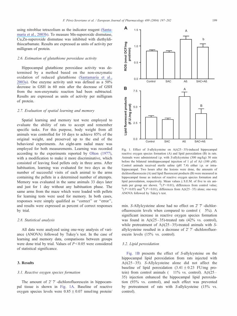
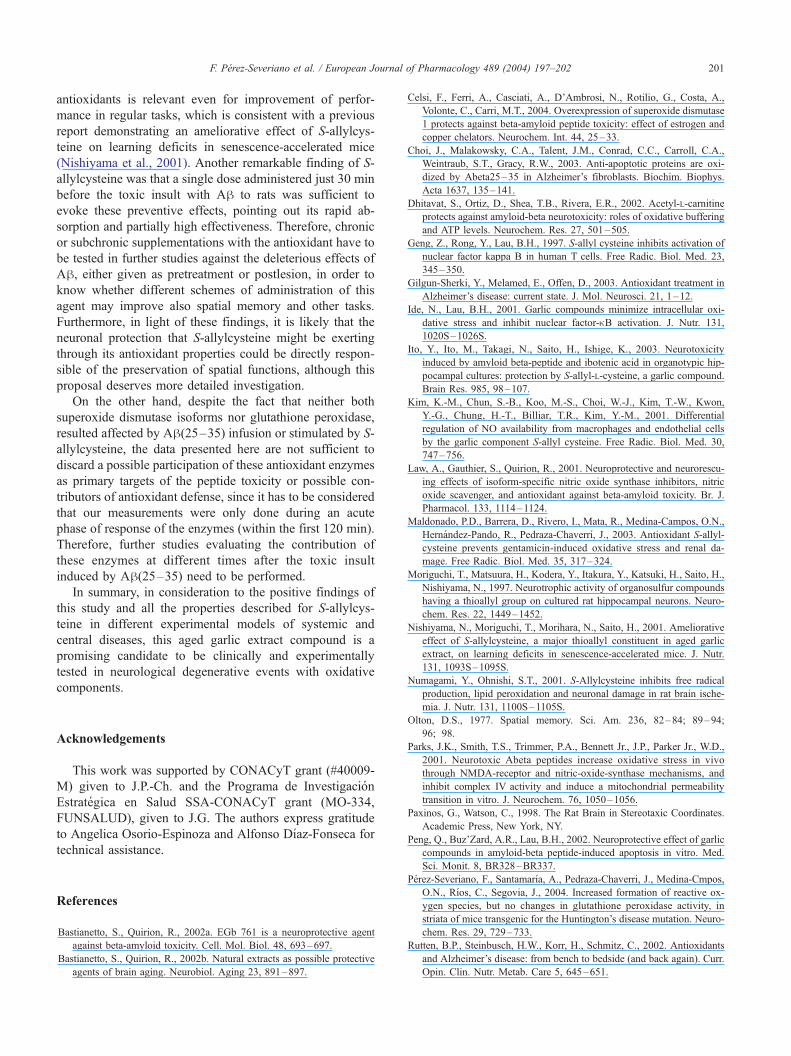
Fig. 1. Effect of S-allylcysteine on Ah(25–35)-induced hippocampal
reactive oxygen species formation (A) and lipid peroxidation (B) in rats.
Animals were administered i.p. with S-allylcysteine (300 mg/kg) 30 min
before the bilateral intrahippocampal injection of 1 Al of Ah (100 AM).
Control animals received sterile saline (pH 7.4) either i.p. or intra-
hippocampal. Two hours after the lesions were done, the amounts of
dichlorofluorescein (A) and lipid fluorescent products (B) were measured in
hippocampal tissue as indexes of reactive oxygen species formation and
lipid peroxidation, respectively. Mean valuesF S.E.M. of five to six ani-
mals per group are shown. A( P < 0.01), differences from control value;b( P< 0.05) and B( P < 0.01), differences from Ah(25–35) alone; one-wayANOVA followed by Tukey’s test.
F. Perez-Severiano et al. / European Journal of Pharmacology 489 (2004) 197–202 199
using nitroblue tetrazolium as the indicator reagent (Santa-
marıa et al., 2003b). To measure Mn-superoxide dismutase,
Cu,Zn-superoxide dismutase was inhibited with diethyldi-
thiocarbamate. Results are expressed as units of activity per
milligram of protein.
2.6. Estimation of glutathione peroxidase activity
Hippocampal glutathione peroxidase activity was de-
termined by a method based on the non-enzymatic
oxidation of reduced glutathione (Santamarıa et al.,
2003a). One enzyme activity unit was defined as a 50%
decrease in GSH in 60 min after the decrease of GSH
from the non-enzymatic reaction had been subtracted.
Results are expressed as units of activity per milligram
of protein.
2.7. Evaluation of spatial learning and memory
Spatial learning and memory test were employed to
evaluate the ability of rats to accept and remember
specific tasks. For this purpose, body weight from all
animals was controlled for 10 days to achieve 85% of the
original weight, and preserved up to the end of the
behavioral experiments. An eight-arm radial maze was
employed for both measurements. Learning was recorded
according to the experiments reported by Olton (1977),
with a modification to make it more discriminative, which
consisted of leaving food pellets only in three arms. After
habituation, learning was evaluated for two days as the
number of successful visits of each animal to the arms
containing the pellets in a determined number of attempts.
Memory was evaluated in the same animals 33 days later
and just for 1 day without any habituation phase. The
same arms from the maze which were loaded with pellets
for learning tests were used for memory. In both cases,
responses were simply qualified as ‘‘correct’’ or ‘‘error’’,
and results were expressed as percent of correct responses
by trial.
2.8. Statistical analysis
All data were analyzed using one-way analysis of vari-
ance (ANOVA) followed by Tukey’s test. In the case of
learning and memory data, comparisons between groups
were done trial by trial. Values of P < 0.05 were considered
of statistical significance.
3. Results
3.1. Reactive oxygen species formation
The amount of 2V7V-dichlorofluorescein in hippocam-
pal tissue is shown in Fig. 1A. Baseline of reactive
oxygen species levels were 0.85F 0.07 nmol/mg protein/
min. S-Allylcysteine alone had no effect on 2V7V-dichlor-ofluorescein levels when compared to control (� 5%). A
significant increase in reactive oxygen species formation
was found in Ah(25–35)-treated rats (42% vs. control),
while pretreatment of Ah(25–35)-treated animals with S-
allylcysteine resulted in a decrease of 2V7V-dichlorofluor-escein levels (15% vs. control).
3.2. Lipid peroxidation
Fig. 1B presents the effect of S-allylcysteine on the
hippocampal lipid peroxidation from rats injected with
Ah(25–35). S-Allylcysteine alone did not affect the
baseline of lipid peroxidation (3.41F 0.23 FU/mg pro-
tein) from control animals (� 11% vs. control). Ah(25–35) injection enhanced the hippocampal lipid peroxida-
tion (93% vs. control), and such effect was prevented
by pretreatment of rats with S-allylcysteine (15% vs.
control).
urnal
3.3. Superoxide dismutase and glutathione peroxidase
activities
Baseline enzyme activities were: 4.70F 0.62 U/mg
protein for Mn-superoxide dismutase, 9.09F 0.58 U/mg
protein for Cu,Zn-superoxide dismutase and 0.012F 0.001
U/mg protein for glutathione peroxidase. No significant
effects were found with any of the agents tested, or their
combination, when compared with control values (data
not shown).
F. Perez-Severiano et al. / European Jo200
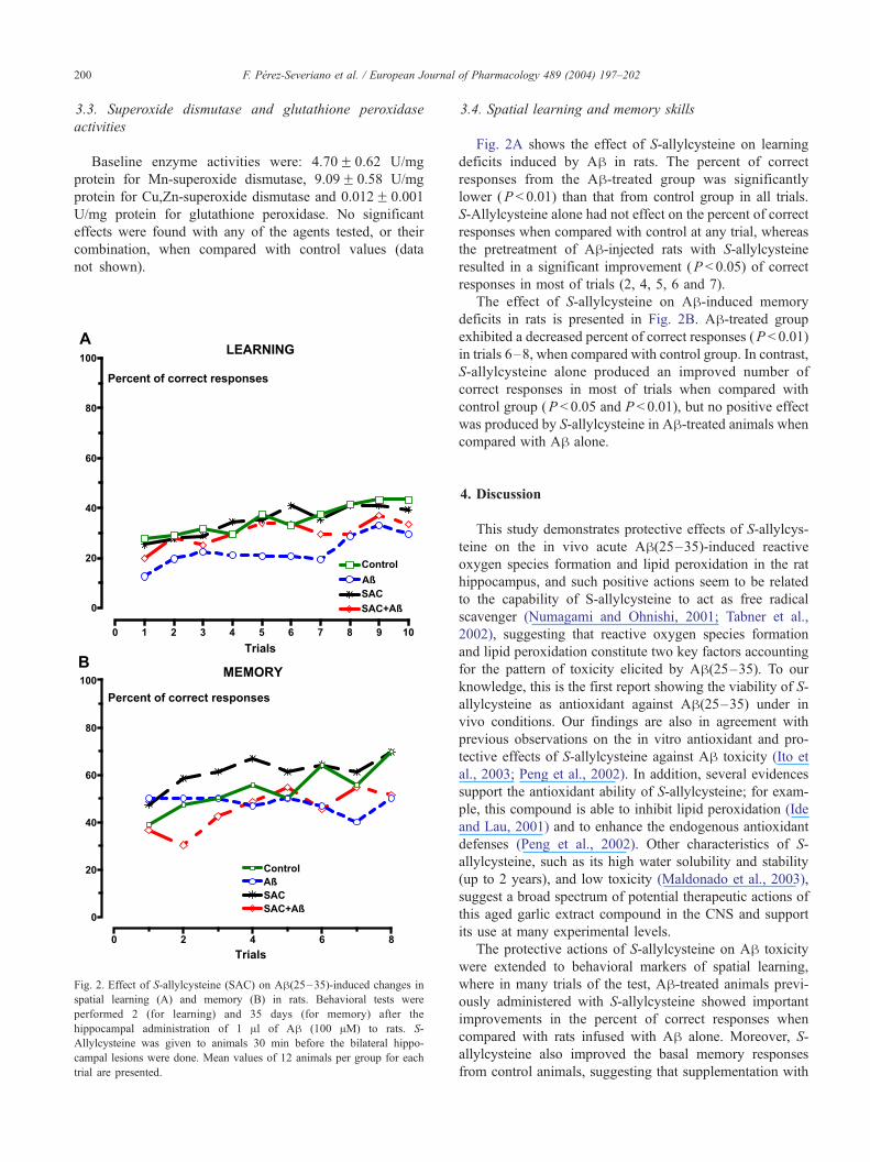
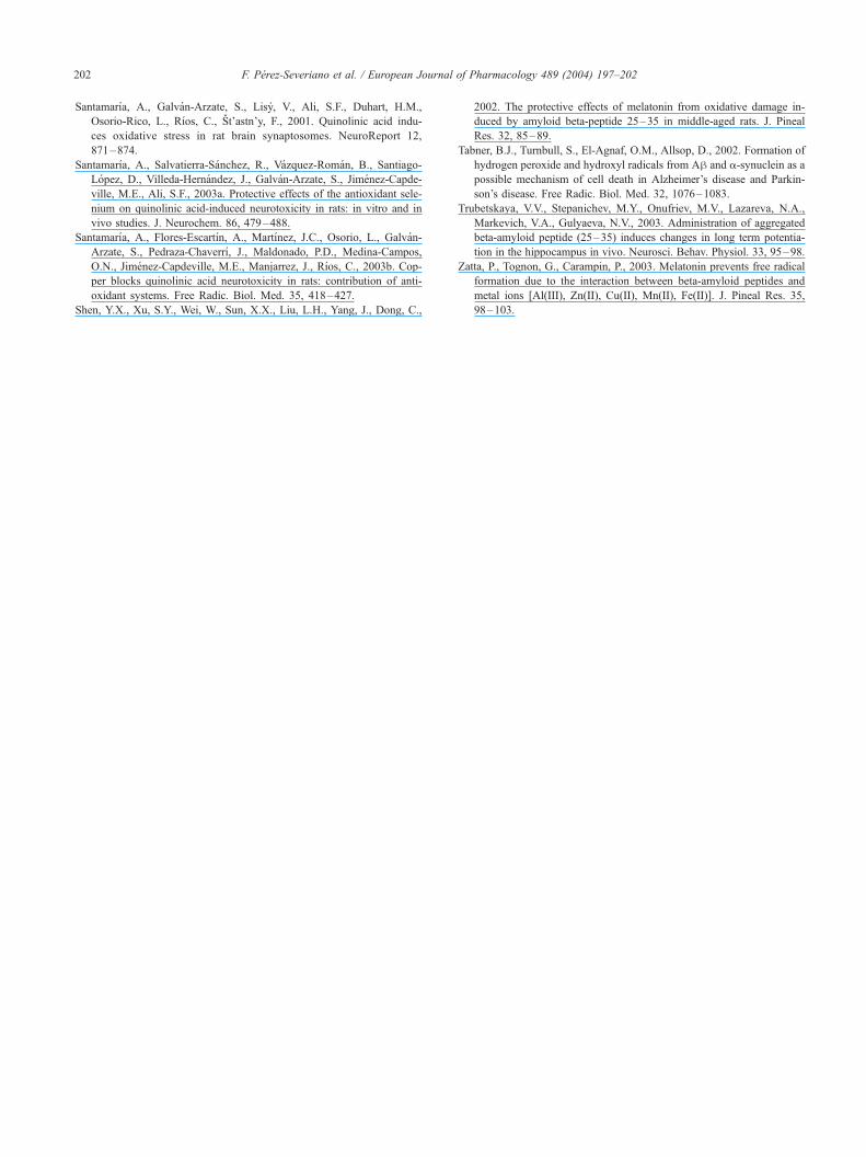
Fig. 2. Effect of S-allylcysteine (SAC) on Ah(25–35)-induced changes in
spatial learning (A) and memory (B) in rats. Behavioral tests were
performed 2 (for learning) and 35 days (for memory) after the
hippocampal administration of 1 Al of Ah (100 AM) to rats. S-
Allylcysteine was given to animals 30 min before the bilateral hippo-
campal lesions were done. Mean values of 12 animals per group for each
trial are presented.
3.4. Spatial learning and memory skills
Fig. 2A shows the effect of S-allylcysteine on learning
deficits induced by Ah in rats. The percent of correct
responses from the Ah-treated group was significantly
lower (P < 0.01) than that from control group in all trials.
S-Allylcysteine alone had not effect on the percent of correct
responses when compared with control at any trial, whereas
the pretreatment of Ah-injected rats with S-allylcysteine
resulted in a significant improvement (P < 0.05) of correct
responses in most of trials (2, 4, 5, 6 and 7).
The effect of S-allylcysteine on Ah-induced memory
deficits in rats is presented in Fig. 2B. Ah-treated group
exhibited a decreased percent of correct responses (P < 0.01)
in trials 6–8, when compared with control group. In contrast,
S-allylcysteine alone produced an improved number of
correct responses in most of trials when compared with
control group (P < 0.05 and P < 0.01), but no positive effect
was produced by S-allylcysteine in Ah-treated animals when
compared with Ah alone.
of Pharmacology 489 (2004) 197–202
4. Discussion
This study demonstrates protective effects of S-allylcys-
teine on the in vivo acute Ah(25–35)-induced reactive
oxygen species formation and lipid peroxidation in the rat
hippocampus, and such positive actions seem to be related
to the capability of S-allylcysteine to act as free radical
scavenger (Numagami and Ohnishi, 2001; Tabner et al.,
2002), suggesting that reactive oxygen species formation
and lipid peroxidation constitute two key factors accounting
for the pattern of toxicity elicited by Ah(25–35). To our
knowledge, this is the first report showing the viability of S-
allylcysteine as antioxidant against Ah(25–35) under in
vivo conditions. Our findings are also in agreement with
previous observations on the in vitro antioxidant and pro-
tective effects of S-allylcysteine against Ah toxicity (Ito et
al., 2003; Peng et al., 2002). In addition, several evidences
support the antioxidant ability of S-allylcysteine; for exam-
ple, this compound is able to inhibit lipid peroxidation (Ide
and Lau, 2001) and to enhance the endogenous antioxidant
defenses (Peng et al., 2002). Other characteristics of S-
allylcysteine, such as its high water solubility and stability
(up to 2 years), and low toxicity (Maldonado et al., 2003),
suggest a broad spectrum of potential therapeutic actions of
this aged garlic extract compound in the CNS and support
its use at many experimental levels.
The protective actions of S-allylcysteine on Ah toxicity
were extended to behavioral markers of spatial learning,
where in many trials of the test, Ah-treated animals previ-
ously administered with S-allylcysteine showed important
improvements in the percent of correct responses when
compared with rats infused with Ah alone. Moreover, S-
allylcysteine also improved the basal memory responses
from control animals, suggesting that supplementation with
F. Perez-Severiano et al. / European Journal of Pharmacology 489 (2004) 197–202 201
antioxidants is relevant even for improvement of perfor-
mance in regular tasks, which is consistent with a previous
report demonstrating an ameliorative effect of S-allylcys-
teine on learning deficits in senescence-accelerated mice
(Nishiyama et al., 2001). Another remarkable finding of S-
allylcysteine was that a single dose administered just 30 min
before the toxic insult with Ah to rats was sufficient to
evoke these preventive effects, pointing out its rapid ab-
sorption and partially high effectiveness. Therefore, chronic
or subchronic supplementations with the antioxidant have to
be tested in further studies against the deleterious effects of
Ah, either given as pretreatment or postlesion, in order to
know whether different schemes of administration of this
agent may improve also spatial memory and other tasks.
Furthermore, in light of these findings, it is likely that the
neuronal protection that S-allylcysteine might be exerting
through its antioxidant properties could be directly respon-
sible of the preservation of spatial functions, although this
proposal deserves more detailed investigation.
On the other hand, despite the fact that neither both
superoxide dismutase isoforms nor glutathione peroxidase,
resulted affected by Ah(25–35) infusion or stimulated by S-
allylcysteine, the data presented here are not sufficient to
discard a possible participation of these antioxidant enzymes
as primary targets of the peptide toxicity or possible con-
tributors of antioxidant defense, since it has to be considered
that our measurements were only done during an acute
phase of response of the enzymes (within the first 120 min).
Therefore, further studies evaluating the contribution of
these enzymes at different times after the toxic insult
induced by Ah(25–35) need to be performed.
In summary, in consideration to the positive findings of
this study and all the properties described for S-allylcys-
teine in different experimental models of systemic and
central diseases, this aged garlic extract compound is a
promising candidate to be clinically and experimentally
tested in neurological degenerative events with oxidative
components.
Acknowledgements
This work was supported by CONACyT grant (#40009-
M) given to J.P.-Ch. and the Programa de Investigacion
Estrategica en Salud SSA-CONACyT grant (MO-334,
FUNSALUD), given to J.G. The authors express gratitude
to Angelica Osorio-Espinoza and Alfonso Dıaz-Fonseca for
technical assistance.
References
Bastianetto, S., Quirion, R., 2002a. EGb 761 is a neuroprotective agent
against beta-amyloid toxicity. Cell. Mol. Biol. 48, 693–697.
Bastianetto, S., Quirion, R., 2002b. Natural extracts as possible protective
agents of brain aging. Neurobiol. Aging 23, 891–897.
Celsi, F., Ferri, A., Casciati, A., D’Ambrosi, N., Rotilio, G., Costa, A.,
Volonte, C., Carri, M.T., 2004. Overexpression of superoxide dismutase
1 protects against beta-amyloid peptide toxicity: effect of estrogen and
copper chelators. Neurochem. Int. 44, 25–33.
Choi, J., Malakowsky, C.A., Talent, J.M., Conrad, C.C., Carroll, C.A.,
Weintraub, S.T., Gracy, R.W., 2003. Anti-apoptotic proteins are oxi-
dized by Abeta25–35 in Alzheimer’s fibroblasts. Biochim. Biophys.
Acta 1637, 135–141.
Dhitavat, S., Ortiz, D., Shea, T.B., Rivera, E.R., 2002. Acetyl-L-carnitine
protects against amyloid-beta neurotoxicity: roles of oxidative buffering
and ATP levels. Neurochem. Res. 27, 501–505.
Geng, Z., Rong, Y., Lau, B.H., 1997. S-allyl cysteine inhibits activation of
nuclear factor kappa B in human T cells. Free Radic. Biol. Med. 23,
345–350.
Gilgun-Sherki, Y., Melamed, E., Offen, D., 2003. Antioxidant treatment in
Alzheimer’s disease: current state. J. Mol. Neurosci. 21, 1–12.
Ide, N., Lau, B.H., 2001. Garlic compounds minimize intracellular oxi-
dative stress and inhibit nuclear factor-nB activation. J. Nutr. 131,
1020S–1026S.
Ito, Y., Ito, M., Takagi, N., Saito, H., Ishige, K., 2003. Neurotoxicity
induced by amyloid beta-peptide and ibotenic acid in organotypic hip-
pocampal cultures: protection by S-allyl-L-cysteine, a garlic compound.
Brain Res. 985, 98–107.
Kim, K.-M., Chun, S.-B., Koo, M.-S., Choi, W.-J., Kim, T.-W., Kwon,
Y.-G., Chung, H.-T., Billiar, T.R., Kim, Y.-M., 2001. Differential
regulation of NO availability from macrophages and endothelial cells
by the garlic component S-allyl cysteine. Free Radic. Biol. Med. 30,
747–756.
Law, A., Gauthier, S., Quirion, R., 2001. Neuroprotective and neurorescu-
ing effects of isoform-specific nitric oxide synthase inhibitors, nitric
oxide scavenger, and antioxidant against beta-amyloid toxicity. Br. J.
Pharmacol. 133, 1114–1124.
Maldonado, P.D., Barrera, D., Rivero, I., Mata, R., Medina-Campos, O.N.,
Hernandez-Pando, R., Pedraza-Chaverrı, J., 2003. Antioxidant S-allyl-
cysteine prevents gentamicin-induced oxidative stress and renal da-
mage. Free Radic. Biol. Med. 35, 317–324.
Moriguchi, T., Matsuura, H., Kodera, Y., Itakura, Y., Katsuki, H., Saito, H.,
Nishiyama, N., 1997. Neurotrophic activity of organosulfur compounds
having a thioallyl group on cultured rat hippocampal neurons. Neuro-
chem. Res. 22, 1449–1452.
Nishiyama, N., Moriguchi, T., Morihara, N., Saito, H., 2001. Ameliorative
effect of S-allylcysteine, a major thioallyl constituent in aged garlic
extract, on learning deficits in senescence-accelerated mice. J. Nutr.
131, 1093S–1095S.
Numagami, Y., Ohnishi, S.T., 2001. S-Allylcysteine inhibits free radical
production, lipid peroxidation and neuronal damage in rat brain ische-
mia. J. Nutr. 131, 1100S–1105S.
Olton, D.S., 1977. Spatial memory. Sci. Am. 236, 82–84; 89–94;
96; 98.
Parks, J.K., Smith, T.S., Trimmer, P.A., Bennett Jr., J.P., Parker Jr., W.D.,
2001. Neurotoxic Abeta peptides increase oxidative stress in vivo
through NMDA-receptor and nitric-oxide-synthase mechanisms, and
inhibit complex IV activity and induce a mitochondrial permeability
transition in vitro. J. Neurochem. 76, 1050–1056.
Paxinos, G., Watson, C., 1998. The Rat Brain in Stereotaxic Coordinates.
Academic Press, New York, NY.
Peng, Q., Buz’Zard, A.R., Lau, B.H., 2002. Neuroprotective effect of garlic
compounds in amyloid-beta peptide-induced apoptosis in vitro. Med.
Sci. Monit. 8, BR328–BR337.
Perez-Severiano, F., Santamarıa, A., Pedraza-Chaverri, J., Medina-Cmpos,
O.N., Rıos, C., Segovia, J., 2004. Increased formation of reactive ox-
ygen species, but no changes in glutathione peroxidase activity, in
striata of mice transgenic for the Huntington’s disease mutation. Neuro-
chem. Res. 29, 729–733.
Rutten, B.P., Steinbusch, H.W., Korr, H., Schmitz, C., 2002. Antioxidants
and Alzheimer’s disease: from bench to bedside (and back again). Curr.
Opin. Clin. Nutr. Metab. Care 5, 645–651.
F. Perez-Severiano et al. / European Journal of Pharmacology 489 (2004) 197–202202
Santamarıa, A., Galvan-Arzate, S., Lisy, V., Ali, S.F., Duhart, H.M.,
Osorio-Rico, L., Rıos, C., St’astn’y, F., 2001. Quinolinic acid indu-
ces oxidative stress in rat brain synaptosomes. NeuroReport 12,
871–874.
Santamarıa, A., Salvatierra-Sanchez, R., Vazquez-Roman, B., Santiago-
Lopez, D., Villeda-Hernandez, J., Galvan-Arzate, S., Jimenez-Capde-
ville, M.E., Ali, S.F., 2003a. Protective effects of the antioxidant sele-
nium on quinolinic acid-induced neurotoxicity in rats: in vitro and in
vivo studies. J. Neurochem. 86, 479–488.
Santamarıa, A., Flores-Escartın, A., Martınez, J.C., Osorio, L., Galvan-
Arzate, S., Pedraza-Chaverrı, J., Maldonado, P.D., Medina-Campos,
O.N., Jimenez-Capdeville, M.E., Manjarrez, J., Rıos, C., 2003b. Cop-
per blocks quinolinic acid neurotoxicity in rats: contribution of anti-
oxidant systems. Free Radic. Biol. Med. 35, 418–427.
Shen, Y.X., Xu, S.Y., Wei, W., Sun, X.X., Liu, L.H., Yang, J., Dong, C.,
2002. The protective effects of melatonin from oxidative damage in-
duced by amyloid beta-peptide 25–35 in middle-aged rats. J. Pineal
Res. 32, 85–89.
Tabner, B.J., Turnbull, S., El-Agnaf, O.M., Allsop, D., 2002. Formation of
hydrogen peroxide and hydroxyl radicals from Ah and a-synuclein as a
possible mechanism of cell death in Alzheimer’s disease and Parkin-
son’s disease. Free Radic. Biol. Med. 32, 1076–1083.
Trubetskaya, V.V., Stepanichev, M.Y., Onufriev, M.V., Lazareva, N.A.,
Markevich, V.A., Gulyaeva, N.V., 2003. Administration of aggregated
beta-amyloid peptide (25–35) induces changes in long term potentia-
tion in the hippocampus in vivo. Neurosci. Behav. Physiol. 33, 95–98.
Zatta, P., Tognon, G., Carampin, P., 2003. Melatonin prevents free radical
formation due to the interaction between beta-amyloid peptides and
metal ions [Al(III), Zn(II), Cu(II), Mn(II), Fe(II)]. J. Pineal Res. 35,
98–103.


