Embed Size (px)
Citation preview

IMEC 2009
Science and engineering of electrospun nanofibers for advancesin clean energy, water filtration, and regenerative medicine
S. Ramakrishna • R. Jose • P. S. Archana • A. S. Nair •
R. Balamurugan • J. Venugopal • W. E. Teo
Received: 15 January 2010 / Accepted: 9 April 2010 / Published online: 27 April 2010
� Springer Science+Business Media, LLC 2010
Abstract Nanostructured materials with high aspect ratio
and one-dimensional (ID) morphology are nature’s choices
when high degree of functional performances and flexible
properties are concerned. Two examples are extracellular
matrices in tissues of living organism, and light harvest-
ing rods of the retina and chlorophyll. Electrospinning
(E-spinning) is a simple processing technique that allows
fabrication of high aspect ratio nanofibers (NFs) in a
commercial scale. Electrospun nanofibers (E-spun NFs)
combine a number of physical properties such as guided
electron transport, strain-induced electronic properties,
high mechanical strength, high degree of flexibility, large
specific surface area, high electron and thermal diffusivity,
and tailorable pore distribution. Our laboratory has been
involved in fabrication of E-spun polymeric, inorganic, and
polymer-nanocomposite fibers in random, aligned, cross-
aligned, sheaths, tubes, yarns, core/shell, and trilayer
morphologies. This article focuses on application of the
E-spun fibers in the areas of clean energy, water treatment,
and regenerative medicine in the authors’ laboratory. In
addition, the article briefly reviews the progress made in
these areas using E-spun NFs.
Introduction
One-dimensional (ID) nanostructures are choice of the
nature when high degree of functional performances and
flexible properties are concerned as in extracellular matri-
ces, neural networks, and light harvesting rods of the retina
and chlorophyll. Fabrication of 1D structures of the new
generation materials could, therefore, provide new oppor-
tunities to improve and/or modify performances of the
advanced functional devices and structures. Electrospin-
ning (E-spinning), which works under the principle of
asymmetric bending of a charged liquid jet when acceler-
ated by a longitudinal electric field, is a technique that
allows fabrication of continuous NFs of polymers and
advanced functional materials. In the E-spinning process, a
polymer solution is injected from a needle in the presence
of an electric field. When the applied electric field over-
comes surface tension of the liquid, a continuous jet is
ejected, which on subsequent solvent evaporation and
bending produces NFs on a collector surface [1]. Figure 1
shows a schematic of the processes during E-spinning.
Various fiber morphologies could be produced by E-spin-
ning such as random, aligned, core–shell, bundles, and
mats, and these are done by merely changing the collection
and/or injection strategy [2–5]. If the polymer solution
contains respective metal ions for forming an inorganic
solid, then appropriate post-electrospinning heat treatment
yields continuous inorganic NFs [6, 7].
The E-spinning as a method to produce continuous
fibers has been known since 1902 [8]. Figure 2 shows the
E-spinning timeline showing its evolution as an acceptable
S. Ramakrishna (&) � R. Jose � P. S. Archana �A. S. Nair � R. Balamurugan � J. Venugopal � W. E. Teo
Healthcare and Energy Materials Laboratory, National
University of Singapore, 117576 Singapore, Singapore
e-mail: [email protected]
S. Ramakrishna
Institute of Materials Research and Engineering (IMRE),
A-STAR, 3 Research Link, 117602 Singapore, Singapore
S. Ramakrishna
King Saud University, Riyadh 11451, Kingdom of Saudi Arabia
R. Jose
Faculty of Industrial Science and Technology, Universiti
Malaysia Pahang, 26300 Pahang, Malaysia
123
J Mater Sci (2010) 45:6283–6312
DOI 10.1007/s10853-010-4509-1

technology since its inception. There is a revived interest in
E-spinning in academia from mid-1990s with the advances
in nanotechnology [9], which is possibly due to the
potential of the process to fabricate 1D nanostructures of a
wide range of materials systems.
E-spun NFs (NFs) have interesting physical character-
istics: (i) high specific surface area and interesting pore
distribution; (ii) polymer NFs are opaque due to high
packing density of monomers; and (iii) metal oxide NFs are
with controllable crystallinity and are characterized by
diameter-dependent strain. The E-spun NFs of lead zir-
conate titanate showed a piezoelectric response that
is *200 times higher than that of corresponding single
crystals [10]. High efficiency photovoltaic devices were
also demonstrated using E-spun metal oxide NFs [11] in
addition to study some of the factors affecting the con-
version efficiency of dye-sensitized solar cells (DSCs) [12].
Owing to their high scientific and commercial potentials,
E-spinning as a method for producing 1D nanostructures is
gaining increased interest both in academia and industry.
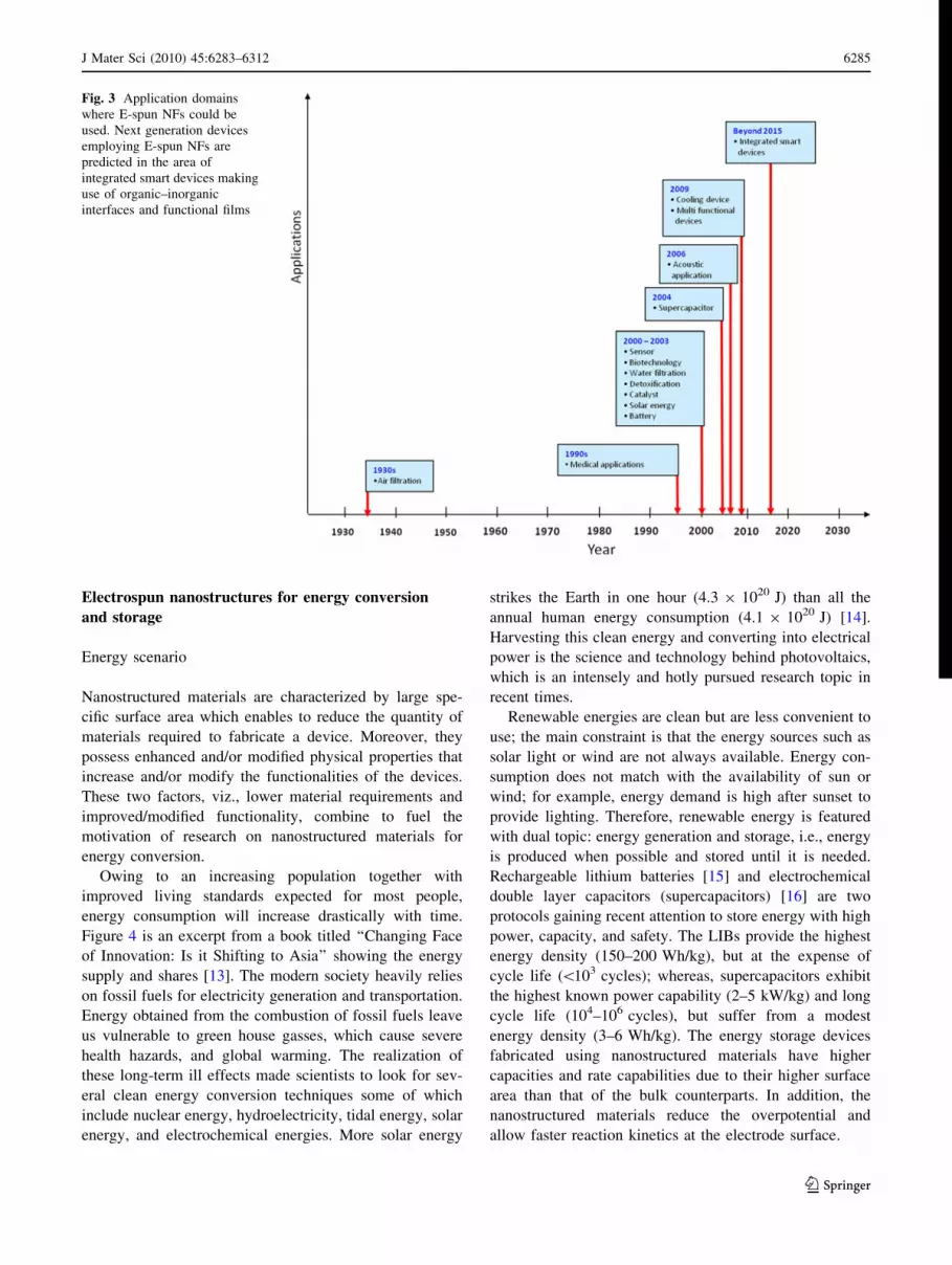
Applications envisaged using E-spun NFs since its dis-
covery are summarized in Fig. 3.
Studies have been carried out in our laboratory to
utilize E-spun NFs to address global challenges requiring
immediate attention such as clean energy, water treat-
ment, and regenerative medicine. This article is organized
to provide an overview of advances in E-spinning and the
application of NFs in the areas of photovoltaics, water
filtration, and healthcare. ‘‘Electrospun nanostructures for
energy conversion and storage’’ section of this article
deals with application of E-spun TiO2 nanostructures in
energy, both photovoltaic and lithium ion batteries
(LIBs), starting from global energy scenario; ‘‘Nanofibers
for water filtration’’ section focuses on the application of
E-spun membranes for water filtration; and ‘‘Nanofibers
for regenerative medicine’’ section concentrates on their
applications in regenerative medicine. Notable drawbacks
of E-spinning are also included. A brief summary, future
prospects, and limitations are given at the end of each
section.
Fig. 1 Schematics of the electrospinning (E-spinning) process. The
experimental set-up consists of a high voltage power supply, a
spinneret, and a collector. The three processes, viz., formation of
tailor cone (1), bending due to various instabilities (2), and collection
of solid samples (3) are shown. The qE is the electrostatic force, g is
the viscosity, and T is the surface tension. Conventionally, E-spinning
produces a fiber cloth consisting of randomly oriented nano/microf-
ibers, a typical SEM image of which is also shown
Fig. 2 Electrospinning timeline
6284 J Mater Sci (2010) 45:6283–6312
123
Electrospun nanostructures for energy conversion
and storage
Energy scenario
Nanostructured materials are characterized by large spe-
cific surface area which enables to reduce the quantity of
materials required to fabricate a device. Moreover, they
possess enhanced and/or modified physical properties that
increase and/or modify the functionalities of the devices.
These two factors, viz., lower material requirements and
improved/modified functionality, combine to fuel the
motivation of research on nanostructured materials for
energy conversion.
Owing to an increasing population together with
improved living standards expected for most people,
energy consumption will increase drastically with time.
Figure 4 is an excerpt from a book titled ‘‘Changing Face
of Innovation: Is it Shifting to Asia’’ showing the energy
supply and shares [13]. The modern society heavily relies
on fossil fuels for electricity generation and transportation.
Energy obtained from the combustion of fossil fuels leave
us vulnerable to green house gasses, which cause severe
health hazards, and global warming. The realization of
these long-term ill effects made scientists to look for sev-
eral clean energy conversion techniques some of which
include nuclear energy, hydroelectricity, tidal energy, solar
energy, and electrochemical energies. More solar energy
strikes the Earth in one hour (4.3 9 1020 J) than all the
annual human energy consumption (4.1 9 1020 J) [14].
Harvesting this clean energy and converting into electrical
power is the science and technology behind photovoltaics,
which is an intensely and hotly pursued research topic in
recent times.
Renewable energies are clean but are less convenient to
use; the main constraint is that the energy sources such as
solar light or wind are not always available. Energy con-
sumption does not match with the availability of sun or
wind; for example, energy demand is high after sunset to
provide lighting. Therefore, renewable energy is featured
with dual topic: energy generation and storage, i.e., energy
is produced when possible and stored until it is needed.
Rechargeable lithium batteries [15] and electrochemical
double layer capacitors (supercapacitors) [16] are two
protocols gaining recent attention to store energy with high
power, capacity, and safety. The LIBs provide the highest
energy density (150–200 Wh/kg), but at the expense of
cycle life (\103 cycles); whereas, supercapacitors exhibit
the highest known power capability (2–5 kW/kg) and long
cycle life (104–106 cycles), but suffer from a modest
energy density (3–6 Wh/kg). The energy storage devices
fabricated using nanostructured materials have higher
capacities and rate capabilities due to their higher surface
area than that of the bulk counterparts. In addition, the
nanostructured materials reduce the overpotential and
allow faster reaction kinetics at the electrode surface.
Fig. 3 Application domains
where E-spun NFs could be
used. Next generation devices
employing E-spun NFs are
predicted in the area of
integrated smart devices making
use of organic–inorganic
interfaces and functional films
J Mater Sci (2010) 45:6283–6312 6285
123

In the next sections, detailed descriptions on these two
types of energy devices fabricated using E-spun NFs are
given.
Solar energy conversion––the dye-sensitized solar cells
Currently available solar cells can be broadly classified into
p–n junction solar cells and excitonic solar cells (ESCs)
[17]. In the p–n junction cells, the photogenerated electrons
are accelerated by a built-in electric field at the junction
between the p- and the n-type semiconductors. In the ESCs,
light absorption results in the generation of excitons, which
are quasi particles with an electron in the conduction band
(or the lowest unoccupied molecular orbital, LUMO, in the
case of molecules and clusters) and a hole in the valence
band (or the highest occupied molecular orbital, HOMO, in
the case of molecules and nanoclusters). The excitons thus
generated in a semiconductor are dissociated at a material
interface into free carriers in excitonic solar cells. Popular
examples of excitonic solar cells are the DSCs [18],
organic solar cells [19], and quantum dot solar cells [20].
Organic solar cells employ conjugated conducting poly-
mers and/or organic materials. The photovoltaic effect in
DSC occurs at the interface between a dye-anchored wide
bandgap oxide semiconductor and an electrolyte. In
quantum dot solar cells, semiconductor nanocrystals with
sizes less than their exciton’s Bohr radius have been used
to harvest light, i.e., the quantum dots simply replace dyes,
so far.
The DSCs are fabricated by (i) sintering a wide bandgap
metal oxide semiconductor, typically mesoporous TiO2, on
a conducting glass plate, usually fluorine-doped tin oxide
(FTO); (ii) dye anchoring on the sintered metal oxide layer;
(iii) counter electrode fabrication, which is typically a Pt
sputtered FTO; (iii) sealing; and (iv) electrolyte filling. The
optimized processes for fabrication of highly efficient
Fig. 5 Typical materials’ arrangement and processes in DSCs. Light
absorption by the dye results in an electron at the unoccupied
molecular orbital of the dye leaving a hole at its occupied molecular
orbitals (process 1). The photogenerated electrons are then injected to
the metal oxide semiconductor (process 2) due to the overlapping of
the wave functions [12] achieved through chemisorption of the dyes
on the metal oxide. The photoelectrons received by the metal oxide
cause its Fermi level to rise and subsequently result in photoconduc-
tivity. In metal oxides, the electron diffuses randomly; the surviving
electrons are collected, and they use their energy to do an external
work and then deliver them at the counter electrode, from where they
are injected to the dye to neutralize the hole. Catalysts are used at the
counter electrode, usually platinum, for faster regeneration of the dye.
The random electron diffusion imposes severe restrictions on the
scalability of the DSCs as the chances of collecting the electrons
decreases with increase in the device area. One dimensional metal
oxides are likely a remedy to increase the charge collection and
scalability of the device
Fig. 4 Fuel share for primary
energy supply [13]. The figures
were from those authors’ study
6286 J Mater Sci (2010) 45:6283–6312
123

DSCs were recently published [21]. The reader may refer
to the processes and rate limiters in DSCs in recent reviews
[22, 23]. In brief, upon irradiation, the electrons in the dye
are excited to the higher energy levels from where they are
injected into the metal oxide (Fig. 5) due to the overlap-
ping of their electronic wave functions. These electrons are
transported through the metal oxide and collected by the
FTO, which is coated with a thin layer (B100 nm) of TiO2
that acts as blocking layer to the holes. Role of the elec-
trolyte is two-fold: (i) regeneration of the dye, and (ii)
blocking electrons from reaching the counter electrode. It
should be noted that the photoconversion efficiency (g) of
DSCs is much higher than that of the other ESCs and p–n
junction solar cells fabricated using amorphous or nano-
crystalline silicon [24]. Certified g as high as 8.4% in
submodules [25] (area *18 cm2) and 11.5% in laboratory
scale (area *0.22 cm2) has been achieved using meso-
porous TiO2-based DSCs [24, 26]. The DSCs have the
potential for becoming cost-effective means for producing
electricity, capable of competing with available solar
electric technologies and, eventually, with today’s
conventional power technologies [27]. An update of pho-
tovoltaic conversion efficiencies of various solar cells at
single cell, submodule, and module levels with or without
solar concentrators is summarized in Table 1.
Role of 1D nanostructures in DSCS
It is generally accepted that photoelectrons injected into
mesoporous TiO2 in the DSCs move via trap-limited dif-
fusion process [28–34]; i.e., the electrons undergo a series
of trapping and detrapping events as the energy states lie
within the bandgap during diffusion. These inner-lying
energy states are a characteristic of the nanomaterials
which are arising from an increased number of defects such
as packing defects, unsaturated bonds, and deviation from
the bulk long-range order [22, 28, 35]. The trapping and
detrapping events result in lower electron mobility (ln) and
diffusivity (diffusion coefficient, Dn) than the bulk mate-
rials and thereby increasing the diffusion time [28]. Longer
diffusion time causes the electrons to undergo a series of
recombination processes with electrolyte, oxidized dye,
Table 1 Solar energy conversion efficiencies of various solar cells at cell (area \1 cm2), submodule (area \25 cm2), and modules
(area [50 cm2). The table is summarized from the recent photovoltaic update provided by Green et al. [24]
Type of cell Description Efficiency (%) at AM1.5 spectrum
Without concentrator
[area (cm2)]
With concentrator
[area (cm2), intensity (suns)]
p–n (Single junction) Silicon
Crystalline 25.0 ± 0.5 [4.00 (da)] 27.6 ± 1 [1.00 (da), 92]
Polycrystalline 20.4 ± 0.5 [1.002 (ap)]
Amorphous 9.5 ± 0.3 [1.070 (ap)]
Nanocrystalline 10.1 ± 0.2 [1.199 (ap)]
GaAs
Thin film 26.1 ± 0.8 [1.001 (ap)] 28.8 ± 1.2 [0.0504 (da), 232]
Polycrystalline 18.4 ± 0.5 [4.011 (t)]
InP
Crystalline 22.1 ± 0.7 [4.02 (t)]
p–n (Multi junction) GaInP/GaAs/Ge 32.0 ± 1.5 [3.989 (t)] 40.7 ± 2.4 [0.267 (da), 240]
20.7 ± 1.5 [34 (da), 10]
GaInP/GaAs 30.3 [4.0 (t)]
GaInP/GaInAs/Ge 41.1 ± 2.5 [0.0509 (da), 454]
Thin film CuInGaSe2 (cell) 19.4 ± 0.6 [0.994 (ap)] 21.8 ± 1.5 [0.102 (da), 14]
CuInGaSe2 (submodule) 16.7 ± 0.4 [16.0 (ap)]
CdTe 16.7 ± 0.5 [1.032 (ap)]
Si 16.7 ± 0.4 [4.017 (ap)]
Si (submodule) 10.5 ± 0.3 [94.0 (ap)]
Excitonic Dye solar cells (cell) 11.2 ± 0.3 [0.219 (ap)]
Dye solar cells (submodule) 8.4 ± 0.2 [17.11 (ap)]
Organic polymer 5.15 ± 0.3 [1.021 (ap)]
Organic (submodule) 2.05 ± 0.3 [223.5 (ap)]
Da designated illumination area, ap aperture area, t total area
J Mater Sci (2010) 45:6283–6312 6287
123

and phonon relaxation [22]. The distance traveled by the
electrons before being recombined is known as the diffu-
sion length, L ¼ffiffiffiffiffiffiffiffi
Dnsp
, where s is the electron lifetime,
which for conventional nanoparticle-based DSCs is
L * 30 lm [36]. The film thickness should be *L/3 for
efficient charge collection.
Hence, an increase in the nanoparticle film thickness
significantly above 10 lm does not increase the g. On the
other hand, g of the DSCs could be increased by increasing
the thickness of the mesoporous network using materials
and architectures of higher Dn. As shown in Fig. 6, 1D
structures, such as nanowires, nanotubes, nanorods, and
NFs in the place of nanoparticles, have been proposed to
have longer L due to the directed charge transport through
them [37–42]. There are two types of these 1D nano-
structures: (i) ordered nanotube or nanowire arrays verti-
cally aligned on conducting glass substrates and (ii)
random network of 1D nanostructures. While ordered
structures are attractive in terms of the device performance
parameters, such as diffusion length and time, their scala-
bility is a major issue. On the other hand, disordered
structures could be developed in large scale through
nanofabrication techniques such as E-spinning. TiO2 is an
archetypical nanostructure exploited for the DSC applica-
tion; therefore, this article is restricted on the synthesis of
1D TiO2 nanostructures by E-spinning, characterization of
the resulting nanostructures and evaluation of their prop-
erties, as well as device fabrication and testing.
TiO2 nanofibers by E-spinning
The TiO2 NFs were first prepared using E-spinning by Li
and Xia in 2003 [43]. Those authors have dispersed a sol of
titanium isopropoxide [Ti(iPrO)4] in poly(vinyl pyrroli-
done) (PVP), and the resultant solution was E-spun to
prepare polymeric NFs which upon heat treatment pro-
duced continuous NFs of TiO2. The diameter of the final
TiO2 NFs was controlled through varying the solution
viscosity, solution feed rate, intensity of the electric field,
and concentration of the Ti(iPrO)4 precursor [43].
Decomposition of the composite fiber in which Ti ions are
dispersed in PVP and crystallization behavior are deter-
mined by simultaneous differential thermal (DTA) and
thermogravimetric analysis (TGA) (Fig. 7). The thermal
analyses showed an exothermic event in the DTA curve
and a weight loss (*20%) in the TGA curve at *60 �C.
These events result from the liberation of surface-adsorbed
ethanol. Although the bending instabilities during the
E-spinning increases the jet path length enormously which
helps in solvent evaporation and subsequent solidification,
a small amount of solvent is expected to adhere to the
surface. Following the exothermic event at 60 �C, a major
endothermic event and substantial weight loss (35%) were
observed at *300 �C, which results from crystallization of
the anatase phase and/or decomposition of the polymer.
Wang et al. [44] showed that crystallization of TiO2 in
E-spun PVP occurs at *210 �C using 1-tetra-n-butyl
Fig. 6 Differences in the
diffusion process between
nanoparticles and 1D
morphologies. The
nanoparticles are too small to
support macroscopic electric
fields; therefore, the charge
transport is diffusion limited.
On the other hand, 1D
nanostructures with solid cross
section can have a partially
depleted space charge region.
Band edge type conduction,
similar to bulk semiconductors,
could be expected from the 1D
nanostructure. Besides, sintering
of mesoporous particles leaves
pores that increases the
recombination pathways which
is reduced in 1D nanostructures
6288 J Mater Sci (2010) 45:6283–6312
123

titanate as the titanium precursor; therefore, the present
assignment is acceptable. Two more endothermic events
are observed in the DTA curve, which are believed to arise
due to the complete decomposition of the leftover polymer
and grain growth, respectively.
From the observed thermal analyses’ curves, formation
of metal oxides from the composite polymer on heat
treatment involve at least three processes, viz. (i) nucle-
ation of the crystals, (ii) growth, and (iii) directional mass
transport, in other words, sintering of the grains. The first
two processes typically occur during growth of nanocrys-
tals from solutions. These processes are schematically
shown in Fig. 8.
Mesoporous TiO2 NFs with high specific BET surface
area (*200 m2/g) using a triblock co-polymer, pluronic
P-123, have also been produced by E-spinning [45, 46].
Nuansing et al. [47] systematically studied the growth of
TiO2 as well as phase transition from anatase to rutile.
They observed that the anatase to rutile transition occurs at
temperatures \600 �C. Kumar et al. [48] showed that
lowering of the anatase-to-rutile phase transition is a
function of fiber diameter and arises from the increased
strain due to the capillary forces. All the above studies used
conventional random E-spun fiber morphology. Fabrication
of well-aligned TiO2 NFs was reported using near field
E-spinning [49]. Recent articles of E-spun TiO2 NFs or
their composites with other materials have focused on the
application potential of this morphology. These areas
include sensing [50–53], lithium-ion batteries [54, 55],
catalysis [56–59], and DSCs [12, 41, 60–66].
Morphology of the annealed TiO2 fibers examined by
field emission scanning electron microscope (SEM) and high
resolution transmission electron microscope (HRTEM) is
shown in Fig. 9. The NFs maintained cross-sectional uni-
formity throughout the length indicating a smooth injection
of fine TiO2 sol dispersed in the polymer matrix.
One of the most attracting properties of the E-spun NFs
is its viability to tailor the crystallinity by controlling the
solution chemistry for E-spinning [63] or heat treatment
without disturbing the fiber diameter [67]. As majority of
the application areas of the E-spun TiO2 NFs, such as
sensors, solar cells, and batteries, depend on the electrical
resistance, the crystallinity (i.e., particle size) of the fibers
is of crucial importance. Reduced crystallinity increases
the grain boundaries from where electrons scatter addi-
tionally and thereby increases the electrical resistance.
Figure 10 shows the HRTEM images and selected area
diffraction patterns of three typical TiO2 NFs of radius
*150 nm, which display the difference in crystallinity due
to heat treatment.
Crystal structure evolutions in E-spun TiO2 nanofibers
The powder X-ray diffraction (XRD) and selected area
electron diffraction (SAED) techniques are powerful tools
to identify the crystal structure of the materials. Figure 11
shows the phase evolution of E-spun TiO2 NFs as a func-
tion of heat treatment. No XRD peaks were detected for the
as-spun polymeric fibers [48]. Well-crystallized pure ana-
tase TiO2 NFs without any polymeric part were obtained
by heating the as-spun fibers at *500 �C for *60 min.
The lattice parameters of the anatase phase were
a = 3.784 A and c = 9.514 A, which are close to the
earlier reported values [68].
The rutile phase was observed to nucleate and grow in
the fiber at temperatures at *550 �C. The lattice parame-
ters calculated from the rutile phase were a = 4.594 A and
c = 2.959 A, which are also close to the earlier reported
values [69]. Lowering of anatase-to-rutile phase transition
temperature (Ta) was observed compared to the one
observed for nanoparticles [70–73], which is assigned to
Fig. 7 Simultaneous thermal analysis of the composite fiber where a
sol for TiO2 is uniformly dispersed in PVP
Fig. 8 Cartoon depicting the nucleation and growth of pure TiO2
fiber from (1) a composite fiber in which titanium ions are dispersed;
(2) heating at a critical temperature leads to a threshold supersatu-
ration for nucleation of TiO2—nucleation of the crystals relieves the
supersaturation of monomers; (3) the crystal grows from the available
monomers, and the grains undergo a directional mass transport with
simultaneous evaporation of polymers; and (4) the crystals ultimately
form pure TiO2 NFs. Although the E-spinning is a top-down approach
for nanofabrication, the formation of an inorganic species from the
polymeric fiber follows bottom-up principles
J Mater Sci (2010) 45:6283–6312 6289
123

the increased capillary forces acting on the fiber surface
[48]. The fibers heated up to 650 �C had the presence of
anatase phase although the volume fraction is less than 5%.
Fabrication of DSCs using E-spun TiO2 nanofibers
Initial attempts to fabricate DSCs using E-spun TiO2 NFs
were directed to develop the composite polymeric NFs films
on FTO and subsequent sintering. One of the crucial prob-
lems in this protocol was the development of films with
appreciable thickness (C10 lm). This is because the E-spun
metal oxide NFs suffer a volume decrease due to the evap-
oration of polymers. The final TiO2 film shrinks as a result of
this volume change and peels off from the substrate.
Continuous TiO2 nanofibers on FTO Song et al. [74]
fabricated fibrous films of polyvinylacetate (PVAc) con-
taining Ti4? by E-spinning on FTO, and the resulting films
were partially dissolved using tetrahydrofuran (THF)
before sintering. The sintered films showed conventional
E-spun web-like fibrous structure; however, fibers had
core-sheath structure similar to multi-strand electrical
wires. The core-sheath fiber formation was explained as
due to phase separation during gelation in the presence of
moisture. The TiO2 sol precursor is converted to TiO2 gel
when the E-spun fibers were exposed to moisture. The
solidification occurred faster in the sheath, than in the core,
in which the microphase separation between TiO2 sol and
PVAc solution occurred during solidification. Considerable
film thickness (*20 lm) was achieved using this tech-
nique. The DSCs fabricated using the above electrode, N3
dye, and LiI/I2 electrolyte gave the following photovoltaic
properties: open circuit voltage (VOC) of *0.77 V; current
density (JSC) of *8.67 mA/cm2; fill factor (FF) of *60%;
and g *4.01%. This team of authors has further improved
the performance of DSCs by giving an additional hot
pressing during the TiO2 film fabrication stage. A record g*6.2%—with VOC *0.70 V; JSC *14.77 mA/cm2; and
FF *60%—was achieved in E-spun TiO2-based quasi-
solid-state DSCs [63]. Although the original web structure
of the as-spun fibers was retained in the hot-pressed fiber
film, each fiber was composed of TiO2 nanofibrils of sev-
eral tens of nanometer length. Very recently, this group of
authors reported g of *11% similar to the conventional
nanoparticulate-based DSCs optimizing the nanorod for-
mation, purified dyes, and electrolyte composition [11].
Onozuka et al. [62] further optimized the amount of
THF and dimethylforamamide (DMF) and studied the
effect of processing conditions on the photovoltaic
parameters. The additional treatments of the pre-deposited
Fig. 9 Top panel: SEM
micrograph of the TiO2 NFs
showing layers of fiber sheet
and magnified portion
displaying the continuous
morphology. Bottom panel:TEM images showing the grain
arrangements and interparticle
porosity
6290 J Mater Sci (2010) 45:6283–6312
123

TiO2 film and the adhesion treatment using DMF were
shown to be efficient for improving the photoelectric
characteristics of the DSCs. A high JSC of *9.88 mA/cm2
and g *4.14% were obtained for relatively lower TiO2
film thickness (*3.9 lm). Kokubo et al. [65] studied the
dependence of pressure used during the hot-pressing stage
on the photovoltaic parameters. The DSCs fabricated using
TiO2 films of thickness *9.2 lm obtained by pressing the
composite fiber films at 8 MPa, N3 dye, and LiI/I2 elec-
trolyte gave VOC *0.73 V, JSC *16.09 mA/cm2, FF
*49%, and g *5.77%. A high JSC observed in this study
was a result of an enhanced BET surface area (*100 m2/g)
of the resulting films. Rui et al. [75] fabricated TiO2 NFs
films on FTO by directly E-spinning the polymeric solution
on in situ-heated FTO and subsequent sintering. A thick-
ness of *1 lm was achieved by repeating the above
E-spinning–sintering steps for a number of times.
Increasing the fiber thickness caused the NF films to peel
off from the substrate. The solid-state DSCs fabricated
using N719 dye anchored to the TiO2 electrode and poly(3-
hexylthiophene-2,5-diyl) (P3HT) as the hole-conductor
gave JSC of *1.3 mA/cm2 and g of *0.3%.
Shim et al. [76] fabricated aligned and cross-aligned
TiO2 NFs on FTO by E-spinning and fabricated solid-state
DSCs using poly[2-methoxy-5-(20-ethyl-hexyloxy)–1,4-
phenylene vinylene] (MEH–PPV) and poly(3,4-ethylenedi-
oxythiophene)–poly-(styrenesulfonate) (PEDOT–PSS). The
JSC (*1.2 mA/cm2) and g (*0.5%) of the aligned and
cross-aligned NFs were 50% higher than that of the random
NFs. It was concluded that the power conversion efficiency
could be significantly improved by at least 70% under one
sun condition depending on the degree of aligning TiO2
nanowire arrays through enhancing charge collection and
transport rate, as well as facilitating the polymer infiltration
as compared to the random counterparts. Despite this
promising result, no further efforts could be found in the
published literature on the use of aligned NFs for DSC
applications.
Charge transport through continuous TiO2 nanofibers on
FTO There are two types of charge movement in DSCs,
viz. (i) charge transport, which refers the motion of charge
through a single chemical species such as TiO2 and (ii)
charge transfer, which is charge motion through an interface.
This article focuses only on charge transport through the
E-spun NFs. A large number of factors affect the charge
transfer in DSCs, which is the subject of a recent review [23].
Mukherjee et al. [41] recently reported charge transport
behavior through random NF DSCs. The device was pre-
pared by developing continuous TiO2 NFs on FTO substrates
duly spun coated with a thin (*100 nm) layer of TiO2
nanoparticles and subsequent sintering. The TiO2 NFs were
polycrystalline with diameter *150 nm composing of
Fig. 10 HRTEM images and SAED patterns displaying increase in
crystallinity of the TiO2 NFs when the fibers are heated for prolonged
duration. The figures were from those authors’ study and reproduced
from J Phys Chem C 113:21538, 2009 with permission
Fig. 11 XRD patterns showing the evolution of anatase and rutile
phases. The A’s in the figure indicate the anatase phase, and the R’s
indicate the rutile phase
J Mater Sci (2010) 45:6283–6312 6291
123

particles of size *12–15 nm. The g of the resulting device
was 4.2% with JSC *9.45 mA/cm2, VOC *0.78 V, and FF
*57%. The electrochemical impedance spectroscopic (EIS)
investigations of the above cell revealed that the charge
transport resistance of the NF samples is lesser compared to
the spherical nanoparticles. The lower transport resistance
could be attributed to the one-dimensionality of the NFs.
However, the Dn of the continuous NFs was slightly lesser
than that of the random nanoparticles reported by Wang et al.
[77]. The lowering of the Dn followed from longer transit
time (*100 ms). It means that the enhanced charge collec-
tion due to the continuous NFs that are parallel to the
substrate effectively reduced the Dn despite of its lower
transport resistance. However, inferior crystallinity of the
TiO2 films adversely decreased the electron lifetime; and
therefore, increased the recombination pathways in the NF
cell. According to the transmission line model, charge
transfer resistance and capacitance indicate the rate of
recombination with the electrolyte. The charge transfer
resistance of the NF-based cell was lower compared to the
nanoparticle-based cell. The capacitance of the NF-based
cell was nearly an order of magnitude higher than that of the
nanoparticle cell. The E-spun NFs have a diameter large
enough to support a radial electric field and thus should
exhibit space charge capacitance in parallel with the capac-
itance from electronic states. The decreased charge transport
resistance was explained as a consequence of this space
charge-depleted region [67].
The electron lifetime measurement using EIS and VOC
decay (OCVD) measurements showed that the charge
transport mechanism through NF DSCs is similar to that
through nanoparticle-based ones, i.e., by trappining–
detrapping mechanism. The OCVD measures the electron
lifetime as a function of Fermi level or VOC [34, 78].
Figure 12 shows the OCVD curve of the NF-based DSCs
[41] compared to that of the nanoparticles [34] and other 1D
nanostructures such as TiO2 nanotubes [39]. While the
electron lifetime showed a linear dependence on quasi-Fermi
level in single crystalline nanotubes [39], it showed an
exponential dependence in nanoparticles and E-spun NFs
indicative of the trap-limited diffusion process. A clear
increase in electron lifetime was observed in the NF-based
cell whose magnitude was larger than the corresponding
event in the nanoparticle-based cell. This increase in lifetime
is due to high density of trap states in the nanocrystalline
materials; the mobile electrons live longer times in the trap
states and directly undergo recombination before they are
detrapped by thermal excitations. Consequently, the con-
version efficiency decreases. It is worth noting here that Song
et al. reported relatively higher efficiency in single crystal-
line nanorods [11, 63]. Most frequently used method for
improving the crystallinity is by heat treatment for longer
duration; however, this would lead to an increase in the sheet
resistance, as the E-spun NFs deposited on FTO, if heated for
extended duration, ultimately increases the series resistance
and loss of efficiency.
Ex-situ annealed fibers on FTO
In order to increase the crystallinity and thereby improve
the charge transport, we have developed methods for
deposition of the ex-situ-annealed NFs on FTO [61, 67]. In
this method, the E-spun NFs were heated for longer dura-
tion and then either were mechanically ground to short
nanorods (NRs) (aspect ratio *1:3) [61] or ultrasonically
dispersed in a solution to nanowires (NWs, aspect ratio
*1:10) [67]. These NWs and NRs were developed into a
film on FTO by conventional film fabrication processes
such as spray deposition or doctor blade technique [12, 60,
61, 67]. As shown in Fig. 13, no appreciable change in the
fiber diameter was observed when prolonging the heating;
however, their crystallinity could be greatly increased. The
crystallinity of the fibers increased considerably due to
grain growth from *10–15 nm (1 h) to 25–50 nm (24 h),
which was well reflected in the SAED patterns and HREM
images. Microstructure of typical NWs obtained using the
annealed fibers after ultrasonication are in Fig. 14.
The ex-situ annealing and subsequent film fabrication
have several advantages compared to those using direct
spinning of film on FTO; most important one is the via-
bility for large area of deposition of thicker films (thickness
[10 lm) with higher degree of uniformity. In addition,
tailoring the charge transport properties is possible by
controlling the aspect ratio of the fibers. With regard to the
charge transport properties, Archana et al. [67] recently
showed enhanced Dn in NW films fabricated using ex-situ-
annealed TiO2 NFs compared to those of the continuous
NFs. Furthermore, the Dns of the 24-h heated samples
determined using the transient photocurrent measurements
Fig. 12 The OCVD curves for various nanostructures. The OCVD
curve of the NFs is from this study, whereas the others are reproduced
from J Phys D Appl Phys 39:2498–2503, 2006 with permission
6292 J Mater Sci (2010) 45:6283–6312
123

[67] were several orders of magnitude higher than those
determined for conventional nanoparticles [79].
Figure 15 shows the OCVD curves displaying the
electron lifetime as a function of VOC for 1-h and 24-h
heated samples. Shape of the OCVD curve for 1-h heated
sample shows a dependence on the quasi-Fermi level
confirming a trap-assisted conduction mechanism similar
to those of nanoparticles. Similar OCVD was reported for
E-spun continuous NFs directly spun on FTO followed by
annealing for 1 h [41]. The depression seen in the curve at
around 0.3 V indicates the presence of surface trap states
that could result in recombination of electrons with the
Fig. 13 TEM images of the
TiO2 NFs sintered at 500 �C for
a 1 h and b 24 h. The figures
are from those authors’ study
and reproduced from J Phys
Chem C 113:21538, 2009 with
permission
Fig. 14 SEM images of the
NWs obtained by ultrasonically
dispersing the TiO2 NFs in
acetic acid for 30 min when
thick films developed using the
NW paste [67] (top panel).Bottom panel displays the
magnified images of the
corresponding samples. The
figures were from those authors’
study and reproduced from
J Phys Chem C 113:21538,
2009 with permission
J Mater Sci (2010) 45:6283–6312 6293
123

electrolyte through tunneling. Furthermore, the strength of
deviation from linear mode suggests a high rate constant
for such recombination. Interestingly, the dependence of
lifetime on VOC of 24-h heated samples was found to be
linear indicating the removal of the surface traps. The
DSCs with g *5.8% were reported using ex-situ-annealed
samples [60].
The above mentioned efforts on fabricating E-spun TiO2
NF/wire/rod DSCs require the use of an adhesion layer,
hot-pressing, and TiCl4 treatments to realize high effi-
ciencies. We have developed a simple procedure based on
a Pechini-type sol of E-spun TiO2 NRs to realize high
efficiency in the DSCs without the need for the above
mentioned steps [80]. The E-spun fibers were ground to
NRs and mixed with a suitable amount of polyester, and
sonicated for *12 h. The side carboxylic acid groups of
the polyester would chemically bind to the TiO2 surfaces
which results in a Pechini-type sol ideal for doctor blading.
The sol was doctor-bladed to a thickness of *15 lm on an
FTO and annealed at 450 �C for 1 h, when the polymer
evaporated giving a highly porous (high inner porosity)
NRs network ideal for excellent anchoring of the sensitizer.
The best DSCs fabricated on an area of 0.28 cm2 by the
methodology gave g *4.3%, and an incident photon-to-
electron conversion efficiency (IPCE) of 50%. Details of
the charge transport mechanism analyzed through imped-
ance measurements showed a lower charge transport
resistance (0.81 V), and hence, a good fill factor (62%).
More studies on increasing the specific surface area while
keeping the higher charge transport properties of the ex-
situ-annealed NRs and NWs are currently underway at our
laboratory.
Photovoltaic fibers by electrospinning––solar cloths
Development of DSC and polymer-based photovoltaic
fibers (PVFs) is a challenging research topic of current
interest as these find extensive applications in smart tex-
tiles, medical field, sensors industries, etc. Powering
wearable and implantable electronic devices in fabrics and
sensors is still a daunting task. Several research efforts
were directed toward the development of PVFs. Photo-
electric response of E-spun NFs consisting of TiO2 nano-
particles, phthalocyanine, N3, Azo, and Congo red dyes
in polyacrylonitrile matrix was reported by Senecal et al.
[81, 82]. The E-spun membranes with the azo dye and
Congo red showed photoelectric response with the short-
circuit current of the order of several nA/cm2.
We propose that the use of 1D NWs in place of nano-
particles would enhance the photoconversion efficiency.
Efficiency of photovoltaic conversion depends on the
degree to which the two species, i.e., dye-anchored semi-
conductor and the polymeric hole-conductor, form a per-
colating network in addition to the efficiency of charge
transport and collection. Figure 16 shows a schematic of
this concept.
Owing to their large aspect ratio, NWs are expected to
have better connectivity and percolation behavior; and
Fig. 16 Concept of photovoltaic fibers. The PVFs are visualized as a
non-woven fiber cloth (a). The fiber is a percolating network of 1D
nanostructures such as nanowires in a polymeric hole-conducting
medium (b). Organic flourophores such as dyes or semiconducting
quantum dots could be anchored to the nanowires to widen the
absorption wavelength window (c). Light absorption by the flouro-
phores leads to excitation of electrons to unoccupied molecular
orbitals which are then injected to the conduction band of the wide
bandgap metal oxide semiconductor; the oxidized flourophores are
regenerated by a hole conductor
Fig. 15 The OCVD curve measured for the samples heated for 1 h
(black dots) and 24 h (red dots). The figures were from those authors’
study and reproduced from J Phys Chem C 113:21538, 2009 with
permission
6294 J Mater Sci (2010) 45:6283–6312
123

therefore, NWs were used in this study as electron transport
medium. Moreover, NWs have higher electron diffusion
coefficients. We note that no photocurrent was observed
when dye-anchored nanoparticles were dispersed in a
polymeric hole-conducting medium possibly due to the
increased percolation threshold of the nanoparticle–poly-
mer system or higher fiber diameters (500 nm–1 lm)
observed in this study. Main hurdle in this study was to
optimize the electrospinnable solution with the dye-
anchored NWs of relatively large diameter and length.
The N3 dye-anchored TiO2 NWs were dispersed in a
polymeric solution containing polyaniline (PANi) and
polyethylene oxide (PEO), lithium iodide/iodine (LiI/I2)
PANi/PEO, and polyhexyl thiophene (P3HT). The result-
ing solutions were E-spun at *30 kV with solution feed
rate of 2 mL/h. Figure 17 shows the TEM images of the
NFs developed by E-spinning the above solutions. Images
a and b show a large area and magnified images, respec-
tively, of the fiber cloths from PANi/PEO/LiI/I2 and c and
d are the corresponding images from PANi/PEO/P3HT
system. Beads were observed in the SEM images; however,
the fibers were continuous. Possible aggregations in the
TiO2 nanowires could have also increased the bead for-
mation. A closer examination using TEM revealed islands
of interconnected TiO2 nanowires of length *2 lm dis-
persed in the polymeric matrix (images e, f). Constraints in
the present experiments are the comparatively larger
diameter (150 nm) of the starting TiO2 nanowires likely
rendering good percolation among the nanowires difficult.
The best one of the PVFs fabricated by this methodology
showed an efficiency of *10-3%.
One of the factors to be improved for better photovoltaic
parameters is to reduce the percolation threshold thereby
providing better connectivity with the nanowires. This
could be achieved through using long length (4–10 lm)
and lower diameter (B50 nm) nanowires in the hole-con-
ducting polymeric matrix. Other possible ways to improve
the efficiency should be to use highly conducting polymers
for hole/electron transport. These efforts are currently
underway at our laboratory.
Energy storage––lithium ion batteries
As stated before, the renewable energy comes with a dual
topic of energy conversion and storage. Many electro-
chemical devices that integrate different energy storage
modes have been investigated in recent years for reversible
energy storage, a brief account of which is available in
recent reviews [83–85]. The intercalation storage of lithium
ions is widely used due to its potentially large operational
voltage window, high energy density, fast power capabil-
ity, safety, and long cycle life. Conventional lithium ion
battery (LIB) consists of LiCoO2 (cathode), graphite
(anode), and a non-aqueous Li-ion-conducting electrolyte.
The lithium ion is inserted in the anode from the cathode
(intercalation) during the charge cycle and vice versa
during the discharge cycle (deintercalation) due to the
electrochemical reactions happening at the respective
electrodes (Fig. 18). Nanostructured materials come into
picture due to their high specific surface area which will
enable absoption of the volume changes due to the smaller
number of atoms in the nanograins of the electrodes. Also,
an increased access of Li to the alloy-forming metal par-
ticles and short diffusion path length for Li ions will enable
improved electrode kinetics, solid electrolyte interphase
formation, and current-rate capability [86].
Electrospun polymeric membranes for ion transport
The first reports on the use of E-spun fibers [poly(viny-
lidenefluoride), PVDF] were on their use as membranes for
ion transport or as separators [87, 88]. The membranes
exhibited a high uptake of the electrolyte solution (320–
350%) and a high ionic conductivity at room temperature
(*103 S/cm). A decrease in the fiber diameter constituting
the membranes led to an increase in the conductivity due to
the high electrolyte uptake. Interestingly, the fibrous
polymer electrolyte that contained a 1 M LiPF6-EC/DMC/
DEC (1/1/1 by weight) solution showed a high electro-
chemical stability of above 5.0 V, which increased with the
decrease in the fiber diameters. The interfacial resistance
(R-i) between the polymer electrolyte and the lithium
electrode slightly increased with the storage time, com-
pared with the higher increase in the interfacial resistance
of other gel polymer electrolytes. The prototype cell
showed stable charge–discharge behavior with minimal
capacity loss under constant current and voltage conditions.
Later improved performances were observed in
poly(vinylidenefluoride–co-hexafluoropropylene) (PVDF–
HFP) membranes [89].
Electrospun cathode materials––LiCoO2
The E-spun LiCoO2 NFs were evaluated for LIB electro-
chemical cycling in 2005 [90]. The cyclic voltammogram
curves indicated faster diffusion and migration of Li?
cations in the nanostructured LiCoO2 fiber electrode [90–
93]. In the first charge–discharge process, the LiCoO2
fibers showed the initial charge and discharge capacities of
216 and 182 (mA h)/g, respectively. After the 20th cycle,
the discharge capacity decreased to 123 (mA h)/g. A
comprehensive study on the dependence of crystallinity
and surface area on the specific capacity and cycling sta-
bility is still lacking.
J Mater Sci (2010) 45:6283–6312 6295
123

Electrospun anode materials
The E-spun carbon fibers and their composites were tested
as anodes in the place of conventional graphite [94–97] in
the LIBs. Resultant electrodes were mechanically tough
due to random web structure; a large accessible surface
area due to nanometer-sized diameters; and relatively
good electrical conductivity due to 1D fiber morphology.
These parameters make it possible to improve the rate
capability of LIBs. These studies reached a conclusion
that the constant and slightly inclined charge potentials,
high reversible capacity, and enhanced cycling at high
rate capabilities of the E-spun carbon fiber make them
Fig. 17 a, b, c, and d are the
SEM and e and f are the TEM
images of the photovoltaic
fibers (PVFs) developed by
electrospinning. The SEM
images were measured using a
FESEM (Quanta 200 FEG
System: FEI Company, USA)
and the TEM images by JEOL
2010Fas
Fig. 18 Structure of the lithium ion battery. The electrochemical
reaction at the cathode during charging cycle is LiCoO2 ? Li1-x
CoO2 ? xLi? ? xe- and that at the anode is xLi? ? xe- ? LixC
(Graphite). The overall reaction during the charging cycle could be
written as LiCoO2 ? C ? Li1-xCoO2 ? LixC. A reverse reaction
occurs during the discharge cycle
6296 J Mater Sci (2010) 45:6283–6312
123

ideal candidates to be employed as anodes for high-power
LIBs.
More recently, E-spun metal oxide anodes such as V2O5
[98], MnOx [99], TiO2 [54, 55], Nb2O5 [100, 101], and
Co3O4 [102, 103] were used as anodes for the LIB. The
specific capacity and cycling stability of the resultant LIB
depends on the crystallinity and surface area of the
respective anode materials (Fig. 19). Incomplete phase
formation or highly defective crystalline structure
adversely affected lithium intercalation/de-intercalation
behaviors both in terms of specific capacity and cycling
stability. The peculiar porous structure of E-spun NFs
could be beneficial for the observed cycling stability of the
LIB which favors a complete desorption of lithium ions
during the discharge cycle.
Summary
In order to summarize, significant achievements are made
in synthesizing metal oxide NFs with controllable diame-
ter, crystallinity, and surface area. The case of TiO2 NFs is
presented here as a typical example. The TiO2 NFs with
diameter as low as *20 nm and surface area as high as
200 m2/g have been synthesized. The DSCs with efficiency
as high as 11% has been reported using E-spun TiO2 NRs
through a simple configuration relative to that achieved
using mesoporous nanoparticles. The charge transport
measurements determined in the presence of an electrolyte
using electrochemical impedance spectroscopy as well as
transient photocurrent measurements showed that the
charge transport resistance through the NFs is much lesser
than that due to the nanoparticles, and this property could
be exploited to determine a suitable aspect ratio for giving
efficient electron diffusion coefficient. Further, it has been
shown that the electron diffusion through the NFs could be
controlled by tailoring the crystallinity. When dye-
anchored wires are dispersed in a polymeric hole-con-
ducting medium and the resultant solution is E-spun to
produce a non-woven fiber mat, it could act as a PVF.
Some preliminary results were generated at our laboratory.
Furthermore, E-spun metal oxide NFs could be efficiently
used for electrochemical storage of energy. The LIBs,
fabricated using metal oxide NFs as electrodes or poly-
meric membranes as ion-conducting channels, demon-
strated higher operational stability. Since the E-spinning
process has the potential to fabricate metal oxide NFs, with
superior crystallinity and surface properties and the
remarkable properties of the fibers thereby produced, this
process, i.e., E-spinning, promises to be a candidate pro-
cess for material fabrication for future electronic and
energy industry.
Nanofibers for water filtration
Fresh water scarcity is a major problem in different parts of
the world. Many technologies are widely employed to get
usable water from brackish, waste, and salt water. Tradi-
tional approaches like distillation, sand filtration, sedi-
mentation, and treatment with chlorine are widely used
[104]. Filtration using membrane technology is a relatively
new [105] with advantages over traditional water purifi-
cation methods such as scalability, low power consump-
tion, and the non-usage of chemicals. Inevitably,
membranes can be operated at ambient temperatures which
are favorable for biological, drug, and food applications
[106]. In general, membrane acts as a very specific filter
that will pass water flow through, while it separates sus-
pended solids and other substances.
Membrane filtration can be broadly divided into two
parts based on the particle sizes filtered: micro and ultra
filtration for the removal of larger particles as membranes
operate at low pressures with high productivity. On the
other hand, nano filtration and reverse osmosis (RO)
remove salts from water. In nano and RO membranes,
separation takes place mainly by diffusion through the
membrane and not by the principle of pores. The pressure
required to operate nano filtration and RO is much higher
than that required for micro and ultra filtration, while
productivity is much lower. Figure 20 shows a schematic
of membrane filtration processes.
Fig. 19 Capacity versus cycle number plots of a bare H, O, and
M-Nb2O5; current rate: 50 mAg-1. b M-Nb2O5 heat-treated electrode
at 220 �C at 6 h in Ar; current rate: 50 and 400 mAg-1. Voltage range:
1.0–2.6 V, Li-metal as counter and reference electrode [100, 101]
J Mater Sci (2010) 45:6283–6312 6297
123

However, main disadvantages of the membrane mate-
rials are fouling which forms quickly when exposed to the
organic species in waste water feed streams. The main
method of preventing membrane fouling includes surface
grafting of hydrophilic polymers onto the membranes. The
former approach works fairly well, but it requires a mod-
ified number of fabrication steps and the surface treatments
tend to lose their membrane strength.
Membranes and materials
Filtration is a process of removing suspended particulates
from water by applying pressure to drive the water through
a porous media. With the proven success of membranes in
the water filtration media, membrane technology continues
to make advancement. Even though advanced membranes
are developed, major problems still needing attention are
the membrane fouling and chemical stability. Reduced
fouling would make membranes become cost effective by
extending their operational lifetime and lowering energy
requirements. The size of particles removed and permeated
by the types of membranes are summarized in Table 2.
Most of the microfiltration (MF), ultrafiltration (UF),
nanofiltration, and RO membranes are synthetic organic
polymers, although other forms of membranes including
ceramic and metallic are available. Currently, almost all
the membranes manufactured for drinking water produc-
tion are made of polymeric material, since they are sig-
nificantly less expensive than membranes constructed of
other materials. MF and UF membranes are often prepared
from same materials, under different membrane formation
conditions so that different pore sizes are produced [107].
The polymers for MF and UF include polysulfone,
poly(acrylonitrile), poly(vinylidene fluoride), poly(acrylo-
nitrile)-poly(vinyl chloride) copolymers [108] and poly
(ether sulfone). The MF membranes also include cellulose
acetate–cellulose nitrate blends, nylons, and poly (tetra-
fluoroethylene). The RO membranes are typically made up
of either cellulose acetate or polysulfone coated with aro-
matic polyamides [108]. The NF membranes are made
from cellulose acetate blends or polyamide composites like
the RO membranes.
Methodology
E-spinning [109] is one of the most effective and versatile
method to produce polymer NFs [5, 110–113]. E-spinning
is a fiber-forming process that utilizes a high-voltage
electric field to produce an electrically charged jet of
polymer fluid, which, while solidifying, produces a fibrous
web comprising fibers from a few nanometers to sub-
micron in diameter. E-spun NFs were successfully used as
high performance filters for air filtration [114] and also in
protective textiles [115–117], advanced composites [118–
120], photovoltalic cells [81], scaffolds in tissue engi-
neering [121–125], solar cells in space [126], and recently
as membranes in affinity separation [127].
Our research group has extensively studied the impact
and application of E-spun nanofibrous membranes for
various separation technologies. Investigations have
revealed E-spun nanofibrous membranes (ENMs) possess
high-flux rates and low trans-membrane pressure [128].
These characteristics are due to (a) high porosity, (b)
interconnected open pore structure, and (c) tailorable
membrane thickness. In general, membrane processes
operate in two modes: dead-end or cross-flow methods as
shown in (Fig. 21). In dead end flow, the feed is forced
through the membrane, increasing concentration of the
rejected components in the feed. In cross flow, the
Table 2 Summary of different membrane processes
Microfiltration Ultrafiltration Nanofiltration Reverse osmosis
Type of particles removed Colloids
bacteria
Large organic molecules,
viruses
Small organic molecules,
divalent ions
All dissolved
species
Operating pressure (bar) 0.2–2 1–5 5–20 20–80
Typical flux (l/m2/h) 100–1,000 50–200 20–50 10–50
Driving force
(CP)
Raw Feed
Permeate
Micro particlesrejected
Deposition ofparticles rejected
Membrane MembraneSupport
Fig. 20 Schematic diagram of membrane filtration process depicting
various fouling methods: concentration polarization (CP), deposition
of particles rejected on the feed side, fouling of the membrane on the
skin void space
6298 J Mater Sci (2010) 45:6283–6312
123

occurrence of concentration polarization and fouling of the
membrane is reduced.
Using the developed ENMs, the liquid entry pressure of
water was determined using pore size measurement set-up,
which indicates that beyond the upper pressure limit, the
flux is directly related to pressure (Fig. 22).
Later, the developed ENMs were subjected to micro
particle separation using polystyrene micro particles at
different concentration. A comparison of the results shows
that the developed ENMs have characteristics similar to
that of the MF membranes (Table 3).
The chemical and physical properties of the membrane’s
surface play an important role in determining the flux and
selectivity of a separation process. Hence, surface modifi-
cation is a powerful tool in membrane technology, which
can be used to enhance the performance of membranes.
Polymers suited for membrane applications should prefer-
ably be chemically stable and mechanically strong. Tradi-
tionally, polymers with the best solvent resistance or those
which provide the most convenient pore structure should
be too hydrophobic for use as a filter in aqueous media
[129]. Conversely, polymers with the desired active sur-
faces do not possess adequate mechanical stability, and
hence, cannot be used as a support or base membrane
[130]. Thus, surface modification is frequently employed to
combine the attributes of a desirable surface chemistry and
adequate mechanical stability.
Blending, interfacial polymerization, grafting, and sur-
face coating are the various methods available for surface
modification. Among these, plasma-induced graft copoly-
merization is an efficient and versatile method for intro-
ducing a selective polymeric layer on the surface of a
hydrophobic membrane. The surface pores of the ENMs
can be reduced significantly by plasma-induced grafting,
without compromising on its bulk porosity. The thickness
of the modified layer can be controlled up to the angstrom
level [131].
Kaur et al. [132] utilized plasma-induced grafting to
reduce the surface pores of the ENMs while maintaining
the base nanofiber architecture. This modification tech-
nique created an asymmetric membrane structure. Various
grafting techniques had also been employed to impart
specific surface chemistry on the fiber surface. The com-
monly available PVDF and PSU polymers were electro-
spun, and these ENMs were exposed to plasma at different
power and exposure time. Further, polymerization was
carried out at certain temperature in monomer solution
such as methacrylic acid (MAA) for a specified time to
create the high performance UF membrane [132]. This
study evaluated the application of plasma-induced graft
copolymerization to reduce the surface pores of ENMs.
The available polymer poly(vinylidene) fluoride (PVDF)
was E-spun as the base membrane, and a hydrophilic
monomer methacrylic acid (MAA) was grafted on the
surface of the ENM to develop an asymmetric membrane
with enhanced flux performance. Concomitantly, grafting
was performed on a commercial phase-inverse PVDF
(HVLP) membrane, and membrane performance of the
Table 3 Separation results of 1-, 5-, and 10-lm polystyrene micro particles using ENM
Particle size
(lm)
Membrane area
(910-4m2)
Pressure
(psi)
Feed
(ppm)
Water flux start
of expt (kg/m2h)
Feed flux
(kg/h)
Water flux end
of expt (kg/h)
Separation
factor (%)
10 3 8.3 133 133 133 96
1 3 8.3 500 200 200–133 200 91
1 3 8.3 – 467 Drop in flux – –
1 3 9.5 100 1300 1066–530 530 98
Fig. 22 Pressure versus flux for E-spun (PVDF) membrane [128]
Feed
Permeate
1) Dead End method 2) Cross flow method
Permeate
etatneteRdeeF
Fig. 21 Schematic representation of modes of flow
J Mater Sci (2010) 45:6283–6312 6299
123

ENM with the grafted layer was compared with the com-
mercial membranes. More significantly, water filtration
results revealed that the grafted ENM had a better flux
throughput than the HVLP membrane, which proved that
ENMs were successfully engineered through surface
modification to achieve smaller pores while retaining their
high flux performance (Fig. 23).
In order to further advance the role of ENMs in liquid
separation, several approaches to introduce a polyamide
layer on the surface of the ENM through interfacial poly-
merization to develop membranes suitable for nanofiltra-
tion. The nanofiltration (NF) membrane is a type of
pressure-driven membrane with properties in between RO
and UF membranes. NF offers several advantages such as
low operation pressure, high flux, high retention of multi-
valent anion salts, relatively low investment and low
operation and maintenance costs. Because of these
advantages, the applications of NF worldwide have
increased.
Very recently, Kaur et al. [133] fabricated polyamide
composite membranes based on an E-spun porous non-
woven support. Through surface modification, these
membranes could be used beyond the microfiltration range.
A polyamide layer was introduced on the surface of the
porous E-spun NF membrane through interfacial poly-
merization. Different approaches to facilitate the formation
of the thin film on the ENMs have been systematically
explored and interesting surface morphologies have been
summarized. Owing to extreme hydrophobic nature of
PVDF base membrane, common practice of soaking the
membrane in an aqueous phase followed by organic phase
were reversed, which paved the way for the best separation
results.
E-spun PVDF polymeric fiber membranes in two dif-
ferent concentrations were developed. The membranes
were heated from room temperature to 60 �C for 1 h at a
rate of 1 �C/min to ensure complete removal of adhered
organic solvents such as acetone. Subsequently, the mem-
branes were then heated up to 157 �C/min at the same rate
for 3 h to improve the structural integrity of the membrane.
A polyamide thin film layer was formed through interfacial
polymerization reaction of p-phenylenediamine (PPD)/
aqueous phase and trimesoyl chloride (TMC)/organic
phase.
The membranes were characterized for morphology and
pore distributions. The surface and the cross-section of the
membranes were observed by FESEM. The pore size dis-
tribution of the support membrane was evaluated using a
capillary flow poromoter (Porous Materials Inc., USA).
Circular composite ENM 25 mm in diameter were used for
flux and separation studies All the tests were conducted on
Amicon-stirred cell model 8010 at an operating condition
of 70 psi.
The best interfacial polymerization condition performed
on the surface of the ENM resulted in the rejection of
80.7% of MgSO4 and 60% of NaCl (Table 4).
Using the modified approach, a polyamide thin film was
successfully produced (Fig. 24). The ratio of monomers
played an important role in the rejection of the salts, and by
varying the ratio between two monomers, the rejection of
the salts was increased. The preliminary results proved the
potential of ENM as self-supporting nanofilters. However,
with careful optimization of the surface film, the flux may
be greatly improved. With better optimization and under-
standing of their separation behavior, efficient nano-filters
based on E-spun membranes can be designed and
developed.
Summary
In order to summarize, E-spun NFs have high potential in
the improvement of water filtration membranes. Fouling of
membranes by organic micro pollutants, heavy metals,
pharmaceutical and personal care products, and biofouling
of membranes caused by the bacterial incumbrance in
Fig. 23 Water flux permeation results of ENM, grafted ENM, HVLP,
and HVHP [132]
Table 4 Flux and separation
profile of the ENM-based
composite membranes
a Solute concentration in feed
2000 ppm, operation pressure
70 psi
Ratio of TMC conc:
PPD concentration
MgSO4 experimenta NaCl experimenta
Rejection (%) Flux (L/m2h) Rejection (%) Flux (L/m2h)
1:1 0 – 0 –
1:2 42.0 1.24 42.5 1.2
1:4 75.3 0.66 61.6 0.56
1:16 80.7 0.51 67 0.52
6300 J Mater Sci (2010) 45:6283–6312
123

water reduce the quality of drinking water and has become
a major problem in water treatment. Several studies
showed inhibition of these bacteria to reduce the fouling/
biofouling after exposure to NFs with functionalized sur-
faces. As the sustainability of drinking water availability is
challenged worldwide, nanofiltration is becoming one of
the most important technologies in this century for water
treatment due to its superb efficacy in producing a high
quality of water at relatively low costs. By E-spinning,
ultrafine fibers were created from various polymers and
other materials, with diameters ranging from a few lm
down to tens of nm. The nonwoven webs of fibers formed
through this process typically have nano-scale pore sizes,
high and controllable porosity, high specific surface areas,
and extreme flexibility with regard to the materials used.
Surface chemistry of the E-spun NFs could be modified by
blending, grafting, coating, and interfacial polymerization.
Finally, future research with E-spun NF membranes could
pursue new pretreatment technologies that can improve the
quality of treated water and, more importantly, can control
membrane fouling in a cost-effective and environmental-
friendly manner.
Nanofibers for regenerative medicine
Before the turn of this century, E-spinning had only been
used commercially in air filtration, and not much attention
was paid to it in the research community. In the mid-1990s,
interests in nanotechnology and tissue engineering have
caught the attention of researchers. For the first time, a
scaffold that has the same nano-building blocks of extra-
cellular matrix (ECM) can be fabricated easily. The NFs
have been shown to yield cellular response that differs
from that of traditional smooth-surfaced substrate.
Advances in E-spinning have allowed construction of NF
scaffolds, which mimics more complex micro-structural
arrangement of certain ECMs. Bone, nerve, and cardiac
regenerative scaffold will be described here to illustrate the
advancement of biomimetic scaffolds. Taking into account
the benefits of NFs in tissue regeneration, NFs can be
applied to existing biomedical devices. An illustration of
this capability will be shown using NF-coated stent as an
example.
Advantage of E-spun fibers
Common materials that have been E-spun for biomedical
applications include biodegradable synthetic polymers,
such as polylactic acid, polycaprolactone, and polyglycolic
acid, and non-biodegradable materials such as polyure-
thane, and natural polymers such as chitosan, collagen, and
alginate. Therefore, specific material can be chosen based
on the criteria that match the requirement of scaffold.
However, in certain biodegradable materials, the property
of the material may differ from the bulk property and must
be verified at nanometer scale. For example, polylactic acid
NF does not undergo accelerated catalytic hydrolysis
which is commonly seen in bulk polylactic acid [134]. This
may result in less severe reaction in the tissue from the
degraded PLA product due to slower degradation rate.
Mimicking ECM has been one of the main motivations
for exploring its use as tissue regenerative scaffolds
(Fig. 25). Till date, many different cell lineages and stem
Fig. 25 Motivation of using E-spun NFs for tissue engineering.
Functionalized hierarchically organized nanofibrous scaffold has the
potential to enhance cell adhesion, promote cell proliferation, and
differentiation as well as migration and physical morphology
Fig. 24 a Structural
architecture of PVDF ENM.
b Polyamide thin film on the
surface of ENM
J Mater Sci (2010) 45:6283–6312 6301
123

cells have been cultured on E-spun NFs and have yielded
fair to excellent results. In terminally differentiated cells
such as smooth muscle cells, osteoblast, chondrocytes,
cardiomyocytes, and endothelial cells, the E-spun NFs
have been demonstrated to show enhanced proliferation
and adhesion [135]. Gene expression studies on endothelial
cells cultured on NFs showed upregulated expression of
ICAM-1, VCAM-1, and E-selectin compared to cells cul-
tured on tissue culture plates [136]. Nur-E-Kamal et al.
showed that fibroblast and rat kidney cells cultured on NFs
resulted in the activation of GTPase Rac which is vital in
normal cellular activity in contrast to higher level of Rho
kinase released by cells cultured on 2D surfaces [137].
Mechanical stimulation of rat skeletal myoblasts cultured
on NF scaffold and 2D film showed earlier and higher
expression of integrin b1 and integrin-linked kinase (ILK)
level for cells cultured on NFs [138]. Stromal cells, such as
mesenchymal stem cells [139, 140], haemopoetic stem
cells [141], embryonic stem cells [142], and neural pro-
genitor cells [143] have been cultured on NFs. Li et al.
have shown that human mesenchymal stem cells cultured
on NF scaffold can be induced to differentiate along adi-
pogenic, chondrogenic, or osteogenic lineages by culturing
in specific differentiation media [140]. Prabhakaran et al.
showed that mesenchymal stem cells cultured on NFs can
be induced to differentiate into neuronal cells in neuronal-
inducing factors.
Contact guidance of cell migration and proliferation has
been demonstrated using various aligned nanostructures.
Cell organization in ECM is also influenced by its under-
lying ECM organization. Through modification of the
E-spinning setup, scaffold consisting of aligned NFs can be
fabricated, and this has been shown to influence cell
behavior. In vitro culture of dorsal root ganglia by Corey
et al. has shown that neurites’ outgrowth on highly aligned
substrate were 20% longer than that of neurites on ran-
domly oriented NFs [144]. Chew et al. showed that human
Schwann cells had not only aligned to the orientation of
NF, but also shown upregulation of the myelin-specific
gene, P0, which was absent in randomly oriented NFs
[145]. Other cells such as endothelial cells, smooth muscle
cells and fibroblast have been shown to orient in the
direction of the underlying aligned NF direction [123, 146,
147].
Mimicking ECM chemical composition
Although ECM was previously thought to give just struc-
tural support to cells, it is now recognized that integrins on
ECM and cytokines bound to ECM are able to interact and
influence cell behavior. Therefore, just mimicking the
physical structure of ECM may not be sufficient. Chemical
composition and addition of proteins are sometimes
necessary to regulate cell behavior. Studies have shown
that cells have better adhesion and proliferation on the NFs
containing proteins than the purely synthetic NFs [136].
Since the addition of biochemical cues have a positive
influence on cell behavior, various methods have been
explored for this purpose. Polymer solution blending has
proven to be one of the most commonly used methods, as it
does not require any additional setup or equipment. In this
technique, only one of the materials needs to be E-spin-
nable, and the others can be small biological molecules and
fragments. Proteins, such as fibrin [148] and collagen
[121], DNA [149], virus [150], and GAG [151], have been
successfully mixed and E-spun to give functional NFs.
Although a common solvent is not required, the distribu-
tion of the two components in the fluid should be uniform.
Another common technique is through surface modifica-
tion, where biological entity is attached only to the surface
of NFs. An advantage of this technique is that, usually, a
small amount of biological cue is required to evoke a
desirable response from the cells. Surface modification can
be achieved through plasma treatment on the NF surface
and dipping NF into a solution containing the biomolecules
[152]. More elaborate setup such as using core–shell
spinneret to generate core–shell NFs has also been used. In
this case, the spinneret contains a dual bore where the inner
solution is usually different from the outer solution. With
this, solvent-sensitive biomolecules can be incorporated
through the inner core using water, while the outer shell
may use solvent-based polymer.
Native bone is known to contain up to 70% nano-
hydroxyapatite (nHA) with the rest consisting predomi-
nantly of collagen NFs. Therefore, in the construction of a
biomimetic regenerative bone scaffold, it is important to
incorporate hydroxyapatite. Studies have shown that the
presence of hydroxyapatite stimulates secretion of bone-
like minerals by osteoblasts on NF scaffold compared to
those without nHA [153]. While blending would incorpo-
rate nHA in the NF matrix, other mineralization technique
has been successful in introducing nHA on the surface.
Simulated body fluid has been shown to deposit nHA on
the surface of NFs after a week or two of immersion [154].
Ngiam et al. used the alternate dipping method, where the
NFs were first dipped in calcium solution followed by
phosphate solution, to form nHA on the NF surface in a
few hours [155].
Mimicking ECM hierarchical structure
Many biological structures are built in a hierarchical
manner with nanostructures coming together to give
microstructures which are then organized to a macro-scale
level. Researches have shown that nanotopography is able
to influence cell functions. Therefore, in the construction of
6302 J Mater Sci (2010) 45:6283–6312
123

regenerative graft, it is important to consider incorporation
of nanoscale surface features. Nanoscale topographic
modification of dental and orthopedic implants has shown
great promises in clinical applications [156, 157]. How-
ever, the ideal scaffold should replicate the nano to micro-
scale organization in native ECM.
Progress in the E-spinning process has allowed us to
replicate some of the simpler structural organization from
the nanoscale. The chaotic jet which is intrinsic to
E-spinning generally yields a nonwoven mesh of NFs.
Over the last decade, more innovative setups and config-
urations have led to an evolution in the NF architectures
and applications. Figure 26 shows the advancement in
nanofibrous membrane organization. At the simplest level
of organization, E-spun NF membrane with its nonwoven
NF organization resembles connective tissues of ECM.
Advantage of aligned NFs in contact guidance of cells has
prompted other researchers to develop various methods to
fabricate them. Generally, fabricating aligned NF mem-
brane can be achieved through mechanical rotation, air gap
between parallel electrodes or their combination. Other
researchers have used metal grids to collect E-spun NFs
[158, 159], and these materials have been shown to yield
better burst strength [158, 159].
While membrane can be used in in vitro cell culture
studies and as dermal patch, the NF assembly needs to be
modified for it to be used in other regenerative graft
application. In vascular graft and peripheral nerve graft, a
tubular scaffold is required. Using a rod as a collector
during E-spinning has been shown to be a simple method
of fabricating tubular NF construct. In the beginning, only
randomly oriented NF tubular graft have been used as
vascular or nerve graft [160–162]. However, increasing
knowledge on mechanical and materials properties have
led to the development of multi-layered and multi-func-
tional tubes to mimic the strength and physical structure of
blood vessels [163]. Teo et al. have demonstrated the
ability to E-spin tubular NF structure with controlled fiber
alignment through auxiliary electrodes to guide the flight of
E-spinning jet [164]. These properties of the resultant graft
can be altered through material selection and NF orienta-
tion. Figure 27 shows the advancement in vascular NF
graft design. A 12-week study by Nottelet et al. showed
that the NF graft implanted in a rat aorta model demon-
strated good patency, full endothelialization, and cells’
ingrowth [165].
In peripheral nerve graft, biomimicking of the ECM has
evolved from just using a conduit with randomly oriented
NFs [161, 166] to the inclusion of intra-luminal guidance
channel. Native nerve lumen is not an empty conduit-like
blood vessel. Instead, it is filled with endoneurium tubes
which support the axon and other tissues such as blood
vessels and fascicles. As aligned NFs have been shown to
guide neurite outgrowth, Chew et al. fabricated a conduit
with E-spun fibers aligned along the length of the tube and
compared it with circumferentially aligned NF conduits in
a rat sciatic nerve model. However, no significant differ-
ence in the recovery was found at the end of 3 months
although they perform significantly better than empty tube
(smooth wall) of the same material [167]. In an effort to
mimic the intraluminal support of native nerve ECM,
Clements et al. inserted thin film of longitudinally aligned
NFs into the lumen of semi-permeable polysulfone tubing.
In vivo study using rat sciatic nerve model showed that a
single film performs better than 3-film conduit [168].
Where intra-luminal guidance channel is used, there must
be a compromise in the number of guidance channel to be
inserted versus the blockage of axon growth due to the
guidance channel. Figure 28 shows advancement in the
nerve NF graft design.
Other than film and tubular structures, NF structures
such as yarn and 3D block have been developed. Such
structures have the potential to be used in other tissue
regenerative graft. In an unpublished study, micro-yarn
Fig. 26 Nanofiber organization in membrane
J Mater Sci (2010) 45:6283–6312 6303
123

consisting of aligned NFs as shown in Fig. 29 has been
used as intra-luminal guidance channel in peripheral nerve
repair graft. Various yarn-spinning techniques have been
developed; in particular, the technique where E-spun NFs
were first deposited onto a non-solvent before drawing into
yarn has been highly successful in fabricating aligned NF
micro-yarn [169, 170]. The development of process to
fabricate 3D block NF’s scaffold is a significant develop-
ment in the use of NFs as regenerative graft. Although in
vitro study with NF membrane has demonstrated to
encourage cell adhesion and proliferation, many tissues
exists as3D block. Therefore, with the process to fabri-
cate3D block NF scaffold, the synthetic graft has the
potential to be used in clinical applications. Teo et al. has
demonstrated that it is possible to fabricate a hierarchically
organized block NF scaffold consisting of yarn micro-
structures [171]. Mineralization technique can be carried
out to deposit nano-hydroxyapatite on the scaffold. With
this, a hierarchically organized block construct consisting
of nano-hydroxyapatite on NFs can be fabricated as shown
in Fig. 30.
Medical devices
Currently, most research studies focus on new product
development, in particular, regenerative graft. However,
Fig. 28 Advancement in peripheral nerve repair nanofibrous graft
Fig. 27 Advancement in
nanofibrous vascular graft
Fig. 29 Aligned nanofibrous yarn
6304 J Mater Sci (2010) 45:6283–6312
123

E-spinning can also be used to incorporate NFs with
existing biomedical devices. Investigation of NFs as vas-
cular conduit has demonstrated that the endothelial cells
proliferate well on NF surface. This beneficial effect has
been applied to bare metal stent by coating the stent with
NFs. Since E-spinning can be used to fabricate NF conduit
by coating on a metallic rod, the same method has been
used to coat NFs on bare metal stent. Cases of in-stent
restenosis due to proliferation of smooth muscle cells into
the bare metal stent has prompted the development of
covered stent. Covered stent has also found applications in
treating aneurysms. Current commercially available cov-
ered stent is mainly made out of Teflon which is hydro-
phobic and discourages endothelial cell proliferation into
the lumen of the stent. This poses a risk of thrombosis in
the long term as Teflon is non-biodegradable. In contrast, if
a layer of endothelial cells are able to cover the lumen of
the stent, the risk of in-stent restenosis and thrombosis can
be significantly reduced.
Kuraishi et al. carried out an in vivo study of a bare
metal stent coated with a layer of randomly oriented NFs in
a rabbit aneurysms model. The covered stent was found to
be effective in occluding the aneurysms and patent. At day
10, a neointimal layer was formed in the coated stent [172].
In order to reduce the risk of NF coating tear and to min-
imize the impedance of NF-coating on the stent during
expansion, longitudinally aligned NFs can be deposited on
the NFs. This will allow stiffer biodegradable polymers to
be used for stent coating. Figure 31 shows the bare metal
stent coated with polylactide-co-e-caprolactone NF which
has been expanded.
Cardiac tissue engineering
Heart failure following myocardial infarction continues to
be the leading causes of death in the United States and
industrialized countries. The critical cause of heart failure
is myocardial ischemia, resulting in dysfunction and death
of cardiomyocytes. The main cardiac response to myo-
cardial infarction (MI) can be seen as cardiomyocyte
hypertrophy, apoptotic myocyte loss, progressive collagen
replacement, and enlargement of the left ventricle [173].
The cardiomyocytes are starved of oxygen, and nutrients
undergo necrosis and apoptosis leading to MI. Cardio-
myocytes are essential for the contractile activity of the
heart, death of which can eventually lead to congestive
heart failure (CHF) and other cardiac dysfunctions [174].
Left ventricular dilatation is a well-recognized precursor of
ventricular dysfunction and CHF after myocardial infarc-
tion. The damaged left ventricle undergoes progressive
‘‘remodeling’’ and chamber dilation, with myocyte slip-
page and fibroblast proliferation. The enlargement in ven-
tricular volume leads to progressive structural and
functional changes in ventricles (called ventricular
Fig. 31 Nanofiber-coated stent crimped on a balloon (left) and an expanded stent (right) (Photo courtesy: A. Paneerselvan)
Fig. 30 3D nanofibrous
scaffold with nano-
hydroxyapatite
J Mater Sci (2010) 45:6283–6312 6305
123

remodeling). Ventricular remodeling is compensatory at
the initial stages, but adds further inefficiency to the
mechanical pumping of the ventricular muscle, predispos-
ing toward the final stage of CHF, a condition in which the
heart cannot pump sufficient amount of blood to meet the
metabolic requirements of the body [175]. Restoration of
heart function by replacement of diseased myocardium
with functional cardiomyocytes is an intriguing strategy
because it offers a potential cure. There is an increasing
body of experimental approaches to restore/regenerate
failing myocardium. Two of the promising pathways are
direct implantation of primordial type of cells into the
injured heart and the replacement portions of the heart
muscle with tissue-engineered bioartificial grafts. Several
promising tissues on engineered technologies have already
been developed and are presently being tested in animal
models, and also in stem cell therapy that has successfully
proved in human clinical trials. The ultimate goal in
therapeutic cardiac tissue engineering is to generate bio-
compatible, non-immunogenic heart muscle with morpho-
logical and functional properties of natural myocardium.
Cell therapy for myocardial infarction
Cell therapy is a novel treatment to prevent ventricular
dilation and cardiac dysfunction in patients suffering from
MI. Cell therapy presents new opportunities to enhance
cardiac performance through differentiation of injected
cells into cardiomyocytes. Although early studies opened
the plausibility of cell therapy to regenerate cardiomyo-
cytes, subsequent studies failed to advance through follow-
up on these initial observations. Cell transplantation is
considered to be a novel and hopeful therapeutic alternative
to support endogenous regenerative mechanisms in ische-
mic heart disease and heart failure. Potential cell sources
used for MI include skeletal myoblasts, crude bone mar-
row, endothelial progenitor cells, hematopoietic stem cells,
mesenchymal stem cells, smooth muscle cells, umbilical
cord cells, fibroblasts, human embryonic stem cells, fetal
cardiomyocytes, and myocardial progenitors. Dib et al.
[176] assessed safety and feasibility of using minimally
invasive cardiac transcatheter for acute myocardial
infarction to previously infarcted myocardium [176].
Autologous myoblast transplantation has the capacity to
replace lost myocardial contractile cells [177] plus arrest
and reverse ventricular dilation [178]. Opie and Dib [179]
observed the improvement in left ventricular ejection
fraction (LVEF), wall motion score index, and electrical
activity in infarcted myocardium suggesting that the
myoblasts remain viable after transplantation for the
overall activity of heart function [179]. Mesenchymal stem
cells (MSCs) have substantial capacity to facilitate cardiac
repair following acute MI, participate in vasculogenesis
and angiogenesis by paracrine signaling, incorporation into
newly formed vessels, and most likely transdifferentiation
into vascular endothelium and smooth muscle cells [180,
181]. Ramakrishna et al. are focusing research on using rat
cardiomyocytes and MSCs for the repair of MI (Fig. 32).
Biomaterial scaffolds for cardiac tissue engineering
The challenges in heart muscle tissue engineering include
the design and fabrication of noninvasive biomaterial
constructs and hydrogels for the regeneration of MI. The
criteria and objectives for developing a cardiac patch for
cardiac tissue engineering are: (1) it is highly porous with
large interconnected pores (to facilitate mass transport), (2)
hydrophilic (to enhance cell attachment), (3) structurally
stable (to withstand the shearing forces during bioreactor
cultivation), (4) degradable (to provide ultimate biocom-
patibility of the tissue graft), (5) elastic (to enable trans-
mission of contractile forces), and (6) non-immunogenic
having the ability to differentiate into mature, functional
cardiomyocytes [182]. Scaffold structure determines the
transport of nutrients, metabolites, and regulatory mole-
cules to and from the cells, whereas the scaffold chemistry
has an important role in cell attachment and differentiation.
Mechanical properties of the scaffold should comparably
match to those of the native tissue, to provide mechanical
integrity of the forming tissue and support an in vivo-like
mechanotransduction between cells and their environment.
The cardiac tissue constructs express structural and phys-
iological features characteristic of native cardiac muscle
have been engineered in vitro using fetal CM on collagen
fibers, poly(glycolic acid) fibrous scaffolds, and porous
collagen scaffolds. The stress–strain curves of heart muscle
and the poly(glycerol sebacate) scaffolds tailored to match
either the stiffness of the heart muscle at the beginning of
diastole (stiffness is 10–20 kPa) or the stiffness at the end
of diastole (200–500 kPa) [183, 184]. The pore size in the
range of 50–100 lm was sufficient to allow the vascular-
ization of the scaffold following transplantation. In order to
promote functional assembly and electrochemical cell
coupling, synchronous contractions of cultured constructs
were induced by applying electrical signals designed to
mimic those in native heart. Leor et al. (2009) reported
cardiac grafts using alginate scaffolds attenuated left ven-
tricular remodeling and heart function deterioration in
animal models of myocardial infarction [185]. Jin et al.
[186] observed poly(lactide-co-caprolactone) served as a
mechanical ECM, allowing the seeded bone marrow MSCs
to survive and differentiate into cardiomyocytes, thereby
regenerating the myocardium and ultimately ameliorating
cardiac function [186]. Zimmermann et al. [187] created
engineered heart tissue construct made from heart cells,
liquid collagen I, and Matrigel as well as growth
6306 J Mater Sci (2010) 45:6283–6312
123

supplements, reconstituted in circular molds for the
regeneration of diseased myocardium [187]. Our objective
is to fabricate poly (L-lactic acid)-co-poly (e-caprolactone)
(70:30) and polycaprolactone/collagen (1:1) NF scaffolds
(Fig. 33) modified with vascular endothelial growth factor,
stromal derived factor-1, and antioxidants (ascorbic acid)
for the normal functioning of left ventricular (LV) function
in MI.
Injectable biomaterials for MI
The greatest advantage of injectable biomaterials is the
opportunity to mix the cells and biologically active mole-
cules prior to implantation for the regeneration of myo-
cardial tissue. These materials which are thermally
responsive biopolymer hydrogels are particularly attractive
for the injection therapy following MI since it is possible to
inject the necessary fluid volumes from a syringe main-
tained below body temperature. Upon injection of the
thermoresponsive polymers, the mechanical property of the
materials at the injection site is to facilitate mechanical
benefit to increase the thickness of cardiac wall of the
infarcted heart. Bio-absorbable thermoresponsive polymer
systems have been achieved by incorporation of
biodegradable segments such as hyaluronic acid, alginate,
fibrin, peptides, collagen, and poly(NIPAAm-co-AAc-co-
HEMAPTMC) hydrogel [175, 188]. This in situ approach
utilizes an injectable biomaterial to deliver cells directly
into the infarct wall to increase cell survival. An injectable
treatment is more minimally invasive than implanting in
vitro-engineered tissue or an epicardial patch, and is,
therefore, more clinically appealing. Venugopal et al. have
fabricated an injectable photopolymerized gelatin hydro-
gels with mesenchymal stem cells/cardiomyocytes for MI
(Fig. 34). Christman et al. [189] demonstrated improved
cell survival when transplanted cells are delivered in an
injectable biopolymer fibrin glue to induced neovasculari-
zation within the ischemic myocardium and reduced infarct
expansion [189]. Ryu et al. [190] further demonstrated the
beneficial effects of an injectable fibrin glue scaffold by
injecting bone marrow mononuclear cells in the matrix
[190]. Kofidis et al. [191] examined an in situ approach
using Matrigel� to deliver mouse embryonic stem cells
[191]. An LV pouch was formed, similar to their study
using an in vitro approach, and the gel was injected into the
infracted area. They demonstrated improved LV function
in those animals that received the Cell–Matrigel mixture
compared to those that received either the biomaterial
alone or cells-in-cell culture medium. Davis et al. [192]
developed a novel injectable scaffold for myocardium
using self-assembling peptides, which form NFs upon
injection, creating a microenvironment that is suitable for
Fig. 32 Cardiomyocytes
cultured in Dulbecco’s Modified
Eagles Medium. a Rat
cardiomyocytes.
b Mesenchymal stem
cells
Fig. 33 Electrospun NFs.
a Poly (l-lactic acid)-co-poly
(e-caprolactone) (10%, fiber
diameter 436 ± 76 nm).
b Polycaprolactone/collagen
(10%, fiber diameter
267 ± 56 nm)
J Mater Sci (2010) 45:6283–6312 6307
123

cell and vessel ingrowth [192]. After injection of the
peptides alone into infarct, progenitor cells expressing
endothelial cell markers and vascular smooth-muscle cells
were recruited into the NFs. Such a noninvasive scaffold
may prevent heart failure by increasing the mechanical
strength of the infarct, thereby preventing remodeling and
deterioration of cardiac function in a similar fashion to LV
restraints. Overall, design and fabrication of a noninvasive
catheter-based strategy of injectable bioresorbable implant
biomaterials/cells into the infarct myocardium is the fea-
sible way to prevent mechanical complications and heart
failure to save patient’s life.
Summary
Before E-spinning was identified as a process for fabri-
cating NFs, studies on nanotopographical effect on cellular
response are mainly restricted to basic research with lim-
ited clinical applications. However, within the last decade,
NFs have attracted attention both from researchers and
clinicians due to the ease of fabrication. Today, tissue
engineering and regeneration continues to dominate the use
of E-spun NFs. Significant advances have been made in the
construction of nerve and bone regenerative scaffold. Pat-
terning of NF architecture by aligned, random, spiral form
could fabricate biocompatible scaffolds for myocardial
tissue engineering. Hybrid constructs combining biocom-
patible materials with stem cells as injectables are the
successful noninvasive approaches for cardiac regenera-
tion. Already, some clinical trials have been carried out to
test the safety and efficacy of NFs. FDA has not given a
clear definition on nanotechnology, and its long-term tox-
icity effects remain unknown. The unique characteristics of
nanoparticles and nanomaterials are responsible for their
toxicity and interaction with biological macromolecules
within the human body. Nanotechnology has achieved
tremendous progress in a relatively short time period in
medical applications. Nanoceramics are commercially
available as new bone grafts or as implant coating materials
(i.e., nano-HA paste-Ostim� from Obernburg, Germany,
nano-beta-tricalcium phosphate-Vitoss from Orthovita,
USA). Electrospun NFs’ effects are greatly reduced due to
their significant length and also not being able to cross the
serous membrane. Numerous in vivo studies have yet to
report migration of NFs away from the immediate vicinity.
In the foreseeable future, biomedical devices made of NFs
or with NF components can be expected.
Drawbacks of electrospinning
Although E-spinning is established as a scalable process to
fabricate random and aligned fibers of a wide range of
organic and inorganic materials using multi-needle arrays
and needle-less E-spinning introduced by Elmarco Inc. for
a wide range of applications including the three types
described here, it suffers from several limitations. Forma-
tion of nanosized fibers requires lower feed rate of the
solution; which imposes severe drawbacks for their eco-
nomic production at commercial scales compared to the
conventional submicron fiber producing techniques such as
melt blowing and island-in-the-sea processes. Although
E-spinning has been scaled up at an industrial level and
used commercially, notably in Donaldson high perfor-
mance filters, the cost of producing them using E-spinning
is still unattractive to compete with the larger diameter
fibers. Other competing fiber spinning technologies such as
melt blowing and island-in-the-sea process have shown to
give greater yield. However, in terms of simplicity of
producing long, continuous strands of relatively high
quality NFs of uniform diameter, E-spinning is still the
preferred technology.
Similar to many top-down techniques, E-spinning also
suffers from the difficulty of achieving diameter of
B10 nm. In other words, a continuous quantum wire using
E-spinning has not been reported till today. The diameters
of majority of E-spun polymeric fibers are still in the 100–
500 nm range while inorganic NFs could be *20 nm in
diameter which provide opportunities for quantum wires.
While long strands of NFs can be easily fabricated using
Fig. 34 Cardiomyocytes and
mesenchymal stem cells
cultured in gelatin hydrogel
(day 5). a Mesenchymal stem
cells, b Rabbit cardiomyocytes
6308 J Mater Sci (2010) 45:6283–6312
123

E-spinning, 1D nanostructures with controlled aspect ratio
are difficult to fabricate and often require post-spinning
processes.
Although E-spun aligned NFs are consistently produced
in the laboratory using simple modified setup, this has not
been shown to be feasible for production at an industrial
level. Extreme precise deposition of NFs for rapid proto-
typing to lay down structures using E-spinning has yet to
be demonstrated due to the difficulty in controlling the
E-spinning jet. Nevertheless, there has been steady pro-
gress in achieving precision E-spinning. Several advanced
applications such as solar cells and sensors requires the 1D
nanostructures to be patterned vertically on a substrate,
which appears to be a major challenge for the E-spinning
community. The difficulty in controlling the unsymmetrical
bending required for fiber diameter reduction and removal
of the momentum of the fibers upon reaching the collector
substrates are issues that need to be addressed and solved.
Concluding remarks
E-spinning has been emerging as an acceptable technology
for fabrication of 1D nanostructures for a number of
material systems including polymers, metals, inorganic
non-metals such as carbon fibers, metal oxides, carbides,
and organic–inorganic composites. The E-spinning time-
line since its initial discovery and potential applications of
E-spun NFs are presented in this article. Conventionally,
the E-spinning process is a top-down nanofabrication
technique; however, the E-spun polymeric NFs act as a
template for the fabrication of 1D nanostructures of inor-
ganic materials through bottom-up principles. E-spun NFs
combine a number of physical properties such as high
mechanical strength, high degree of flexibility, channeled
charge transport, occurrence of partially depleted space
charge region, strain-induced electronic properties, large
specific surface area, tailorable crystallinity, high electron
and thermal diffusivity, tailorable pore-distribution, and so
on. Our laboratory has been involved in fabrication of
E-spun polymeric, inorganic, and polymer–nanocomposite
fibers in random, aligned, cross-aligned, sheaths, tubes,
yarns, core/shell, and trilayer morphologies.
Owing to the never-ending need for cleaner energy,
water, and regenerative medicine, E-spun NFs are used
globally to find solutions for these issues. The guided
electron transport, availability of space charge-depleted
space charge region, and enhanced electron diffusivity of
electrons were utilized to fabricate 3D with high photo-
electric conversion efficiency in limited number of fabri-
cation steps. Similar efficiencies were achieved otherwise,
e.g., using nanoparticles, involveing complex multi-step
processes. Peculiar pore size distribution and tailorable
crystallinity made E-spun NF-based LIBs to perform
longer durations without appreciable loss of capacity.
Owing to the adjustable pore size distribution by tai-
loring the fiber diameter and morphology, E-spun NF
membranes are used in emerging separating technology in
water filtration. The pore sizes of the membranes prepared
could be adjusted by altering the E-spinning conditions and
through surface modification techniques like plasma-
induced grafting, interfacial polymerization, and surface
coating. The water flux of the grafted E-spun NF mem-
branes has been found to be higher than that of commercial
membranes. Nanofiltration membrane has been fabricated
through interfacial polymerization on the surface of the
E-spun NF membranes. The composite membrane was able
to separate approximately 80% of divalent salts and 60% of
monovalent salts.
E-spinning is a simple technology to mimic the archi-
tecture of natural extracellular matrix, and higher-order
constructs for regenerating tissues such as bone and nerve
have also been fabricated using E-spinning. E-spinning can
also be used to integrate NFs with existing medical devices
such as stent, and innovative application of E-spun NFs
have been explored in cardiac tissue engineering. While
bone and nerve regenerative scaffold aims to bridge a
critical size defect and injury, cardiac tissue engineering
aims to design and fabricate noninvasive, non-immuno-
genic, and biocompatible cardiac patch/injectables for the
repair of myocardial infarction.
It is worth noting here that the environmental/biological
hazards of nanomaterials are a major issue for their
application in regenerative medicine even though nanoce-
ramics are commercially available as new bone grafts or as
implant coating materials (i.e., nano-HA paste-Ostim�
from Obernburg, Germany, nano-beta-tricalcium phos-
phate-Vitoss from Orthovita, USA). The smaller size of
some nanomaterials, such as quantum dots, can easily
permeate through the serous membrane and can cause
hazardous effects depending on their chemistry. Much
research is currently conducted worldwide on nanotoxi-
cology. However, continuous network of E-spun nanofi-
brous structure imposes serious constraints to cross the
serous membrane. Nevertheless, numerous in vivo studies
have yet to report migration of NFs away from the
immediate vicinity.
E-spinning is a technology that world researches and
innovators are now realizing and taking advantage.
Acknowledgements Authors acknowledge fellow colleagues at the
Healthcare and Energy Materials Laboratory, Nanoscience and
Nanotechnology Initiative, National University of Singapore. Photo-
voltaic and lithium ion battery researches are supported by the
National Research Foundation, Singapore through the clean energy
program office (NRF-CRP4-2008-03) and Competitive Research
Project (NRF2007EWT-CERP01-0531). The clean water program is
J Mater Sci (2010) 45:6283–6312 6309
123

supported by the Environment and Water Industry (EWI) Develop-
ment Council, Government of Singapore through the funded project
‘‘Development of low pressure, high flux UF and NF membranesbased on electrospun nanofibers for water treatment (EDB (EWI)-0601-IRIS-062-04)’’. The clean regenerative medicine program is
supported by NRF––Technion project (R-398-001-063-281).
References
1. Reneker DH, Yarin AL (2008) Polymer 49:2387
2. Greiner A, Wendorff JH (2007) Angew Chem 46:5670
3. Teo WE, Ramakrishna S (2006) Nanotechnol 17:R89
4. Teo WE, Ramakrishna S (2009) Compos Sci Technol 69:1804
5. Reneker DH, Chun I (1996) Nanotechnology 7:216
6. Ramasheshan R, Sundarrajan S, Jose R, Ramakrishna S (2007)
J Appl Phys 102:111101
7. Sigmund W, Yuh J, Park H, Maneeratana V, Pyrgiotakis G,
Daga A, Taylor J, Nino JC (2006) J Am Ceram Soc 89:395
8. Cooley JF (1902) Apparatus for electrically dispersing fluids.
US Patent Specification 692631
9. Li D, Xia YN (2004) Adv Mater 16:1151
10. Zhou ZH, Gao XS, Wang J, Fujihara K, Ramakrishna S,
Natarajan V (2007) Appl Phys Lett 90:052902
11. Lee BH, Song MY, Jang S-Y, Jo SM, Kwak S-Y, Kim DK
(2009) J Phys Chem C 113:21435
12. Jose R, Kumar A, Thavasi V, Fujihara K, Uchida S, Rama-
krishna S (2008) Appl Phys Lett 93:023125
13. Ramakrishna S, Ng DJT (2009) Changing face of Innovation: is
it Shifting to Asia. World Scientific Publishers, Singapore
14. Lewis NS, Crabtree GW, Nozik AJ et al (Eds) (2005) Basic
energy sciences workshop on solar energy utilization. Office of
Science, U.S. Department of Energy, Washington, DC
15. Kim MG, Cho J (2009) Adv Funct Mater 19:1
16. Simon P, Gogotsi Y (2008) Nat Mater 7:845
17. Gregg BA (2003) J Phys Chem B 107:4688
18. Gratzel M (2003) Nature 414:338
19. Yu G, Gao J, Hummelen JC, Wudl F, Heeger AJ (1995) Science
270:1789
20. Vogel R, Pohl K, Weller H (1990) Chem Phys Lett 174:241
21. Ito S, Murakami TN, Comte P, Liska P, Gratzel C, Nazeeruddin
MK, Gratzel M (2008) Thin Solid Films 516:4613
22. Jose R, Thavasi V, Ramakrishna S (2009) J Am Ceram Soc
92:289
23. Thavasi V, Renugopalakrishnan V, Jose R, Ramakrishna S
(2009) Mater Sci Eng R Rep 63:81
24. Green MA, Emery K, Hishikawa Y, Warta W (2009) Progr
Photovolt 17:320
25. Morooka M, Noda K (2008) In: 88th spring meeting of the
chemical society of Japan, Tokyo
26. Han L, Fukui A, Fuke N, Koide N, Yamanaka R (2006) In: 4th
world conference on photovoltaic energy conversion (WCEP-4),
Hawai
27. McConnell RD (2002) Renew Sustain Energy Rev 6:271
28. Bisquert J (2008) Phys Chem Chem Phys 10:49
29. Bisquert J (2008) Phys Chem Chem Phys 10:3175
30. Bisquert J, Fabregat-Santiago F, Mora-Sero I, Garcia-Belmonte
G, Barea EM, Palomares E (2008) Inorg Chim Acta 361:684
31. Bisquert J, Garcia-Belmonte G, Bueno P, Longo E, Bulhoes
LOS (1998) J Electroanal Chem 452:229
32. Bisquert J, Garcia-Belmonte G, Fabregat-Santiago F, Ferriols
NS, Bogdanoff P, Pereira EC (2000) J Phys Chem B 104:2287
33. Bisquert J, Gratzel M, Wang Q, Fabregat-Santiago F (2006)
J Phys Chem B 110:11284
34. Bisquert J, Zaban A, Greenshtein M, Mora-Sero I (2004) J Am
Chem Soc 126:13550
35. Jose R, Zhanpeisov NU, Fukumura H, Baba Y, Ishikawa M
(2006) J Am Chem Soc 128:629
36. Fisher AC, Peter LM, Ponomarev EA, Walker AB, Wijayantha
KGU (2000) J Phys Chem B 104:949
37. Adachi M, Jiu J, Isoda S (2007) Curr Nanosci 3:285
38. Enache-Pommer E, Boercker JE, Aydil ES (2007) Appl Phys
Lett 91:3
39. Paulose M, Shankar K, Varghese OK, Mor GK, Grimes CA
(2006) J Phys D Appl Phys 39:2498
40. Varghese OK, Paulose M, Grimes CA (2009) Nat Nanotechnol
4:592
41. Mukherjee K, Teng TH, Jose R, Ramakrishna S (2009) Appl
Phys Lett 95:012101
42. Jennings JR, Ghicov A, Peter LM, Schmuki P, Walker AB
(2008) J Am Chem Soc 130:13364
43. Li D, Xia Y (2003) Nano Lett 3:555
44. Wang C, Tong Y, Sun Z, Xin Y, Yan E, Huang Z (2007) Mater
Lett 61:5125
45. Madhugiri S, Sun B, Smirniotis PG, Ferraris JP, Balkus KJ
(2004) Micropor Mesopor Mater 69:77
46. Macıas M, Chacko A, Ferraris JP, Balkus Jr KJ (2005) Micropor
Mesopor Mater 86:1
47. Nuansing W, Ninmuang S, Jarernboon W, Maensiri S, Seraphin
S (2006) Mater Sci Eng B 131:147
48. Kumar A, Jose R, Fujihara K, Wang J, Ramakrishna S (2007)
Chem Mater 19:6536
49. Rinaldi M, Ruggieri F, Lozzi L, Santucci S (2009) J Vac Sci
Technol B 27:1829
50. Wang Y, Jia WZ, Strout T, Schempf A, Zhang H, Li BK, Cui
JH, Lei Y (2009) Electroanalysis 21:1432
51. Wang Y, Jia WZ, Strout T, Ding Y, Lei Y (2009) Sensors
9:6752
52. Wal RLV, Berger GM, Kulis MJ, Hunter GW, Xu JC, Evans L
(2009) Sensors 9:7866
53. Qi Q, Feng YL, Zhang T, Zheng XJ, Lu GY (2009) Sens
Actuators B Chem 139:611
54. Reddy MV, Jose R, Teng TH, Chowdari BVR, Ramakrishna S
(2010) Electrochim Acta 55:3109
55. Lu HW, Zeng W, Li YS, Fu ZW (2007) J Power Sour 164:874
56. Zhang XW, Xu SY, Han GR (2009) Mater Lett 63:1761
57. Yang Y, Wang HY, Li X, Wang C (2009) Mater Lett 63:331
58. Im JS, Il Kim M, Lee YS (2008) Mater Lett 62:3652
59. Lee SH, Sigmund WM (2006) J Nanosci Nanotechnol 6:554
60. Jose R, Kumar A, Thavasi V, Ramakrishna S (2008) Nano-
technol 19:424004
61. Fujihara K, Kumar A, Jose R, Ramakrishna S, Uchida S (2007)
Nanotechnol 18:365709
62. Onozuka K, Ding B, Tsuge Y, Naka T, Yamazaki M, Sugi S,
Ohno S, Yoshikawa M, Shiratori S (2006) Nanotechnol 17:1026
63. Song MY, Ahn YR, Jo SM, Kim DY, Ahn JP (2005) Appl Phys
Lett 87:11311364. Song MY, Kim DK, Ihn KJ, Jo SM, Kim DY (2005) Synth Met
153:77
65. Kokubo H, Ding B, Naka T, Tsuchihira H, Shiratori S (2007)
Nanotechnol 18:165604
66. Chuangchote S, Sagawa T, Yoshikawa S (2008) Appl Phys Lett
93:033310
67. Archana PS, Jose R, Vijila C, Ramakrishna S (2009) J Phys
Chem C 113(52)21538–21542
68. ICDD Powder Diffraction Database. PDF#211272 (Ed)
PCPDFWIN, PDF#211272
69. ICDD Powder Diffraction Database. PDF#211276 (Ed)
PCPDFWIN, PDF#211276
6310 J Mater Sci (2010) 45:6283–6312
123

70. Orendorz ABA, Losch J, Bai LH, Chen ZH, Le YK, Ziegler C,
Gnaser H (2006) Surf Sci 600:4347
71. Banfield HZaJF (2000) J Mater Res 15:437
72. Ranade ANMR, Zhang HZ, Banfield JF, Elder SH, Zaben A,
Borse PH, Kulkarni SK, Doran GS, Whitfield HJ (2001) Proc
Nat Acad Sci 99(Suppl. 2):6476–6481
73. Ding XZ, Liu XH, He YZ (1996) J Mater Sci Lett 15:1789
74. Song MY, Kim DK, Ihn KJ, Jo SM, Kim DY (2004) Nano-
technol 15:1861
75. Rui Z, Jiang CY, Liu X, Liu B, Kumar A, Ramakrishna S (2008)
Appl Phys Lett 93:013102
76. Shim HS, Na SI, Nam SH, Ahn HJ, Kim HJ, Kim DY, Kim WB
(2008) Appl Phys Lett 92:183107
77. Wang Q, Ito S, Gratzel M, Fabregat-Santiago F, Mora-Sero I,
Bisquert J, Bessho T, Imai H (2006) J Phys Chem B
110:25210
78. Zaban A, Greenshtein M, Bisquert J (2003) Chemphyschem
4:859
79. Kopidakis N, Benkstein KD, van de Lagemaat J, Frank AJ,
Yuan Q, Schiff EA (2006) Phys Rev B 73:045326
80. Nair AS, Jose R, Shengyuan Y, Ramakrishna S (unpublished
results)
81. Drew C, Wang XY, Senecal K, Schreuder-Gibson H, He JN,
Kumar J, Samuelson LA (2002) J Macromol Sci A 39:1085
82. Senecal KJ, Ziegler DP, He JN, Mosurkal R, Schreuder-Gibson
H, Samuelson LA (2001) Mater Res Soc Symp Proc
708:BB9.5.1
83. Gratzel M (2001) Nature 414:338
84. Armand M, Tarascon JM (2008) Nature 451:652
85. Tarascon JM, Armand M (2001) Nature 414:359
86. Bruce PG, Scrosati B, Tarascon JM (2008) Angewandte
Chemie-Int Ed 47:2930
87. Kim JR, Choi SW, Jo SM, Lee WS, Kim BC (2004) Electrochim
Acta 50:69
88. Choi SS, Lee YS, Joo CW, Lee SG, Park JK, Han KS (2004)
Electrochim Acta 50:339
89. Kim JR, Choi SW, Jo SM, Lee WS, Kim BC (2005) J Electr
Chem 152:A295
90. Gu YX, Chen DR, Jiao ML (2005) J Phys Chem B 109:17901
91. Shao CL, Yu N, Liu YC, Mu RX (2006) J Phys Chem Solids
67:1423
92. Zhan SH, Li Y, Yu HB (2008) J Dispers Sci Technol 29:702
93. Lu HW, Yu L, Zeng W, Li YS, Fu ZW (2008) Electrochem
Solid State Lett 11:A140
94. Kim C, Yang KS, Kojima M, Yoshida K, Kim YJ, Kim YA,
Endo M (2006) Adv Funct Mater 16:2393
95. Wang L, Yu Y, Chen PC, Chen CH (2008) Scripta Mater 58:405
96. Lu HW, Li D, Sun K, Li YS, Fu ZW (2009) Solid State Sci
11:982
97. Fan X, Zou L, Zheng YP, Kang FY, Shen WC (2009) Electro-
chem Solid State Lett 12:A199
98. Ban CM, Chernova NA, Whittingham MS (2009) Electrochem
Commun 11:522
99. Fan Q, Whittingham MS (2007) Electrochem Solid State Lett
10:A48
100. Viet AL, Reddy MV, Jose R, Chowdari BVR, Ramakrishna S
(2010) J Phys Chem C 114:664
101. Le Viet A, Reddy MV, Jose R, Chowdari BVR, Ramakrishna S
(unpublished results)
102. Gu YX, Jian FF, Wang X (2008) Thin Solid Films 517:652
103. Ding YH, Zhang P, Long ZL, Jiang Y, Huang JN, Yan WJ, Liu
G (2008) Mater Lett 62:3410
104. Brennan MB (2001) Chem Eng News 79:32
105. Baker RW, Ruthven DM (1997) Encycl Sep Technol 2:1212
106. Starthmann H, Bungay PM, Lonsdale HK, de Pinho MN (eds)
(1983) Kluwer Academic Publishers, Dordrecht; Boston
107. Pinnau I, Freeman BD (eds) (2000) Formation and modification
of polymeric membranes. American Chemical Society, Wash-
ington DC
108. Baker RW (2004) Membrane technology and applications, 2nd
edn. John Wiley & Sons. Ltd, Chichester
109. Formhals A (1943) US Patent
110. Baumgarten PK (1971) J Colloid Interface Sci 36:71
111. Jaeger R, Schoenherr H, Vansco G (1996) J Macromol 29:7634
112. Reneker DH, Yarin AL, Fong H, Koombhongse S (2000) J Appl
Phys 87:4531
113. Huang ZM, Zhang YZ, Kotaki M, Ramakrishna S (2003)
Compos Sci Technol 63:2223
114. Hajra MG, Mehta K, Chase GG (2003) Sep Purif Technol 30:79
115. Gibson PW, Schreuder-Gibson HL, Rivin D (1999) Aiche J
45:190
116. Schreuder-Gibson H, Gibson P, Senecal K, Sennett M, Walker J,
Yeomans W, Ziegler D, Tsai PP (2002) J Adv Mater 34:44
117. Gibson P, Schreuder-Gibson H, Rivin D (2001) Colloid Surf A-
Physicochem Eng Aspect 187:469
118. Kim JS, Reneker DH (1999) Polym Eng Sci 39:849
119. Bergshoef MM, Vancso GJ (1999) Adv Mater 11:1362
120. Fong H (2004) Polymer 45:2427
121. Matthews JA, Wnek GE, Simpson DG, Bowlin GL (2002)
Biomacromolecules 3:232
122. Yoshimoto H, Shin YM, Terai H, Vacanti JP (2003) Biomate-
rials 24:2077
123. Xu CY, Inai R, Kotaki M, Ramakrishna S (2004) Biomaterials
25:877
124. Min BM, Lee G, Kim SH, Nam YS, Lee TS, Park WH (2004)
Biomaterials 25:1289
125. Shin M, Ishii O, Sueda T, Vacanti JP (2004) Biomaterials
25:3717
126. Kumar S, Dang TD, Arnold FE, Bhattacharyya AR, Min BG,
Zhang XF, Vaia RA, Park C, Adams WW, Hauge RH, Smalley
RE, Ramesh S, Willis PA (2002) Macromolecules 35:9039
127. Ma ZW, Kotaki M, Ramakrishna S (2005) J Membr Sci 265:115
128. Gopal R, Kaur S, Ma ZW, Chan C, Ramakrishna S, Matsuura T
(2006) J Membr Sci 281:581
129. Nunes SP, Peinemann KV (2001) Membrane technology in the
chemical industry. Wiley-VCH, Weinheim, Germany
130. Gopal R, Ma Z, Kaur S, Ramakrishna S, Manssori AG, George
TF, Zhang G, Assoufid L (Eds) (2006) Surface modification and
application of functionalized polymer nanofibers. Springer, New
York
131. Chan CM, Ko TM, Hiraoka H (1996) Surf Sci Rep 24:3132. Kaur S, Ma Z, Gopal R, Singh G, Ramakrishna S, Matsuura T
(2007) Langmuir 23:13085
133. Kaur GSS, Gopal R, Barhate RS, Ng WJ, Matsuura T, Rama-
krishna S, Unpublished work
134. Dong Y, Yong T, Liao S, Chan CK, Ramakrishna S (2008) J R
Soc Interface/R Soc 5:1109
135. Teo WE, He W, Ramakrishna S (2006) Biotechnol J 1:918
136. He W, Yong T, Teo WE, Ma ZW, Ramakrishna S (2005) Tissue
Eng 11:1574
137. Nur-E-Kamal A, Ahmed I, Kamal J, Schindler M, Meiners S
(2005) Biochem Biophys Res Commun 331:428
138. Fan X, Zou R, Zhao Z, Yang P, Li Y, Song J (2009) Tissue Cell
41:266
139. Prabhakaran MP, Venugopal JR, Ramakrishna S (2009) Bio-
materials 30:4996
140. Li W-J, Tuli R, Huang X, Laquerriere P, Tuan RS (2005) Bio-
materials 26:5158
141. Chua K-N, Chai C, Lee P-C, Tang Y-N, Ramakrishna S, Leong
KW, Mao H-Q (2006) Biomaterials 27:6043
142. Xie J, Willerth SM, Li X, Macewan MR, Rader A, Sakiyama-
Elbert SE, Xia Y (2009) Biomaterials 30:354
J Mater Sci (2010) 45:6283–6312 6311
123
143. Li W, Guo Y, Wang H, Shi D, Liang C, Ye Z, Qing F, Gong J
(2008) J Mater Sci Mater Med 19:847
144. Corey JM, Lin DY, Mycek KB, Chen Q, Samuel S, Feldman EL,
Martin DC (2007) J Biomed Mater Res A 83:636
145. Chew SY, Mi R, Hoke A, Leong KW (2008) Biomaterials
29:653
146. Zhong S, Teo WE, Zhu X, Beuerman RW, Ramakrishna S,
Yung LYL (2006) J Biomed Mater Res A 79:456
147. Ma Z, He W, Yong T, Ramakrishna S (2005) Tissue Eng
11:1149
148. Wnek GE, Carr ME, Simpson DG, Bowlin GL (2003) Nano Lett
3:213
149. Fang X, Reneker DH (1997) J Macromol Sci B Phys B36:169
150. Lee SW, Belcher AM (2004) Nano Lett 4:387
151. Zhong S, Teo W, Zhu X, Beuerman R, Ramakrishna S, Yung L
(2007) Mater Sci Eng C 27:262
152. He W, Ma Z, Yong T, Teo WE, Ramakrishna S (2005) Bio-
materials 26:7606
153. Venugopal J, Vadgama P, Kumar TSS, Ramakrishna S (2007)
Nanotechnol 18:055101
154. Chen J, Chu B, Hsiao BS (2006) J Biomed Mater Res A 79:307
155. Ngiam M, Liao S, Patil AJ, Cheng Z, Yang F, Gubler MJ,
Ramakrishna S, Chan CK (2009) Tissue Eng A 15:535
156. Mendonca G, Mendonca DBS, Arago FJL, Cooper LF (2008)
Biomaterials 29:3822–3835
157. Webster TJ, Ejiofor JU (2004) Biomaterials 25:4731–4739
158. Neves NM, Campos R, Pedro A, Cunha J, Macedo F, Reos RL
(2007) Int J Nanomed 2:433
159. Gibson P, Schreuder-Gibson H (2004) INJ 13:34
160. Matsuda T, Ihara M, Inoguchi H, Kown IK, Takamizawa K,
Kidoaki S (2005) J Biomed Mater Res 73A:125
161. Bini TB, Gao S, Tan TC, Wang S, Lim A, Lim BH, Rama-
krishna S (2004) Nanotechnol 15:1459
162. Stitzel JD, Pawlowski J, Wnek GE, Simpson DG, Bowlin GL
(2001) J Biomater Appl 16:22
163. Thomas V, Zhang X, Catledge SA, Vohra YK (2007) Biomed
Mater 2:224
164. Teo WE, Kotaki M, Mo XM, Ramakrishna S (2005) Nano-
technol 16:918
165. Nottelet B, Pektok E, Mandracchia D, Tille JC, Walpoth B,
Gurny R, Moller M (2008) J Biomed Mater Res A 89A:865
166. Panseri S, Cunha C, Lowery J, Del Carro U, Taraballi F,
Amadio S, Vescovi A, Gelain F (2008) BMC Biotechnol 8:39
167. Chew SY, Mi R, Hoke A, Leong KW (2007) Adv Funct Mater
17:1288
168. Clements IP, Kim Y-t, English AW, Lu X, Chung A, Bel-
lamkonda RV (2009) Biomaterials 30:3834
169. Teo WE, Gopal R, Ramaseshan R, Fujihara K, Ramakrishna S
(2007) Polymer 48:3400
170. Smit E, Buttner U, Sanderson RD (2005) Polym Commun
46:2419
171. Teo WE, Liao S, Chan CK, Ramakrishna S (2008) Curr Nanosci
4:361
172. Kuraishi K, Iwata H, Nakano S, Kubota S, Tonami H, Toda M,
Toma N, Matsushima S, Hamada K, Ogawa S, Taki W (2009)
J Biomed Mater Res B Appl Biomater 88:230
173. Choi D, Hwang KC, Lee KY, Kim YH (2009) J Control Release
140:194
174. Beltrami AP, Barlucchi L, Torella D, Baker M, Limana F,
Chimenti S, Kasahara H, Rota M, Musso E, Urbanek K, Leri A,
Kajstura J, Nadal-Ginard B, Anversa P (2003) Cell 114:763
175. Wu J, Zeng F, Weisel DR, Li R-K (2009) Adv BioChem Engin/
Biotechnol 114:107
176. Dib N, Dinsmore J, Lababidi Z, Diethrich E et al (2009) JACC
Cardiovasc Interv 2:9
177. Menasche P, Hagege AA, Vilquin JT, Desnos M, Abergel E,
Pouzet B, Bel A, Sarateanu S, Scorsin M, Schwartz K, Bruneval
P, Benbunan M, Marolleau JP, Duboc D (2003) J Am Coll
Cardiol 41:1078
178. McConnell PI, del Rio CL, Jacoby DB, Pavlicova M, Kwiat-
kowski P, Zawadzka A, Dinsmore JH, Astra L, Wisel S, Michler
RE (2005) J Thorac Cardiovasc Surg 130:1001
179. Opie SR, Dib N (2006) Nat Clin Pract Cardiovasc Med Suppl
1:S42
180. Silva GV, Litovsky S, Assad JAR, Sousa ALS, Martin BJ, Vela
D, Coulter SC, Lin J, Ober J, Vaughn WK, Branco RVC,
Oliveira EM, He RM, Geng YJ, Willerson JT, Perin EC (2005)
Circulation 111:150
181. Tomita S, Mickle DAG, Weisel RD, Jia ZC, Tumiati LC,
Allidina Y, Liu P, Li RK (2002) J Thorac Cardiovasc Surg
123:1132
182. Park H, Radisc M, Lim JO, Chang BH, Vunjak-Novakovic G
(2005) In Vitro Cell Dev Biol Animal 41:188
183. Omens JH (1998) Prog Biophys Mol Biol 69:559
184. Misra SK, Mohn D, Brunner TJ, Stark WJ, Philip SE, Roy I,
Salih V, Knowles JC, Boccaccini AR (2008) Biomaterials
29:1750
185. Leor J, Tuvia S, Guetta V, Manczur F, Castel D, Willenz U,
Petnehazy O, Landa N, Feinberg MS, Konen E, Goitein O,
Tsur-Gang O, Shaul M, Klapper L, Cohen S (2009) J Am Coll
Cardiol 54:1014
186. Jin JY, Jeong SI, Shin YM, Lim KS, Shin HS, Lee YM, Koh
HC, Kim KS (2009) Eur J Heart Fail 11:147
187. Zimmermann WH, Melnychenko I, Wasmeier G, Didie M,
Naito H, Nixdorff U, Hess A, Budinsky L, Brune K, Michaelis
B, Dhein S, Schwoerer A, Ehmke H, Eschenhagen T (2006) Nat
Med 12:452
188. Fujimoto KL, Ma ZW, Nelson DM, Hashizume R, Guan JJ,
Tobita K, Wagner WR (2009) Biomaterials 30:4357
189. Christman KL, Vardanian AJ, Fang QZ, Sievers RE, Fok HH,
Lee RJ (2004) J Am Coll Cardiol 44:654
190. Ryu JH, Kim IK, Cho SW, Cho MC, Hwang KK, Piao H, Piao S,
Lim SH, Hong YS, Choi CY, Yoo KJ, Kim BS (2005) Bioma-
terials 26:319
191. Kofidis T, de Bruin JL, Hoyt G, Lebl DR, Tanaka M, Yamane T,
Chang CP, Robbins RC (2004) J Thorac Cardiovasc Surg
128:571
192. Davis ME, Motion JPM, Narmoneva DA, Takahashi T, Hakuno
D, Kamm RD, Zhang SG, Lee RT (2005) Circulation 111:442
6312 J Mater Sci (2010) 45:6283–6312
123


