Embed Size (px)
Citation preview

Segregated Populations of Hippocampal Principal CA1 NeuronsMediating Conditioning and Extinction of Contextual Fear
Natalie C. Tronson1, Christina Schrick1, Yomayra F. Guzman1, Kyu Hwan Huh1, Deepak P.Srivastava2, Peter Penzes2, Anita L. Guedea1, Can Gao1, and Jelena Radulovic11Department of Psychiatry and Behavioral Sciences, The Asher Center for the Study and Treatmentof Depressive Disorders, Feinberg School of Medicine, Northwestern University, Chicago, Illinois606112Department of Physiology, Feinberg School of Medicine, Northwestern University, Chicago, Illinois60611
AbstractLearning processes mediating conditioning and extinction of contextual fear require activation ofseveral key signaling pathways in the hippocampus. Principal hippocampal CA1 neurons respond tofear conditioning by a coordinated activation of multiple protein kinases and immediate early genes,such as cFos, enabling rapid and lasting consolidation of contextual fear memory. The extracellularsignal-regulated kinase (Erk) additionally acts as a central mediator of fear extinction. It is not knownhowever, whether these molecular events take place in overlapping or nonoverlapping neuronalpopulations. By using mouse models of conditioning and extinction of fear, we set out to determinethe time course of cFos and Erk activity, their cellular overlap, and regulation by afferent cholinergicinput from the medial septum. Analyses of cFos+ and pErk+ cells by immunofluorescence revealedpredominant nuclear activation of either protein during conditioning and extinction of fear,respectively. Transgenic cFos-LacZ mice were further used to label in vivo Fos+ hippocampal cellsduring conditioning followed by pErk immunostaining after extinction. The results showed that thesesignaling molecules were activated in segregated populations of hippocampal principal neurons.Furthermore, immunotoxin-induced lesions of medial septal neurons, providing cholinergic inputinto the hippocampus, selectively abolished Erk activation and extinction of fear without affectingcFos responses and conditioning. These results demonstrate that extinction mechanisms based onErk signaling involve a specific population of CA1 principal neurons distinctively regulated byafferent cholinergic input from the medial septum.
IntroductionExtinction of contextual fear takes place when aversive events associated with a particularenvironment consistently stop occurring. This process is essential for the cessation of ongoingfear responses rooted in past stressful experiences. Significant theoretical and experimentaleffort has been undertaken to elucidate the mechanisms underlying extinction to advancestrategies for treatment of anxious patients displaying persistent contextual fear in the absenceof aversive reinforcement (Grillon, 2002). Presently, extinction is thought to reflect a learningprocess regulated among the basolateral amygdala, prefrontal cortex and hippocampus
Copyright © 2009 Society for NeuroscienceCorrespondence should be addressed to Jelena Radulovic, Associate Professor and Dunbar Scholar, Department of Psychiatry andBehavioral Sciences, The Asher Center, Feinberg School of Medicine, Northwestern University, 303 East Chicago Avenue, Ward 9-221,Chicago, IL 60611-3008. [email protected].
NIH Public AccessAuthor ManuscriptJ Neurosci. Author manuscript; available in PMC 2010 June 25.
Published in final edited form as:J Neurosci. 2009 March 18; 29(11): 3387–3394. doi:10.1523/JNEUROSCI.5619-08.2009.
NIH
-PA Author Manuscript
NIH
-PA Author Manuscript
NIH
-PA Author Manuscript

(Bouton, 2004; Maren and Quirk, 2004; Sotres-Bayon et al., 2004). Whereas the prefrontalcortex involves excitatory glutamatergic-mediated mechanisms to form new extinctionmemories (Milad and Quirk, 2002), and the amygdala recruits learning mechanisms involvingboth excitatory and inhibitory neurotransmission to regulate expression of fear (Falls et al.,1992; Chhatwal et al., 2005; Berlau and McGaugh, 2006; Herry et al., 2008), the mechanismsby which the hippocampus contributes to extinction are not known in detail.
Several processes mediated by the hippocampus have been implicated in extinction ofcontextual fear. Some of them, such as retrieval of the conditioning memory (Ouyang andThomas, 2005) or its destabilization (Lee et al., 2008) involve modifications of the neuronalsubsets involved in conditioning (Reijmers et al., 2007). New extinction learning, however, islikely to recruit distinctive cellular and molecular mechanisms.
At a molecular level, principal hippocampal CA1 neurons respond to fear conditioning by acoordinated activation of multiple signaling pathways and immediate early genes, such as cFos,enabling rapid and lasting consolidation of contextual fear memory (Radulovic et al., 1998;Matsuo et al., 2008). Most of these pathways, involving protein kinase C (Tronson et al.,2008), cAMP-dependent protein kinase (Isiegas et al., 2006), and cyclin-dependent kinase 5(Sananbenesi et al., 2007), show downregulated activity during extinction. The extracellularsignal-regulated kinase (Erk), however, acting as a central mediator of fear extinction (Szapiroet al., 2003; Chen et al., 2005; Tronson et al., 2008; Ryu et al., 2008), shows robust and sustainedsomatonuclear phosphorylation (pErk) (Fischer et al., 2007).
The main objective of the present study was to determine the cellular overlap of cFos+
hippocampal neurons activated by fear conditioning and pErk+ neurons activated by extinctionand establish their regulation by afferent hippocampal input. We demonstrated that Erk-dependent extinction mechanisms are fully distinguishable at a molecular, cellular and circuitlevel from mechanisms underlying conditioning of fear.
Materials and MethodsAnimals
Nine-week old C57BL/6 mice were obtained at 9 weeks of age. Frozen embryos of cFos-LacZ transgenic mice (Schilling et al., 1991) were re-derived in the Northwestern Universitytransgenic core facility. Experiments were replicated in 3 backcrosses of cFos-LacZ mice inthe C57BL/6 strain. The mice were individually housed in a satellite facility adjacent to thebehavioral equipment. The facility was provided with a separate ventilation system (15 airexchanges per hour), a 12 h dark/light cycle (7:00 A.M.–7:00 P.M.), 40–50% humidity, and20 ± 2°C temperature. All studies were approved by the Animal Care and Use Committee ofNorthwestern University in compliance with National Institutes of Health standards. Thenumber of mice per group was 8 –15.
AntibodiesSingle labeling was performed using rabbit polyclonal anti-cFos (Oncogene, 1:20,000), mousemonoclonal anti-di-phosphoErk (Sigma, 1:16,000) or goat polyclonal anti-AChE antibodies(1:200). Double labeling was performed using rabbit polyclonal anti-cFos (Oncogene, 1:5000),mouse monoclonal anti-di-phosphoErk (Sigma,1:4000), rabbit polyclonal anti-fluorescein(Genetex) reacting with fluorescein-di-β-D-galactopyranoside (FDG) (1:500) and rabbitpolyclonal anti-GAD65,67 (Millipore Bioscience Research Reagents, 1:100). Immunotoxin,consisting of saporin (SAP) conjugated to a rabbit anti-mouse neurotrophin low affinity (p75)receptor antibody and unconjugated SAP were purchased from Advanced Targeting Systems.
Tronson et al. Page 2
J Neurosci. Author manuscript; available in PMC 2010 June 25.
NIH
-PA Author Manuscript
NIH
-PA Author Manuscript
NIH
-PA Author Manuscript
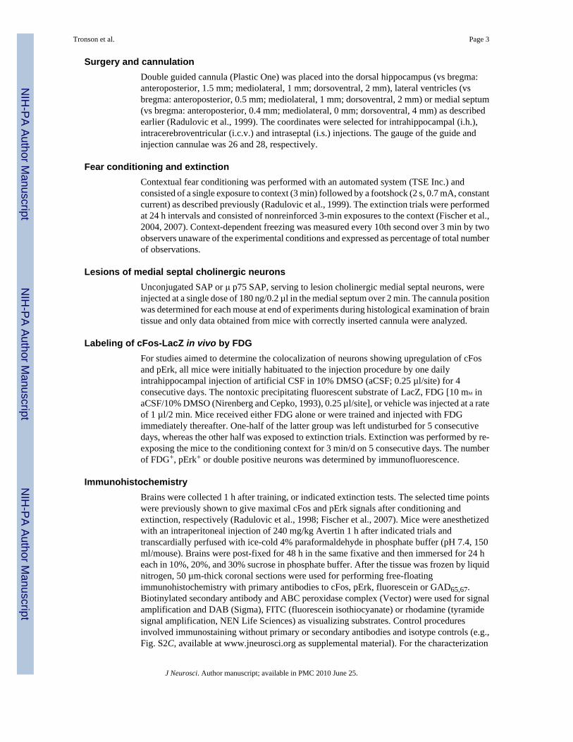
Surgery and cannulationDouble guided cannula (Plastic One) was placed into the dorsal hippocampus (vs bregma:anteroposterior, 1.5 mm; mediolateral, 1 mm; dorsoventral, 2 mm), lateral ventricles (vsbregma: anteroposterior, 0.5 mm; mediolateral, 1 mm; dorsoventral, 2 mm) or medial septum(vs bregma: anteroposterior, 0.4 mm; mediolateral, 0 mm; dorsoventral, 4 mm) as describedearlier (Radulovic et al., 1999). The coordinates were selected for intrahippocampal (i.h.),intracerebroventricular (i.c.v.) and intraseptal (i.s.) injections. The gauge of the guide andinjection cannulae was 26 and 28, respectively.
Fear conditioning and extinctionContextual fear conditioning was performed with an automated system (TSE Inc.) andconsisted of a single exposure to context (3 min) followed by a footshock (2 s, 0.7 mA, constantcurrent) as described previously (Radulovic et al., 1999). The extinction trials were performedat 24 h intervals and consisted of nonreinforced 3-min exposures to the context (Fischer et al.,2004, 2007). Context-dependent freezing was measured every 10th second over 3 min by twoobservers unaware of the experimental conditions and expressed as percentage of total numberof observations.
Lesions of medial septal cholinergic neuronsUnconjugated SAP or μ p75 SAP, serving to lesion cholinergic medial septal neurons, wereinjected at a single dose of 180 ng/0.2 µl in the medial septum over 2 min. The cannula positionwas determined for each mouse at end of experiments during histological examination of braintissue and only data obtained from mice with correctly inserted cannula were analyzed.
Labeling of cFos-LacZ in vivo by FDGFor studies aimed to determine the colocalization of neurons showing upregulation of cFosand pErk, all mice were initially habituated to the injection procedure by one dailyintrahippocampal injection of artificial CSF in 10% DMSO (aCSF; 0.25 µl/site) for 4consecutive days. The nontoxic precipitating fluorescent substrate of LacZ, FDG [10 mM inaCSF/10% DMSO (Nirenberg and Cepko, 1993), 0.25 µl/site], or vehicle was injected at a rateof 1 µl/2 min. Mice received either FDG alone or were trained and injected with FDGimmediately thereafter. One-half of the latter group was left undisturbed for 5 consecutivedays, whereas the other half was exposed to extinction trials. Extinction was performed by re-exposing the mice to the conditioning context for 3 min/d on 5 consecutive days. The numberof FDG+, pErk+ or double positive neurons was determined by immunofluorescence.
ImmunohistochemistryBrains were collected 1 h after training, or indicated extinction tests. The selected time pointswere previously shown to give maximal cFos and pErk signals after conditioning andextinction, respectively (Radulovic et al., 1998; Fischer et al., 2007). Mice were anesthetizedwith an intraperitoneal injection of 240 mg/kg Avertin 1 h after indicated trials andtranscardially perfused with ice-cold 4% paraformaldehyde in phosphate buffer (pH 7.4, 150ml/mouse). Brains were post-fixed for 48 h in the same fixative and then immersed for 24 heach in 10%, 20%, and 30% sucrose in phosphate buffer. After the tissue was frozen by liquidnitrogen, 50 µm-thick coronal sections were used for performing free-floatingimmunohistochemistry with primary antibodies to cFos, pErk, fluorescein or GAD65,67.Biotinylated secondary antibody and ABC peroxidase complex (Vector) were used for signalamplification and DAB (Sigma), FITC (fluorescein isothiocyanate) or rhodamine (tyramidesignal amplification, NEN Life Sciences) as visualizing substrates. Control proceduresinvolved immunostaining without primary or secondary antibodies and isotype controls (e.g.,Fig. S2C, available at www.jneurosci.org as supplemental material). For the characterization
Tronson et al. Page 3
J Neurosci. Author manuscript; available in PMC 2010 June 25.
NIH
-PA Author Manuscript
NIH
-PA Author Manuscript
NIH
-PA Author Manuscript

of FDG signals, selected brain sections were coverslipped with Vectashield (Vector) containingthe nuclear counterstain 4′,6-diamidino-2-phenylindole (DAPI).
Quantification of immunostaining signals was performed as described previously (Fischer etal., 2007). Digital images were captured with a cooled color charge-coupled device camera(RTKE Diagnostic Instruments) and SPOT software for Macintosh. Image J was used for imageprocessing. Cell counts from the dorsohippocampal CA1 subfield was performed using threeconsecutive dorsohippocampal sections per mouse (Brown et al., 1998). For double labeling,the separate FITC and rhodamine captures were digitally combined to produce compositeimages. Equal cutoff thresholds were applied to all captures to remove backgroundautofluorescence. For each capture, cell counts were performed within 100 µm2 grids (~7 grids/section) for three sections of each CA1. Counts of individual signals were first performedseparately, followed by identification and counting of double positive cells on the compositeimages. An overlapping signal of FITC and rhodamine fluorescence in nuclei that were sharplyin focus in a single focal plane was used as a criterion for nuclear colocalization of FDG andpErk (Patterson et al., 2001). The measures for each capture were averaged to give the numberof pErk+, cFos+ and FDG+ nuclei and expressed per 0.1 mm2 area. Finally, the proportionalnumber (percentage) of double positive neurons from total pErk immunopositive neurons wascalculated. Representative images (4–5 per group) were captured using a Zeiss LSM5 Pascalconfocal microscope. Between 7 and 12 z-slices were obtained for each channel which werethen converted into a z-projection, using a maximum projection algorithm (MetaMorph,Molecular Devices); thus, there was no loss of signal from any slice when the z-stack isproduced (Schrick et al., 2007).
Data analysisStatistically significant differences were determined by one- (molecular analyses, factorGroup) or two-way ANOVA (behavioral analyses, Group × Test interactions) followed byScheffe′s test for post hoc comparisons. The results are presented as mean ± SEM.
ResultsPredominant nuclear activation of cFos after conditioning and pErk during extinction of fearin hippocampal neurons
Several studies have shown that retrieval of the contextual fear memory (Ouyang and Thomas,2005) and temporary destabilization (Lee et al., 2008) or depotentiation (Kim et al., 2007) ofits underlying circuits are needed for fear extinction. These processes involve, at least in part,the neuronal subset encoding the conditioning fear memory. Fear conditioning potentlyactivates the c-fos promoter causing an increase of the cFos protein in hippocampal neuronsduring fear conditioning (Radulovic et al., 1998; Reijmers et al., 2007). We therefore firstexamined whether cFos and pErk are coactivated in hippocampal neurons in response toconditioning and extinction of fear. A naive group consisted of mice that were left undisturbedin their home cages throughout the experiments. Mice repeatedly exposed to context alonewere not used in the present study because our previous analyses revealed that this treatmentdid not trigger cFos (Radulovic et al., 1998) or pErk responses (our unpublished observations).Brain sections were analyzed 1 h after training (T) or 1 h after the fifth extinction test (E5)(Fig. 1A). This time point was selected because when they are activated by conditioning orextinction of fear, both cFos and pErk show maximal hippocampal levels ~1 h later (Atkins etal., 1998; Radulovic et al., 1998; Chen et al., 2005; Fischer et al., 2007). Thus, colocalizationanalyses are possible in this time frame. Immunohistochemical analyses of cFos and pErkrevealed that the upregulation of these proteins represented transient and temporally dissociatedmolecular alterations the T and E5 groups (Fig. 1B,C). Furthermore, although detectable signalsof both proteins were observed after E2, <5% of either cFos or pErk+ cells showed
Tronson et al. Page 4
J Neurosci. Author manuscript; available in PMC 2010 June 25.
NIH
-PA Author Manuscript
NIH
-PA Author Manuscript
NIH
-PA Author Manuscript
colocalization of both proteins. This lack of overlap was specific for the hippocampus whencompared with the amygdala, in which 29 and 41% of cFos and pErk+ cells, respectively, weredouble positive (Fig. 2A–C). Two possibilities could account for the segregated cFos and pErksignals: (1) pErk was upregulated instead of cFos within cells previously involved in fearconditioning, suggesting that conditioning and extinction are processed by the same cells usingdifferent molecular mechanisms, or (2) pErk upregulation during extinction took place withina separate subset of hippocampal cells.
Upregulation of cFos and pErk in segregated hippocampal principal CA1 neuronsTo delineate between these possibilities, we tagged the hippocampal neurons of miceexpressing a cFos-LacZ fusion protein driven by the c-fos promoter (Schilling et al., 1991).The fusion protein, responding to exogenous stimuli in a manner similar to that of endogenouscFos (Smeyne et al., 1992), was visualized with FDG, a nontoxic precipitating substrate ofLacZ allowing for stable in vivo labeling of activated neurons (Nirenberg and Cepko, 1993).Indirect immunofluorescence was used to enhance the signal intensity. Untreated cFos-LacZmice acquired and extinguished conditioned fear in a manner similar to that of their wild-typelittermates and C57BL/6 mice (F(2,27) = 0.45, p = 0.98) (Fig. S1A, available atwww.jneurosci.org as supplemental material), and these behaviors remained unaltered aftertreatment with FDG or vehicle (F(3,28) = 0.76, p = 1.02) (Fig. S1B, available atwww.jneurosci.org as supplemental material). Furthermore, extinction of fear was also Erk-dependent in cFos-LacZ mice, as it was reported earlier for the C57BL/6 strain, as revealed bysignificant persistence of fear after inhibition of the Erk up-stream activator mitogen-activatedand extracellular signal regulated kinase by U0126 (F(1,14) = 8.23, p < 0.05) (Fig. S1C, availableat www.jneurosci.org as supplemental material). The possibility that genetically introducedcFos-LacZ protein might involve somewhat smaller cell population than endogenous cFos(Smeyne et al., 1992) or interferes with Erk activation was examined by comparing thesemolecular responses between cFos-LacZ, wild-type and C57BL/6 mice. We did not observesignificant differences in the number of cFos (F(3,16) = 1.88, p = 1.92) and pErk (F(2,12) = 1.25,p = 1.13) signals determined after conditioning and E5, respectively, between these mousestrains (Fig. S1D,E, available at www.jneurosci.org as supplemental material). The FDGsignals (Fig. S1D, available at www.jneurosci.org as supplemental material) were notsignificantly reduced when compared with cFos signals in cFos-LAcZ mice despite showinga tendency toward a decrease. Possibly, earlier studies showed a greater difference becauseLacZ activity was compared with general fos-like immunoreactivity rather than specific cFossignals. The observed labeling pattern thus suggested that the cFos-LacZ mouse model wouldbe appropriate for the analyses of FDG+ and pERK+ cells. We subsequently performed i.h.injections of FDG alone or in conjunction with training and extinction (Fig. 3A). Whereasinjection of FDG alone did not cause an increase of immunofluorescent signals above baseline(Fig. 3B, top), injection of FDG after fear conditioning resulted in a significant increase ofFDG+ signals in the CA1 hippocampal subfield, F(2,38) = 43.93, p < 0.001, that persisted over5 consecutive days with or without exposure to extinction trials (Fig. 3B, middle and bottom).The FDG puncta were smaller than typical cFos nuclear labeling, probably because thesubstrate becomes diluted by diffusion through the brain tissue. Accordingly, a controlexperiment using i.c.v. injection of FDG revealed signals of large nuclear size (similar to cFos)in the lateral septal area adjacent to the ventricles, followed by a size reduction within thehipocampal tissue (Fig. 3C). FDG injections did not cause a lesion within the dorsalhippocampus, as revealed by cresyl violet staining (Fig. 3D). TheFDG+ signals were specific(Fig. 3E) and nuclear, as revealed by their colocalization with cFos (Fig. 3F) and the nuclearcounterstain DAPI (Fig. 3G). Because in 98% of the counted nuclei (from a total of 500 CA1neurons randomly picked from sections of different experiments) we detected only one FDGpunctum/nucleus, each punctum was counted as one cell. The somatonuclear pERKimmunostaining, upregulated in CA1 cells of the extinction group (F(2,38) = 87.53, p < 0.001)
Tronson et al. Page 5
J Neurosci. Author manuscript; available in PMC 2010 June 25.
NIH
-PA Author Manuscript
NIH
-PA Author Manuscript
NIH
-PA Author Manuscript

(Fig. 4A,B; Fig. S2A, available at www.jneurosci.org as supplemental material) showed lessthen 10% overlap with FDG. Namely, the number of FDG+/pErk+ neurons was not significantlyelevated above baseline levels, F(2,38) = 0.757, p = 0.47 (Fig. 4A–C), as determined by bothconventional and confocal microscopy. This is unlikely the result of pErk localization ininterneurons, because pErk, as it is well known for cFos, was predominantly increased inexcitatory, principal CA1 neurons (Fig. 4D). We also examined whether the absence of pErksignals was caused by inhibitory effects of cFos-LacZ or FDG on Erk activation withinindividual cells. In a separate group of mice, we injected FDG i.c.v. to obtain FDG signals inthe cortical areas adjacent to the hippocampus. Contrary to the hippocampus, FDG+ andpErk+ signals in the parietal cortex exhibited 42% overlap (7–9 double positive cells from 20pErk+ cells/0.1 mm2) in this brain area (Fig. 4E; Fig. S2B, available at www.jneurosci.org assupplemental material). These findings indicated that cFos-LacZ did not interfere with Erkactivity. Rather, segregated subsets of excitatory cFos+ and pErk+CA1 neurons responded tofear conditioning and extinction, respectively. Thus, Erk activity triggered by nonreinforcedtrials most likely did not involve modifications of the conditioning fear memory but insteadprocessed a new extinction memory.
Given that in some conditioning paradigms one-trial training is insufficient to trigger ceilingbehavioral and molecular responses (Navarro et al., 2000), a possibility remained that the usedexperimental approach did not allow for optimal codetection of cFos and pErk (each showingupregulation in 9–15% of CA1 principal cells). By increasing the number of training trials orshock intensity, we did not see further enhancement of conditioned freezing (Fig. S3A,B,available at www.jneurosci.org as supplemental material). Notably, the use of a 1.5 mA shockresulted in slower extinction, indicating a stronger fear memory, however this effect wasaccompanied by reduced pErk and unaltered cFos responses of CA1 neurons (Fig. S3C,available at www.jneurosci.org as supplemental material). So far, we were not able to identifyconditioning/extinction conditions causing increased levels of both cFos and pErk thusenhancing the odds of their codetection. Nevertheless, the consistent lack of overlap in the one-trial conditioning paradigm is strongly suggestive of disparate cell populations upregulatingcFos and pErk in the hippocampus as opposed to control brain areas.
Selective regulation of pErk+ neurons by afferent cholinergic hippocampal input from themedial septum
We next studied the regulation of pErk and cFos responses by hippocampal input. Two keyinputs from the entorhinal cortex and medial septum (Wheal and Miller, 1980) are thought toprovide the hippocampus with signals on actual and predicted events, respectively (Buhusi andSchmajuk, 1996; Gray and Mc-Naughton, 2000; Vinogradova, 2001). Because the latter signalshave been selectively implicated in extinction of conditioned responses (Gray andMcNaughton, 2000), we hypothesized that elimination of hippocampal cholinergic input fromthe medial septum would be critical for pErk upregulation and fear extinction. To test thishypothesis, we performed cholinergic lesions of the medial septum. The toxin SAP, conjugatedto a rabbit anti-mouse antibody recognizing the low affinity p75 neurotrophin receptorexpressed on cholinergic neurons was injected into the medial septum (Fig. 5A,B). The injectioncaused a significant loss of cholinergic markers within the medial septum and cholinergicdenervation of the hippocampus (Fig. 5C,G), as determined at the end of the experiment foreach mouse. Twenty days after injection of toxin or immunotoxin, mice with septohippocampalcholinergic lesions did not exhibit alterations of activity (t18 = 1.32, p = 0.83) shock response(t18 = 1.44, p = 0.79) or freezing behavior during training (Fig. 5D,E). The mice acquiredcontextual fear normally but did not show fear extinction even after multiple nonreinforcedtrials (Fig. 5E) when compared with control mice injected with unconjugated SAP (Group ×Extinction interaction: F(6,84) = 3.693, p < 0.01), alone or training followed by extinction.Notably, the lesions did not affect cFos responses triggered by fear conditioning, t4 = 0.43, p
Tronson et al. Page 6
J Neurosci. Author manuscript; available in PMC 2010 June 25.
NIH
-PA Author Manuscript
NIH
-PA Author Manuscript
NIH
-PA Author Manuscript

= 0.65, but significantly attenuated pErk responses, t12 = 10.966, p < 0.01, triggered byextinction (Fig. 4F,G). On the basis of these findings, the septal cholinergic input was identifiedan important regulator of the hippocampal pErk+ cell subset and fear extinction.
DiscussionBy using stable visualization of cFos-LacZ+ cells, we established that hippocampal principalneurons responding to fear conditioning (cFos+) and extinction (pErk+) representednonoverlapping cell subsets. Together with data demonstrating selective regulation of pErk+
neurons by afferent cholinergic input, we isolated and extinction-specific mechanism involvingdistinctive molecular, cellular and circuit regulation when compared with cFos-dependentconditioning mechanisms. Although the data do not rule out the possibility that pErkmechnisms may exhibit cellular overlap with cFos-independent mechanisms, the observeddissociation from cFos is significant given its strong causal association to fear conditioning(Fleischmann et al., 2003).
Taking into consideration the established role of Erk signaling in both conditioning (Atkins etal., 1998) and extinction (Szapiro et al., 2003), it was surprising that pErk+ cells activated byextinction represented a segregated neuronal subset. Codetection of cFos/FDG and pErk mighthave been reduced because we used single trial fear conditioning, nevertheless this paradigmconditions were sufficient to trigger robust behavioral and molecular effects during bothconditioning and extinction. We therefore hypothesize that the observed specificity is primarilybased on the subcellular localization of pErk. Namely, whereas transient somatonuclearincrease of pErk has been reported in some fear conditioning paradigms (Trifilieff et al.,2006; Sindreu et al., 2007), we and others have shown that hippocampal responses to synapticpotentiation (Winder et al., 1999), contextual aversive conditioning (Sananbenesi et al.,2002; Feld et al., 2005), or stress hormones (Kovalovsky et al., 2002) primarily requiresomatodendritic pErk. Extinction, however, triggers rapid, robust and sustained somatonuclearErk activation (Fischer et al., 2007). Immunobolot studies further reveal that Erkphosphorylation triggered by contextual extinction is significantly faster and stronger whencompared with fear conditioning, revealing maximal levels shortly before and during initialextinction while returning to baseline levels once extinction has been fully established (Fischeret al., 2007; Ryu et al., 2008). Because cellular and subcellular localization constraining Erk’sproximity to interacting molecules is a critical determinant of its actions (Sweatt, 2004; Schricket al., 2007), the unique contribution of this kinase to fear extinction versus conditioning maytherefore rely on sustained nuclear activity in the absence of co-upregulation of cFos, cAMPresponse element binding protein (Tronson et al., 2008), Cdk5 (Sananbenesi et al., 2007), andpossibly other molecules required for conditioning but not extinction of fear. The down-streameffects of nuclear pErk, likely to involve distinctive gene responses and structural modificationsof the identified neuronal subset, remain to be elucidated.
The dissociation of the identified hippocampal mechanism underlying extinction versusconditioning of fear was additionally documented by specific regulation via medial septalcholinergic input. As has been previously reported for the immediate early gene Arc (Fletcheret al., 2007), cFos and somatodendritic pErk were unaffected by permanent immunotoxin-induced lesions of cholinergic afferents. The levels of somatonuclear pErk, however, weresignificantly reduced. Accordingly, mice acquired conditioned fear normally but showedresistance to extinction. These findings provide strong molecular and cellular support for thecomparator theory (Buhusi and Schmajuk, 1996; Gray and McNaughton, 2000; Vinogradova,2001) proposing that the hippocampal responses to mismatch between anticipated anddelivered reinforcement provided by septal and cortical inputs, respectively, is critical forextinction of conditioned responses. Interestingly, the septal cholinergic input facilitatessomatic but depresses dendritic field potentials of hippocampal pyramidal neurons (Rovira et
Tronson et al. Page 7
J Neurosci. Author manuscript; available in PMC 2010 June 25.
NIH
-PA Author Manuscript
NIH
-PA Author Manuscript
NIH
-PA Author Manuscript

al., 1982). Such effects could account for the sustained somatonuclear Erk activity duringextinction when compared with conditioning of fear. The identification of the main cholinergicreceptor subtypes contributing to hippocampal Erk activation will help to further developspecific tools serving to facilitate fear extinction.
Together, our findings are consistent with recent observations obtained by electrophysiologicalrecordings identifying separate amygdalar excitatory neurons activated during conditioningand extinction of fear (Herry et al., 2008). However, whereas the activity of those cells hasbeen predominantly linked to the expression fear, as revealed by high correlation with freezingbehavior, the hippocampal formation is unlikely to be directly involved in fear regulation.Accordingly, the increase of pErk activity in the hippocampal CA1 neurons occurs in a specifictime window shortly preceding the decline of freezing and lasting only until extinction hasbeen established (Fischer et al., 2007; Ryu et al., 2008). These findings strongly suggest thatpErk activation within a distinctive subset of principal cells initiates new extinction learning.Lack of pErk signals in interneurons additionally indicated that this learning process involvesexcitatory but not inhibitory neurotransmission. Based on the known connections of thehippocamal CA1 subfield, the identified hippocampal Erk+ neurons may provide input to theprefrontal cortex and basolateral amygdala thereby initiating broad neuronal alterations withinthe fear extinction circuitry.
AcknowledgmentsThis work was supported by National Institute of Mental Health Grant MH073669 and Dunbar Funds to J.R. We thankDr. James I. Morgan (St. Joseph Hospital, Memphis, TN) for providing frozen embryos of cFos-LacZ mice, WarrenTourtelotte and the Northwestern University Transgenic Core Facility for deriving the cFos-LacZ mouse lines fromfrozen embryos, Dr. Eva Redei for reading and discussing this manuscript, and Dan Sylvester for assistance with thepreparation of this manuscript.
ReferencesAtkins CM, Selcher JC, Petraitis JJ, Trzaskos JM, Sweatt JD. The MAPK cascade is required for
mammalian associative learning. Nat Neurosci 1998;1:602–609. [PubMed: 10196568]Berlau DJ, McGaugh JL. Enhancement of extinction memory consolidation: the role of the noradrenergic
and GABAergic systems within the basolateral amygdala. Neurobiol Learn Mem 2006;86:123–132.[PubMed: 16458544]
Bouton ME. Context and behavioral processes in extinction. Learn Mem 2004;11:485–494. [PubMed:15466298]
Brown HE, Garcia MM, Harlan RE. A two focal plane method for digital quantification of nuclearimmunoreactivity in large brain areas using NIH-image software. Brain Res Brain Res Protoc1998;2:264–272. [PubMed: 9630665]
Buhusi CV, Schmajuk NA. Attention, configuration, and hippocampal function. Hippocampus1996;6:621–642. [PubMed: 9034850]
Chen X, Garelick MG, Wang H, Lil V, Athos J, Storm DR. PI3 kinase signaling is required for retrievaland extinction of contextual memory. Nat Neurosci 2005;8:925–931. [PubMed: 15937483]
Chhatwal JP, Myers KM, Ressler KJ, Davis M. Regulation of gephyrin and GABAA receptor bindingwithin the amygdala after fear acquisition and extinction. J Neurosci 2005;25:502–506. [PubMed:15647495]
Falls WA, Miserendino MJ, Davis M. Extinction of fear-potentiated startle: blockade by infusion of anNMDA antagonist into the amygdala. J Neurosci 1992;12:854–863. [PubMed: 1347562]
Feld M, Dimant B, Delorenzi A, Coso O, Romano A. Phosphorylation of extra-nuclear ERK/MAPK isrequired for long-term memory consolidation in the crab Chasmagnathus. Behav Brain Res2005;158:251–261. [PubMed: 15698891]
Tronson et al. Page 8
J Neurosci. Author manuscript; available in PMC 2010 June 25.
NIH
-PA Author Manuscript
NIH
-PA Author Manuscript
NIH
-PA Author Manuscript

Fischer A, Sananbenesi F, Schrick C, Spiess J, Radulovic J. Distinct roles of hippocampal de novo proteinsynthesis and actin rearrangement in extinction of contextual fear. J Neurosci 2004;24:1962–1966.[PubMed: 14985438]
Fischer A, Radulovic M, Schrick C, Sananbenesi F, Godovac-Zimmermann J, Radulovic J. HippocampalMek/Erk signaling mediates extinction of contextual freezing behavior. Neurobiol Learn Mem2007;87:149–158. [PubMed: 16979915]
Fleischmann A, Hvalby O, Jensen V, Strekalova T, Zacher C, Layer LE, Kvello A, Reschke M, SpanagelR, Sprengel R, Wagner EF, Gass P. Impaired long-term memory and NR2A-type NMDA receptor-dependent synaptic plasticity in mice lacking c-Fos in the CNS. J Neurosci 2003;23:9116–9122.[PubMed: 14534245]
Fletcher BR, Baxter MG, Guzowski JF, Shapiro ML, Rapp PR. Selective cholinergic depletion of thehippocampus spares both behaviorally induced Arc transcription and spatial learning and memory.Hippocampus 2007;17:227–234. [PubMed: 17286278]
Gray, J.; McNaughton, N. The neuropsychology of anxiety. Ed 2. Oxford: Oxford UP; 2000.Grillon C. Startle reactivity and anxiety disorders: aversive conditioning, context, and neurobiology. Biol
Psychiatry 2002;52:958–975. [PubMed: 12437937]Herry C, Ciocchi S, Senn V, Demmou L, Müller C, Lüthi A. Switching on and off fear by distinct neuronal
circuits. Nature 2008;454:600–606. [PubMed: 18615015]Isiegas C, Park A, Kandel ER, Abel T, Lattal KM. Transgenic inhibition of neuronal protein kinase A
activity facilitates fear extinction. J Neurosci 2006;26:12700–12707. [PubMed: 17151273]Kim J, Lee S, Park K, Hong I, Song B, Son G, Park H, Kim WR, Park E, Choe HK, Kim H, Lee C, Sun
W, Kim K, Shin KS, Choi S. Amygdala depotentiation and fear extinction. Proc Natl Acad Sci U SA 2007;104:20955–20960. [PubMed: 18165656]
Kovalovsky D, Refojo D, Liberman AC, Hochbaum D, Pereda MP, Coso OA, Stalla GK, Holsboer F,Arzt E. Activation and induction of NUR77/NURR1 in corticotrophs by CRH/cAMP: involvementof calcium, protein kinase A, and MAPK pathways. Mol Endocrinol 2002;16:1638–1651. [PubMed:12089357]
Lee SH, Choi JH, Lee N, Lee HR, Kim JI, Yu NK, Choi SL, Lee SH, Kim H, Kaang BK. Synaptic proteindegradation underlies destabilization of retrieved fear memory. Science 2008;319:1253–1256.[PubMed: 18258863]
Maren S, Quirk GJ. Neuronal signalling of fear memory. Nat Rev Neurosci 2004;5:844–852. [PubMed:15496862]
Matsuo N, Reijmers L, Mayford M. Spine-type-specific recruitment of newly synthesized AMPAreceptors with learning. Science 2008;319:1104–1107. [PubMed: 18292343]
Milad MR, Quirk GJ. Neurons in medial prefrontal cortex signal memory for fear extinction. Nature2002;420:70–74. [PubMed: 12422216]
Navarro M, Spray KJ, Cubero I, Thiele TE, Bernstein IL. cFos induction during conditioned taste aversionexpression varies with aversion strength. Brain Res 2000;887:450–453. [PubMed: 11134640]
Nirenberg S, Cepko C. Targeted ablation of diverse cell classes in the nervous system in vivo. J Neurosci1993;13:3238–3251. [PubMed: 8340805]
Ouyang M, Thomas SA. A requirement for memory retrieval during and after long-term extinctionlearning. Proc Natl Acad Sci U S A 2005;102:9347–9352. [PubMed: 15947076]
Patterson SL, Pittenger C, Morozov A, Martin KC, Scanlin H, Drake C, Kandel ER. Some forms ofcAMP-mediated long-lasting potentiation are associated with release of BDNF and nucleartranslocation of phospho-MAP kinase. Neuron 2001;32:123–140. [PubMed: 11604144]
Radulovic J, Kammermeier J, Spiess J. Relationship between fos production and classical fearconditioning: effects of novelty, latent inhibition, and unconditioned stimulus preexposure. JNeurosci 1998;18:7452–7461. [PubMed: 9736664]
Radulovic J, Rühmann A, Liepold T, Spiess J. Modulation of learning and anxiety by corticotropin-releasing factor (CRF) and stress: differential roles of CRF receptors 1 and 2. J Neurosci1999;19:5016–5025. [PubMed: 10366634]
Reijmers LG, Perkins BL, Matsuo N, Mayford M. Localization of a stable neural correlate of associativememory. Science 2007;317:1230–1233. [PubMed: 17761885]
Tronson et al. Page 9
J Neurosci. Author manuscript; available in PMC 2010 June 25.
NIH
-PA Author Manuscript
NIH
-PA Author Manuscript
NIH
-PA Author Manuscript

Rovira C, Cherubini E, Ben-Ari Y. Opposite actions of muscarinic and nicotinic agents on hippocampaldendritic negative fields recorded in rats. Neuropharmacology 1982;21:933–936. [PubMed:7145042]
Ryu J, Futai K, Feliu M, Weinberg R, Sheng M. Constitutively active Rap2 transgenic mice display fewerdendritic spines, reduced extracellular signal-regulated kinase signaling, enhanced long-termdepression, and impaired spatial learning and fear extinction. J Neurosci 2008;28:8178–8188.[PubMed: 18701680]
Sananbenesi F, Fischer A, Schrick C, Spiess J, Radulovic J. Phosphorylation of hippocampal Erk-1/2,Elk-1, and p90-Rsk-1 during contextual fear conditioning: interactions between Erk-1/2 and Elk-1.Mol Cell Neurosci 2002;21:463–476. [PubMed: 12498787]
Sananbenesi F, Fischer A, Wang X, Schrick C, Neve R, Radulovic J, Tsai LH. A hippocampal Cdk5pathway regulates extinction of contextual fear. Nat Neurosci 2007;10:1012–1019. [PubMed:17632506]
Schilling K, Luk D, Morgan JI, Curran T. Regulation of a fos-lacZ fusion gene: a paradigm for quantitativeanalysis of stimulus-transcription coupling. Proc Natl Acad Sci U S A 1991;88:5665–5669. [PubMed:1648227]
Schrick C, Fischer A, Srivastava DP, Tronson NC, Penzes P, Radulovic J. N-cadherin regulatescytoskeletally associated IQGAP1/ERK signaling and memory formation. Neuron 2007;55:786–798.[PubMed: 17785185]
Sindreu CB, Scheiner ZS, Storm DR. Ca2+stimulated adenylyl cyclases regulate ERK-dependentactivation ofMSK1during fear conditioning. Neuron 2007;53:79–89. [PubMed: 17196532]
Smeyne RJ, Schilling K, Robertson L, Luk D, Oberdick J, Curran T, Morgan JI. fos-lacZ transgenic mice:mapping sites of gene induction in the central nervous system. Neuron 1992;8:13–23. [PubMed:1730004]
Sotres-Bayon F, Bush DE, LeDoux JE. Emotional perseveration: an update on prefrontal-amygdalainteractions in fear extinction. Learn Mem 2004;11:525–535. [PubMed: 15466303]
Sweatt JD. Mitogen-activated protein kinases in synaptic plasticity and memory. Curr Opin Neurobiol2004;14:311–317. [PubMed: 15194111]
Szapiro G, Vianna MR, McGaugh JL, Medina JH, Izquierdo I. The role of NMDA glutamate receptors,PKA, MAPK, and CAMKII in the hippocampus in extinction of conditioned fear. Hippocampus2003;13:53–58. [PubMed: 12625457]
Trifilieff P, Herry C, Vanhoutte P, Caboche J, Desmedt A, Riedel G, Mons N, Micheau J. Foregroundcontextual fear memory consolidation requires two independent phases of hippocampal ERK/CREBactivation. Learn Mem 2006;13:349–358. [PubMed: 16705140]
Tronson NC, Schrick C, Fischer A, Sananbenesi F, Pagès G, Pouysségur J, Radulovic J. Regulatorymechanisms of fear extinction and depression-like behavior. Neuropsychopharmacology2008;33:1570–1583. [PubMed: 17712345]
Vinogradova OS. Hippocampus as comparator: role of the two input and two output systems of thehippocampus in selection and registration of information. Hippocampus 2001;11:578–598.[PubMed: 11732710]
Wheal HV, Miller JJ. Pharmacological identification of acetylcholine and glutamate excitatory systemsin the dentate gyrus of the rat. Brain Res 1980;182:145–155. [PubMed: 6243231]
Winder DG, Martin KC, Muzzio IA, Rohrer D, Chruscinski A, Kobilka B, Kandel ER. ERK plays aregulatory role in induction of LTP by theta frequency stimulation and its modulation by beta-adrenergic receptors. Neuron 1999;24:715–726. [PubMed: 10595521]
Tronson et al. Page 10
J Neurosci. Author manuscript; available in PMC 2010 June 25.
NIH
-PA Author Manuscript
NIH
-PA Author Manuscript
NIH
-PA Author Manuscript
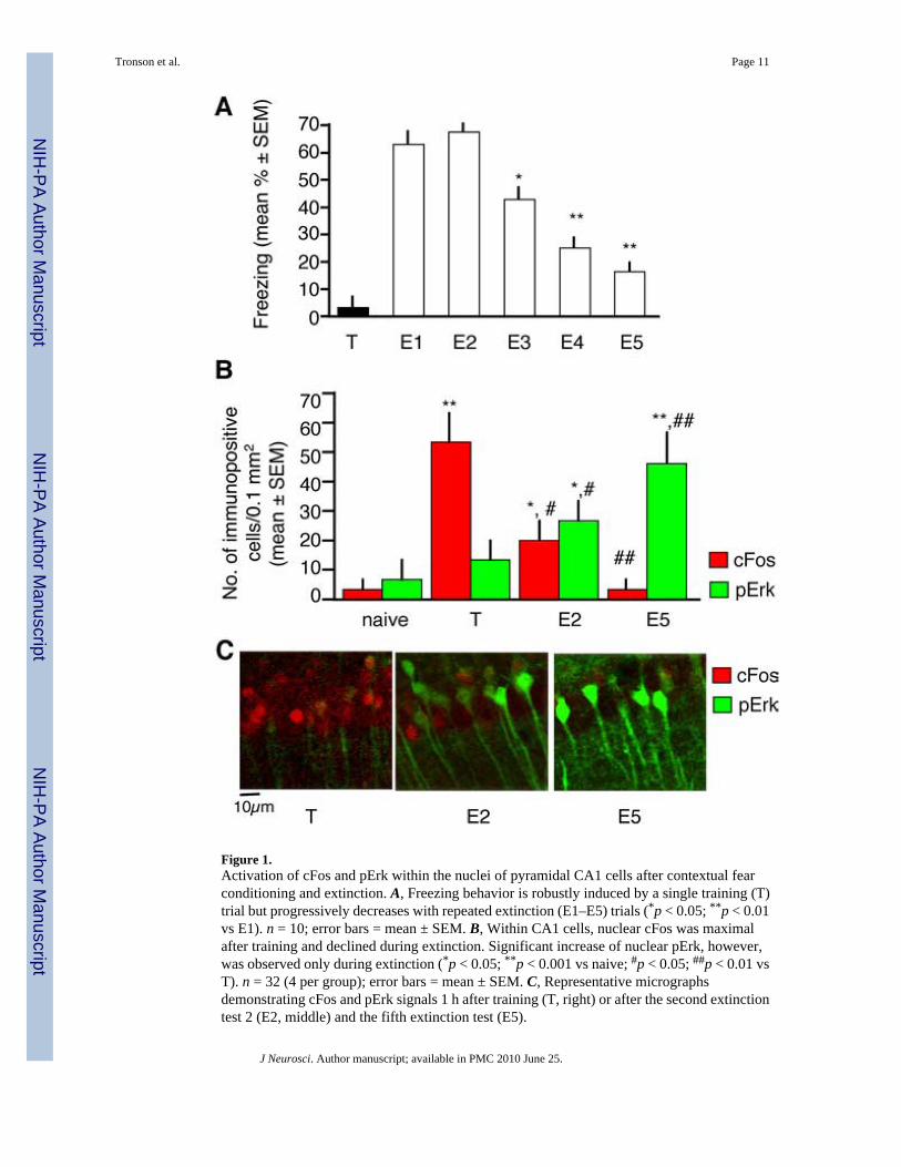
Figure 1.Activation of cFos and pErk within the nuclei of pyramidal CA1 cells after contextual fearconditioning and extinction. A, Freezing behavior is robustly induced by a single training (T)trial but progressively decreases with repeated extinction (E1–E5) trials (*p < 0.05; **p < 0.01vs E1). n = 10; error bars = mean ± SEM. B, Within CA1 cells, nuclear cFos was maximalafter training and declined during extinction. Significant increase of nuclear pErk, however,was observed only during extinction (*p < 0.05; **p < 0.001 vs naive; #p < 0.05; ##p < 0.01 vsT). n = 32 (4 per group); error bars = mean ± SEM. C, Representative micrographsdemonstrating cFos and pErk signals 1 h after training (T, right) or after the second extinctiontest 2 (E2, middle) and the fifth extinction test (E5).
Tronson et al. Page 11
J Neurosci. Author manuscript; available in PMC 2010 June 25.
NIH
-PA Author Manuscript
NIH
-PA Author Manuscript
NIH
-PA Author Manuscript

Figure 2.Lack of cFos/pErk colocalization in the hippocampus but not the amygdala. Colocalization ofcFos and pErk was determined 1 h after E2. A, Areas selected for analyses are outlined. B,cFos and pErk+ cells did not colocalize in the hippocampal CA1 subfield, whereas colabelingwas observed in the basolateral amygdala (BLA) as revealed by 30 ± 3% of pErk+ neurons; 43± 8 cFos+ neurons showing overlap (*p < 0.05; **p < 0.001 vs naive). The naive group ispresented as a dashed line. C, Representative micrographs.
Tronson et al. Page 12
J Neurosci. Author manuscript; available in PMC 2010 June 25.
NIH
-PA Author Manuscript
NIH
-PA Author Manuscript
NIH
-PA Author Manuscript

Figure 3.Long-term labeling of cFos-LacZ+ neurons activated by fear conditioning. A, Experimentaldesign for cFos-LacZ labeling. Four aCSF injections were performed once a day for fourconsecutive days to habituate the cFos response to the injection procedure before FDGadministration. Control mice were injected with FDG alone without prior training orsubsequent contextual exposures. Mice of the training group were injected with FDGimmediately after training and left undisturbed for 5 consecutive days. The extinction groupconsisted of mice exposed to training, FDG injection, and five extinction trials. B, FDG signals(green) were weak in the control group (top). Note strong FDG signals in the groups injectedwith FDG immediately after training with (middle panel) or without extinction (bottom panel).Background was subtracted using the same threshold for each section to eliminate interferenceof autofluorescence. C, Control section obtained from a mouse with FDG injected into thebrain ventricles showing the effect of a diffusion gradient from the ventricles into thehippocampal tissue on the size of FDG+ signals. The size of the signals was smaller than wasobserved in vitro or after surface application (Nirenberg and Cepko, 1993), probably becauseof dilution after diffusing within the brain tissue. The size of FDG signals decreased from 10µm(neurons close to the chorioid plexus) to 0.5–3 µm(100 µm laterally within the hippocampaltissue). D, Image of cresyl violet staining showing an intact CA1 area immediately dorsal tothe injection site. E, Micrographs showing lack of cFos-LacZ+ labeling under controlconditions when primary (left) or secondary (middle) antibody (Ab) was omitted or FDG wasnot injected (right). F, Micrographs showing nuclear colocalization of FDG and cFos signals.G, Using DAPI as a nuclear counterstain, we determined that most nuclei contained one FDGpunctum. Red arrows, cFos+ cells; blue arrows, DAPI+ nuclei.
Tronson et al. Page 13
J Neurosci. Author manuscript; available in PMC 2010 June 25.
NIH
-PA Author Manuscript
NIH
-PA Author Manuscript
NIH
-PA Author Manuscript

Figure 4.Conditioning and extinction of fear activate segregated cFos+ and pErk+ cell subsets. A,Confocal images reveal stably labeled cFos-LacZ (FDG+) cells in mice exposed to training ortraining followed by extinction, but not in untrained mice injected with FDG alone. cFos-LacZand pErk were upregulated (**p < 0.001 vs FDG control) in nonoverlapping cell subsets afterconditioning and extinction, respectively, as revealed by lack of FDG and pErk colabeling. n= 41 (13–15 per group); error bars = mean ± SEM. The naive group is presented as a dashedline. B, Representative micrograph showing very low overlap between pErk (red, occasionallywhite because of intense fluorescence) and FDG (green) nuclear signals (only one indicatedcell in the field is pErk+/FDG+). C, Orthogonal projections along the FDG-positive cell. Theprojections are marked with an arrow along the YZ (ii–iv) and XZ (v–vii) planes. The channelsfor FDG (green; ii and v), pERK (red; iii and vi) and pERK + FDG (overlay; iv–vii) arepresented. Arrows indicate FDG-positive cell nuclei. D, Confocal images excluding pErk+
labeling of CA1 interneurons. Micrographs obtained during confocal microscopy of arepresentative section labeled for pErk (red) and GAD65,67 (green) after extinction wereoverlaid revealing lack of colocalization. E, Confocal images demonstrating colocalization ofFDG and pErk signals in the parietal cortex. Dashed squares outlined cells or puncta forcolocalization analysis; green stars, FDG+ cells; yellow stars, pErk+/FDG+ cells; yellow arrow,overlays of pErk+ fibers and FGD puncta (these signals were not counted as nuclearcolocalization).
Tronson et al. Page 14
J Neurosci. Author manuscript; available in PMC 2010 June 25.
NIH
-PA Author Manuscript
NIH
-PA Author Manuscript
NIH
-PA Author Manuscript

Figure 5.Lesions of medial septal cholinergic neurons impair contextual fear extinction and Erksignaling while leaving fear conditioning and cFos responses intact. A, Experimental designdescribing the timing of surgery, toxin injection, training and extinction. B, Localization ofinjection sites of SAP and μ p75-SAP in the medial septum. C, Immunostaining for AChE (top)in the medial septum of SAP and μ p75-SAP-injected mice. Sections were collectedimmediately posterior to the injection site. The same sections counterstained with cresyl violet(bottom) do not reveal gross cell loss in the medial septum. LS, lateral septum; MS, medialseptum. D, Mice with μ p75-SAP-induced cholinergic lesions did not show alterations ofexploratory activity or shock responses during training. E, Permanent immunotoxic lesion ofcholinergic cells of the medial septum initiated 20 d before training (n = 7 per group) preservedfear conditioning but impaired fear extinction (**p < 0.01). F, Anatomical level of the dorsalhippocampus (left) used for quantification of the number of cFos+ (middle) and pErk+ (right)CA1 neurons after training and extinction, respectively, in sections of mice injected with SAP-and μ p75-SAP into the medial septum. Note specific impairment of the pErk response(***p < 0.01). Error bars = mean ± SEM. G, Effects of septally injected μ p75-SAP on AChE(n = 10 per group), pErk (n = 7 per group), and cFos immunoreactivity (n = 3 per group) in thehippocampus. μ p75-SAP produced hippocampal cholinergic denervation (left) resulting inintact responses of cFos+ cells after training (middle) but impaired responses of pErk+ cellsafter extinction (right).
Tronson et al. Page 15
J Neurosci. Author manuscript; available in PMC 2010 June 25.
NIH
-PA Author Manuscript
NIH
-PA Author Manuscript
NIH
-PA Author Manuscript


