Embed Size (px)
Citation preview

THE JOURNAL OF COMPARATIVE NEUROLOGY 353:427-438 (1995)
Serotonin Inputs to Rabbit Sympathetic Preganglionic Neurons Projecting to the
Superior Cervical Ganglion or Adrenal Medulla
IWONA JENSEN, IDA J. LLEWELLYN-SMITH, PAUL PILOWSKY, JANE B. MINSON, AND JOHN CHALMERS
Department of Medicine and Centre for Neuroscience, School of Medicine, Flinders University, Bedford Park, South Australia 5042, Australia
ABSTRACT The input from serotonin-containing nerve fibres to rabbit sympathetic preganglionic
neurons projecting to either the superior cervical ganglion or the adrenal medulla was investigated by combining retrograde tracing with the B subunit of cholera toxin and immunocytochemistry for serotonin. There were pronounced rostrocaudal variations in the density of serotonin fibres in the rabbit intermediolateral cell column from T1 to L4; maximum numbers of fibres were found in T3-6 and L3-4 and minimum numbers in T1 and T10-12. By light microscopy, retrogradely labelled sympathetic preganglionic neurons projecting to the superior cervical ganglion or the adrenal medulla received variable densities of close appositions from serotonin-immunoreactive fibres. Some neurons from each population received many close appositions, whereas others received moderate numbers or few appositions. Appositions occurred on the cell bodies, dendrites, and occasionally axons of sympathetic preganglionic neurons. Rare neurons in both groups of retrogradely labelled cells received no appositions from serotonin-containing nerve fibres. At the ultrastructural level, synapses were found between serotonin-positive boutons and sympathetic preganglionic neurons projecting either to the superior cervical ganglion or to the adrenal medulla. These results indicate that, through direct synaptic contacts, serotonin-immunoreactive, presumably bulbospinal, nerve fibres affect the activity of the vast majority of sympathetic preganglionic neurons that send axons either to the superior cervical ganglion or to the adrenal medulla. This serotonin input may be sympathoex- citatory and could mediate increases in sympathetic nerve activity and in the release of catecholamines from the adrenal medulla. o 1995 WiIey-Liss, Inc.
Indexing terms: blood pressure, cholera toxin B subunit, immunocytochemistry, spinal cord, ultrastructure
Sympathetic preganglionic neurons (SPN) play a pivotal role in the central control of blood pressure. With the exception of the sinoatrial node of the heart, the mass of cardiac and vascular smooth muscle receives its most important innervation from the sympathetic nervous sys- tem, as does the adrenal medulla. For this reason, knowl- edge about the control of the activity of SPN is important for understanding how blood pressure is regulated. SPN receive synaptic inputs from both intraspinal and supraspi- nal neurons (Laskey and Polosa, 1988; Chalmers and Pilowsky, 1991). Of the supraspinal neurons, bulbospinal neurons are believed to be most important in regulating the ongoing activity of SPN. Bulbospinal neurons with axons that project to the intermediolateral cell column (presympa- thetic neurons) are located in the ventrolateral region of the
medulla, the raphe nuclei, and the dorsomedial medulla (Bowker et al., 1981a,b, 1982a,b; Loewy and McKellar, 1981; Loewy, 1982; Minson et al., 1984, 1987; Pilowsky et al., 1986a; Millhorn et al., 1987; Laskey and Polosa, 1988).
Serotonin is known to be an important neurotransmitter regulating the activity of SPN in the cat, rabbit, and rat (Laskey and Polosa, 1988; Chalmers and Pilowsky, 1991). Physiological and pharmacological studies largely support a sympathoexcitatory role for bulbospinal serotoninergic path- ways (Howe et al., 1983; Minson et al., 1987; Yusof and
Accepted August 22,1994 Address reprint requests to Dr. I.J. Llewellyn-Smith, Department of
Medicine, Flinders Medical Centre, Bedford Park, South Australia 5042, Australia.
O 1995 WILEY-LISS, INC.
428
Coote, 1988; Lewis and Coote, 1990; Gilbey and Stein, 1991). Stimulation of the rostra1 ventromedial medulla, from which serotonin-containing neurons project to the intermediolateral cell column, evokes a release of serotonin in the spinal cord, hypertension, and a sympathoexcitation that can be blocked with methysergide or by destruction of serotoninergic neurons with 5,7- or 5,6-dihydroxytrypta- mine (Howe et al., 1983; Minson et al., 1984, 1987; Pi- lowsky et al., 1986a; Mills et al., 1988). In the unanaesthe- tised rabbit, intracisternal administration of 5,7- dihydroxytryptamine causes an acute release of serotonin from nerve endings that is associated with hypertension, an augmentation of renal sympathetic nerve activity, and a massive release of adrenal hormones (Korner and Head, 1981; Head and Korner, 1982; Pilowsky et al., 198613). The acute phase of serotonin release is followed by a destruction of serotoninergic nerves and a fall in arterial blood pressure (Wing and Chalmers, 1974).
Anatomical studies in the rat and other species have shown a serotoninergic projection from the medulla to various levels of the spinal cord (Bowker et al., 1981a,b, 1982a,b; Loewy and McKellar, 1981; Loewy, 1982; Millhorn et al., 1987; Li et al., 1992; Ding et al., 1993). The distribu- tion of serotonin-immunoreactive terminals in the interme- diolateral cell column and other autonomic subnuclei paral- lels that of SPN somata identified by retrograde tracing (Appel and Elde, 1988; Strack et al., 1988; Anderson et al., 1989; Pilowsky et al., 1992b; Jensen et al., 1992). In the rat, serotonin-containing fibres have been found closely ap- posed to retrogradely Iabelled SPN (Holets and Elde, 1982; Appel et al., 1986, 1987; Appel and Elde, 1988) and ultrastructural studies have shown direct synaptic contacts between serotonin-immunoreactive terminals and SPN (Ba- con and Smith, 1988; Chiba, 1989; Vera et al., 1990).
Many physiological and pharmacological studies in the rabbit have demonstrated an important role for bulbospinal serotonin neurons in the control of the cardiovascular system (Wingand Chalmers, 1974; Korner and Head, 1981; Head and Korner, 1982; Pilowsky et al., 198613). Bulbospi- nal serotonin pathways controlling sympathetic outflow in the rabbit have also been studied anatomically (Li et al., 1992; Ding et al., 1993). However, neither the distribution of serotonin fibres in rabbit spinal cord nor the density of the serotoninergic input to rabbit SPN has been investi- gated. In this study, we first defined the patterns of serotonin innervation in autonomic areas of rabbit thoracic and upper lumbar spinal cord and then studied the seroto- nin input to rabbit SPN retrogradely labelled with cholera toxin B subunit either from the superior cervical ganglion or from the adrenal medulla. These two groups of SPN were chosen because SPN projecting to the superior cervical ganglion constitute a heterogeneous group of neurons innervating postganglionic neurons that supply a variety of target organs, whereas SPN projecting to the adrenal medulla form a functionally homogenous population of neurons that innervates only the adrenal medulla. We were particularly interested in determining whether or not these two populations of SPN differed in their innervation from serotonin-containing nerve fibres. Previous immunohisto- chemical studies have shown that SPN are not all sur- rounded by the same chemical types of nerve fibres. For example, rat sympathoadrenal neurons are preferentially contacted by somatostatin fibres (Holets and Elde, 1982), and rabbit SPN projecting to the lumbar sympathetic chain are associated with dense plexuses of neuropeptide Y-
I. JENSEN ET AL.
containing nerve fibres, whereas those projecting to the pelvic viscera are not (Anderson et al., 1987). Hence, differences in the serotonin innervation patterns of differ- ent groups of rabbit SPN could provide support for the idea that qualitative or quantitative differences in the synaptic inputs to SPN underlie functional specificity in the control of sympathetic outflow.
MATERIALS AND METHODS Injection of retrograde tracers
Experiments were conducted on 11 male New Zealand white rabbits (3.0-4.1 kg). Anaesthesia was induced with sodium thiopentone (40-60 mg i.v.; Abbott) and main- tained with halothane via an endotracheal tube. Seven rabbits were used to examine the relationship between serotonin-immunoreactive nerve fibres and retrogradely labelled SPN. Three rabbits received injections of cholera toxin B subunit (CTB; List Biological Laboratories, Camp- bell, CA) into both the left adrenal medulla (Jensen et al., 1992) and the right superior cervical ganglion (Pilowsky et al., 1992b), as described previously. In three rabbits CTB was injected only into the adrenal medulla and, in another rabbit, only into the superior cervical ganglion. The remain- ing four rabbits were used to examine the rostrocaudal distribution of serotonin-immunoreactive nerve fibres in the spinal cord, without prior injection of retrograde tracer.
Tissue processing Seventy-two hours after injection of CTB into the supe-
rior cervical ganglion and/or the adrenal medulla, rabbits were deeply anaesthetised with sodium pentobarbitone (40-60 mgikg), heparinised, and perfused through the heart with 1 litre of oxygenated tissue culture medium (Sigma D-8900) followed by 2 litres of fixative containing either 4% formaldehyde or 4% formaldehyde, 0.5% glutaral- dehyde, and 0.2% picric acid in 0.1 M buffer, pH 7.4 (FAGLU). Four percent formaldehyde was prepared as a 1: 10 dilution of analytical reagent-grade formaldehyde solu- tion stabilised with methanol (Univar). The spinal cords from T1 to L4 were carefully removed and cut into blocks of one to three segments in length. For spinal cords fixed with FAGLU, the segments were split dorsoventrally along the midline and postfixed in the same fixative overnight at 4°C. Spinal cords that had been perfused with 4% formaldehyde were postfixed for 1-2 days in 4% formaldehyde and then transferred serially to 4% formaldehyde plus 10% sucrose, 4% formaldehyde plus 20% sucrose, and 4% formaldehyde plus 30% sucrose over several weeks. These spinal cords were sectioned parasagittally or transversely at 50 km on a freezing microtome. Spinal cords fixed with FAGLU were cut parasagittally at 70 pm on a Vibratome.
Sections were exposed to 50% ethanol in distilled water (10 minutes for tissue fixed with 4% formaldehyde; 30 minutes for tissue fixed with FAGLU) to improve the penetration of reagents (Llewellyn-Smith and Minson, 1992) and then washed in several changes of phosphate buffer. To block nonspecific binding sites, sections were immersed in 10% normal horse serum (NHS) diluted with 10 mM phosphate-buffered saline, pH 7.4, containing 10 mM Tris and 0.05% thimerosal (TPBS). Sections containing retro- gradely labelled SPN were incubated for 2 days in rat monoclonal antiserotonin (Seralab; 1:500 or 1:1,000) com- bined with goat anti-CTB (List; 1:50,000) diluted with 10% NHS-TPBS. The sections were then incubated for 24 hours

SEROTONIN INPUTS TO RABBIT SPN 429
in biotinylated antirat immunoglobulin (1:500 or 1:1,000; Jackson ImmunoResearch, West Grove, PA) in 1% NHS- TPBS followed by an overnight incubation in a 1:1,500 dilution of ExtrAvidin-horseradish-peroxidase (Sigma E-2886) in TPBS. All incubations were at room tempera- ture with continuous gentle agitation. Sections were washed in three changes of TPBS after each incubation. For light microscopy, serotonin-immunoreactive nerve fibres were visualised by nickel-intensified diaminobenzidine (nickel- DAB) reaction (Llewellyn-Smith et al., 1993). For electron microscopy, a tungstate-stabilised tetramethylbenzidine (TMB) reaction (Llewellyn-Smith et al., 1993) or a nickel- DAB reaction was used to reveal serotonin-containing nerve fibres. Sections treated with TMB-tungstate were subsequently stabilised with DAB, cobalt chloride, and peroxide ions (Llewellyn-Smith et al., 1993).
After visualisation of serotonin immunoreactivity, sec- tions were incubated for 16-24 hours in 1:200 biotinylated antisheep immunoglobulin (Sigma B-73901, followed by an overnight incubation in 1: 1,500 ExtrAvidin-horseradish- peroxidase. Alternatively, the sections received a 16-24 hours incubation in 1500 or 1:1,000 unlabelled antigoat immunoglobulin (Jackson ImmunoResearch) in 1% NHS- TPBS, followed by a 4 hour or overnight incubation in 1:500 or 1:1,000 goat PAP (Jackson ImmunoResearch) in TPBS. Imidazole-intensified DAB reactions (Llewellyn- Smith et al., 1992a) were used to reveal CTB immunoreac- tivity for light microscopy, and nickel-intensified DAB reactions were used for electron microscopy. With these procedures, the second peroxidase reaction did not alter the character of the reaction product deposited during the first peroxidase reaction. In sections prepared for light micros- copy, the color of the black serotonin fibres was not changed by the DAB-imidazole reaction to reveal brown retrogradely labelled neurons. In tissue processed for electron micros- copy, the Ni-DAB reaction did not cause the deposition of amorphous reaction product in serotonin fibres visualised previously with a TMB-tungstate reaction. Sections from rabbits that did not receive tracer injections were incubated for 2 days in a 1:500 dilution of antiserotonin in 10% NHS-TPBS, for 24 hours in a 1:200 dilution of biotinylated antimouse immunoglobulin (Sigma B-4765) in 1% NHS- TPBS, and for 24 hours in 1: 1,500 ExtrAvidin-horseradish- peroxidase and then treated with nickel-DAB.
As control, sections of formaldehyde-fixed rabbit spinal cord were incubated with antiserotonin antibody (1: 1,000 dilution) that had been preabsorbed overnight at 4°C with
M, M, or M serotonin or with serotonin conjugated to bovine serum albumin through formalde- hyde. The conjugate was prepared according to the method of Steinbusch et al. (1978). Pretreatment of the anti-CTB antiserum with CTB has previously been shown to abolish staining of SPN retrogradely labelled with CTB (Llewellyn- Smith et al., 1992b).
Sections for light microscopy were mounted onto gelatin- coated slides, air dried, dehydrated, cleared, and cover- slipped with DePeX (GURR). Sections for electron micros- copy were pcstfixed for 1 hour in 0.5% osmium tetroxide in 0.1 M phosphate buffer, stained en bloc with 2% aqueous uranyl acetate, and dehydrated with ethanol and propylene oxide. The Vibratome sections were then embedded in Durcupan (Fluka) on glass slides under Aclar (Pelco) coverslips. After polymerisation, retrogradely labelled neu- rons for ultrastructural study were cut out of the Vibra- tome sections and reembedded on flat blank blocks. Ultra-
thin sections were cut with a diamond knife, collected on mesh grids, counterstained with Reynold’s lead citrate, and examined with a Jeol 1200EX electron microscope.
RESULTS Light microscopy
Serotonin-immunoreactive nerve fibres. Parasagittal and transverse sections of all spinal cord segments con- tained black serotonin-immunoreactive nerve fibres. In transverse section, serotonin fibres could be seen in the dorsolateral funiculus of the white matter and in the dorsal horn, intermediate zone, and ventral horn of the gray matter. Serotonin-immunoreactive nerve fibres were found in all spinal sympathetic subnuclei at all rostrocaudal levels. Within the intermediate zone, serotonin-immunore- active fibres were especially concentrated in the intermedio- lateral cell column (Fig. 1) and ran rostrocaudally parallel to its longitudinal axis; serotonin fibres also occurred in transverse bands that linked the intermediolateral cell column to the plexus of fibres around the central canal.
Most varicose immunoreactive axons in the intermediolat- era1 cell column had irregularly shaped varicosities with short intervaricose segments, whereas others had spherical varicosities. Rare axons had unusually large varicosities throughout their length in comparison to the majority of positive fibres. No cell bodies were found that contained serotonin immunoreactivity.
The distribution of the serotonin-immunoreactive nerve fibres within the intermediolateral cell column varied rostro- caudally along the length of the spinal cord from segment T1 through segment L4 (Fig. 1). The highest density of serotonin-containing nerve fibres was observed in spinal cord segments T3--6 (Fig. 1B) and L1-4 (Fig. 1D). In these segments the density of fibres was so high that it was often difficult to discern individual labelled varicosities. Spinal cords segments T2 and T7-9 contained fewer serotonin- immunoreactive fibres, whereas the density of serotonin- positive fibres was lowest in the segments T1 (Fig. 1A) and T10-12 (Fig. 1C).
In control experiments, a few serotonin-immunoreactive nerve fibres could still be found when sections of rabbit spinal cord were stained with antiserotonin antibody that had been preabsorbed with M serotonin; the staining pattern for serotonin antibody absorbed with M or
M serotonin was identical to that seen with unab- sorbed antiserum. However, staining of nerve fibres was totally abolished by overnight incubation of the serotonin antibody with 200 mg of a serotonin-formaldehyde-bovine serum albumin conjugate.
Retrogradely labelled sympathetic preganglionic neu- rons. At the light microscopic level, SPN retrogradely labelled with CTB contained an amber brown reaction product that filled their cell bodies and dendrites (Figs. 2, 3). The vast majority of labelled neurons projecting either to the superior cervical ganglion or to the adrenal medulla was found in clusters within the intermediolateral cell column (Fig. 2A). Some retrogradely labelled neurons were observed in the dorsolateral funiculus, whereas occasional cells were seen in the intercalated nucleus or the central autonomic area. The morphology of rabbit SPN projecting either to the superior cervical ganglion or to the adrenal medulla has been described elsewhere (Jensen et al., 1992; Pilowslry et al., 1992b). Occasional neurons showed CTB immunoreactivity in their axons (Fig. 41, which could often

430 I. JENSEN ET AL.
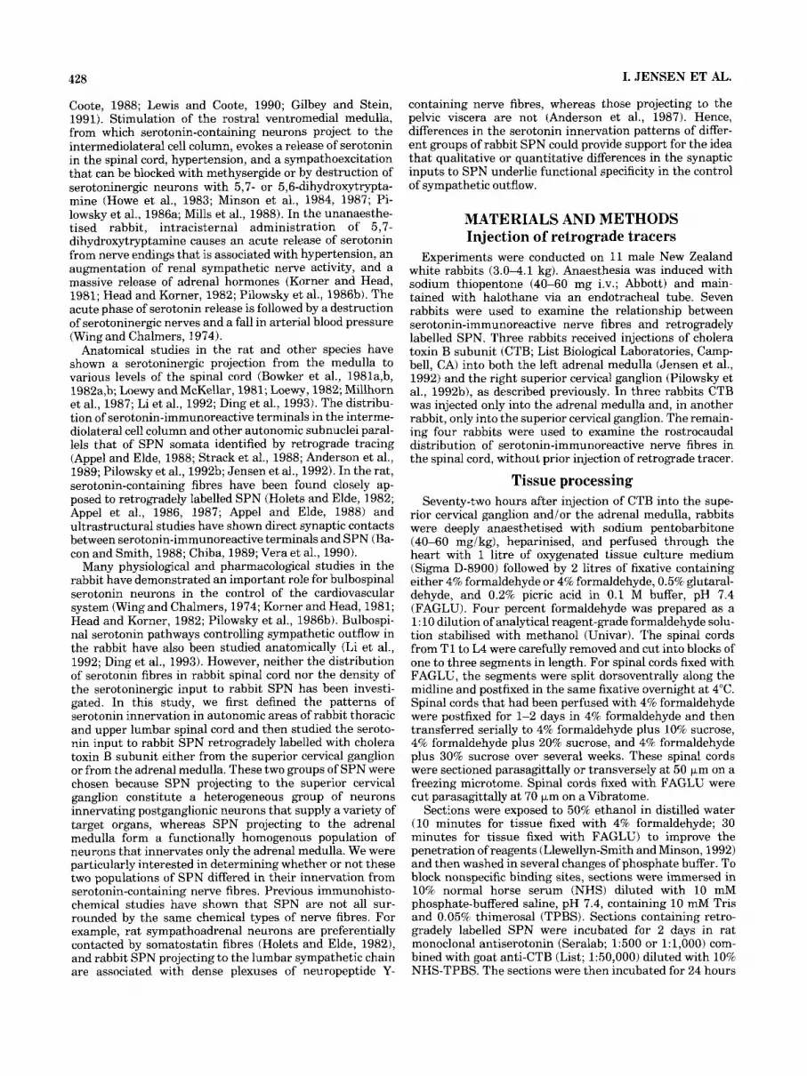
Fig. 1. Light micrographs illustrating the rostrocaudal distribution of serotonin-immunoreactive nerve fibres along the length of the rabbit intermediolateral cell column from spinal cord segments Tl-L4. Trans- verse sections. Fixative, 4% formaldehyde. A: Rostra1 spinal cord
segment T1. B: Spinal cord segment T5. C: Spinal cord segment T10. D: Spinal cord segment L4. Segments T3-T5 and L3-L4 were most densely innervated; segments T10-Tl2 were least densely innervated. Scale bar = 50 Km.
be followed for several hundred micrometres through the ventral horn.
After injection of CTB into the superior cervical ganglion (n = 4), retrogradely labelled SPN were observed in spinal cord segments T1-8 and concentrated in T3-5. After injection of CTB into the adrenal medulla (n = 61, retro- gradely labelled SPN were seen in spinal cord segments T2-L3, with the bulk located in segments T6-11. Retro- gradely labelled SPN projecting to the superior cervical ganglion were much more densely packed than retrogradely labelled sympathoadrenal SPN (Fig. 2). These results con- firm our previous work. The data of Pilowsky et al. (1992b) indicate that there are about 100-120 SPN per millimetre
projecting to the superior cervical ganglion in rabbit seg- ment T3 (length about 5-6 mm), and those of Jensen et al. (1992) imply that there about 30-40 sympathoadrenal neurons per millimetre in rabbit segments T7 (length about 7.5-9 mm).
Relationship of serotonin-immunoreactive nerve fibres to CTB-labelled SPN. Black serotonin-immunoreactive nerve fibres formed close appositions with brown SPN containing CTB retrogradely transported from either the superior cervical ganglion or the adrenal medulla (Figs. 2, 3). Serotonin appositions occurred on SPN whose cell bodies were located in the intermediolateral cell column, in the dorsolateral funiculus, in the intercalated region, and in

SEROTONIN INPUTS TO RABBIT SPN 431
Fig. 2. Light micrographs showing the relationships between seroto- nin-immunoreactive nerve fibres and groups of retrogradely labelled sympathetic preganglionic neurons (SPN) in the rabbit intermediolat- era1 cell column. Parasagittal sections. Fixative, 4% formaldehyde. A SPN in spinal cord segment T3 retrogradely labelled with cholera toxin B subunit (CTB) from the superior cervical ganglion (SCG). There are
10 CTB-immunopositive neurons (asterisks) in the area shown; most are embedded within the plexus of serotonin (5-HT)-immunoreactive nerve fibres, and this makes them difficult to discern. B: Three SPN (asterisks) in spinal cord segment T7 retrogradely labelled with CTB from the adrenal medulla (AM) lie within a moderately dense plexus of 5-HT fibres. Scale bars = 50 mm.
the central autonomic area. Single serotonin-immunoposi- tive nerve fibres could be traced for long distances, and, in some cases, individual fibres were closely apposed to more than one CTB-labelled neuron. In parasagittal sections
through the intermediolateral cell column of T2-5, SPN projecting to the superior cervical ganglion usually lay within the dense plexus of serotonin fibres (Fig. 2A). The cell bodies of sympathoadrenal neurons were concentrated

432 I. JENSEN ET AL.
Fig. 3. Light micrographs showing the relationships between seroto- nin-immunoreactive nerve fibres and individual retrogradely labelled SPN whose cell bodies lie within the intermediolateral cell column. Parasagittal sections. Fixative, 4% formaldehyde. A-C: CTB retro- gradely transported from the adrenal medulla. D,E: CTB retrogradely transported from the superior cervical ganglion. Some serotonin- immunoreactive varicosities that are closely apposed to the CTB- labelled SPN are indicated by arrows. (Not all appositions are in the plane of focus of the micrographs). A Labelled SPN with many serotonin appositions on its cell body and dendrites. Spinal cord segment T7. B: Serotonin-immunoreactive varicosities form a pericellu- lar basket around a retrogradely labelled SPN. Thirty-five to forty
varicosities made close appositions with the labelled neuron, only some of which are in the plane of focus of the micrograph. An adjacent unlabelled neuron (asterisk) also lies within a basket formed by serotonin-immunoreactive varicosities. Spinal cord segment T12. C: Two labelled SPN with few appositions from serotonin-immunoreactive varicosities. The top neuron received direct appositions from five immunoreactive varicosities, the bottom neuron from three varicosi- ties. Spinal cord segment T8. D: Labelled SPN that receives appositions from a serotonin-immunoreactive fibre with large boutons. Spinal cord segment T4. E: Two labelled SPN that received no appositions from serotonin-positive nerve fibres. Spinal cord segment T1. Scale bars = 20 fim (bar in E also applies to Band C).

SEROTONIN INPUTS TO RABBIT SPN 433
D
50um
Fig. 4. Appositions by serotonin-immunoreactive varicosities (ar- rows) onto the axon of an SPN in spinal cord segment T5. Parasagittal section. Fixative, 4% formaldehyde. CTB retrogradely transported from the adrenal medulla. A Camera lucida reconstruction of the labelled SPN cell body and its axon. B: Light micrograph showing that the cell body of the labelled SPN receives many close appositions from serotonin-
immunoreactive nerve fibres. C: Light micrograph of a cluster of appositions by serotonin varicosities onto the axon of the SPN near Its point of origin from the cell body. D: Light micrograph of a serotonin apposition onto the axon of the SPN ahout 140 Fm distal to its point of origin from the cell body. Scale bars = 50 pm in A, 10 pm in B, 5 pm in C and D.
in the area of the intermediolateral cell column where the Appositions density of serotonin immunoreactive nerve fibres was lower by serotonin-positive nerve fibres were present on the cell (Fig. 2B). bodies and dendrites of SPN projecting either to the
Appositions on cell bodies and dendrites.

I. JENSEN ET AL. 434
superior cervical ganglion or to the adrenal medulla (Fig. 3). Both groups of retrogradely labelled SPN had a similar range of densities of input from serotonin-containing nerve fibres. Some CTB-positive neurons in each group received many appositions (Figs. 3A,B, 4B), with serotonin-positive nerve fibres sometimes forming pericellular baskets around CTB-containing SPN (Fig. 3B). Other SPN had moderate numbers or few serotonin appositions (Fig. 3C,D). Rare SPN did not receive any appositions from serotonin- immunoreactive nerve fibres (Fig. 3E). The SPN without appositions occurred singly or in groups and were most frequently found in T1 after injection of CTB into the superior cervical ganglion. After either superior cervical ganglion or adrenal medulla injections, SPN with dense appositions and SPN with no appositions were inter- mingled with SPN that had moderate or sparse appositions. Serotonin-immunoreactive nerve fibres also occurred around neurons that were not retrogradely labelled and sometimes formed baskets around these cells (Fig. 3B).
Appositions by serotonin-immu- noreactive nerve fibres were also found on the axons of SPN (Fig. 4). Appositions were easiest to identify on the axons of sympathoadrenal neurons, although only a small subset of sympathoadrenal axons received appositions from serotonin- immunoreactive varicosities. These neurons were more sparsely distributed than SPN projecting to the superior cervical ganglion and occurred in areas of the intermediolat- eral cell column where serotonin innervation was less dense; consequently, their axons could often be traced from their point of origin for several hundred micrometres through the ventral horn. Some of the serotonin apposi- tions occurred near the point of origin of an axon, either from a primary dendrite or from a cell body (Fig. 4A,C). Other appositions occurred more distally on SPN axons within lamina 7, but no appositions were ever observed within lamina 8 / 9 of the ventral horn. Where appositions were present, more than one serotonin-immunoreactive varicosity was usually apposed to each individual axon.
Appositions on axons.
Electron microscopy Sections containing serotonin-immunoreactive terminals
in close apposition to retrogradely labelled sympathetic neurons were selected for ultrastructural study. CTB- immunoreactive SPN were identified at the ultrastructural level by the presence of electron-dense peroxidase reaction product in their cytoplasm (Fig. 5 ) . Variably sized deposits of peroxidase reaction product were seen in the CTB- positive cell bodies. The deposits were concentrated in and around the Golgi apparatus (see also Pilowsky et al., 199213) and also occurred in the rough endoplasmic reticulum. The cytoplasmic matrix either had no label or contained homoge- neously distributed moderate to faint deposits of immunore- activity. Reaction product was never seen in the nucleus.
At the electron microscopic level, varicosities were found to contain synaptic vesicles. Serotonin immunoreactivity was indicated by the presence of amorphous electron-dense reaction product in the varicosities after nickel-DAB reac- tions (Fig. 5 ) or electron-dense crystals after TMB- tungstate reactions. Synaptic vesicles were irregularly dis- tributed in the axon profiles positive for serotonin. Most synaptic vesicles were small, were spherical or oval, and had reaction product associated with their membranes after nickel-DAB reactions. A few large dense-core vesicles were found in some immunoreactive axon terminals.
Vesicle-containing serotonin-immunoreactive axon pro- files formed synapses on the cell bodies and dendrites of CTB-immunoreactive neurons that projected either to the superior cervical ganglion or to the adrenal medulla (Fig. 5). The criteria used for the identification of synapses were the clustering of vesicles presynaptically and the presence of an electron-dense thickening associated with the postsynaptic membrane. Serotonin-containing varicosities also directly contacted SPN without the presence of postsynaptic densi- ties. In addition, contacts and synapses by serotonin- immunopositive varicosities were frequently found on nerve cell bodies and dendrites in the intermediolateral cell column that were not retrogradely labelled.
The range of densities of serotonin input to individual SPN that was seen at the light microscopic level was also apparent ultrastructurally. Single ultrathin sections through 53 SPN projecting to the superior cervical ganglion from T I and T2 of two rabbits were examined. In T1, few serotonin-immunoreactive varicosities were encountered, and direct contacts or synapses from serotonin-immunore- active varicosities were found on only 1 of 20 retrogradely labelled SPN. In contrast, T2 contained numerous seroto- nin varicosities. Of 33 CTB-immunoreactive SPN examined from this segment, 17 received contacts or synapses from serotonin varicosities and 16 did not. Nine serotonin- immunoreactive varicosities synapsed on or contacted 1 of the 17 neurons in a single section, suggesting that this neuron lay within a pericellular basket of serotonin- immunoreactive nerve fibres. Five serotonin boutons con- tacted or synapsed on one dendrite along 14 mm of its length. There were also a multitude of serotonin synapses and contacts on unlabelled nerve cell bodies and processes. Nine retrogradely labelled sympathoadrenal neurons from T6 or T9 of two rabbits were examined in single ultrathin section. Five neurons were innervated by at least one serotonin-containing terminal, and two of these had five or more serotonin contacts or synapses in a single section. Four neurons received no contacts or synapses.
DISCUSSION The experiments described here have produced three
main findings. First, this study is the first to demonstrate serotonin immunoreactivity in rabbit spinal cord and has revealed marked rostrocaudal variation in the density of serotonin-immunoreactive nerve fibres in the rabbit inter- mediolateral cell column, where the cell bodies of SPN are located. Second, this work has shown that rabbit SPN projecting either to the superior cervical ganglion or to the adrenal medulla receive variable densities of appositions from serotonin-immunoreactive nerve fibres. Third, axons containing serotonin immunoreactivity were observed to form synaptic contacts with the dendrites and somata of both populations of retrogradely labelled rabbit SPN.
Distribution of serotonin-immunoreactive nerve fibres in the rabbit intermediolateral
cell column The present study has demonstrated serotonin-immuno-
reactive nerve fibres in the intermediolateral cell column of rabbit spinal cord segments Tl-L4 and has shown that the density of serotonin-immunoreactive nerve fibres varies rostrocaudally within this region. We found dense plexuses of serotonin fibres in the intermediolateral cell column of upper thoracic (T3-5) and lumbar (L3-4) segments but a

SEROTONIN INPUTS TO RABBIT SPN 435
Fig. 5. A synapse formed by a serotonin-immunoreactive varicosity onto an SPN retrogradely labelled with CTB from the adrenal medulla. Fixative, 4% formaldehyde, 0.5% glutaraldehyde. A: Low-magnification electron micrograph showing a portion of the retrogradely labelled SPN. Electron-dense peroxidase reaction product (arrow) indicates the presence of CTB immunoreactivity in the rough endoplasmic reticu-
sparse supply in T1 and lower thoracic segments (T10-12). Dahlstrom and Fuxe (1964) were the first to describe dense networks of nerve fibres exhibiting serotonin fluorescence in the lateral horn of the spinal cord, an observation subsequently confirmed by immunohistochemistry (Stein- busch, 1981). Furthermore, biochemical data (Zivin et al., 1975) have shown that in rabbit the lateral horn contains the highest concentration of serotonin in the spinal cord. In the cat, serotonin-immunoreactive nerve fibres exhibit a distribution similar, but not identical, to that observed in rabbits (Krukoff et al., 1985). The greatest accumulation of serotonin fibres in the cat occurs in spinal cord segments T1-5. A dense plexus is also present in segments L2-3, a moderate plexus in T6-11 and L1, and a sparse plexus in T12-13 (Krukoff et al., 1985). In the rat, the density of serotonin fibres in the intermediolateral cell column is high in T1-5 and from segment T11 through the upper lumbar segments. There is a drop in density between T6 and T10, but it is not as marked as in the cat or rabbit (Newton and Hamill, 1989; Fuxe et al., 1990a,b). In the dog and the monkey, the intermediolateral cell column is also heavily innervated by serotonin-immunoreactive nerve fibres, but there is apparently no variation in the density of labelled fibres from T1 to L4 (Kojima et al., 1982, 1983).
lum. The serotonin-immunoreactive varicosity marked by arrow B is shown in Figure 5B. B: High-magnification micrograph of the serotonin- immunoreactive varicosity, which contains amorphous nickel-intensi- fied 3,3'-diaminobenzidine (nickel-DAB) reaction product. Vesicles are clustered presynaptically and the arrowhead indicates a postsynaptic specialisation. Scale bars = 1 km in A, 250 nm in B.
The source of the serotonin-immunoreactive nerve fibres innervating rabbit SPN in this study is uncertain, but the serotonin fibres are most likely of medullary origin. In other species, serotonin-containing neurons of the raphe nuclei have axons that project to the spinal cord (Bowker et al., 1981a,b, 1982a,b; Loewy and McKellar, 1981; Loewy, 1982; Millhorn et al., 1987). In the rat, a release of serotonin in the spinal cord has been demonstrated after stimulation of the portion of the B3 group of medullary serotonin-containing neurons (Pilowsky et al., 1986a). Fur- thermore, physiological studies in the rat have shown that cardiovascular effects can be elicited by stimulation in the same area as the lateral B3 neurons or in the midline raphe (Howe et al., 1983; Minson et al., 1984, 1987).
Innervation of rabbit sympathetic preganglionic neurons by
serotonin-immunoreactive varicosities In the present study, we observed close appositions
between serotonin-immunoreactive varicosities and rabbit SPN projecting either to the superior cervical ganglion or to the adrenal medulla at the light microscopic level. Both populations of retrogradely labelled SPN received variable

436
densities of appositions from serotonin varicosities, with some neurons in each group receiving many appositions and rare neurons receiving no appositions at all. At the electron microscopic level, rabbit SPN containing CTB retrogradely transported from either the superior cervical ganglion or the adrenal medulla received synapses and direct contacts from boutons that showed immunoreactiv- ity for serotonin. Differences in the innervation patterns of individual SPN were also apparent at the electron micro- scopic level. Serotonin-containing varicosities have previ- ously been reported to appose retrogradely labelled rat SPN in the intermediolateral cell column at the light microscopic level (Holets and Elde, 1982; Appel et al., 1987; Appel and Elde, 1988; Bacon and Smith, 1988) and, at the ultrastruc- tural level, to form synapses on guinea pig thoracic SPN (Chiba, 1989) and rat SPN whose axons innervate the adrenal medulla or travel through the cervical sympathetic trunk (Bacon and Smith, 1988; Vera et al., 1990).
In the rabbit, we found that SPN received appositions from two morphologically distinct types of serotonin- immunoreactive nerve fibres. Most appositions were made by serotonin-containing fibres with small varicosities. How- ever, occasional fibres with large varicosities were also closely apposed to rabbit SPN. Similar findings have been made for serotonin boutons innervating cat phrenic motor neurons (Pilowsky et al., 1990). In the rat, anterograde tracing to the cerebral cortex has shown that median raphe neurons have axons characterised by large varicosities (Kosofsky and Molliver, 1987). If the rabbit is like the rat, then median raphe neurons might provide only a small proportion of the serotonin input to rabbit SPN projecting either to the superior cervical ganglion or to adrenal medulla, with most of the serotonin innervation of rabbit SPN coming from ventral raphe nuclei. In the rat, retro- grade labelling from tracer injections into the spinal cord occurs predominantly in ventral raphe neurons (Bowker et al., 1981a,b, 1982; Loewy and McKellar, 1981; Loewy, 1982).
In the present study, the cell bodies and dendrites of rabbit SPN received the vast majority of appositions by serotonin-immunoreactive varicosities. However, close ap- positions from serotonin-containing nerve fibres also some- times occurred on the axons of SPN. We have recently shown that synapses are present on the axons of both rat and rabbit SPN (Llewellyn-Smith et al., 19951, and our present results suggest that a proportion of these synapses may contain serotonin. Serotonin synapses in this location would be particularly effective at regulating the output of SPN, since they occur at or near the site of generation of action potentials (Araki and Otani, 1955; Coombs et al., 1955).
Is there a relationship between the functions of rabbit sympathetic preganglionic neurons
and their serotonin innervation? It is unclear why there are such pronounced rostrocaudal
gradations in the density of serotonin-immunoreactive nerve fibres in the rabbit intermediolateral cell column. One possibility is that the density of serotonin innervation may be correlated with the peripheral targets of SPN. In the present study, serotonin innervation was very dense in spinal cord segments T3-T6, whereas segments T7-Tl2 showed markedly lower densities of serotonin-positive nerve fibres. Segments T3-T6 are where most of the SPN project- ing to the superior cervical ganglion and stellate ganglion
I. JENSEN ET AL.
have been identified in rabbit spinal cord (Pilowsky et al., 1992b), whereas segments T7-Tll contain the majority of SPN projecting to the adrenal medulla and aorticorenal ganglion (Jensen et al., 1992). Since distinct functional groups of SPN can be distinguished on the basis of their physiological characteristics (Janig, 1986; Janig and McLa- chlan, 1992), it would not be surprising to find that bulbospinal serotonin neurons selectively innervate specific functional groups of SPN. In rats, this is probably the case for the oxytocin-immunoreactive pathway supplying the intermediolateral cell column: Oxytocin fibres are associ- ated with SPN whose axons travel through the cervical sympathetic trunk but not with sympathoadrenal neurons (Holets and Elde, 1982; Appel and Elde, 1988).
Our observations on the relationships of serotonin- immunoreactive fibres with CTB-labelled SPN do not sup- port the idea that serotonin fibres preferentially innervate either of the two populations of rabbit SPN examined here. First, although we found the density of serotonin fibres in the intermediolateral cell column to be higher in upper thoracic than in lower thoracic segments, the data of Pilowsky et al. (1992b) and of Jensen et al. (1992) suggest that SPN projecting to the superior cervical ganglion are about three times more densely packed than SPN supplying the adrenal medulla. Thus, if both groups of SPN receive a similar degree of serotonin innervation, the greater packing density of SPN in the upper thoracic cord could account for the higher fibre density in that area. Second, both SPN projecting to the superior cervical ganglion and those projecting to the adrenal medulla received a similarly variable input from serotonin-containing nerve fibres at both light and electron microscopic levels. Some neurons in each group were heavily supplied with serotonin varicosi- ties, whereas others received no appositions from serotonin fibres. Third, although SPN projecting to the adrenal medulla are generally assumed to be a functionally homoge- neous population, we found light microscopically that sero- tonin appositions on individual sympathoadrenal neurons varied from dense to none. A similar variability in innerva- tion has been reported for the substance P-immunoreactive input to thoracic SPN in the cat (Pilowsky et al., 1992a). Although it appears from the present work that SPN projecting to the superior cervical ganglion and sympathoad- renal SPN are similar in their serotonin innervation pat- terns, other groups of SPN may differ in their input from serotonin-immunoreactive varicosities. In the intermedio- lateral cell column of T2, where the number of serotonin boutons is high, many immunoreactive contacts and syn- apses were found on neurons that were not retrogradely labelled. Since SPN projecting to the stellate ganglion are also concentrated in upper thoracic segments of rabbit spinal cord (Pilowsky et al. 1992b), our ultrastructural observations may suggest that the serotonin input to SPN projecting to the stellate ganglion is heavier than that to SPN that send axons to the superior cervical ganglion.
Despite our present results, there may still be a correla- tion between the serotonin innervation pattern of SPN and their function. Serotonin is known to be colocalised in medullary neurons with GABA, phosphate-activated gluta- minase (PAG; an enzyme involved in glutamate synthesis) and a variety of neuropeptides, including substance P, thyrotropin-releasing hormone, and enkephalin (e.g., Johan- sson et al., 1981; Belin et al., 1983; Leger et al., 1986; Millhorn et al., 1989; Minson et al., 1991). However, it seems unlikely that each serotonin neuron contains all of

SEROTONIN INPUTS TO RABBIT SPN 437
Araki, T., and T. Otani (1955) Response of single motoneurons to direct stimulation of toad's spinal cord. J. Neurophysiol. 18:472-485.
Bacon, S.J., and A.D. Smith (1988) Preganglionic sympathetic neurons innervating the rat adrenal medulla: Immunohistochemical evidence of synaptic input from nerve terminals containing substance P, GABA or 5-hydroxytryptamine. J. Auton. Nerv. Syst. 24:97-122.
Belin, M.F., D. Nanopoulos, M. Didier, M. Aguers, H. Steinbusch, A. Verhofstad, M. Maitre, and F.J. Pujol (1983) Immunohistochemical evidence for the presence of y-aminobutyric acid and serotonin in one nerve cell. A study on the raphe nuclei of the rat using antibodies to glutamate decarboxylase and serotonin. Brain Res. 275329-339.
Bowker, R.M., H.W.M. Steinbusch, and J.D. Coulter (1981a) Serotoninergic and peptidergic projections to the spinal cord demonstrated by a combined retrograde HRP histochemical and immunocytochemical stain- ing method. Brain Res. 21 1:412417.
Bowker, R.M., K.N. Westlund, and J.D. Coulter (1981b) Origins of serotoner- gic projections to the spinal cord in rat: An immunocytochemical- retrograde transport study. Brain Res. 226187-199.
Bowker, R.M., K.N. Westlund, and J.D. Coulter (1982a) Origins of serotoner- gic projections to the lumbar spinal cord in the monkey using a combined retrograde transport and immunocytochemical technique. Brain Res. Bull. 9:271-278.
Bowker, R.M., K.N. Westlund, M.C. Sullivan, J.F. Wilber, and J.D. Coulter (1982b) Transmitters in the raphe-spinal complex: Immunohistochemi- cal studies. Peptides 3.291-298.
Chalmers, J.P., and P.M. Pilowsky (1991) Brainstem and bulbospinal neurotransmitter systems in the control of blood pressure. J. Hypertens. 9:675-694.
Chiba, T. (1989) Direct synaptic contacts of 5-hydroxytryptamine-, neuropep- tide Y-, and somatostatin-immunoreactive nerve terminals on the pregan- glionic sympathetic neurons of the guinea pig. Neurosci. Lett. 105:281- 286.
Coomhs, J.S., D.R. Curtis, and J.C. Eccles (1955) Thegeneration ofimpulses in motoneurons. J. Physiol. (London) 139:232-249.
Dahlstrom, A,, and K. Fuxe (1964) Evidence for existence of monoamine- containing neurons in the central nervous system. I. Demonstration of monoamines in cell bodies of brain neurons. Acta Physiol. Scand. 62 Suppl. 232t1-55.
Ding, Z.-Q., Y.-W. Li, S. L. Wesselingh and W. W. Blessing (1993) Transneu- ronal labelling of neurons in rabbit brain after injection of Herpes simplexvirus type 1 into the renal nerve. J. Auton. Nerv. Syst. 4223-32.
Fuxe, K., B. Tinner, B. Bjelke, L.F. Agnati, A. Verhofstad, H.W.M. Stein- busch, M. Goldstein, and M. Kalia (1990a) Monoaminergic and peptider- gic innervation of the intermediolateral horn of the spinal cord. I. Distribution patterns of nerve terminal networks. Eur. J. Neurosci. 2430-450.
Fuxe, K., B. Tinner, B. Bjelke, L.F. Agnati, A. Verhofstad, H.G.W. Stein- busch, M. Goldstein, L. Hersh, and M. Kalia (1990b) Monoaminergic and peptidergic innervation of the intermediolateral horn of the spinal cord. 11. Relationship to preganglionic sympathetic neurons. Eur. J. Neurosci. 2:451460.
Gilbey, M.P., and R.D. Stein (1991) Characteristics of sympathetic pregangli- onic neurones in the lumbar spinal cord of the cat. J. Physiol. (London) 432427443.
Head, G.A., and P.I. Korner (1982) Cardiovascular functions of brain serotonergic neurons in the rabbit as analysed from the acute and chronic effects of 5,6-dihydroxytryptamine. J. Cardiovasc. Pharmacol. 4:398-408.
Helke, C.J., S.C. Sayson, J.R. Keeler, and C.G. Charlton (1986) Thyrotropin- releasing hormone-immunoreactive neurons project from the ventral medulla to the intermediolateral cell column: Partial coexistence with serotonin. Brain Res. 381:l-7.
Holets, V., and R. Elde (1982) The differential distribution and relationship of serotoninergic and peptidergic fibres to sympathoadrenal neurons in the intermediolateral cell column in the rat: A combined retrograde axonal transport and immunofluorescence study. Neuroscience 7:1155- 1174.
Howe, P.R.C., D.M. Kuhn, J.B. Minson, B.H. Stead, and J.P. Chalmers (1983) Evidence for a bulbospinal serotonergic pressor pathway in the rat brain. Brain Res. 27029-36.
J h i g , W. (1986) Spinal cord integration of visceral sensory systems and sympathetic nervous system reflexes. Progr. Brain Res. 67.255-276.
Jiinig, W., and E.M. McLachlan (1992) Characteristics of function-specific pathways in the sympathetic nervous system. TINS 15475481.
the other amino acids and neuropeptides. Wu et al. (1993) have shown that virtually all serotonin fibres apposing somatic motoneurons contain thyrotropin-releasing hor- mone and substance P, whereas substance P is lacking from all but 1% of the serotonin fibres apposing parasympathetic preganglionic neurons. Thyrotropin-releasing hormone and substance P have been shown to occur in serotonin fibres that appose SPN (Appel et al., 1987) but Helke and colleagues (1986) have presented biochemical evidence sug- gesting that not all serotonin fibres in the intermediolateral cell column contain thyrotropin-releasing hormone. Thus, serotonin fibres innervating SPN may show a neurochemi- cal diversity similar to that of those innervating somatic or parasympathetic preganglionic motoneurons. Further study is obviously needed to clarify whether subpopulations of serotoninergic neurons, distinguishable on the basis of their content of GABA, PAG, andlor neuropeptides, have selective inputs to discrete functional groups of SPN.
In summary, we have provided the first description of the serotoninergic innervation of the rabbit spinal cord and established that there are marked variations in the density of serotonin-containing nerve fibres along the length of the intermediolateral cell column in thoracic and upper lumbar segments. By retrogradely labelling SPN with CTB from the superior cervical ganglion or the adrenal medulla in combination with serotonin immunohistochemistry, we have also provided evidence for the existence of appositions and direct synaptic contacts between SPN and serotoniner- gic nerve terminals. These serotonin synapses are likely to mediate the increases in blood pressure, sympathetic nerve activity, and plasma levels of adrenal catecholamines that occur when serotonin is released from nerve terminals in the spinal cord (Wing and Chalmers, 1974; Head and Korner, 1982; Pilowsky et al., 1986b).
ACKNOWLEDGMENTS This work was supported by the National Health and
Medical Research Council of Australia, the National Heart Foundation of Australia, the National SIDS Council of Australia, and the Flinders Medical Centre Research Foun- dation. I.J. was a Vacation Scholar of the National Heart Foundation. Carolyn Martin, Margaret McLaren, Claudine Frisby, and Rachael Coffey provided expert technical assis- tance.
LITERATURE CITED Anderson, C.R., E.M. McLachlan, and 0. Srb-Christie (1987) The relation-
ship of terminals containing neuropeptide Y-like immunoreactivity to lumbar sympathetic vasoconstrictor neurones in the rabbit. Neurosci. Lett. 80:33-38.
Anderson, C.R., E.M. McLachlan, and 0. Srb-Christie (1989) Distribution of sympathetic preganglionic neurons and monoaminergic nerve terminals in the spinal cord of the rat. J. Comp. Neurol. 283:269-284.
Appel, N.M., and R.P. Elde (1988) The intermediolateral cell column of the thoracic spinal cord is comprised of target-specific subnuclei: Evidence from retrograde transport studies and immunohistochemistry. J. Neuro- sci. 8:1767-1775.
Appel, N.M., M.W. Wessendorf, and R.P. Elde (1986) Coexistence of seroto- nin- and substance P-like immunoreactivity in nerve fibres apposing identified sympathoadrenal preganglionic neurons in rat intermediolat- era1 cell column. Neurosci. Lett. 65241-246.
Appel, N.M., M.W. Wessendorf, and R.P. Elde (1987) Thyrotropin-releasing hormone in spinal cord: Coexistence with serotonin and with substance P in fibres and terminals apposing identified preganglionic sympathetic neurons. Brain Res. 415137-143.

438
Jensen, I . , P. Pilowsky, I. Llewellyn-Smith, J. Minson, and J. Chalmers (1992) Sympathetic preganglionic neurons projecting to the adrenal medulla and aorticorenal ganglion in the rabbit. Brain Res. 586:125-129.
Johansson, O., T. Hokfelt, B. Pernow, S.L. Jeffcoate, N. White, H.W.M. Steinbusch, A.A.J. Verhofstad, P.C. Emson, and E. Spindel (1981) Immunohistochemical support for three putative transmitters in one neuron: Coexistence of 5-hydroxytryptamine, substance P- and thyrotro- pin-releasing hormone-like immunoreactivity in medullary neurons projecting to the spinal cord. Neuroscience 6r1857-1881.
Kojima, M., Y. Takeuchi, M. Gotn, andY. Sano (1982) Immunohistochemical study on the distribution of serotonin fibres in the spinal cord of the dog. Cell Tissue Res. 226:477-491.
Kojima, M., Y. Takeuchi, M. Goto, andY. Sano (1983) Immunohistochemical study on the localization of serotonin fibres and terminals in the spinal cord of the monkey (Macaca fuscata). Cell Tissue Res. 229:23-36.
Korner, P.I., and G.A. Head (1981) Effects of noradrenergic and serotonergic neurons on blood pressure, heart rate and baroreceptor-heart rate reflex of the conscious rabbit. J. Auton. Nerv. Syst. 3:511-523.
Kosofsky, B.E., and M.E. Molliver (1987) The serotoninergic innervation of the cerebral cortex: Different classes of axon terminals arise from dorsal and median raphe nuclei. Synapse 1:153-168.
Krukoff, T.L., J. Ciriello, and F.R. Calaresu (1985) Segmental distribution of pcptide- and 5-HT-like immunoreactivity in nerve terminals and fibres of the thoracolumbar sympathetic nuclei of the cat. J. Comp. Neurol. 240:103-116.
Laskey, W., and C. Polosa (1988) Characteristics of the sympathetic preganglionic neurons and its synaptic input. Progr. Neurobiol. 31r47-84.
Leger, L., Y. Charnay, P.M. Dubois, and M. Jouvet (1986) Distribution of enkephalin-immunoreactive cell bodies in relation to serotonin-contain- ing neurons in the raphe nuclei of the cat: Immunohistochemical evidence for the coexistence of enkephalins and serotonin in certain cells. Brain Res. 36263-73.
Lewis, D.I., and J.H. Coote (1990) The influence of 5-hydroxytryptamine agonists and antagonists on identified sympathetic preganglionic neu- rons in the rat, in vivo. Br. J. Pharmacol. 99:667-672.
Li, Y.-W., S.L. Wesselingh, and W.W. Blessing (1992) Projections from rabbit caudal medulla to C1 and A5 sympathetic premotor neurons, demon- strated with Phaseolus leucoagglutinin and herpes simplex virus. J. Comp. Neurol. 317r379-395.
Llewcllyn-Smith, I J . , and J.B. Minson (1992) Complete penetration of antibodies into Vibratome sections after glutaraldehyde fixation and ethanol treatment: Light and electron microscopy for neuropeptides. J. Histochem. Cytochem. 40: 1741-1749.
Llewellyn-Smith, I.J., P.M. Pilowsky, J.B. Minson, and J.P. Chalmers (1995) Synapses on axons of sympathetic preganglionic neurons in rat and rabbit spinal cord. J. Comp. Neurol. (in press).
Llewellyn-Smith, I.J., J.B. Minson, and P.M. Pilowsky (1992a) Retrograde tracing with cholera toxin B-gold or with immunocytochemically- detected cholera toxin B in the central nervous system. In P.M. Conn (ed): Methods in Neurosciences, Vol. 8, Neurotoxins. New York: Aca- demic Press, pp. 180-201.
Llewellyn-Smith, I.J., K.D. Phend, J.B. Minson, P.M. Pilowsky and J.P. Chalmers (1992b) Glutamate immunoreactive synapses on retrogradely labelled sympathetic preganglionic neurons in rat thoracic spinal cord. Brain Res. 581:67-80.
Llewellyn-Smith, I.J., P. Pilowsky, and J.B. Minson (1993) The tungstate- stabilized tetramethylbenzidine reaction for light and electron micro- scopic immunocytochemistry and for revealing biocytin-filled neurons. J. Neurosci. Methods 4697-40.
Loewy, A.D. (1982) Raphe pallidus and raphe obscurus projections to the intermediolateral cell column in the rat. Brain Res. 22:129-133.
Loewy, A.D., and S. McKellar (1981) Scrotonergic projections from the ventral medulla to the intermediolateral cell column in the rat. Brain Res. 211:146-152.
Millhorn, D.E., T. Hokfelt, K. Seroogy, W. Oertel, A.A.J. Verhofstad, and J.Y. Wu (1987) Immunohistochemical evidence for colocalization of y-aminobutyric acid and serotonin in neurons of the ventral medulla oblongata projecting to the spinal cord. Brain Res. 410:179-185.
I. JENSEN ET AL.
Millhorn, D.E., T. Hokfelt, A.A. Verhofstad, and I,. Terenius (1989) Indi- vidual cells in the raphe nuclei of the medulla oblongata in rat that contain immunoreactivities for both serotonin and enkephalin project to the spinal cord. Exp. Brain. Res. 75: 5 3 6 4 2
Mills, E.H., J.B. Minson, and J.P. Chalmers (1988) Effects of intrathecal administration of methysergide, phentolamine, and pindolol on pressor responses to electrical stimulation of the rostral ventrolateral medulla. J. Cardiovasc. Pharmacol. 11:456460.
Minson, J.B., V.J. Choy, and J.P. Chalmers (1984) Bulbospinal serotonin neurons and hypotensive effects of methyldopa in the spontaneously hypertensive rat. J. Cardiovasc. Pharmacol. 6:312-317.
Minson, J.B., J.P. Chalmers, A.C. Caon, and B. Renaud (1987) Separate areas of rat medulla oblongata with populations of serotonin- and adrenaline-containing neurons alter blood pressure after 1-glutamate stimulation. J. Auton. Nerv. Syst. 19:39-50.
Minson, J., P. Pilowsky, I. Llewellyn-Smith, T. Kaneko, V. Kapoor, and J. Chalmers (1991) Glutamate in spinally projecting neurons of the rostral ventral medulla. Brain Res 555:326-331.
Newton, B.W., and R.W. Hamill (1989) Immunohistochemical distribution of serotonin in spinal autonomic nuclei: I. fibre patterns in the adult rat. J. Comp. Neurol. 279:68-81.
Pilowsky, P.M., V. Kapoor, J.B. Minson, M.J. West, and J.P. Chalmers (1986a) Spinal cord serotonin release and raised blood pressure after brainstem kainic acid injection. Brain Res. 366:354-357.
Pilowsky, P.M., M.J. Morris, V. Kapoor, M.J. West, and J.P. Chalmers (1986b) Role of renal nerve activity, plasma catecholamines and plasma vasopressin in cardiovascular responses to intracisternal neurotoxins in the rabbit. J. Auton. Nerv. Syst. 17:109-120.
Pilowsky P.M., D. de Castro, I.J. Llewellyn-Smith, J. Lipski, and M. Voss (1990) Serotonin immunoreactive boutons make synapses with feline phrenic motor neurons. J. Neurosci. 10:1091-1098.
Pilowsky, P.M., I.J. Llewellyn-Smith, J. Lipski, and J. Chalmers (1992a) Substance P immunoreactive boutons form synapses with feline sympa- thetic preganglionic neurons. J. Comp. Neurol. 3220: 121-135.
Pilowsky, P.M., I.J. Llewellyn-Smith, J. Minson, and J.P. Chalmers (1992b) Sympathetic preganglionic neurons in rabbit spinal cord that project to the stellate or the superior cervical ganglion. Brain Res. 577:181-188.
Steinbusch, H.W.M. (1981) Distribution of serotonin-immunoreactivity in the central nervous system of the rat-cell bodies and terminals. Neurosci- ence 6:557-618.
Steinbusch, H.W.M., A.A.J. Verhofstad, and H.W.J. Joosten (1978) Localiza- tion of serotonin in the central nervous system by immunohistochemis- try: Description of a specific and sensitive technique and some applica- tions. Neuroscience 3:811-819.
Strack, A.M., L.M. Sawyer, L.M. Marubio, and A.D. Loewy (1988) Spinal origin of sympathetic preganglionic neurons in the rat. Brain Res. 455187-191.
Vera, P.L., V.R. Holets, and K.E. Miller (1990) Ultrastructural evidence of synaptic contacts between substance P-, enkephalin-, and serotonin- immunoreactive terminals and retrogradely labelled sympathetic pregan- glionic neurons in the rat: A study using a double-peroxidase procedure. Synapse 6 2 2 1-229.
Wing, L.M.H., and J.P. Chalmers (1974) Participation of central serotoner- gic neurons in the control of the circulation of the unanaesthetized rabbit. Circ. Res. 35:504-513.
Wu, W., R. Elde, and M.W. Wessendorf (1993) Organization of the serotoner- gic innervation of spinal neurons in rats 111. Differential serotonergic innervation of somatic and parasympathetic preganglionic motoneurons as determined by patterns of coexisting peptides. Neuroscience 55223- 233.
Yusof, A.P.M., and J.H. Coote (1988) Excitatory and inhibitory actions of intrathecally administered 5-hydroq4ryptamine on sympathetic nerve activity in the rat. J. Auton. Nerv. Syst. 22229-236.
Zivin, J.A., J.L. Reid, J.M. Saavedra, and I.J. Kopin (1975) Quantitative localization of biogenic amines in the spinal cord. Brain Res. 99293-301.


