Embed Size (px)
Citation preview

SHROUD-LIKE COLORATION OF LINEN BY NANOSECOND
LASER PULSES IN THE VACUUM ULTRAVIOLET
P. DI LAZZARO, D. MURRA, A. SANTONI
ENEA – Unità Tecnica Sviluppo di Applicazioni delle Radiazioni
Laboratorio Sorgenti di Radiazione
Centro Ricerche Frascati, Roma
E. NICHELATTI
ENEA – Unità Tecnica Tecnologie dei Materiali
Laboratorio Sviluppo e Realizzazione di Componenti Ottici
Centro Ricerche Casaccia, Roma
G. BALDACCHINI
ENEA – Guest
RT/2012/16/ENEA
AGENZIA NAZIONALE PER LE NUOVE TECNOLOGIE,LʼENERGIA E LO SVILUPPO ECONOMICO SOSTENIBILE
SHROUD-LIKE COLORATION OF LINEN BY NANOSECONDLASER PULSES IN THE VACUUM ULTRAVIOLET
P. DI LAZZARO, D. MURRA, A. SANTONI
ENEA – Unità Tecnica Sviluppo di Applicazioni delle Radiazioni
Laboratorio Sorgenti di Radiazione
Centro Ricerche Frascati, Roma
E. NICHELATTI
ENEA – Unità Tecnica Tecnologie dei Materiali
Laboratorio Sviluppo e Realizzazione di Componenti Ottici
Centro Ricerche Casaccia, Roma
G. BALDACCHINI
ENEA – Guest
RT/2012/16/ENEA

I contenuti tecnico-scientifici dei rapporti tecnici dell'ENEA rispecchiano l'opinione degli autori enon necessariamente quella dell'Agenzia.
The technical and scientific contents of these reports express the opinion of the authors but notnecessarily the opinion of ENEA.
I Rapporti tecnici sono scaricabili in formato pdf dal sito web ENEA alla paginahttp://www.enea.it/it/produzione-scientifica/rapporti-tecnici

SHROUD-LIKE COLORATION OF LINEN BY NANOSECOND LASER PULSES IN THE VACUUM ULTRAVIOLET P. DI LAZZARO, D. MURRA, E. NICHELATTI, A. SANTONI, G. BALDACCHINI Abstract We present a survey on five-years experiments of excimer laser irradiation of linen fabrics, seeking for a coloration mechanism able to reproduce the microscopic complexity of the body image embedded onto the Shroud of Turin. We achieved a superficial Shroud-like coloration in a narrow range of irradiation parameters. We also obtained latent coloration that appears after artificial or natural aging of linen following laser irradiations that at first did not generate any visible effect. Most importantly, we have recognized distinct photo-chemical processes that account for both coloration and latent coloration. These processes may have played a role in the generation of the body image on the Shroud of Turin. Keywords: Excimer laser, Latent image, Coloration depth, Photo-chemistry, Shroud of Turin COLORAZIONE SIMILSINDONICA DI TESSUTI DI LINO TRAMITE IMPULSI LASER DI BREVE DURATA NEL LONTANO ULTRAVIOLETTO Riassunto Presentiamo un compendio di 5 anni di esperimenti di irraggiamento di tessuti di lino tramite laser a eccimeri, allo scopo di trovare un meccanismo di colorazione in grado di riprodurre la complessità microscopica dell’immagine che appare sulla Sindone di Torino. Abbiamo ottenuto una colorazione superficiale e simil-sindonica in un ristretto intervallo di parametri laser. Abbiamo anche ottenuto immagini latenti che appaiono dopo invecchiamento naturale o artificiale del lino, a seguito di irraggiamenti laser che non producono nell’immediato una colorazione visibile. Il risultato forse più importante è aver individuato processi fotochimici che spiegano la formazione sia della colorazione che della colorazione latente. E’ possibile che questi processi possano aver contribuito alla formazione dell’immagine sulla Sindone. Parole chiave: Laser eccimero, Immagine latente, Profondità di colorazione, Fotochimica, Sindone di Torino


INDEX
1. INTRODUCTION 7
2. THE ‘RADIATIVE HYPOTHESIS’ 9
3. EXPERIMENTAL RESULTS BY UV LASER RADIATION 10
4. EXPERIMENTAL RESULTS BY VUV LASER RADIATION 11
5. LATENT COLORATION 14
6. FLUORESCENCE BY ULTRAVIOLET ILLUMINATION 15
7. FURTHER EXPERIMENTS 15
7.1 How much different is our linen from the Shroud? 15
7.2 Can UV laser radiation make linens older? 17
7.3 The coloration induced by excimer laser irradiation is a photochemical or
a thermal effect? 18
8. ANALYSIS OF RESULTS 19
8.1 Chemical processes 19
8.2 Physical processes 20
9. SUMMARY AND REMARKS 21
ACKNOWLEDGEMENTS 23


7
SHROUD-LIKE COLORATION OF LINEN BY NANOSECOND LASER PULSES IN THE VACUUM
ULTRAVIOLET
1. INTRODUCTION The front and back images of a scourged man, barely visible on the linen cloth of the Shroud of Turin (see Fig. 1) possess particular physical and chemical characteristics [1] such that nobody was yet able to create an image identical in all its microscopic details, as discussed in a number of papers [2 - 21]. Figure 1. Photograph of the Shroud of Turin and its negative black/white obtained by Jasc Software. The image is clearly a negative, not a positive. The dimensions of the Shroud are of about 441 cm in length and 113 cm in width. From www.sindone.org The inability to replicate (and therefore falsify) the image on the Shroud prevents formulating a reliable hypothesis on the process of the image formation. As a partial justification, scientists complain the Shroud has been seldom accessible. Indeed, the most recent in-depth experimental analysis of the body image of the Shroud was carried out in 1978 by the multidisciplinary team of

8
the Shroud of Turin Research Project, Inc. (STURP). They used the most advanced instruments available at that time, which were supplied by various manufactures, having a commercial value of over two million dollars. The Shroud was examined by ultraviolet, visible and infrared spectrometry, X-ray fluorescence spectrometry, microscopy, thermography, pyrolysis-mass-spectrometry, laser-microprobe Raman analyses, microchemical testing, fibers sampling [3 - 13]. These analyses did not find pigments or artist's media on the Shroud, except for some iron oxide particles, micrometer-size cinnabar, small traces of vermilion (HgS) [13]. However, there is a large body of scientific evidence [8, 11, 12, 16, 17] that the microscopic observations reported in [13] cannot support the Shroud image is painted. It is likely these debris particles have been transferred by contact of pigments from artist’s copies of the Shroud that have been “sanctified” by pressing the two images together [17]. After years of exhaustive study and data evaluation, STURP team achieved the following results:
a) The body image is not painted, nor printed. X-ray, fluorescence and microchemistry on the fibers preclude the possibility of paint being used as a method for creating the image [7, 8, 11, 12]. Ultraviolet and infrared evaluation confirm these studies [3, 5, 6, 10].
b) Both kinetics studies and fluorescence measurements support the image was formed by a low-temperature process. In fact, the temperature was not high enough to change cellulose within the time available for image formation, and no char was produced [12, 14].
c) The Shroud's image is superficial as the color resides on the outer surface of the fibers that make up the threads of the cloth [8, 11]. Recent measurements on image-fibers of the Shroud [19] confirmed that the coloration depth is extremely thin, approximately 200 nm, which corresponds to the thickness of the primary cell wall of the linen fiber [22]. In a single linen thread there are some 200 fibers.
d) The colored (image) fibers are brittle, show “corroded” surfaces and are more fragile than uncolored fibers [8, 11].
e) The shallow coloration of the Shroud image was formed by an unknown process that caused oxidation, dehydration and conjugation of polysaccharide structure of fibers, to produce a conjugated carbonyl group as the chromophore [8, 11, 12]. In other words, the color is a result of an accelerated aging process of the flax.
f) The image seen at the macroscopic level is an areal density image. This means shading is not accomplished by varying the color, but by varying the number of colored fibers per unit area at the microscopic level [5, 9, 11, 12].
g) The blood test positive for human blood, and no image formed under the blood stains [12, 23]. Independent works of forensic pathologist Baima Bollone confirmed STURP’s findings [24]. The image formation mechanism did not damage nor char the blood.
h) The image fading has three-dimensional information of the body encoded in it [14]. Countless attempts to create a Shroud-like image have failed to reproduce adequately the above characteristics. Some researchers have obtained coloration/images that look similar to the Shroud [5, 18, 20, 21] but no one has created images that match all microscopic and macroscopic characteristics. The answer to the question of how the image was produced or what produced the image is still unknown. This is the main point of the "mystery of the Shroud". Regardless of the age of the Shroud, either medieval (1260-1390) as dated by radiocarbon test [25, 26] or older as it results from other measurements [27], and whatever the real importance of historical documents about the existence

9
of the Shroud before 1260 [28 - 30], the most important question remains the same: how came the image of a man to be on the Shroud? In this paper we approach this unsolved problem, summarizing the main results of experiments carried out at the ENEA Frascati Centre in the years 2005-2010, aimed at identifying the physical and chemical processes able to generate a Shroud-like coloration.
2. THE ‘RADIATIVE HYPOTHESIS’ The results of STURP measurements, briefly summarized in §1, have important consequences when seeking for possible mechanisms of image formation. Let us discuss some of these consequences, assuming that (according to the STURP conclusion http://www.shroud.com/78conclu.htm) the Shroud covered a real man when the body image and blood stains were formed. • The front and back images do not show the typical deformations of a 3-D body put in contact
with a 2-D cloth. As a consequence, we deduce that the image was NOT formed by contact with the body. This observation suggests the Shroud was loosely draping the body, that is, it was not wrapped around and in full contact with the whole body. This mode is also supported by the gradations of image brightness that correlate with expected cloth-body distances: in fact, there is a geometrical relationship that corresponds to a body shape and a cloth draping naturally over that shape [14]. Moreover, this “draping mode” is consistent with the lack of image of the body’s sides. These considerations, combined with the extreme shallowness of the image color, the absence of pigments and the subliminal, microscopic complexity of image at the fiber level, make unlikely obtaining a Shroud-like image by contact methods (i.e., by chemicals), either in a modern laboratory [20], or a fortiori by a medieval forger. The hypothesis of the medieval forger is also ruled out by the anatomical consistency of blood and serum versus wounds, including the presence of birilubin, which is invisible at the naked eye. This subliminal feature is only visible by ultraviolet fluorescence photography [10], and requires knowledge of anatomy and of forensic medicine [31] not available in the middle age.
• Under the blood there is no image. This means that the blood stains occurred physically on the Shroud before the body image [8, 12]. As a consequence, the image was formed after the deposition of the corpse. Moreover, we observe that all the blood stains have sharp outlines and are flawless, and this could be explained assuming that the corpse was not removed from the cloth.
• On the Shroud there are no signs of putrefactions, which occur at the orifices about 40 hours after death. This means that the image does not depend on the gases of putrefaction and the corpse was wrapped in the Shroud not longer than two days.
In order to satisfy the conditions posed by these experimental observations, some papers [14, 15, 18, 19] have suggested that an electromagnetic energy incident on a flax could reproduce the main characteristics of the Shroud image, such as the absence of pigments, the shallowness of the coloration, the image in areas not in contact with the body, the gradient of the color, and the absence of image under the blood stains. The first attempts to reproduce a Shroud-like coloration by an electromagnetic energy used a CO2 laser emitting infrared radiation (wavelength λ = 10.6 μm). These experiments produced an image on a linen fabric similar to the Shroud at macroscopic level [21]. However, microscopic analyses showed a bulky coloration and many fibers carbonized, which are not compatible with the Shroud

10
image [1, 12]. In fact, the CO2 infrared radiation excites the vibrational energy levels of the irradiated material, with consequent release of thermal energy which heats the irradiated area up to carbonize the linen threads in the bulk of the fabric. On the contrary, it is well known that the energy carried by short-wavelength radiation breaks the chemical bonds of the irradiated material without inducing a significant heating (photochemical reaction). Moreover, flax has a molar absorptivity which increases when decreasing the radiation wavelength. Consequently, the smaller the wavelength, the thinner the material necessary to absorb all the radiation. Therefore, we have considered the ultraviolet (UV) radiation as a candidate to obtain two of the main characteristics of the Shroud image: a thin coloration depth and a low-temperature image-formation. We first irradiated linen fabrics with two XeCl excimer lasers (wavelength emission λ = 0.308 μm) emitting pulses lasting, respectively, 120 ns and 33 ns [32 - 34]. The analysis of the results, summarized in §3, suggested that a radiation with shorter than UV wavelength would have allowed a coloration more similar to that of the Shroud. Our choice was the ArF excimer laser that emits in the vacuum ultraviolet (VUV) at λ = 0.193 μm, and the results [35 - 38] are summarized in §4.
3. EXPERIMENTAL RESULTS BY UV LASER RADIATION Figure 2 shows the setup of the laser irradiations. The excimer laser emits radiation pulses that are focused by a lens onto a linen fabric fixed on a frame. The energy per unit area (fluence) and the power per unit area (intensity) of the laser pulses are varied by changing the surface of linen irradiated, simply moving the fabric with respect to the lens that focuses the laser radiation, as shown in Fig. 2. Figure 2. Setup of excimer laser irradiations of linens. The laser pulses are focused by the lens ℓ on the linen fabric L. The laser fluence/intensity incident on the flax is varied by moving L along the optical axis of the lens. A small part of the laser pulse is reflected by the beam splitter BS and monitored by the photodiode PD and the oscilloscope OS connected to a personal computer PC, which records the train of pulses and processes the data taking into account the shot to shot energy fluctuations. When we irradiated linen with the Hercules XeCl laser (λ = 0.308 μm, single pulse energy 5 J, time duration of each pulse 120 ns) we could not get any coloration. Linens irradiated with high fluence/intensity were carbonized, while at intermediate and low fluence/intensity values we did not observe any change. Then, we irradiated the linen with the radiation emitted by another XeCl laser that emits pulses 4 times shorter and an energy per pulse 12 times smaller than the Hercules laser. The irradiated area was chosen to have the same values of laser fluence incident on linen of the previous irradiations.

11
In this configuration we achieved a permanent coloration of the linen in a narrow range of pulse duration, intensity, number of laser pulses and time interval between successive pulses. In summary we have shown that the combination of laser parameters necessary to color the linen (the time-width of the laser pulses, the intensity/ fluence, the number of pulses, the repetition rate) is very narrow. In fact, to obtain the coloring of the flax, pulses must be shorter than 50 ns, and small changes of any other laser parameter may lead to lack of linen coloration. However, the hue of the color (brown to light brown to dark yellow, depending on the intensity and number of laser shots, see e.g., Fig. 3) was darker than the yellowish image of the Shroud. The linen coloration was superficial, but the depth of color was still larger than that of the Shroud image. Figure 3. Micro-photographs of the cloth irradiated with 100 XeCl laser pulses. Intensity (fluence) on linen 16 MW/cm2 (0.5 J/cm2) per pulse.
4. EXPERIMENTAL RESULTS BY VUV LASER RADIATION The ArF laser (λ = 0.193 μm, 0.08 J per pulse, 12 ns) emits radiation in the VUV spectral region with smaller energy and shorter pulse duration than XeCl lasers. In this experiment we used the same setup shown in Fig. 2. The linen was irradiated in a wide range of laser parameters, as summarized in the Table. The Table reports the observations of the irradiated flax as a function of the number N of consecutive laser pulses, of the spatially averaged fluence F of each pulse, of the total fluence FT = N × F and of the spatially averaged intensity I of each laser pulse, defined as follows:
F = (1/A) × ∫∫s F (x,y) dxdy, (1) and
FT = (N/A) × ∫∫s F (x, y) dxdy, (2)
where A = area of the flax irradiated by the laser and F (x, y) = fluence at points x, y of the transverse area s of the laser beam. An equation analogous to (1) can be written for I. The Table shows that the coloration of linen is proportional to the total fluence FT and is not related to the intensity I nor to the fluence F of each pulse. A possible reason of this unexpected behavior is that the single laser pulse affects to some extent the linen surface, so that each laser pulse interacts with a linen slightly modified by the previous laser pulse. This cumulative effect becomes visible only when FT > 22 J/cm2, which is the threshold value for coloration. In particular, a yellow color as in Fig. 4 is obtained when the combination of fluence of individual pulses and number of pulses generates a FT ≈ (25 - 27) J/cm2. When FT > 51 J/cm2 linen is ablated, and when FT > 66 J/cm2 linen is vaporized and holed.

12
TABLE. Summary of main results observed on linen as a function of the parameters of ArF laser irradiation.
N I (MW/cm2/pulse)
F (J/cm2/pulse)
FT (J/cm2) Macroscopic findings on linen
30 35 0.420 12.6 No change
100 14 0.168 16.8 Coloration only visible at grazing incidence
50 36 0.432 21.6 Light yellowing
200 10.5 0.126 25.2 Yellow color
200 11.2 0.134 26.8 Yellow color
402 6.6 0.073 29.3 Yellow-sepia
600 6 0.066 39.6 Yellow-sepia
500 13.3 0.146 73.0 Ablation
Figure 4. Photomicrograph of a warp thread of flax irradiated with ArF laser at a total laser fluence FT = 26.4 J/cm2. The thread was crushed with forceps to separate the fibers and highlight the yellow color of some of them. At the center of the thread there is a uncolored zone due to a weft thread that shadowed the laser radiation. An interesting property of the irradiated linen is the hue of color, which continuously varies from light yellow to yellow-sepia when increasing FT. This means we obtain a fine adjustment of the values of RGB and of the chromatic coordinates (http://en.wikipedia.org/wiki/RGB_color_model) by varying FT, e.g., simply by changing N. As an example, let us consider the third row of the Table. In this case 50 laser pulses produced a light yellow linen coloration. This means that each laser pulse varies the contrast and the RGB value of the color by a very small amount, equal in average to 1/50 ≈ 2%, and consequently we have a very fine control of the chromatic coordinates. In fact, a variation of 2% cannot be appreciated, considering that after 50 pulses (i.e. at 100% of color variation) the color is barely perceptible. Similar arguments can be extended from the second to the seventh row of the Table.

13
Equations. 1 and 2 show that the F values in the Table are averaged over the irradiated area. Because of the "bell-shaped" spatial profile of the laser fluence and intensity, however, the local value of F(x,y) and I(x,y) may differ from the average F and I. Consequently, in some cases we have observed all the possible effects on the linen in the same area. For example, Fig. 5 shows damaged threads in the linen region at the center of the laser beam (where F(x,y) is higher), while about one millimeter away (where F(x,y) is smaller) there are yellow colored threads. Near the outer edge of the laser beam, where F(x,y) is too small, threads are unaffected. Figure 5. The area of the linen irradiated by the ArF laser beam shows different characteristics that depend on the local values of F(x,y). 1) colored area; 2) ablated area; 3) area irradiated below the threshold for coloration. Concerning the thickness of coloration, photomicrographs (an example is shown in Fig. 6a) show coloration depths ranging between 7 μm and 26 μm in threads irradiated with different intensities [36]. This is a range of thicknesses 11 to 3 times thinner than the coloration depth achieved by the radiation at λ = 0.308 μm, see Fig. 6b [33, 34]. This experimental evidence confirm that a shorter λ generates a more superficial coloration. The threads of our linen fabric have an average diameter of 300 μm, and we deduce that the light at λ = 0.193 μm penetrates 2% to 9% of the diameter of the linen yarn, depending on specific conditions of irradiation. Figure 6. Photomicrographs of the cross section of two linen threads, respectively colored by ArF (a) and XeCl (b) laser beams "from top" of photos. In a) the VUV colors a very thin topmost part of the thread, corresponding to few fibers. In b) the UV colors more than one half of the section of the thread. Both threads have an average diameter of 300 μm. Among the analyzed fibers, we found one showing a colorless inner part, see Fig. 7, and in this case the color could affect only the outermost film of the same fiber, the so called primary cell
a) b
14
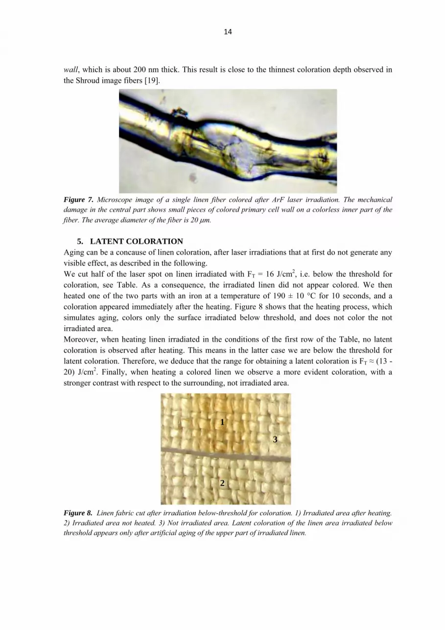
wall, which is about 200 nm thick. This result is close to the thinnest coloration depth observed in the Shroud image fibers [19]. Figure 7. Microscope image of a single linen fiber colored after ArF laser irradiation. The mechanical damage in the central part shows small pieces of colored primary cell wall on a colorless inner part of the fiber. The average diameter of the fiber is 20 μm.
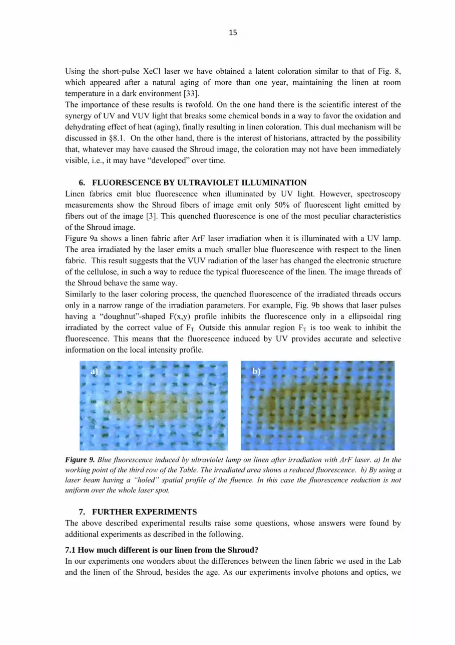
5. LATENT COLORATION Aging can be a concause of linen coloration, after laser irradiations that at first do not generate any visible effect, as described in the following. We cut half of the laser spot on linen irradiated with FT = 16 J/cm2, i.e. below the threshold for coloration, see Table. As a consequence, the irradiated linen did not appear colored. We then heated one of the two parts with an iron at a temperature of 190 ± 10 °C for 10 seconds, and a coloration appeared immediately after the heating. Figure 8 shows that the heating process, which simulates aging, colors only the surface irradiated below threshold, and does not color the not irradiated area. Moreover, when heating linen irradiated in the conditions of the first row of the Table, no latent coloration is observed after heating. This means in the latter case we are below the threshold for latent coloration. Therefore, we deduce that the range for obtaining a latent coloration is FT ≈ (13 - 20) J/cm2. Finally, when heating a colored linen we observe a more evident coloration, with a stronger contrast with respect to the surrounding, not irradiated area. Figure 8. Linen fabric cut after irradiation below-threshold for coloration. 1) Irradiated area after heating. 2) Irradiated area not heated. 3) Not irradiated area. Latent coloration of the linen area irradiated below threshold appears only after artificial aging of the upper part of irradiated linen.
1
2
3
15
Using the short-pulse XeCl laser we have obtained a latent coloration similar to that of Fig. 8, which appeared after a natural aging of more than one year, maintaining the linen at room temperature in a dark environment [33]. The importance of these results is twofold. On the one hand there is the scientific interest of the synergy of UV and VUV light that breaks some chemical bonds in a way to favor the oxidation and dehydrating effect of heat (aging), finally resulting in linen coloration. This dual mechanism will be discussed in §8.1. On the other hand, there is the interest of historians, attracted by the possibility that, whatever may have caused the Shroud image, the coloration may not have been immediately visible, i.e., it may have “developed” over time.
6. FLUORESCENCE BY ULTRAVIOLET ILLUMINATION Linen fabrics emit blue fluorescence when illuminated by UV light. However, spectroscopy measurements show the Shroud fibers of image emit only 50% of fluorescent light emitted by fibers out of the image [3]. This quenched fluorescence is one of the most peculiar characteristics of the Shroud image. Figure 9a shows a linen fabric after ArF laser irradiation when it is illuminated with a UV lamp. The area irradiated by the laser emits a much smaller blue fluorescence with respect to the linen fabric. This result suggests that the VUV radiation of the laser has changed the electronic structure of the cellulose, in such a way to reduce the typical fluorescence of the linen. The image threads of the Shroud behave the same way. Similarly to the laser coloring process, the quenched fluorescence of the irradiated threads occurs only in a narrow range of the irradiation parameters. For example, Fig. 9b shows that laser pulses having a “doughnut”-shaped F(x,y) profile inhibits the fluorescence only in a ellipsoidal ring irradiated by the correct value of FT. Outside this annular region FT is too weak to inhibit the fluorescence. This means that the fluorescence induced by UV provides accurate and selective information on the local intensity profile. Figure 9. Blue fluorescence induced by ultraviolet lamp on linen after irradiation with ArF laser. a) In the working point of the third row of the Table. The irradiated area shows a reduced fluorescence. b) By using a laser beam having a “holed” spatial profile of the fluence. In this case the fluorescence reduction is not uniform over the whole laser spot.
7. FURTHER EXPERIMENTS The above described experimental results raise some questions, whose answers were found by additional experiments as described in the following.
7.1 How much different is our linen from the Shroud? In our experiments one wonders about the differences between the linen fabric we used in the Lab and the linen of the Shroud, besides the age. As our experiments involve photons and optics, we
a) b)

16
measured some additional optical characteristics of our linen to be compared with the linen of the Shroud. We used a spectrophotometer Perkin-Elmer Lambda 950TM, equipped with a 15-cm-diameter integrating sphere [39]. The interior of the sphere is covered with a plastic material known as Spectralon, whose characteristics of reflection are almost 100% Lambertian and constant over the whole spectrum UV-visible-near infrared. Additionally, this instrument has an internal calibration of the Spectralon, which allows to directly obtain absolute reflectance spectra [40]. We measured the hemispherical absolute spectral reflectance R(λ) (i.e. the percentage of light reflected by our linen with respect to the incident light) and the results are shown in Fig. 10, together with the results of spectral reflectance measured on the Shroud as reported in [3]. Figure 10. The solid lines show the absolute reflectance of the linen of the Shroud in areas of no-image as a function of the wavelength [3]. The dashed line shows the absolute hemispherical reflectance of the linen used in our experiments. Figure 10 shows that the reflectance spectrum of our linen is similar to that of the Shroud. We note a small difference in the spectral region between 520 nm and 600 nm, showing our linen is less yellowish than the Shroud, possibly because of the different age. Most important, the absolute reflectance of the two linens at the laser wavelengths we used, 193 nm and 308 nm, is almost the same. Thus, from the optical point of view, when irradiated in the UV and VUV our linen behaves like the linen of the Shroud. The unexpected similarity between the STURP results and our results obtained using a much more advanced spectrophotometer than that available in 1978, highlights the extreme care of the STURP scientists when making measurements in situ on the Shroud. Using the same spectrophotometer we measured the hemispherical transmittance T(λ) of the linen (i.e. the percentage of light transmitted by our linen with respect to the incident light) as a function of wavelength. Then, we can deduce the spectral absorbance A(λ) as follows:
A(λ) = 1 - R(λ) - T(λ) (3)
that is, A(λ) is the amount of light absorbed by linen as a function of wavelength, and the results are shown in Fig. 11.

17
Figure 11. Plot of the absolute value of the absorbance of the linen vs. the wavelength, according with our experimental values inserted in eq. (3). 7.2 Can UV laser radiation make linens older? The process of dehydrative oxidation which produces the Shroud image can be considered a kind of premature aging of linen [8]. To check if the laser irradiation produces a similar aging effect we observed some linen fibers placed between two crossed polarizers in a petrographic microscope to detect changes induced by laser irradiation in the crystal structure of the fibers. The petrographic microscope allows to observe a pattern of isochromatic lines that depend on several parameters such as age of the sample, mechanical stress, presence of defects. When the fibers are aligned to the axis of polarization of the analyzer, we see a dark image: in this case the fibers are placed "to extinction", and there is no birefringence. If a part of the fiber aligned to extinction is damaged, it becomes birefringent and appears bright, because damaged regions have a different crystalline orientation than the fiber. Figure 12 shows a partially irradiated fiber of linen as observed in cross polarization to identify the stressed regions of the fiber. Figure 12. Petrographic microscope observation of a linen fibers. In the middle there is a partly colored fiber of linen in 1.515-index immersion oil between crossed polarizers to detect stress characteristics. The left part of the fiber is the non-irradiated region, which is enlarged in the inset below. On the right is the region irradiated by XeCl laser, enlarged in the inset above.

18
The non-irradiated region has the usual aspect of a recent linen. In contrast, the irradiated (colored) region of the fiber shows bright spots and tracks corresponding to stressed areas coupled with high-fragility zones. The dehydration of the fiber cellulose is in fact associated with an increment of the fiber fractures pointed out by birefringence at the location of the defects. A similar behavior is observed on very ancient linen fibers, like those used to wrap Egyptian mummies. We can therefore infer that short and high-intensity UV pulses change the crystalline structure of cellulose in a similar manner as aging and low-intensity radiation (radon, natural radioactivity, secondary particles from cosmic rays) accumulated in a long term period do. 7.3 The coloration induced by excimer laser irradiation is a photochemical or a thermal effect? In order to verify experimentally whether the UV and VUV light interacts with the linen by photochemical or thermal processes, we used the infrared camera ThermoShot F30 [http://www.nec-avio.co.jp/en/products/ir-thermo/pdf/catalog-f30-e.pdf] equipped with micro-bolometers sensitive in the spectral range 8 μm - 13 μm. This camera is able to measure the surface temperature of objects with the uncertainty of ± 0.2 °C. The camera was aligned in front of the linen during laser irradiation, monitoring in real time the temperature of the whole linen fabric as shown in Figures 13a and 13b. During laser irradiations the room temperature ranged between 20 and 21 °C, and the linen region irradiated by the UV XeCl laser was heated up to 33 °C, while the linen irradiated by the VUV ArF laser up to 25 °C. It is known that thermal effects can color the linen only when the linen temperature approaches 200 °C [11], and we can conclude that excimer laser coloration is a photochemical process that does not involve significant thermal effects. Figure 13a. Left: photo of the linen during XeCl laser irradiation. On the right, the same picture seen in infrared light. The color scale at the bottom shows that the warmest region of the linen (in the middle of the laser spot) reaches 33 °C, while the non-irradiated area is at the room temperature of 20 °C. Figure 13b. Left: photo of the linen during ArF laser irradiation. On the right, the same frame seen in infrared light. The color scale reveals that the warmest area of the linen (in the middle of the laser spot) is at 25 °C, while the non-irradiated area is at the room temperature of 21 °C.

19
8. ANALYSIS OF RESULTS We obtained a linen coloration that approaches some of the characteristics of the image on the Shroud. We also achieved a latent coloration that appears in a relatively long period (one year) or at once by an accelerated aging, following a laser irradiation that at first does not generate a visible image. We showed that the UV laser light produces a fragility and a stress of the irradiated fibers equivalent to an accelerated aging of the fabric. Finally, we have shown that the coloration is not due to a thermal effect, in analogy with the features of the Shroud image. Obviously, no one can hypothesize that the body image of the Shroud was produced by a burst of flashes of light emitted from an excimer laser. Rather, our results show that the laser is a powerful tool to simulate the physical and chemical processes that might have caused the peculiar coloration of the Shroud image. In order to gain a deeper insight into these processes, we need to detail some chemical and physical properties of linen fabrics and of the excimer lasers. 8.1 Chemical processes Each thread of linen consists of about 200 fibers, rod-like structures with an average length of 30 mm and average diameter of 20 μm. Each linen fiber has an inner part (the secondary cell wall) of cellulose, and a thin (0.2 μm) outer skin (the primary cell wall) composed of hemicellulose (predominantly xyloglucan) bound with pectin to cellulose [22]. Let’s recall that hemicellulose is a polysaccharide similar to cellulose, but it consists of shorter chains (500-3,000 sugar units) as opposed to 7,000-15,000 glucose molecules per polymer seen in cellulose. While cellulose is crystalline and strong, hemicelluloses has an amorphous structure with little strength. To some extent, hemicellulose can be considered a degraded form of cellulose. The STURP results show the image chromophore is a conjugated carbonyl produced in the polysaccharide structure of fibers by a dehydrative oxidation process [8, 11, 12]. The color of the Shroud image is a result of an accelerated aging process of the linen, similar to the yellowing of ancient papers [41]. Figure 14 shows two different chemical paths possibly involved in the formation of the image on the Shroud. Figure 14. a) Main molecular structure common to both cellulose and hemicellulose. There are two possible transitions (a) → (b) → (c) and (a) → (d) → (e) that generate chromophores after oxidation and dehydration. The C=C and C=O double bonds in (c) and (e) act as the chromophore and are responsible for the yellow color of the fibers of image on the Shroud of Turin. The different thickness of coloration obtained with the XeCl and ArF lasers (see Fig. 6 and §3, §4) may be due to the different λ respectively emitted. In fact, the shorter the wavelength, the larger the energy absorbed per unit volume. However, Fig. 11 shows there is only a difference of 11%
20
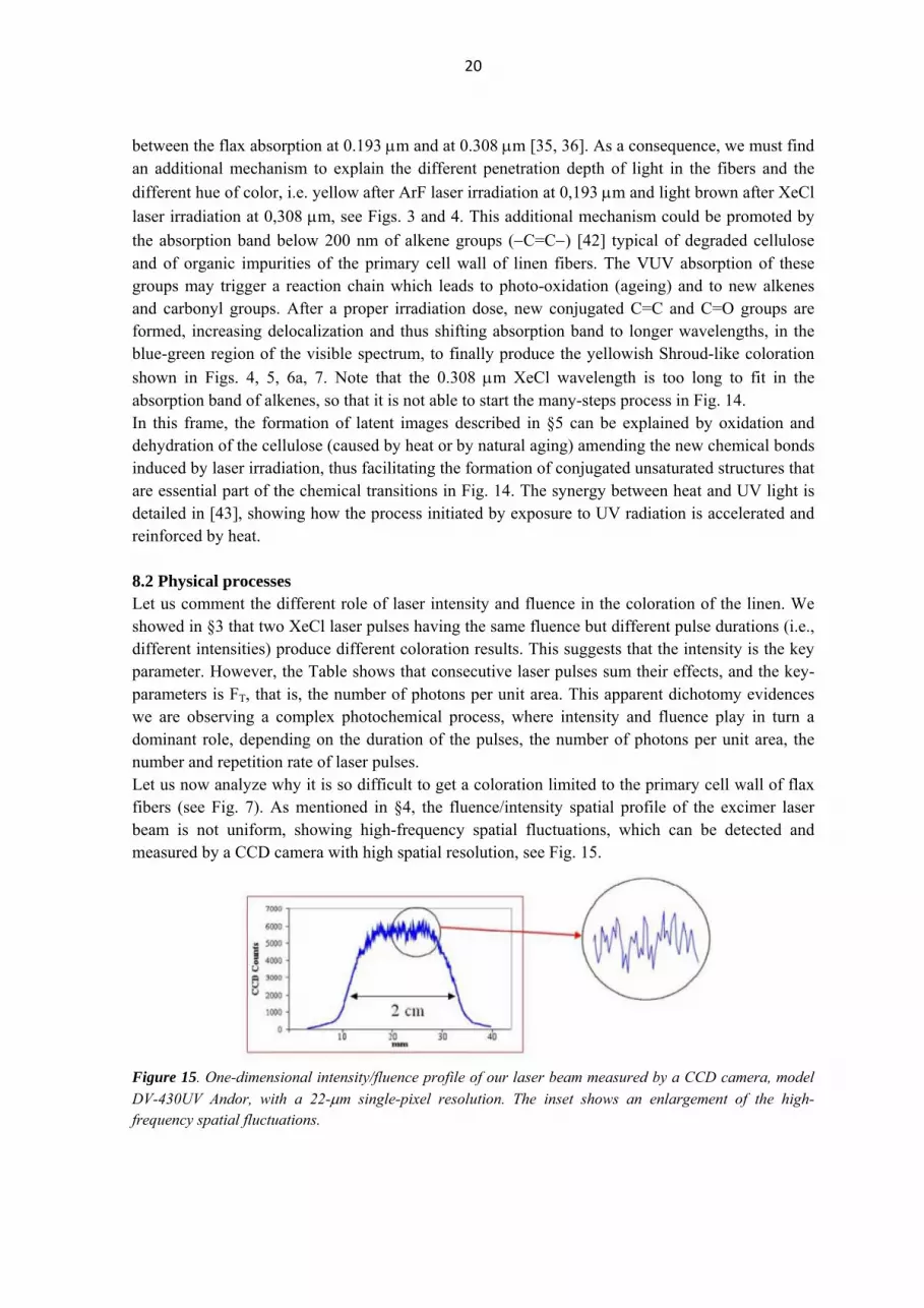
between the flax absorption at 0.193 μm and at 0.308 μm [35, 36]. As a consequence, we must find an additional mechanism to explain the different penetration depth of light in the fibers and the different hue of color, i.e. yellow after ArF laser irradiation at 0,193 μm and light brown after XeCl laser irradiation at 0,308 μm, see Figs. 3 and 4. This additional mechanism could be promoted by the absorption band below 200 nm of alkene groups (−C=C−) [42] typical of degraded cellulose and of organic impurities of the primary cell wall of linen fibers. The VUV absorption of these groups may trigger a reaction chain which leads to photo-oxidation (ageing) and to new alkenes and carbonyl groups. After a proper irradiation dose, new conjugated C=C and C=O groups are formed, increasing delocalization and thus shifting absorption band to longer wavelengths, in the blue-green region of the visible spectrum, to finally produce the yellowish Shroud-like coloration shown in Figs. 4, 5, 6a, 7. Note that the 0.308 μm XeCl wavelength is too long to fit in the absorption band of alkenes, so that it is not able to start the many-steps process in Fig. 14. In this frame, the formation of latent images described in §5 can be explained by oxidation and dehydration of the cellulose (caused by heat or by natural aging) amending the new chemical bonds induced by laser irradiation, thus facilitating the formation of conjugated unsaturated structures that are essential part of the chemical transitions in Fig. 14. The synergy between heat and UV light is detailed in [43], showing how the process initiated by exposure to UV radiation is accelerated and reinforced by heat. 8.2 Physical processes Let us comment the different role of laser intensity and fluence in the coloration of the linen. We showed in §3 that two XeCl laser pulses having the same fluence but different pulse durations (i.e., different intensities) produce different coloration results. This suggests that the intensity is the key parameter. However, the Table shows that consecutive laser pulses sum their effects, and the key-parameters is FT, that is, the number of photons per unit area. This apparent dichotomy evidences we are observing a complex photochemical process, where intensity and fluence play in turn a dominant role, depending on the duration of the pulses, the number of photons per unit area, the number and repetition rate of laser pulses. Let us now analyze why it is so difficult to get a coloration limited to the primary cell wall of flax fibers (see Fig. 7). As mentioned in §4, the fluence/intensity spatial profile of the excimer laser beam is not uniform, showing high-frequency spatial fluctuations, which can be detected and measured by a CCD camera with high spatial resolution, see Fig. 15. Figure 15. One-dimensional intensity/fluence profile of our laser beam measured by a CCD camera, model DV-430UV Andor, with a 22-μm single-pixel resolution. The inset shows an enlargement of the high-frequency spatial fluctuations.

21
The fluctuations in Fig. 15 have an irregular period, with gradients of intensity/fluence up to 350 MW/cm2 per centimeter (4 J/cm2 per centimeter). The value of laser intensity (fluence) incident on two points of the linen at, say, one millimeter distance can vary up to 35 MW/cm2 (0.4 J/cm2). This huge value of the intensity/fluence gradient can explain why it is possible to get the narrow "right" value of intensity for sub-micrometer coloration only in a very limited area, which is difficult to be found by photomicrographs. As mentioned in §1, point f), photomicrographs show that shading of the Shroud image is accomplished by varying the number of colored fibers per unit area and not by varying the color [5, 12], the so called “half tone effect”. In addition, the image area has a discontinuous distribution of color along the threads of the Shroud [9]. Some of these features can be found in our irradiated linens, see e.g., Fig. 16 which shows colored fibers next to uncolored ones in the same thread. However, we never fully achieved a “half tone effect” comparable with that observed on the Shroud. In principle, it would be possible to replicate exactly this characteristic by laser pulses having a spatial intensity distribution "sawtooth"-like with variable period. This distribution can be achieved by state-of-the-art diffractive optics which allow to arbitrarily modulate the spatial distribution of laser beams [44]. Figure 16. Microphotograph of linen threads after ArF laser irradiation. Single colored fibers are visible next to uncolored fibers, like in the Shroud image.
9. SUMMARY AND REMARKS In this paper we summarized the current state of knowledge on the Shroud image, and explained the reasons of the difficulty to create an image that matches its peculiar superficiality and chemistry at the microscopic level. After countless attempts, the inability to replicate the image on the Shroud prevents formulating a reliable hypothesis on the process of the image formation. Due to these scientific and technological difficulties, the hypothesis of a medieval forger does not seem reasonable. We then summarized the studies and experiments done at the ENEA Research Center of Frascati, which have demonstrated the ability of VUV light pulses lasting few nanoseconds to generate a Shroud-like coloration on linen that matches many (although not all) characteristics of the Shroud image. By the way, the ability of VUV light to generate a Shroud-like coloration helps to clarify the controversy between two scientists of the STURP team: John Jackson, who foresaw the possibility of coloring flax by VUV radiation [15] and Ray Rogers who believed that laser pulses would have

22
heated and vaporized flax, without any coloration effect [45]. Rogers's opinion was based on the failure of experiments made at Los Alamos using excimer lasers, but our results demonstrate that their failure was due to laser parameters (e.g., pulsewidth) outside the narrow range of values able to generate linen coloration. Let us summarize in the following the main results we achieved.
I. We obtained a linen coloration only in a narrow range of laser parameters. In particular, the temporal duration of the single laser pulse must be shorter than 50 ns [32, 33].
II. The most interesting results were obtained with VUV light. The permanent linen coloration is a threshold effect, i.e. the color is obtained only when FT > 22 J/cm2, see Table. When FT > 60 J/cm2 the linen is ablated and/or vaporized, while when FT < 13 J/cm2 the linen does not change color. Even when FT is in the coloration range, not all the irradiated fibers are colored (Figs. 5 and 16) due to the spatial fluctuations of energy density of the laser pulses shown in Fig. 15.
III. We triggered a photochemical coloration process. In fact, the thermal heating associated with UV and VUV radiation is within a few degrees centigrade and therefore irrelevant for the purpose of coloring by scorching linens, see Fig. 13. This result fits with the “cold” coloration process of the Shroud estimated in [8, 11, 12].
IV. The hue of color mainly depends on the wavelength of the radiation and on the number of pulses incident on linen, which is proportional to FT. Irradiations at 0.308 μm generate a brownish coloration, while the 0.193 μm photons produce a yellow color, see Fig. 4, similar to the color of the Shroud image. In both cases, the contrast slowly increases with the number of laser pulses, allowing an accurate control of the RGB value by varying FT.
V. The different hue of color obtained by UV and VUV radiation is due to different chains of photochemical reactions respectively triggered. In particular, the VUV radiation at 0,193 μm is absorbed by alkene groups in degraded cellulose, whose number increases with FT, thus inducing a photolysis of the cellulose which promotes the formation of chromophores, see Fig. 14. These chromophores determine the yellow coloration of the fibers [8, 12, 41, 42].
VI. We observed an irradiated fiber whose coloration was confined in the primary cell wall [36, 37], which is comparable with the thinnest coloration depth observed in the fibers image of the Shroud of Turin [8, 11, 19].
VII. After laser irradiations that do not produce a visible coloration of linen, a latent coloration appears either by artificial (Fig. 8) or natural ageing of linen [33, 36]. Latent coloration is interesting on the one hand for the synergy between UV, oxidation and the dehydrating effect of heat (or of aging) which triggers the coloration process, and on the other hand for historians, attracted by the possibility that the Shroud image may have developed over time (years) from the moment the process of latent coloration acted.
VIII. The partial inhibition of fluorescence induced by VUV laser radiation (Fig. 9) is an additional feature of our coloration similar to the Shroud image. The induced fluorescence is also capable to selectively recognize the uniformity of FT incident on linen, cf. Figs. 9a and 9b.
IX. Both UV and VUV light coloring linen is compatible with the absence of image under the bloodstains on the Shroud, because in this spectral region light is absorbed by very thin layers of blood hemoglobin. According to [46] the UV light may be responsible for another very special feature of the Shroud, the red color of blood stains after so much time since their deposition.

23
X. Using a petrographic microscope, we have observed some defects induced by UV radiation in the structure of irradiated linen fibers, see Fig. 12, similarly to very old linen fabrics, including the image fibers of the Shroud [11, 47].
XI. A highly unconventional hypothesis about the origin of the Shroud image was proposed in [15], which assumes a corpse emitting electromagnetic radiation. Although this hypothesis is out the realm of Science, we note VUV light is compatible with both the shading correlation with cloth-body distance and the absence of side images expected from it. This is because VUV photons are strongly absorbed by air, so the greater the distance in air the VUV light must travel, the lower the percentage of light impinging on linen, and less likely the intensity of the residual light is above threshold to color the linen (see item II above). Since the sides of the body are more distant from the cloth than frontal and dorsal area, it is obvious that the alleged light emitted from the body’s sides would have had a low probability to color the linen.
XII. Absolute reflectance measurements show that when irradiated in the UV and VUV, our linen behaves like the linen of the Shroud.
In summary, our results demonstrate that a short and intense burst of directional VUV radiation can color a linen cloth so as to reproduce many of the peculiar characteristics of the image on the Shroud of Turin, including the hue of color, the shallow penetration depth of the color, the inhibition of fluorescence. The work summarized in this paper shows that the excimer laser is a tool suitable to study in detail the physical and chemical processes that might have played a role in the generation of the Shroud body image, regardless of the source of radiation (or energy) that may have caused this image. The Shroud image has characteristics that we have been able to reproduce only in part, for example the gross shading structure that is determined by the ratio of yellow to uncolored fibers in a given area, see point f) in §1 and Fig. 16. As discussed in §8.1, there are sophisticated diffractive optics that allow replicating these features, but this effort is far beyond our goal. In fact, our purpose was not to demonstrate that a battery of ten thousand lasers can accurately reproduce the image on the Shroud. Our main purpose was to perform accurate, controlled and reproducible experiments, apt to understand the details of the physical and chemical mechanisms that have produced the Shroud image, thanks to a powerful and versatile tool such as the laser. In this frame, our experimental data can be helpful to scholars seeking a linen coloration with experiments such as corona discharge [18] or electrostatic discharge and radon emitted during seismic events [48] which involve UV and VUV light but are difficult to control and characterize.
We are not the conclusion, we are composing pieces of a fascinating and complex scientific puzzle. The enigma of the body image of the Shroud of Turin is still "a challenge to our intelligence" [49]. ACKNOWLEDGEMENTS The Authors thank Prof. Giulio Fanti (University of Padua) for his support for the microphotographs of fibers, for photos of Fig. 13 and for useful discussions of the manuscript contents. One of the Authors (PDL) gratefully acknowledges Prof. John Jackson (University of Colorado) for useful discussion about the role of fluence and intensity on linen irradiations.

24
REFERENCES [1] G. Fanti, J.A. Botella, F. Crosilla, F. Lattarulo, N. Svensson, R. Schneider, A. Wanger: “List of evidences of the Turin Shroud” Proc. IWSAI, P. Di Lazzaro ed. (ENEA 2010) pp. 67-75. ISBN 978-88-8286-232-9 www.acheiropoietos.info/proceedings/proceedings.php
[2] B.J. Culliton: “The mystery of the Shroud challenges 20th-century science” Science 201, 235-239 (1978).
[3] R. Gilbert, M. Gilbert: “Ultraviolet visible reflectance and fluorescence spectra of the Shroud of Turin” Appl. Opt. 19, 1930-1936 (1980).
[4] E.J. Jumper, W. Mottern: “Scientific Investigation of the Shroud of Turin” Appl. Opt. 19, 1909-1912 (1980).
[5] S.F. Pellicori: “Spectral properties of the Shroud of Turin” Appl. Opt. 19, 1913-1920 (1980).
[6] J.S. Accetta, J.S. Baumgart: “Infrared reflectance spectroscopy and thermographic investigations of the Shroud of Turin” Appl. Opt. 19, 1921-1929 (1980).
[7] R.A. Morris, L.A. Schwalbe, J.R. London: “X-ray fluorescence investigation on the Shroud of Turin” X-Ray Spectrometry 9, 40-47 (1980).
[8] J.H. Heller, A.D. Adler: “A chemical investigation of the Shroud of Turin” Can. Soc. Forens. Sci. J. 14, 81-103 (1981).
[9] S.F. Pellicori and M.S. Evans: “The Shroud of Turin through the microscope” Archaeology 34, 34-43 (1981).
[10] V.D. Miller, S.F. Pellicori, "Ultraviolet Fluorescence Photography of the Shroud of Turin" J. of Biol. Phot. 49, 71-85 (1981).
[11] L.A. Schwalbe, R.N. Rogers: “Physics and chemistry of the Shroud of Turin, a summary of the 1978 investigation” Analytica Chimica Acta 135, 3-49 (1982).
[12] E.J. Jumper, A.D. Adler, J.P. Jackson, S.F. Pellicori, J.H. Heller, and J.R. Druzik, “A comprehensive examination of the various stains and images on the Shroud of Turin”, Archaeological Chemistry III: ACS Advances in Chemistry 205 (American Chemical Society, Washington, 1984), pp. 447-476.
[13] W.C. Mc Crone, C. Skirius: “Light microscopical study of the Shroud of Turin” Microscope 28, 105-111 (1980).
[14] J.P. Jackson, E.J. Jumper, W.R. Ercoline: “Correlation of image intensity on the Turin Shroud with the 3-D structure of a human body shape” Appl. Opt. 23, 2244-2270 (1984).
[15] J.P. Jackson: “Is the image on the Shroud due to a process heretofore unknown to modern science?” Shroud Spectrum International 34, 3-29 (1990).
[16] T. Heimburger: “A detailed critical review of the chemical studies on the Turin Shroud: facts and interpretations” www.shroud.com/pdfs/thibault%20final%2001.pdf (2001).
[17] I. Piczek: “Is the Shroud of Turin a painting?” www.shroud.com/piczek.htm (1995).
[18] G. Fanti, “Can corona discharge explain the body image of the Turin Shroud?” J. Imaging Sci. Technol. 54 020508-020508-11 (2010).

25
[19] G. Fanti, J. Botella, P. Di Lazzaro, R. Schneider, N. Svensson: “Microscopic and macroscopic characteristics of the Shroud of Turin image superficiality” J. Imaging Sci. Technol. 54, 040201-040201(8) (2010).
[20] L. Garlaschelli: “Life-size Reproduction of the Shroud of Turin and its Image” J. Imaging Sci. Technol. 54, 040301-040301(14) (2010). See also: T. Heimburger, G. Fanti: “A scientific comparison between the Turin Shroud and the first handmade whole copy” Proc. IWSAI, P. Di Lazzaro ed. (ENEA 2010) pp. 19-28. ISBN 978-88-8286-232-9. www.acheiropoietos.info/proceedings/proceedings.php
[21] F. Ferrero, F. Testore, C. Tonin, R. Innocenti: “Surface degradation of linen textiles induced by laser treatment” AUTEX Research Journal 2, 109-114 (2002).
[22] S. Perez, K. Mazeau: Chapter 2 of Polysaccharides: structural diversity and functional versatility, S. Dumitriu Editor (M. Dekker Inc. 2004).
[23] J.H Heller, A.D. Adler: "Blood on the Shroud of Turin" Applied Optics 19, 2742-2744 (1980).
[24] P.L. Baima Bollone.: "Indagini identificative su fili della Sindone", Giornale della Accademia di Medicina di Torino, n. 1-12, 228-239 (1982).
[25] P.E. Damon, et al.: “Radiocarbon dating of the Shroud of Turin” Nature 337, 611-615 (1989).
[26] R. Van Haelst: “A critical review of the radiocarbon dating of the Shroud of Turin” Proc. IWSAI, P. Di Lazzaro ed. (ENEA 2010) pp. 267-273. ISBN 978-88-8286-232-9. www.acheiropoietos.info/proceedings/proceedings.php
[27] R.N. Rogers: “Studies on the radiocarbon sample from the Shroud of Turin” Thermochimica Acta 425, 189-194 (2005).
[28] D. Scavone: “Documenting the Shroud’s missing years” Proc. Int. Workshop on the Scientific approach to the Acheiropoietos Images, IWSAI, P. Di Lazzaro ed. (ENEA 2010) pp. 87-94. ISBN 978-88-8286-232-9. www.acheiropoietos.info/proceedings/proceedings.php
[29] A. Piana: “Missing years of the Holy Shroud” Proc. IWSAI, P. Di Lazzaro ed. (ENEA 2010) pp. 95-102. www.acheiropoietos.info/proceedings/proceedings.php
[30] A. Nicolotti: “I Templari e la Sindone. Storia di un falso” (Salerno ed., Roma 2011) Chapters. 1, 3 and 4.
[31] N. Svensson: “Medical and forensic aspects on the man depicted on the Shroud of Turin” Proc. IWSAI, P. Di Lazzaro ed. (ENEA 2010) pp. 181-186. ISBN 978-88-8286-232-9. www.acheiropoietos.info/proceedings/proceedings.php
[32] G. Baldacchini, P. Di Lazzaro, D. Murra, G. Fanti: “Colorazione di tessuti di lino con laser ad eccimeri e confronto con l’immagine sindonica” Technical Report ENEA RT/2006/70/FIM (2006).
[33] G. Baldacchini, P. Di Lazzaro, D. Murra, G. Fanti: “Coloring linens with excimer lasers to simulate the body image of the Turin Shroud” Appl. Opt. 47, 1278-1285 (2008).
[34] P. Di Lazzaro, G. Baldacchini, G. Fanti, D. Murra, A. Santoni: “Colouring fabrics with excimer lasers to simulate encoded images: the case of the Shroud of Turin” Proc. SPIE vol. 7131 (2009) pp. 71311R-1–71311R-6.

26
[35] P. Di Lazzaro, G. Baldacchini, G. Fanti, D. Murra, E. Nichelatti, A. Santoni: “A physical hypothesis on the origin of the body image embedded into the Turin Shroud” Proc. Int. Conf. on The Shroud of Turin: Perspectives on a Multifaceted Enigma, G. Fanti ed. (Libreria Progetto Padova 2009) pp. 116-125. www.ohioshroudconference.com/papers/p01.pdf
[36] P. Di Lazzaro, D. Murra, A. Santoni, G. Fanti, E. Nichelatti, G. Baldacchini: “Deep Ultraviolet radiation simulates the Turin Shroud image” J. of Imaging Sci. Technol. 54, 040302-040302(06) (2010).
[37] P. Di Lazzaro, D. Murra, A. Santoni, G. Baldacchini: “Sub-micrometer coloration depth of linens by vacuum ultraviolet radiation”, Proc. IWSAI, P. Di Lazzaro ed. (ENEA, 2010) pp. 3-10. ISBN 978-88-8286-232-9. www.acheiropoietos.info/proceedings/proceedings.php
[38] P. Di Lazzaro, D. Murra, A. Santoni, G. Baldacchini: “Colorazione simil-sindonica di tessuti di lino tramite radiazione nel lontano ultravioletto” Technical Report ENEA RT/2011/14/ENEA (2011). http://opac.bologna.enea.it:8991/RT/2011/2011_14_ENEA.pdf
[39] www.perkinelmer.com/CMSResources/Images/44-74789SPC_LAMBDA1050LAMBDA950.pdf
[40] E. Nichelatti: “Caratterizzazione spettrofotometrica di due tessuti di lino” Internal Report (April 2008). Unpublished.
[41] A. Mosca Conte, O. Pulci, A. Knapik, J. Bagniuk, R. Del Sole, J. Lojewska, M. Missori: “Role of Cellulose Oxidation in the Yellowing of Ancient Paper” Phys. Rev. Lett. 108, 158301-5 (2012).
[42] A. Bos: “The UV spectra of cellulose and some model compounds” J. Appl. Polymer Science 16, 2567-2576 (1972).
[43] M. Yatagai, S.H. Zeronian: “Effect of ultraviolet light and heat on the properties of cotton cellulose” Cellulose 1, 205-214 (1994).
[44] See, e.g., F. Gori: “Diffractive optics, an introduction” in Diffractive optics and optical microsystems, S. Martellucci and A.N. Chester eds. (Plenum Press NY 1997) pp. 3-23. ISBN 0-306-45770-9.
[45] R.N. Rogers: “Testing the Jackson "theory" of image formation” www.shroud.com/pdfs/rogers6.pdf (2004).
[46] C. Goldoni: “The Shroud of Turin and the bilirubin blood stains” Proc. Int. Conf. on The Shroud of Turin: Perspectives on a Multifaceted Enigma, G. Fanti ed. (Libreria Progetto Padova 2009) pp. 442-445. www.ohioshroudconference.com/papers/p04.pdf
[47] R.N. Rogers: “The Shroud of Turin: radiation effects, aging and image formation” http://www.shroud.com/pdfs/rogers8.pdf (2005 b).
[48] G. De Liso: “Shroud-like experimental image formation during seismic activity”, Proc. IWSAI, P. Di Lazzaro ed. (ENEA 2010) pp. 11-18. ISBN 978-88-8286-232-9 www.acheiropoietos.info/proceedings/proceedings.php
[49] “The Shroud is a challenge to our intelligence” Address of Pope John Paul II during the visit to Turin (May 24, 1998).

Edito dall’
Servizio Comunicazione
Lungotevere Thaon di Revel, 76 - 00196 Roma
www.enea.it
Stampa: Tecnografico ENEA - CR Frascati
Pervenuto il 8.8.2012Finito di stampare nel mese di settembre 2012