Embed Size (px)
Citation preview

Review
Signal perception and transduction: the role of protein kinases
Paul W. Schenk 1, B. Ewa Snaar-Jagalska *Section of Cell Biology, Institute of Molecular Plant Sciences, Leiden University, P.O. Box 9505, 2300 RA Leiden, Netherlands
Received 13 August 1998; received in revised form 9 December 1998; accepted 10 December 1998
Abstract
Cells can react to environmental changes by transduction of extracellular signals, to produce intracellular responses.Membrane-impermeable signal molecules are recognized by receptors, which are localized on the plasma membrane of thecell. Binding of a ligand can result in the stimulation of an intrinsic enzymatic activity of its receptor or the modulation of atransducing protein. The modulation of one or more intracellular transducing proteins can finally lead to the activation orinhibition of a so-called `effector protein'. In many instances, this also results in altered gene expression. Phosphorylation by
0167-4889 / 99 / $ ^ see front matter ß 1999 Elsevier Science B.V. All rights reserved.PII: S 0 1 6 7 - 4 8 8 9 ( 9 8 ) 0 0 1 7 8 - 5
Abbreviations: AC, adenylyl cyclase; ANP, atrial natriuretic peptide; aPKC, atypical PKC; cAK, cAMP-dependent protein kinase;CaMK, Ca2�/calmodulin-dependent kinase; cAMP, cyclic AMP; CDK, cyclin-dependent kinase; cGK, cGMP-dependent protein kinase;cGMP, cyclic GMP; CK1, casein kinase I; CK2, casein kinase II; Clk, Cdc-like kinase; cPKC, classical or conventional PKC; CREB,cAMP responsive element binding protein; Csk, C-terminal Src kinase; DAG, diacylglycerol; EGF, epidermal growth factor; EGFR,EGF receptor; ERK, extracellular signal regulated kinase; FAK, focal adhesion kinase; GKi, inhibitory GK subunit; GAIP, G alphainteracting protein; GAP, GTPase activating protein; GKs, stimulatory GK subunit ; GC, guanylyl cyclase; GEF, G-nucleotide exchangefactor; GNRP, G-nucleotide releasing protein; GPCR, G-protein-coupled receptor; G-protein, guanine-nucleotide binding protein; Grb2,growth factor receptor-bound protein 2; GSK-3, glycogen synthase kinase-3; His K, histidine kinase; Hog1p, high osmolarity glycerolresponse 1 protein; Ins, insulin; IP3, inositol 1,4,5-trisphosphate; IRS, insulin receptor substrate; JAK, Janus kinase; JNK, cJun N-terminal kinase; KHD, protein kinase homology domain; Ksr-1; kinase suppressor of Ras 1; LIM domain, lin-11/ISL1/mec-3 homologydomain; LIMK, LIM kinase; LPA, lysophosphatidic acid; MAPK, mitogen-activated protein kinase; MAPKAP2, MAPK activatedprotein kinase 2; MAPKK, MAPK kinase; MAPKKK, MAPKK kinase; MEK, MAPK/ERK kinase; MEKK, MEK kinase; MKK,MAPK kinase; MKP-1, MAPK-speci¢c phosphatase 1; MTK1, MAP three kinase 1; Myr modi¢cation, myristoyl modi¢cation; nPKC,non-classical or novel PKC; PAK, p21-activated kinase; PDGF, platelet-derived growth factor; PDK1, 3-phosphoinositide-dependentprotein kinase 1; PDZ domain, PSD-95/Dlg/ZO-1 homology domain; PI3K, phosphatidylinositol 3-kinase; PIP2, phosphatidylinositol4,5-bisphosphate; PIP3, phosphatidylinositol 3,4,5-trisphosphate; PKA, cAMP-dependent protein kinase; PKB, protein kinase B; PKC,protein kinase C; PKG, cGMP-dependent protein kinase; PLC, phospholipase C; PMA, phorbol 12-myristate 13-acetate; PTK, proteintyrosine kinase; PY, phosphotyrosine; RACK, receptor for activated C-kinase (PKC); RBD, Ras binding domain; RGS, regulator of G-protein signaling; RICK, receptor for inactive C-kinase (PKC); RPTP, receptor-like protein tyrosine phosphatase; RSK, p90 ribosomalS6 kinase; RTK, receptor tyrosine kinase; SAPK, stress-activated protein kinase; SEK1, SAPK/ERK kinase 1, SH2, Src homologydomain 2; Shc, Src homology and collagen; S6K, p70 S6 protein kinase; Smad, Sma/Mad homolog; Sos, son of sevenless; STAT, signaltransducer and activator of transcription; TAK1, TGF-L activated kinase 1; TESK1, testis-speci¢c protein kinase 1; TGF-L, trans-forming growth factor-L ; TGFR, activin/TGF-L receptor; TM domain, transmembrane domain; TPA, 12-O-tetradecanoyl phorbol 13-acetate
* Corresponding author. Fax: +31 (71) 527-4999; E-mail : [email protected] Present address. Department of Medical Oncology, Josephine Nefkens Institute^Daniel den Hoed Cancer Center, University Hospital
Rotterdam, Dr. Molewaterplein 50, 3015 GE Rotterdam, The Netherlands.
BBAMCR 14438 1-2-99
Biochimica et Biophysica Acta 1449 (1999) 1^24

protein kinases is one of the most common and important regulatory mechanisms in signal transmission. This reviewdiscusses the non-channel transmembrane receptors and their downstream signaling, with special focus on the role of proteinkinases. ß 1999 Elsevier Science B.V. All rights reserved.
Keywords: Protein kinase; Signal perception; Signal transduction
Contents
1. Introduction . . . . . . . . . . . . . . . . . . . . . . . . . . . . . . . . . . . . . . . . . . . . . . . . . . . . . . . . . . 2
2. Receptors with enzymatic activity . . . . . . . . . . . . . . . . . . . . . . . . . . . . . . . . . . . . . . . . . . 32.1. Receptor tyrosine kinases . . . . . . . . . . . . . . . . . . . . . . . . . . . . . . . . . . . . . . . . . . . . . 32.2. Receptor-like protein tyrosine phosphatases . . . . . . . . . . . . . . . . . . . . . . . . . . . . . . . . 42.3. Receptor serine/threonine kinases . . . . . . . . . . . . . . . . . . . . . . . . . . . . . . . . . . . . . . . . 52.4. The two-component regulatory system: histidine kinases . . . . . . . . . . . . . . . . . . . . . . 52.5. Guanylyl cyclases . . . . . . . . . . . . . . . . . . . . . . . . . . . . . . . . . . . . . . . . . . . . . . . . . . . 6
3. Receptors without enzymatic activity . . . . . . . . . . . . . . . . . . . . . . . . . . . . . . . . . . . . . . . . 63.1. Cytokine receptors . . . . . . . . . . . . . . . . . . . . . . . . . . . . . . . . . . . . . . . . . . . . . . . . . . 63.2. Integrins . . . . . . . . . . . . . . . . . . . . . . . . . . . . . . . . . . . . . . . . . . . . . . . . . . . . . . . . . . 73.3. G-protein-coupled receptors . . . . . . . . . . . . . . . . . . . . . . . . . . . . . . . . . . . . . . . . . . . . 7
4. Protein kinases . . . . . . . . . . . . . . . . . . . . . . . . . . . . . . . . . . . . . . . . . . . . . . . . . . . . . . . . 94.1. Phosphorylation: a type of protein modi¢cations . . . . . . . . . . . . . . . . . . . . . . . . . . . . 94.2. Conserved protein kinase catalytic domains: some mechanistic aspects . . . . . . . . . . . . 94.3. The AGC group of protein kinases . . . . . . . . . . . . . . . . . . . . . . . . . . . . . . . . . . . . . . 114.4. The CaMK group of protein kinases . . . . . . . . . . . . . . . . . . . . . . . . . . . . . . . . . . . . . 114.5. The CMGC group of protein kinases . . . . . . . . . . . . . . . . . . . . . . . . . . . . . . . . . . . . 124.6. The conventional protein tyrosine kinase group . . . . . . . . . . . . . . . . . . . . . . . . . . . . . 124.7. The group of `other' protein kinases . . . . . . . . . . . . . . . . . . . . . . . . . . . . . . . . . . . . . 124.8. Protein kinase C . . . . . . . . . . . . . . . . . . . . . . . . . . . . . . . . . . . . . . . . . . . . . . . . . . . . 144.9. Mitogen-activated protein kinases . . . . . . . . . . . . . . . . . . . . . . . . . . . . . . . . . . . . . . . 16
5. Looking ahead . . . . . . . . . . . . . . . . . . . . . . . . . . . . . . . . . . . . . . . . . . . . . . . . . . . . . . . . 17
Acknowledgements . . . . . . . . . . . . . . . . . . . . . . . . . . . . . . . . . . . . . . . . . . . . . . . . . . . . . . . . . 18
References . . . . . . . . . . . . . . . . . . . . . . . . . . . . . . . . . . . . . . . . . . . . . . . . . . . . . . . . . . . . . . . 18
1. Introduction
Cells react to environmental changes, which theyperceive through extracellular signals. These signalscan be either physical (e.g. light, temperature, pres-sure and electricity) or chemical (e.g. food, hormonesand neurotransmitters). Cells can both sense andproduce signals. This makes it possible that theycommunicate with each other. In order to achievethis, there are complex signal-sensing and -producingmechanisms in uni- and multi-cellular organisms.
Two groups of chemical signals can be distin-guished: membrane-permeable and membrane-im-permeable signals. The membrane-permeable signalmolecules comprise the large family of steroid hor-mones, such as estrogens, progesterone and andro-gens. Steroids pass the plasma membrane and bindto speci¢c receptors, which are localized in the cyto-plasm or nucleus of the cell. After binding of thehormone, the receptor undergoes a conformationalchange. The receptor is then able to bind to DNAitself or to proteins which can in turn interact with
BBAMCR 14438 1-2-99
P.W. Schenk, B.E. Snaar-Jagalska / Biochimica et Biophysica Acta 1449 (1999) 1^242

DNA. In general, steroid hormones can directly reg-ulate gene expression by means of this process [1].The membrane-impermeable signal molecules includeacetylcholine, growth factors, extracellular matrixcomponents, thrombin, lysophosphatidic acid, theyeast mating factors and, for the social amoeba Dic-tyostelium discoideum, folic acid and cyclic AMP.They are recognized by receptors, which are localizedon the plasma membrane of the cell. The receptorsare speci¢c for one particular signal molecule or afamily of closely related signal molecules. Uponbinding of their ligands, these receptors transducethe signals by several mechanisms.
Binding of a ligand to an ion channel receptor candirectly lead to its altered opening, which results in achanged membrane potential. There are, for instance,acetylcholine-regulated cation channels in nerve ter-minals [2]. Alternatively, binding of a ligand mayresult in the stimulation of an intrinsic enzymaticactivity of its receptor or the modulation of a trans-ducing protein. The modulation of one or more in-tracellular transducing proteins can ¢nally lead to theactivation or inhibition of a so-called `e¡ector pro-tein'. Activity modulations can be achieved by cova-lent modi¢cations at the molecular level. Among themost common and important modi¢cations are pro-tein phosphorylation and dephosphorylation on ser-ine, threonine or tyrosine residues. Phosphorylationand dephosphorylation are carried out by kinasesand phosphatases, respectively. As described below,changes in the phosphorylation state of cellular com-ponents can alter their properties in several ways. Aninteresting yet complicating phenomenon is cross-talk: intracellular responses to extracellular signalswhich in£uence each other. While stimulation of acell with one speci¢c ligand may lead to more thanone response, di¡erent signals may lead to an anal-ogous response via identical components.
The non-channel transmembrane receptors andtheir downstream signaling will be discussed, withspecial focus on the role of protein kinases. Par-ticular attention will be drawn to protein kinase Cand mitogen-activated protein kinases. We willpresent current knowledge and attempt to placethis into the perspective of possible future develop-ments.
2. Receptors with enzymatic activity
2.1. Receptor tyrosine kinases
Extracellular signal molecules, like epidermalgrowth factor, platelet derived growth factor and in-sulin bind to receptor tyrosine kinases (RTKs). Thesereceptors possess one or two intracellular tyrosinekinase regions (Figs. 1A and 2B). Upon ligand bind-ing, RTKs auto-phosphorylate. The resulting phos-photyrosine (PY) residues act as highly selectivebinding sites for so-called `SH2' (Src homology do-main 2)-containing proteins, which transduce the sig-nal by changing their enzymatic activity or recruitingother proteins. Among these SH2-containing pro-teins are Ras-GTPase activating protein (GAP) andphospholipase C-Q. The latter hydrolyzes phospha-tidylinositol 4,5-bisphosphate (PIP2) into inositol1,4,5-trisphosphate (IP3) and diacylglycerol (DAG).IP3 releases Ca2� from intracellular stores; DAGactivates protein kinase C (PKC) (Fig. 1A) [3]. TheSH2-containing adapter molecules, Shc and Grb2(Src homology and collagen and growth factor re-ceptor-bound protein 2, respectively), also bind tophosphorylated RTKs. They are importantly in-volved in the recruitment of the son of sevenless(Sos) protein towards the plasma membrane [4].Sos, which is called a GEF (G-nucleotide exchangefactor) or GNRP (G-nucleotide releasing protein),stimulates the exchange of GDP for GTP on thesmall G-protein Ras, thereby activating it. Ras-GTP in turn stimulates a protein kinase cascade re-sulting in mitogen-activated protein kinase (MAPK,also called ERK, for extracellular signal regulatedkinase) activation (Fig. 1A) [5].
The p85 subunit of phosphatidylinositol 3-kinase(PI3K) is recruited to PY residues on insulin recep-tors by IRS (insulin receptor substrate) adapters. ThePI3K p85 subunit is linked to the p110 subunit,which is able to phosphorylate PIP2. This generatesphosphatidylinositol 3,4,5-trisphosphate (PIP3),which has several intracellular targets, such as PKCand protein kinase B (PKB)/Akt. The activation ofPKB/Akt requires phosphorylation by PDK1 (3-phosphoinositide-dependent protein kinase 1) andleads to serine phosphorylation and inactivation ofGSK-3 (glycogen synthase kinase-3) (Figs. 1A and2B) [6,7]. The C-terminus of the cytoplasmic PDZ
BBAMCR 14438 1-2-99
P.W. Schenk, B.E. Snaar-Jagalska / Biochimica et Biophysica Acta 1449 (1999) 1^24 3

domain protein, Enigma, interacts with Tyr-contain-ing motifs outside the tyrosine kinase cores of theRet RTK and the insulin receptor. Enigma does sovia two of its three so-called `LIM domains' (seebelow). The LIM2^Ret and LIM3^insulin receptorinteractions are required for mitogenic signaling
by Ret/ptc2 and receptor endocytosis, respectively[8,9].
2.2. Receptor-like protein tyrosine phosphatases
Receptor-like protein tyrosine phosphatases
BBAMCR 14438 1-2-99
P.W. Schenk, B.E. Snaar-Jagalska / Biochimica et Biophysica Acta 1449 (1999) 1^244

(RPTPs) contain a variable extracellular domain, atransmembrane domain and one or two intracellulartyrosine phosphatase domains (Fig. 1B). Dependingon the RPTP subtype, the extracellular domain ex-hibits immunoglobulin- and ¢bronectin-like regionsand other sequence motifs involved in cell^cell adhe-sion [10,11]. The subtype I RPTP CD45 is involvedin the activation of B- and T-lymphocytes. It stimu-lates the Src-like kinases Lck and Fyn by dephos-phorylating them [12]. The subtype II RPTPs Wand U have been shown to modulate cell^cell inter-action. There is some debate on the association ofthe cytoplasmic domain of RPTPW with a proteincomplex containing cadherin transmembrane adhe-sion molecules and catenins (which are linked tothe underlying actin cytoskeleton). It appears thatBrady-Kalnay et al. recently reinforced their originalobservation, that cadherins are part of such com-plexes, which has been doubted by other authors(Fig. 1B) [11,13]. The subtype IV RPTPK tightlybinds to Grb2 (Fig. 1B). Although the precise bio-logical function of the resulting complex remains tobe determined, it has already been suggested thatbinding of Grb2 may inhibit RPTPK activity [14].The establishment of the exact repercussions of lig-and binding to RPTPs is currently in progress.
2.3. Receptor serine/threonine kinases
The e¡ects of transforming growth factor-L (TGF-L) superfamily signal molecules are mediated by re-ceptors with a cysteine-rich extracellular domain anda cytoplasmic serine/threonine kinase activity (Figs.
1C and 2BC). In addition to the three TGF-L iso-forms, this superfamily comprises activins, bonemorphogenetic proteins and other secreted factors.Members of the TGF-L superfamily have been im-plicated as being crucial for many speci¢c develop-mental events in vertebrates, Drosophila melanogasterand Caenorhabditis elegans ; TGF-L induces growtharrest in epithelial cells. TGF-L-related factors signalthrough type I and type II receptor serine/threoninekinases, which form a heteromeric complex. Whereastype II receptors bind TGF-L1 independently, thisligand does not bind to the type I receptor in theabsence of the type II receptor. In the heteromericcomplex, the type II receptor phosphorylates the typeI receptor [15,16]. A type I^type II complex (but nottype II alone) can associate with the e¡ector Smad3(for Sma/Mad homolog 3; formerly called hMAD-3), which is phosphorylated in vitro by type I. Acomplex of Smad3 and its homolog Smad4 isthought to ¢nally function as a transcriptional regu-lator of downstream genes in the nucleus (Fig. 1C)[17]. The Smad2 protein has recently been reportedto act as a common positive e¡ector of both receptorserine/threonine kinases and RTKs; this providesone of the many examples of cross-talk during signaltransduction from the cell surface to the nucleus [18].
2.4. The two-component regulatory system: histidinekinases
Prokaryotic organisms commonly employ the two-component regulatory system; homologous path-ways have recently been identi¢ed in eukaryotes, in-
Fig. 1. Structures of cell surface receptors and schematic diagrams of their downstream signaling. In plasma membrane receptors, theN-terminus is generally extracellular, while the C-terminus is cytoplasmic. Main structural features are indicated. Open ovals, cysteine-rich regions; shaded boxes, protein kinase catalytic domains; semi-circles, immunoglobulin-like regions; horizontally striped boxes, ¢-bronectin-like regions. Double and triple arrows represent multiple signal transduction steps (activation via second messenger forma-tion or a cascade). (A) Receptor tyrosine kinases (RTKs). Structures of the EGF (epidermal growth factor), PDGF (platelet derivedgrowth factor) and Ins (insulin) receptor subclasses. The protein tyrosine kinase domain of the PDGF receptor contains an insertionsequence. (B) Receptor-like protein tyrosine phosphatases (RPTPs). Structures of RPTPW and RPTPK. Vertically striped box, speci¢ccell^cell interaction domain; solid boxes, protein tyrosine phosphatase catalytic domains. Some RPTPs contain a single phosphatasedomain. (C) Receptor serine/threonine kinases. Structure of the activin/TGF-L type II receptor (TGFR II). (D) Histidine kinases. TheSaccharomyces Sln1p His K (histidine kinase) contains an extracellular input region (double diagonally striped box) and an intracellu-lar transmitter module (diagonally striped box) and receiver domain (open box). (E) Transmembrane guanylyl cyclases (GCs). Dottedbox, protein kinase homology domain; black and white box, cyclase catalytic domain. (F) Cytokine receptors. Solid oval, membrane-proximal JAK binding box1/box2 motifs. (G) Integrins. Structures of L and K subunits. The extracellular ligand binding region ismade up from both subunits in K/L heterodimers. (H) G-protein-coupled receptors (GPCRs). The C-terminal intracellular domains ofGPCRs greatly vary in sequence and length. For further abbreviations, details and references, see main text.6
BBAMCR 14438 1-2-99
P.W. Schenk, B.E. Snaar-Jagalska / Biochimica et Biophysica Acta 1449 (1999) 1^24 5

cluding Saccharomyces cerevisiae, Arabidopsis thali-ana, Neurospora crassa and Dictyostelium discoideum.The prototypical two-component pathway consists oftwo proteins: a protein histidine kinase (also calledsensor kinase) and a response regulator. The N-ter-minal part of the histidine kinase functions as aninput domain, detecting extracellular signals directlyor via an upstream receptor. The C-terminal portioncontains the transmitter module. This domain in-cludes a histidine residue at which the protein auto-phosphorylates. The response regulator has a re-ceiver domain, which catalyzes the transfer of thephosphoryl group from the histidine of the sensorkinase to a conserved aspartate residue on the re-ceiver. The phosphorylation state of the regulatormodulates the activity of its output domain, whichis often involved in the regulation of transcription[19]. Hybrid histidine kinases combine an input re-gion, a transmitter module and a response regulatorin one molecule. Sensing domains can be integratedwithin the histidine kinase or contained within a sep-arate protein [20]. Histidine kinases are very distinctfrom the superfamily of conventional protein serine/threonine and tyrosine kinases.
There are complex signal transduction pathwaysbuilt from two-component circuit elements: one im-portant motif is the His^Asp^His^Asp phosphorelay.One of the most striking examples is the Sln1p^Ypd1p^Ssk1p pathway, which governs osmoregula-tion in the budding yeast S. cerevisiae. The trans-membrane protein Sln1p contains an extracellularinput region and cytoplasmic histidine kinase andreceiver domains (Fig. 1D); the cytoplasmic Ssk1pprotein has a receiver domain. Ypd1p binds toboth Sln1p and Ssk1p and mediates the phosphore-lay. During this phosphorelay, a phosphate is ¢rsttransferred from a His in the Sln1p kinase domainto an Asp in its receiver domain, then to a His inYpd1p, and ¢nally to an Asp in Ssk1p. Ssk1p in turnmodulates the activity of the downstream Ssk2p/Ssk22p^Pbs2p^Hog1p (for high osmolarity glycerolresponse) MAPK cascade (Fig. 1D) [21^23].
2.5. Guanylyl cyclases
Guanylyl cyclases (GCs) usually serve as receptorsthat produce cyclic GMP (cGMP) from GTP in re-sponse to ligand binding. In general, the plasma
membrane forms of GC possess a variable extracel-lular domain, a single transmembrane domain, anintracellular protein kinase homology domain(KHD) and an intracellular cyclase catalytic domain(Fig. 1E). The known ligands for mammalian trans-membrane GCs fall into two families: (1) the Esche-richia coli heat-stable enterotoxins and their endoge-nous homologs; and (2) the natriuretic peptides, suchas the vasorelaxing ANP (atrial natriuretic peptide).Binding of ATP to the KHD is believed to potentiatethe activation of the GC catalytic domain upon ANPbinding; protein kinase activity is, however, not re-quired [24,25]. ANP is able to inhibit the prolifera-tion of mesangial cells in a cGMP-dependent man-ner. Interestingly, it does so by inducing theexpression of the downstream MAPK-speci¢c phos-phatase MKP-1, which inhibits MAPK [26]. Intra-cellular cGMP can activate cGMP-dependent proteinkinase (cGK or PKG) I or II. The action of PKG Ican then, for instance, reduce cytosolic Ca2� levels inseveral cell types (Fig. 1E) [27].
3. Receptors without enzymatic activity
3.1. Cytokine receptors
Many cytokine receptors lack intrinsic catalyticdomains. These receptors consist of a conserved ex-tracellular domain, a transmembrane region and anintracellular domain containing the membrane-prox-imal so-called `box1' and `box2' motifs (Fig. 1F).Cytokine receptors couple ligand binding to tyrosinephosphorylations by using non-covalently associatedprotein tyrosine kinases: the Janus kinases (JAKs).These JAKs bind to the box1 and box2 motifs. Theknown members of the JAK family (JAK1, JAK2,JAK3 and TYK2) have a C-terminal kinase domain,which is immediately preceded by a pseudo-kinasedomain; several other homologous regions in theN-terminal sequences have been implicated in inter-actions with various cytokine receptors. Among thesignaling proteins that are recruited to the receptorcomplex and tyrosine phosphorylated are the cyto-plasmic signal transducers and activators of tran-scription (STATs) (Fig. 1F). Phosphorylated STATsdimerize through reciprocal SH2^PY interactionsand translocate to the nucleus, where they bind to
BBAMCR 14438 1-2-99
P.W. Schenk, B.E. Snaar-Jagalska / Biochimica et Biophysica Acta 1449 (1999) 1^246

speci¢c DNA elements and stimulate transcription.STATs are not only activated by cytokine receptors,but also by RTKs and the so-called `serpentine' an-giotensin II receptor (see below). There is evidencethat STATs are at least partly modulated by theRas^MAPK pathway; conversely, JAK2 has beenshown to activate MAPK through the upstream kin-ase Raf-1 [28^30].
Recently, the STAT genes dstA and stat92E werecloned from Dictyostelium and the Drosophila fruit£y, respectively. Like its metazoan counterparts, theDictyostelium STAT protein functions via a recipro-cal SH2^PY interaction; it is activated by a serpen-tine cyclic AMP receptor. In the £y, the STAT92Eprotein is believed to be activated by a JAK homologencoded by hopscotch. In both model organisms,pathways employing STATs have been implicatedin developmental decisions [31,32].
3.2. Integrins
Integrins are the major type of cell surface recep-tors that bind to ligands on adjacent cells or in theextracellular matrix. Integrins are heterodimers of Kand L subunits, which consist of a large extracellular,a single transmembrane and a short cytoplasmic do-main (Fig. 1G). Most integrins bind extracellular ma-trix components like ¢bronectin, collagen or vitro-nectin. Upon ligand binding, the integrins cluster.This leads to the formation of focal adhesions, whereintegrins link to intracellular cytoskeletal complexes.The cytoplasmic domains of integrins do not haveintrinsic enzymatic activities and appear to activateintracellular tyrosine kinases by clustering [33,34].Integrin engagement elevates focal adhesion kinase(FAK) PY levels and FAK-associated tyrosine kin-ase activity. The non-receptor tyrosine kinase Src isthought to associate with an `auto-phosphorylated'form of FAK and to phosphorylate FAK in turn.Both FAK and Src phosphorylate the adapter mol-ecule Shc at multiple sites to create SH2 binding sitesfor the Grb2 adapter protein; phosphorylation of aFAK tyrosine residue near the C-terminus (by a Srcfamily kinase) also promotes Grb2 binding. This pro-vides a link to the proliferative Ras^MAPK cascade(Fig. 1G). Certain levels of integrin-dependent signal-ing to MAPK can also be achieved in the absence ofeither Src family or FAK activity [35^37].
3.3. G-protein-coupled receptors
A third, large group of receptors without intrinsiccatalytic activity is that of the G-protein-coupled re-ceptors (GPCRs). The GPCRs include adrenergic,muscarinic, serotonin, dopamine, adenosine, angio-tensin II, thrombin, extracellular Ca2�, lysophospha-tidic acid (LPA), yeast mating factor and Dictyoste-lium cyclic AMP receptors [38^41]. These receptorsall contain seven transmembrane regions; they are,therefore, also referred to as serpentine receptors.The N-termini of GPCRs are extracellular, whilethe C-termini are found in the cytoplasm (Fig. 1H).Upon binding of its ligand, a GPCR generally inter-acts with a heterotrimeric guanine-nucleotide bindingprotein (G-protein). GPCRs like the L2-adrenergicreceptor can be desensitized by uncoupling fromtheir G-proteins and internalization. This process isinitiated by phosphorylation of the agonist-occupiedreceptor. Strikingly, GPCR endocytosis is requiredfor the L2-adrenergic receptor-dependent activationof MAPKs [42].
Heterotrimeric G-proteins consist of three sub-units: K, L and Q. The K subunit binds the G-nucleo-tide, contains a GTPase activity and modulates e¡ec-tor enzymes. The L/Q complex enhances receptorinteraction with K subunits and also regulates a ple-thora of e¡ectors directly. Upon ligand binding to aGPCR, the interacting G-protein exchanges boundGDP for GTP and undergoes a conformationalchange. This results in the dissociation of the GTP-bound K subunit from the L/Q subunit complex. TheK and L/Q subunits are then able to activate or inhibitenzymes which produce an intracellular signal. Theintrinsic GTPase activity of the K subunit terminatesits signaling. The K subunit dissociates from the tar-get molecule to reunite with the L/Q complex, so thatthe resting state is reached again [43]. RGS (regula-tors of G-protein signaling) family members such asGAIP (G alpha interacting protein) and RGS4 havebeen identi¢ed as GAPs for GK subunits [44,45].Several lines of evidence suggest that RGS can switcho¡ signaling through L/Q complexes by modulatingthe activity of the K subunits [46].
The activation of G-proteins leads to the regula-tion of channels and second messenger-producingenzymes such as adenylyl cyclase and phospholipaseC. Active adenylyl cyclase (AC) produces the second
BBAMCR 14438 1-2-99
P.W. Schenk, B.E. Snaar-Jagalska / Biochimica et Biophysica Acta 1449 (1999) 1^24 7

messenger cyclic AMP (cAMP) from ATP. All ninecloned mammalian ACs can be activated by stimu-latory K subunits (GKs) ; several are modulated byinhibitory K subunits (GKi) and/or GL/Q complexes.cAMP can activate the cAMP-dependent proteinkinase (cAK or PKA) (Fig. 1H), which in turn phos-phorylates a wide range of substrates, such as thecAMP responsive element binding protein (CREB).When PKA translocates to the nucleus and phos-phorylates CREB, the latter is stimulated to regulategene transcription. CREB does so by binding to thecis-acting cAMP responsive elements of several genes[47,48].
There are three mammalian phospholipase C(PLC) isoform families: PLC-L, PLC-Q and PLC-N.Members of the PLC-L family are activated by ser-pentine receptors and PLC-Q isoforms are stimulatedthrough RTKs. The mechanisms involved in PLC-Nactivation have been poorly understood for a longtime; it was only recently reported that PLC-N1 isan e¡ector for GKh-dependent GPCR signaling[49]. All known forms of PLC-L are stimulated tovarious extents by the GKq family of K subunits.PLC-L2 and -L3 are also activated by G-protein L/Qsubunits. There is cross-talk between AC and PLCpathways: PKA can speci¢cally inhibit GL/Q-stimu-lated PLC-L2 (Fig. 1H) [50]. Active PLCs catalyzethe hydrolysis of PIP2 to generate the second mes-sengers IP3 and DAG. On the endoplasmatic reticu-lum, there are IP3-speci¢c receptors, which regulateCa2� release into the cytoplasm [51]. Cytosolic Ca2�
can modulate the activity of serine/threonine Ca2�/calmodulin-dependent kinases (CaMKs) via calmo-dulin. Neuronal CaMK is, for instance, stimulatedto phosphorylate and activate tyrosine hydroxylase,which is the rate-limiting enzyme in the synthesis ofcatecholamine neurotransmitters [52]. One of theother e¡ects mediated by Ca2� is the regulation ofthe conventional PKC isoforms (see below). In-creased intracellular Ca2� promotes binding ofCa2� to inactive PKC in the cytosol. This leads tomembrane-association of PKC, which then bindsDAG. Binding of DAG and Ca2� increases the af-¢nity of PKC for phosphatidyl-serine, resulting ina tighter association of PKC with the membraneand full physiological activation of PKC (Fig. 1H)[53].
Mitogenic GPCRs, such as those for the lipid mes-senger LPA, can also activate the Ras^MAPK cas-cade. At least four G-protein mediated signalingpathways, involved in the action of LPA, havebeen identi¢ed: (1) stimulation of PLC; (2) inhibi-tion of AC; (3) activation of Ras and the down-stream Raf^MAPK pathway; and (4) remodelingof the actomyosin cytoskeleton in a GK12/GK13-and Rho-dependent manner [54]. Gq links the recep-tor to PLC, while the inhibition of AC is mediatedby Gi [55,56]. The activation of the Ras^MAPK cas-cade involves GiL/Q subunits, tyrosine kinase activityand recruitment of the adapter protein Grb2 (andtherefore Sos, etc.) to the plasma membrane (Fig.1H) [57^59]. The p110 PI3K Q subunit is thoughtto be involved in connecting Gi to the Ras^MAPKcascade in COS-7 cells [56,60]. A recent paper byLuttrell et al. [58] suggests that Src family tyrosinekinases, Shc and Grb2 link LPA receptors and GiL/Qsubunits to this cascade in COS-7 cells ; the authorspropose that, during this process, the epidermalgrowth factor receptor is used as a sca¡old. Databy Kranenburg et al. [59] and Roche et al. [61], onthe other hand, indicate that Src and Shc are notinvolved in linking LPA receptors and Gi to theRas^MAPK pathway in ¢broblasts and COS-7 cells.The results of Kranenburg et al. suggest that a 100-kDa tyrosine phosphorylated protein^Grb2 complex,together with an upstream non-Src tyrosine kinaseand PI3K, couples Gi to Ras^MAPK activation. Ac-tomyosin cytoskeleton remodeling results from ser-ine/threonine phosphorylation and inhibition of my-osin light chain phosphatase [62].
After years of unsuccessful e¡orts, several groupshave recently reported the isolation of cDNA clonesencoding putative functional LPA receptors, withpredicted molecular masses of approximately 40kDa. The most recent evidence now strongly sub-stantiates that LPA is indeed able to exert multipleactions in its target cells via these GPCRs. It hasbeen shown that LPA speci¢cally activates the Sac-charomyces pheromone response MAPK pathway inyeast cells functionally expressing the human puta-tive LPA receptor Edg-2 (Vzg-1) [40]. In addition,heterologous expression of mouse VZG-1 in neuro-nal and non-neuronal cells is both necessary andsu¤cient in mediating multiple e¡ects of LPA [41].
BBAMCR 14438 1-2-99
P.W. Schenk, B.E. Snaar-Jagalska / Biochimica et Biophysica Acta 1449 (1999) 1^248

4. Protein kinases
4.1. Phosphorylation: a type of protein modi¢cations
One of the common scenarios in signal transduc-tion is the modi¢cation of proteins leading to thealteration of their properties (e.g. activity). Thereare several types of modi¢cations. They create newforms of amino acid residues and can thus expandthe repertoire of chemistry that proteins are able toperform.
Isoprenylation (mostly farnesylation and geranyl-geranylation), myristoyl and palmitoyl lipidation andmethylation are often important for correct proteinlocalization [63^67]. In several cases, glycosylationmodulates the activity of a protein. The reversiblelinkage of N-acetylglucosamine to serines and threo-nines is also thought to be important for the assem-bly of protein complexes and to act as an antagonistof phosphorylation: glycosylation and phosphoryla-tion may be competing for the same sites on manyproteins [68,69].
Phosphorylation is an extremely important type ofmodi¢cation. When a molecule is phosphorylated,the Q-phosphate of ATP is transferred onto it by akinase. Phosphorylation of a protein can either acti-vate or inhibit it. The AP-1 (Fos/Jun) transcriptionfactor cJun contains phosphorylation sites at its N-terminus and near the downstream DNA bindingregion. N-terminal phosphorylation (by MAPK fam-ily members) is involved in cJun activation, whereasphosphorylation near the DNA binding site (byGSK-3) silences cJun [70]. Phosphorylation of tyro-sine residues can also create highly selective dockingsites for SH2 or other PY binding domain containingproteins [3]. The presence of protein phosphatasesallows switching between active and inactive statesof proteins and their complexes: they dephospho-rylate their targets by removing phosphate modi¢ca-tions. Although many phosphatases have a widerange of substrates, speci¢c protein phosphataseshave also been described recently (see below).
4.2. Conserved protein kinase catalytic domains: somemechanistic aspects
Enzymes belonging to the superfamily of proteinkinases are related by virtue of their kinase catalytic
domains [71]. In S. cerevisiae (for which the completegenome has been sequenced) alone, there are 113conventional protein kinase genes. It is estimatedthat there are more than a thousand human proteinkinases [72]. For many years, the kinase proteinshave been divided into two classes: kinases thatphosphorylate serine and/or threonine residues andkinases that phosphorylate tyrosine amino acids.The enzymes were grouped into families and sub-families, solely on the basis of sequence alignments.After the elucidation of several crystal structures ofprotein kinases in the active and/or inactive state,they are now also structurally and mechanisticallyanalyzed and compared [73]. Although serine/threo-nine and tyrosine kinases di¡er in the residues towhich they transfer phosphate, their catalytic do-mains appear to have many common features anda basically identical mode of action [74].
Histidine kinases were originally identi¢ed in pro-karyotes and are very distinct from the superfamilyof conventional protein kinases described in this sec-tion. They auto-phosphorylate on histidine residuesand are involved in the phosphorylation of aspartateamino acids in their targets. The eukaryotic histidinekinases include the Saccharomyces Sln1p, ArabidopsisETR1 and CKI1, and Dictyostelium DokA, DhkAand DhkB proteins [75,76]. Although the mammalianmitochondrial branched-chain K-ketoacid dehydro-genase and pyruvate dehydrogenase kinases phos-phorylate their target proteins on serines, they struc-turally belong to the histidine kinases as well [77].
The kinase catalytic domain of the members of thesuperfamily of conventional protein kinases consistsof 250^300 amino acids. This domain is further di-vided into twelve smaller subdomains (indicated byRoman numerals; Fig. 2A). The subdomains arevery rarely interrupted by large amino acid insertionsand contain characteristic patterns of conserved res-idues. The overall kinase domain folds into a two-lobed structure. The smaller N-terminal lobe (subdo-mains I^IV) is primarily involved in anchoring andorienting ATP. The larger C-terminal lobe (subdo-mains VIa^XI) is largely responsible for bindingthe substrate and initiating phosphotransfer. Thesubdomain V residues span the two lobes. Thedeep cleft between the two lobes is the site of catal-ysis. There is £exibility between the two lobes inresponse to substrate binding, via a so-called `in-
BBAMCR 14438 1-2-99
P.W. Schenk, B.E. Snaar-Jagalska / Biochimica et Biophysica Acta 1449 (1999) 1^24 9

duced ¢t mechanism': the transition from open (in-active) to closed (active) conformations proceeds bya de¢ned route [73].
Subdomain I contains the consensus motifGxGxxGxV, in which the second glycine (G) is in-variant. This region can act as a £exible clamp, thatanchors the non-transferred phosphates of ATP.Subdomain II includes an invariant lysine, which isessential for maximal kinase activity. This lysine isalmost immediately preceded by an alanine residue(AxK motif). This region importantly contributes tothe anchoring and orienting of ATP. The invariantlysine residue is localized by interaction with thenearly invariant glutamate (E) amino acid in subdo-main III. The subdomains IV and V contain no in-variant or nearly invariant residues.
Subdomain VIa seems to act mainly as a supportstructure. Subdomain VIb includes invariant aspar-tate and asparagine amino acids within aHRDLKxxN or corresponding motif (the sequencementioned here represents the consensus for serine/threonine kinases: see below). The loop containingthe invariant aspartate (D) has been named the cata-lytic loop, since this residue is the most likely candi-date for being the catalytic base. The lysine (K) inthe loop contacts the ATP Q-phosphate and may helpfacilitate phosphotransfer. The side chain of the in-variant asparagine (N) and the invariant aspartate(D) amino acid of the highly conserved DFG tripletin subdomain VII are involved in binding ATP che-lating metal. Subdomain VIII includes the APE mo-tif ; this subdomain folds into a loop, which faces thecatalytic cleft. The nearly invariant glutamate (E)forms an ion pair with an arginine (R) in subdomainXI, thereby stabilizing the C-terminal lobe.
Subdomain IX contains a nearly invariant aspar-tate (D) residue within a DxWxxG consensus motif.This aspartate amino acid stabilizes the catalyticloop. Subdomain X is poorly conserved. In theMAPK family member ERK2, conformationalchanges in the so-called `MAPK insertion region'(at the C-terminus of subdomain X) are thought tohave a regulatory role [78]. As already mentioned,subdomain XI assists in stabilization of the largekinase domain lobe, through interaction with subdo-main VIII (for consensus motifs, see Fig. 2A) [71,73].
The region between the DFG and APE motifs(subdomains VII and VIII, respectively) has been
Fig. 2. (A) Typical 300 amino acid protein serine/threonine kin-ase catalytic domain. The 12 conserved subdomains are indi-cated by Roman numerals. Consensus sequences found in thesubdomains are shown with invariant or nearly invariant resi-dues in bold. In conventional protein tyrosine kinases, the Kxxpart of the motif in subdomain VIb is replaced by RAA orAAR. (B) Schematic diagram of some major families of proteinkinases. The kinase catalytic domains are shaded. Where applic-able, monomeric forms of the proteins are shown. The AGCgroup includes the cyclic nucleotide-dependent kinase (PKA,PKG) and PKC families. For PKA, the catalytic subunit isshown. The cGMP binding domain of PKG and the regulatorydomain of PKC are diagonally striped. The autoregulatory do-main (which is able to bind Ca2�/calmodulin) of CaMK is alsodiagonally striped. The CMGC group contains the CDK,MAPK/ERK and GSK-3 families. The regulatory serine (S),threonine (T) and tyrosine (Y) amino acids are indicated. Theconventional protein tyrosine kinase (PTK) group includes theSrc family and the EGFR RTK family. The regulatory tyrosine(Y) residue of Src and its SH2 and SH3 domains are indicated.The N-terminal myristoyl (Myr) modi¢cation is required for itsmembrane attachment. The RTKs contain ligand binding ex-tracellular domains (double diagonally striped) and transmem-brane (TM) domains. Among the group of `other' kinases arethe MEK/Ste7p, Raf and activin/TGF-L receptor (TGFR) fami-lies and the LIM kinases. The regulatory SxxxxxS/T motif ofthe MEK/Ste7p proteins and the Ras binding domain (RBD) ofthe Raf proteins are indicated. The activin/TGF-L receptorshave ligand binding extracellular domains (double diagonallystriped) and transmembrane (TM) domains. Finally, the LIMand PDZ domains of the LIM kinases are shown. For furtherdetails and references, see main text.
BBAMCR 14438 1-2-99
P.W. Schenk, B.E. Snaar-Jagalska / Biochimica et Biophysica Acta 1449 (1999) 1^2410

de¢ned as the activation segment. The residues, thatlie in the central part of this segment, are often well-conserved among the members of individual proteinkinase families. This is, for instance, the case for theTxY motif of the MAPK family: this is the site onwhich the family members are phosphorylated bytheir kinase regulators [78,79]. In the conformationof inactive kinases, interactions between the DFGregion and the N-terminal lobe keep the lobesopen. Modi¢cation of the segment between theDFG and APE motifs is thought to trigger the cor-rect disposition of the two kinase domain lobes foractivation. The interaction with the substrate alsodepends upon a de¢ned conformation of the activa-tion segment. There is thus a crucial role for thissegment in substrate recognition [73]. Obviously,the serine/threonine or tyrosine residue undergoingphosphorylation and the surrounding kinase andsubstrate residues in£uence the catalytic event aswell. Especially for serine/threonine protein kinases,many substrate sequence elements, which direct e¤-cient phosphorylation, have been identi¢ed. Theseare often referred to as speci¢city determinants,which have been reviewed extensively by Pinna andRuzzene [80]. Moreover, it is well possible that thethree-dimensional structure of the substrate proteinis at least as important in many instances.
Phylogenetic trees, derived from an alignment ofcatalytic domains, served as the basis for a classi¢-cation of protein kinases by Hanks and Hunter [71].They have divided the superfamily of eukaryotic pro-tein kinases into ¢ve groups. It turned out that themembers of each group not only show similarities inthe catalytic domain, but also in other characteris-tics: they are often similar in overall structural top-ology (Fig. 2B) and frequently display analogousmodes of regulation and substrate speci¢cities. Wewill only present a brief overview of these groupsof protein kinases; links to more detailed informa-tion can, for instance, be found in the Protein KinaseResource on the World Wide Web (http://www.sdsc.edu/Kinases/pk_home.html) [81].
4.3. The AGC group of protein kinases
The AGC group serine/threonine protein kinasestend to be basic amino acid-directed enzymes
[71,80]. Many of these kinases are activated upon therelease of second messengers. The AGC group in-cludes the cyclic nucleotide regulated protein kinase(PKA, PKG) family, the PKC family (see below), the`RAC' (PKB/Akt) family, the GPCR kinase familyand the ribosomal S6 protein kinase family. ThecAMP activated mammalian PKA enzymes aretetramers composed of two regulatory (cAMP bind-ing) and two catalytic (kinase) subunits [82]. GPCRkinases such as the L-adrenergic receptor kinase areonly active when a ligand induced conformationalchange of the substrate receptor has occurred [42].The PIP3-dependent p70 S6 protein kinase (S6K) isstimulated by means of serine/threonine phosphoryl-ation by upstream kinases; S6K can in turn phos-phorylate the 40S ribosomal protein S6. Only in1997, it has been demonstrated that S6K is an invivo and in vitro substrate for the recently identi¢edenzyme PDK1 (which also phosphorylates PKB/Akt). In a more recent paper, Weng et al., however,suggested that in vivo S6K activity could be moreclosely related to another phosphorylation event[6,83^86].
4.4. The CaMK group of protein kinases
The CaMK group serine/threonine protein kinasestend to be basic amino acid-directed as well [71,80].Regulation via second messenger pathways is alsocommon for this group. The CaMK group includesthe Ca2�/calmodulin-dependent kinase (CaMK) andSNF1/AMPK families. Ca2� is mainly responsiblefor the regulation of CaMKs. It can bind to theso-called `EF-hands' of calmodulin, which in turnregulates CaMK activity by direct interaction (Fig.2B) [52,87]. The myosin light chain kinases and theplant calcium-dependent protein kinases (which con-tain an intrinsic calmodulin-like domain) belong tothe family of CaMKs too [88,89]. The SNF1/AMPKfamily contains the yeast and plant SNF1 and verte-brate AMPK homologs. The latter are activated byelevated AMP levels. The SNF1/AMPK proteins arethought to function in cellular responses to environ-mental changes, by regulating key metabolic path-ways [90]. An additional important member of theCaMK group is MAPKAP2 (MAPK activated pro-tein kinase 2).
BBAMCR 14438 1-2-99
P.W. Schenk, B.E. Snaar-Jagalska / Biochimica et Biophysica Acta 1449 (1999) 1^24 11
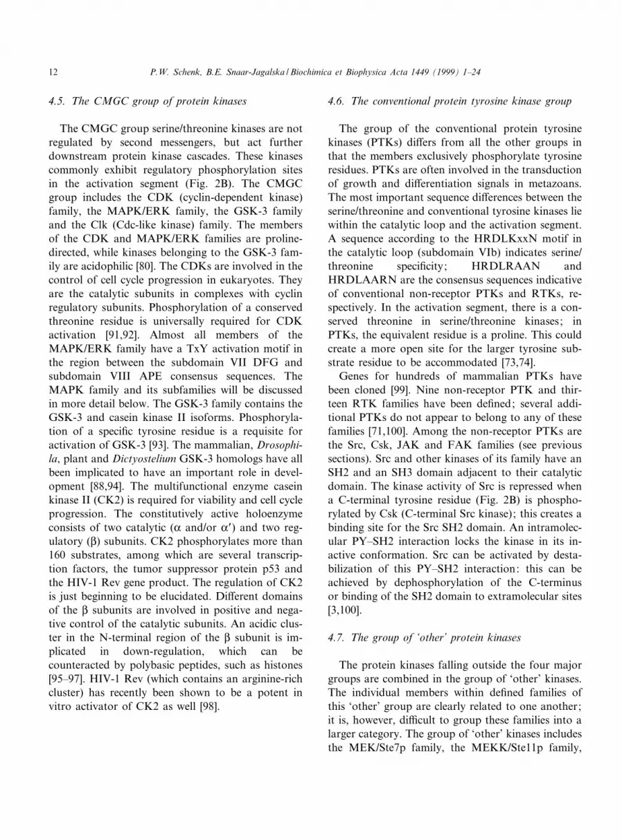
4.5. The CMGC group of protein kinases
The CMGC group serine/threonine kinases are notregulated by second messengers, but act furtherdownstream protein kinase cascades. These kinasescommonly exhibit regulatory phosphorylation sitesin the activation segment (Fig. 2B). The CMGCgroup includes the CDK (cyclin-dependent kinase)family, the MAPK/ERK family, the GSK-3 familyand the Clk (Cdc-like kinase) family. The membersof the CDK and MAPK/ERK families are proline-directed, while kinases belonging to the GSK-3 fam-ily are acidophilic [80]. The CDKs are involved in thecontrol of cell cycle progression in eukaryotes. Theyare the catalytic subunits in complexes with cyclinregulatory subunits. Phosphorylation of a conservedthreonine residue is universally required for CDKactivation [91,92]. Almost all members of theMAPK/ERK family have a TxY activation motif inthe region between the subdomain VII DFG andsubdomain VIII APE consensus sequences. TheMAPK family and its subfamilies will be discussedin more detail below. The GSK-3 family contains theGSK-3 and casein kinase II isoforms. Phosphoryla-tion of a speci¢c tyrosine residue is a requisite foractivation of GSK-3 [93]. The mammalian, Drosophi-la, plant and Dictyostelium GSK-3 homologs have allbeen implicated to have an important role in devel-opment [88,94]. The multifunctional enzyme caseinkinase II (CK2) is required for viability and cell cycleprogression. The constitutively active holoenzymeconsists of two catalytic (K and/or KP) and two reg-ulatory (L) subunits. CK2 phosphorylates more than160 substrates, among which are several transcrip-tion factors, the tumor suppressor protein p53 andthe HIV-1 Rev gene product. The regulation of CK2is just beginning to be elucidated. Di¡erent domainsof the L subunits are involved in positive and nega-tive control of the catalytic subunits. An acidic clus-ter in the N-terminal region of the L subunit is im-plicated in down-regulation, which can becounteracted by polybasic peptides, such as histones[95^97]. HIV-1 Rev (which contains an arginine-richcluster) has recently been shown to be a potent invitro activator of CK2 as well [98].
4.6. The conventional protein tyrosine kinase group
The group of the conventional protein tyrosinekinases (PTKs) di¡ers from all the other groups inthat the members exclusively phosphorylate tyrosineresidues. PTKs are often involved in the transductionof growth and di¡erentiation signals in metazoans.The most important sequence di¡erences between theserine/threonine and conventional tyrosine kinases liewithin the catalytic loop and the activation segment.A sequence according to the HRDLKxxN motif inthe catalytic loop (subdomain VIb) indicates serine/threonine speci¢city; HRDLRAAN andHRDLAARN are the consensus sequences indicativeof conventional non-receptor PTKs and RTKs, re-spectively. In the activation segment, there is a con-served threonine in serine/threonine kinases; inPTKs, the equivalent residue is a proline. This couldcreate a more open site for the larger tyrosine sub-strate residue to be accommodated [73,74].
Genes for hundreds of mammalian PTKs havebeen cloned [99]. Nine non-receptor PTK and thir-teen RTK families have been de¢ned; several addi-tional PTKs do not appear to belong to any of thesefamilies [71,100]. Among the non-receptor PTKs arethe Src, Csk, JAK and FAK families (see previoussections). Src and other kinases of its family have anSH2 and an SH3 domain adjacent to their catalyticdomain. The kinase activity of Src is repressed whena C-terminal tyrosine residue (Fig. 2B) is phospho-rylated by Csk (C-terminal Src kinase); this creates abinding site for the Src SH2 domain. An intramolec-ular PY^SH2 interaction locks the kinase in its in-active conformation. Src can be activated by desta-bilization of this PY^SH2 interaction: this can beachieved by dephosphorylation of the C-terminusor binding of the SH2 domain to extramolecular sites[3,100].
4.7. The group of `other' protein kinases
The protein kinases falling outside the four majorgroups are combined in the group of `other' kinases.The individual members within de¢ned families ofthis `other' group are clearly related to one another;it is, however, di¤cult to group these families into alarger category. The group of `other' kinases includesthe MEK/Ste7p family, the MEKK/Ste11p family,
BBAMCR 14438 1-2-99
P.W. Schenk, B.E. Snaar-Jagalska / Biochimica et Biophysica Acta 1449 (1999) 1^2412

the PAK/Ste20p family, the Raf family, the activin/TGF-L receptor family, the £owering plant receptorkinase family, the casein kinase I family and the LIMkinases.
The members of the ¢rst four families function inthe MAPK family protein kinase cascades (Fig. 3).The mammalian MEK1a (MAPK/ERK kinase 1a)and Saccharomyces Ste7p isoforms are activated bymeans of phosphorylation on conserved serine/threo-nine residues by Raf-1 and Ste11p, respectively (Fig.2B) [101]. MAPK kinases, like MEK1a, are able tophosphorylate both threonine and tyrosine residueson MAPKs by themselves; MAPKKs are thereforecalled dual speci¢city kinases [102]. These dual spe-ci¢city kinases have a lysine (K) within the catalyticloop motif in subdomain VIb, which is otherwiseindicative of serine/threonine speci¢city. MammalianMEKKs (MEK kinases) activate the MAPK familySAPKs or JNKs (stress-activated protein kinases orcJun N-terminal kinases) via the dual speci¢city kin-ase SEK1 (SAPK/ERK kinase 1) [79]. MEKK1 alsobinds the small G-protein Ras and kinase-inactiveMEKK1 inhibits ERK activation in response toEGF. The PAKs (p21-activated kinases) are acti-vated by interaction with the small Rho family G-
proteins Rac1 and Cdc42Hs. PAKs are able to me-diate the activation of SAPKs, independent ofMEKKs [103]. The Saccharomyces Ste20p proteinphosphorylates Ste11p in vitro [104,105]. The Raffamily kinases (which include the Arabidopsis CTR1protein) have a large N-terminal section, which isinvolved in regulation of the kinase activity and in-teraction with other proteins. Mammalian Raf-1 candirectly bind Ras and Rap1a at its Ras binding do-main (Fig. 2B). Ras-GTP is involved in the trans-location of Raf-1 to the plasma membrane, whichis the ¢rst step required for full Raf activation[106,107]. Ras-GTP is assumed to displace half of aso-called `14-3-3 dimer' from the N-terminus of in-active Raf-1. After phosphorylation of Raf-1, the 14-3-3 dimer is then proposed to stabilize the activeconformation of Raf-1 by binding to newly phos-phorylated sites [108].
Many members of the £owering plant (putative)receptor kinase family have been identi¢ed recently.The physiological functions of most of these kinasesare presently unknown. This does not apply to theBrassica S-receptor kinase: its activity is required forself-incompatibility [88,109]. The casein kinase I(CK1) isozymes are a family of constitutively active
Fig. 3. Schematic diagram of some major MAPK family pathways in Saccharomyces (left) and vertebrates (middle). Generic names ofthe transduction components are indicated on the right. Dotted arrows represent interactions, which have not been fully elucidated.The double arrow represents multiple steps. For abbreviations, details and references, see main text.
BBAMCR 14438 1-2-99
P.W. Schenk, B.E. Snaar-Jagalska / Biochimica et Biophysica Acta 1449 (1999) 1^24 13

monomeric enzymes, which are phosphate-directed.CK1 is an exceptional protein kinase, in that its pri-mary structure contains neither the APE motif insubdomain VIII, nor the interacting arginine (R) insubdomain XI (Fig. 2A). Its three-dimensional fold-ing is, however, still very similar to that of otherprotein kinases [80,110]. CK1 has been implicatedin the pathogenesis of Alzheimer's disease, throughhyperphosphorylation of the d protein (which is themajor component of the abnormal paired helical ¢l-aments); it also phosphorylates a proteolytic frag-ment of the disease associated protein presenilin-2in vitro and in vivo [111,112]. In lower eukaryotes,individual CK1 isoforms are involved in the regula-tion of repair pathways, cell proliferation and mor-phogenesis [113,114].
When Hanks and Hunter [71] de¢ned their classi-¢cation of protein kinases, the ¢rst LIM kinases(LIMKs) were just being discovered. They containtwo N-terminal LIM domains, an internal PDZ do-main (see below) and a C-terminal protein kinasedomain (Fig. 2B) with an unusual catalytic region(subdomain VIb: HRDLNSHN); a similar catalyticregion (HRDLTSKN) is found in the human and rattestis-speci¢c protein kinase TESK1. These kinasesare most related to members of the activin/TGF-Lreceptor family. Their catalytic activity is towardsserine/threonine residues [115,116]. LIM domainscontain a cysteine-rich motif that was ¢rst identi¢edin the products of the C. elegans lin-11, the rat ISL1and the C. elegans mec-3 genes; the term LIM is acombination of the ¢rst letters of the gene names.LIM domains are de¢ned by a conserved pattern ofcysteine, histidine or alternative metal-coordinatingresidues; they coordinate two zinc ions each. Thereis ample evidence that these domains mediate intra-or intermolecular protein^protein interactions.Although the similarity to zinc ¢ngers might suggestso, there is no evidence for DNA binding by LIMdomains [117^119].
Human, rat, mouse and chicken LIMKs have beenfound. They possess unique structural features whencompared to known protein kinases. Therefore,LIMKs have already been thought to play a speci¢crole in previously uncharacterized signaling path-ways, since they were ¢rst identi¢ed. HumanLIMK1 tightly interacts with PKC-Q and -j ; theLIM2 domain is critical for the binding between
LIMK1 and PKC-Q [120]. LIMK1 may negativelyregulate its catalytic function by interaction betweenLIM and kinase catalytic domains; LIMK1 has alsobeen demonstrated to associate with LIMK2 [121].LIMK1 has recently been shown to be involved inthe accumulation of actin ¢laments: LIMK1 is acti-vated by Rac1 to phosphorylate and inactivate co¢-lin, which, in its active state, promotes disassemblyof actin ¢laments [122,123]. Our laboratory hascloned a cDNA encoding a structural homolog ofthe LIMKs from D. discoideum (P.W. Schenk etal., unpublished results), which is the ¢rst non-verte-brate LIMK to be identi¢ed.
4.8. Protein kinase C
Protein kinase C (PKC) has diverse functions ingrowth, di¡erentiation and the control of membraneprocesses. The PKC family includes 11 individualisoforms. All PKC isoforms consist of a regulatoryand a catalytic domain (Fig. 2B). There are fourconserved (C1^C4) and ¢ve variable (V1^V5) re-gions. C1 and C2 regions are situated in the regula-tory domain; the C3 and C4 regions are containedwithin the catalytic domain. The regulatory domaincontains a pseudo-substrate site, which is involved inblocking the kinase. The association between thepseudo-substrate site and the C4 region results inan inactive PKC conformation [124,125]. The C1 re-gion has one or two cysteine-rich domains, whichform zinc ¢nger structures. These structures bind ac-tivating compounds, such as the lipid DAG (diacyl-glycerol) and the phorbol ester PMA (phorbol 12-myristate 13-acetate, also named TPA, for 12-O-tet-radecanoyl phorbol 13-acetate) [126]. The C2 regionis involved in the binding of Ca2� : its canonical formis absent in Ca2�-independent PKC isoforms [127].This region also contains a so-called `pseudo-anchor-ing site'. This site is involved in the regulation ofPKC binding to receptors for activated C-kinase(RACKs). RACKs are localized in the particulatefraction and bind to the C2 region of activatedPKC. These components are important in regulatingPKC function: the inhibition of RACK^PKC inter-actions disrupts PKC activation [128]. The pseudo-anchoring site binds to the RACK binding site,which contributes to the formation of an inactivePKC conformation [129]. Several anchoring proteins,
BBAMCR 14438 1-2-99
P.W. Schenk, B.E. Snaar-Jagalska / Biochimica et Biophysica Acta 1449 (1999) 1^2414

which assist in keeping PKCs inactive (RICKs, forreceptors for inactive C-kinase), are also thought toplay their parts. PKC's isozyme speci¢city seems tobe partly mediated by association of each isoformwith speci¢c RICKs and RACKs [130]. PKC's C3and C4 regions comprise the 12 conserved kinasesubdomains: the C3 region contains the ATP bind-ing site, while the C4 region is responsible for sub-strate binding [125].
The presence of DAG causes a striking and selec-tive increase in PKC's a¤nity for membranes, whichis accompanied by activation and pseudo-substraterelease. Membrane translocation is mediated byDAG binding to the C1 domain and phosphatidyl-serine binding to the C2 domain. For conventionalPKCs, the a¤nity for acidic lipids is increased byCa2� [125]. Exploration of the time course of PKC'sactivation state under speci¢c cell stimulation is aprerequisite for the clari¢cation of its physiologicalroles. Very few studies have, however, succeeded inthe direct monitoring of its kinase activity in intactcells. A method, that has therefore been used fre-quently, is measuring the signal-dependent changein the intracellular state of the protein. Most impor-tantly, this involves redistribution of PKC, which isoften from the cytosol to the plasma membrane[131].
Signals that stimulate GPCRs, RTKs or non-re-ceptor PTKs can cause DAG production. PKC hasbeen shown to be rapidly activated by a transient risein DAG levels, resulting from PLC stimulation.There is also a sustained increase in DAG andPKC activity, which is thought to be the result of(among others) phosphatidyl-choline hydrolysis byphospholipase D [124,125,132]. Other lipid metabo-lites, such as PIP3, can activate PKC as well. SeveralPKC isotypes are activated independently in a redun-dant manner through the PLC and the PI3K path-way, which generate DAG and PIP3, respectively.PKC-O activated via the PLC pathway translocatesto the cytoskeletal fraction, whereas that activatedvia PI3K does not. This indicates a functional di¡er-ence between PKC activated through di¡erent path-ways [131].
Di¡erent PKC isozymes are also believed to havedistinct biological functions. On the basis of struc-tural elements and activational characteristics, thePKC family has been divided into three subfamilies.
PKC was originally identi¢ed as a phospholipid- andcalcium-dependent protein kinase [133]. PKC-K, -LI,-LII and -Q are the members of the so-called `classi-cal' or `conventional PKC' (cPKC) subfamily. Thesekinases have two zinc ¢ngers in the C1 region andare phosphatidyl-serine-, DAG- and Ca2�-depend-ent. They have a molecular mass of 77^78 kDa[124]. For several years, it has been established thatPKC-K directly phosphorylates and activates the ser-ine/threonine kinase Raf-1 in vitro and in vivo [134].It is, however, still unclear how this phosphorylationcontributes to Raf activation. Although dominantnegative Ras expression can not block Raf-1 activa-tion by signals that stimulate PKC, this activationwas recently reported to depend on the formationof Ras-GTP^Raf-1 complexes [135]. PKC-K, -LIand -Q speci¢cally inactivate GSK-3L by phosphory-lation; this leads to derepression of the cJun tran-scription factor [136]. The subfamily of non-classicalor novel PKCs (nPKCs) includes the N, O, R and aisoforms. Their activity is only dependent on phos-phatidyl-serine and DAG; it is independent of Ca2�.The nPKCs have a molecular mass of approximately80 kDa [124]. Like PKC-K, PKC-N and -O have beensuggested to activate the MEK^ERK pathway viaRaf [137,138]. PKC-a has recently been shown tosynergize with the Ca2�-dependent phosphatase cal-cineurin to stimulate JNK1 via Rac1 [139]. The re-lated sequence PKC-W/protein kinase D is also acti-vated by DAG in the absence of Ca2�. The 115-kDaPKC-W protein contains an insert of 74 amino acidsbetween the two zinc ¢ngers of the C1 region, whichis associated with ine¤cient phorbol ester bindingand apparently constitutive in vitro kinase activity.It has two unique N-terminal hydrophobic domainsand lacks a typical pseudo-substrate site. Taken to-gether, its characteristics place PKC-W somewherebetween the novel and the atypical PKCs [140].The remaining PKC isoforms (j and S/V) are placedin the subfamily of the atypical PKCs (aPKCs). TheaPKCs have a molecular mass of approximately 67kDa. They lack the canonical C2 region and one ofthe cysteine-rich domains in the C1 region. This re-sults in DAG- and Ca2�-independent PKCs[124,132]. PKC-j and -S/V can be activated by PIP3
[141,142]. A direct interaction between PKC-j andRas is required for the stimulation of ERKs by an-giotensin II in vascular smooth muscle cells [143].
BBAMCR 14438 1-2-99
P.W. Schenk, B.E. Snaar-Jagalska / Biochimica et Biophysica Acta 1449 (1999) 1^24 15

Finally, both TPA-sensitive and -insensitive PKCsplay a role in the regulation of gene expression byso-called `TPA response elements' [142,144].
4.9. Mitogen-activated protein kinases
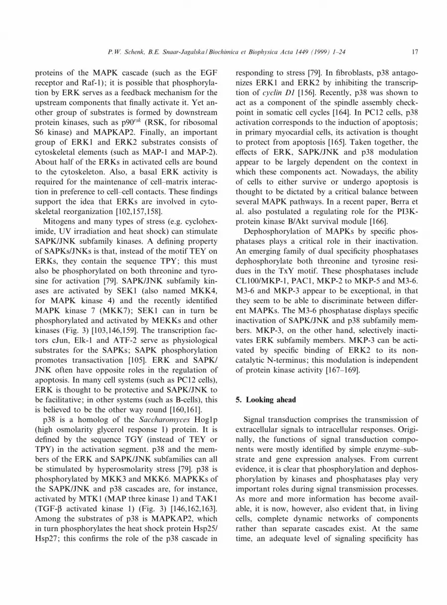
The mitogen-activated protein kinases (MAPKs)form a family of well-conserved serine/threonine kin-ases, with a molecular mass of 38^55 kDa [79]. Mem-bers of the MAPK family have a central role in awide variety of protein kinase cascades. These cas-cades are found in all eukaryotic organisms and con-sist of a three-kinase module that includes a MAPK,a MAPK kinase (MAPKK) and a MAPK kinasekinase (MAPKKK): the MAPK is activated by aMAPKK, which is in turn activated by a MAPKKK(Fig. 3). Stimulation of MAPKs often results in tran-scriptional activation [145]. MAPKs are grouped onthe basis of sequence similarity, mechanism of up-stream regulation and sensitivity to activation by dif-ferent MAPKKs. Until recently, the MAPK familywas just subdivided into three subfamilies: the ERK(extracellular signal regulated kinase), the SAPK/JNK (stress-activated protein kinase/cJun N-terminalkinase) and the p38 subfamily. The most studiedsubfamily is that including ERK1 (42 kDa) andERK2 (44 kDa). These are involved in cascades con-sisting of Raf, MEK1a/MEK2 and ERK1/ERK2 iso-forms. Raf is in turn activated by a dynamic combi-nation of phosphorylation (by PKC and/or otherprotein kinases) and interactions with Ras-GTPand 14-3-3 proteins [108]. The Raf^MEK^MAPK/ERK pathway has e¡ects in non-proliferating cells,but mitogenic signals especially stimulate the path-way; proliferation can be blocked by inhibiting it.The SAPKs/JNKs and the p38 subfamily kinasesmediate responses to cellular stress. During the lastfew years, genes encoding novel MAPKs, such asERK5 and ERK6, have been cloned from verte-brates. ERK5, ERK6 and the previously identi¢edkinase ERK3 are probably not regulated like themembers of the three subfamilies mentioned. Thissuggests the existence of more MAPK pathways inmetazoans than was originally realized [146]. ERK5was recently shown to mediate a Ras-dependent,Raf-independent pathway activating the transcrip-tion factor cMyc (which is also a substrate forERK1 and ERK2) [147].
In S. cerevisiae, there are six MAPKs. Five ofthese have been associated with biological responsesto speci¢c stimuli. Inappropriate cross-talk is, forinstance, precluded by Fus3p. This MAPK channelsGL/Q-dependent signals from the pheromone ligandthrough the mating pathway and prevents the signalfrom activating ¢lamentation di¡erentiation via theKss1p MAPK [148]. The yeast probably uses Ste5pas a sca¡old for the Ste11p^Ste7p^Fus3p cascadecomplex [149]. Interestingly, functional binding be-tween GL and the LIM domain-like region ofSte5p has recently also been demonstrated to be re-quired for the activation of Ste11p (Fig. 3) [150].
The vertebrate Ras^Raf^MEK^MAPK/ERKpathway can be switched on by extracellular signalmolecules binding to either RTKs or GPCRs. Oneactivation mechanism involving GPCRs is mediatedby G-protein L/Q subunits stimulating Ras-GTP for-mation [57,151]. The GPCR ligand angiotensin IIcan, for instance, also stimulate ERK via a putativeRas- and Raf-independent, PKC-dependent pathway[152]. The members of the ERK subfamily have aTEY motif in the activation segment between thesubdomain VII DFG and subdomain VIII APE con-sensus sequences. MEK1a and MEK2 are able tophosphorylate ERK1 and ERK2 on both threonineand tyrosine in this TEY motif. The activating phos-phorylation of these ERKs by these MEKs is highlyspeci¢c and requires the native form of MAPK[79,102]. The Ksr-1 (kinase suppressor of Ras 1) pro-tein has been proposed to serve as a sca¡old forMEK and ERK [153]. The dual phosphorylationon TEY seems to be the main requirement for trans-location of ERK1 and ERK2 to the nucleus; cata-lytic MAPK activity is not required. Moreover,phosphorylation induces ERK2 dimerization; this isalso necessary for its nuclear localization [154]. In-terestingly, unlike the other MAPK family members,ERK3 is constitutively nuclear [155].
Because of their broad range of substrate recogni-tion, activated ERK1 and ERK2 can phosphorylatea large number of proteins. Among the substrates ofthese ERKs are transcription factors and other nu-clear proteins (such as the ternary complex factorElk-1 and cMyc). In ¢broblasts, ERK1 and ERK2promote entry into the cell cycle, at least in part, bypositively regulating the transcription of cyclin D1[156]. Other substrates for these ERKs are upstream
BBAMCR 14438 1-2-99
P.W. Schenk, B.E. Snaar-Jagalska / Biochimica et Biophysica Acta 1449 (1999) 1^2416
proteins of the MAPK cascade (such as the EGFreceptor and Raf-1); it is possible that phosphoryla-tion by ERK serves as a feedback mechanism for theupstream components that ¢nally activate it. Yet an-other group of substrates is formed by downstreamprotein kinases, such as p90rsk (RSK, for ribosomalS6 kinase) and MAPKAP2. Finally, an importantgroup of ERK1 and ERK2 substrates consists ofcytoskeletal elements (such as MAP-1 and MAP-2).About half of the ERKs in activated cells are boundto the cytoskeleton. Also, a basal ERK activity isrequired for the maintenance of cell^matrix interac-tion in preference to cell^cell contacts. These ¢ndingssupport the idea that ERKs are involved in cyto-skeletal reorganization [102,157,158].
Mitogens and many types of stress (e.g. cyclohex-imide, UV irradiation and heat shock) can stimulateSAPK/JNK subfamily kinases. A de¢ning propertyof SAPKs/JNKs is that, instead of the motif TEY onERKs, they contain the sequence TPY; this mustalso be phosphorylated on both threonine and tyro-sine for activation [79]. SAPK/JNK subfamily kin-ases are activated by SEK1 (also named MKK4,for MAPK kinase 4) and the recently identi¢edMAPK kinase 7 (MKK7); SEK1 can in turn bephosphorylated and activated by MEKKs and otherkinases (Fig. 3) [103,146,159]. The transcription fac-tors cJun, Elk-1 and ATF-2 serve as physiologicalsubstrates for the SAPKs; SAPK phosphorylationpromotes transactivation [105]. ERK and SAPK/JNK often have opposite roles in the regulation ofapoptosis. In many cell systems (such as PC12 cells),ERK is thought to be protective and SAPK/JNK tobe facilitative; in other systems (such as B-cells), thisis believed to be the other way round [160,161].
p38 is a homolog of the Saccharomyces Hog1p(high osmolarity glycerol response 1) protein. It isde¢ned by the sequence TGY (instead of TEY orTPY) in the activation segment. p38 and the mem-bers of the ERK and SAPK/JNK subfamilies can allbe stimulated by hyperosmolarity stress [79]. p38 isphosphorylated by MKK3 and MKK6. MAPKKs ofthe SAPK/JNK and p38 cascades are, for instance,activated by MTK1 (MAP three kinase 1) and TAK1(TGF-L activated kinase 1) (Fig. 3) [146,162,163].Among the substrates of p38 is MAPKAP2, whichin turn phosphorylates the heat shock protein Hsp25/Hsp27; this con¢rms the role of the p38 cascade in
responding to stress [79]. In ¢broblasts, p38 antago-nizes ERK1 and ERK2 by inhibiting the transcrip-tion of cyclin D1 [156]. Recently, p38 was shown toact as a component of the spindle assembly check-point in somatic cell cycles [164]. In PC12 cells, p38activation corresponds to the induction of apoptosis;in primary myocardial cells, its activation is thoughtto protect from apoptosis [165]. Taken together, thee¡ects of ERK, SAPK/JNK and p38 modulationappear to be largely dependent on the context inwhich these components act. Nowadays, the abilityof cells to either survive or undergo apoptosis isthought to be dictated by a critical balance betweenseveral MAPK pathways. In a recent paper, Berra etal. also postulated a regulating role for the PI3K-protein kinase B/Akt survival module [166].
Dephosphorylation of MAPKs by speci¢c phos-phatases plays a critical role in their inactivation.An emerging family of dual speci¢city phosphatasesdephosphorylate both threonine and tyrosine resi-dues in the TxY motif. These phosphatases includeCL100/MKP-1, PAC1, MKP-2 to MKP-5 and M3-6.M3-6 and MKP-3 appear to be exceptional, in thatthey seem to be able to discriminate between di¡er-ent MAPKs. The M3-6 phosphatase displays speci¢cinactivation of SAPK/JNK and p38 subfamily mem-bers. MKP-3, on the other hand, selectively inacti-vates ERK subfamily members. MKP-3 can be acti-vated by speci¢c binding of ERK2 to its non-catalytic N-terminus; this modulation is independentof protein kinase activity [167^169].
5. Looking ahead
Signal transduction comprises the transmission ofextracellular signals to intracellular responses. Origi-nally, the functions of signal transduction compo-nents were mostly identi¢ed by simple enzyme^sub-strate and gene expression analyses. From currentevidence, it is clear that phosphorylation and dephos-phorylation by kinases and phosphatases play veryimportant roles during signal transmission processes.As more and more information has become avail-able, it is now, however, also evident that, in livingcells, complete dynamic networks of componentsrather than separate cascades exist. At the sametime, an adequate level of signaling speci¢city has
BBAMCR 14438 1-2-99
P.W. Schenk, B.E. Snaar-Jagalska / Biochimica et Biophysica Acta 1449 (1999) 1^24 17

to be maintained. The latter is often essential to thephysiological roles of cells. In other words: cross-talkis de¢nitely present, but, in many cases, has to beminimized in order to obtain the right response atthe right time and place.
Of course, a considerable amount of speci¢city isobtained by direct recognition of protein substratesites by kinases [73,80]. In addition, phylogeneticanalyses (such as that performed by Hanks andHunter [71]), the elucidation of `active' crystal struc-tures (such as those of PKA and ERK2 [73,78]), two-hybrid screens [170], co-immunoprecipitation experi-ments (such as those performed by Brady-Kalnay etal. [13]) and overlay assays have demonstrated thepresence of an ever-growing number of interactionsand interaction motifs for many signal transductioncomponents. Examples of these motifs are SH2 andother PY binding domains, proline-rich sequencebinding SH3 domains (such as that present in Src),so-called `pleckstrin homology domains' (such asthose in Sos and IRS-1) [3], LIM domains and so-called `PDZ' (for PSD-95/Dlg/ZO-1 homology) do-mains [171].
In the ¢rst place, several interactions between sig-nal transduction and cytoskeletal components (whichmay assure correct localization) have been described.In many other cases we mentioned above, transmis-sion molecules, which are often modi¢ed themselves,constitute docking sites or sca¡olds for other com-ponents or complexes. Yet another nice example isprovided by the Drosophila InaD protein, which ba-sically consists of ¢ve PDZ domains. InaD serves asa sca¡old to assemble di¡erent components of a pho-totransduction cascade. It is believed that its PDZdomains function as key elements in the organizationof transduction complexes in vivo [171]. Other mech-anisms to obtain higher speci¢city do, of course, alsoexist. The pathway-dedicated yeast MAPK Fus3p is,for instance, thought to impart speci¢city by simplychanneling a particular signal through the right path-way; by doing so, it prevents the signal from activat-ing another [148].
Also, simple ligand^receptor interactions are oftennot enough to activate underlying cascades. The re-ceptor-like protein tyrosine phosphatase-L is, for ex-ample, assumed to be activated by lateral mobility ofspeci¢c ligands: this induces formation of monomersfrom inactive dimers [172]. Receptor clustering (such
as that of integrins [33,34]) can be essential as well.In general, a combination of sensitivity and widedynamic range of response is obtained if a cell hasboth clusters and single receptors on its surface, es-pecially if the propagation of the signal can adapt toexternal conditions [173].
Much of the current knowledge has been obtainedusing in vitro approaches and transfected cell lines.Taken together, the re£ections above lead to the no-tion that an adequate picture of signal transductionby, for example, protein kinases, has to be obtainedfrom living cells, which have been manipulated aslittle as possible. The use of model organisms, suchas Saccharomyces and Dictyostelium, can be of greatvalue: they potentially provide true in vivo situa-tions. Modern imaging techniques, like those em-ploying green £uorescent protein [174] and its deriv-atives (which are shifted in their excitation andemission spectra), will also prove very valuablewhen analyzing localization and interactions of pro-teins. The combined use of such systems and meth-ods will greatly contribute to a better understandingof how signal transduction components work in thedynamic environment of intact cells.
Acknowledgements
We are grateful to Theo Konijn for critically read-ing the manuscript.
References
[1] M.-J. Tsai, B.W. O'Malley, Molecular mechanisms of actionof steroid/thyroid receptor superfamily members, Annu.Rev. Biochem. 63 (1994) 451^486.
[2] I.B. Levitan, Modulation of ion channels by protein phos-phorylation and dephosphorylation, Annu. Rev. Physiol. 56(1994) 193^212.
[3] T. Pawson, Protein modules and signalling networks, Nature373 (1995) 573^580.
[4] L. Bon¢ni, E. Migliaccio, G. Pelicci, L. Lanfrancone, P.G.Pelicci, Not all Shc's roads lead to Ras, Trends Biochem.Sci. 21 (1996) 257^261.
[5] K.-L. Guan, The mitogen activated protein kinase signaltransduction pathway: from the cell surface to the nucleus,Cell. Signal. 6 (1994) 581^589.
[6] D.R. Alessi, S.R. James, C.P. Downes, A.B. Holmes, P.R.J.Ga¡ney, C.B. Reese, P. Cohen, Characterization of a 3-
BBAMCR 14438 1-2-99
P.W. Schenk, B.E. Snaar-Jagalska / Biochimica et Biophysica Acta 1449 (1999) 1^2418

phosphoinositide-dependent protein kinase which phospho-rylates and activates protein kinase BK, Curr. Biol. 7 (1997)261^269.
[7] D.R. Alessi, P. Cohen, Mechanism of activation and func-tion of protein kinase B, Curr. Opin. Genet. Dev. 8 (1998)55^62.
[8] R. Wu, K. Durick, Z. Songyang, L.C. Cantley, S.S. Taylor,G.N. Gill, Speci¢city of LIM domain interactions with re-ceptor tyrosine kinases, J. Biol. Chem. 271 (1996) 15934^15941.
[9] K. Durick, G.N. Gill, S.S. Taylor, Shc and Enigma are bothrequired for mitogenic signaling by Ret/ptc2, Mol. Cell. Biol.18 (1998) 2298^2308.
[10] E.H. Fischer, H. Charbonneau, N.K. Tonks, Protein tyro-sine phosphatases: a diverse family of intracellular andtransmembrane enzymes, Science 253 (1991) 401^406.
[11] G.C.M. Zondag, W.H. Moolenaar, Receptor protein tyro-sine phosphatases: involvement in cell^cell interaction andsignaling, Biochimie 79 (1997) 477^483.
[12] I.S. Trowbridge, M.L. Thomas, CD45: an emerging role as aprotein tyrosine phosphatase required for lymphocyte acti-vation and development, Annu. Rev. Immunol. 12 (1994)85^116.
[13] S.M. Brady-Kalnay, T. Mourton, J.P. Nixon, G.E. Pietz, M.Kinch, H. Chen, R. Brackenbury, D.L. Rimm, R.L. DelVecchio, N.K. Tonks, Dynamic interaction of PTPW withmultiple cadherins in vivo, J. Cell Biol. 141 (1998) 287^296.
[14] J. den Hertog, T. Hunter, Tight association of GRB2 withreceptor protein-tyrosine phosphatase K is mediated by theSH2 and C-terminal SH3 domains, EMBO J. 15 (1996)3016^3027.
[15] R. Derynck, TGF-L-receptor-mediated signaling, TrendsBiochem. Sci. 19 (1994) 548^553.
[16] R. Derynck, X.-H. Feng, TGF-L receptor signaling, Bio-chim. Biophys. Acta 1333 (1997) F105^F150.
[17] Y. Zhang, X.-H. Feng, R.-Y. Wu, R. Derynck, Receptor-associated Mad homologues synergize as e¡ectors of theTGF-L response, Nature 383 (1996) 168^172.
[18] M.P. de Caestecker, W.T. Parks, C.J. Frank, P. Castagnino,D.P. Bottaro, A.B. Roberts, R.J. Lechleider, Smad2 trans-duces common signals from receptor serine-threonine andtyrosine kinases, Genes Dev. 12 (1998) 1587^1592.
[19] J.L. Appleby, J.S. Parkinson, R.B. Bourret, Signal transduc-tion via the multi-step phosphorelay: not necessarily a roadless traveled, Cell 86 (1996) 845^848.
[20] R.V. Swanson, L.A. Alex, M.I. Simon, Histidine and aspar-tate phosphorylation: two-component systems and the limitsof homology, Trends Biochem. Sci. 19 (1994) 485^490.
[21] T. Maeda, M. Takekawa, H. Saito, Activation of yeast PBS2MAPKK by MAPKKKs or by binding of an SH3-contain-ing osmosensor, Science 269 (1995) 554^558.
[22] F. Posas, S.M. Wurgler-Murphy, T. Maeda, E.A. Witten,T.C. Thai, H. Saito, Yeast HOG1 MAP kinase cascade isregulated by a multistep phosphorelay mechanism in theSLN1-YPD1-SSK1 `two-component' osmosensor, Cell 86(1996) 865^875.
[23] F. Posas, H. Saito, Activation of the yeast SSK2 MAP kin-ase kinase kinase by the SSK1 two-component response reg-ulator, EMBO J. 17 (1998) 1385^1394.
[24] D.L. Garbers, D.G. Lowe, Guanylyl cyclase receptors,J. Biol. Chem. 269 (1994) 30741^30744.
[25] B.J. Wedel, D.L. Garbers, New insights on the functions ofthe guanylyl cyclase receptors, FEBS Lett. 410 (1997) 29^33.
[26] T. Sugimoto, M. Haneda, M. Togawa, M. Isono, T. Shika-no, S. Araki, T. Nakagawa, A. Kashiwagi, K.-L. Guan, R.Kikkawa, Atrial natriuretic peptide induces the expression ofMKP-1, a mitogen-activated protein kinase phosphatase, inglomerular mesangial cells, J. Biol. Chem. 271 (1996) 544^547.
[27] S.M. Lohmann, A.B. Vaandrager, A. Smolenski, U. Walter,H.R. de Jonge, Distinct and speci¢c functions of cGMP-dependent protein kinases, Trends Biochem. Sci. 22 (1997)307^312.
[28] J.N. Ihle, Cytokine receptor signalling, Nature 377 (1995)591^594.
[29] D.W. Leaman, S. Leung, X. Li, G.R. Stark, Regulation ofSTAT-dependent pathways by growth factors and cytokines,FASEB J. 10 (1996) 1578^1588.
[30] M.B. Marrero, B. Schie¡er, B. Li, J. Sun, J.B. Harp, B.N.Ling, Role of Janus kinase/signal transducer and activator oftranscription and mitogen-activated protein kinase cascadesin angiotensin II- and platelet-derived growth factor-inducedvascular smooth muscle cell proliferation, J. Biol. Chem. 272(1997) 24684^24690.
[31] X.S. Hou, N. Perrimon, The JAK-STAT pathway in Droso-phila, Trends Genet. 13 (1997) 105^110.
[32] T. Araki, M. Gamper, A. Early, M. Fukuzawa, T. Abe, T.Kawata, E. Kim, R.A. Firtel, J.G. Williams, Developmen-tally and spatially regulated activation of a DictyosteliumSTAT protein by a serpentine receptor, EMBO J. 17(1998) 4018^4028.
[33] R.O. Hynes, Integrins: versatility, modulation, and signalingin cell adhesion, Cell 69 (1992) 11^25.
[34] E.A. Clark, J.S. Brugge, Integrins and signal transductionpathways: the road taken, Science 268 (1995) 233^239.
[35] S.K. Hanks, T.R. Polte, Signaling through focal adhesionkinase, BioEssays 19 (1997) 137^145.
[36] T.H. Lin, A.E. Aplin, Y. Shen, Q. Chen, M. Schaller, L.Romer, I. Aukhil, R.L. Juliano, Integrin-mediated activationof MAP kinase is independent of FAK: evidence for dualintegrin signaling pathways in ¢broblasts, J. Cell Biol. 136(1997) 1385^1395.
[37] D.D. Schlaepfer, K.C. Jones, T. Hunter, Multiple Grb2-mediated integrin-stimulated signaling pathways to ERK2/mitogen-activated protein kinase: summation of both c-Src- and focal adhesion kinase-initiated tyrosine phospho-rylation events, Mol. Cell. Biol. 18 (1998) 2571^2585.
[38] A.D. Strosberg, Structure/function relationship of proteinsbelonging to the family of receptors coupled to GTP-bindingproteins, Eur. J. Biochem. 196 (1991) 1^10.
[39] E.M. Brown, G. Gamba, D. Riccardi, M. Lombardi, R.Butters, O. Kifor, A. Sun, M.A. Hediger, J. Lytton, S.C.
BBAMCR 14438 1-2-99
P.W. Schenk, B.E. Snaar-Jagalska / Biochimica et Biophysica Acta 1449 (1999) 1^24 19

Hebert, Cloning and characterization of an extracellularCa2�-sensing receptor from bovine parathyroid, Nature366 (1993) 575^580.
[40] J.R. Erickson, J.J. Wu, J.G. Goddard, G. Tigyi, K. Kawa-nishi, L.D. Tomei, M.C. Kiefer, Edg-2/Vzg-1 couples to theyeast pheromone response pathway selectively in response tolysophosphatidic acid, J. Biol. Chem. 273 (1998) 1506^1510.
[41] N. Fukushima, Y. Kimura, J. Chun, A single receptor en-coded by vzg-1/lpA1/edg-2 couples to G proteins and medi-ates multiple cellular responses to lysophosphatidic acid,Proc. Natl. Acad. Sci. USA 95 (1998) 6151^6156.
[42] Y. Daaka, L.M. Luttrell, S. Ahn, G.J. Della Rocca, S.S.G.Ferguson, M.G. Caron, R.J. Lefkowitz, Essential role for G-protein-coupled receptor endocytosis in the activation of mi-togen-activated protein kinase, J. Biol. Chem. 273 (1998)685^688.
[43] H.E. Hamm, The many faces of G protein signaling, J. Biol.Chem. 273 (1998) 669^672.
[44] D.M. Berman, T.M. Wilkie, A.G. Gilman, GAIP and RGS4are GTPase-activating proteins for the Gi subfamily of Gprotein K subunits, Cell 86 (1996) 445^452.
[45] D.M. Berman, A.G. Gilman, Mammalian RGS proteins:barbarians at the gate, J. Biol. Chem. 273 (1998) 1269^1272.
[46] R. Iyengar, There are GAPs and there are GAPs, Science275 (1997) 42^43.
[47] M. Hagiwara, P. Brindle, A. Harootunian, R. Armstrong, J.Rivier, W. Vale, R. Tsien, M.R. Montminy, Coupling ofhormonal stimulation and transcription via the cyclicAMP-responsive factor CREB is rate limited by nuclear en-try of protein kinase A, Mol. Cell. Biol. 13 (1993) 4852^4859.
[48] M.J. Smit, R. Iyengar, Mammalian adenylyl cyclases, Adv.Second Messenger Phosphoprotein Res. 32 (1998) 1^21.
[49] S.G. Rhee, Y.S. Bae, Regulation of phosphoinositide-speci¢cphospholipase C isozymes, J. Biol. Chem. 272 (1997) 15045^15048.
[50] M. Liu, M.I. Simon, Regulation by cAMP-dependent pro-tein kinease of a G-protein-mediated-phospholipase C, Na-ture 382 (1996) 83^87.
[51] M.J. Berridge, Inositol trisphosphate and calcium signalling,Nature 361 (1993) 315^325.
[52] A.P. Braun, H. Schulman, The multifunctional calcium/calmodulin-dependent protein kinase: from form to func-tion, Annu. Rev. Physiol. 57 (1995) 417^445.
[53] N.R. Murray, L.J. Thompson, A.P. Fields, The role of pro-tein kinase C in cellular proliferation and cell cycle control,in: P.J. Parker, L.V. Dekker (Eds.), Protein Kinase C, R.G.Landes, USA, 1997, pp. 97^120.
[54] A.M. Buhl, N.L. Johnson, N. Dhanasekaran, G.L. Johnson,GK12 and GK13 stimulate Rho-dependent stress ¢ber forma-tion and focal adhesion assembly, J. Biol. Chem. 270 (1995)24631^24634.
[55] W.H. Moolenaar, Lysophosphatidic acid, a multifunctionalphospholipid messenger, J. Biol. Chem. 270 (1995) 12949^12952.
[56] W.H. Moolenaar, O. Kranenburg, F.R. Postma, G.C.M.
Zondag, Lysophosphatidic acid: G-protein signalling andcellular responses, Curr. Opin. Cell Biol. 9 (1997) 168^173.
[57] W.J. Koch, B.E. Hawes, L.F. Allen, R.J. Lefkowitz, Directevidence that Gi-coupled receptor stimulation of mitogen-activated protein kinase is mediated by GLQ activation ofp21ras, Proc. Natl. Acad. Sci. USA 91 (1994) 12706^12710.
[58] L.M. Luttrell, G.J. Della Rocca, T. van Biesen, D.K. Lut-trell, R.J. Lefkowitz, GLQ subunits mediate Src-dependentphosphorylation of the epidermal growth factor receptor,J. Biol. Chem. 272 (1997) 4637^4644.
[59] O. Kranenburg, I. Verlaan, P.L. Hordijk, W.H. Moolenaar,Gi-mediated activation of the Ras/MAP kinase pathway in-volves a 100 kDa tyrosine-phosphorylated Grb2 SH3 bind-ing protein, but not Src nor Shc, EMBO J. 16 (1997) 3097^3105.
[60] M. Lopez-Ilasaca, P. Crespo, P.G. Pellici, J.S. Gutkind, R.Wetzker, Linkage of G protein-coupled receptors to theMAPK signaling pathway through PI 3-kinase Q, Science275 (1997) 394^397.
[61] S. Roche, M. Koegl, M.V. Barone, M.F. Roussel, S.A.Courtneidge, DNA synthesis induced by some but not allgrowth factors requires Src family protein tyrosine kinases,Mol. Cell. Biol. 15 (1995) 1102^1109.
[62] K. Kimura, M. Ito, M. Amano, K. Chihara, Y. Fukata, M.Nakafuku, B. Yamamori, J. Feng, T. Nakano, K. Okawa,A. Iwamatsu, K. Kaibuchi, Regulation of myosin phospha-tase by Rho and Rho-associated kinase (Rho-kinase), Sci-ence 273 (1996) 245^248.
[63] S. Clarke, Protein isoprenylation and methylation at car-boxy-terminal cysteine residues, Annu. Rev. Biochem. 61(1992) 355^386.
[64] M.D. Resh, Myristylation and palmitylation of Src familymembers: the fats of the matter, Cell 76 (1994) 411^413.
[65] M.D. Resh, Regulation of cellular signalling by fatty acidacylation and prenylation of signal transduction proteins,Cell. Signal. 8 (1996) 403^412.
[66] A.D. Cox, C.J. Der, Farnesyltransferase inhibitors and can-cer treatment: targeting simply Ras?, Biochim. Biophys.Acta 1333 (1997) F51^F71.
[67] J.M. Aletta, T.R. Cimato, M.J. Ettinger, Protein methyl-ation: a signal event in post-translational modi¢cation,Trends Biochem. Sci. 23 (1998) 89^91.
[68] S.P. Jackson, R. Tjian, O-glycosylation of eukaryotic tran-scription factors: implications for mechanisms of transcrip-tional regulation, Cell 55 (1988) 125^133.
[69] D.M. Snow, G.W. Hart, Nuclear and cytoplasmic glyco-sylation, Int. Rev. Cytol. 181 (1998) 43^74.
[70] J.R. Woodgett, B.J. Pulverer, E. Nikolakaki, S. Plyte, K.Hughes, C.C. Franklin, A.S. Kraft, Regulation of Jun/AP-1 oncoproteins by protein phosphorylation, Adv. SecondMessenger Phosphoprotein Res. 28 (1993) 261^269.
[71] S.K. Hanks, T. Hunter, The eukaryotic protein kinase super-family: kinase (catalytic) domain structure and classi¢cation,FASEB J. 9 (1995) 576^596.
[72] T. Hunter, G.D. Plowman, The protein kinases of budding
BBAMCR 14438 1-2-99
P.W. Schenk, B.E. Snaar-Jagalska / Biochimica et Biophysica Acta 1449 (1999) 1^2420

yeast : six score and more, Trends Biochem. Sci. 22 (1997)18^22.
[73] L.N. Johnson, E.D. Lowe, M.E.M. Noble, D.J. Owen, Thestructural basis for substrate recognition and control by pro-tein kinases, FEBS Lett. 430 (1998) 1^11.
[74] S.S. Taylor, E. Radzio-Andzelm, T. Hunter, How do proteinkinases discriminate between serine/threonine and tyrosine?Structural insights from the insulin receptor protein-tyrosinekinase, FASEB J. 9 (1995) 1255^1266.
[75] W.F. Loomis, G. Shaulsky, N. Wang, Histidine kinases insignal transduction pathways of eukaryotes, J. Cell Sci. 110(1997) 1141^1145.
[76] M.J. Zinda, C.K. Singleton, The hybrid histidine kinasedhkB regulates spore germination in Dictyostelium discoi-deum, Dev. Biol. 196 (1998) 171^183.
[77] K.M. Popov, J.W. Hawes, R.A. Harris, Mitochondrial K-ketoacid dehydrogenase kinases, Adv. Second MessengerPhosphoprotein Res. 31 (1997) 105^111.
[78] B.J. Canagarajah, A. Khokhlatchev, M.H. Cobb, E.J. Gold-smith, Activation mechanism of the MAP kinase ERK2 bydual phosphorylation, Cell 90 (1997) 859^869.
[79] E. Cano, L.C. Mahadevan, Parallel signal processing amongmammalian MAPKs, Trends Biochem. Sci. 20 (1995) 117^122.
[80] L.A. Pinna, M. Ruzzene, How do protein kinases recognizetheir substrates?, Biochim. Biophys. Acta 1314 (1996) 191^225.
[81] C.M. Smith, I.N. Shindyalov, S. Veretnik, M. Gribskov, S.S.Taylor, L.F. ten Eyck, P.E. Bourne, The protein kinase re-source, Trends Biochem. Sci. 22 (1997) 444^446.
[82] S.O. DÖskeland, E. Maronde, B.T. Gjertsen, The geneticsubtypes of cAMP-dependent protein kinase ^ functionallydi¡erent or redundant?, Biochim. Biophys. Acta 1178 (1993)249^258.
[83] D.R. Alessi, M.T. Kozlowski, Q.-P. Weng, N. Morrice, J.Avruch, 3-Phosphoinositide-dependent protein kinase 1(PDK1) phosphorylates and activates the p70 S6 kinase invivo and in vitro, Curr. Biol. 8 (1997) 69^81.
[84] N. Pullen, P.B. Dennis, M. Andjelkovic, A. Dufner, S.C.Kozma, B.A. Hemmings, G. Thomas, Phosphorylation andactivation of p70s6k by PDK1, Science 279 (1998) 707^710.
[85] J. Downward, Lipid-regulated kinases: some commonthemes at last, Science 279 (1998) 673^674.
[86] Q.-P. Weng, M. Kozlowski, C. Belham, A. Zhang, M.J.Comb, J. Avruch, Regulation of the p70 S6 kinase by phos-phorylation in vivo, J. Biol. Chem. 273 (1998) 16621^16629.
[87] S. Nakayama, R.H. Kretsinger, Evolution of the EF-handfamily of proteins, Annu. Rev. Biophys. Biomol. Struct. 23(1994) 473^507.
[88] J.M. Stone, J.C. Walker, Plant protein kinase families andsignal transduction, Plant Physiol. 108 (1995) 451^457.
[89] K. Kohama, L.-H. Ye, K. Hayakawa, T. Okagaki, Myosinlight chain kinase: an actin-binding protein that regulates anATP-dependent interaction with myosin, Trends Pharmacol.Sci. 17 (1996) 284^287.
[90] D.G. Hardie, D. Carling, The AMP-activated protein kin-ase: Fuel gauge of the mammalian cell?, Eur. J. Biochem.246 (1997) 259^273.
[91] D.O. Morgan, Principles of CDK regulation, Nature 374(1995) 131^134.
[92] E.A. Nigg, Cyclin-dependent protein kinases: key regula-tors of the eukaryotic cell cycle, BioEssays 17 (1995) 471^480.
[93] K. Hughes, E. Nikolakaki, S.E. Plyte, N.F. Totty, J.R.Woodgett, Modulation of the glycogen synthase kinase-3family by tyrosine phosphorylation, EMBO J. 12 (1993)803^808.
[94] A.J. Harwood, S.E. Plyte, J. Woodgett, H. Strutt, R.R.Kay, Glycogen synthase kinase 3 regulates cell fate in Dic-tyostelium, Cell 80 (1995) 139^148.
[95] F. Meggio, B. Boldyre¡, O.-G. Issinger, L.A. Pinna, Caseinkinase 2 down-regulation and activation by polybasic pep-tides are mediated by acidic residues in the 55-64 region ofthe L-subunit. A study with calmodulin as phosphorylat-able substrate, Biochemistry 33 (1994) 4336^4342.
[96] L.A. Pinna, F. Meggio, Protein kinase CK2 (`casein kinase-2') and its implication in cell division and proliferation,Prog. Cell Cycle Res. 3 (1997) 77^97.
[97] L.A. Pinna, Protein kinase CK2, Int. J. Biochem. Cell Biol.29 (1997) 551^554.
[98] K. Ohtsuki, T. Maekawa, S. Harada, A. Karino, Y. Mor-ikawa, M. Ito, Biochemical characterization of HIV-1 Revas a potent activator of casein kinase II in vitro, FEBSLett. 428 (1998) 235^240.
[99] L.C. Cantley, Z. Songyang, Speci¢city in protein-tyrosinekinase signaling, Adv. Second Messenger PhosphoproteinRes. 31 (1997) 41^48.
[100] K. Neet, T. Hunter, Vertebrate non-receptor protein-tyro-sine kinase families, Genes Cells 1 (1996) 147^169.
[101] C.-F. Zheng, K.-L. Guan, Activation of MEK family kin-ases requires phosphorylation of two conserved Ser/Thrresidues, EMBO J. 13 (1994) 1123^1131.
[102] R. Seger, E.G. Krebs, The MAPK signaling cascade, FA-SEB J. 9 (1995) 726^735.
[103] G.R. Fanger, N.L. Johnson, G.L. Johnson, MEK kinasesare regulated by EGF and selectively interact with Rac/Cdc42, EMBO J. 16 (1997) 4961^4972.
[104] A.B. Vojtek, J.A. Cooper, Rho family members: activatorsof MAP kinase cascades, Cell 82 (1995) 527^529.
[105] J.M. Kyriakis, J. Avruch, Sounding the alarm: protein kin-ase cascades activated by stress and in£ammation, J. Biol.Chem. 271 (1996) 24313^24316.
[106] G. Daum, I. Eisenmann-Tappe, H.-W. Fries, J. Troppmair,U.R. Rapp, The ins and outs of Raf kinases, Trends Bio-chem. Sci. 19 (1994) 474^480.
[107] B.M.T. Burgering, J.L. Bos, Regulation of Ras-mediatedsignalling: more than one way to skin a cat, Trends Bio-chem. Sci. 20 (1995) 18^22.
[108] G. Tzivion, Z. Luo, J. Avruch, A dimeric 14-3-3 protein isan essential cofactor for Raf kinase activity, Nature 394(1998) 88^92.
BBAMCR 14438 1-2-99
P.W. Schenk, B.E. Snaar-Jagalska / Biochimica et Biophysica Acta 1449 (1999) 1^24 21

[109] D.M. Braun, J.M. Stone, J.C. Walker, Interaction of themaize and Arabidopsis kinase interaction domains with asubset of receptor-like protein kinases: implications fortransmembrane signaling in plants, Plant J. 12 (1997) 83^95.
[110] R.-M. Xu, G. Carmel, R.M. Sweet, J. Kuret, X. Cheng,Crystal structure of casein kinase-1, a phosphate-directedprotein kinase, EMBO J. 14 (1995) 1015^1023.
[111] I.J. Vincent, P. Davies, A protein kinase associated withpaired helical ¢laments in Alzheimer disease, Proc. Natl.Acad. Sci. USA 89 (1992) 2878^2882.
[112] J. Walter, J. Gru«nberg, A. Schindzielorz, C. Haass, Proteo-lytic fragments of the Alzheimer's disease associated prese-nilins-1 and -2 are phosphorylated in vivo by distinct cel-lular mechanisms, Biochemistry 37 (1998) 5961^5967.
[113] X. Wang, M.F. Hoekstra, A.J. DeMaggio, N. Dhillon, A.Vancura, J. Kuret, G.C. Johnston, R.A. Singer, Prenylatedisoforms of yeast casein kinase I, including the novelYck3p, suppress the gcs1 blockage of cell proliferationfrom stationary phase, Mol. Cell. Biol. 16 (1996) 5375^5385.
[114] Y. Ho, S. Mason, R. Kobayashi, M. Hoekstra, B. An-drews, Role of the casein kinase I isoform, Hrr25, andthe cell cycle-regulatory transcription factor, SBF, in thetranscriptional response to DNA damage in Saccharomycescerevisiae, Proc. Natl. Acad. Sci. USA 94 (1997) 581^586.
[115] I. Okano, J. Hiraoka, H. Otera, K. Nunoue, K. Ohashi, S.Iwashita, M. Hirai, K. Mizuno, Identi¢cation and charac-terization of a novel family of serine/threonine kinases con-taining two N-terminal LIM motifs, J. Biol. Chem. 270(1995) 31321^31330.
[116] J. Toshima, K. Ohashi, I. Okano, K. Nunoue, M. Kishio-ka, K. Kuma, T. Miyata, M. Hirai, T. Baba, K. Mizuno,Identi¢cation and characterization of a novel protein kin-ase, TESK1, speci¢cally expressed in testicular germ cells,J. Biol. Chem. 270 (1995) 31331^31337.
[117] I. Sanchez-Garc|a, T.H. Rabbitts, The LIM domain: a newstructural motif found in zinc-¢nger-like proteins, TrendsGenet. 10 (1994) 315^320.
[118] I.B. Dawid, R. Toyama, M. Taira, LIM domain proteins,C.R. Acad. Sci. Paris 318 (1995) 295^306.
[119] I.B. Dawid, J.J. Breen, R. Toyama, LIM domains: multipleroles as adapters and functional modi¢ers in protein inter-actions, Trends Genet. 14 (1998) 156^162.
[120] S. Kuroda, C. Tokunaga, Y. Kiyohara, O. Higuchi, H.Konishi, K. Mizuno, G.N. Gill, U. Kikkawa, Protein-pro-tein interaction of zinc ¢nger LIM domains with proteinkinase C, J. Biol. Chem. 271 (1996) 31029^31032.
[121] J. Hiraoka, I. Okano, O. Higuchi, N. Yang, K. Mizuno,Self-association of LIM-kinase 1 mediated by the interac-tion between an N-terminal LIM domain and a C-terminalkinase domain, FEBS Lett. 399 (1996) 117^121.
[122] S. Arber, F.A. Barbayannis, H. Hanser, C. Schneider, C.A.Stanyon, O. Bernard, P. Caroni, Regulation of actin dy-namics through phosphorylation of co¢lin by LIM-kinase,Nature 393 (1998) 805^809.
[123] N. Yang, O. Higuchi, K. Ohashi, K. Nagata, A. Wada, K.Kangawa, E. Nishida, K. Mizuno, Co¢lin phosphorylationby LIM-kinase 1 and its role in Rac-mediated actin reor-ganization, Nature 393 (1998) 809^812.
[124] Y. Nishizuka, Intracellular signaling by hydrolysis of phos-pholipids and activation of protein kinase C, Science 258(1992) 607^614.
[125] A.C. Newton, Protein kinase C: structure, function, andregulation, J. Biol. Chem. 270 (1995) 28495^28498.
[126] G. Zhang, M.G. Kazanietz, P.M. Blumberg, J.H. Hurley,Crystal structure of the cys2 activator-binding domain ofprotein kinase CN in complex with phorbol ester, Cell 81(1995) 917^924.
[127] N. Srinivasan, T.L. Blundell, Insights into the structures offunctional modules in the protein kinase C family, in: P.J.Parker, L.V. Dekker (Eds.), Protein Kinase C, R.G.Landes, USA, 1997, pp. 11^24.
[128] B.L. Smith, D. Mochly-Rosen, Inhibition of protein kinaseC function by injection of intracellular receptors for theenzyme, Biochem. Biophys. Res. Commun. 188 (1992)1235^1240.
[129] D. Ron, D. Mochly-Rosen, An autoregulatory region inprotein kinase C: the pseudoanchoring site, Proc. Natl.Acad. Sci. USA 92 (1995) 492^496.
[130] D. Mochly-Rosen, A.S. Gordon, Anchoring proteins forprotein kinase C: a means for isozyme selectivity, FASEBJ. 12 (1998) 35^42.
[131] S. Ohno, The distinct biological potential of PKC isotypes,in: P.J. Parker, L.V. Dekker (Eds.), Protein Kinase C, R.G.Landes, USA, 1997, pp. 75^95.
[132] Y. Nishizuka, Protein kinase C and lipid signaling for sus-tained cellular responses, FASEB J. 9 (1995) 484^496.
[133] Y. Takai, A. Kishimoto, Y. Iwasa, Y. Kawahara, T. Mori,Y. Nishizuka, Calcium-dependent activation of a multi-functional protein kinase by membrane phospholipids,J. Biol. Chem. 254 (1979) 3692^3695.
[134] W. Kolch, G. Heidecker, G. Kochs, R. Hummel, H. Vahi-di, H. Mischak, G. Finkenzeller, D. Marme, U.R. Rapp,Protein kinase CK activates RAF-1 by direct phosphoryla-tion, Nature 364 (1993) 249^252.
[135] R. Marais, Y. Light, C. Mason, H. Paterson, M.F. Olson,C.J. Marshall, Requirement of Ras-GTP^Raf complexesfor activation of Raf-1 by protein kinase C, Science 280(1998) 109^112.
[136] N. Goode, K. Hughes, J.R. Woodgett, P.J. Parker, Di¡er-ential regulation of glycogen synthase kinase-3L by proteinkinase C isotypes, J. Biol. Chem. 267 (1992) 16878^16882.
[137] Y. Ueda, S. Hirai, S. Osada, A. Suzuki, K. Mizuno, S.Ohno, Protein kinase C N activates the MEK-ERK path-way in a manner independent of Ras and dependent onRaf, J. Biol. Chem. 271 (1996) 23512^23519.
[138] H. Cai, U. Smola, V. Wixler, I. Eisenmann-Tappe, M.T.Diaz-Meco, J. Moscat, U. Rapp, G.M. Cooper, Role ofdiacylglycerol-regulated protein kinase C isotypes in growthfactor activation of the Raf-1 protein kinase, Mol. Cell.Biol. 17 (1997) 732^741.
BBAMCR 14438 1-2-99
P.W. Schenk, B.E. Snaar-Jagalska / Biochimica et Biophysica Acta 1449 (1999) 1^2422

[139] G. Werlen, E. Jacinto, Y. Xia, M. Karin, Calcineurin pref-erentially synergizes with PKC-a to activate JNK and IL-2promoter in T lymphocytes, EMBO J. 17 (1998) 3101^3111.
[140] F.-J. Johannes, J. Prestle, S. Dieterich, P. Oberhagemann,G. Link, K. P¢zenmaier, Characterization of activators andinhibitors of protein kinase CW, Eur. J. Biochem. 227 (1995)303^307.
[141] H. Nakanishi, K.A. Brewer, J.H. Exton, Activation of the jisozyme of protein kinase C by phosphatidylinositol 3,4,5-trisphosphate, J. Biol. Chem. 268 (1993) 13^16.
[142] K. Akimoto, R. Takahashi, S. Moriya, N. Nishioka, J.Takayanagi, K. Kimura, Y. Fukui, S. Osada, K. Mizuno,S. Hirai, A. Kazlauskas, S. Ohno, EGF or PDGF receptorsactivate atypical PKCV through phosphatidylinositol 3-kin-ase, EMBO J. 15 (1996) 788^798.
[143] D.-F. Liao, B. Monia, N. Dean, B.C. Berk, Protein kinaseC-j mediates angiotensin II activation of ERK1/2 in vas-cular smooth muscle cells, J. Biol. Chem. 272 (1997) 6146^6150.
[144] A. Hata, Y. Akita, K. Suzuki, S. Ohno, Functional diver-gence of protein kinase C (PKC) family members, J. Biol.Chem. 268 (1993) 9122^9129.
[145] R. Treisman, Regulation of transcription by MAP kinasecascades, Curr. Opin. Cell Biol. 8 (1996) 205^215.
[146] M.J. Robinson, M.H. Cobb, Mitogen-activated proteinkinase pathways, Curr. Opin. Cell Biol. 9 (1997) 180^186.
[147] J.M. English, G. Pearson, R. Baer, M.H. Cobb, Identi¢ca-tion of substrates and regulators of the mitogen-activatedprotein kinase ERK5 using chimeric protein kinases, J. Biol.Chem. 273 (1998) 3854^3860.
[148] H.D. Madhani, G.R. Fink, The riddle of MAP kinase sig-naling speci¢city, Trends Genet. 14 (1998) 151^155.
[149] F. Banuett, Signalling in the yeasts: an informational cas-cade with links to the ¢lamentous fungi, Microbiol. Mol.Biol. Rev. 62 (1998) 249^274.
[150] Y. Feng, L.Y. Song, E. Kincaid, S.K. Mahanty, E.A. Elion,Functional binding between GL and the LIM domain ofSte5 is required to activate the MEKK Ste11, Curr. Biol. 8(1998) 267^278.
[151] J. Inglese, W.J. Koch, K. Touhara, R.J. Lefkowitz, GLQ
interactions with PH domains and Ras-MAPK signalingpathways, Trends Biochem. Sci. 20 (1995) 151^156.
[152] X. Li, J.W. Lee, L.M. Graves, H.S. Earp, Angiotensin IIstimulates ERK via two pathways in epithelial cells : pro-tein kinase C suppresses a G-protein coupled receptor-EGFreceptor transactivation pathway, EMBO J. 17 (1998)2574^2583.
[153] W. Yu, W.J. Fantl, G. Harrowe, L.T. Williams, Regulationof the MAP kinase pathway by mammalian Ksr throughdirect interaction with MEK and ERK, Curr. Biol. 8 (1998)56^64.
[154] A.V. Khokhlatchev, B. Canagarajah, J. Wilsbacher, M.Robinson, M. Atkinson, E. Goldsmith, M.H. Cobb, Phos-phorylation of the MAP kinase ERK2 promotes its homo-dimerization and nuclear translocation, Cell 93 (1998) 605^615.
[155] M. Cheng, T.G. Boulton, M.H. Cobb, ERK3 is a consti-tutively nuclear protein kinase, J. Biol. Chem. 271 (1996)8951^8958.
[156] J.N. Lavoie, G. L'Allemain, A. Brunet, R. Mu«ller, J.Pouyssegur, Cyclin D1 expression is regulated positivelyby the p42/44MAPK and negatively by the p38/HOGMAPK
pathway, J. Biol. Chem. 271 (1996) 20608^20616.[157] A.A. Reszka, R. Seger, C.D. Diltz, E.G. Krebs, E.H. Fisch-
er, Association of mitogen-activated protein kinase with themicrotubule cytoskeleton, Proc. Natl. Acad. Sci. USA 92(1995) 8881^8885.
[158] Q. Lu, M. Paredes, J. Zhang, K.S. Kosik, Basal extracel-lular signal-regulated kinase activity modulates cell^cell andcell^matrix interactions, Mol. Cell. Biol. 18 (1998) 3257^3265.
[159] Y.T. Ip, R.J. Davis, Signal transduction by the c-JunN-terminal kinase (JNK) ^ from in£ammation to develop-ment, Curr. Opin. Cell Biol. 10 (1998) 205^219.
[160] Z. Xia, M. Dickens, J. Raingeaud, R.J. Davis, M.E. Green-berg, Opposing e¡ects of ERK and JNK-p38 MAP kinaseson apoptosis, Science 270 (1995) 1326^1331.
[161] N. Sakata, H.R. Patel, N. Terada, A. Aru¡o, G.L. John-son, E.W. Gelfand, Selective activation of c-Jun kinase mi-togen-activated protein kinase by CD40 on human B cells,J. Biol. Chem. 270 (1995) 30823^30828.
[162] K. Yamaguchi, K. Shirakabe, H. Shibuya, K. Irie, I. Oishi,N. Ueno, T. Taniguchi, E. Nishida, K. Matsumoto, Iden-ti¢cation of a member of the MAPKKK family as a po-tential mediator of TGF-L signal transduction, Science 270(1995) 2008^2011.
[163] M. Takekawa, F. Posas, H. Saito, A human homolog ofthe yeast Ssk2/Ssk22 MAP kinase kinase kinases, MTK1,mediates stress-induced activation of the p38 and JNKpathways, EMBO J. 16 (1997) 4973^4982.
[164] K. Takenaka, T. Moriguchi, E. Nishida, Activation of theprotein kinase p38 in the spindle assembly checkpoint andmitotic arrest, Science 280 (1998) 599^602.
[165] D. Zechner, R. Craig, D.S. Hanford, P.M. McDonough,R.A. Sabbadini, C.C. Glembotski, MKK6 activates myo-cardial cell NF-UB and inhibits apoptosis in a p38 mitogen-activated protein kinase-dependent manner, J. Biol. Chem.273 (1998) 8232^8239.
[166] E. Berra, M.T. Diaz-Meco, J. Moscat, The activation ofp38 and apoptosis by the inhibition of Erk is antagonizedby the phosphoinositide 3-kinase/Akt pathway, J. Biol.Chem. 273 (1998) 10792^10797.
[167] M. Muda, A. Theodosiou, N. Rodrigues, U. Boschert, M.Camps, C. Gillieron, K. Davies, A. Ashworth, S. Arkin-stall, The dual speci¢city phosphatases M3/6 and MKP-3are highly selective for inactivation of distinct mitogen-ac-tivated protein kinases, J. Biol. Chem. 271 (1996) 27205^27208.
[168] M. Muda, A. Theodosiou, C. Gillieron, A. Smith, C. Cha-bert, M. Camps, U. Boschert, N. Rodrigues, K. Davies, A.Ashworth, S. Arkinstall, The mitogen-activated protein kin-ase phosphatase-3 N-terminal noncatalytic region is respon-
BBAMCR 14438 1-2-99
P.W. Schenk, B.E. Snaar-Jagalska / Biochimica et Biophysica Acta 1449 (1999) 1^24 23

sible for tight substrate binding and enzymatic speci¢city,J. Biol. Chem. 273 (1998) 9323^9329.
[169] M. Camps, A. Nichols, C. Gillieron, B. Antonsson, M.Muda, C. Chabert, U. Boschert, S. Arkinstall, Catalyticactivation of the phosphatase MKP-3 by ERK2 mitogen-activated protein kinase, Science 280 (1998) 1262^1264.
[170] S. Fields, O. Song, A novel genetic system to detect pro-tein^protein interactions, Nature 340 (1989) 245^246.
[171] S. Tsunoda, J. Sierralta, Y. Sun, R. Bodner, E. Suzuki, A.Becker, M. Socolich, C.S. Zuker, A multivalent PDZ-do-main protein assembles signalling complexes in a G-pro-tein-coupled cascade, Nature 388 (1997) 243^249.
[172] E. Peles, J. Schlessinger, M. Grumet, Multi-ligand interac-tions with receptor-like protein tyrosine phosphatase L : im-plications for intercellular signaling, Trends Biochem. Sci.23 (1998) 121^124.
[173] D. Bray, M.D. Levin, C.J. Morton-Firth, Receptor cluster-ing as a cellular mechanism to control sensitivity, Nature393 (1998) 85^88.
[174] X. Feng, J. Zhang, L.S. Barak, T. Meyer, M.G. Caron,Y.A. Hannun, Visualization of dynamic tra¤cking of aprotein kinase C LII/green £uorescent protein conjugatereveals di¡erences in G protein-coupled receptor activationand desensitization, J. Biol. Chem. 273 (1998) 10755^10762.
BBAMCR 14438 1-2-99
P.W. Schenk, B.E. Snaar-Jagalska / Biochimica et Biophysica Acta 1449 (1999) 1^2424


